Embed Size (px)
Citation preview

Management of Adult Cataract
Prof. Naimatullah Khan KundiHead, Department of Ophthalmology
Khyber Teaching HospitalPeshawar
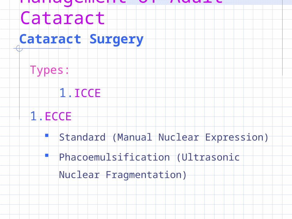
Cataract Surgery
Types:
1. ICCE
1. ECCE
Standard (Manual Nuclear Expression)
Phacoemulsification (Ultrasonic Nuclear
Fragmentation)
Management of Adult Cataract
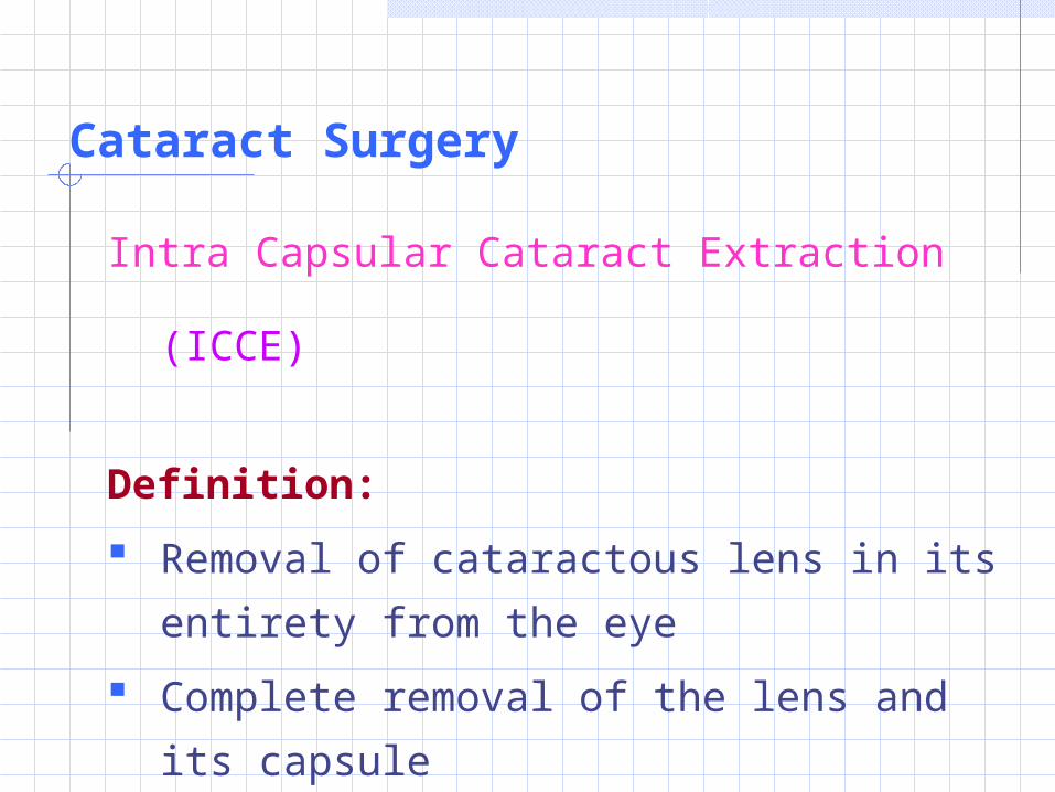
Cataract Surgery
Intra Capsular Cataract Extraction (ICCE)
Definition:
Removal of cataractous lens in its
entirety from the eye
Complete removal of the lens and its
capsule
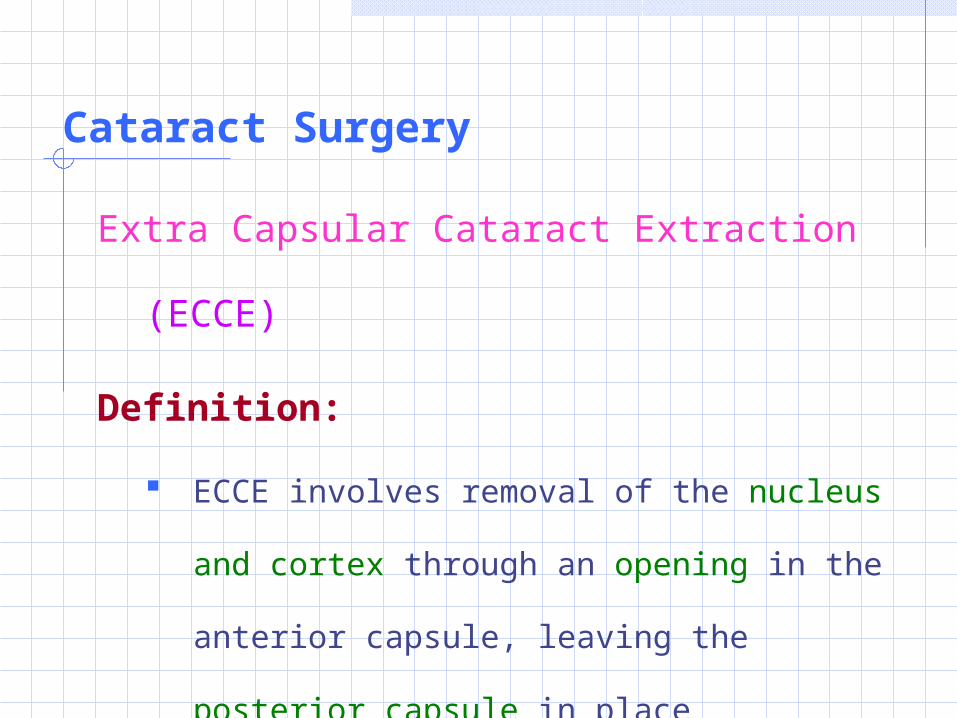
Cataract Surgery
Extra Capsular Cataract Extraction (ECCE)
Definition:
ECCE involves removal of the nucleus and
cortex through an opening in the anterior
capsule, leaving the posterior capsule in
place
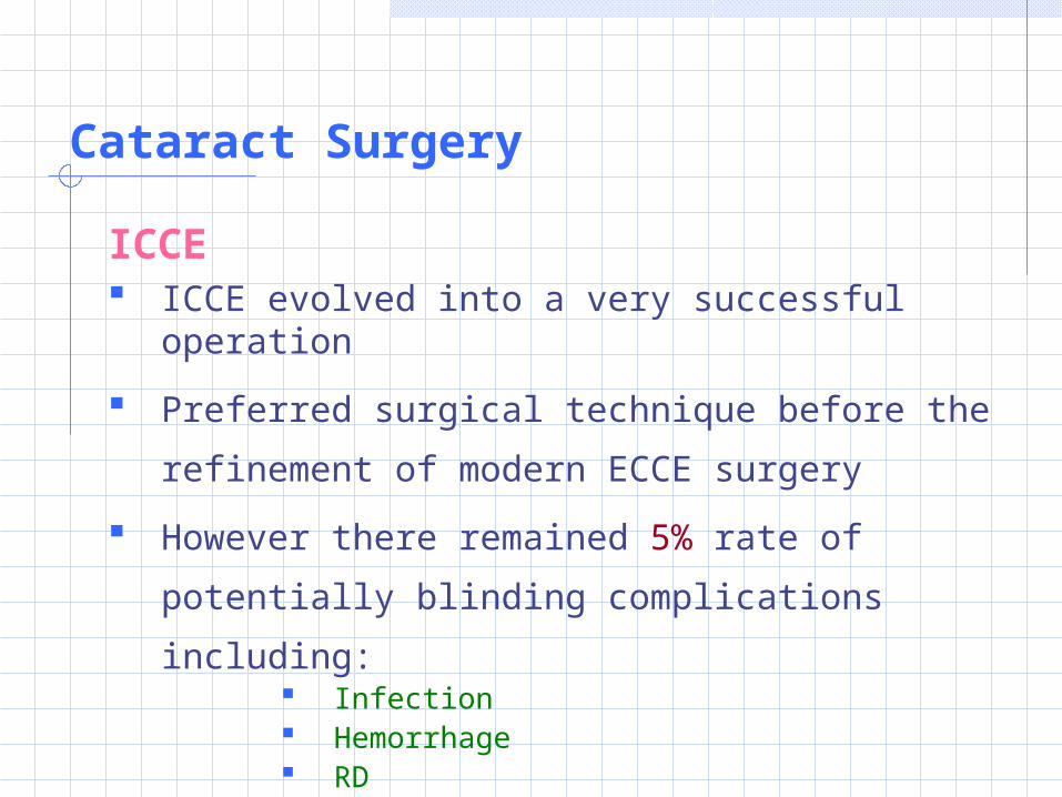
Cataract Surgery
ICCE ICCE evolved into a very successful operation
Preferred surgical technique before the
refinement of modern ECCE surgery
However there remained 5% rate of
potentially blinding complications including: Infection Hemorrhage RD CME
Cataract Surgery
ECCE has replaced ICCE, almost entirely in
most parts of the world:
1. Better operating microscopes
2. More sophisticated surgical aspiration
systems
3. More sophisticated IOL implants
Pre-operative evaluation and information
General health
Drug History
Ocular and social histories
Ocular examination
Measurement of visual function
Preoperative measurement
Pre-operative evaluation and information
General health
A complete medical history starting point
Ophthalmic surgeon should work with
patient’s primary care physician to achieve
optimal management of all medical problems
like: DM IHD COPD Bleeding Disorders Adrenal Suppression by Corticosteroids
Pre-operative evaluation and information
Awareness of any Drug sensitivities and
medications:
Immunosuppressants
Anticoagulants:
These may alter the outcome of surgery
Pre-operative evaluation and information
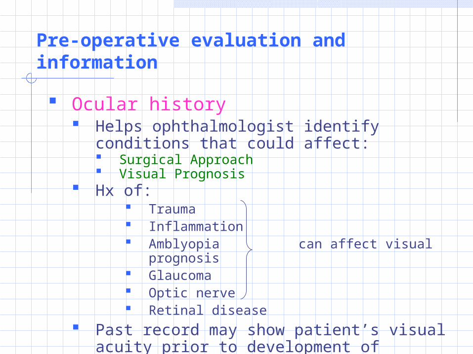
Ocular history Helps ophthalmologist identify conditions
that could affect: Surgical Approach Visual Prognosis
Hx of: Trauma Inflammation Amblyopia can affect visual
prognosis Glaucoma Optic nerve Retinal disease
Past record may show patient’s visual acuity prior to development of cataract
Pre-operative evaluation and information
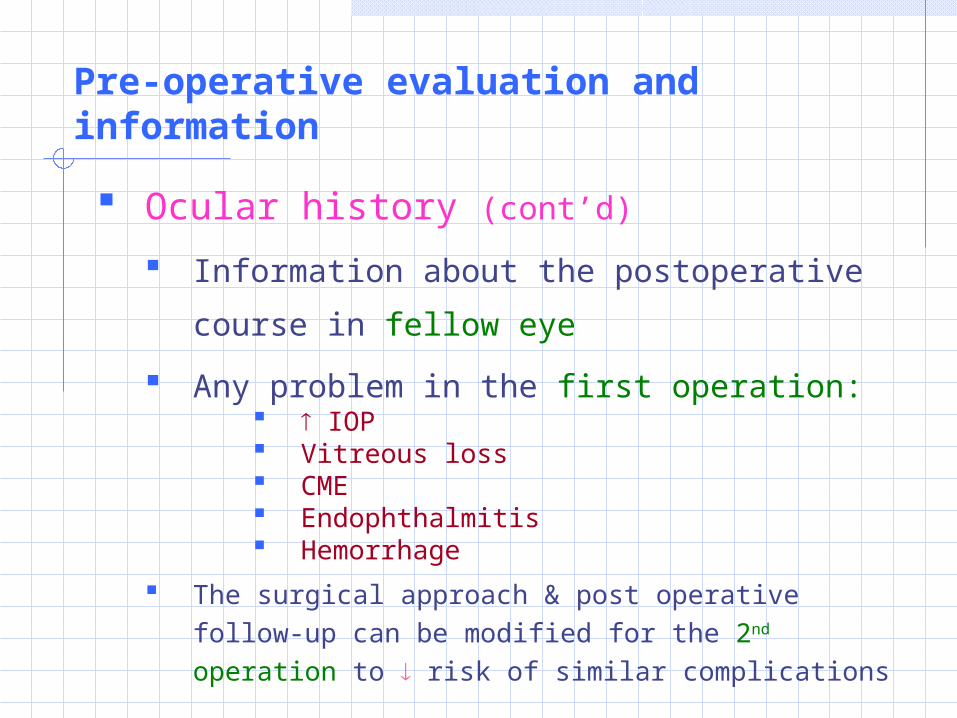
Ocular history (cont’d)
Information about the postoperative course
in fellow eye
Any problem in the first operation: IOP Vitreous loss CME Endophthalmitis Hemorrhage
The surgical approach & post operative follow-up can be modified for the 2nd operation to risk of similar complications
Pre-operative evaluation and information
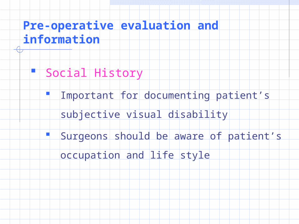
Social History
Important for documenting patient’s
subjective visual disability
Surgeons should be aware of patient’s
occupation and life style
External examination (pre-op Evaluation)
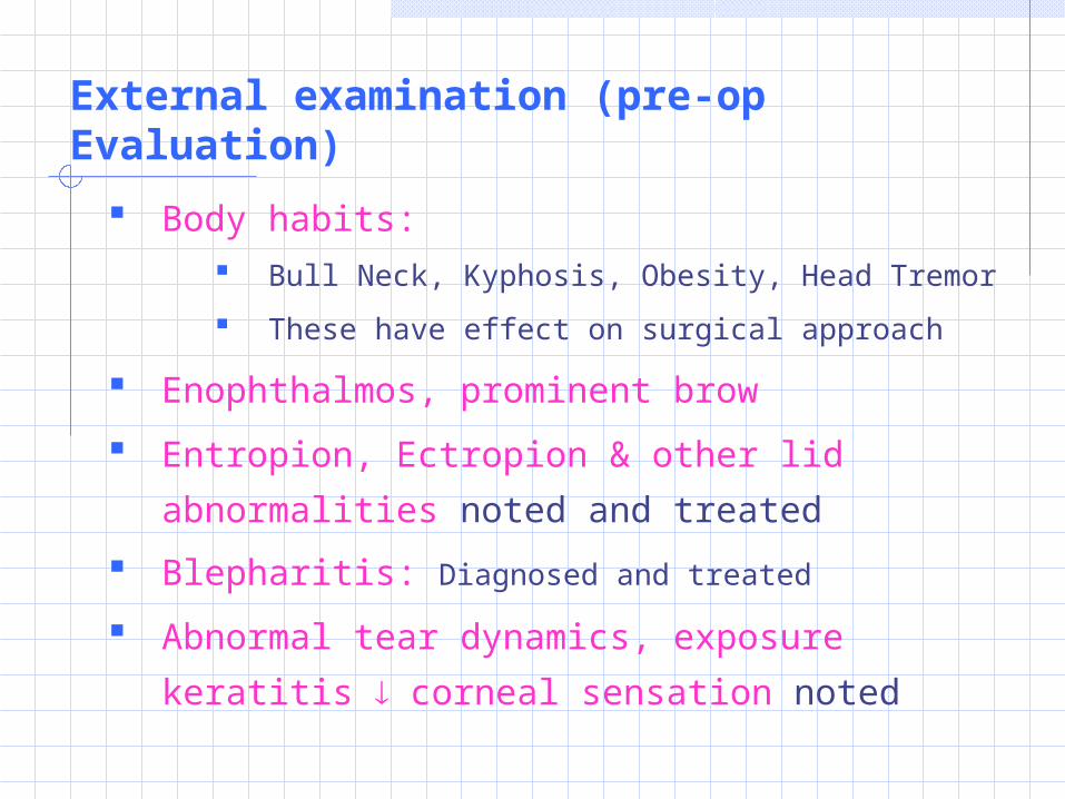
Body habits: Bull Neck, Kyphosis, Obesity, Head Tremor
These have effect on surgical approach
Enophthalmos, prominent brow
Entropion, Ectropion & other lid abnormalities
noted and treated
Blepharitis: Diagnosed and treated
Abnormal tear dynamics, exposure keratitis
corneal sensation noted

External examination (pre-op Evaluation)
Motility:
Ocular alignment evaluated
EOM tested for their range of movements
Cover testing (muscle balance):
Any abnormality might suggest pre-existing
strabismus with amblyopia as cause of visual
loss
Tropia: may result in diplopia following
surgery
External examination (pre-op Evaluation)
Pupil
Pupillary responses to light and
accommodation evaluated
Direct & consensual constriction of pupil
Swinging-flashlight Test:
To detect RAPD (Indicative of serious
retinal / optic nerve dysfunction)
External examination (pre-op Evaluation)
Biomicroscopic examination
Conjunctiva Scarring / lack of mobility over sclera
Symblepharon / shortening of fornices (underlying systemic/ocular surface disease): Can limit surgical approach
Loss of vascularization (Previous chemical injury / scarring from ocular surgery): Change in surgical approach
External examination (pre-op Evaluation)
Biomicroscopic examination
Conrnea
Corneal thickness, presence of Guttata
and marked abnormalities of
endothelium
Specular reflection and SL examination
provide estimate of endothelial cell count
and morphology
External examination (pre-op Evaluation)
Biomicroscopic examination
Conrnea (cont’d)
Thickness> 600 µm suggest poor
prognosis for corneal clarity following
cataract surgery. Surgery tailored to
minimize trauma to corneal endothelium
Cornea inspected for corneal arcus /
stromal opacities (may limit view during
surgery)
External examination (pre-op Evaluation)
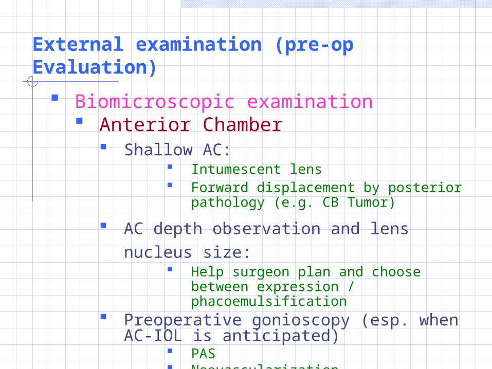
Biomicroscopic examination Anterior Chamber
Shallow AC: Intumescent lens Forward displacement by posterior
pathology (e.g. CB Tumor)
AC depth observation and lens nucleus size:
Help surgeon plan and choose between expression / phacoemulsification
Preoperative gonioscopy (esp. when AC-IOL is anticipated)
PAS Neovascularization Prominent major arterial circle
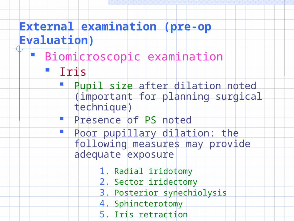
External examination (pre-op Evaluation) Biomicroscopic examination
Iris Pupil size after dilation noted (important
for planning surgical technique) Presence of PS noted Poor pupillary dilation: the following
measures may provide adequate exposure
1. Radial iridotomy2. Sector iridectomy3. Posterior synechiolysis4. Sphincterotomy5. Iris retraction
External examination (pre-op Evaluation)
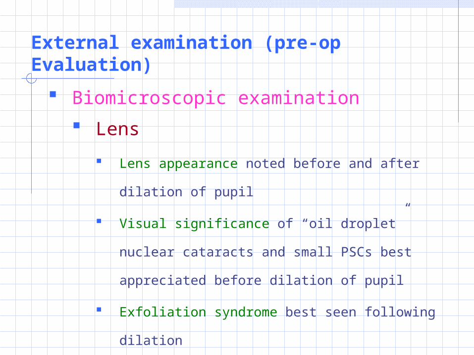
Biomicroscopic examination
Lens
Lens appearance noted before and after dilation
of pupil
Visual significance of “oil droplet” nuclear
cataracts and small PSCs best appreciated before
dilation of pupil
Exfoliation syndrome best seen following dilation
Nuclear size and brunescence evaluated for
phacoemulcification (after dilation)
External examination (pre-op Evaluation)
Biomicroscopic examination
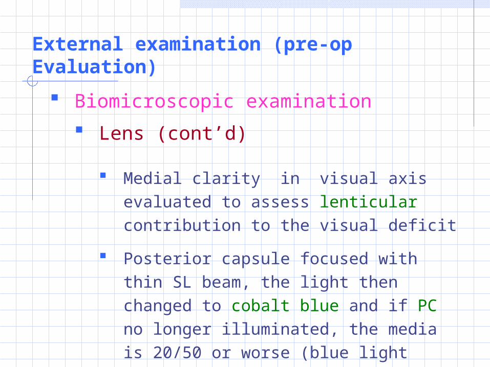
Lens (cont’d)
Medial clarity in visual axis evaluated to assess lenticular contribution to the visual deficit
Posterior capsule focused with thin SL beam, the light then changed to cobalt blue and if PC no longer illuminated, the media is 20/50 or worse (blue light scatter)
External examination (pre-op Evaluation)
Biomicroscopic examination
Lens (cont’d)
PSC (small) may cause severe visual
loss:
Conversely dense brunescent nuclear
sclerotic cataracts may allow surprisingly
good visual acuity
External examination (pre-op Evaluation)
Biomicroscopic examination Lens (cont’d)
Lens position and zonular fibers integrity also evaluated Lens decentration Excessive distance between lens and
pupillary margin (may indicate subluxation)
Indentation/flattening of lens periphery might indicate focal loss of zonular support
Fundus Evaluation
Ophthalmoscopy (Direct & Indirect)
1. Anatomical integrity of posterior segment
assessed
2. Media clarity (direct opthalmoscope)
3. Macular, ON, Retinal vessels, Retinal
periphery evaluated
4. ARM may limit visual rehabilitation after
otherwise uneventful cataract ext.
Fundus Evaluation
Ophthalmoscopy (Direct & Indirect) (cont’d)
5. Diabetic patients examined carefully for:
Macular edema
Retinal ischaemia
Neovascularization ±
Retinal ischaemia may progress to posterior or
anterior neovascularization in case of
ICCE or
ECCE (with PC rupture)
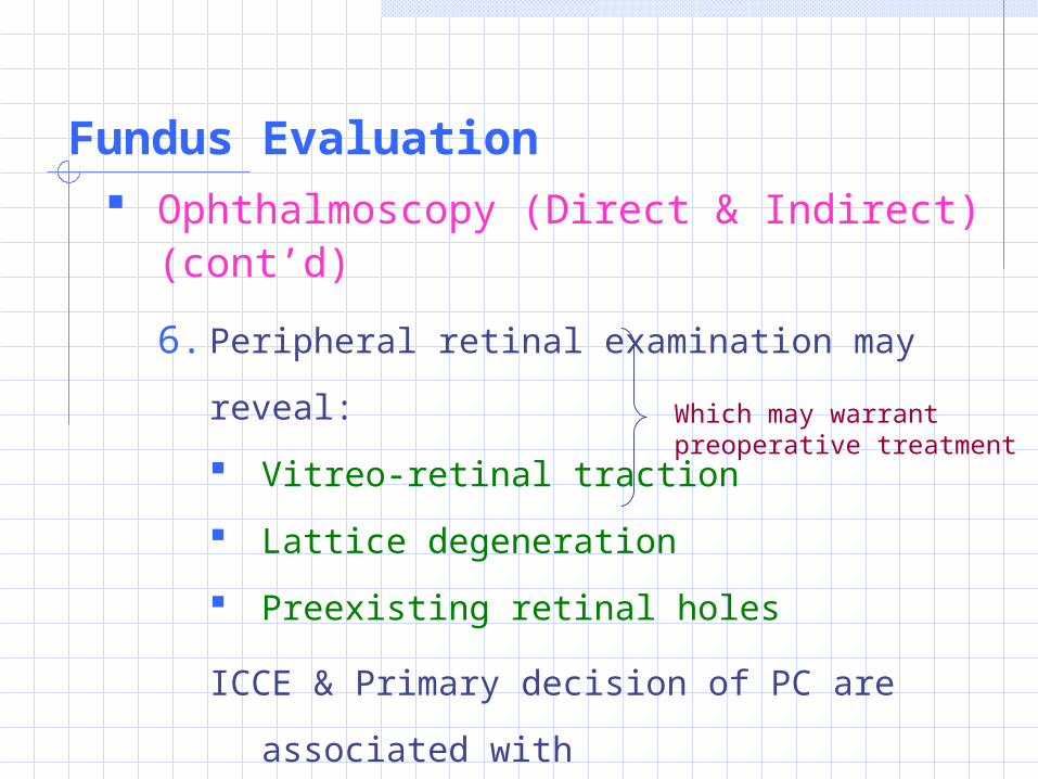
Fundus Evaluation Ophthalmoscopy (Direct & Indirect)
(cont’d)
6. Peripheral retinal examination may reveal:
Vitreo-retinal traction
Lattice degeneration
Preexisting retinal holes
ICCE & Primary decision of PC are associated
with
incidence of RD and CME
Which may warrant preoperative treatment

Optic Nerve
Examined for color, CD ratio or any other
abnormality
ON functions further evaluated by:
VA
Confrontation VF testing
Pupillary Examination
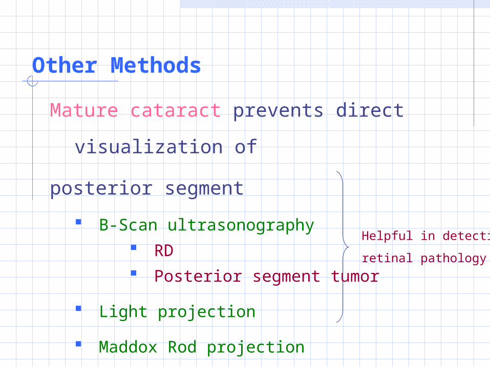
Other Methods
Mature cataract prevents direct visualization
of
posterior segment
B-Scan ultrasonography RD Posterior segment tumor
Light projection
Maddox Rod projection
Helpful in detecting
retinal pathology
Measurements of visual function
1. VA Testing
2. Brightness Acuity
3. Contrast Sensitivity
4. Visual Field Testing
Measurements of visual function
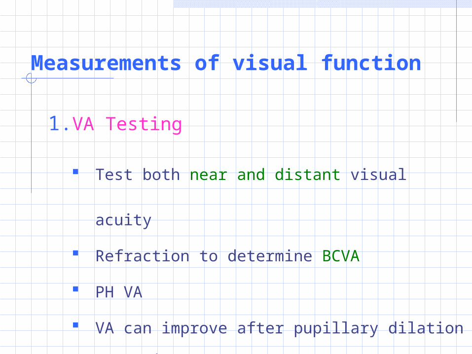
1. VA Testing
Test both near and distant visual acuity
Refraction to determine BCVA
PH VA
VA can improve after pupillary dilation (esp. in
PSC)
Measurements of visual function
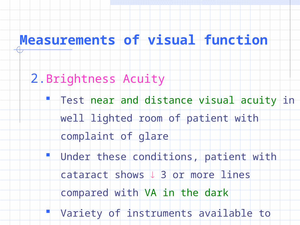
2. Brightness Acuity
Test near and distance visual acuity in well
lighted room of patient with complaint of glare
Under these conditions, patient with cataract
shows 3 or more lines compared with VA in
the dark
Variety of instruments available to standardize
and facilitate this measurement
Measurements of visual function
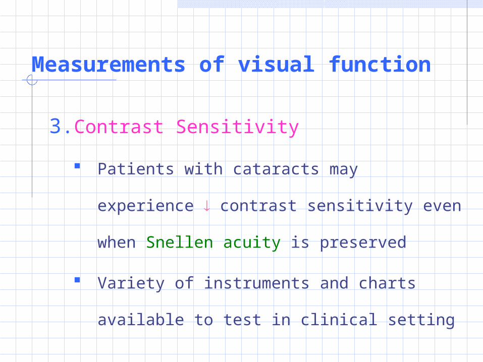
3. Contrast Sensitivity
Patients with cataracts may experience
contrast sensitivity even when Snellen
acuity is preserved
Variety of instruments and charts available
to test in clinical setting
Measurements of visual function
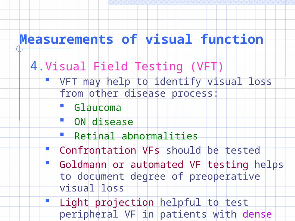
4. Visual Field Testing (VFT) VFT may help to identify visual loss from other
disease process: Glaucoma ON disease Retinal abnormalities
Confrontation VFs should be tested Goldmann or automated VF testing helps to
document degree of preoperative visual loss Light projection helpful to test peripheral VF in
patients with dense cataracts
Measurements of visual function
5. Special Tests
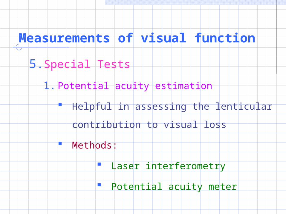
1. Potential acuity estimation
Helpful in assessing the lenticular
contribution to visual loss
Methods:
Laser interferometry
Potential acuity meter
Measurements of visual function
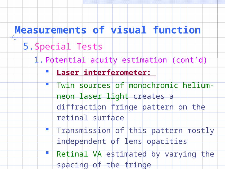
5. Special Tests
1. Potential acuity estimation (cont’d)
Laser interferometer:
Twin sources of monochromic helium-neon laser light creates a diffraction fringe pattern on the retinal surface
Transmission of this pattern mostly independent of lens opacities
Retinal VA estimated by varying the spacing of the fringe
Measurements of visual function
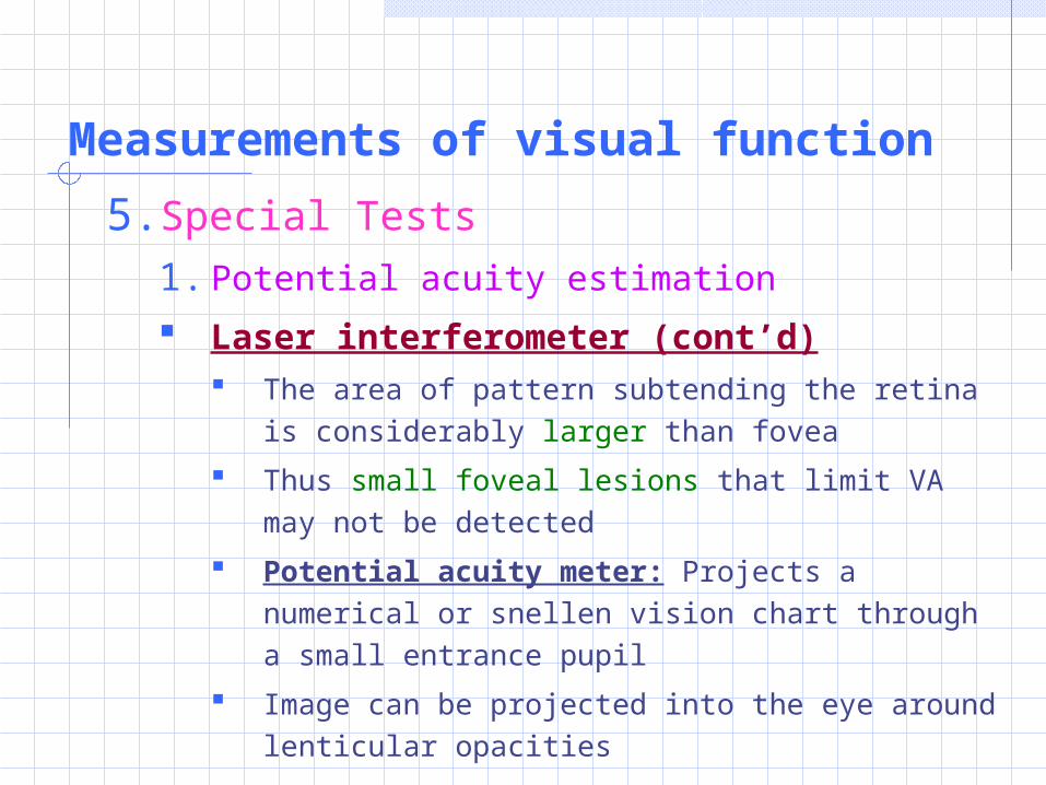
5. Special Tests
1. Potential acuity estimation
Laser interferometer (cont’d) The area of pattern subtending the retina is
considerably larger than fovea
Thus small foveal lesions that limit VA may not be detected
Potential acuity meter: Projects a numerical or snellen vision chart through a small entrance pupil
Image can be projected into the eye around lenticular opacities
Measurements of visual function
5. Special Tests
1. Potential acuity estimation
Potential acuity meter
Projects a numerical or Snellen vision chart
through a small entrance pupil
Image can be projected into the eye around
lenticular opacities
Measurements of visual function5. Special Tests
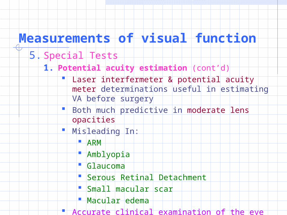
1. Potential acuity estimation (cont’d) Laser interfermeter & potential acuity meter
determinations useful in estimating VA before surgery
Both much predictive in moderate lens opacities Misleading In:
ARM Amblyopia Glaucoma Serous Retinal Detachment Small macular scar Macular edema
Accurate clinical examination of the eye is as good a predictor of the visual outcome as these tests
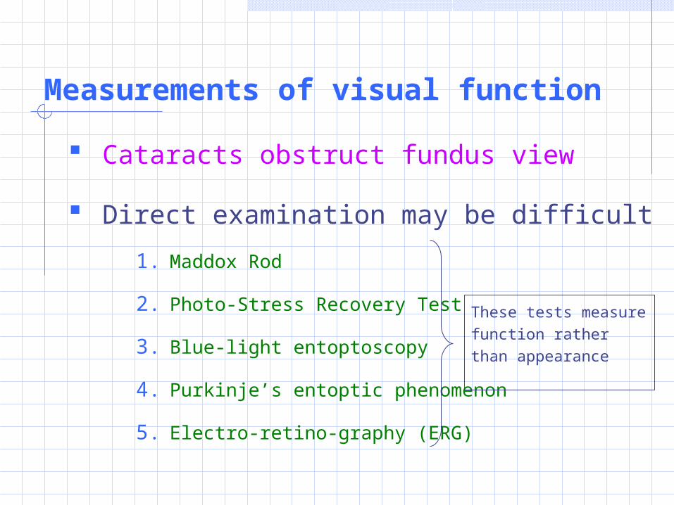
Measurements of visual function
Cataracts obstruct fundus view
Direct examination may be difficult
1. Maddox Rod
2. Photo-Stress Recovery Test
3. Blue-light entoptoscopy
4. Purkinje’s entoptic phenomenon
5. Electro-retino-graphy (ERG)
These tests measure function rather than appearance
Measurements of visual function
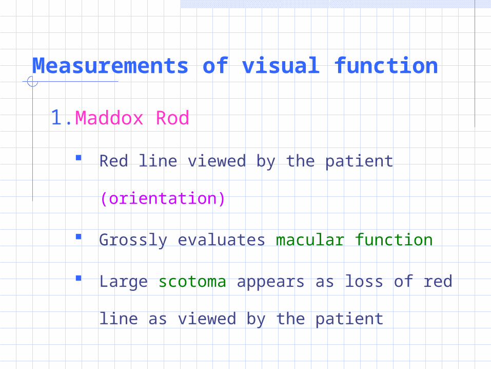
1. Maddox Rod
Red line viewed by the patient (orientation)
Grossly evaluates macular function
Large scotoma appears as loss of red line as
viewed by the patient
Measurements of visual function
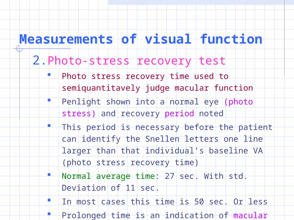
2. Photo-stress recovery test Photo stress recovery time used to semiquantitavely
judge macular function
Penlight shown into a normal eye (photo stress) and recovery period noted
This period is necessary before the patient can identify the Snellen letters one line larger than that individual’s baseline VA (photo stress recovery time)
Normal average time: 27 sec. With std. Deviation of 11 sec.
In most cases this time is 50 sec. Or less
Prolonged time is an indication of macular disease
Measurements of visual function
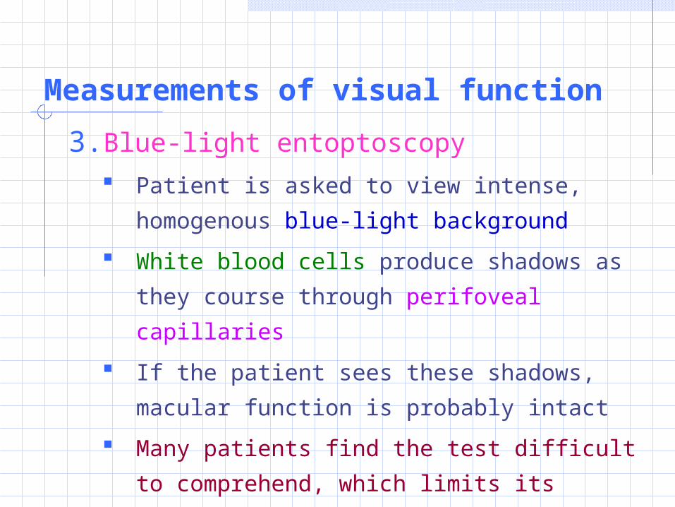
3. Blue-light entoptoscopy
Patient is asked to view intense,
homogenous blue-light background
White blood cells produce shadows as they
course through perifoveal capillaries
If the patient sees these shadows, macular
function is probably intact
Many patients find the test difficult to
comprehend, which limits its usefulness
Measurements of visual function
4. Purkingje’s Entoptic Phenomenon
Subjective test
Rapidly oscillating point source of light is
shown through closed eye lids
Ability of the patient to detect shadow
images of his/her retinal vasculature
provides a very rough indication that retina
is attached
Measurements of visual function
5. Electro-retino-Graphy (ERG) & Visual
Evoked Response (VER)
In rare cases these tests can be
done to evaluate retinal and or ON
function where other testing is
inconclusive


![L5-KinematicAnalysis-II.ppt€¦ · Title: Microsoft PowerPoint - L5-KinematicAnalysis-II.ppt [Compatibility Mode] Author: Erik Created Date: 11/28/2008 11:23:18 AM](https://img.pdfslide.us/doc/110x75/5f105a8e7e708231d448b15c/l5-kinematicanalysis-iippt-title-microsoft-powerpoint-l5-kinematicanalysis-iippt.jpg)


![[RE-POSTED]Week 11 - Data analysis-Part II.ppt](https://img.pdfslide.us/doc/110x75/55cf8e0d550346703b8e072a/re-postedweek-11-data-analysis-part-iippt.jpg)