Embed Size (px)
Citation preview
Dr.Yajuvendra S Rathore
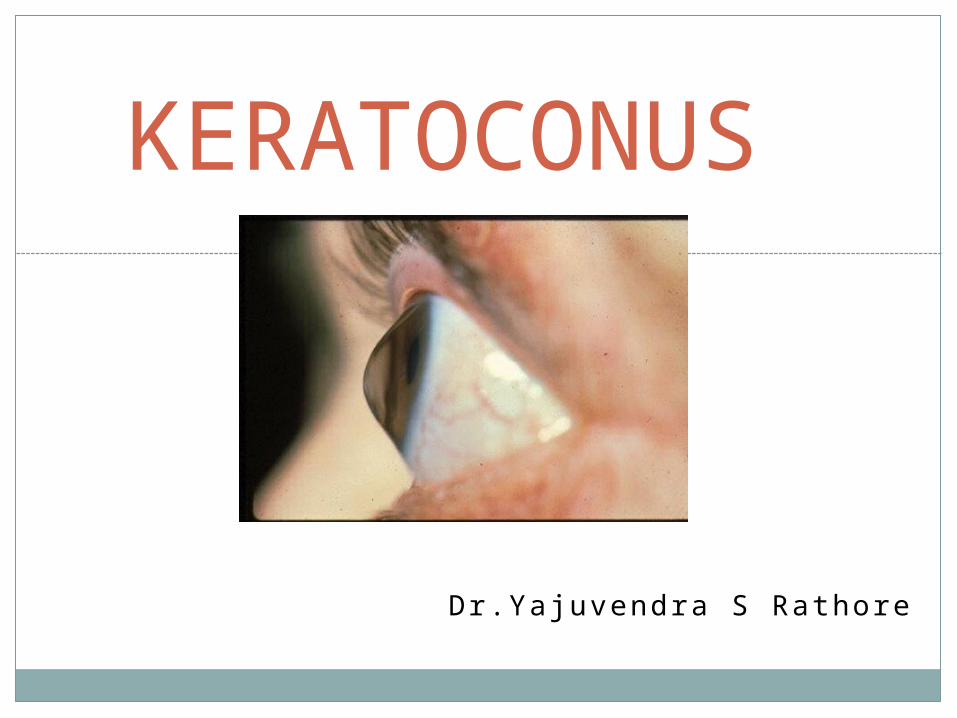
KERATOCONUS

KERATOCONUS
It is a clinical entity characterized by noninflammatory and noninfective, progressive, bilateral thinning of the cornea with ectasia of conical shape.
This is mostly bilateral condition, girls between 15-20 years affected more, overall incidence rate estimated to be 0.15 to 0.20 percent.
It manifests by adolescence, resulting in considerable visual impairment owing to the development of high degree irregular myopic astigmatism.
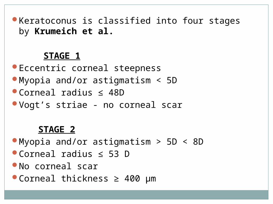
Keratoconus is classified into four stages by Krumeich et al.
STAGE 1Eccentric corneal steepnessMyopia and/or astigmatism < 5DCorneal radius ≤ 48DVogt’s striae - no corneal scar STAGE 2Myopia and/or astigmatism > 5D < 8DCorneal radius ≤ 53 DNo corneal scarCorneal thickness ≥ 400 µm

STAGE 3Myopia and/or astigmatism > 8D < 10DCorneal radius > 53DNo corneal scarCorneal thickness 200-400 µm STAGE 4Refractive error not measurableCorneal radius > 55DCorneal scar/perforationCorneal thickness 200 µm

ETIOLOGYDefinite etiology is unknown. 95% of patients do not
show evidence of specific hereditary pattern. Pattern of inheritance is variable. Several theories have been put forward to explain the etiology of keratoconus.
ENZYME THEORY Alteration in the levels of following enzymes have been
noted: Increased level of epithelial lysosomal enzymes. Decreased level of alpha-1 proteinase inhibitor in the epithelium. Decreased levels of glucose-6 phosphate
dehydrogenase in the epithelium.

CONNECTIVE TISSUE ABNORMALITY THEORY There is association of keratoconus with some
connective tissue disorders.
GENETIC THEORY Due to the occasional association of trisomy-21 with
keratoconus, genetic abnormality may be the cause.
HORMONAL THEORY It is proposed because of the manifestations of the
disease in adolescence. EYE RUBBING Habitual eye rubbing in some diseases like vernal
catarrh, Down syndrome and poorly sighted patients of Leber’s tapetoretinal degeneration are associated with keratoconus.

ASSOCIATIONS
SYSTEMIC DISEASES
Connective tissue and mesodermal dysplasia Marfan’s syndromeEhlers-Danlos syndromeOsteogenesis imperfectaCongenital hip dysplasiaOculodento digital syndromeRieger’s syndromeCrouzon’s syndromeFloppy eyelid syndromeDown syndromeTurners syndromeAtopic dermatitis
Atopic dermatitis Down syndrome Ehlers-Danlos syndrome
Marfan syndrome Crouzon syndromeOsteogenesis imperfecta

ASSOCIATED OCULAR DISORDERSRetinitis pigmentosaLebers congenital amaurosisBlue scleraeCongenital cataractAniridiaRetinopathy of prematurityFuchs dystrophyPosterior polymorphous dystrophyGranular and lattice dystrophy ALLERGIC CONDITIONSSpring catarrhAsthma

CLINICAL FEATURESGradual decrease in vision, photophobia,
monocular diplopia or monocular polyopia.
Severe photophobia and watering is seen in cases of hydrops. Particularly adolescent females are affected
There is conical protrusion of the cornea with central thinning and the apex of the cone is usually directed inferonasally.

Munson’s sign is a V-shaped conformation of the lower lid produced by the ectatic cornea in downgaze.
Rizzuti’s sign is a sharply focused beam of light near the nasal limbus, produced by lateral illumination of the cornea in patients with advanced keratoconus.

Slit lamp examination
Prominent corneal nerves

Slit lamp examination
Fleischer's Ring The Fleischer ring is a yellow-brown
to olive-green ring of pigment which may or may not completely surround the base of the cone
Formed when hemosiderin (iron) pigment is deposited deep in the epithelium
Fleischer's ring often becomes thinner and more discrete with progression

Seen approximately 50% of all cases.Locating this ring initially may be made
easier by using a cobalt filter and carefully focusing on the superior half of the cornea's epithelium.
Once located, the ring should be viewed in white light to assess its extent.


Lines of Vogt:
Small and brushlike lines, generally vertical but they can be oblique.
Found in the deep layers of the stroma and form along the meridian of greatest curvature.
Disappear when gentle pressure is exerted on the globe through the lid.


Corneal Thinning:Significant thinning (up to 1/5th cornea
thickness) in the advanced stages of the disease and a diagnostic criterion based on comparison of central and peripheral corneal thickness has been proposed.
Additionally, as the disease progresses, the cone is often displaced inferiorly. The steepest part of the cornea (apex) is generally the thinnest.


Corneal Scarring:Sub-epithelial corneal scarring, not generally
seen early, may occur as keratoconus progresses because of ruptures in Bowman's membrane which is then filled with connective tissue
Deep opacity of the cornea are also common in keratoconus.


Corneal Hydrops:
Corneal hydrops occurs in advanced cases, when Descemet's membrane ruptures, aqueous flows into the cornea and reseals
Keratoconus patients who are having an acute episode of corneal hydrops report a sudden loss of vision and a visible white spot on the cornea.
Corneal hydrops causes edema and opacification.
As Descemet's regenerates, edema and opacification decreases. Occasionally, hydrops can benefit keratoconus patients who have extremely steep corneas. If the cornea scars, a flatter cornea often results, making it easier to fit with a contact lens.
An increased incidence of hydrops has also been reported in keratoconus patients with Down's syndrome.


Diagnosis
Early keratoconus usually manifests as a small island of irregular astigmatism in the inferior paracentral cornea.
As the cornea bulges outward, the amount of astigmatism increases due to the progressive distortion of the corneal surface.
These changes can easily be seen as irregular mires on keratometry readings and on corneal topography.

Many objective signs are present in keratoconus.
Retinoscopy shows a scissoring reflex.
On direct ophthalmoscopy there is a dark round shadow in the corneal midperiphery due to total internal reflection of the light surrounding the central bright red fundus reflex and separating it from the normal red peripheral reflex. It is called a Charleux oil droplet reflex.

The photokeratoscope or topographer placido disc can provide an overview of the cornea and can show the relative steepness of any corneal area.
There is even separation of the rings in the spherical cornea .

In astigmatic cornea uneven spacing of the rings,especially inferiorly-in the keratoconic cornea should be noted
The central rings may show a tear-drop configuration termed "keratokyphosis".
DOS Times - Vol.10, No. 7 January 2005

With the handheld keratoscopes, such as the Klein keratoscope, early keratoconus is characterized by a downward deviation of the horizontal axis of the Placido disk reflection

The Keratometer also aids diagnosis.
o Classification based on keratometry-1. Mild- <45D in both meridians2. Moderate- 45-52D in both meridians3. Advanced- >52D in both meridians4. Severe- >62D in both meridians
The initial keratometric sign of keratoconus is absence of parallelism and inclination of the mires. These can easily be missed in mild or early cases.

COMPUTER ASSISTED VIDEO KERATOSCOPY
It is one of the most important diagnostic aids for very early as well as abortive forms of keratoconus in the other eye of the patients with unilateral keratoconus or in the family members.
Data of videokeratoscopic image are analyzed by computers and depicted as color coded maps. Red color indicates myopic refraction or ectasia and blue color indicates hypermetropic refraction or flattening of the cornea.

PACHYMETRY
Slit lamp pachymetry shows thinning in the centre of the apex. Ultrasonic pachymetry shows exact thickness of cornea at different places.
Thinning in the inferior quadrant can be diagnostic of keratoconus. Central or paracentral corneal thickness of less than 450 µm is abnormal.
If the reading decreases by nearly 20 µm towards the inferior periphery on successive pachymetric readings, it is suspicious of keratoconus. Increase in the progressive thinning of the cornea is a true index of keratoconus.

Corneal topography
Provides a color coded map of the corneal surface.
The power in diopters of the steepest and flattest meridians and their axes are calculated and displayed
Steep curvatures are marked orange or red
Flat curvature in blue or violet
Normal curvatures in green or yellow

Keratoconus is a clinical diagnosis and Forme Fruste KC is a subtle topographic abnormality before clinical manifestation of the disease.
The aim of topography and tomography in refractive surgery clinic is to rule out keratoectatic disease either in form of frank keratoconus or subtle FFKC as they are contraindications to the procedure.

The suspicious signs for keratoconus include:Axial map abnormalities1. K greater than 48 D.2. Skewed radial axis greater than 21 degrees.3. Inferior -Superior ratio greater than 1.42D.4. Corneal astigmatism greater than 6 D.5. Against the rule astigmatism.6. Superior-Inferior difference at the 5-mm
zone >2.5 D.

On the elevation maps1. Isolated island or tongue-like extension on either surface2. Elevation values greater than 12 microns on the anterior
elevation map in the central 5 mm.3. Elevation values greater than 15 microns on the posterior
elevation map
Pachymetry/corneal thickness map: On Scheimpflug devices1. Thinnest location less than 470 microns.2. Displacement of the thinnest point >500 microns from the
center.3. Pachymetry difference asymmetry in two eyes at thinnest point
>30 microns.4. S-I difference at the 5 mm circle >30 microns.5. Cone-like pattern on the thickness map.
The most elevated points on the anterior and the posterior elevation maps should be correlated to the highest power on Axial/Saggital curvature map and the thinnest point on the global pachymetry map.
If all the above match, it is called as the “fourpoint touch” and is a hallmark of suspect cornea, especially if the apex is decentered by more than 500 microns and the peripheral thickness readings of the upper and lower half at the 7-mm zone also show a significant difference of greater than 100 microns.

Swirl staining
In patients who have never worn contact lenses .
Occur due to basal epithelial cell drop out as a result the epithelium slides from the periphery as the cornea regenerates.
Thus a hurricane, vortex or swirl stain may occur.

Rabinovitz criteria for diagnosis of keratoconus
1. Central corneal power >47.2D2. Inferior superior dioptric assymetry over
1.2D3. Skewed radial axes of astigmatism by more
than 21Degrees4. Difference in central power of more than 1D
between the fellow eye.

DIFFERENTIAL DIAGNOSIS
1. KERATOGLOBUS This is due to thinning of periphery of the cornea which
gradually progresses towards the centre. It is present at or soon after birth. So it is thought to be a developmental anomaly. Perforation can occur in this condition with minimal ocular trauma.
2. POSTERIOR KERATOCONUS It is nonprogressive, dome shaped posterior excavation in
the cornea which may be small and circumscribed( keratoconus posticus circumscriptus) or may be diffuse( keratoconus posticus totalis). It is considered to be a congenital defect. Origin is prior to 5th or 6th month of gestation. Traumatic etiology is also reported.

3. PELLUCID MARGINAL DEGENERATION It is a bilateral peripheral corneal ectatic
disorder characterized by a band of thinning of 1-2 mm width typically in the inferior cornea. Maximum corneal protrusion occurs superior to the area of thinning.
There is no other abnormality and it occurs in 2nd to 5th decade.

Management
1. Glasses2. Contact lenses3. Collagen cross linking4. Intracorneal rings5. LASIK Xtra6. Deep anterior lamellar Keratoplasty7. Penetrating Keratoplasty

Spectacles
Mild keratoconus can be corrected with spectacles.
Retinoscopy is difficult; a normal subjective refraction is required.
Monocular keratoconus is usually best dealt with using spectacle correction.
In this group of patients, motivation for contact lens wear tends to be poor.

Contact lenses
Contact lenses are considered when vision is not correctible to 6/9 by spectacles and patients become symptomatic.
Rigid gas permeable (RGP) contact lenses are the lenses of first choice.
The aim is to provide the best vision possible with the maximum comfort so that the lenses can be worn for a long period of time.

Based on shape of cone
Nipple cone : small diameter (5 mm.); round shape; easiest to fit with contact lenses
Oval large diameter(>5 mm.); often displaced inferiorly; more difficult to fit with lenses
Globus largest diameter (>6 mm.); 75% of cornea affected; most difficult to fit with lens

Nipple cone oval cone globus
Fitting methods
1. Three-point-touch design
Three-point-touch actually refers to the area of apical central contact and two other areas of bearing or contact at the mid-periphery in the horizontal direction.
The three-point-touch design is the most popular and the most widely fitted design
The aim is to distribute the weight of the contact lens as evenly as possible between the cone and the peripheral cornea.

The ideal fit should show an apical contact area of 2-3mm with mid-peripheral contact.
Adequate edge clearance is required to ensure tear exchange.

2. Apical clearance
In this type of fitting technique, the lens vaults the cone and clears the central cornea, resting on the paracentral cornea.
These lenses tend to be small in diameter and have small optic zones
The potential advantages of reducing central corneal scarring are outweighed by the disadvantages like poor tear film, corneal oedema, and poor visual acuity as a result of bubbles becoming trapped under the lens.

3. Flat fitting
The flat fitting method places almost the entire weight of the lens on the cone.
The lens tends to be held in position by the top lid.
Good visual acuity is obtained as a result of apical touch.
Alignment can be obtained in early keratoconus; however, flat fitting lenses can lead to progression/ acceleration of apical changes and corneal abrasions.
This type of fitting is useful where the apex of the cone is displaced.

Piggy back lenses - RGP-CL over a SOFT CL
Can be used in pts who are uncomfortable with RGP wear, more so in pts prone to epithelial erosion at apex of cone
ROSE-K design RGP -are specially designed for keratoconic eyes with a diagnostic set of 26 lenses with base curves ranging from 5.1 to 7.6 mm in 0.1 increments, a std lens diameter of 8.7mm
Scleral lenses
Scleral lenses play a very significant role in cases of advanced keratoconus where corneal lenses do not work and corneal surgery is contra-indicated.
Scleral lenses completely neutralise any corneal irregularity and can help patients maintain a normal quality of life

Soper lens Custom made lens
Two zones in the peripheral posterior curvature
a) Central zone : to vault steep central cornea .It is of varying steepness depending of the patients cornea.
b) Peripheral zone is with a 45D curvature designed to vault the mid periphery and limbal cornea

Boston scleral lens prosthetic device (BSLPD)
Fluid ventilated scleral lens
Designed to enclose a bubble free reservoir of fluid over the corneal surface
Series of breaches are created between haptic bearing surface of the lens and underlying sclera.

This will facilitate the aspiration of surface tears into the reservoir so that intrusion of air bubble during a blink is prevented.
Shape of haptic confirms exactly to that of underlying sclera to maintain functionality and prevents intrusion of air bubbles.
Very expensive

Collagen cross linking
A newer and less invasive technique that shows promise in keratoconus management is combined riboflavin-ultraviolet type A rays( UVA ) collagen cross-linking.
This procedure consists of photopolymerization of corneal stroma by combining vitamin B2 (photosensitizing substance) with UVA.
This process increases rigidity of corneal collagen and thus reduces the likelihood of further ectasia.

•Using topical anaesthesia, 7mm circle is marked on the cornea using a thornton marker.
•Epithelium of the marked area is scraped off using a blunt spatula.
•A corneal abrasion is created to facilitate riboflavin diffusion into the cornea.
•One drop of riboflavin 0.1% and 20% dextran ophthalmic solution is instilled topically in the eye every 2 minutes for 30 minutes.
•At the end of the 30-minute pretreatment period, the eye is examined with blue light for the presence of a yellow flare in the anterior chamber, indicating adequate riboflavin saturation of the corneal tissue.
Technique

When the yellow flare in the anterior chamber is confirmed, the eye is aligned under the UV-A light with the treatment plane at 50 mm from the UV-A beam aperture. Focussed on the apex of cornea at a distance of 10-12m to obtain a radiant energy of 5.4J/cm2 for 5 min
The correct aperture setting is selected for the size of the eye; the eye is irradiated for 30 minutes, during which time instillation of riboflavin is continued (one drop every 5 minutes).

After completion of the procedure,eye is washed with BSS , an antibiotic drop is instilled and a bandage contact lens is applied.
The contact lens is removed once the
abrasion has healed.
Postoperative medications include an antibiotic and a steroid for 2 weeks postoperatively.

Complications of C3R
Corneal hazeDiffuse lamellar keratitisReactivation of viral keratitis and iritisInfective keratitisCorneal scarringPersistent corneal edemaCorneal melt

LASIK Xtra
LASIK Xtra basically means LASIK combined with C3R to treat Keratoconus and also for ectasia following LASIK surgery.
LASIK Xtra embodies a proper evolution of LASIK technique, the refractive surgery technique which has received the most enthusiastic acclaim worldwide. This technique uses the excimer laser to remodel the curve of the cornea and surgically correct myopia, hypermetropia, astigmatism and presbyopia in a rapid and safe manner.
Just like LASIK procedure, the LASIK Xtra technique is also successfully employed to re-treat previous interventions that were partly or incompletely satisfactory. Generally, the procedure is bilateral, i.e. the sight defect is corrected in both eyes in a single operating session.

Advantages
It is pain-free, both during and after the procedure.
In addition to standard LASIK results, the LASIK Xtra technique restores the strength of corneas weakened by LASIK.
It enables normal activity to be resumed immediately: e.g. work and sport.
Furthermore, bilateral correction also notably facilitates postoperative adjustment.
WHO CAN USE LASIK XTRA?
The ideal candidate for LASIK Xtra has a stable refraction, healthy corneas and certain physiological characteristics (essentially, an adequate corneal thickness); in addition the candidate must be highly motivated to reduce or eliminate any dependence on glasses and lenses.
There must be no other eye diseases present.
For women at an advanced stage of pregnancy, it is preferable that they do not undergo laser sight correction until their eyesight stabilises, after the birth.
Previous surgery is not a contraindication. Indeed, LASIK Xtra often perfects unsatisfactory results of previous surgery.

LASIK Xtra strengthening effect allows to broaden the inclusion criteria for potential patients, opening the possibility for many ineligible LASIK patients to become candidates for LASIK Xtra – although this evaluation is specifically carried out by the surgeon on a case-by-case basis.
To assess suitability for the procedure and the nature of the eyesight defect, it is indispensable that the patient undergo a thorough preoperative check. On the basis of this the specialist can plan a tailor-made treatment.

RISKS AND COMPLICATIONS
With the LASIK technique, no major or serious events have ever been reported, involving loss of the eye or of eyesight. Infection is extremely rare, but can be resolved with antibiotic treatment.
On rare occasions it may be the case that when vision has settled, the results obtained do not fully meet expectations. This depends on the natural reactivity of the eye which, if the treatments are equal, differs from person to person.
In these rare cases, termed “under-corrections” or “over-corrections”, the extreme flexibility of the LASIK Xtra technique allows for a subsequent intervention to perfect the results obtained, with no risk for the patient

Intracorneal stromal rings
Act as passive spacing agents which flatten the cornea
Made of PMMA
Amount of correction depends on the ring thickness, more thicker the ring more correction.
On insertion they shorten the arc of ant corneal surface, iron out gross irregularities and in effect create a second limbus.
Various corneal ring-kerarings, intacs.

An important potential benefit of treating keratoconus with INTACS inserts is to delay or eliminate the need for a corneal graft.
Patients with mild to moderate keratoconus appear to be the best candidates.

ComplicationsUndercorrection
Overcorrection
Migration of rings
Extrusion or progressive thinning
New vessel formation
glare /halos

Penetrating Keratoplasty
The gold standard surgery
Success rate is more than 90%.
In this procedure, the keratoconic cornea is prepared by removing the central area of the cornea, and a full-thickness corneal button is sutured in its place.
Usually trephines between 8.0-8.5 mm are used.
Fleischer’s ring can be used as the limit of the conical cornea.
Contact lenses are often required after this procedure for best visual rehabilitation.

Deep Anterior Lamellar Keratoplasty
Partial corneal transplant.
The cornea is removed to the depth of posterior stroma, and the donor button is sutured in place.
This technique is technically difficult, and visual acuity is inferior to that obtained after penetrating keratoplasty.
As a result, use of lamellar keratoplasty is largely confined to the treatment of large cones or keratoglobus when tectonic support is needed.
This technique requires less recovery time, and poses less chance for corneal graft rejection or failure.
Its disadvantages include vascularization and haziness of the graft

Thermokeratoplasty
Rare procedure
It involves placing a hot ring (Holmium yag laser, 2100nm) along the base of the cone to heat and traumatize the cornea, resulting in a corneal scar which reduces the corneal curvature.
It allows a flatter contact lens to be fitted..
The disadvantages of the procedureo transitory corneal haze o development of corneal scarring

Phakic iols
Used to correct high myopia and associated astigmatism of selected keratoconus patients.
Anterior chamber phakic intraocular lens have also been combined with intacs with good results.
The Intacs implantation is followed by toric phakic intraocular lens implantation to correct the residual myopic and astigmatic refractive error.

“Five point” management algorithm for keratoconus

THANK YOU