Embed Size (px)
Citation preview
Corneal collagen cross-linking and keratoconus
Faculty of Medicine, University of Oslo Faiza Abbas
Abstract
Keratoconus is a condition resulting in corneal ectasia. Abnormal protrusion of the cornea
leads to reduced visual acuity. Riboflavin induced collagen cross-linking (CXL) has received
significant attention the last few years and has shown to stop the progression of keratoconus.
The original CXL procedure involves epithelial debridement, application of topical riboflavin
drops and UVA exposure at 370 nm for approximately 30 minutes. However, not all patients
are suitable for this treatment. Collagen cross-linking is a relatively new method and several
complications of the treatment have been reported. The treatment may greatly reduce the need
for corneal transplantations. This article discusses CXL. Current studies show that the
stiffening effect of CXL appears to remain stable after 6 years. However, the exact duration
of the corneal stiffening effect is as yet unknown, and more prospective randomized
controlled trials in the future are desirable.
Introduction
Cornea
The light passing through the main refractive media of the human eye, consisting of the
cornea, crystalline lens, and vitreous, must focus on the retina in order to create a clear image.
The cumulative refractive power of the whole eye equals 59 diopters (D), two thirds of which
is provided by the cornea, making its normal physiological shape and curvature essential for
normal vision. Any minor morphological irregularity of the corneal surface will therefore lead
to optical distortion and will affect the vision. Hence, a healthy cornea with a regular shape
and biomechanical stability is essential for normal stable vision.
The cornea is made of transparent, avascular tissue that measures 11-12.5 mm horizontally
and 10-11.5 mm vertically. The corneal stroma consists of 200 to 500 layers of flattened
collagenous lamellae extending from limbus to limbus (1). The collagen structure in the
stroma provides the cornea with biomechanical strength, and thus is responsible for the
curvature and shape of the cornea. It appears that stiffness of a keratoconic cornea is only
60% of that of the normal cornea, and the conical shape assumed by a keratoconic cornea is
the result of decreased biomechanical stability (2).
Keratoconus
Keratoconus is a degenerative non-inflammatory disease of the cornea, leading to in distortion
of the cornea and apical thinning. These corneal changes result in decreased vision due to
high irregular astigmatism and less frequently, central corneal scarring. The condition usually
begins at puberty, but tends to progress during adolescence. In brief, treatment consists of
glasses, rigid contact lenses and intracorneal rings early in the disease, however none of these
modalities affect progression of the condition. Eventually, penetrating keratoplasty may be
required in advanced cases to restore vision. A recent technology, corneal collagen cross-
linking, might stop the progression of keratoconus, thereby reducing the need for penetrating
keratoplasty.
Demographics: An equal distribution between men and women is found, and all ethnic
groups may be affected (3). However, incidences of keratoconus in western population are
found to be lower compared to incidences in the Asian and Arabic populations (table 1.1). It
typically affects the young, presenting in adolescence (4).
Table 1: The table gives a distribution of keratoconus incidences based on ethnicity (per
100 000).
Region/country Ethnicity Number of incidents
(average)
UK (5) Western 1-3.5
Japan (5) Asian 7.6
Arabia (6) Arab 20
UK (3) Indian 19.6
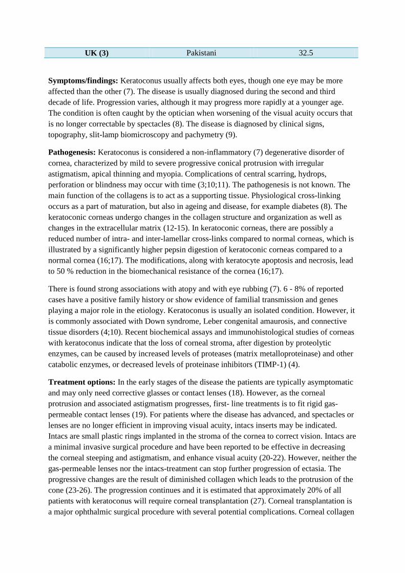
UK (3) Pakistani 32.5
Symptoms/findings: Keratoconus usually affects both eyes, though one eye may be more
affected than the other (7). The disease is usually diagnosed during the second and third
decade of life. Progression varies, although it may progress more rapidly at a younger age.
The condition is often caught by the optician when worsening of the visual acuity occurs that
is no longer correctable by spectacles (8). The disease is diagnosed by clinical signs,
topography, slit-lamp biomicroscopy and pachymetry (9).
Pathogenesis: Keratoconus is considered a non-inflammatory (7) degenerative disorder of
cornea, characterized by mild to severe progressive conical protrusion with irregular
astigmatism, apical thinning and myopia. Complications of central scarring, hydrops,
perforation or blindness may occur with time (3;10;11). The pathogenesis is not known. The
main function of the collagens is to act as a supporting tissue. Physiological cross-linking
occurs as a part of maturation, but also in ageing and disease, for example diabetes (8). The
keratoconic corneas undergo changes in the collagen structure and organization as well as
changes in the extracellular matrix (12-15). In keratoconic corneas, there are possibly a
reduced number of intra- and inter-lamellar cross-links compared to normal corneas, which is
illustrated by a significantly higher pepsin digestion of keratoconic corneas compared to a
normal cornea (16;17). The modifications, along with keratocyte apoptosis and necrosis, lead
to 50 % reduction in the biomechanical resistance of the cornea (16;17).
There is found strong associations with atopy and with eye rubbing (7). 6 - 8% of reported
cases have a positive family history or show evidence of familial transmission and genes
playing a major role in the etiology. Keratoconus is usually an isolated condition. However, it
is commonly associated with Down syndrome, Leber congenital amaurosis, and connective
tissue disorders (4;10). Recent biochemical assays and immunohistological studies of corneas
with keratoconus indicate that the loss of corneal stroma, after digestion by proteolytic
enzymes, can be caused by increased levels of proteases (matrix metalloproteinase) and other
catabolic enzymes, or decreased levels of proteinase inhibitors (TIMP-1) (4).
Treatment options: In the early stages of the disease the patients are typically asymptomatic
and may only need corrective glasses or contact lenses (18). However, as the corneal
protrusion and associated astigmatism progresses, first- line treatments is to fit rigid gas-
permeable contact lenses (19). For patients where the disease has advanced, and spectacles or
lenses are no longer efficient in improving visual acuity, intacs inserts may be indicated.
Intacs are small plastic rings implanted in the stroma of the cornea to correct vision. Intacs are
a minimal invasive surgical procedure and have been reported to be effective in decreasing
the corneal steeping and astigmatism, and enhance visual acuity (20-22). However, neither the
gas-permeable lenses nor the intacs-treatment can stop further progression of ectasia. The
progressive changes are the result of diminished collagen which leads to the protrusion of the
cone (23-26). The progression continues and it is estimated that approximately 20% of all
patients with keratoconus will require corneal transplantation (27). Corneal transplantation is
a major ophthalmic surgical procedure with several potential complications. Corneal collagen

cross-linking with riboflavin is a relatively new treatment that shows good results in
preventing progression of keratoconus (8). Corneal collagen cross-linking targets the
biochemical properties (28), prevents visual loss and significantly reduces the number of
patients requiring surgical treatments (8).

Methods
This literature study is based on selected articles on CXL and keratoconus. The information is
provided by PubMed with search words “keratoconus” and “corneal collagen Cross-linking”.
The search of articles has not been limited by year of publication, but papers in other
languages than English have been excluded considering publish-year.
Corneal Collagen Cross-linking and Keratoconus
1. Corneal collagen cross-linking
Corneal collagen cross-linking is a treatment designed to increase the rigidity and the
structural integrity of the cornea. This prevents further progressive ectasia (4). Collagen cross-
linking is indicated in progressive keratectasia, such as keratoconus (29) and the associated
variants (post-LASIK ectasia (LASIK = laser-assisted in situ keratomileusis), corneal melting
conditions or infectious keratitis) (18).
Spoerl and Huhle et al. (30) were the first to prospect the use of riboflavin and UVA to obtain
cross-linking of corneal collagen. By using this method, Wollensak (31) and Kohlhaas et al.
(32) demonstrated a clearly positive effect of cross-linking of both porcine and human corneal
tissue. The results showed that CXL increased the rigidity of corneal tissue (31). It is thought
that the riboflavin has double function; it acts as a photosynthesizer for the production of
oxygen-free radicals, in addition to absorbing the UVA irradiation and preventing damage to
deeper ocular structures such as the corneal endothelium, the lens, and the retina (33). When
riboflavin is excited into a triplet state by UV exposure it releases highly reactive oxygen
species into the stroma (34) These oxygen species react with surrounding molecules and
amongst several non-specific interactions also result in the formation of cross-links consisting
of intra- and intermicrofibrillar covalent bonds (35). The process is dependent on oxygen, and
enhanced by deuterium oxide (36), which differs it from the formation of cross-links that are
made with increased age and diabetes (37). The cross-links cause increase in the collagen
diameter and in spacing between collagen fibrils (34;38).
Corneal collagen cross-linking was initially thought to increase the biomechanical stability of
the corneal stroma and its resistance to enzymatic digestion, by inducing cross linkage
between the stromal collagen molecules. However, Meek et al. (39) suggested as a result of x-
ray diffraction studies, that CXL is instead strengthening the interlamellar collagen fibril
adhesion of adjacent lamellae originally weakened by keratoconus. The technique has shown
to be safe with no endothelial damage, presupposed that the cornea is thicker than 400 µm,
with no loss of corneal stromal transparency and no damage to deeper ocular structures
(40;41). In several studies, riboflavin /UVA cross linkage has been shown to increase stress-
strain measurements, reduce the swelling rates, increase shrinkage temperature and increase
the resistance against enzymatic degeneration (pepsin) of corneal stromal tissue (31;42;43).
Morphometric computer software has shown increased diameter of the collagen fibers with
most changes occurring in the anterior 200 µm without any damage to the corneal
endothelium (32;34). Recent data show an actual statistically significant modification in the
modulus of elasticity, increased by 4.5 times, and 328 % increase in tensile rigidity, in the
human cornea after Riboflavin/UVA CXL (41;44). Corneal cross-linking is the latest
treatment which may be a potential intervention slowing down the progression of the disease
(8).
Individuals who are in “early-to-moderate keratoconus-phase” and are showing signs of
progression are better candidates for CXL than those at the end-stage of the disease (45).
Corneal cross-linking is also indicated in patients with keratectasia after corneal laser
corrective surgery (29). Recommended limitations in order to perform CXL are maximum
keratometric (K) readings less than 60 diopters (D) and a central corneal thickness (CCT) of
at least 400 µm. The latter concerns the thickness of the deepithelialized cornea when epi-off
CXL is used. Also, CXL done on patients with keratoconus younger than 18 years have
shown positive results 3 years post-treatment(46).
2. History
Collagen cross-linking, involving corneal collagen was first investigated in diabetics in the
1990s (45). Researchers in Dresden noted that this group reduced the incidence of
keratoconus because of glycosylation-mediated cross-linking which strengthened the tissue
(37;45;47;48), and found that diabetes may be a protective factor against the development of
keratoconus (48). To study and show this, Maillard reactions (addition of an aldehyde sugar to
amino group of protein chains), were investigated in the corneas of diabetics compared to
controls (37). It was found that diabetics accumulate more and more glycation end-products
(AGEs) and collagen modification by AGEs results in covalent cross-linking. Another study
found that diabetics had increased AGEs and increased cross links from pentosidine, which is
one of the AGEs formed (37).
In addition to the AGEs related CXL, cross linking has been found to occur in the cornea and
crystalline lens with increasing age (37;49). In a normal cornea it exist covalently bonded
molecular bridges or cross-links between nearby collagen-helices and between microfibrils
and fibrils along their length (50;51). Corneal fibril diameter increases with 4,5 % through life
(52) and the types of cross-linking seen in aging has been shown to be due to enzyme lysyl
oxidase, which leads to aldehyde formation and further post-translational modifications (37).
With aging, the range of natural cross-linking increases (52;53) with modifications in the
biomechanical properties in the tissue and a measurable enhance in Young’s modulus (54).
This may explain the flexibility of the infant cornea compared to adult tissue and the slowing
progression of keratoconus observed with increasing age (27).
The concept of photopolymerisation to strengthen tissues has been around for some time in
industry, as a means to harden various plastic and other long chain molecules (35). In 1968
Christopher Foote published the mechanisms by which photosensitized oxidation happens in
biological systems (55). In 1988 Fujimori disclosed a third mechanism of cross-linkage in
type I collagen involving either oxidation by ozone or photo-oxidation by UV-light (56). It is
also the same technology that has been used to harden bioprosthetic materials (57), as for
example artificial heart valves that are stiffened using glutaraldehyde (8), and as a component
in dental filling materials (57). Indeed a visit to a dentist who used UV irradiation to harden
filling material led to the inspiration to stiffen the cornea (58).
Sporl et al. (30) published the result of their study that induced cross-linkage in porcine eyes
in German in 1997 (followed by in English in 1998 (49)). Their study included 8 test groups
with 10 eyes in each group. The eyes were de-epithelialized and the cornea was treated with
UV light (254 nm), 0.5 % riboflavin, 0.5 % riboflavin and UV light (365 nm), blue light (436
nm), sunlight, glutaraldehyde (0.1 %, 10 min), glutaraldehyde (1 %, 10 min) or Karnovsky’s
solution (a fixative used in electron microscopy which contains paraformaldehyde, sodium
hydroxide and glutaraldehyde) (0.1%, 10 min). A ninth group served as control. The results of
this study showed; riboflavin and UV, glutaraldehyde and Karnovsky’s caused an increase in
stiffness of the treated cornea compared to the control group (p<0.05) (49), and ultraviolet at
365 nm was found to strengthen the cornea best compared to ultraviolet at 254 nm and 436
nm (18). The toxicity and the inability to control depth and reach of diffusion of
glutaraldehyde makes it impractical in clinical content (35). While the riboflavin-UVA
showed promising results for the next stage of human studies (59).
All studies thereafter were done in context of this, with the exception of the riboflavin
concentrate that was changed to 0,1 % in all future studies (18). The next big experiment
consisted of 20 porcine eyes treated with 0.1 % riboflavin and ultraviolet light 370 nm, 3
mW/cm2 for 30 minutes, compared to 20 untreated porcine eyes (32). In a similar manner 5
human donor corneas were treated the same way. The experiment concluded that the
stiffening effect was marked in the treated eyes (32). The stiffening effect was increased by a
factor of 4.5 compared to1.8 in the porcine cornea (31). In addition they discovered that 65-70
% of the UV light was absorbed in the anterior 200 µm of the stroma and only about 20 %
reached the next 200 µm (32), which was later used to determine the effect on deeper
structures for in-vivo studies (18).
In 2004 Seiler et al. (34) published results of increase of collagen fiber diameter in rabbit
corneas. They found that in the treated anterior stroma the collagen fiber was significantly
increased by 12.2% compared to control eyes, while in the treated posterior cornea it was
increased by 4.6 %. Enzymatic digestive studies and electrophoresis were done to analyze the
biomechanical aspects of the CXL (42;60). The former study compared 60 riboflavin-induced
cross linked porcine eyes to control group, to investigate their time to be digested fully by
pepsin, trypsin and collagenase. The results concluded that it took 13 days to digest pepsin in
treated eyes vs. 6 days in the control eyes (42). The results for trypsin and collagenase were
similar and the conclusion was a marked resistance to enzymatic digestion, suggesting high
numbers of cross linked bonds requiring digestion (18). In a study performed by Wollensak et
al.(60), the type I collagen was extracted from the corneas, run on sodium deodecyl sulfate-
polyacrylamide gels and separated by electrophoresis (60). In the cross linked cornea-extracts
they found another much larger band (1000 kDa in contrast to usual band sizes of 130 kDa,
200 kDa, and 300 kDa) compared to the control corneas that only showed the usual type I
collagen pattern. This larger band was further found to be chemical stable; resistant to heat
denaturation, pepsin treatment and mercaptoethanol treatment.
Another study by Wollensak et al. (61), before he did in-vivo human studies, was a hydration
study and second analysis of the depth of treatment using optical coherence tomography
(OCT). Wollensak et al. treated 20 porcine eyes with the riboflavin ultraviolet protocol. The
eyes were incubated for 24 hours in a moist chamber, then 15 eyes were examined by
biomicroscopy and OCT and 5 eyes were examined by light microscopy. The results of this
study were concluded with a characteristic swelling pattern that affected the anterior cornea
portion greater than the posterior portion. The cornea was divided into 3 zones; anterior zone
(242 µm), intermediate zone (next 238 µm) and a posterior zone (The remainder of the tissue,
1355 µm for porcine corneas). The swelling was found to be increased by a factor of 2.2 in
the intermediate zone and by a factor of 2.7 in the posterior zone (61). The light microscopy
concluded cross linking pattern. These results were profitable in analyzing the amount of
swelling following treatment and the treatment depth (18).
Wollensak et al. (41) began in 1998 to treat patients using the riboflavin UVA cross-linking
technique, with the support of all of the in-vivo and ex-vivo animal and human corneal
studies. The results of the pilot study were published in 2003, with a follow-up ranging from 3
months to 4 years. The study showed that all treated eyes had an obstacle of the progression
of the keratoconus and that 70 % had regression and a reduction in the maximal keratometry-
readings by an average of 2 diopters (41). Since then the idea spread to other centers and
resulted in patients with keratoconus being treated worldwide (18).
In the Dresden study they found statistically significant changes after the first year post-
operatively and remained stable for the remainder of the follow-up, which extended up to 6
years (62). The explanation to the observation that many of the patients had improvement in
visual acuity felt to be a combination of a decrease in the amount of irregular astigmatism, a
decrease in the corneal curvature, and a secondary improvement in the ability to fit contact
lenses. They found no statistically significant change in IOP (intra ocular pressure) at one year
(62). Small differences were found in the effect of the central corneal thickness (CCT) change
(44).
3. Technique
3.1. Before the treatment
Before the treatment the patient needs to go through an eye examination including refraction,
keratometry, topography and regional pachymetry (33;63;64). It is also by some
recommended to discontinue the use of contact lenses 1 month before treatment (65-67) and
use broad-spectrum antibiotics 1 to 3 days before treatment.
3.2. Treatment
The CXL treatment protocol most widely used is based on the work of Wollensak, Spoerl,
Seiler and co-workers. It is optimized based on extensive laboratory data to maximize the
crosslinking effect and minimize any damages to ocular tissue. The CXL procedure is
conducted under sterile conditions, after the patient having 4 drops of Ofloxacin 0.3 % and 2
drops of Pilocarpine 2% instilled in the eye to be treated. Then the patient’s eye is
anesthetized with Oxybuprocaine 0,4% drops (4). The central 7-9 mm of the corneal
epithelium are removed to allow better diffusion of riboflavin into the stroma (68). The
epithelium acts as a barrier and without epithelial removal the biomechanical effect is reduced
to be under 50 % of the standard cross-linking procedure (28). Baiocchi et al. (69) showed
that the necessary stromal concentration of riboflavin is only obtained after epithelial removal.
Iso-osmolar riboflavin 0, 1 % solution with dextran 20 % is then applied to the cornea every 2
to 3 minutes for 30 minutes (68). After this time an ultrasound pachymetry (5 repetitive
measurements) on the cornea at the thinnest is done to confirm that it is at least 400 µm in
depth (68). If the cornea is too thin, riboflavin 0, 1 % solution in hypo-osmolar saline is used
instead. Ultrasound pachymetry has to be performed to make sure that the thickness is at least
400 µm (4). Successful penetration of riboflavin through the cornea is certified by
visualization of riboflavin in the anterior chamber by slit-lamp biomicroscopy, using cobalt
blue light, before irradiation (4).
UVA irradiation is commenced using wavelength of 370 nm for 30 minutes at working
distance of 54 mm (68). There are at least three widely available UV light sources for CXL;
the IROC UV-X system (IROC AG, Switzerland), the CSO VEGA system (CSO Italy) and
OptoXlink (Opto Global Pty Ltd, Australia). The three devices come with separate
luminescence-measuring device and have somewhat different operating instructions and
focusing distances (70). Through the irradiation riboflavin solution is applied every 2-3
minutes while ensuring the stroma is thick enough (68).Topical anesthetic agent
(Oxybuprocaine 0,4 %) is applied whenever necessary (4).
3.3. After the treatment
Antibiotic drops (Ofloxacin 0,3 %) are applied and a bandage contact lens is inserted until
the reepithelialization is complete (4). Antibiotic drops (Ofloxacin 0,3 % ) 4 drops a day for a
week were administered and analgesics by mouth as necessary (4). Corticosteroids are used to
minimize the inflammatory response (58). The patient should be closely examined in the early
postoperative period, typically at day 1, 3 and further as required to ensure epithelial healing
(70). Later examinations were performed in month 1, 3, 6 and 12 (45;66;67;71)
postoperatively. Patients wearing gas permeable contact lenses may interpret the results of
CXL since lens wear improves the topographic parameters (72).
4. Contraindications
4.1. Absolute contraindications for treatment with CXL
Corneal thickness of less than 400 µm: Patients with corneas thinner than 400 µm should
not undergo CXL because of possible endothelial cell density decrease postoperatively
(29;73). In advanced keratoconus progressive corneal thinning often leads to thinning of
remaining stromal thickness by less than 400 µm. Hafezi el al. (74) changed the current
treatment protocol by preoperatively swelling thin corneas to a stromal thickness of at least
400 µm using riboflavin 0,1 % solution in hypo-osmolar saline. This treatment protocol was
performed in case series of 20 patients, and no complications were observed (74).
Central corneal opacities and severe dry eye; can be a hinder to re-epithalization (29).
Pregnancy, nursing, having systemic collagen vascular disease (29), concomitant
autoimmune disease (28), history of herpetic keratitis, corneal scarring or concomitant
autoimmune diseases are all conditions were undergoing CXL should be avoided since these
populations have not been appropriately tested (29).
4.2. Relative Contraindications
Maximum K reading more than 58 D: the efficiency of CXL would likely increase if the
treatment was limited to eyes with a maximum K reading of less than 58.00 D (75).
Comprehensive corneal scarring is associated with poor existing best-corrected visual
acuity: Patients with an already limited vision may not advantage from CXL unless it is
combined with a refractive treatment option (for example INTACS). Alternative treatments
should then be considered (for example graft surgery) (4).
Patients with primary or secondary keratectasia, especially corneal guttata or other
endothelial irregularities, history of recurrent erosions, ocular surface disorders, and
connective tissue disease: should be advised and the risks ought to be discussed with the
patient (4).
5. Complications
Blurry vision, lacrimation and foreign body sensation: Can be expected for 24-48 hours
(34). Blurry vision can last up to 1 month or longer depending on the corneal edema.
Stromal haze: Haze occurring after collagen cross-linking usually subsides completely the
first postoperative year. It reaches up to approximately 60 % depth in the anterior stroma
(equal to absolute depth of 300 mm) (64;76). The nature of mild stromal haze is related to
transient corneal fibroblast generation rather than more persistent haze due to generation of
myofibroblasts (77). The demarcation line was described by Seiler and Hafezi to represent the
transition zone between the CXL-treated stroma and the untreated posterior layers (64).
During healing phase: The cornea is vulnerable to infection and melting. Several studies
have published case reports, of infectious keratitis and limited corneal melting, associated
with riboflavin/UVA CXL (78-85). It is more likely that corneal infections after CXL break
out during the early postoperative period rather than during surgery because CXL kills
bacteria and fungi (86-89).
Limbal epithelial cells: Riboflavin-UVA should not be used on the limbus during CXL since
the oxygen radicals can have toxic effect, and reduced cell expansion of limbal epithelial cells
has been reported (90).
Postoperative pain: Pain can be seen the first 3 days after treatment, and can be intense. The
pain has been found to be more intense the younger the patient is (91;92).
IOP: Higher IOP-values are found after CXL when measured by tonometry. These are
probably due to increased stiffness of the cornea after treatment (93).
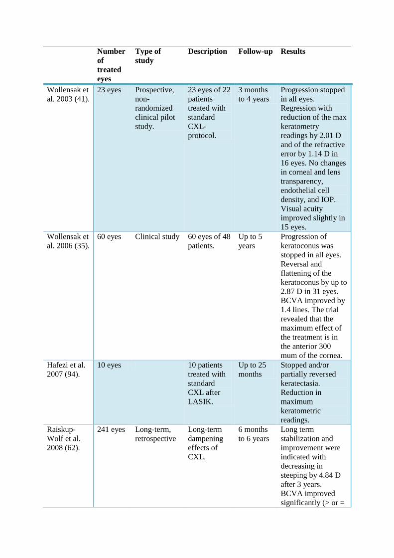
6. Results
Table 2: The table presents an overview of some of the CXL-trials on progressive
keratoconus.
Number
of
treated
eyes
Type of
study
Description Follow-up Results
Wollensak et
al. 2003 (41).
23 eyes Prospective,
non-
randomized
clinical pilot
study.
23 eyes of 22
patients
treated with
standard
CXL-
protocol.
3 months
to 4 years
Progression stopped
in all eyes.
Regression with
reduction of the max
keratometry
readings by 2.01 D
and of the refractive
error by 1.14 D in
16 eyes. No changes
in corneal and lens
transparency,
endothelial cell
density, and IOP.
Visual acuity
improved slightly in
15 eyes.
Wollensak et
al. 2006 (35).
60 eyes Clinical study 60 eyes of 48
patients.
Up to 5
years
Progression of
keratoconus was
stopped in all eyes.
Reversal and
flattening of the
keratoconus by up to
2.87 D in 31 eyes.
BCVA improved by
1.4 lines. The trial
revealed that the
maximum effect of
the treatment is in
the anterior 300
mum of the cornea.
Hafezi et al.
2007 (94).
10 eyes 10 patients
treated with
standard
CXL after
LASIK.
Up to 25
months
Stopped and/or
partially reversed
keratectasia.
Reduction in
maximum
keratometric
readings.
Raiskup-
Wolf et al.
2008 (62).
241 eyes Long-term,
retrospective
Long-term
dampening
effects of
CXL.
6 months
to 6 years
Long term
stabilization and
improvement were
indicated with
decreasing in
steeping by 4.84 D
after 3 years.
BCVA improved
significantly (> or =
1 line), 57% of 66
eyes or remained
stable in 24% after 2
years. Two patients
had to repeat CXL.
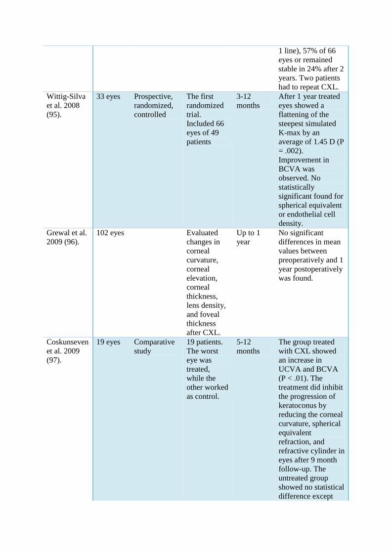
Wittig-Silva
et al. 2008
(95).
33 eyes Prospective,
randomized,
controlled
The first
randomized
trial.
Included 66
eyes of 49
patients
3-12
months
After 1 year treated
eyes showed a
flattening of the
steepest simulated
K-max by an
average of 1.45 D (P
= .002).
Improvement in
BCVA was
observed. No
statistically
significant found for
spherical equivalent
or endothelial cell
density.
Grewal et al.
2009 (96).
102 eyes Evaluated
changes in
corneal
curvature,
corneal
elevation,
corneal
thickness,
lens density,
and foveal
thickness
after CXL.
Up to 1
year
No significant
differences in mean
values between
preoperatively and 1
year postoperatively
was found.
Coskunseven
et al. 2009
(97).
19 eyes Comparative
study
19 patients.
The worst
eye was
treated,
while the
other worked
as control.
5-12
months
The group treated
with CXL showed
an increase in
UCVA and BCVA
(P < .01). The
treatment did inhibit
the progression of
keratoconus by
reducing the corneal
curvature, spherical
equivalent
refraction, and
refractive cylinder in
eyes after 9 month
follow-up. The
untreated group
showed no statistical
difference except
UCVA and BCVA
which decreased.
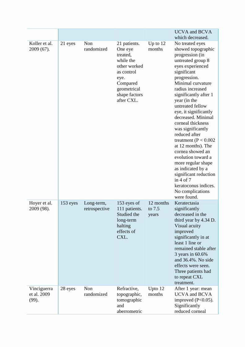
Koller et al.
2009 (67).
21 eyes Non
randomized
21 patients.
One eye
treated,
while the
other worked
as control
eye.
Compared
geometrical
shape factors
after CXL.
Up to 12
months
No treated eyes
showed topographic
progression (in
untreated group 8
eyes experienced
significant
progression.
Minimal curvature
radius increased
significantly after 1
year (in the
untreated fellow
eye, it significantly
decreased. Minimal
corneal thickness
was significantly
reduced after
treatment (P < 0.002
at 12 months). The
cornea showed an
evolution toward a
more regular shape
as indicated by a
significant reduction
in 4 of 7
keratoconus indices.
No complications
were found.
Hoyer et al.
2009 (98).
153 eyes Long-term,
retrospective
153 eyes of
111 patients.
Studied the
long-term
halting
effects of
CXL.
12 months
to 7.5
years
Keratectasia
significantly
decreased in the
third year by 4.34 D.
Visual acuity
improved
significantly in at
least 1 line or
remained stable after
3 years in 60.6%
and 36.4%. No side
effects were seen.
Three patients had
to repeat CXL
treatment.
Vinciguerra
et al. 2009
(99).
28 eyes Non
randomized
Refractive,
topographic,
tomographic
and
aberrometric
Upto 12
months
After 1 year: mean
UCVA and BCVA
improved (P<0.05).
Significantly
reduced corneal
outcomes 12
months after
CXL.
average papillary
power (APP), apical
keratometry (AK),
and corneal and total
wave front
aberrations. No
changes in
endothelial cell
counts were seen.
Hafezi et al.
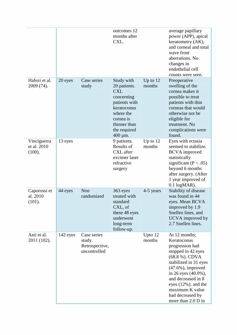
2009 (74).
20 eyes Case series
study
Study with
20 patients.
CXL
concerning
patients with
keratoconus
where the
cornea is
thinner than
the required
400 µm.
Up to 12
months
Preoperative
swelling of the
cornea makes it
possible to treat
patients with thin
corneas that would
otherwise not be
eligible for
treatment. No
complications were
found.
Vinciguerra
et al. 2010
(100).
13 eyes 9 patients.
Results of
CXL after
excimer laser
refractive
surgery
Up to 12
months
Eyes with ectasia
seemed to stabilize.
BCVA improved
statistically
significant (P < .05)
beyond 6 months
after surgery. (After
1 year improved of
0.1 logMAR).
Caporossi et
al. 2010
(101).
44 eyes Non
randomized
363 eyes
treated with
standard
CXL, of
these 48 eyes
underwent
long-term
follow-up.
4-5 years Stability of disease
was found in 44
eyes. Mean BCVA
improved by 1.9
Snellen lines, and
UCVA improved by
2.7 Snellen lines.
Asri et al.
2011 (102).
142 eyes Case series
study.
Retrospective,
uncontrolled
Upto 12
months
At 12 months;
Keratoconus
progression had
stopped in 42 eyes
(68.8 %). CDVA
stabilized in 31 eyes
(47.6%), improved
in 26 eyes (40.0%),
and decreased in 8
eyes (12%). and the
maximum K value
had decreased by
more than 2.0 D in
13 eyes (21.3%).
Complication rate
with loss of vision;
3.5%.
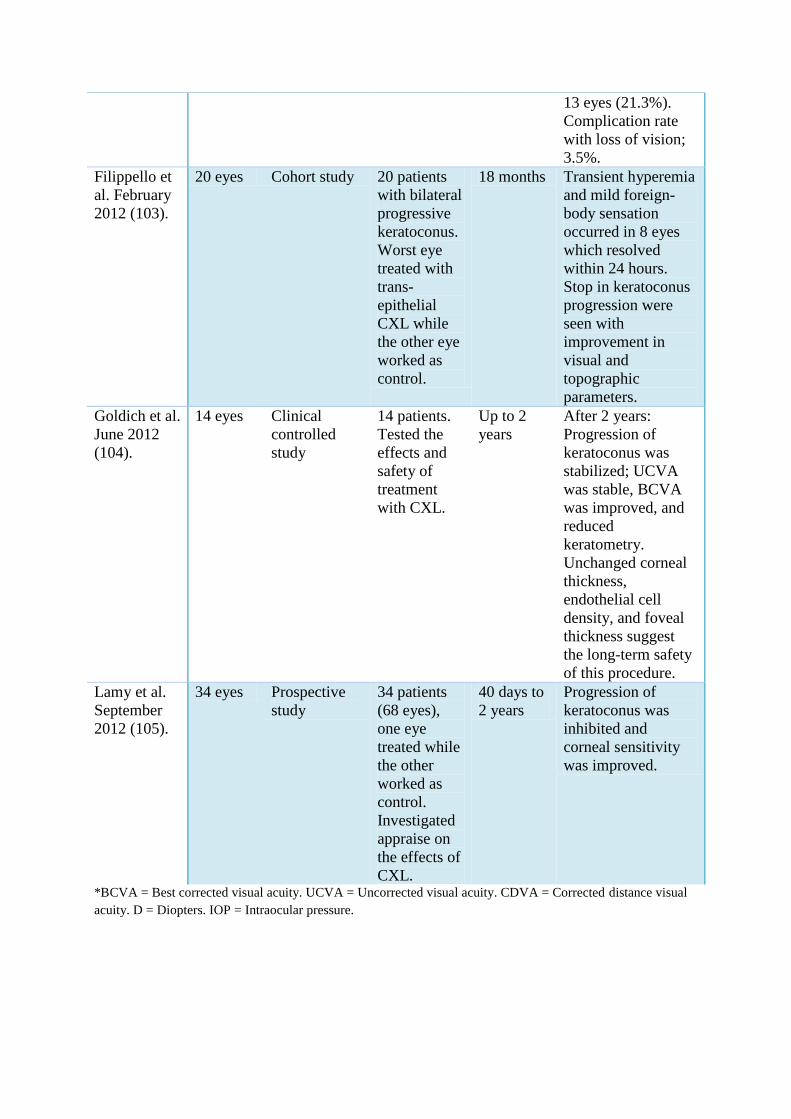
Filippello et
al. February
2012 (103).
20 eyes Cohort study 20 patients
with bilateral
progressive
keratoconus.
Worst eye
treated with
trans-
epithelial
CXL while
the other eye
worked as
control.
18 months Transient hyperemia
and mild foreign-
body sensation
occurred in 8 eyes
which resolved
within 24 hours.
Stop in keratoconus
progression were
seen with
improvement in
visual and
topographic
parameters.
Goldich et al.
June 2012
(104).
14 eyes Clinical
controlled
study
14 patients.
Tested the
effects and
safety of
treatment
with CXL.
Up to 2
years
After 2 years:
Progression of
keratoconus was
stabilized; UCVA
was stable, BCVA
was improved, and
reduced
keratometry.
Unchanged corneal
thickness,
endothelial cell
density, and foveal
thickness suggest
the long-term safety
of this procedure.
Lamy et al.
September
2012 (105).
34 eyes Prospective
study
34 patients
(68 eyes),
one eye
treated while
the other
worked as
control.
Investigated
appraise on
the effects of
CXL.
40 days to
2 years
Progression of
keratoconus was
inhibited and
corneal sensitivity
was improved.
*BCVA = Best corrected visual acuity. UCVA = Uncorrected visual acuity. CDVA = Corrected distance visual
acuity. D = Diopters. IOP = Intraocular pressure.
Discussion
The primary challenge in identifying a permanent treatment for keratoconus is the
pathophysiology of the disease not being fully understood. The corneal changes of
keratoconus can vary across a wide spectrum from stability to rapid progression. Thus
variation in progression can be seen in the same patient at different points, and can even vary
significantly between each eye in the same patient.
When it comes to the safety and long-term efficacy of the treatment, there are areas of
concern. The treatment criteria for patients who should be treated have to be specified
accurately. More studies are necessary to assemble a list of indications regarding patients’
age, diagnosis and the stage of keratectasia. In future studies the effects of other ocular
parameters should be addressed in further detail: parameters like tear function, corneal
sensitivity, alterations in conjunctival epithelium and goblet cells, the long-term effect on
direct UVA irradiation on ocular surface limbal stem cells, and potentially occurrence of
metaplastic disorders of the ocular surface.
The clinical studies of CXL have mostly been limited to small uncontrolled retrospective
series with relatively short follow up. However, the last few years there has been a rapid
growth in interest in this treatment worldwide, with clinical outcomes now reported from
several countries, and the findings of all studies being remarkable consistent.
A disadvantage of CXL is that complete epithelial debridement prolongs early postoperative
discomfort. In addition it prolongs epithelialization, thus increasing the chance of bacterial
and or fungal infiltration. However, adequate stromal absorption of riboflavin may not be
achieved without epithelial debridement. Even if the secondary infection is treated it may
cause corneal opacification and decrease the corrected distance visual acuity (106-108). There
are also concerns about an important aspect of the CXL-technique. By actually increasing
rigidity, the cornea is being aged, and the long-term results of that might not be beneficial.
The induced rigidity of the cornea may promote premature ocular rigidity in general, creating
future severe ocular problems for example age related macular degeneration (109).
Regarding safety considerations, riboflavin, which is also known as vitamin B2, is considered
to be non-toxic. It is water-soluble and is able to penetrate corneal stroma after the removal of
epithelium. There has not been any evidence of riboflavin toxicity in human-studies with oral
intake up to 400 mg/day (49). Given that all studies have shown minimal effect on intraocular
pressure and the optic nerve, it is assumed that riboflavin is filtered efficiently through the
trabecular meshwork (18). Other safety considerations have been the effects on the
keratocytes. Studies have shown that keratocyte loss could be seen down to a depth of 340 µm
in the entire CXL treated corneas (110) compared to the kerotoconus corneas that have not
been treated with CXL and the normal corneas. Corneal cross-linking can cause morphologic
corneal changes up to 30 months after treatment, especially a long-lasting, maybe permanent
keratocyte loss in the anterior and middle corneal stroma involving the central and peripheral
cornea. (111). However, it is suggested that corneal cross-linking does not induce significant
cellular epithelial damage (112) CXL with epithelial debrievement have resulted in nerve
fiber damage and reduced sensitivity, but the nerves have in the mentioned complications
regenerated and appeared normal by 180 days after treatment (113). Changes in the structure
and cellularity of the cornea have been observed up to 3 years after CXL treatment with
confocal microscopy. The alterations can be related to increased cross-link formation,
synthesis of well-structured collagen, or other lamellar interconnections (110). When it comes
to photochemical damage of keratocytes the endothelial thresholds are 0.45 and 0.35
mW/cm2. In a 400 µm thick cornea saturated with riboflavin, the irradiance at the endothelial
level is calculated to be 0.18 mW/cm, which is a factor of 2 smaller than the damage threshold
(33). This is the reason why a cornea less than 400 µm is recommended not to be treated (1).
If the patient is less than 35 years old and the best corrected visual acuity is worse than 6/7,5
CXL seems to be a safe treatment that would yield a complication rate of approximately 1 %
(75). A temporary loss of visual acuity can be observed in some cases. A permanent loss of 2
or more Snellen lines at an appropriate time after surgery (6 months or 1 year) is considered a
complication. Koller et al, found a permanent loss in 2.9 % of their patients. Other studies
show a rate of less than 1 % assumed the patient age is above 35 years and best spectacle
corrected vision better than 6/7,5 (75).
Publication of the results of the randomized controlled trials currently in progress will enable
a more confident assessment of the efficacy of this procedure. Also the safety of CXL will
take time to fully ascertain and long-term observations will be required. Corneal collagen
cross-linking may represent a breakthrough in the treatment of keratoconus, significantly
reducing the need for corneal transplantation.
Conclusion
Collagen cross-linking with riboflavin and UVA have shown to be a very promising method
of treating keratoconus. It is the first therapeutic modality that is shown to halt the progression
of the ectatic process in keratoconus. The treatment is a minimal invasive technique,
promising therapeutic intervention for tissue stabilization.
This treatment appears to have the potential to reduce incidence of progressive forms of
keratoconus. There is also a theoretical basis for the use of CXL in several other corneal
diseases. Both factors may ultimately reduce the need for corneal transplantations. It is
approximated that 20 % of patients with untreated keratoconus will eventually end up having
corneal transplantation. In addition to the clinical benefits, there are also economic and
psychosocial benefits of CXL.
Clinical trials of CXL on progressive keratoconus have shown interesting results. The results
presented shows that moderate to advanced progressive keratoconus is effectively halted,
often with some regression of the corneal steepness resulting in improvement in vision. Only
a few cases needed to repeat treatment. In particular no damage to corneal endothelium, lens
or retina was noted in any of the clinical trials. The data also indicates that the treatment
should only be performed in patients with documented progression of keratoconus in the
preoperative months. A Few cases of keratitis after CXL have been published. Infection
occurs early in the immediate postoperative period and requires strict vigilance during this
time.
Current studies indicate the stiffening effect of CXL appears to remain stable after 6 years,
and long term data are awaited. However, the exact duration of the corneal stiffening effect is
still unknown due to the relative novelty of the treatment, and long term data are yet to be
evaluated. More prospective randomized controlled trials in the future are desirable.
Reference List
(1) Reinstein DZ, Gobbe M, Archer TJ, Couch D, Bloom B. Epithelial, stromal, and corneal pachymetry changes during orthokeratology. Optom Vis Sci 2009 August;86(8):E1006-E1014.
(2) Andreassen TT, Simonsen AH, Oxlund H. Biomechanical properties of keratoconus and normal corneas. Exp Eye Res 1980 October;31(4):435-41.
(3) Rabinowitz YS. Keratoconus. Surv Ophthalmol 1998 January;42(4):297-319.
(4) Samaras KE, Lake DB. Corneal collagen cross linking (CXL): a review. Int Ophthalmol Clin 2010;50(3):89-100.
(5) Ota R, Fujiki K, Nakayasu K. [Estimation of patient visit rate and incidence of keratoconus in the 23 wards of Tokyo]. Nihon Ganka Gakkai Zasshi 2002 June;106(6):365-72.
(6) Assiri AA, Yousuf BI, Quantock AJ, Murphy PJ. Incidence and severity of keratoconus in Asir province, Saudi Arabia. Br J Ophthalmol 2005 November;89(11):1403-6.
(7) McMonnies CW. Mechanisms of rubbing-related corneal trauma in keratoconus. Cornea 2009 July;28(6):607-15.
(8) Ashwin PT, McDonnell PJ. Collagen cross-linkage: a comprehensive review and directions for future research. Br J Ophthalmol 2010 August;94(8):965-70.
(9) Kok YO, Tan GF, Loon SC. Review: keratoconus in Asia. Cornea 2012 May;31(5):581-93.
(10) Krachmer JH, Feder RS, Belin MW. Keratoconus and related noninflammatory corneal thinning disorders. Surv Ophthalmol 1984 January;28(4):293-322.
(11) McGhee CN. 2008 Sir Norman McAlister Gregg Lecture: 150 years of practical observations on the conical cornea--what have we learned? Clin Experiment Ophthalmol 2009 March;37(2):160-76.
(12) Cheng EL, Maruyama I, SundarRaj N, Sugar J, Feder RS, Yue BY. Expression of type XII collagen and hemidesmosome-associated proteins in keratoconus corneas. Curr Eye Res 2001 May;22(5):333-40.
(13) Kenney MC, Nesburn AB, Burgeson RE, Butkowski RJ, Ljubimov AV. Abnormalities of the extracellular matrix in keratoconus corneas. Cornea 1997 May;16(3):345-51.
(14) Radner W, Zehetmayer M, Skorpik C, Mallinger R. Altered organization of collagen in the apex of keratoconus corneas. Ophthalmic Res 1998;30(5):327-32.
(15) Tuori AJ, Virtanen I, Aine E, Kalluri R, Miner JH, Uusitalo HM. The immunohistochemical composition of corneal basement membrane in keratoconus. Curr Eye Res 1997 August;16(8):792-801.
(16) Andreassen TT, Simonsen AH, Oxlund H. Biomechanical properties of keratoconus and normal corneas. Exp Eye Res 1980 October;31(4):435-41.
(17) Cannon DJ, Foster CS. Collagen crosslinking in keratoconus. Invest Ophthalmol Vis Sci 1978 January;17(1):63-5.
(18) Keating A, Pineda R, Colby K. Corneal cross linking for keratoconus. Semin Ophthalmol 2010 September;25(5-6):249-55.
(19) Hwang JS, Lee JH, Wee WR, Kim MK. Effects of multicurve RGP contact lens use on topographic changes in keratoconus. Korean J Ophthalmol 2010 August;24(4):201-6.
(20) Boxer Wachler BS, Christie JP, Chandra NS, Chou B, Korn T, Nepomuceno R. Intacs for keratoconus. Ophthalmology 2003 May;110(5):1031-40.
(21) Colin J, Malet FJ. Intacs for the correction of keratoconus: two-year follow-up. J Cataract Refract Surg 2007 January;33(1):69-74.
(22) Siganos CS, Kymionis GD, Kartakis N, Theodorakis MA, Astyrakakis N, Pallikaris IG. Management of keratoconus with Intacs. Am J Ophthalmol 2003 January;135(1):64-70.
(23) Al-Yousuf N, Mavrikakis I, Mavrikakis E, Daya SM. Penetrating keratoplasty: indications over a 10 year period. Br J Ophthalmol 2004 August;88(8):998-1001.
(24) Javadi MA, Motlagh BF, Jafarinasab MR, Rabbanikhah Z, Anissian A, Souri H et al. Outcomes of penetrating keratoplasty in keratoconus. Cornea 2005 November;24(8):941-6.
(25) Mamalis N, Anderson CW, Kreisler KR, Lundergan MK, Olson RJ. Changing trends in the indications for penetrating keratoplasty. Arch Ophthalmol 1992 October;110(10):1409-11.
(26) Reeves SW, Stinnett S, Adelman RA, Afshari NA. Risk factors for progression to penetrating keratoplasty in patients with keratoconus. Am J Ophthalmol 2005 October;140(4):607-11.
(27) Tuft SJ, Moodaley LC, Gregory WM, Davison CR, Buckley RJ. Prognostic factors for the progression of keratoconus. Ophthalmology 1994 March;101(3):439-47.
(28) Gkika M, Labiris G, Kozobolis V. Corneal collagen cross-linking using riboflavin and ultraviolet-A irradiation: a review of clinical and experimental studies. Int Ophthalmol 2011 August;31(4):309-19.
(29) Dahl BJ, Spotts E, Truong JQ. Corneal collagen cross-linking: an introduction and literature review. Optometry 2012 January;83(1):33-42.
(30) Sporl E, Huhle M, Kasper M, Seiler T. [Increased rigidity of the cornea caused by intrastromal cross-linking]. Ophthalmologe 1997 December;94(12):902-6.
(31) Wollensak G, Spoerl E, Seiler T. Stress-strain measurements of human and porcine corneas after riboflavin-ultraviolet-A-induced cross-linking. J Cataract Refract Surg 2003 September;29(9):1780-5.
(32) Kohlhaas M, Spoerl E, Schilde T, Unger G, Wittig C, Pillunat LE. Biomechanical evidence of the distribution of cross-links in corneas treated with riboflavin and ultraviolet A light. J Cataract Refract Surg 2006 February;32(2):279-83.
(33) Spoerl E, Mrochen M, Sliney D, Trokel S, Seiler T. Safety of UVA-riboflavin cross-linking of the cornea. Cornea 2007 May;26(4):385-9.
(34) Wollensak G, Wilsch M, Spoerl E, Seiler T. Collagen fiber diameter in the rabbit cornea after collagen crosslinking by riboflavin/UVA. Cornea 2004 July;23(5):503-7.
(35) Wollensak G. Crosslinking treatment of progressive keratoconus: new hope. Curr Opin Ophthalmol 2006 August;17(4):356-60.
(36) McCall AS, Kraft S, Edelhauser HF, Kidder GW, Lundquist RR, Bradshaw HE et al. Mechanisms of corneal tissue cross-linking in response to treatment with topical riboflavin and long-wavelength ultraviolet radiation (UVA). Invest Ophthalmol Vis Sci 2010 January;51(1):129-38.
(37) Sady C, Khosrof S, Nagaraj R. Advanced Maillard reaction and crosslinking of corneal collagen in diabetes. Biochem Biophys Res Commun 1995 September 25;214(3):793-7.
(38) Dhaliwal JS, Kaufman SC. Corneal collagen cross-linking: a confocal, electron, and light microscopy study of eye bank corneas. Cornea 2009 January;28(1):62-7.
(39) Meek KM, Tuft SJ, Huang Y, Gill PS, Hayes S, Newton RH et al. Changes in collagen orientation and distribution in keratoconus corneas. Invest Ophthalmol Vis Sci 2005 June;46(6):1948-56.
(40) Wollensak G, Spoerl E, Wilsch M, Seiler T. Endothelial cell damage after riboflavin-ultraviolet-A treatment in the rabbit. J Cataract Refract Surg 2003 September;29(9):1786-90.
(41) Wollensak G, Spoerl E, Seiler T. Riboflavin/ultraviolet-a-induced collagen crosslinking for the treatment of keratoconus. Am J Ophthalmol 2003 May;135(5):620-7.
(42) Spoerl E, Wollensak G, Seiler T. Increased resistance of crosslinked cornea against enzymatic digestion. Curr Eye Res 2004 July;29(1):35-40.
(43) Spoerl E, Wollensak G, Dittert DD, Seiler T. Thermomechanical behavior of collagen-cross-linked porcine cornea. Ophthalmologica 2004 March;218(2):136-40.
(44) Caporossi A, Baiocchi S, Mazzotta C, Traversi C, Caporossi T. Parasurgical therapy for keratoconus by riboflavin-ultraviolet type A rays induced cross-linking of corneal collagen: preliminary refractive results in an Italian study. J Cataract Refract Surg 2006 May;32(5):837-45.
(45) Mazzotta C, Balestrazzi A, Traversi C, Baiocchi S, Caporossi T, Tommasi C et al. Treatment of progressive keratoconus by riboflavin-UVA-induced cross-linking of corneal collagen: ultrastructural analysis by Heidelberg Retinal Tomograph II in vivo confocal microscopy in humans. Cornea 2007 May;26(4):390-7.
(46) Caporossi A, Mazzotta C, Baiocchi S, Caporossi T, Denaro R, Balestrazzi A. Riboflavin-UVA-induced corneal collagen cross-linking in pediatric patients. Cornea 2012 March;31(3):227-31.
(47) Kuo IC, Broman A, Pirouzmanesh A, Melia M. Is there an association between diabetes and keratoconus? Ophthalmology 2006 February;113(2):184-90.
(48) Seiler T, Huhle S, Spoerl E, Kunath H. Manifest diabetes and keratoconus: a retrospective case-control study. Graefes Arch Clin Exp Ophthalmol 2000 October;238(10):822-5.
(49) Spoerl E, Huhle M, Seiler T. Induction of cross-links in corneal tissue. Exp Eye Res 1998 January;66(1):97-103.
(50) Robins SP. Biochemistry and functional significance of collagen cross-linking. Biochem Soc Trans 2007 November;35(Pt 5):849-52.
(51) Sung HW, Chang WH, Ma CY, Lee MH. Crosslinking of biological tissues using genipin and/or carbodiimide. J Biomed Mater Res A 2003 March 1;64(3):427-38.
(52) Daxer A, Misof K, Grabner B, Ettl A, Fratzl P. Collagen fibrils in the human corneal stroma: structure and aging. Invest Ophthalmol Vis Sci 1998 March;39(3):644-8.
(53) Malik NS, Moss SJ, Ahmed N, Furth AJ, Wall RS, Meek KM. Ageing of the human corneal stroma: structural and biochemical changes. Biochim Biophys Acta 1992 March 20;1138(3):222-8.
(54) Elsheikh A, Wang D, Brown M, Rama P, Campanelli M, Pye D. Assessment of corneal biomechanical properties and their variation with age. Curr Eye Res 2007 January;32(1):11-9.
(55) Foote CS. Mechanisms of photosensitized oxidation. There are several different types of photosensitized oxidation which may be important in biological systems. Science 1968 November 29;162(3857):963-70.
(56) Fujimori E. Cross-linking of collagen CNBr peptides by ozone or UV light. FEBS Lett 1988 August 1;235(1-2):98-102.
(57) Wollensak G, Sporl E, Seiler T. [Treatment of keratoconus by collagen cross linking]. Ophthalmologe 2003 January;100(1):44-9.
(58) Snibson GR. Collagen cross-linking: a new treatment paradigm in corneal disease - a review. Clin Experiment Ophthalmol 2010 March;38(2):141-53.
(59) Sporl E, Schreiber J, Hellmund K, Seiler T, Knuschke P. [Studies on the stabilization of the cornea in rabbits]. Ophthalmologe 2000 March;97(3):203-6.
(60) Wollensak G, Redl B. Gel electrophoretic analysis of corneal collagen after photodynamic cross-linking treatment. Cornea 2008 April;27(3):353-6.
(61) Wollensak G, Aurich H, Pham DT, Wirbelauer C. Hydration behavior of porcine cornea crosslinked with riboflavin and ultraviolet A. J Cataract Refract Surg 2007 March;33(3):516-21.
(62) Raiskup-Wolf F, Hoyer A, Spoerl E, Pillunat LE. Collagen crosslinking with riboflavin and ultraviolet-A light in keratoconus: long-term results. J Cataract Refract Surg 2008 May;34(5):796-801.
(63) Chan CC, Sharma M, Wachler BS. Effect of inferior-segment Intacs with and without C3-R on keratoconus. J Cataract Refract Surg 2007 January;33(1):75-80.
(64) Seiler T, Hafezi F. Corneal cross-linking-induced stromal demarcation line. Cornea 2006 October;25(9):1057-9.
(65) Goldich Y, Barkana Y, Morad Y, Hartstein M, Avni I, Zadok D. Can we measure corneal biomechanical changes after collagen cross-linking in eyes with keratoconus?--a pilot study. Cornea 2009 June;28(5):498-502.
(66) Goldich Y, Marcovich AL, Barkana Y, Avni I, Zadok D. Safety of corneal collagen cross-linking with UV-A and riboflavin in progressive keratoconus. Cornea 2010 April;29(4):409-11.
(67) Koller T, Iseli HP, Hafezi F, Vinciguerra P, Seiler T. Scheimpflug imaging of corneas after collagen cross-linking. Cornea 2009 June;28(5):510-5.
(68) Sporl E, Raiskup-Wolf F, Pillunat LE. [Biophysical principles of collagen cross-linking]. Klin Monbl Augenheilkd 2008 February;225(2):131-7.
(69) Baiocchi S, Mazzotta C, Cerretani D, Caporossi T, Caporossi A. Corneal crosslinking: riboflavin concentration in corneal stroma exposed with and without epithelium. J Cataract Refract Surg 2009 May;35(5):893-9.
(70) Kolli S, Aslanides IM. Safety and efficacy of collagen crosslinking for the treatment of keratoconus. Expert Opin Drug Saf 2010 November;9(6):949-57.
(71) Agrawal VB. Corneal collagen cross-linking with riboflavin and ultraviolet - a light for keratoconus: results in Indian eyes. Indian J Ophthalmol 2009 March;57(2):111-4.
(72) Koppen C, Gobin L, Mathysen D, Wouters K, Tassignon MJ. Influence of contact lens wear on the results of ultraviolet A/riboflavin cross-linking for progressive keratoconus. Br J Ophthalmol 2011 October;95(10):1402-5.
(73) Kymionis GD, Portaliou DM, Diakonis VF, Kounis GA, Panagopoulou SI, Grentzelos MA. Corneal collagen cross-linking with riboflavin and ultraviolet-A irradiation in patients with thin corneas. Am J Ophthalmol 2012 January;153(1):24-8.
(74) Hafezi F, Mrochen M, Iseli HP, Seiler T. Collagen crosslinking with ultraviolet-A and hypoosmolar riboflavin solution in thin corneas. J Cataract Refract Surg 2009 April;35(4):621-4.
(75) Koller T, Mrochen M, Seiler T. Complication and failure rates after corneal crosslinking. J Cataract Refract Surg 2009 August;35(8):1358-62.
(76) Mazzotta C, Balestrazzi A, Baiocchi S, Traversi C, Caporossi A. Stromal haze after combined riboflavin-UVA corneal collagen cross-linking in keratoconus: in vivo confocal microscopic evaluation. Clin Experiment Ophthalmol 2007 August;35(6):580-2.
(77) Salomao MQ, Chaurasia SS, Sinha-Roy A, Ambrosio R, Jr., Esposito A, Sepulveda R et al. Corneal wound healing after ultraviolet-A/riboflavin collagen cross-linking: a rabbit study. J Refract Surg 2011 June;27(6):401-7.
(78) Angunawela RI, Arnalich-Montiel F, Allan BD. Peripheral sterile corneal infiltrates and melting after collagen crosslinking for keratoconus. J Cataract Refract Surg 2009 March;35(3):606-7.
(79) Bagga B, Pahuja S, Murthy S, Sangwan VS. Endothelial Failure After Collagen Cross-Linking With Riboflavin and UV-A: Case Report With Literature Review. Cornea 2012 October;31(10):1197-200.
(80) Koppen C, Vryghem JC, Gobin L, Tassignon MJ. Keratitis and corneal scarring after UVA/riboflavin cross-linking for keratoconus. J Refract Surg 2009 September;25(9):S819-S823.
(81) Kymionis GD, Portaliou DM, Bouzoukis DI, Suh LH, Pallikaris AI, Markomanolakis M et al. Herpetic keratitis with iritis after corneal crosslinking with riboflavin and ultraviolet A for keratoconus. J Cataract Refract Surg 2007 November;33(11):1982-4.
(82) Perez-Santonja JJ, Artola A, Javaloy J, Alio JL, Abad JL. Microbial keratitis after corneal collagen crosslinking. J Cataract Refract Surg 2009 June;35(6):1138-40.
(83) Rama P, Di MF, Matuska S, Paganoni G, Spinelli A. Acanthamoeba keratitis with perforation after corneal crosslinking and bandage contact lens use. J Cataract Refract Surg 2009 April;35(4):788-91.
(84) Sharma N, Maharana P, Singh G, Titiyal JS. Pseudomonas keratitis after collagen crosslinking for keratoconus: case report and review of literature. J Cataract Refract Surg 2010 March;36(3):517-20.
(85) Zamora KV, Males JJ. Polymicrobial keratitis after a collagen cross-linking procedure with postoperative use of a contact lens: a case report. Cornea 2009 May;28(4):474-6.
(86) Galperin G, Berra M, Tau J, Boscaro G, Zarate J, Berra A. Treatment of fungal keratitis from Fusarium infection by corneal cross-linking. Cornea 2012 February;31(2):176-80.
(87) Iseli HP, Thiel MA, Hafezi F, Kampmeier J, Seiler T. Ultraviolet A/riboflavin corneal cross-linking for infectious keratitis associated with corneal melts. Cornea 2008 June;27(5):590-4.
(88) Micelli FT, Leozappa M, Lorusso M, Epifani E, Micelli FL. Escherichia coli keratitis treated with ultraviolet A/riboflavin corneal cross-linking: a case report. Eur J Ophthalmol 2009 March;19(2):295-7.
(89) Moren H, Malmsjo M, Mortensen J, Ohrstrom A. Riboflavin and ultraviolet a collagen crosslinking of the cornea for the treatment of keratitis. Cornea 2010 January;29(1):102-4.
(90) Thorsrud A, Nicolaissen B, Drolsum L. Corneal collagen crosslinking in vitro: inhibited regeneration of human limbal epithelial cells after riboflavin-ultraviolet-A exposure. J Cataract Refract Surg 2012 June;38(6):1072-6.
(91) Ghanem VC, Ghanem RC, de OR. Postoperative Pain After Corneal Collagen Cross-Linking. Cornea 2012 April 27.
(92) Bakke EF, Stojanovic A, Chen X, Drolsum L. Penetration of riboflavin and postoperative pain in corneal collagen crosslinking: excimer laser superficial versus mechanical full-thickness epithelial removal. J Cataract Refract Surg 2009 August;35(8):1363-6.
(93) Livny E, Kaiserman I, Hammel N, Livnat T, Zadok D, Israel K et al. The effect of riboflavin-ultraviolet A-induced collagen cross-linking on intraocular pressure measurement: an experimental study. Br J Ophthalmol 2012 July;96(7):1029-33.
(94) Hafezi F, Kanellopoulos J, Wiltfang R, Seiler T. Corneal collagen crosslinking with riboflavin and ultraviolet A to treat induced keratectasia after laser in situ keratomileusis. J Cataract Refract Surg 2007 December;33(12):2035-40.
(95) Wittig-Silva C, Whiting M, Lamoureux E, Lindsay RG, Sullivan LJ, Snibson GR. A randomized controlled trial of corneal collagen cross-linking in progressive keratoconus: preliminary results. J Refract Surg 2008 September;24(7):S720-S725.
(96) Grewal DS, Brar GS, Jain R, Sood V, Singla M, Grewal SP. Corneal collagen crosslinking using riboflavin and ultraviolet-A light for keratoconus: one-year analysis using Scheimpflug imaging. J Cataract Refract Surg 2009 March;35(3):425-32.
(97) Coskunseven E, Jankov MR, Hafezi F. Contralateral eye study of corneal collagen cross-linking with riboflavin and UVA irradiation in patients with keratoconus. J Refract Surg 2009 April;25(4):371-6.
(98) Hoyer A, Raiskup-Wolf F, Sporl E, Pillunat LE. [Collagen cross-linking with riboflavin and UVA light in keratoconus. Results from Dresden]. Ophthalmologe 2009 February;106(2):133-40.
(99) Vinciguerra P, Albe E, Trazza S, Rosetta P, Vinciguerra R, Seiler T et al. Refractive, topographic, tomographic, and aberrometric analysis of keratoconic eyes undergoing corneal cross-linking. Ophthalmology 2009 March;116(3):369-78.
(100) Vinciguerra P, Camesasca FI, Albe E, Trazza S. Corneal collagen cross-linking for ectasia after excimer laser refractive surgery: 1-year results. J Refract Surg 2010 July;26(7):486-97.
(101) Caporossi A, Mazzotta C, Baiocchi S, Caporossi T. Long-term results of riboflavin ultraviolet a corneal collagen cross-linking for keratoconus in Italy: the Siena eye cross study. Am J Ophthalmol 2010 April;149(4):585-93.
(102) Asri D, Touboul D, Fournie P, Malet F, Garra C, Gallois A et al. Corneal collagen crosslinking in progressive keratoconus: multicenter results from the French National Reference Center for Keratoconus. J Cataract Refract Surg 2011 December;37(12):2137-43.
(103) Filippello M, Stagni E, O'Brart D. Transepithelial corneal collagen crosslinking: bilateral study. J Cataract Refract Surg 2012 February;38(2):283-91.
(104) Goldich Y, Marcovich AL, Barkana Y, Mandel Y, Hirsh A, Morad Y et al. Clinical and corneal biomechanical changes after collagen cross-linking with riboflavin and UV irradiation in patients with progressive keratoconus: results after 2 years of follow-up. Cornea 2012 June;31(6):609-14.
(105) Lamy R, Netto CF, Reis RG, Procopio B, Porco TC, Stewart JM et al. Effects of Corneal Cross-linking on Contrast Sensitivity, Visual Acuity, and Corneal Topography in Patients With Keratoconus. Cornea 2012 September 27.
(106) Pollhammer M, Cursiefen C. Bacterial keratitis early after corneal crosslinking with riboflavin and ultraviolet-A. J Cataract Refract Surg 2009 March;35(3):588-9.
(107) Schaefer F, Bruttin O, Zografos L, Guex-Crosier Y. Bacterial keratitis: a prospective clinical and microbiological study. Br J Ophthalmol 2001 July;85(7):842-7.
(108) Hayes S, O'Brart DP, Lamdin LS, Doutch J, Samaras K, Marshall J et al. Effect of complete epithelial debridement before riboflavin-ultraviolet-A corneal collagen crosslinking therapy. J Cataract Refract Surg 2008 April;34(4):657-61.
(109) Pallikaris IG, Kymionis GD, Ginis HS, Kounis GA, Christodoulakis E, Tsilimbaris MK. Ocular rigidity in patients with age-related macular degeneration. Am J Ophthalmol 2006 April;141(4):611-5.

(110) Mazzotta C, Traversi C, Baiocchi S, Caporossi O, Bovone C, Sparano MC et al. Corneal healing after riboflavin ultraviolet-A collagen cross-linking determined by confocal laser scanning microscopy in vivo: early and late modifications. Am J Ophthalmol 2008 October;146(4):527-33.
(111) Messmer EM, Meyer P, Herwig MC, Loeffler KU, Schirra F, Seitz B et al. Morphological and Immunohistochemical Changes After Corneal Cross-Linking. Cornea 2012 May 10.
(112) Wollensak G, Mazzotta C, Kalinski T, Sel S. Limbal and conjunctival epithelium after corneal cross-linking using riboflavin and UVA. Cornea 2011 December;30(12):1448-54.
(113) Xia Y, Chai X, Zhou C, Ren Q. Corneal nerve morphology and sensitivity changes after ultraviolet A/riboflavin treatment. Exp Eye Res 2011 October;93(4):541-7.