Embed Size (px)
Citation preview
INTRACELLULAR ACCUMULATIONS
Lecture No. 2
INTRACELLULAR ACCUMULATIONS
Lecture No. 2

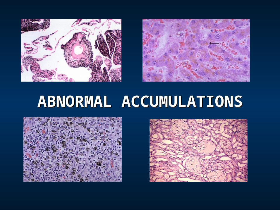
ABNORMAL ACCUMULATIONSABNORMAL ACCUMULATIONS

TYPES OF ACCUMULATIONS
There are 2 basic types of accumulations:1. Excess of substances normal to the
particular cell, and2. Abnormal substances in three
mechanisms: (a) decrease in normal metabolic removal, (b) inability to metabolize the substance, and (c) deposition of abnormal exogenous substance in which the cell has no mechanism to metabolize it.
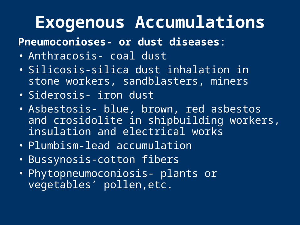
Exogenous AccumulationsPneumoconioses- or dust diseases:• Anthracosis- coal dust• Silicosis-silica dust inhalation in stone workers,
sandblasters, miners• Siderosis- iron dust• Asbestosis- blue, brown, red asbestos and
crosidolite in shipbuilding workers, insulation and electrical works
• Plumbism-lead accumulation• Bussynosis-cotton fibers• Phytopneumoconiosis- plants or vegetables’
pollen,etc.
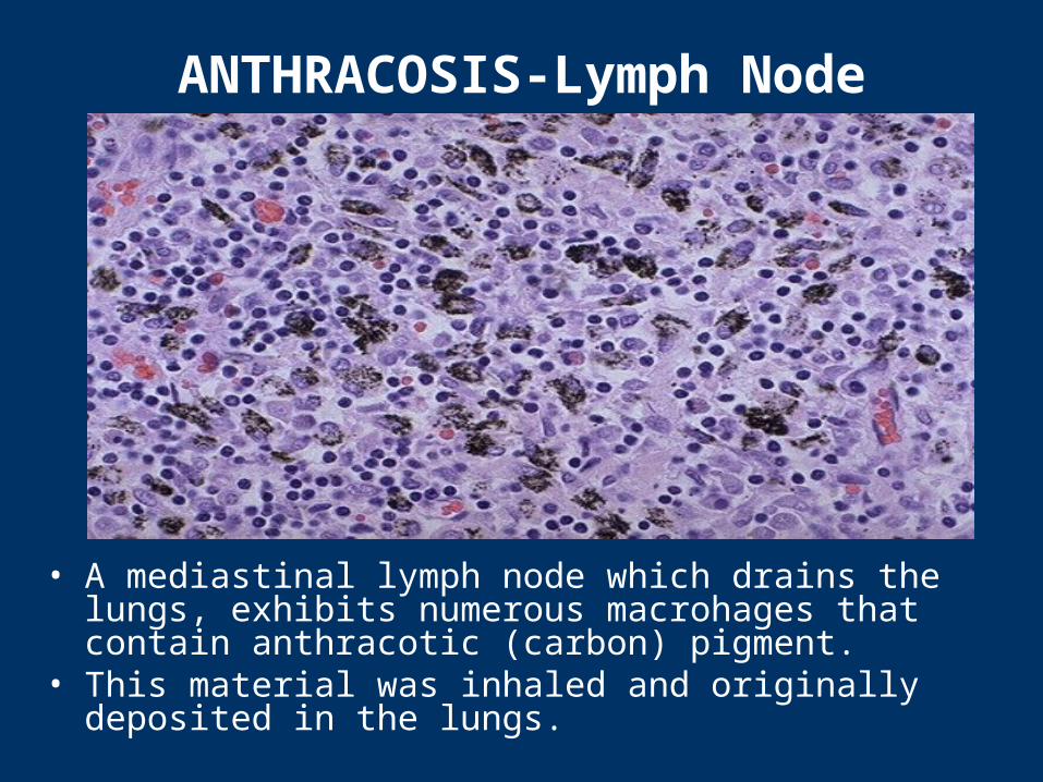
ANTHRACOSIS-Lymph Node
• A mediastinal lymph node which drains the lungs, exhibits numerous macrohages that contain anthracotic (carbon) pigment.
• This material was inhaled and originally deposited in the lungs.
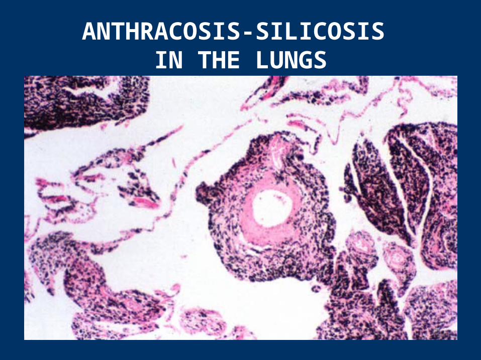
ANTHRACOSIS-SILICOSIS IN THE LUNGS
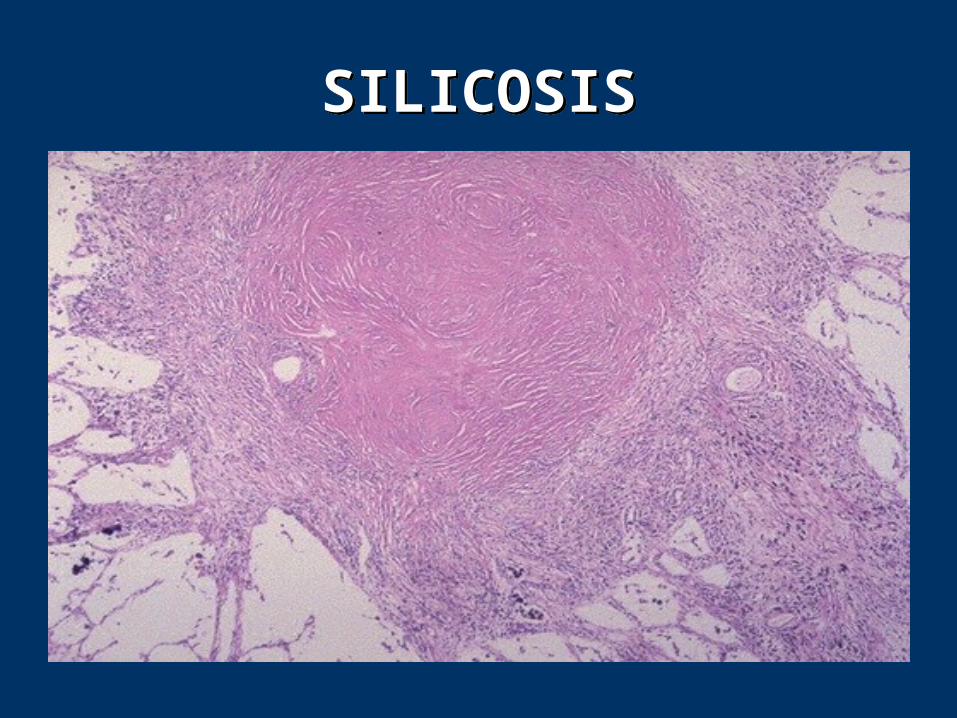
SILICOSISSILICOSIS
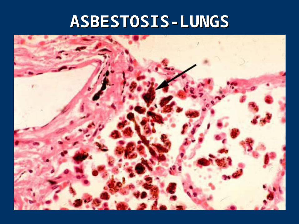
ASBESTOSIS-LUNGSASBESTOSIS-LUNGS
Endogenous Accumulations
• Lipofuchsin- a yellowish brown pigment having a high lipid content, often found in atrophied cells of old age. Also called the “wear-and-tear pigment.” Common in heart muscle called “brown atrophy.”
• Bile pigments-are derived from the degradation of hemoglobin(bilirubin, hemosiderin, hematin, porphyrins, biliverdin)
• Melanin-brown or black pigment of the skin, iris, gums, hair and accumulates in nevus or lentigo or moles
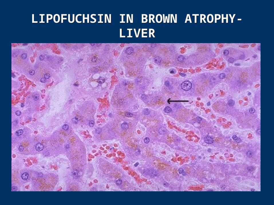
LIPOFUCHSIN IN BROWN ATROPHY-LIVER
LIPOFUCHSIN IN BROWN ATROPHY-LIVER
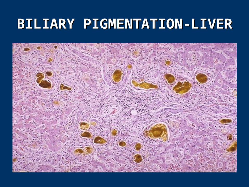
BILIARY PIGMENTATION-LIVERBILIARY PIGMENTATION-LIVER
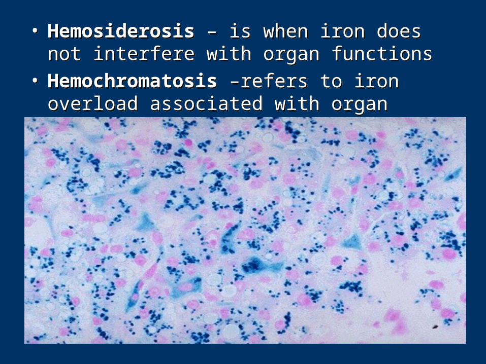
• Hemosiderosis – is when iron does not interfere with organ functions
• Hemochromatosis –refers to iron overload associated with organ failure
• Hemosiderosis – is when iron does not interfere with organ functions
• Hemochromatosis –refers to iron overload associated with organ failure
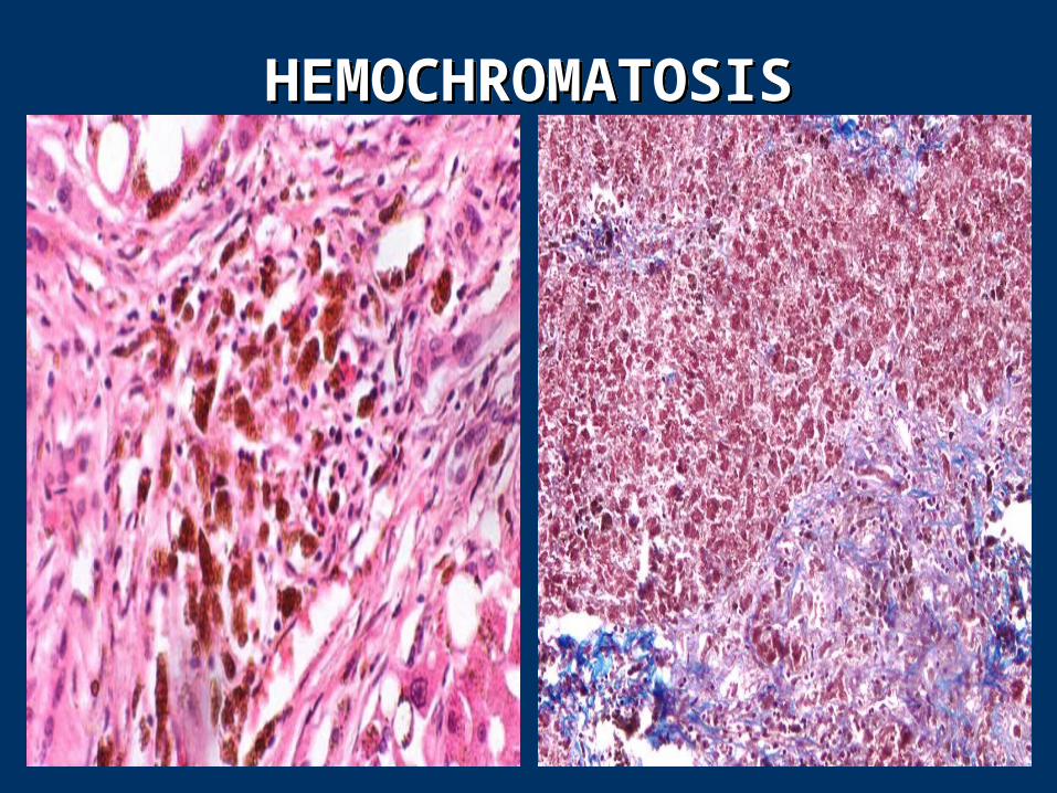
HEMOCHROMATOSISHEMOCHROMATOSIS
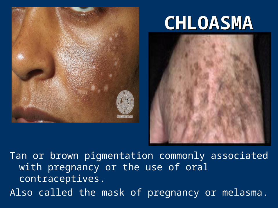
CHLOASMACHLOASMA
Tan or brown pigmentation commonly associated with pregnancy or the use of oral contraceptives.
Also called the mask of pregnancy or melasma.
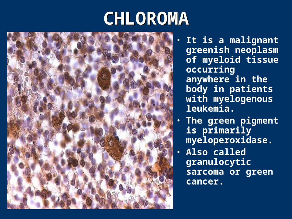
CHLOROMACHLOROMA• It is a malignant
greenish neoplasm of myeloid tissue occurring anywhere in the body in patients with myelogenous leukemia.
• The green pigment is primarily myeloperoxidase.
• Also called granulocytic sarcoma or green cancer.
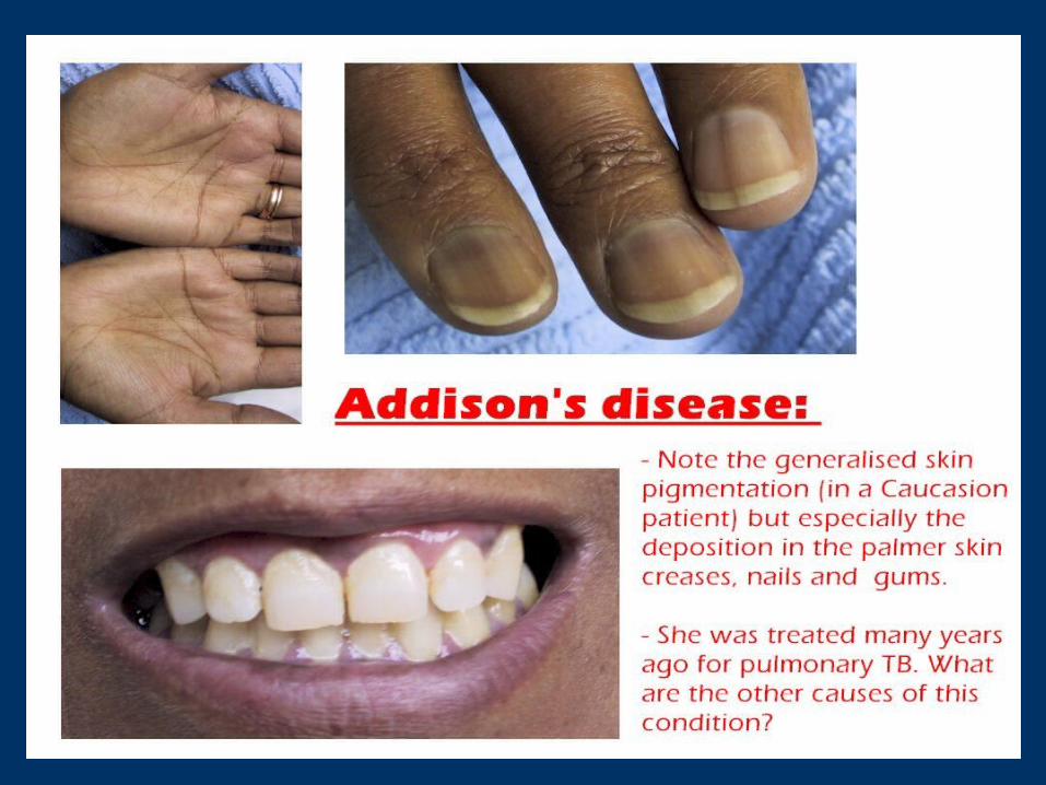
ADDISON’S DISEASE
• Addison’s disease is a life-threatening condition caused by partial or complete adrenocortical function. It is characterized by weakness, decreased endurance, increased pigmentation of the skin and mucous membranes, described as bronzing, anorexia, dehydration and weight loss, GI disturbances, anxiety, depression, and other emotional distress.
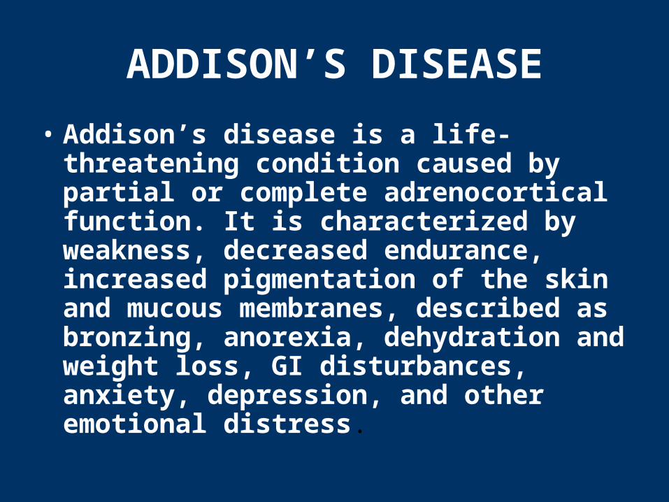
Amyloid Deposits-KidneyAmyloid Deposits-Kidney
Types of Calcification
• Physiologic calcification – laying down of calcium salts in bone-forming tissues
• Pathological calcification - types
1. Dystrophic calcification
2. Metastatic calcification
3. Lithiasis
4. Arterial calcification
5. Calcinosis
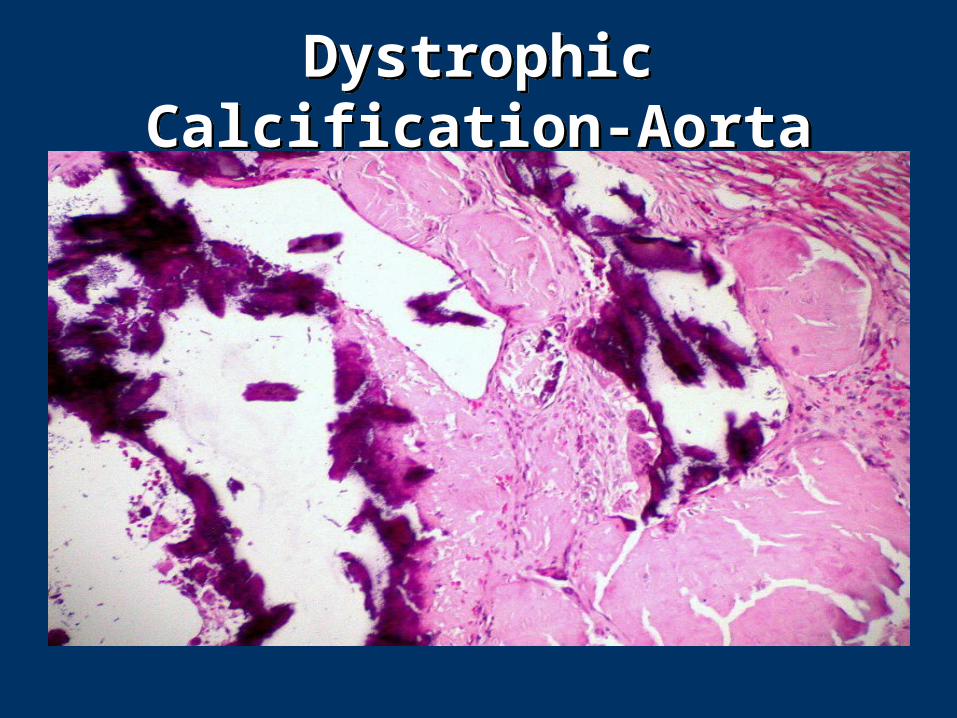
Dystrophic Calcification-AortaDystrophic Calcification-Aorta
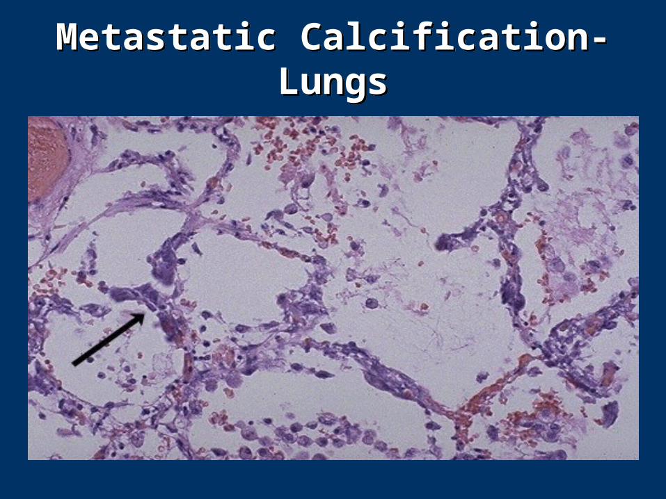
Metastatic Calcification-LungsMetastatic Calcification-Lungs


“Our lives are filled with joys and strife,
And what is death but part of life?
Will come the day that we must die,
And leave behind those learning why?”
by the Pathology Blues
Class 1998, Harvard University
“Our lives are filled with joys and strife,
And what is death but part of life?
Will come the day that we must die,
And leave behind those learning why?”
by the Pathology Blues
Class 1998, Harvard University


![Regulation of the intracellular Ca2+. Regulation of intracellular [H]:](https://img.pdfslide.us/doc/110x75/5a4d1b717f8b9ab0599b56a5/regulation-of-the-intracellular-ca2-regulation-of-intracellular-h.jpg)