Embed Size (px)
Citation preview
Pathology – Chapter 4

60% of lean body weight is waterTwo thirds of the body's water is
intracellularRemainder is in extracellular
compartmentsAbout 5% of total body water is in blood
plasma
Movement of water and low molecular weight solutes (salts)Between the intravascular and
interstitial spaces Controlled primarily by opposing effect
of: Vascular hydrostatic pressure Plasma colloid osmotic pressure


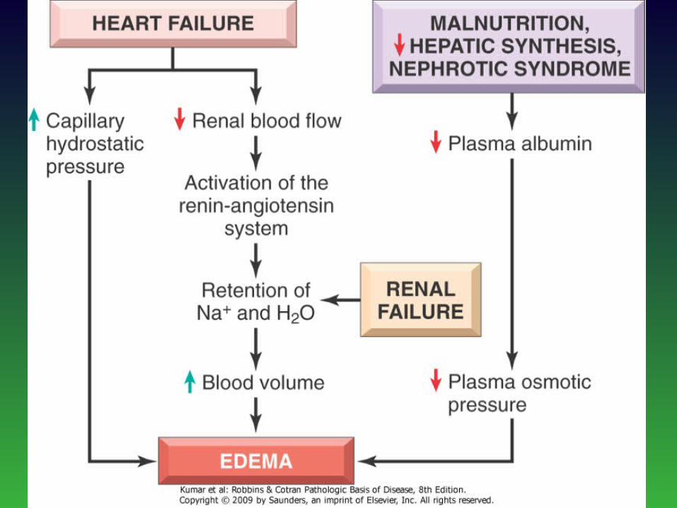
Increased interstitial fluidIncreased capillary pressure Diminished colloid osmotic pressure
Fluid accumulationMovement of water into tissues (or body
cavities) exceeds drainage Abnormal increase in interstitial fluid
within tissues Edema

Fluid collections in the different body cavitiesHydrothoraxHydropericardiumHydroperitoneum (ascites)
AnasarcaSevere and generalized edemaWidespread subcutaneous tissue
swelling

TransudateEdema caused by:
Increased hydrostatic pressure Reduced plasma protein
Typically a protein-poor fluidHeart failure, renal failure, hepatic
failure, and certain forms of malnutrition

Inflammatory edemaProtein-rich exudateResult of increased vascular
permeability

LymphedemaImpaired lymphatic drainageTypically localizedCauses
Chronic inflammation with fibrosis Invasive malignant tumors Physical disruption Radiation damage


Certain infectious agentsParasitic filariasis
Lymphatic obstruction• Extensive inguinal lymphatic and lymph
node fibrosis• Edema of the external genitalia and lower
limbs • Massive = elephantiasis


Severe edema of the upper extremityComplicate surgical removal and/or
irradiation Breast and associated axillary lymph
nodes • Breast cancer
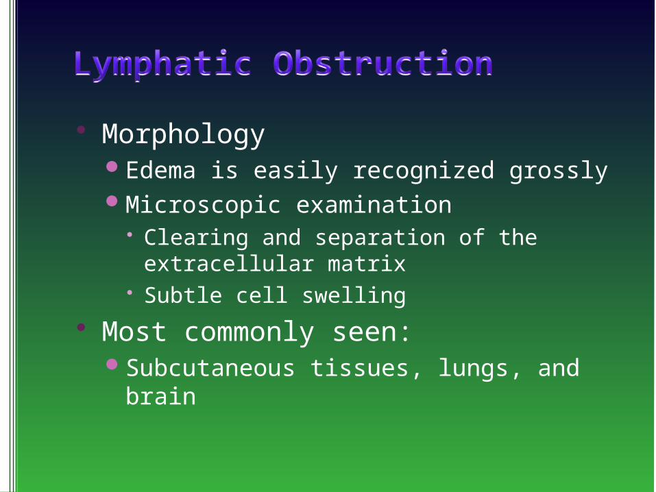
MorphologyEdema is easily recognized grosslyMicroscopic examination
Clearing and separation of the extracellular matrix
Subtle cell swelling
Most commonly seen:Subcutaneous tissues, lungs, and brain

Subcutaneous edemaDiffuse or more conspicuous in regions
with high hydrostatic pressuresDistribution is influenced by gravity
Dependent edema• Legs when standing, the sacrum when
recumbent

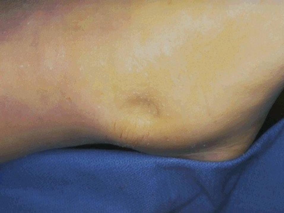
Subcutaneous edemaPitting edema
Finger pressure over substantially edematous subcutaneous tissue
Displaces the interstitial fluid and leaves a depression

Edema secondary to renal dysfunctionAffect all parts of the bodyManifests in tissues with loose connective
tissue matrix (eyelids) Periorbital edema
• Characteristic finding in severe renal disease

Soft tissue edemaImportant because it signals underlying
cardiac or renal diseaseImpairs wound healing or the clearance of
infection
Pulmonary edemaLungs are often two to three times their
normal weightSectioning yields frothy, blood-tinged
fluid Mixture of air, edema, and extravasated
red cellsCommon clinical problemMost frequently seen with left ventricular
failure

Pulmonary edemaLungs are often two to three times their
normal weightSectioning yields frothy, blood-tinged
fluid Mixture of air, edema, and extravasated
red cellsCommon clinical problemMost frequently seen with left ventricular
failure

Brain edemaLocalized or generalized Depending on the nature and extent of
the pathologic process or injuryGeneralized edema
Brain is grossly swollen with narrowed sulci
Distended gyri show evidence of compression against the unyielding skull

Brain edema Life-threateningSevere edema
Brain substance can herniate (extrude)• Foramen magnum
Brain stem vascular supply can be compressed
Either condition can injure the medullary centers• Cause death

Stem from locally increased blood volumes
HyperemiaActive process Arteriolar dilation
Sites of inflammation Skeletal muscle during exercise

Hyperemia Leads to increased blood flow
Affected tissues turn red (erythema) Engorgement of vessels with oxygenated
blood

Congestion Passive processReduced outflow of blood from a tissueSystemic
Cardiac failure

Congestion Local
Isolated venous obstructionDusky reddish-blue color (cyanosis)
Red cell stasis Accumulation of deoxygenated
hemoglobin
Long-standing chronic passive congestionLack of blood flow causes chronic hypoxia
Results in ischemic tissue injury and scarring
Capillary rupture Cause small hemorrhagic foci Subsequent catabolism of extravasated red
cells• Leave residual telltale clusters of hemosiderin-
laden macrophages

MorphologyCut surfaces
Discolored due to the presence of high levels of poorly oxygenated blood
Microscopic examination Acute pulmonary congestion
• Engorged alveolar capillaries• Alveolar septal edema• Focal intra-alveolar hemorrhage


MorphologyMicroscopic examination
Chronic pulmonary congestion • Septa are thickened and fibrotic• Alveoli often contain numerous hemosiderin-
laden macrophages Heart failure cells

MorphologyAcute hepatic congestion
Central vein and sinusoids are distended Centrilobular hepatocytes can be frankly
ischemicChronic passive hepatic congestion
Centrilobular regions are grossly red-brown• Areas are accentuated against uncongested
parenchyma• Nutmeg liver


MorphologyMicroscopic examination
Centrilobular hemorrhage Hemosiderin-laden macrophages Degeneration of hepatocytes


Extravasation of blood into the extravascular space
Increased tendency to hemorrhage (usually with insignificant injury)Occurs in a variety of clinical disordersCollectively called hemorrhagic
diatheses

Distinct patterns of tissue hemorrhageHemorrhage may be external
Contained within a tissue Hematoma
Petechiae Minute 1- to 2-mm hemorrhages into
skin, mucous membranes, or serosal surfaces
Most commonly associated: Locally increased intravascular pressure Low platelet counts (thrombocytopenia) Defective platelet function (as in uremia)


PurpuraSlightly larger (≥3 mm) hemorrhagesAssociated with many of the same
disorders that cause petechiae Secondary to trauma, vascular
inflammation (vasculitis), or increased vascular fragility (amyloidosis)
EcchymosesLarger (>1 to 2 cm) subcutaneous
hematomas (bruises) Red cells in these lesions are degraded
and phagocytized by macrophages Hemoglobin (red-blue color)
• Enzymatically converted into bilirubin (blue-green color) Hemosiderin (gold-brown color), accounting for
the characteristic color changes in a bruise

Large accumulation of blood in a body cavityHemothoraxHemopericardiumHemoperitoneumHemarthrosis (in joints)

Normal hemostasisConsequence of tightly regulated
processes Maintain blood in a fluid state in normal
vesselsPermit the rapid formation of a
hemostatic clot at the site of a vascular injury

ThrombosisPathologic counterpart of hemostasisInvolves blood clot (thrombus) formation
Both hemostasis and thrombosis involve three components: Vascular wall (particularly the
endothelium)PlateletsCoagulation cascade


Endothelial cells Key players in the regulation of
homeostasisExhibit antiplatelet, anticoagulant, and
fibrinolytic propertiesAfter injury or activation
Acquire numerous procoagulant activitiesActivated by infectious agents,
hemodynamic forces, plasma mediators, and cytokines
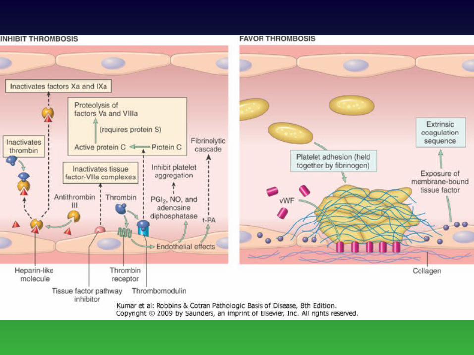

Antiplatelet effectsIntact endothelium prevents platelets
from engaging the highly thrombogenic subendothelial ECM
Nonactivated platelets Do not adhere to endothelial cells
• Even if platelets are activated, prostacyclin (PGI2) and nitric oxide produced by the endothelial cells impede platelet adhesion

Antiplatelet effectsEndothelial cells
Also elaborate adenosine diphosphatase• Degrades adenosine diphosphate (ADP)• Further inhibits platelet aggregation

Anticoagulant effectsMediated by endothelial membrane-
associated heparin-like molecules Thrombomodulin
• Binds to thrombin• Converts it from a procoagulant into an
anticoagulant Via its ability to activate protein C, which
inhibits clotting by inactivating factors Va and VIIIa

Anticoagulant effectsMediated by endothelial membrane-
associated heparin-like molecules Tissue factor pathway inhibitor
• Cell surface protein• Directly inhibits tissue factor-factor VIIa and
factor Xa activities

Anticoagulant effectsHeparin-like molecules
Act indirectly Cofactors that enhance the inactivation of
thrombin and several other coagulation factors• Through the use of plasma protein
antithrombin III


Fibrinolytic effectsEndothelial cells synthesize tissue-type
plasminogen activator (t-PA) Protease that cleaves plasminogen to form
plasmin• Plasmin cleaves fibrin to degrade thrombi

Platelet effectsEndothelial injury allows platelets to
contact the underlying extracellular matrix
Subsequent adhesion occurs through interactions with von Willebrand factor (vWF) Product of normal endothelial cells and an
essential cofactor for platelet binding to matrix elements

Procoagulant effectsResponse to cytokines (TNF or IL-1) or
bacterial endotoxin Endothelial cells synthesize tissue factor
• Major activator of the extrinsic clotting cascade
• Activated endothelial cells Augment the catalytic function of activated
coagulation factors IXa and Xa

Antifibrinolytic effectsEndothelial cells secrete inhibitors of
plasminogen activator (PAIs) Limit fibrinolysis and tend to favor
thrombosis


Intact, nonactivated endothelial cells inhibit platelet adhesion and blood clotting
Endothelial injury or activationResults in a procoagulant phenotype
that enhances thrombus formation

Disc-shaped Anucleate cell fragments Shed from megakaryocytes in the
bone marrow into the blood stream

Play a critical role in normal hemostasisForming the hemostatic plug that
initially seals vascular defectsProviding a surface that recruits and
concentrates activated coagulation factors
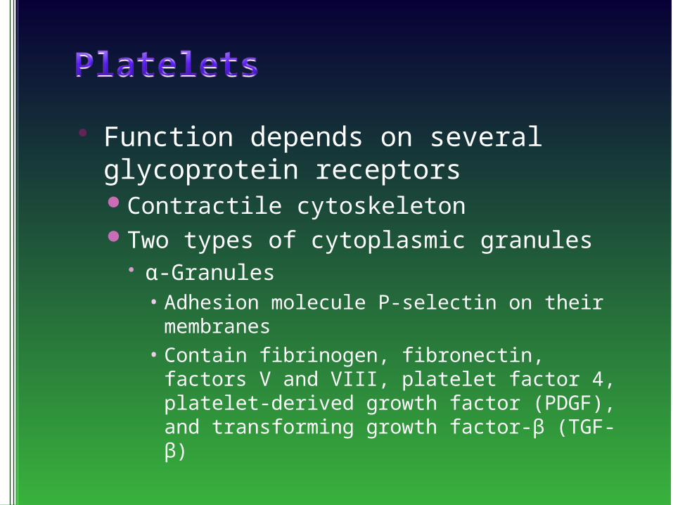
Function depends on several glycoprotein receptorsContractile cytoskeletonTwo types of cytoplasmic granules
α-Granules• Adhesion molecule P-selectin on their
membranes• Contain fibrinogen, fibronectin, factors V and
VIII, platelet factor 4, platelet-derived growth factor (PDGF), and transforming growth factor-β (TGF-β)

Function depends on several glycoprotein receptorsTwo types of cytoplasmic granules
Dense (or δ) granules • Contain ADP and ATP, ionized calcium,
histamine, serotonin, and epinephrine
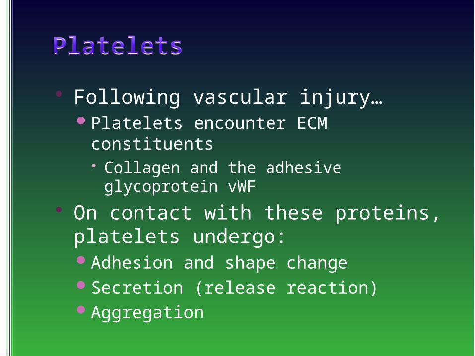
Following vascular injury…Platelets encounter ECM constituents
Collagen and the adhesive glycoprotein vWF
On contact with these proteins, platelets undergo: Adhesion and shape changeSecretion (release reaction)Aggregation

Mediated largely via interactions with vWFActs as a bridge between platelet
surface receptors (glycoprotein Ib) and exposed collagen
vWF-GpIb associations Necessary to overcome the high shear
forces of flowing blood
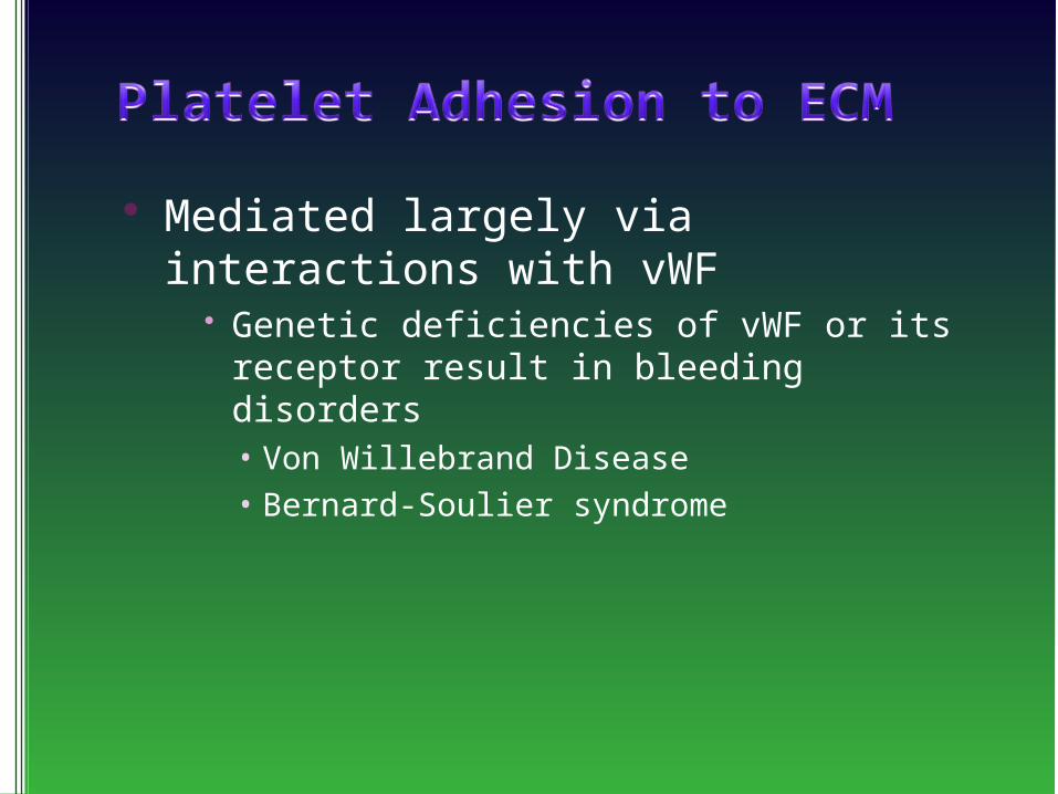
Mediated largely via interactions with vWF
Genetic deficiencies of vWF or its receptor result in bleeding disorders• Von Willebrand Disease• Bernard-Soulier syndrome

Occurs soon after adhesion Various agonists can bind platelet
surface receptorsInitiate an intracellular protein
phosphorylation cascade Leads to degranulation

Various agonists can bind platelet surface receptorsRelease of the contents of dense-bodies
Important Calcium is required in the coagulation
cascade ADP is a potent activator of platelet
aggregation• Causes additional ADP release
Amplifies aggregation process

Platelet activationAppearance of negatively charged
phospholipids (particularly phosphatidylserine) on their surfaces Bind calcium and serve as critical
nucleation sites for the assembly of complexes containing the various coagulation factors

Follows adhesion and granule release
Vasoconstrictor thromboxane A2
Important platelet-derived stimulus Amplifies platelet aggregationFormation of the primary hemostatic
plug Initial wave of aggregation is
reversible

Concurrent activation of the coagulation cascadeGenerates thrombinStabilizes the platelet plug via two
mechanisms: Thrombin binds to a protease-activated
receptor on the platelet membrane• In concert with ADP and TxA2 causes further
platelet aggregation
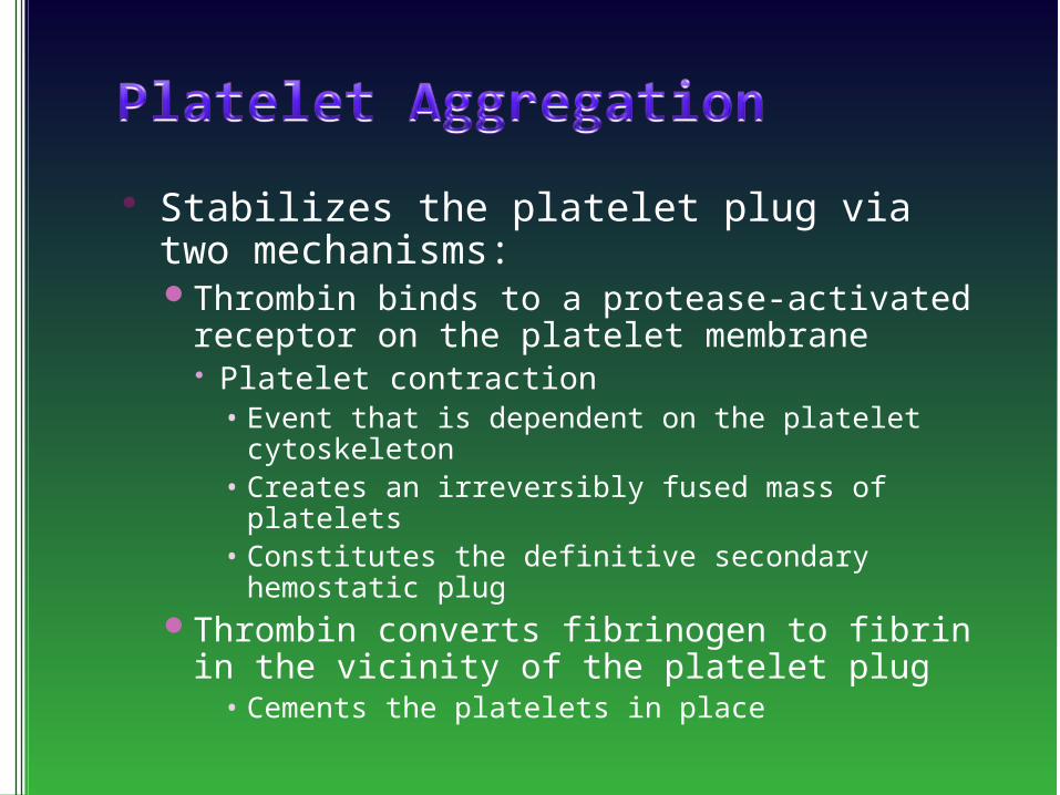
Stabilizes the platelet plug via two mechanisms:Thrombin binds to a protease-activated
receptor on the platelet membrane Platelet contraction
• Event that is dependent on the platelet cytoskeleton
• Creates an irreversibly fused mass of platelets• Constitutes the definitive secondary
hemostatic plugThrombin converts fibrinogen to fibrin in
the vicinity of the platelet plug• Cements the platelets in place

Noncleaved fibrinogenImportant component of platelet
aggregation Platelet activation by ADP
Triggers a conformational change in the platelet GpIIb-IIIa receptors
Induces binding to fibrinogen Large protein that forms bridging
interactions between platelets that promote platelet aggregation

Part 2

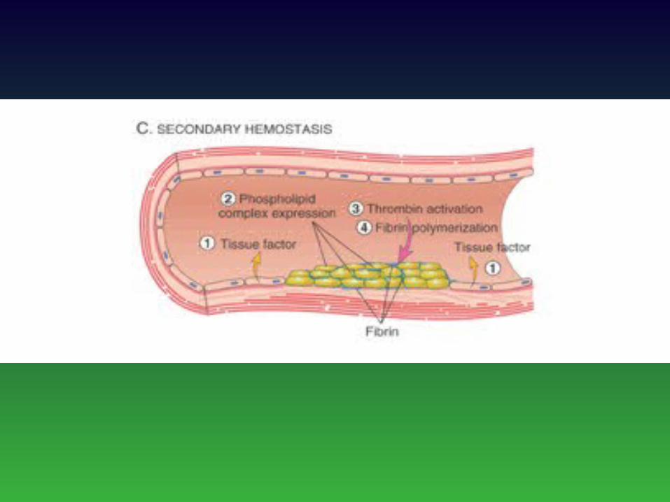
Third arm of the hemostatic process Amplifying series of enzymatic
conversionsEach step proteolytically cleaves an
inactive proenzyme into an activated enzyme Culminates in thrombin formation
Thrombin is the most important coagulation factorCan act at numerous stages in the process

Conclusion of the proteolytic cascadeThrombin converts the soluble plasma
protein fibrinogen into fibrin monomers that polymerize into an insoluble gel Fibrin gel encases platelets and other
circulating cells in the definitive secondary hemostatic plug
Fibrin polymers are covalently cross-linked and stabilized by factor XIIIa (which itself is activated by thrombin)


Assess the function of the two arms of the coagulation pathway Two standard assays
Prothrombin time (PT) Partial thromboplastin time (PTT)

The PT assayAssesses the function of the proteins in
the extrinsic pathway Factors VII, X, II, V, and fibrinogen Accomplished by adding tissue factor and
phospholipids to citrated plasma (sodium citrate chelates calcium and prevents spontaneous clotting)
Coagulation is initiated by the addition of exogenous calcium and the time for a fibrin clot to form is recorded

Partial thromboplastin time (PTT) Screens for the function of the proteins
in the intrinsic pathway Factors XII, XI, IX, VIII, X, V, II, and
fibrinogen Clotting is initiated through the addition of
negative charged particles (ground glass)• Activates factor XII (Hageman factor),
phospholipids, and calcium, and the time to fibrin clot formation is recorded

Thrombin Exerts a wide variety of proinflammatery
effectsMost effects of thrombin occur through
its activation of a family of protease activated receptors (PARs) Belong to the seven-transmembrane G
protein-coupled receptor family PARs are expressed on endothelium,
monocytes, dendritic cells, T lymphocytes, and other cell types

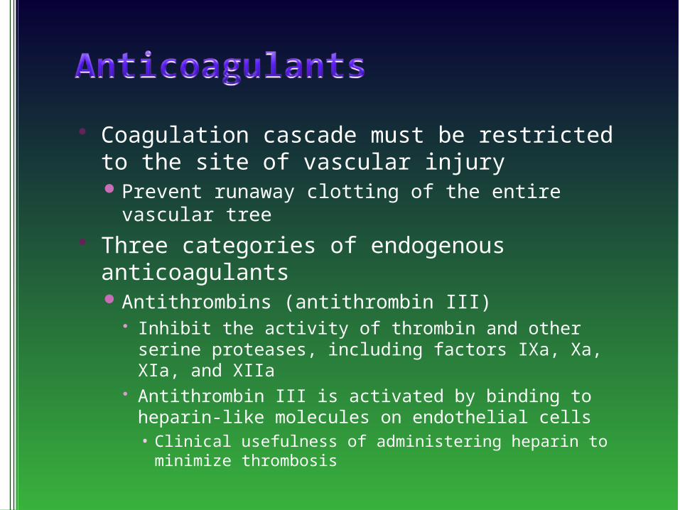
Coagulation cascade must be restricted to the site of vascular injury Prevent runaway clotting of the entire vascular
tree Three categories of endogenous
anticoagulantsAntithrombins (antithrombin III)
Inhibit the activity of thrombin and other serine proteases, including factors IXa, Xa, XIa, and XIIa
Antithrombin III is activated by binding to heparin-like molecules on endothelial cells• Clinical usefulness of administering heparin to
minimize thrombosis

Three categories of endogenous anticoagulantsProteins C and S
Vitamin K-dependent proteins Act in a complex that proteolytically
inactivates factors Va and VIIIaTFPI is a protein produced by endothelium
Inactivates tissue factor-factor VIIa complexes

Fine-tune the coagulation/anticoagulation balance
Releasing plasminogen activator inhibitor (PAI)Blocks fibrinolysis by inhibiting t-PA
binding to fibrinConfers an overall procoagulant effectProduction is increased by thrombin as
well as certain cytokines

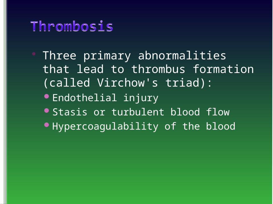
Three primary abnormalities that lead to thrombus formation (called Virchow's triad):Endothelial injuryStasis or turbulent blood flowHypercoagulability of the blood

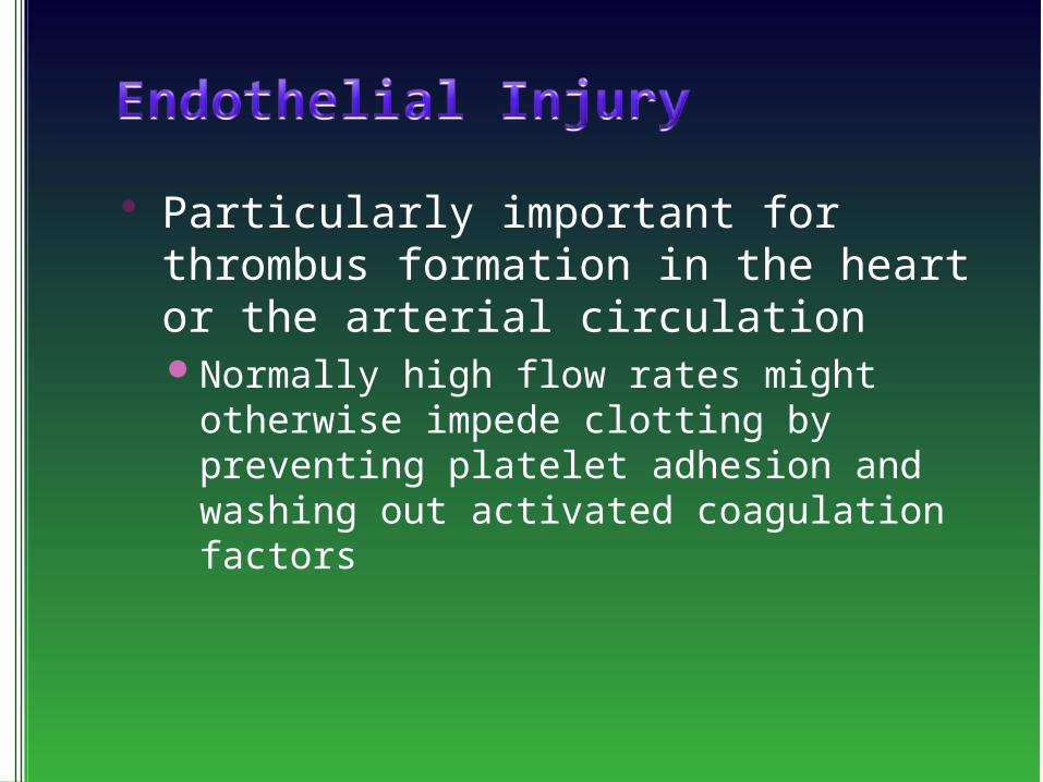
Particularly important for thrombus formation in the heart or the arterial circulationNormally high flow rates might otherwise
impede clotting by preventing platelet adhesion and washing out activated coagulation factors

Endothelial cell injuryThrombus formation within cardiac
chambers (i.e. after endocardial injury due to myocardial infarction)
Over ulcerated plaques in atherosclerotic arteries
Sites of traumatic or inflammatory vascular injury (vasculitis)
Endothelium Does not need to be denuded or
physically disrupted to contribute to the development of thrombosis
Any perturbation in the dynamic balance of the prothombotic and antithrombotic activities of endothelium can influence local clotting events

Endothelial dysfunction Induced by a wide variety of insults,
including hypertension, turbulent blood flow, bacterial endotoxins, radiation injury, metabolic abnormalities such as homocystinemia or hypercholesterolemia, and toxins absorbed from cigarette smoke

Turbulence Contributes to arterial and cardiac
thrombosis by causing endothelial injury or dysfunction
Forming countercurrents and local pockets of stasis Stasis is a major contributor in the
development of venous thrombi

Normal blood flow is laminar Platelets (and other blood cellular
elements) flow centrally in the vessel lumen, separated from endothelium by a slower moving layer of plasma

Stasis and turbulence therefore: Promote endothelial activation
Enhancing pro-coagulant activity through flow-induced changes in endothelial cell gene expression
Disrupt laminar flow and bring platelets into contact with the endothelium
Prevent washout and dilution of activated clotting factors by fresh flowing blood and the inflow of clotting factor inhibitors

AKA thrombophilia Less frequent contributor to
thrombotic states Any alteration of the coagulation
pathways that predisposes to thrombosisDivided into primary (genetic)Secondary (acquired) disorders

Of the inherited causes of hypercoagulabilityMost common
Point mutations in the factor V gene Prothrombin gene

Elevated levels of homocysteine Contribute to arterial and venous
thrombosis Prothrombotic effects of
homocysteineMay be due to thioester linkages formed
between homocysteine metabolites and a variety of proteins, including fibrinogen

Rare inherited causes of primary hypercoagulabilityDeficiencies of anticoagulants
Antithrombin III, protein C, or protein S Present with venous thrombosis and
recurrent thromboembolism beginning in adolescence or early adulthood
Acquired thrombophilic statesHeparin-induced thrombocytopenia (HIT)
syndrome Occurs following the administration of
unfractionated heparin• May induce the appearance of antibodies
Recognize complexes of heparin and platelet factor 4 on the surface of platelets

Heparin-induced thrombocytopenia (HIT) syndrome Occurs following the administration of
unfractionated heparin• Complexes of heparin-like molecules and
platelet factor 4-like proteins on endothelial cells
• Binding of these antibodies to platelets Results in their activation, aggregation, and
consumption Prothrombotic state, even in the face of
heparin administration and low platelet counts
AKA lupus anticoagulant syndrome Clinical manifestations
Recurrent thromboses, repeated miscarriages, cardiac valve vegetations, and thrombocytopenia
Pulmonary embolism, pulmonary hypertension, stroke, bowel infarction, or renovascular hypertension
Fetal loss

Autoantibodies induce a hypercoagulable state Cause endothelial injury by activating
platelets and complement directly Primary and secondary forms
Secondary antiphospholipid syndrome Individuals with a well-defined
autoimmune disease Systemic lupus erythematosus
Primary and secondary formsPrimary antiphospholipid syndrome
Exhibit only the manifestations of a hypercoagulable state
Lack evidence of other autoimmune disorders
Association with certain drugs or infections

Can develop anywhere in the cardiovascular system
Size and shape of thrombi Depend on the site of origin and the
causeArterial or cardiac thrombi
Begin at sites of turbulence or endothelial injury
Venous thrombi Occur at sites of stasis

Focally attached to the underlying vascular surfaceArterial thrombi tend to grow retrograde
from the point of attachmentVenous thrombi extend in the direction
of blood flow

Gross and microscopic laminationsLines of Zahn
Represent pale platelet and fibrin deposits alternating with darker red cell-rich layers
Signify that a thrombus has formed in flowing blood
Presence can therefore distinguish antemortem thrombosis from the bland nonlaminated clots that occur postmortem
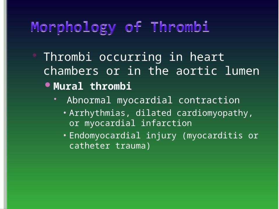
Thrombi occurring in heart chambers or in the aortic lumenMural thrombi
Abnormal myocardial contraction• Arrhythmias, dilated cardiomyopathy, or
myocardial infarction• Endomyocardial injury (myocarditis or
catheter trauma)

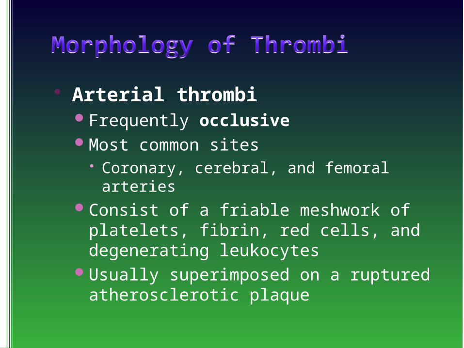
Arterial thrombi Frequently occlusiveMost common sites
Coronary, cerebral, and femoral arteriesConsist of a friable meshwork of
platelets, fibrin, red cells, and degenerating leukocytes
Usually superimposed on a ruptured atherosclerotic plaque

Venous thrombosis (phlebothrombosis) Invariably occlusiveThrombus forming a long cast of the
lumenThrombi form in the sluggish venous
circulationContain more enmeshed red cells
Red, or stasis, thrombiVeins of the lower extremities are most
commonly involved (90% of cases)

Postmortem clotsMistaken for antemortem venous thrombiGelatinous with a dark red dependent
portion where red cells have settled by gravity and a yellow "chicken fat" upper portion
Usually not attached to the underlying wall Red thrombi
Firmer Focally attachedGross and/or microscopic lines of Zahn

VegetationsThrombi on heart valves Blood-borne bacteria or fungi
Adhere to previously damaged valves (rheumatic heart disease)
Directly cause valve damage Infective endocarditis
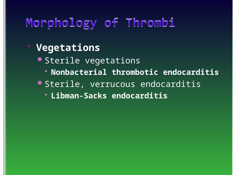
VegetationsSterile vegetations
Nonbacterial thrombotic endocarditisSterile, verrucous endocarditis
Libman-Sacks endocarditis
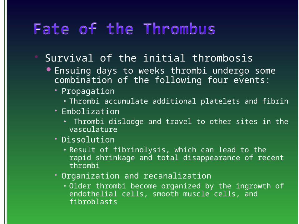
Survival of the initial thrombosisEnsuing days to weeks thrombi undergo some
combination of the following four events: Propagation
• Thrombi accumulate additional platelets and fibrin Embolization
• Thrombi dislodge and travel to other sites in the vasculature
Dissolution• Result of fibrinolysis, which can lead to the rapid
shrinkage and total disappearance of recent thrombi Organization and recanalization
• Older thrombi become organized by the ingrowth of endothelial cells, smooth muscle cells, and fibroblasts
Deep venous thrombosis (DVT)Larger leg veins-at or above the knee Thrombi more often embolize to the lungs
and give rise to pulmonary infarction Venous obstructions from DVTs can be
rapidly offset by collateral channelsDVTs are asymptomatic in approximately
50% of affected individualsRecognized only in retrospect after
embolization

Obstetric complications to advanced malignancy
Sudden or insidious onset of widespread fibrin thrombi in the microcirculation
Not grossly visible Diffuse circulatory insufficiency,
particularly in the brain, lungs, heart, and kidneys

Widespread microvascular thrombosis results in platelet and coagulation protein consumptionFibrinolytic mechanisms are activated
Initially thrombotic disorderEvolve into a bleeding catastrophe

EmbolusDetached intravascular solid, liquid, or
gaseous mass that is carried by the blood to a site distant from its point of origin
ThromboembolismRare forms of emboli include fat droplets,
nitrogen bubbles, atherosclerotic debris (cholesterol emboli), tumor fragments, bone marrow, or even foreign bodies
Unless otherwise specified, emboli should be considered thrombotic in origin
Occlusions—embolic 95% from deep leg veins Indwelling central venous lines
Right atrial thrombi 50,000 deaths/year in US

Origin of emboliLeg or pelvic veins
Large emboli Sudden death
Lodging • Major branches of pulmonary arteries• Saddle emboli
Acute cor pulmonale

Small emboliMinimal symptomsException
Inadequate bronchial circulation • Symptoms
Causes of emboliImmobilized individualsHypercoagulable state (primary vs.
secondary)Heart failure
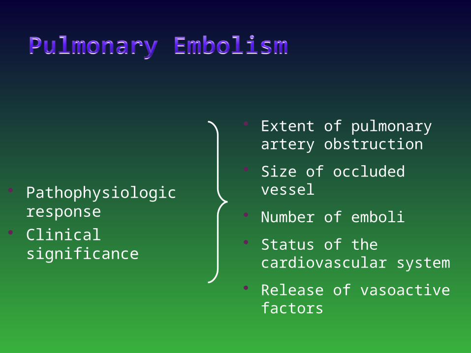
Pathophysiologic response
Clinical significance
Extent of pulmonary artery obstruction
Size of occluded vessel
Number of emboli
Status of the cardiovascular system
Release of vasoactive factors

Pathophysiologic consequencesRespiratory compromise Hemodynamic compromise
Adequate cardiovascular functionBronchial artery compensation
Hemorrhage without infarction
Infarction Inadequate circulationRare in young

Clinical course Cardiopulmonary resuscitation
Electromechanical dissociation• Electrocardiogram has a rhythm• No pulses are palpated
Survival (post-sizable pulmonary embolus) Mimics myocardial infarction

DiagnosisSpiral CTOther diagnostic methods
Ventilation perfusion scanning
Pulmonary angiography Duplex ultrasonography•Deep vein thrombosis


PreventionMajor clinical problem Prophylactic therapy
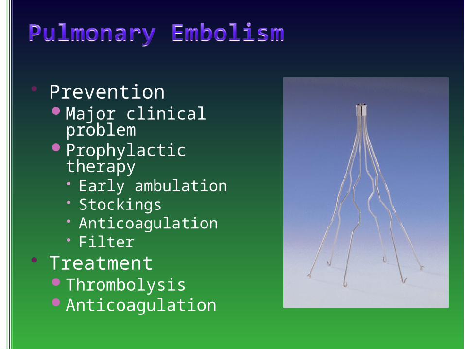
Early ambulation Stockings Anticoagulation Filter
TreatmentThrombolysis Anticoagulation
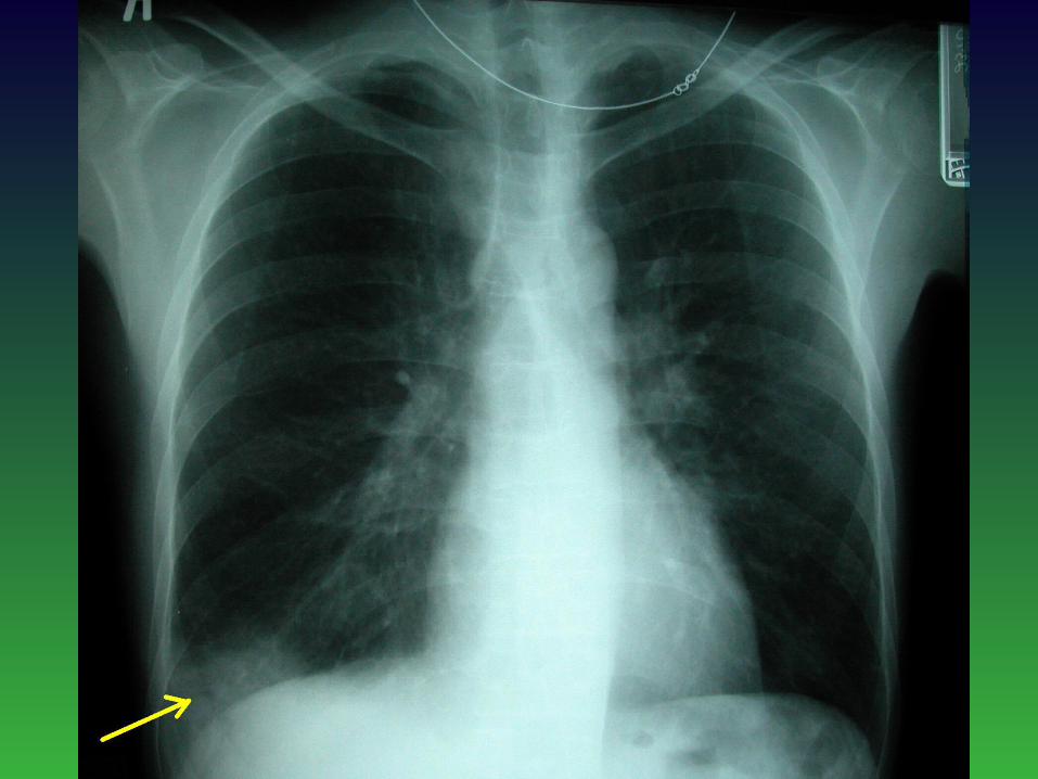
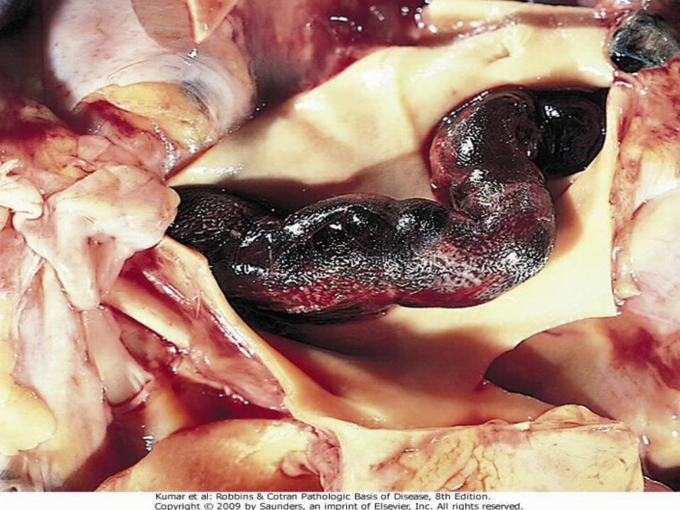
Gross examinationParenchyma
75% of all infarcts affect the lower lobes Greater than 50%--multiple lesions Wedge shaped Hemorrhagic
Fibrinous pleural exudateScarEmbolus


http://www.path.uiowa.edu/cgi-bin-pub/vs/fpx_gen.cgi?slide=714&viewer=java&lay=&jpg=493


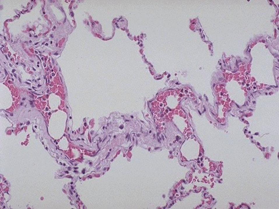
Microscopic examinationIschemic necrosis
Alveolar walls, bronchioles, and vesselsInfected embolus
Intense neutrophilic inflammatory reaction Septic infarct


Microscopic fat globules-with or without associated hematopoietic marrow elements
Fractures of long bones (which have fatty marrow)
Soft tissue trauma and burns Common incidental findings after
vigorous cardiopulmonary resuscitation
No clinical consequence


Gas bubbles within the circulationCoalesce to form frothy masses that
obstruct vascular flow (and cause distal ischemic injury)
More than 100 cc of air are required to have a clinical effect in the pulmonary circulation
Decompression sickness Sudden decreases in atmospheric pressure Scuba and deep sea divers, underwater
construction workers, and individuals in unpressurized aircraft in rapid ascent are all at risk
The bendsRapid formation of gas bubbles within
skeletal muscles and supporting tissues in and about joints
The chokesGas bubbles in the vasculature cause
edema, hemorrhage, and focal atelectasis or emphysema, leading to a form of respiratory distress

Caisson disease Chronic form of decompression sickness
is called (named for the pressurized vessels used in the bridge construction; workers in these vessels suffered both acute and chronic forms of decompression sickness)
Persistence of gas emboli in the skeletal system leads to multiple foci of ischemic necrosis; the more common sites are the femoral heads, tibia, and humeri
Amniotic fluid embolismOminous complication of labor and the
immediate postpartum periodSudden severe dyspnea, cyanosis, and
shock Followed by neurologic impairment ranging
from headache to seizures and coma If the patient survives the initial crisis,
pulmonary edema typically develops, along with (in half the patients) DIC, as a result of release of thrombogenic substances from the amniotic fluid

Underlying cause Infusion of amniotic fluid or fetal tissue
into the maternal circulation via a tear in the placental membranes or rupture of uterine veins
Classic findings Presence of squamous cells shed from
fetal skin, lanugo hair, fat from vernix caseosa, and mucin derived from the fetal respiratory or gastrointestinal tract in the maternal pulmonary microvasculature


Final common pathway for several potentially lethal clinical eventsIncluding severe hemorrhage, extensive
trauma or burns, large myocardial infarction, massive pulmonary embolism, and microbial sepsis
Shock is characterized by systemic hypotension due either to reduced cardiac output or to reduced effective circulating blood volume

The consequences are impaired tissue perfusion and cellular hypoxia
Three general categories Cardiogenic shock Hypovolemic shockSeptic shock

Depend on the precipitating insult Hypovolemic and cardiogenic shock
Patient presents with hypotension Weak, rapid pulse; tachypnea; and cool,
clammy, cyanotic skin
Septic shockSkin may initially be warm and flushed
because of peripheral vasodilation

Rapidly, however, the cardiac, cerebral, and pulmonary changes secondary to shock worsen the problem
Electrolyte disturbances and metabolic acidosis