Embed Size (px)
Citation preview
Department of PeriodonticsI.T.S Dental College, Hospital & Research Centre
Greater Noida
2nd April, 2014
Moderator- Dr. Shivjot Chhina
Presented by:
Dr. Abhishek Gakhar
Once a tooth erupts, various materials gather on its surfaces, these substances are frequently called tooth – accumulated materials/deposits.1
They are classified as: Soft deposits: Acquired pellicleMicrobial plaqueMateria albaFood debris Hard deposits: CalculusStains

Acquired Pellicle : Following tooth eruption or a dental prophylaxis, a thin, saliva- derived layer, called the acquired pellicle, covers the tooth surface.1
Dental Plaque is defined as a specific but highly variable structural entity resulting from sequential colonization and growth of micro organisms on the surfaces of teeth and restoration consisting of micro organisms of various strains and species are embedded in the extra cellular matrix, composed of bacterial metabolic products and substance from serum, saliva and blood. (WHO-1978)1
Materia Alba refers to soft accumulations of bacteria and tissue cells that lack the organized structure of dental plaque.1
Calculus:
*Dental calculus can be considered as an ectopic mineralized structure.2
Dental Calculus consists of mineralized bacterial plaque that forms on the surfaces of natural teeth and
dental prosthesis. [Carranza ]2

*Calculus can be defined as a hard concretion that forms on teeth or dental prostheses through calcification of bacterial plaque [GPT 4th ed.].3
A deposit of inorganic salts composed primarily of calcium carbonate and phosphate mixed with food debris bacteria and desquamated epithelial cells. (Greene 1967)4
Mineralized dental plaque that is permeated with crystals of various calcium phosphates (Schroeder,1969)5
Calculus is also known as odontolithiasis or tartar. It is also called fossilized plaque.1

*Calculus was recognized as a clinical entity in some way related to periodontal disease as far back as the tenth century.1
*In 1683 Van Leeuwenhoek described microorganisms in tartar. He called them ‘animalcules’.1
*Fauchard, in 1728, termed it tartar or slime, and referred to it as “a substance which accumulates on the surface of the teeth and which becomes, when left there, a stony crust of more or less considerable volume.1
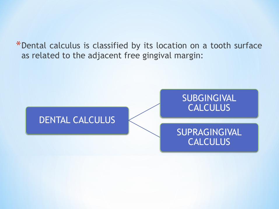
*Dental calculus is classified by its location on a tooth surface as related to the adjacent free gingival margin:
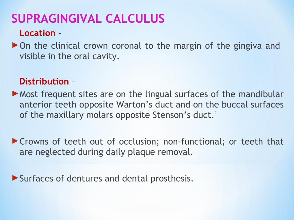
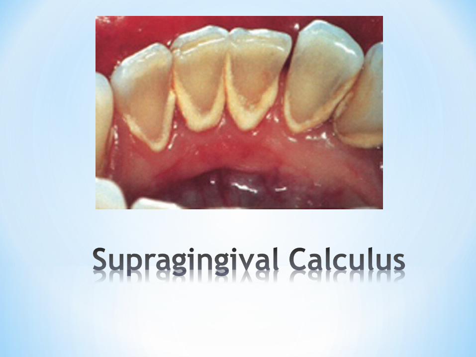
SUPRAGINGIVAL CALCULUS Location – On the clinical crown coronal to the margin of the gingiva and
visible in the oral cavity. Distribution – Most frequent sites are on the lingual surfaces of the mandibular
anterior teeth opposite Warton’s duct and on the buccal surfaces of the maxillary molars opposite Stenson’s duct.6
Crowns of teeth out of occlusion; non-functional; or teeth that are neglected during daily plaque removal.
Surfaces of dentures and dental prosthesis.
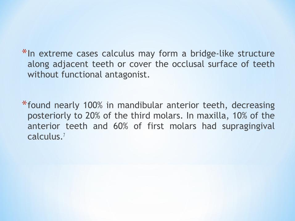
*In extreme cases calculus may form a bridge-like structure along adjacent teeth or cover the occlusal surface of teeth without functional antagonist.
*found nearly 100% in mandibular anterior teeth, decreasing posteriorly to 20% of the third molars. In maxilla, 10% of the anterior teeth and 60% of first molars had supragingival calculus.7
Other names for supragingival calculus -
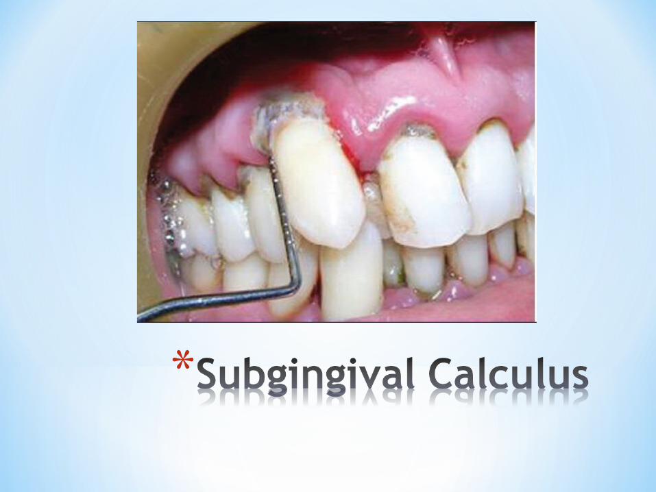
SUBGINGIVAL CALCULUS
Location-
*On the clinical crown apical to the margin of the gingiva, usually in periodontal pockets, not visible upon oral examination.
*Extents to bottom of the pocket and follows contour of soft tissue attachment.
Distribution –
May be generalized or localized on single teeth or a group of teeth.
Proximal surfaces have heaviest deposits, lightest deposits on facial surfaces.(Lovdal et al.1958)1
Occurs with or without associated supragingival deposits.
*Other names for subgingival calculus-

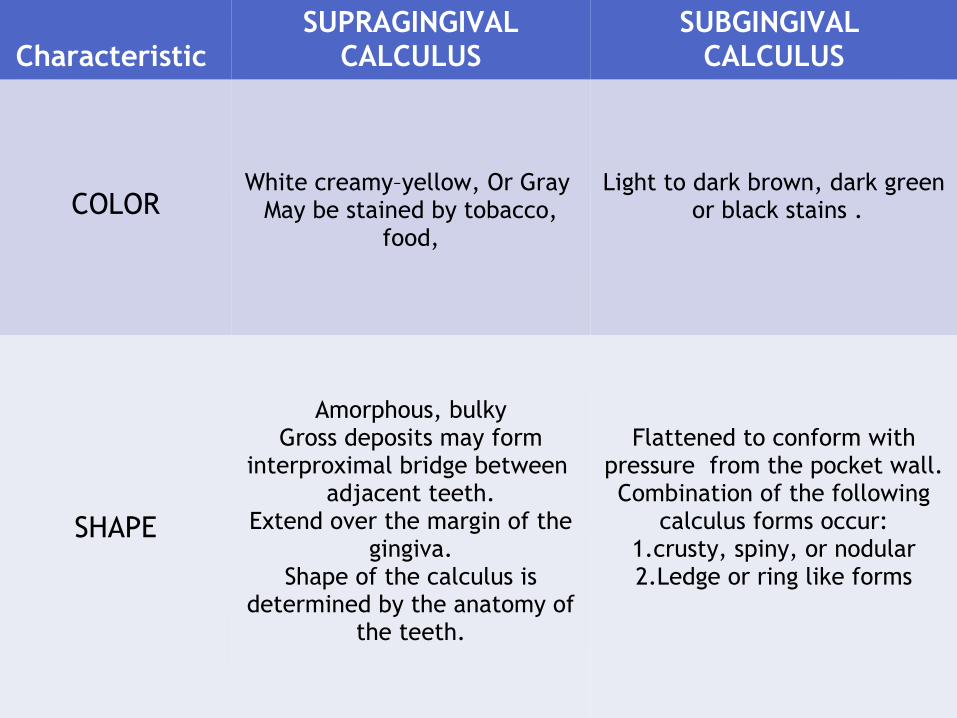
Characteristic SUPRAGINGIVAL
CALCULUSSUBGINGIVAL
CALCULUS
COLORWhite creamy–yellow, Or Gray
May be stained by tobacco, food,
Light to dark brown, dark green or black stains .
SHAPE
Amorphous, bulkyGross deposits may form
interproximal bridge between adjacent teeth.
Extend over the margin of the gingiva.
Shape of the calculus is determined by the anatomy of
the teeth.
Flattened to conform with pressure from the pocket wall.Combination of the following
calculus forms occur:1.crusty, spiny, or nodular2.Ledge or ring like forms

CONSISTENCYAND TEXTURE
Moderately hard newer deposits- less dense and hard
and porous.Surface covered with
nonmineralized plaque
Brittle, flint-like.Hardens and more dense than
supragingival calculus.Newest deposits near bottom of pocket are less dense and
hard.Surface covered with plaque
SIZE AND QUANTITY
Quantity has direct relationship to
1. Personal oral care procedures and plaque control measures.2. Physical character of diet.
3. Individual tendencies.4. Function and use
Increased amount in tobacco smokers.
Related to pocket depthIncreased amount with age because of accumulation
Subgingival is primarily related to the development and
progression of periodontal disease.
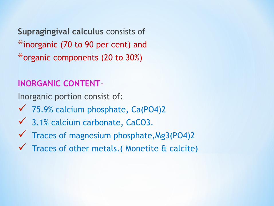
Supragingival calculus consists of
*inorganic (70 to 90 per cent) and
*organic components (20 to 30%)
INORGANIC CONTENT-
Inorganic portion consist of:
75.9% calcium phosphate, Ca(PO4)2
3.1% calcium carbonate, CaCO3.
Traces of magnesium phosphate,Mg3(PO4)2
Traces of other metals.( Monetite & calcite)

Principal components- calcium - 39%phosphorus - 19%carbon dioxide – 1.9%magnesium – 0.8%
Trace elements –sodium tungstenzinc goldstrontium aluminiumbromine siliconcopper iron manganese fluorine
FLOURIDE IN CALCULUS-
*Concentration of fluoride in calculus varies and is influenced by the amount of fluoride, received from fluoride in the drinking water, topical application, dentifrices, or any form that is received by contact with the external surface of calculus.7
*Fluoride concentrations were highest in or near the outermost regions of the calculus.
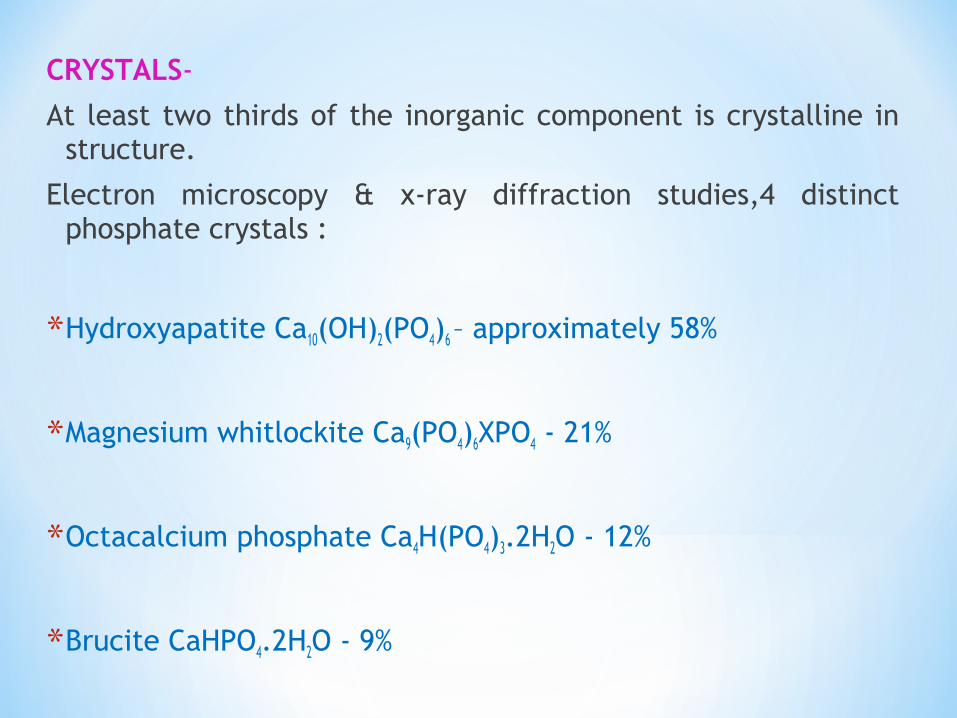
CRYSTALS-
At least two thirds of the inorganic component is crystalline in structure.
Electron microscopy & x-ray diffraction studies,4 distinct phosphate crystals :
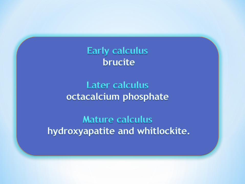
*Hydroxyapatite Ca10(OH)2(PO4)6 – approximately 58%
*Magnesium whitlockite Ca9(PO4)6XPO4 - 21%
*Octacalcium phosphate Ca4H(PO4)3.2H2O - 12%
*Brucite CaHPO4.2H2O - 9%
*Generally, two or more crystal forms are typically found in a sample of calculus.
*Hydroxyapatite and octacalcium phosphate are detected most frequently (in 97-100% of all supragingival calculus) and constitute the bulk of the specimen.
*Brucite is more common in the mandibular anterior region.
*Magnesium whitlockite is common in the posterior areas and sublingually.
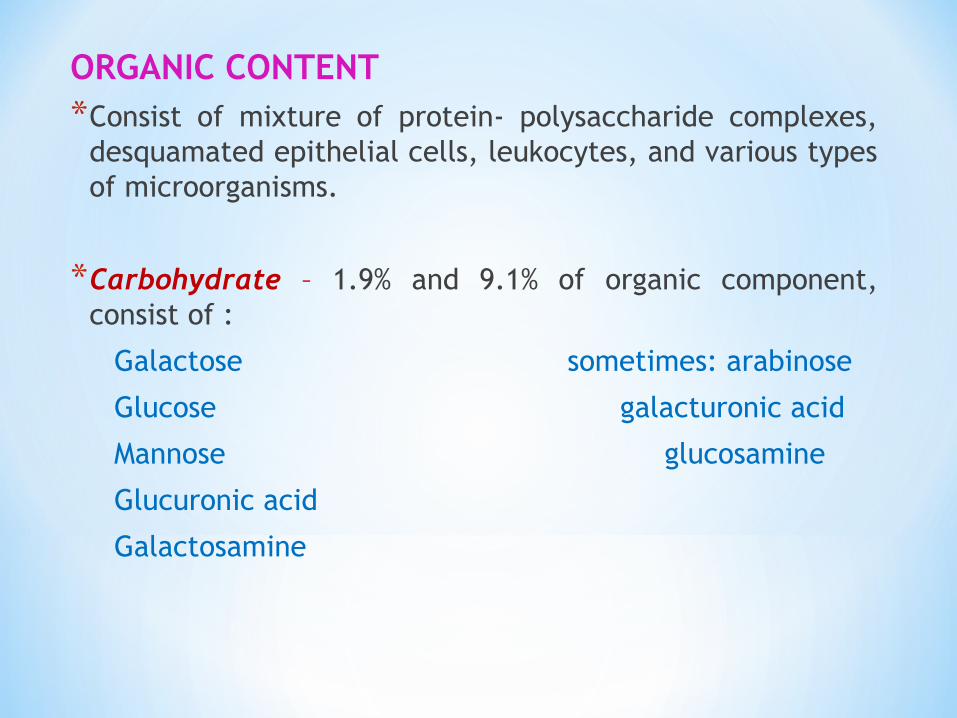
ORGANIC CONTENT*Consist of mixture of protein- polysaccharide complexes,
desquamated epithelial cells, leukocytes, and various types of microorganisms.
*Carbohydrate – 1.9% and 9.1% of organic component, consist of :
Galactose sometimes: arabinose
Glucose galacturonic acid
Mannose glucosamine
Glucuronic acid
Galactosamine
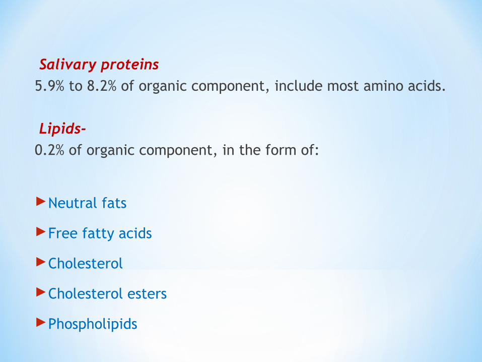
Salivary proteins 5.9% to 8.2% of organic component, include most amino acids.
Lipids-
0.2% of organic component, in the form of:
Neutral fats
Free fatty acids
Cholesterol
Cholesterol esters
Phospholipids

Subgingival calculus-
has composition similar to supragingival calculus, with some differences.
*More homogenous with equally high density of minerals.
*Same hydroxyapatite content, more magnesium
*Less brucite and octacalcium phosphate
*The ratio of calcium to phosphate is higher subgingivally.
*The sodium content increases with the depth of periodontal pockets.
*Salivary protein found in supragingival calculus is not found subgingivally
BACTERIAL CONTENT-
*The percentage of gram positive and gram- negative filamentous organisms is greater within calculus than in the remainder of oral cavity.7
*The microorganisms at the periphery are predominantly gram-negative rods and cocci.
*Most of the organisms within the calculus is nonviable.
*SUPRAGINGIVAL CALCULUS
Predominance of gram-positive filaments.
Next in frequency; gram-negative filaments and cocci.
Gram-positive cocci seen in calculus about which suppuration had taken place.
SUBGINGIVAL CALCULUS
*Superficial layers : gram- negative filaments most numerous
*Deep and middle zones : gram-positive filaments predominant.
Calculus is dental plaque which has undergone mineralization. Calculus formation occurs in three basic steps:
Pellicle formation
*All surfaces of the oral cavity are coated with a pellicle. Following tooth eruption or a dental prophylaxis, a thin, saliva- derived layer, called the acquired pellicle, covers the tooth surface.

Initial adhesion and attachment of bacteria
Transport to the surface – involves the initial transport of the bacterium to the tooth surface.
Initial adhesion – reversible adhesion of the bacterium, initiated by the interaction between the bacterium and the surface , through long-range and short-range forces
Attachment – a firm anchorage between bacterium and surface will be established by specific interactions.
Colonization and Plaque Maturation – when the firmly attached microorganisms start growing and the newly formed bacterial clusters remain attached, microcolonies or a biofilm can develop.
*Gram- positive coccoidal organisms are the first settlers to adhere to the formed enamel pellicle, and subsequently, filamentous bacteria gradually dominate the maturing plaque biofilm (Scheie, 1994).7
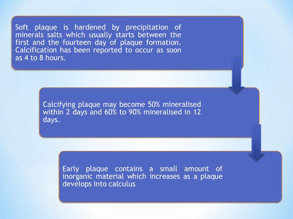
*Mineralization :
rate of formation and accumulation
*Formation of plaque consist of amorphous and/ or finely granular organic matrix containing mass of variety of gram positive and gram negative coccoid bacteria and filamentous form.
*The matrix is a form of mucopolysaccride derived from either saliva or bacteria or both.

*Calculus formation continues until it reaches a maximum from which it may be reduced in amount.
*The time required to reach the maximum level has been reported as 10 weeks, 18 weeks, and 6 months.7
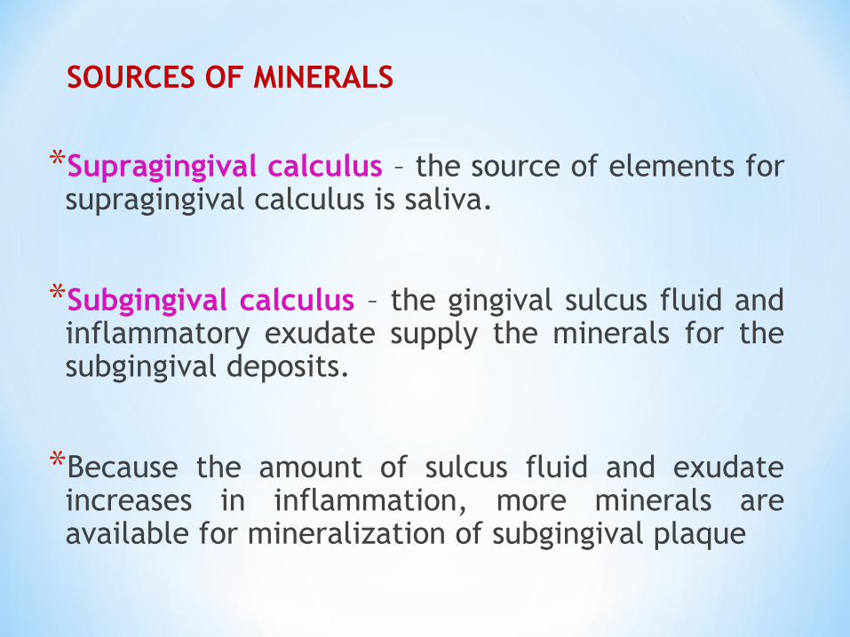
SOURCES OF MINERALS
*Supragingival calculus – the source of elements for supragingival calculus is saliva.
*Subgingival calculus – the gingival sulcus fluid and inflammatory exudate supply the minerals for the subgingival deposits.
*Because the amount of sulcus fluid and exudate increases in inflammation, more minerals are available for mineralization of subgingival plaque
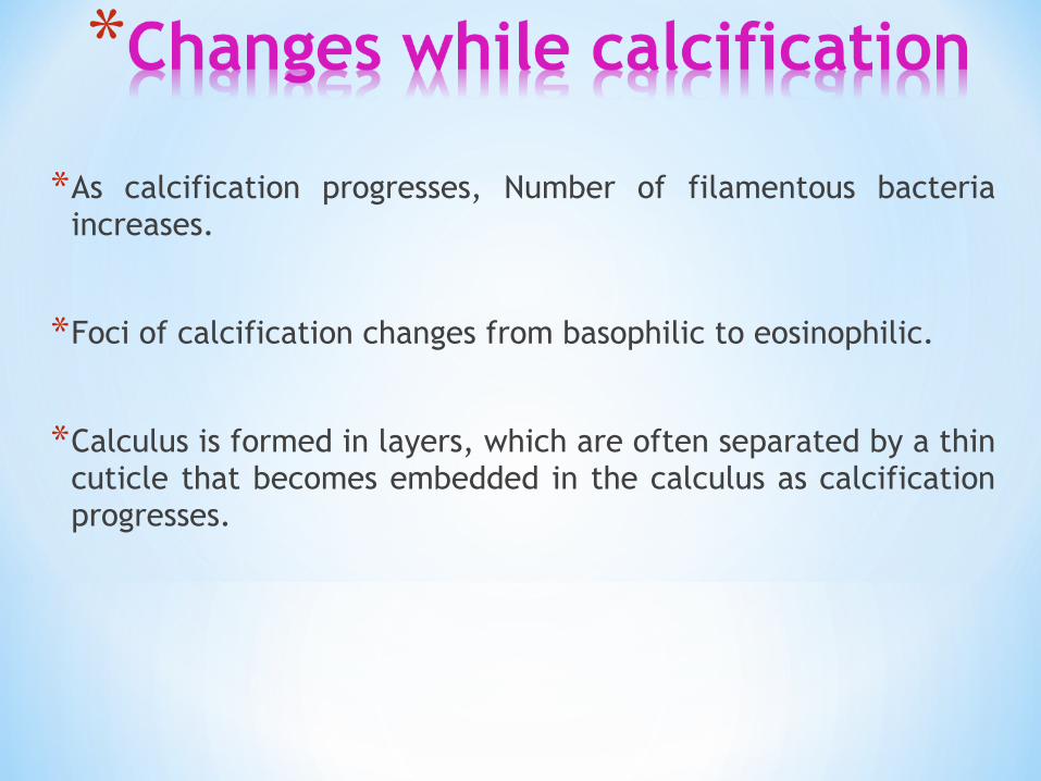
*As calcification progresses, Number of filamentous bacteria increases.
*Foci of calcification changes from basophilic to eosinophilic.
*Calculus is formed in layers, which are often separated by a thin cuticle that becomes embedded in the calculus as calcification progresses.
In 1878, Magitot was of the opinion that tartar consisted mainly of mineral matter formed by the precipitation of earthy carbonates and phosphates from the saliva.
These mineral salts were united with organic matter, epithelial cells, fatty globules, and leukocytes.
*BOOSTER CONCEPT1 according to this concept mineral precipitation results from a local rise in the degree of saturation of calcium and phosphate ions which may be brought about in several ways:
*Salivary pH theory – Hodge & Leung1 1950
A rise in the pH of saliva causes precipitation of calcium phosphate salts by lowering the precipitation constant.
The pH may be elevated by the loss of carbon dioxide, by the formation of ammonia by dental plaque and bacteria, or by protein degradation during stagnation.
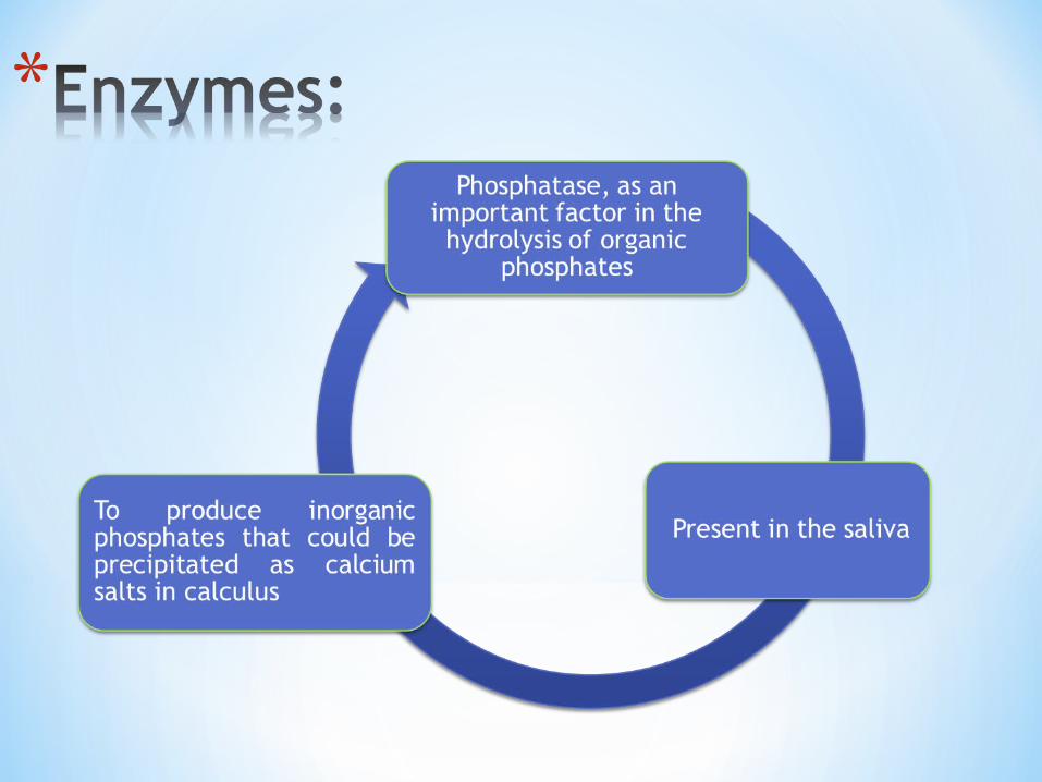
*Another enzyme esterase, present in the cocci, filamentous organisms, leukocytes, macrophages and desquamated epithelial cells of dental plaque, may initiate calcification by hydrolysing fatty esters into free fatty acids.
*The fatty acids forms soaps with calcium and magnesium that are converted later into the less soluble calcium phosphate salts
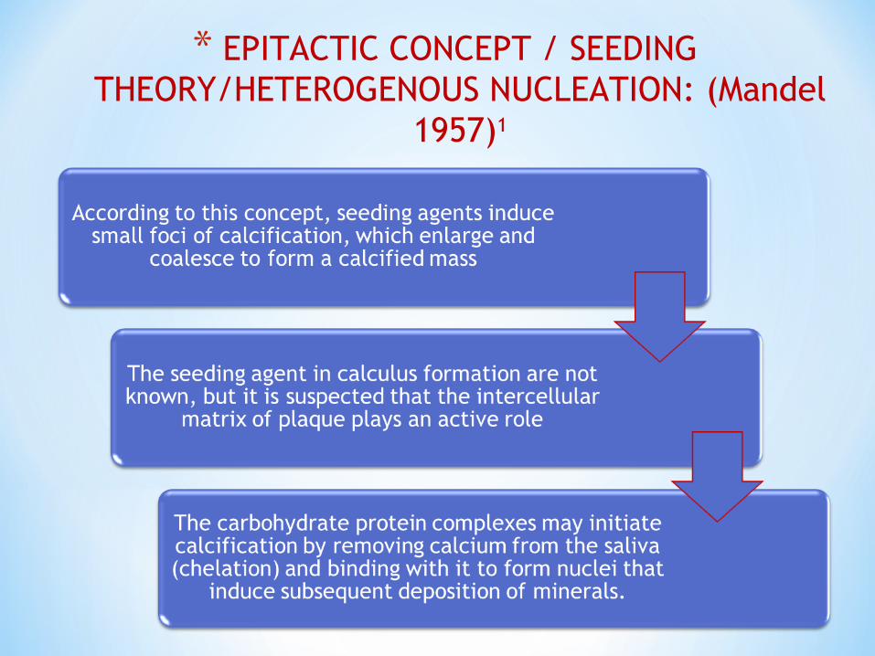
* EPITACTIC CONCEPT / SEEDING THEORY/HETEROGENOUS NUCLEATION: (Mandel
1957)1
*FACTORS AFFECTING THE RATE OF CALCULUS
FORMATION
*Diet and nutrition –the significance of diet in calculus formation depends more upon its consistency than upon its content.
*Increased calculus formation has been associated with deficiencies of vitamin A, niacin, or pyridoxine, and with an increase in dietary calcium, phosphorus, bicarbonate, protein and carbohydrate.
*Age – there is an increase in calculus deposition with an increasing age.(Schroeder et al,1969).5
*This increase, is not only increase in the number of surfaces, but also the size of calculus deposits.
*This may be due to change in quantity and quality of saliva with age, favouring the mineralization properties.
*Habits – In populations that practice regular oral hygiene and with access to regular professional care have low tendency for calculus formation.
*Smoking- is associated with an elevated risk for supragingival calculus deposition. Smoking may exert its influence systemically (elevated levels of salivary calcium and phosphorus) or locally via a conditioning of tooth surfaces.
*Salivary pH- increase pH increases the calculus formation.
*When the calcium phosphate crystals in solution are in kinetic equilibrium, the rate of precipitation is equal to that of dissolution.
* If pH in solution drops (the concentration of hydrogen ions increases), OH- and (PO4)3 - tend to be removed by H+ by forming water and more acidic forms of phosphate, respectively
*Salivary flow rate – increased salivary flow rate decreases the calculus formation. Salivary flow rate affects calcium phosphate saturation.
*Salivary calcium concentration- Elevated salivary calcium concentration, increases the rate of calculus formation.
Higher total salivary lipid levels – is associated with increased calculus formation
Emotional status- increased calculus formation has been associated with disturbed emotional status.
Nucleation inhibitors - Mg blocks apatite crystallization and stabilizes calcium phosphate as amorphous mineral (Ennever and Vogel, 1981)
Calcification promoters –
*Urea is a product from the metabolism of nitrogen -containing substances. Urea can be secreted in normal saliva at concentrations of between 5 and 10 mmol/L but can be as high as 30 mmol/L in patients with renal disease
*Gingival crevicular fluid contains up to 60mmol/L urea.
*Urease is responsible for bacterial urea hydrolysis. At a neutral pH, urea is hydrolyzed by urease to NH4
+ and bicarbonate.
*ULTRASTRUCTURE OF CALCULUS
*LAYERS – calculus forms in layers that are more or less parallel with the tooth surface.6
*The layers are separated by a line that appear to be a pellicle that was deposited over previously formed calculus, and as mineralization progressed, the pellicle became embedded.
*The lines between the layers of calculus can be called incremental lines. The lines are evidence that calculus grows, or increases by apposition of new layers.
*LIGHT MICROSCOPY-
Young supragingival calculus- the interface with tooth surface is fairly smooth where as the external mineralized surface is generally irregular and covered by a layer of plaque.
There are large areas within the calculus that are non- mineralized plaque layer of variable thickness

*Mature supragingival calculus- fewer non- mineralized lacunae, and in some sections, the lacunae formed a continuous connection with the external bacterial plaque.
*The presence of areas of non-mineralization matrix was a frequent finding at all levels within the body of supragingival calculus. two possible explanations have been suggested(Friskopp 1983) :1
*Filamentous bacteria that are predominant in supragingival plaque could have properties that inhibit mineralization, resulting in non-mineralized regions within the calculus.
*Differences in calcification capability between different bacterial colonies located in different parts of supragingival plaque.
*Subgingival calculus- uniform mineralized component. Unlike supragingival calculus, lacunae are not seen within the body of subgingival calculus.
*ATTACHMENT OF CALCULUS TO TOOTH SURFACE
*Differences in the manner in which calculus is attached to the tooth surface affect the relative ease or difficulty encountered in its removal.
*Several modes of attachment has been observed by conventional histological techniques and by electron microscopy.
*On any one tooth and in any one area, more than one mode of attachment may be found.
Attachment by means of an organic pellicle on enamel-
*Calculus attachment is superficial because no interlocking or penetration occurs.
*Pellicle attachment occur most frequently on enamel and newly scaled and planned root surfaces
*Calculus can be readily removed because of smooth attachment

Mechanical locking into surface irregularities.
*Enamel irregularities include cracks, lamellae, and carious defects.
*Cemental irregularities include resorption lacunae , cemental tears.
*Close adaptation of calculus undersurface depressions to the gentle sloping moulds of the unaltered cementum surface.
*Attachment of organic matrix of calculus into minute irregularities that were previously insertion locations of sharpey’s fibres.
*Calculus embedded deeply in cementum may appear morphologically similar to cementum and thus has been termed calculocementum
*Early studies by Goodrich & Moseley1 demonstrated that the presence of long, thick, unbranching filaments, which they called Leptothrix, were most important in the formation of tartar deposits.
*Bulleid1 recognized the Leptotrichia buccalis in calculus.
*Naeslund1. Actinomyces and Leptotrichia are the important organisms in calculus. The Leptotrichia usually formed a distinct, more superficial layer over the deeper Actinomyces colonies.
*The growth of these organisms produces certain biochemical changes that lead to a precipitation of calcium

*The lack of formation of a calcified deposit in the absence of bacteria was confirmed by Bibby’s ( in vitro studies). The important organism in his investigation was Leptotrichia.1
*Yardeni cultured various layers of calculus. His results showed that the deep, well mineralized portions of calculus contained very few if any viable organisms. The bulk of calculus contained mostly gram-positive filaments of the Actinomyces type. 1
Acquired pellicle forms on an implant surface when the metal surface initially comes into contact with tissues (Baier, 1982).1
Adsorption of proteins does not occur on the surface of pure titanium (Ti).
5-6nm oxide layer composed of TiO2 forms on the surface of Ti when exposed to air.
At physiological pH, the TiO2 layer carries net -ve charges, which enable the TiO2 layer to bind cations like Ca2
+
This makes the surface of an implant +vely charged and, consequently, attracts the high-weight molecules carrying -ve charges, notably proteins.
Some irregularities may also be encountered on oral implant surfaces,
The attachment to commercially pure titanium generally is less intimate than to root surface structures.

SUPRAGINGIVAL EXAMINATION :
Direct Examination – supragingival calculus can be seen directly or indirectly.
Using a mouth mirror with a combination of retraction, light and drying with air, small deposits can be seen.
SUBGINGIVAL EXAMINATION:
Visual Examination – dark edge of calculus may be seen at or just beneath the gingival margin.gentle air blast can deflect the margin from the tooth for observation into the pocket.
Gingival tissue colour change – dark calculus may reflect through a thin margin and suggest its presence.
Tactile examination- Clerehugh 1996 used WHO # 621 probe and a fine subgingival explorer is also used.
*Simplified calculus index (Green & Vermillion ’64 )
*Calculus component of periodontal disease index (Ramfjord 1959 )
*Calculus surface index (Ennever J,, Sturzenberger & Radike 1961)
*Calculus surface severity index (Ennever J et al ’61)
*Marginal line calculus index (Muhleman & Villa ’67)
*Volpe- Manhold index (Volpe A R & Manhold J H ’62)

Calculus Detection Only
*Perioscopy™Fiberoptic Endoscopy Based Technology
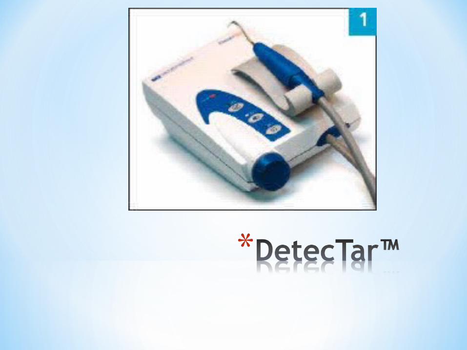
*DetecTar™ Spectro-Optical Technology
*Diagnodent™ Auto fluorescence Based Technology
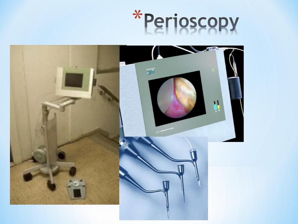
*Perioscopy involves a modified medical endoscope exclusively for periodontal purpose.
*This was developed in year 2000.
*It consists of a fiberoptic bundle surrounded by multiple illumination fibres, a light source and irrigation system.

*Its miniature nature causes minimal tissue trauma.
*Fiberoptic system permits visualization of the subgingival root surface, tooth structure and calculus in real time on a display monitor.8
*DetecTar™ uses a Spectro-optical approach in order to detect subgingival calculus by utilizing a light emitting diode and fiberoptic technology.
*This involves an optical fibre which recognizes the characteristic spectral signature of calculus caused by absorption, reflection and diffraction of red light.8
*Calculus and tooth structure differ in composition.
*This structural difference gives a typical fluorescence to both these structures.
*Calculus contains various non-metal as well as metal porphyrins and chromatophores which make it able to emit fluorescent light when irradiated with a light of certain wavelength.
*Diagnodent™ makes use of this property of calculus to detect its presence.
*Calculus and teeth fluoresce at different wavelength region of 628-685nm & 477-497nm respectively.
*Diagnodent™ involves use of an indium gallium arsenide phosphate (InGaAsP) based red laser diode which emits a wavelength of 655nm through an optical fibre causing fluorescence of tooth surface and calculus.8

DIAGNODENT™
*Perioscan™ Ultrasound technology
*Keylaser3™ Laser Based Technology

*Keylaser3™ combines a 655nm InGaAsP diode for detection of calculus and a 2940nm Er: YAG laser for treatment.
*Previous versions of this system (i.e. Keylaser 1 and 2) can be used for removal of calculus only.
*A scale of 0-99 is used for detection of calculus.
*Values exceeding 40 indicate definite presence of mineralized deposits.
*Er: YAG laser is activated as a certain threshold is reached.
*As soon as the value fall below threshold level Er: YAG laser is switched off.
*Studies done to assess the efficacy of this device have shown that it produces tooth surface comparable to hand and ultrasonic instruments. Cost factor can be a limiting aspect for using lasers for detection and treatment.8

*Perioscan™ can differentiate between calculus and healthy root surfaces.
*It also has a treatment option that can be used to remove these calculus deposits immediately.
*This combination of detection and removal mechanism is advantageous since calculus can be removed just by switching the mode from detection to removal.
*Perioscan™ is an ultrasonic device that works on acoustic principles.
* It is similar to tapping on a glass surface with a hard substance and analysing the sound produced to find out the cracks that are present on glass.
*Tip of the ultrasonic insert is oscillating continuously.

*Different voltages are produced due to changes in oscillations depending on the hardness of the surface.
*Hardness of the calculus differs from the hardness of the tooth surface.
*This difference in hardness can be used to generate the information of the surface that is being touched by the device.8
*This instrument is used in two different modes.
*Whenever ultrasonic tip touches the tooth surface a light signal is displayed on hand-piece and actual unit.
*Light signal is also accompanied by an acoustic signal.
*During calculus detection mode, the instrument shows a blue light when calculus is present. (Fig.2)

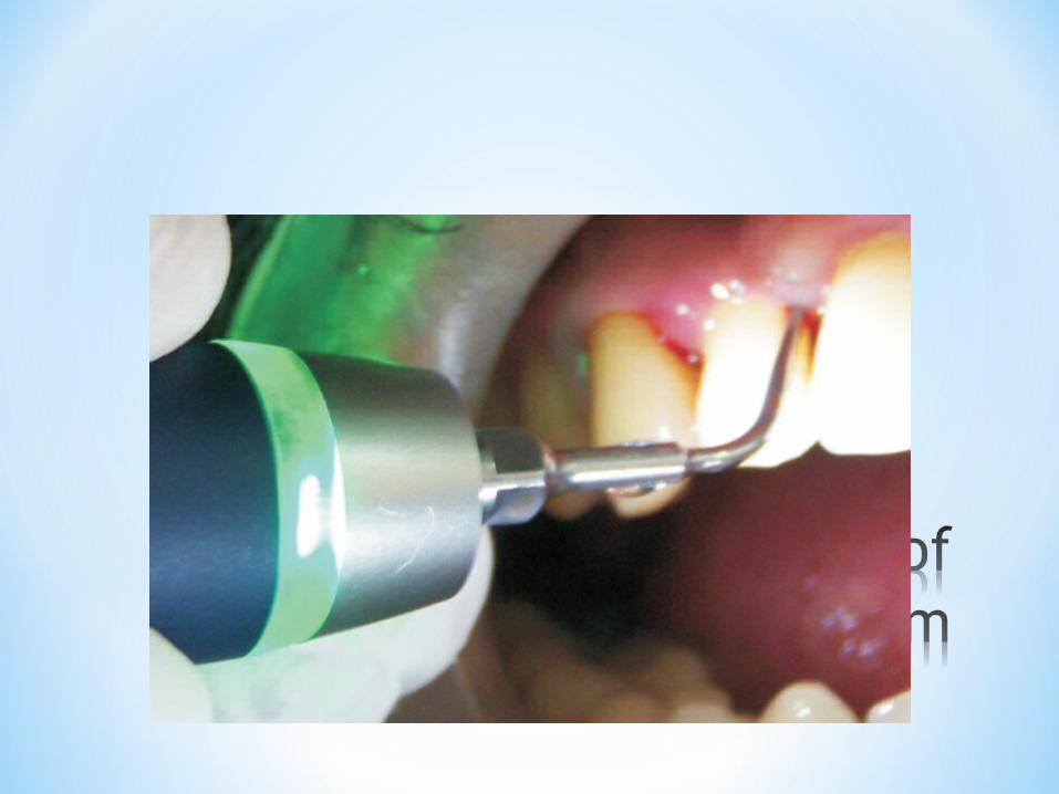
*Once a healthy root surface is attained, green light is displayed when the ultrasonic tip touches healthy cementum. (fig.3)
* Different power settings aid the clinician in removing tenacious calculus
1.Mandel ID, Gaffar A. Calculus revisited- A review. J Clin Periodontol1986;13: 249-257
2.Newmann, Takei, Klokkevold, Carranza: Clinical periodontology. 10th Edition. Noida: Elsevier; 2009.
3.Glossary of periodontal terms (2001). 4th edn. Chicago: The American academy of periodontology.
4.Greene JC. The Oral Hygiene Index—Development and uses. Journal of Periodontology. 1967;38(Suppl):37.
5.Schroeder HE (1969) Formation and inhibition of dental calculus. Vienna: Hans Huber
6.Ten Cate's Oral Histology, Nanci, Elsevier, 2013, page 255
7. Jepsen S, Deschner J, Braun A, Schwarz F, Eberhard J. Calculus removal and the prevention of its formation. Periodontol 2000 (2011);55: 167-188
8. Meissner G, Kocher T. Calculus detection technologies and their clinical application. Periodontol 2000 2011;55:189-204