Embed Size (px)
Citation preview
Colorectal Cancer
M. N. Jalalian
Medical Intern
Tehran University of Medical Sciences
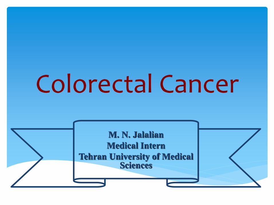
Most common malignancy of GI
Aging Dominant
after age 50
Hereditary Risk Factors 20% with a family history
FAP, HNPCC
Environmental and Dietary Factors Saturated or polyunsaturated fats
Inflammatory Bowel Disease long-standing colitis
Other Risk Factors Cigarette smoking, Ureterosigmoidostomy, Acromegaly,
Pelvic irradiation
Epidemiology and Risk Factors
Familial Adenomatous Polyposis
Attenuated FAP
Hereditary Nonpolyposis Colon Cancer (Lynch Syndrome)
Familial Colorectal Cancer
Inherited Colorectal Carcinoma
Definition:
An autosomal dominant condition with numerous polyps and increased risk of colorectal cancer
A known family history of FAP with even one adenomatous polyp …or
Developing hundreds to thousands of adenomatous polyps shortly after puberty (without a family history)
Familial Adenomatous Polyposis
1% of all colorectal adenocarcinomas
mutation in the APC gene (5q)
75% of cases
25% without a family history
Lifetime risk of colorectal cancer 100% by age 50 years
Treatment is surgical
Most patients elect to have an ileal pouch–anal anastomosis
FAP
fewer polyps (usually 10 to 100)
The right colon
Cancer risk 50%
APC mutation testing + in 60%
Screening by colonoscopy
Unknown family mutation
at age 13–15y, then every 4y to age 28y.
Treatment is surgical
Total abdominal colectomy with ileorectal anastomosis
Attenuated FAP
Definition:
An AD genetic condition
High risk of colorectal carcinoma at an early age (average age: 40–45 years) & other cancers
More common than FAP
70% develop cancer
HNPCC (Lynch Syndrome)
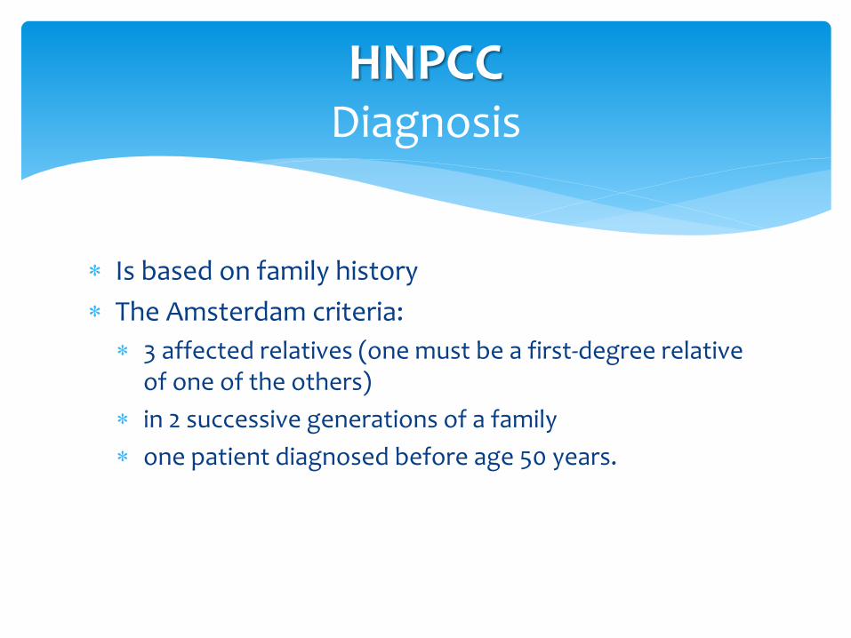
Is based on family history
The Amsterdam criteria:
3 affected relatives (one must be a first-degree relative of one of the others)
in 2 successive generations of a family
one patient diagnosed before age 50 years.
HNPCCDiagnosis
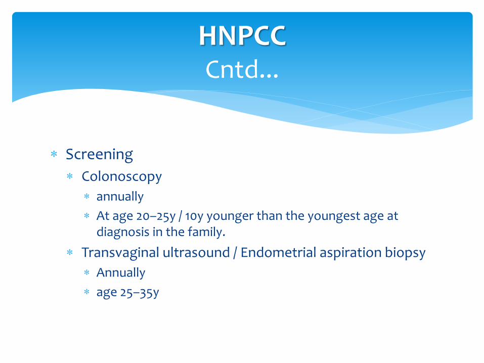
Screening
Colonoscopy
annually
At age 20–25y / 10y younger than the youngest age at diagnosis in the family.
Transvaginal ultrasound / Endometrial aspiration biopsy
Annually
age 25–35y
HNPCCCntd...
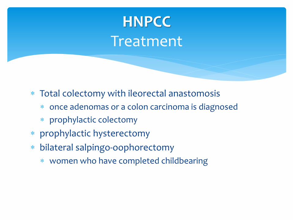
Total colectomy with ileorectal anastomosis
once adenomas or a colon carcinoma is diagnosed
prophylactic colectomy
prophylactic hysterectomy
bilateral salpingo-oophorectomy
women who have completed childbearing
HNPCCTreatment
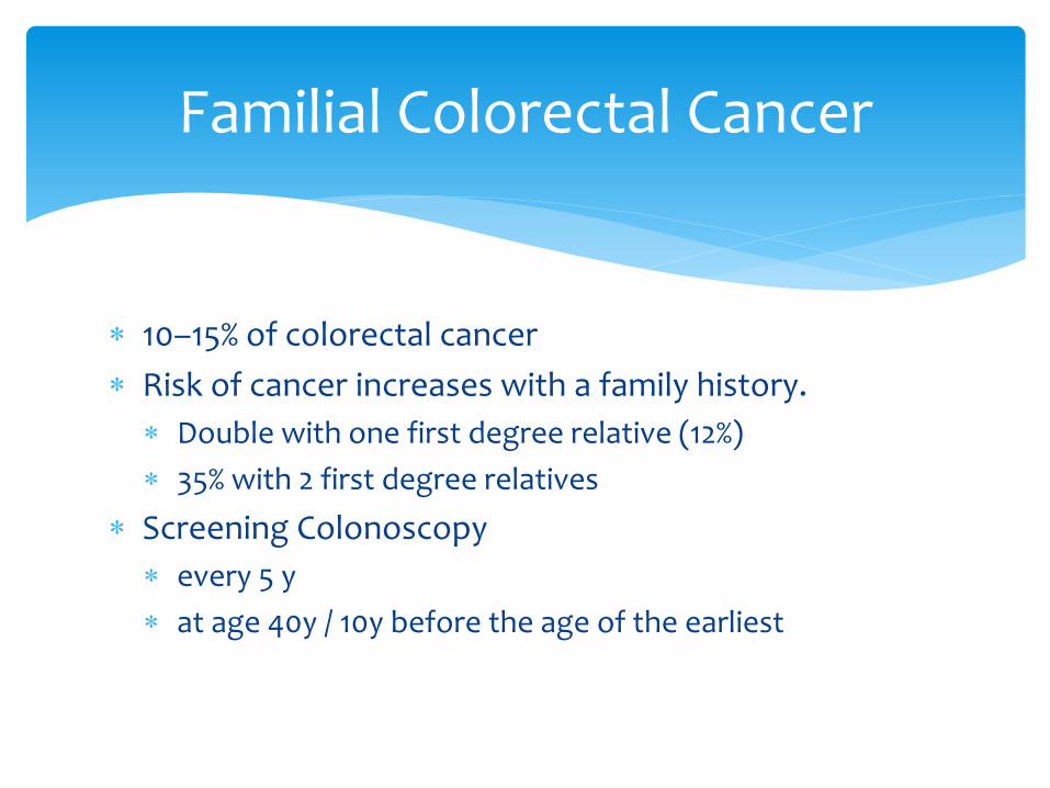
10–15% of colorectal cancer
Risk of cancer increases with a family history.
Double with one first degree relative (12%)
35% with 2 first degree relatives
Screening Colonoscopy
every 5 y
at age 40y / 10y before the age of the earliest
Familial Colorectal Cancer
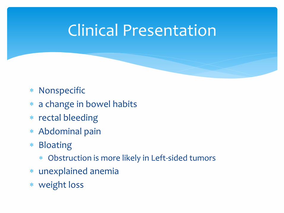
Nonspecific
a change in bowel habits
rectal bleeding
Abdominal pain
Bloating
Obstruction is more likely in Left-sided tumors
unexplained anemia
weight loss
Clinical Presentation
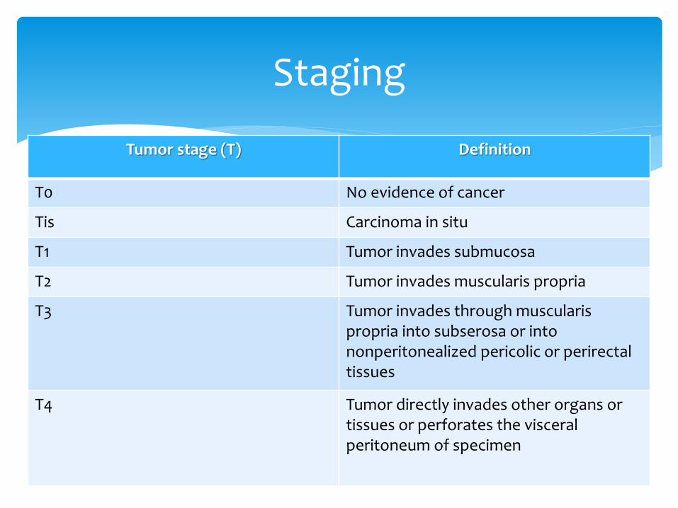
Tumor stage (T) Definition
T0 No evidence of cancer
Tis Carcinoma in situ
T1 Tumor invades submucosa
T2 Tumor invades muscularis propria
T3 Tumor invades through muscularispropria into subserosa or into nonperitonealized pericolic or perirectal tissues
T4 Tumor directly invades other organs or tissues or perforates the visceral peritoneum of specimen
Staging
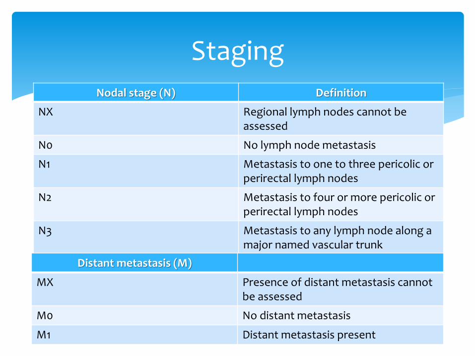
Nodal stage (N) Definition
NX Regional lymph nodes cannot be assessed
N0 No lymph node metastasis
N1 Metastasis to one to three pericolic or perirectal lymph nodes
N2 Metastasis to four or more pericolic or perirectal lymph nodes
N3 Metastasis to any lymph node along a major named vascular trunk
Staging
Distant metastasis (M)
MX Presence of distant metastasis cannot be assessed
M0 No distant metastasis
M1 Distant metastasis present
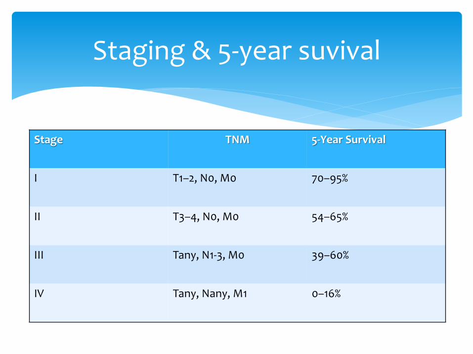
Staging & 5-year suvival
Stage TNM 5-Year Survival
I T1–2, N0, M0 70–95%
II T3–4, N0, M0 54–65%
III Tany, N1-3, M0 39–60%
IV Tany, Nany, M1 0–16%
Colonoscopy
Synchronous disease up to 5%
Chest and Abdominal/pelvic CT scan
distant metastases
Routine Blood tests and CEA
Endorectal ultrasound / Pelvic MRI
The ultrasound T and N stage of rectal cancer
Preoperative Evaluation
The objective is
remove the primary tumor with clean borders
And its lymphovascular supply
Chemotherapy
Stages III and IV
Stage II if
Young patient
Bad histology
Radiotherapy
Greatly used for rectal cancers
Treatment
Stage 0 (Tis, N0, M0)
Polipectomy with clean margins
Stage I: The malignant polyp (T1, N0, M0)
Polipectomy by endoscope (low risk of LN metastasis)
Segmental colectomy
Stages I and II: Localized colon carcinoma (T1–3, N0, M0)
The majority cured with surgical resection
Adjuvant chemotherapy
young patients
“high-risk” histologic
THERAPY FOR COLONIC CARCINOMA
Stage III: Lymph Node Metastasis (T any, N1, M0)
Surgery
adjuvant chemotherapy
Stage IV: Distant metastasis (T any, N any, M1)
metastases limited to the liver
Resection
adjuvant chemotherapy
The remainder
Palliative therapy
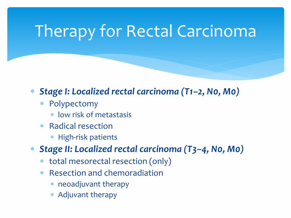
Stage I: Localized rectal carcinoma (T1–2, N0, M0) Polypectomy
low risk of metastasis
Radical resection High-risk patients
Stage II: Localized rectal carcinoma (T3–4, N0, M0) total mesorectal resection (only)
Resection and chemoradiation neoadjuvant therapy
Adjuvant therapy
Therapy for Rectal Carcinoma
Stage III: Lymph node metastasis (T any, N1, M0)
Resection
neoadjuvant chemoradiation
Adjuvant chemoradiation
Stage IV: Distant metastasis (T any, N any, M1)
Mostly palliative
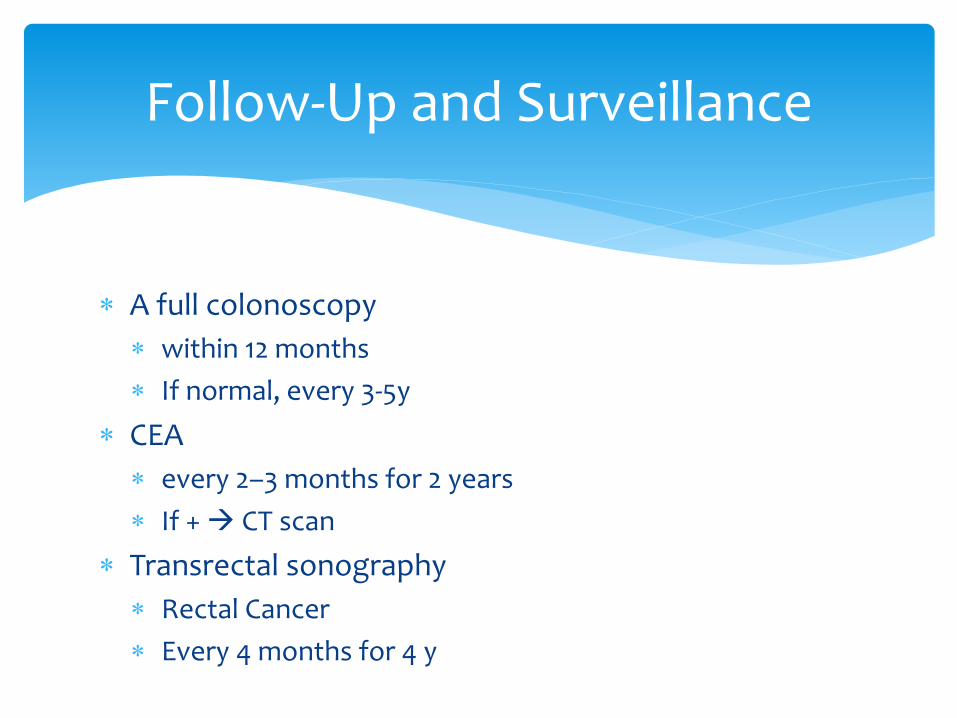
A full colonoscopy
within 12 months
If normal, every 3-5y
CEA
every 2–3 months for 2 years
If + CT scan
Transrectal sonography
Rectal Cancer
Every 4 months for 4 y
Follow-Up and Surveillance

THANK YOU FOR YOUR ATTENTION