Embed Size (px)
Citation preview

Hafiz Waqar Ahmed Baber
560-FBAS/BSBT/S14
Department of Biotechnology
Faculty of Basic and applied sciences
Cell mediated and Antibody mediated immunity
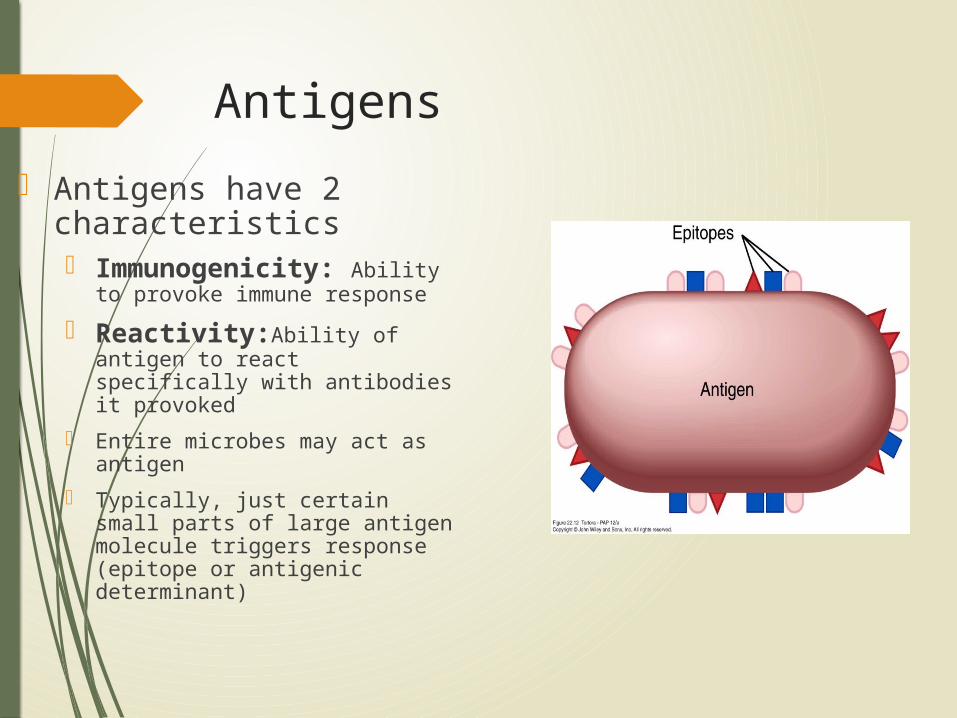
Antigens
Antigens have 2 characteristics Immunogenicity: Ability to
provoke immune response
Reactivity:Ability of antigen to react specifically with antibodies it provoked
Entire microbes may act as antigen
Typically, just certain small parts of large antigen molecule triggers response (epitope or antigenic determinant)
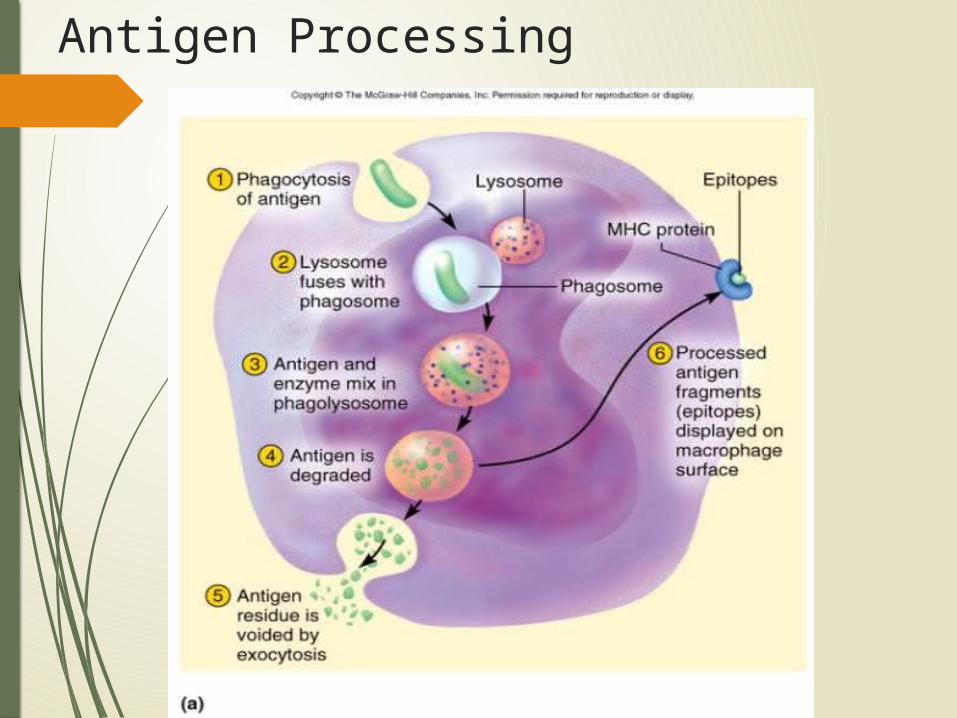
Antigen Processing
Cellular Immunity T cells attack foreign cells and diseased host cells; memory of Ag
Three classes of T cells
1. Cytotoxic T cells (Tc cells) carry out attack
2. Helper T cells: help promote Tc cell and B cell action and nonspecific defense mechanisms
3. Memory T cells: provide immunity from future exposure to antigen
MHC Proteins Membrane glycoproteins, bind to antigens,
encoded by chromosome 6, form MHC. 2 Classes of MHC Proteins Class I: found in membranes of all nucleated
cells Pick up small peptides in cell and carry them
to the surface: T cells ignore normal peptides abnormal peptides or viral proteins activate T
cells to destroy cell
Class II: found in membranes of antigen-presenting cells (APCs) found in lymphocytes Antigenic fragments inserted in cell membrane to stimulate T cells
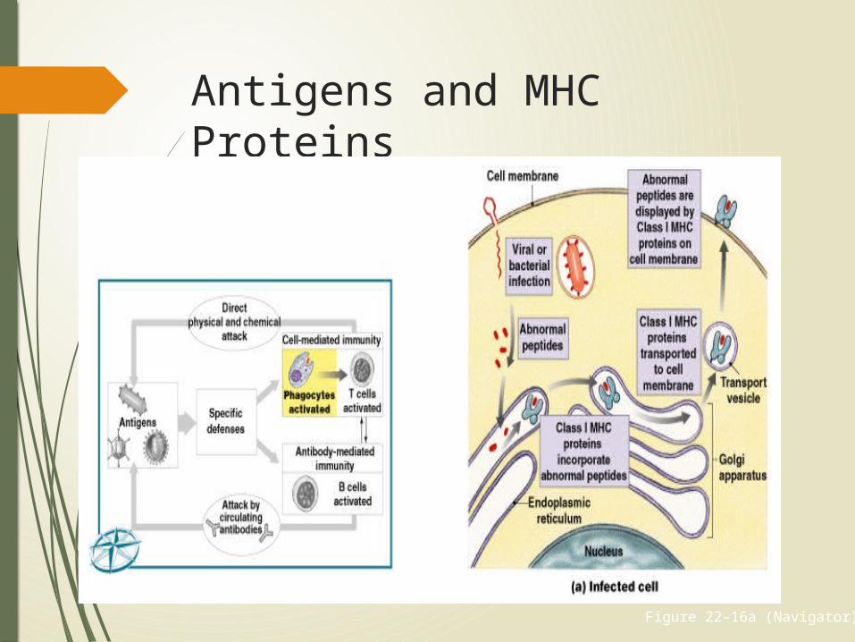
Antigens and MHC Proteins
Figure 22–16a (Navigator)
Antigen-Presenting Cells (APCs) Responsible for activating T cells against foreign cells and proteins
Phagocytic APCs :
Free and fixed macrophages:
in connective tissues
Kupffer cells:
of the liver
Microglia:
in the CNS
Pinocytic APCs
Langerhans cells:
in the skin
Dendritic cells:
in lymph nodes and spleen
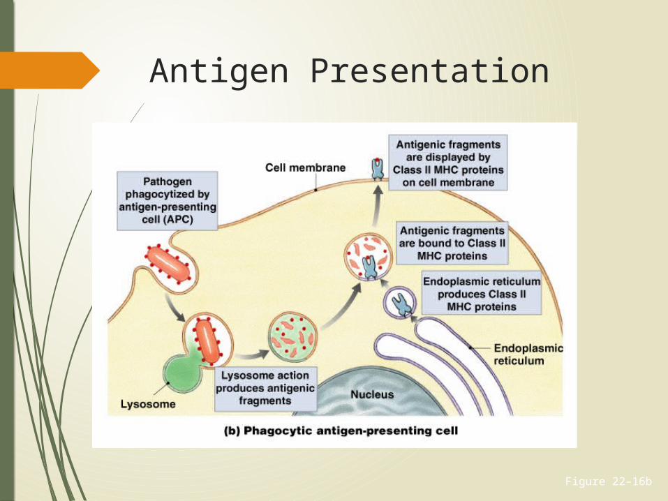
Figure 22–16b
Antigen Presentation
Mechanism of T cell recognition and activation
Antigen Recognition
Inactive T cell receptors:
recognize Class I or Class II MHC proteins
recognize a specific antigen
Binding occurs when MHC protein matches antigen
TC cell Recognition
Antigen presentation MHC-I proteins
found on nearly all nucleated body cells
display peptides produced by host cells
TC cell activation1. binding of cytotoxic T cells (CD8 cells) to abnormal
peptides on MHC-I and
2. costimulation via a cytokine
triggers clonal selection: clone of identical T cells against cells with same epitope
TH cell Recognition
Antigen presentation
role of MHC-II proteins found only on antigen
presenting cells display only foreign antigens stimulate helper T cells (CD4
cells)
CD Markers Also called cluster of differentiation markers:
in T cell membranes
molecular mechanism of antigen recognition
more than 70 types:
designated by an identifying number
CD3 Receptor Complex
Found in all T cells
CD8 Markers Found on cytotoxic T cells and suppressor T cells
Respond to antigens on Class I MHC proteins
CD4 Markers Found on helper T cells
Respond to antigens on Class II MHC proteins
CD8 or CD4 Markers - Bind to CD3 receptor complex
Prepare cell for activation
Costimulation
For T cell to be activated, it must be costimulated:
by binding to stimulating cell at second site
which confirms the first signal
2 Classes of CD8 T Cells
Activated by exposure to antigens on MHC proteins:
one responds quickly:
producing cytotoxic T cells and memory T cells
the other responds slowly:
producing suppressor T cells
TH cell Activation
1. binding of helper T cells (CD4 cells) to epitope displayed on MHC-II of APC
2. costimulation via a cytokine
3. triggers clonal selection
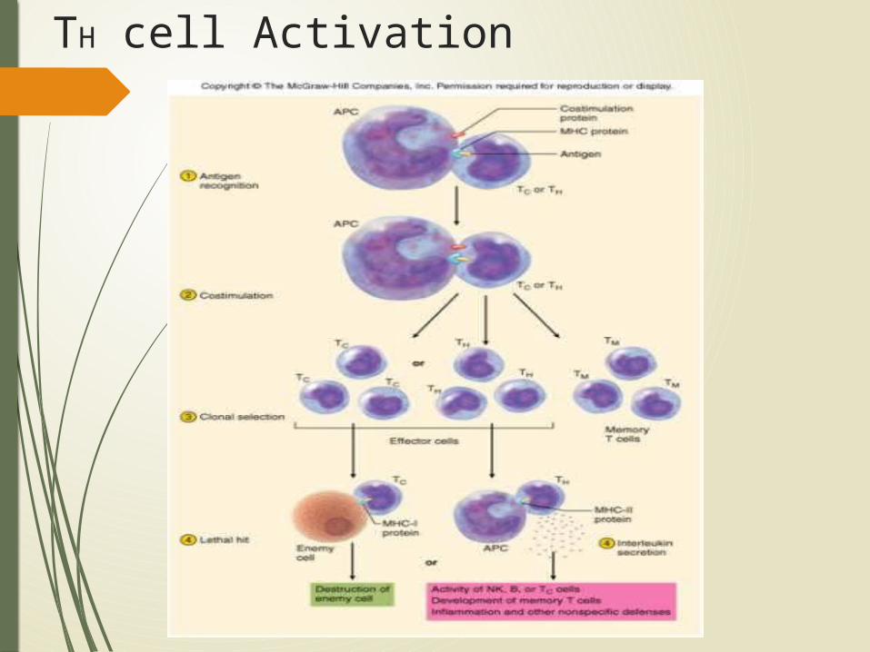
TH cell Activation
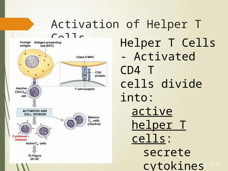
Activation of Helper T Cells
Figure 22–18
Helper T Cells - Activated CD4 T cells divide into:
active helper T cells:
secrete cytokines
memory T cells:
remain in reserve
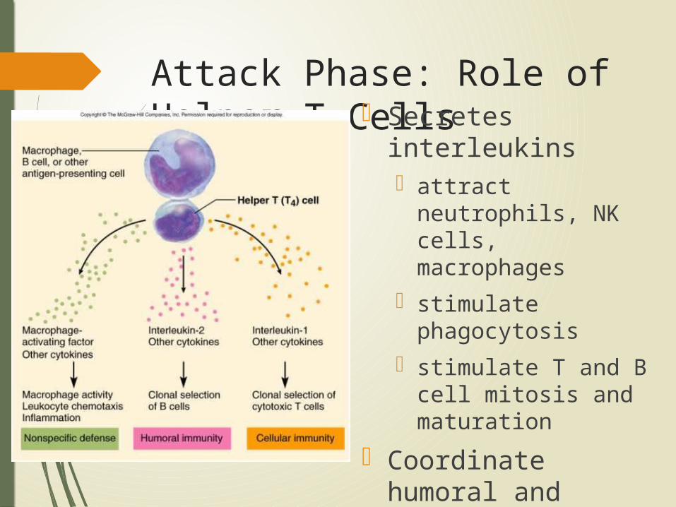
Attack Phase: Role of Helper T Cells Secretes
interleukins attract neutrophils,
NK cells, macrophages
stimulate phagocytosis
stimulate T and B cell mitosis and maturation
Coordinate humoral and cellular immunity
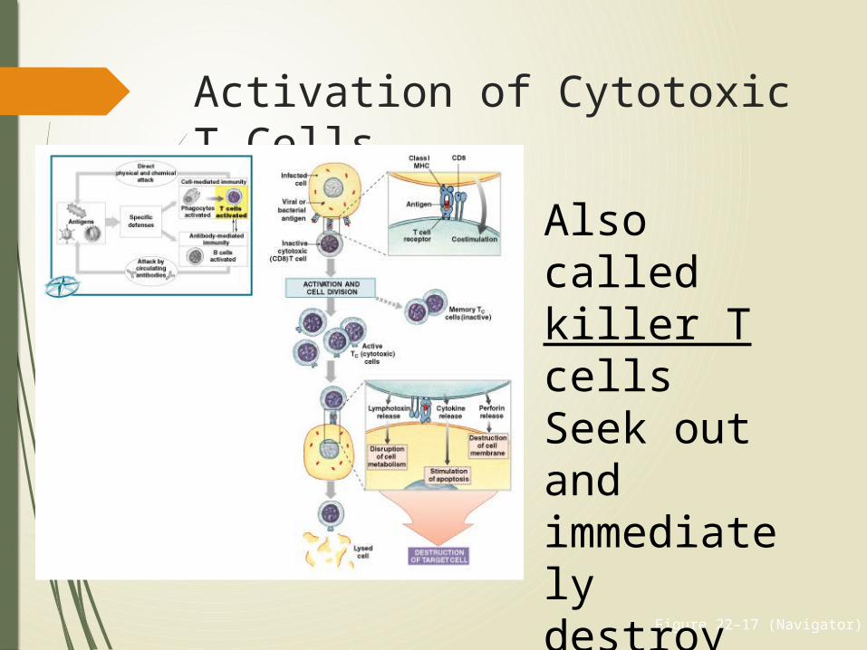
Figure 22–17 (Navigator)
Activation of Cytotoxic T Cells
Also called killer T cells Seek out and immediately destroy target cells
Actions of Cytotoxic T Cells
1. Release perforin:
to destroy antigenic cell membrane
2. Secrete poisonous lymphotoxin:
to destroy target cell
3. Activate genes in target cell:
that cause cell to die
Slow Response
Can take up to 2 days from time of first exposure to an antigen, for cytotoxic T cells to reach effective levels
Memory Tc Cells
Produced with cytotoxic T cells
Stay in circulation
Immediately form cytotoxic T cells:
if same antigen appears again
Suppressor T Cells
Secrete suppression factors
Inhibit responses of T and B cells
After initial immune response
Limit immune reaction to single stimulus
4 Functions of Cytokines
1. Stimulate T cell divisions:
produce memory T cells
accelerate cytotoxic T cell maturation
2. Attract and stimulate macrophages
3. Attract and stimulate NK cells
4. Promote activation of B cells
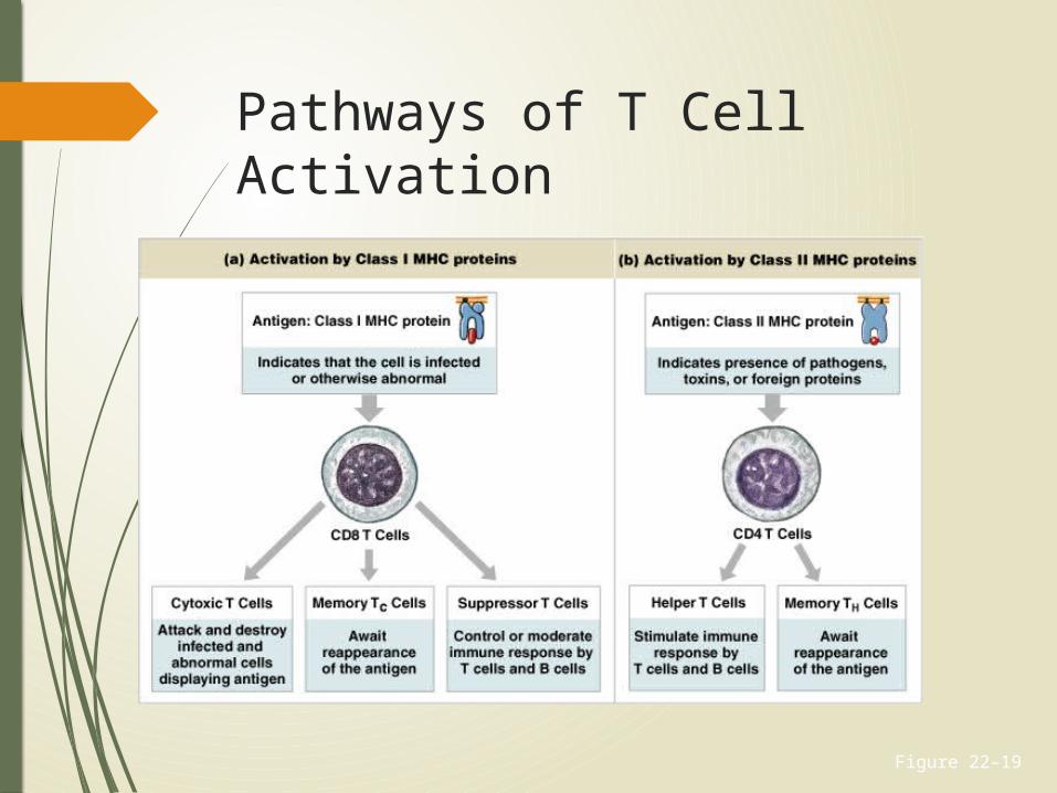
Figure 22–19
Pathways of T Cell Activation
Humoral Immunity Recognition
B cell receptors bind antigen, take in and digest antigen then display epitopes on its MHC-II protein
After costimulation by TH cell, divide repeatedly, differentiate into plasma cells, produce antibodies specific to that antigen
Attack antibodies bind to antigen, render it harmless, ‘tag it’ for destruction
Memory some B cells differentiate into memory cells
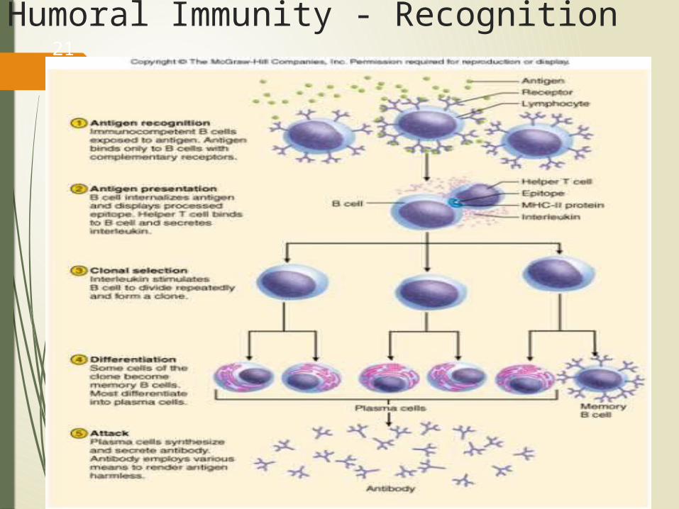
Humoral Immunity - Recognition21
-31
B Cells Responsible for antibody-mediated immunity
Attack antigens by producing specific antibodies
Millions of populations, each with different antibody molecules
Sensitization
Corresponding antigens in interstitial fluids bind to B cell receptors
B cell prepares for activation
Preparation process is sensitization
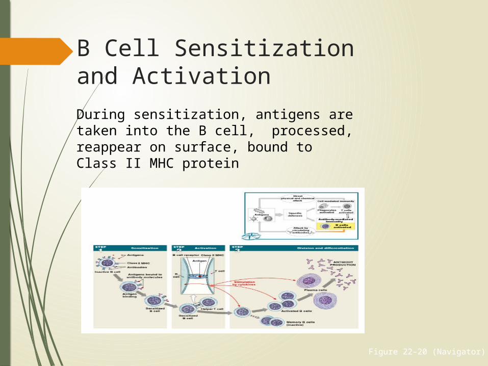
Figure 22–20 (Navigator)
B Cell Sensitization and ActivationDuring sensitization, antigens are taken into the B cell, processed, reappear on surface, bound to Class II MHC protein
Helper T Cells Sensitized B cell is prepared for activation, but needs helper T cell
activated by same antigen
B Cell Activation Helper T cell binds to MHC complex:
secretes cytokines that promote B cell activation and division
B Cell Division
Activated B cell divides into:
plasma cells -Synthesize and secrete antibodies into interstitial fluid
Memory B cells- Like memory T cells remain in reserve to respond to next infection

Any Question

Thank you