Embed Size (px)
Citation preview
design of a self-sustained, completely synthet-ic carbon metabolism in artificial or minimalcells (37).
REFERENCES AND NOTES
1. I. A. Berg, Appl. Environ. Microbiol. 77, 1925–1936(2011).
2. G. Fuchs, Annu. Rev. Microbiol. 65, 631–658 (2011).3. M. Aresta, in Greenhouse Gases: Mitigation and Utilization,
CHEMRAWN-XVII and ICCDU-IX Conference, July 8 to 12, 2007,E. Buncel, Ed. (Queen’s University, Kingston, Ontario,Canada, 2009), pp. 123–149.
4. S. M. Glueck, S. Gümüs, W. M. F. Fabian, K. Faber, Chem. Soc.Rev. 39, 313–328 (2010).
5. L. Rosgaard, A. J. de Porcellinis, J. H. Jacobsen, N. U. Frigaard,Y. Sakuragi, J. Biotechnol. 162, 134–147 (2012).
6. B. J. Walker, A. VanLoocke, C. J. Bernacchi, D. R. Ort, Annu.Rev. Plant Biol. 67, 107–129 (2016).
7. T. J. Erb, J. Zarzycki, Curr. Opin. Chem. Biol. 34, 72–79 (2016).8. D. N. Greene, S. M. Whitney, I. Matsumura, Biochem. J. 404,
517–524 (2007).9. N. E. Kreel, F. R. Tabita, PLOS ONE 10, e0138351 (2015).10. P. M. Shih, J. Zarzycki, K. K. Niyogi, C. A. Kerfeld, J. Biol. Chem.
289, 9493–9500 (2014).11. R. Kebeish et al., Nat. Biotechnol. 25, 593–599 (2007).12. M. Mattozzi, M. Ziesack, M. J. Voges, P. A. Silver, J. C. Way,
Metab. Eng. 16, 130–139 (2013).13. M. W. Keller et al., Proc. Natl. Acad. Sci. U.S.A. 110, 5840–5845 (2013).14. N. Antonovsky et al., Cell 166, 115–125 (2016).15. A. Bar-Even, E. Noor, N. E. Lewis, R. Milo, Proc. Natl. Acad. Sci.
U.S.A. 107, 8889–8894 (2010).16. P. H. Opgenorth, T. P. Korman, J. U. Bowie, Nat. Chem. Biol. 12,
393–395 (2016).17. S. Billerbeck, M. Bujara, C. Hold, R. Pellaux, S. Panke, FEBS J.
277 (suppl. 1), 23 (2010).18. C. E. Hodgman, M. C. Jewett, Metab. Eng. 14, 261–269 (2012).19. J. A. Rollin, T. K. Tam, Y. H. P. Zhang, Green Chem. 15,
1708–1719 (2013).20. S. P. Long, E. A. Ainsworth, A. Rogers, D. R. Ort, Annu. Rev.
Plant Biol. 55, 591–628 (2004).21. R. J. Spreitzer, M. E. Salvucci, Annu. Rev. Plant Biol. 53,
449–475 (2002).22. M. Könneke et al., Proc. Natl. Acad. Sci. U.S.A. 111, 8239–8244
(2014).23. L. Schada von Borzyskowski, R. G. Rosenthal, T. J. Erb,
J. Biotechnol. 168, 243–251 (2013).24. T. J. Erb et al., Proc. Natl. Acad. Sci. U.S.A. 104, 10631–10636
(2007).25. D. M. Peter et al., Angew. Chem. Int. Ed. 54, 13457–13461
(2015).26. T. J. Erb, V. Brecht, G. Fuchs, M. Müller, B. E. Alber, Proc. Natl.
Acad. Sci. U.S.A. 106, 8871–8876 (2009).27. M. Volpers et al., PLOS ONE 11, e0157851 (2016).28. T. J. Erb, G. Fuchs, B. E. Alber, Mol. Microbiol. 73, 992–1008
(2009).29. B. W. Carlson, L. L. Miller, J. Am. Chem. Soc. 105, 7453–7454
(1983).30. V. de Lorenzo, A. Sekowska, A. Danchin, FEMS Microbiol. Rev.
39, 96–119 (2015).31. C. Lerma-Ortiz et al., Biochem. Soc. Trans. 44, 961–971
(2016).32. C. L. Linster et al., J. Biol. Chem. 286, 42992–43003
(2011).33. N. L. Gale, J. V. Beck, J. Bacteriol. 94, 1052–1059
(1967).34. F. Jacob, Science 196, 1161–1166 (1977).35. D. R. Ort et al., Proc. Natl. Acad. Sci. U.S.A. 112, 8529–8536
(2015).36. R. K. Yadav et al., J. Am. Chem. Soc. 134, 11455–11461
(2012).37. C. A. Hutchison III et al., Science 351, aad6253
(2016).38. P. C. Hinkle, BBA Bioenergetics 1706, 1–11 (2005).39. D. M. Kramer, J. R. Evans, Plant Physiol. 155, 70–78
(2011).40. L. Schada von Borzyskowski, M. Remus-Emsermann,
R. Weishaupt, J. A. Vorholt, T. J. Erb, ACS Synth. Biol. 4,430–443 (2015).
ACKNOWLEDGMENTS
We thank G. Fuchs for introducing us to the fundamental principlesof microbial CO2 fixation. We thank U. Oppermann for the
expression plasmid p2BP1, K. Castiglione for the expressionplasmid of Fdh (D221A), J. Andexer for the expression plasmid forPkk2 and the purified enzyme, and J. Zarzycki and B. Vögeli forcontributions during pathway design. The work conducted by theU.S. Department of Energy Joint Genome Institute, a DOE Office ofScience User Facility, is supported under contract DE-AC02-05CH11231. Also supported by European Research Council grantERC 637675 “SYBORG,” Swiss National Science FoundationAmbizione grant PZ00P3_136828/1, ETH Zürich grant ETH-41 12-2,and the Max Planck Society. Supporting data are available in thesupplementary materials. Author contributions: T.J.E. conceivedthe project; T.J.E., L.S.v.B., and T.S. designed the experiments,analyzed the data, and wrote the manuscript; T.J.E., T.S., and
L.S.v.B. performed experiments; S.B. assisted in experiments; andN.S.C. performed mass spectrometry and analyzed the data.
SUPPLEMENTARY MATERIALS
www.sciencemag.org/content/354/6314/900/suppl/DC1Materials and MethodsSupplementary TextFigs. S1 to S14Tables S1 to S8References (41–63)
8 July 2016; accepted 5 October 201610.1126/science.aah5237
◥NEURODEGENERATION
Site-specific phosphorylation of tauinhibits amyloid-b toxicity inAlzheimer’s miceArne Ittner,1* Sook Wern Chua,1 Josefine Bertz,1 Alexander Volkerling,1
Julia van der Hoven,1 Amadeus Gladbach,1 Magdalena Przybyla,1 Mian Bi,1
Annika van Hummel,1,2 Claire H. Stevens,1 Stefania Ippati,1 Lisa S. Suh,1,3
Alexander Macmillan,4 Greg Sutherland,3 Jillian J. Kril,3 Ana P. G. Silva,5
Joel Mackay,5 Anne Poljak,6 Fabien Delerue,1,7 Yazi D. Ke,2 Lars M. Ittner1,7,8*
Amyloid-b (Ab) toxicity in Alzheimer’s disease (AD) is considered to be mediated byphosphorylated tau protein. In contrast, we found that, at least in early disease, site-specificphosphorylation of tau inhibited Ab toxicity.This specific tau phosphorylation was mediatedby the neuronal p38mitogen-activated protein kinase p38g and interfered with postsynapticexcitotoxic signaling complexes engaged by Ab. Accordingly, depletion of p38g exacerbatedneuronal circuit aberrations, cognitive deficits, and premature lethality in amousemodel of AD,whereas increasing the activity of p38g abolished these deficits. Furthermore, mimickingsite-specific tau phosphorylation alleviated Ab-induced neuronal death and offered protectionfrom excitotoxicity. Our work provides insights into postsynaptic processes in AD pathogenesisand challenges a purely pathogenic role of tau phosphorylation in neuronal toxicity.
Alzheimer’s disease (AD), themost prevalentform of dementia, is neuropathologicallycharacterized by extracellular amyloid-b (Ab)plaques and intracellular tau-containingneurofibrillary tangles (1, 2). Growing evi-
dence suggests that Ab and tau together orches-trate neuronal dysfunction in AD (3), althoughtheirmolecular connections remain poorly under-stood. Aberrant tau phosphorylation is the firststep in a cascade leading to its deposition and tocognitive dysfunction (4, 5). Ab is thought to trig-
ger toxic events, including tau phosphorylation(6). Accordingly, the depletion of tau prevents Abtoxicity in ADmodels (7–9). Ab-induced neuronalnetwork and synaptic dysfunction is associatedwith aberrant glutamatergic synaptic transmis-sion (10). N-methyl-D-aspartate (NMDA)–typeglutamatergic receptors (NRs) drive glutamate-induced neuronal excitotoxicity (11) and mediateAb toxicity by downstream responses that pro-mote neuronal dysfunction (12).Multiple factors, including p38 kinases, con-
tribute to NR-mediated toxicity (12). Although in-hibition of p38a and p38b improves Ab-inducedlong-term potentiation deficits (13, 14), it in-creases hyperexcitability in Ab precursor protein(APP) transgenic mice (15). However, the speci-ficity of p38 inhibitors remains debatable (16, 17).Other p38 kinases, p38g and p38d, have not beenstudied in AD. To understand the roles of p38kinases in AD, we induced excitotoxic seizureswith pentylenetetrazole (PTZ), an approach wide-ly used for studying excitotoxicity in AD mousemodels (8, 9). We used mice with individual de-letion of p38a, p38b, p38g, or p38d (fig. S1). Sur-prisingly, only p38g depletion (p38g−/−), but notsystemic p38b, p38d, or neuronal p38a (p38aDneu)
904 18 NOVEMBER 2016 • VOL 354 ISSUE 6314 sciencemag.org SCIENCE
1Dementia Research Unit, School of Medical Sciences, Universityof New South Wales (UNSW), Sydney, New South Wales 2052,Australia. 2Motor Neuron Disease Unit, School of MedicalSciences, UNSW, Sydney, New South Wales 2052, Australia.3Discipline of Pathology, Sydney Medical School, University ofSydney, Sydney, New South Wales 2050, Australia. 4BiomedicalImaging Facility, Mark Wainwright Analytical Centre, UNSW,Sydney, New South Wales 2052, Australia. 5School of MolecularBioscience, University of Sydney, Sydney, New South Wales2050, Australia. 6Biomedical Mass Spectrometry Facility, MarkWainwright Analytical Centre, UNSW, Sydney, New South Wales2052, Australia. 7Transgenic Animal Unit, Mark WainwrightAnalytical Centre, UNSW, Sydney, New South Wales 2052,Australia. 8Neuroscience Research Australia, Sydney, New SouthWales 2031, Australia.*Corresponding author. Email: [email protected] (A.I.); [email protected] (L.M.I.)
RESEARCH | REPORTS
on
Nov
embe
r 24
, 201
6ht
tp://
scie
nce.
scie
ncem
ag.o
rg/
Dow
nloa
ded
from
knockout, changed PTZ-induced seizures (Fig.1A and fig. S2). Pan-p38 inhibition in wild-type(WT) mice augmented seizures, similar to theeffects of p38g depletion (fig. S3). Consistentwith a postsynaptic role, only p38g localized to
dendritic spines and postsynaptic densities(PSDs) of neurons and was enriched in PSDpreparations (Fig. 1B and fig. S4). Hence, of allp38 kinases, only p38g localized to postsynap-ses and limited excitotoxicity.
To test whether p38g−/− augments Ab-induceddeficits, we crossed p38g−/− with Ab-formingAPP23 mice. APP23 mice present with prema-ture mortality, memory deficits, neuronal circuitaberrations with epileptiform brain activity,
SCIENCE sciencemag.org 18 NOVEMBER 2016 • VOL 354 ISSUE 6314 905
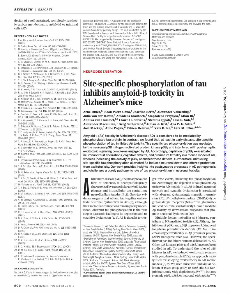
Fig. 1. Depletion of postsynaptic p38g exacerbates excitotoxicity anddeficits in APP transgenicmice. (A) Reduced seizure latency (linear regressionslopes) and increased seizure severity in p38g−/−mice compared with p38g+/+
animals injected with 30 mg/kg pentylenetetrazole (PTZ) (n = 10 to 12 mice).(B) Colocalization of p38g and postsynaptic PSD-95 (arrows), but not pre-synaptic synaptophysin (Syp), in neurons. Scale bars, 1 mm. (C) Mortality inAPP23.p38g+/+ mice was further augmented in APP23.p38g−/− animals,whereas p38g+/+ and p38g−/−mice survived normally (n = 43 to 62). (D to F)Memory deficits in 4-month-old APP23.p38g+/+ and APP23.p38g−/− mice(memory deficits were exacerbated in the APP23.p38g−/− animals). Morriswater maze (MWM) test: (D) Representative MWM path traces to a hiddenplatform. (E) Increased escape latency and (F) reduced time in the target
quadrant during probe trials in APP23.p38g+/+ and, more so, APP23.p38g−/−
mice compared with p38g+/+ and p38g−/− mice (n = 8 to 10). (G to H)Increased (G) spike train and (H) spike frequency in APP23.p38g+/+ andAPP23.p38g−/− mice but not p38g+/+ and p38g−/− mice (n = 6 to 8). (I) Rep-resentative phase-amplitude comodulograms of interictal hippocampallocal field potential recordings showed reduced and virtually lost cross-frequency coupling (CFC) (~8Hz) in APP23.p38g+/+ and APP23.p38g−/−mice,respectively, compared with p38g+/+ and p38g−/− mice. (J) Reduced modu-lation index in APP23.p38g+/+ mice and, more so, in APP23.p38g−/− micecompared with p38g+/+ and p38g−/− animals (n = 6 to 8). For (A), (C), (E) to (H),and (J): ****P <0.0001, ***P < 0.001, **P <0.01, *P < 0.05; ns, not significant.Error bars indicate SEM.
RESEARCH | REPORTS
on
Nov
embe
r 24
, 201
6ht
tp://
scie
nce.
scie
ncem
ag.o
rg/
Dow
nloa
ded
from
906 18 NOVEMBER 2016 • VOL 354 ISSUE 6314 sciencemag.org SCIENCE
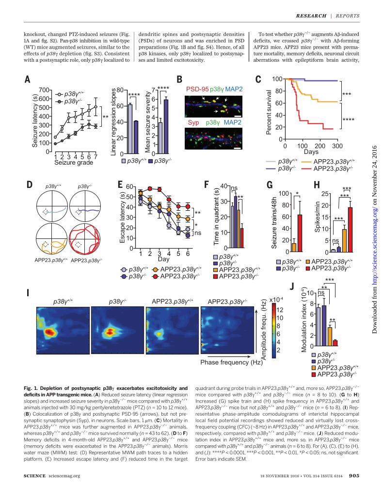
Fig. 2. Tau is requiredfor p38g-mediatedinhibition of Ab andexcitotoxicity.(A) Normal survival ofAPP23.p38g−/−.tau−/−
compared with APP23.p38g−/− and APP23.p38g+/+ mice (n = 42 to62). (B and C) Normalmemory in 12-month-old APP23.p38g−/−.tau−/−
mice compared withAPP23.p38g−/− andAPP23.p38g+/+ mice (n =10 to 12). MWM test:(B) Escape latency and(C) time in target quad-rant during probe trials.(D to F) (D) Further reduc-tion in seizure latencies,shown by linear regressionanalysis (E). (F) Furtherenhanced mean seizureseverity following 30 mg/kgPTZ in Alz17.p38g−/−
mice versus those alreadyreduced in p38g−/−
compared with p38g+/+
and Alz17.p38g+/+ mice(n = 10 to 12). (G to I)(G) Seizure latenciesafter 30 mg/kg PTZ werereduced, as shown by linear regression analysis (H), andmean seizure severity was increased in tau+/+.p38g−/− compared with tau+/+.p38g+/+ mice (I). However,latenciesweremarkedly increased (G) and severities similarly reduced (I) in both tau−/−.p38g+/+ and tau−/−.p38g−/−mice (n= 10 to 12). For all panels: ****P<0.0001,***P < 0.001, **P < 0.01, *P < 0.05; ns, not significant. Error bars indicate SEM.
Fig.3.Activep38g dissociatesPSD-95/tau/Fyn/NRcomplexes. (A) Immuno-precipitation (IP) analysis. More PSD-95/tau/Fyn complexes were immuno-precipitated from the brains of Alz17.p38g−/− than Alz17.p38g+/+ animals, despitecomparable total protein levels. Detection of glyceraldehyde-3-phosphatedehydrogenase (GAPDH) confirmed equal loading (n = 6). Numbers at rightindicate molecular weight. a.i., arbitrary index. (B) p38g (WT) and p38gCA
(CA) failed to disrupt PSD-95/tau/Fyn complexes in the presence of thep38 inhibitor (n = 6). p38 inhibition reduces phosphorylated active p38
levels (p-p38). (C) More tau, Fyn, and NMDA receptor subunits 1 (NR1) and 2B(NR2B) were immunoprecipitated with PSD-95 from p38g−/− versus p38g+/+
brains.This result was further enhanced in APP23.p38g−/−mice comparedwithAPP23.p38g+/+ animals, without changes to total protein levels (n = 6 to 8).(D) Virtually no PSD-95/tau/Fyn complexes were immunoprecipitated fromthe brains of p38gCA-expressing mice (n = 6). For all panels: Representativeblots are shown. Quantification of IP bands relative to PSD-95 IP. ***P < 0.001;**P < 0.01; *P < 0.05. Error bars indicate SEM.
RESEARCH | REPORTS
on
Nov
embe
r 24
, 201
6ht
tp://
scie
nce.
scie
ncem
ag.o
rg/
Dow
nloa
ded
from
and Ab pathology (9, 15, 18). Compared withAPP23.p38g+/+ mice, APP23.p38g−/− animals hadincreased sensitivity to PTZ-induced seizures(fig. S5). Ab pathologywas comparable in thebrainsof APP23.p38g−/− and APP23.p38g+/+ mice (fig. S6),but p38g deletion aggravated premature mortalityandmemory deficits of APP23mice (Fig. 1, C to F,and fig. S7). p38g−/− mice showed no deficits andhad normal motor function (fig. S8). Electroen-cephalography showed enhanced spontaneousepileptiform activity and interictal hypersynchro-
nous discharges in APP23.p38g−/− compared withAPP23.p38g+/+ mice (Fig. 1, G and H, and fig. S9).Hippocampal theta and gamma oscillations andcross-frequency coupling (CFC) through theta-phasemodulation of gamma power are measuresof network activity related to memory, includingin humans (19–22). These measures are compro-mised in APP transgenicmice (15). Compromisedspectral power and CFC of APP23.p38g+/+ micewere significantly more affected in APP23.p38g−/−
recordings (Fig. 1, I and J, and fig. S9). Recordings of
p38g−/− and p38g+/+ mice were indistinguishable.In summary, p38g depletion exacerbated excito-toxicity, neuronal circuit synchronicity, mortal-ity, and memory deficits in APP23 mice, withoutchanges in Ab pathology. In addition, p38g levelswere reduced in aged APP23 and APPNL-G-F miceand humanswith AD (fig. S10), further suggestingthat the loss of p38g-mediated neuroprotectionmay contribute to AD pathogenesis.To determine whether the Ab toxicity–limiting
effects of p38g were tau-dependent, we crossed
SCIENCE sciencemag.org 18 NOVEMBER 2016 • VOL 354 ISSUE 6314 907
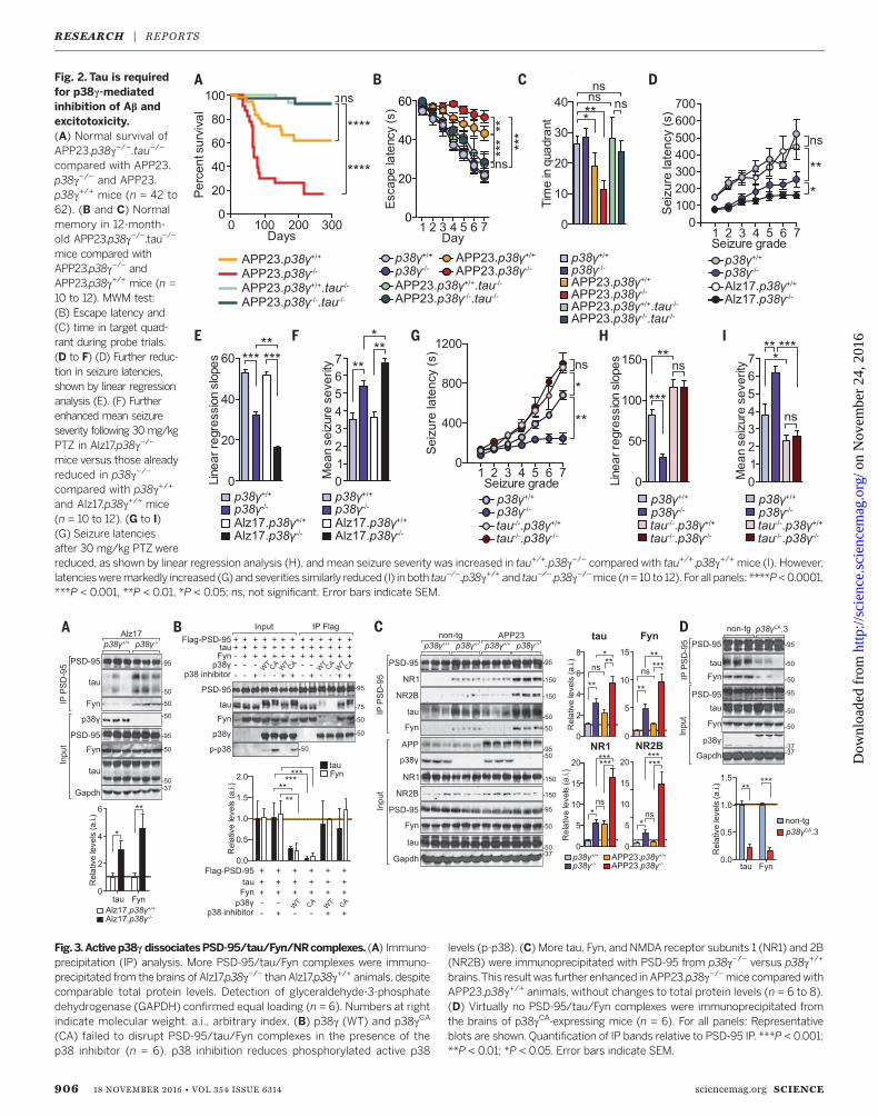
Fig. 4. Site-specific tau phosphorylation disrupts PSD-95/tau/Fyn inter-action and inhibits Ab toxicity. (A) Coexpression of p38g (WT) or p38gCA
(CA) with tau showed phosphorylation at T205, less at S199, and virtuallynone at S396 or S404. Detection of GAPDH confirmed equal loading. (B)Compared with APP23.p38g+/+ animals, APP23.p38g−/− mice showed a lack ofT205 tau phosphorylation (n = 6). Other sites remained phosphorylated.Graph shows quantification of tau phosphorylation. (C) Coexpression ofT205E disrupted PSD95/tau/Fyn IP, whereas T205A tau increased it (n = 6).S199 mutations had no effect. Graph shows quantification of tau/Fyn bound
to PSD-95. D, Asp. (D) AAV-mediated expression of WTand T205A, but notT205E or GFP, restored susceptibility of tau−/− to 50 mg/kg PTZ-inducedseizures,with reduced latency (linear regression) and higher severity (n = 12).(E) Improvedmemory in APP23mice upon AAV-mediated p38gCA expression(APP23.AAVp38gCA). MWM test: (Left) Example traces; (middle) escapelatencies; (right) time in target quadrant during probe trials (n = 8 to 10).(F) Rescued CFC in APP23.AAVp38gCA compared with APP23.AAVGFP mice(n = 5 to 6). For (B) to (F): ***P < 0.001; **P < 0.01; *P < 0.05; ns, not sig-nificant. Error bars indicate SEM.
RESEARCH | REPORTS
on
Nov
embe
r 24
, 201
6ht
tp://
scie
nce.
scie
ncem
ag.o
rg/
Dow
nloa
ded
from
APP23.p38g−/− with tau−/− mice. The exacer-bating effects of p38g loss on reduced survival,memory deficits, and neuronal network dys-function of APP23 mice were virtually abolishedin APP23.p38g−/−.tau−/− mice (Fig. 2, A to C, andfig. S11). These data also showed that, com-pared with APP23 mice, APP23.p38g−/− animalshad aggravated memory deficits that persistedwith aging. In contrast, increasing tau levels inp38g−/− mice [brought about by crossing withnonmutant tau-expressing Alz17 mice (23)] sig-nificantly enhanced PTZ-induced seizures inAlz17.p38g−/− mice (Fig. 2, D to F). Conversely,when compared to tau−/−.p38g+/+ mice, tau−/−.p38g−/− animals showed similar protection fromPTZ-induced seizures (Fig. 2, G to I). Takentogether, the effects of p38g on excitotoxicityand Ab toxicity were tau-dependent.We have previously shown that postsynaptic
PSD-95/tau/Fyn complexes mediate Ab-inducedexcitotoxicity (9). PSD-95/tau/Fyn interaction wasenhanced in Alz17.p38g−/− animals versus Alz17.p38g+/+ mice (Fig. 3A and fig. S12). Conversely, noPSD-95/tau/Fyn complexes were isolated fromtau−/− and tau−/−.p38g−/− brains (fig. S12). In-creasing p38g levels compromised PSD-95/tau/Fyn interaction in cells, and expression of a con-stitutively active p38g variant (p38gCA) com-pletely abolished this interaction (Fig. 3B andfig. S13). Pan-p38 inhibition stopped p38g/p38gCA-induced disruption of PSD-95/tau/Fyn complexes(Fig. 3B). PSD-95 copurified more tau and Fynfrom p38g−/− versus p38g+/+ brains, and evenmore fromAPP23.p38g−/− comparedwith APP23.p38g+/+ and p38g−/− brains (Fig. 3C). Conversely,PSD-95/tau/Fyn interactionwas reduced in trans-genic mice with neuronal expression of p38gCA
(Fig. 3D and fig. S14). PTZ transiently increasedPSD-95/tau/Fyn complex formation in p38g+/+
animals; this effect was even more noticeablein p38g−/− mice (fig. S12). Fyn-mediated NR2Bphosphorylation at Tyr1472 (Y1472) facilitatesPSD-95/NR2B interaction (24). Consistent withincreased PSD-95/tau/Fyn complex formation,NR2B phosphorylation at Y1472 was increasedin p38g−/− brains (fig. S15). Conversely, cellularexpression of p38g and p38gCA—but not p38aCA,p38bCA, or p38dCA—reduced Y1472 phosphoryl-ation of NR2B (fig. S15). Hence, p38g regulatedPSD-95/tau/Fyn complexes, likely at the level ofPSD-95/tau interaction (fig. S16).Although p38g hyperphosphorylates tau dur-
ing long-term in vitro kinase assays (25), thetemporal profile of p38g-induced tau phospho-rylation in acute signaling remains unknown.Short-term in vitro kinase reactions using phos-phorylation site–specific tau antibodies revealedphosphorylation at Ser199 (S199), Thr205 (T205),S396, and S404 (fig. S17). Mass spectrometricanalysis confirmed these and 14 additional,though low-abundant, sites (figs. S17C and S18and table S4). Coexpression of p38g or p38gCA
and tau in cells revealed tau phosphorylation(p) at T205, less at S199, and hardly any at S396or S404 (Fig. 4A). Similarly, T205 (and, less so,S199 and S396) were phosphorylated in p38gCA
transgenic mice (fig. S19). pT205 increased after
PTZ inp38g+/+ animals butwas virtually abolishedin p38g−/−mice, whereas pS199, pS396, and pS404were induced in both p38g+/+ and p38g−/− mice(fig. S19). Similarly, pT205 was markedly reducedin APP23.p38g−/− animals compared with APP23.p38g+/+ mice (Fig. 4B). In primary neurons,pT205 (but not p199) was markedly reduced bypan-p38 inhibition (fig. S20). Taken together,these findings indicate that pT205 was a primaryp38g site in tau.Next, we showed that a phosphorylation-
mimicking Thr205→Glu205 (T205E) tau variantcoprecipitated significantly less with PSD-95 ascompared with nonmutant and T205A (A, Ala) tau(Fig. 4C and fig. S21). In contrast, phosphorylation-mimicking mutants of all other identified siteshad no effect on PSD-95/tau/Fyn interaction (fig.S18). Microscale thermophoresis and glutathioneS-transferase–pulldown in vitro and fluorescence-lifetime imaging microscopy (FLIM)–fluorescenceresonance energy transfer (FRET) analysis inlive cells confirmed the markedly compromisedinteraction of T205E tau with PSD-95 (fig. S22).The T205E mutation did not hinder tau/Fyninteraction (fig. S21). Phosphorylation of T205 byp38gCA was required for disrupting PSD-95/tau/Fyn complexes (fig. S21). Hence, p38g regulatedPSD-95/tau/Fyn complexes via phosphorylatingtau at T205.The disruption of NR/PSD-95/tau/Fyn com-
plexes prevents excitotoxicity and Ab toxicity (9).Hence, phosphorylation of tau at T205 shouldsimilarly mitigate neurotoxicity. Ab caused celldeath in WT and T205A neurons but signifi-cantly less in T205E tau-expressing neurons(fig. S23). Similarly, neurons expressing p38gand, more so, p38gCA were significantly moreresistant to Ab-induced cell death than controls(fig. S24). PTZ-induced seizures are reduced intau−/−mice (8, 9). Adeno-associated virus (AAV)–mediated expression of WT and T205A neurons,but not T205E tau or green fluorescent protein(GFP), in the forebrains of tau−/−mice enhancedPTZ-induced seizures (Fig. 4D and fig. S25). Incontrast, expression of p38gCA in WT mice usingAAV or in Thy1.2-p38gCA transgenic mice de-creased PTZ-induced seizures (fig. S25). AAV-mediated p38gCA expression in APP23 micerescued memory deficits and network aberra-tions; the same was true for crossing APP23with Thy1.2-p38gCA mice (Fig. 4, E and F, andfigs. S26 and S27). In summary, the levels ofactive p38g kinase and tau phosphorylation atT205 determined susceptibility to excitotoxicityand Ab toxicity.Here we have shown that T205 phosphoryl-
ation of tau is part of an Ab toxicity–inhibitingresponse. This is contrary to the current viewthat tau phosphorylation downstream of Ab tox-icity is a pathological response (3). However, thisfinding is in line with the idea that tau is in-volved in normal physiologic NR signaling eventsin neurons (12). Finally, we found that tau-dependent Ab toxicity was modulated by site-specific tau phosphorylation, which inhibitedpostsynaptic PSD-95/tau/Fyn complexes, reveal-ing an Ab toxicity–limiting role of p38g in AD
that is distinct and opposite to the effects ofp38a and p38b (11, 13, 14).
REFERENCES AND NOTES
1. C. Ballatore, V. M. Lee, J. Q. Trojanowski, Nat. Rev. Neurosci. 8,663–672 (2007).
2. C. Haass, D. J. Selkoe, Nat. Rev. Mol. Cell Biol. 8, 101–112(2007).
3. L. M. Ittner, J. Götz, Nat. Rev. Neurosci. 12, 67–72(2011).
4. K. Iqbal, F. Liu, C.-X. Gong, A del C. Alonso,I. Grundke-Iqbal, Acta Neuropathol. 118, 53–69(2009).
5. E. M. Mandelkow, E. Mandelkow, Cold Spring Harb. Perspect.Med. 2, a006247 (2012).
6. E. S. Musiek, D. M. Holtzman, Nat. Neurosci. 18, 800–806(2015).
7. M. Rapoport, H. N. Dawson, L. I. Binder, M. P. Vitek,A. Ferreira, Proc. Natl. Acad. Sci. U.S.A. 99, 6364–6369(2002).
8. E. D. Roberson et al., Science 316, 750–754 (2007).9. L. M. Ittner et al., Cell 142, 387–397 (2010).10. J. J. Palop, L. Mucke, Nat. Neurosci. 13, 812–818
(2010).11. G. E. Hardingham, H. Bading, Nat. Rev. Neurosci. 11, 682–696
(2010).12. L. Mucke, D. J. Selkoe, Cold Spring Harb. Perspect. Med. 2,
a006338 (2012).13. Q. Wang, D. M. Walsh, M. J. Rowan, D. J. Selkoe,
R. Anwyl, J. Neurosci. 24, 3370–3378 (2004).14. S. Li et al., J. Neurosci. 31, 6627–6638 (2011).15. A. A. Ittner, A. Gladbach, J. Bertz, L. S. Suh, L. M. Ittner, Acta
Neuropathol. Commun. 2, 149 (2014).16. M. A. Fabian et al., Nat. Biotechnol. 23, 329–336 (2005).17. M. B. Menon, S. Dhamija, A. Kotlyarov, M. Gaestel, Autophagy
11, 1425–1427 (2015).18. C. Sturchler-Pierrat et al., Proc. Natl. Acad. Sci. U.S.A. 94,
13287–13292 (1997).19. G. Buzsáki, E. I. Moser, Nat. Neurosci. 16, 130–138
(2013).20. R. Goutagny, J. Jackson, S. Williams, Nat. Neurosci. 12,
1491–1493 (2009).21. R. T. Canolty et al., Science 313, 1626–1628 (2006).22. A. B. Tort, R. W. Komorowski, J. R. Manns, N. J. Kopell,
H. Eichenbaum, Proc. Natl. Acad. Sci. U.S.A. 106,20942–20947 (2009).
23. A. Probst et al., Acta Neuropathol. 99, 469–481(2000).
24. M. Aarts et al., Science 298, 846–850 (2002).25. M. Goedert et al., FEBS Lett. 409, 57–62 (1997).
ACKNOWLEDGMENTS
We thank the staff of the Biological ResourcesCentre of UNSW for animal care, E. Hinde for helpwith FLIM-FRET experiments, D. Sullivan and T. Foofor APOE genotyping of human samples, and T. Saitoand T. C. Saido for APPNL-G-F mice. The datafrom mass spectrometry experiments can be foundin the supplementary materials. This work wasfunded by the National Health and Medical ResearchCouncil (grants 1081916, 1037746, and 1003083),the Australian Research Council (grants DP130102027and DE130101591), Alzheimer’s Association (grantNIRG000070035), Alzheimer’s Australia (grantsDGP14-39 and DGP14-95), the NIH (grant R28AA012725),and UNSW Australia. A.I. and L.M.I. are inventorson Australian patent application number APO/2016/900764,submitted by UNSW, which covers increasing p38gactivity to prevent neuronal toxicity.
SUPPLEMENTARY MATERIALS
www.sciencemag.org/content/354/6314/904/suppl/DC1Materials and MethodsFigs. S1 to S27Tables S1 to S5References (26–53)
22 July 2016; resubmitted 9 October 2016Accepted 19 October 201610.1126/science.aah6205
908 18 NOVEMBER 2016 • VOL 354 ISSUE 6314 sciencemag.org SCIENCE
RESEARCH | REPORTS
on
Nov
embe
r 24
, 201
6ht
tp://
scie
nce.
scie
ncem
ag.o
rg/
Dow
nloa
ded
from
(6314), 904-908. [doi: 10.1126/science.aah6205]354Science Ke and Lars M. Ittner (November 17, 2016) Ana P. G. Silva, Joel Mackay, Anne Poljak, Fabien Delerue, Yazi D.Lisa S. Suh, Alexander Macmillan, Greg Sutherland, Jillian J. Kril, Mian Bi, Annika van Hummel, Claire H. Stevens, Stefania Ippati,Julia van der Hoven, Amadeus Gladbach, Magdalena Przybyla, Arne Ittner, Sook Wern Chua, Josefine Bertz, Alexander Volkerling,in Alzheimer's mice
toxicityβSite-specific phosphorylation of tau inhibits amyloid-
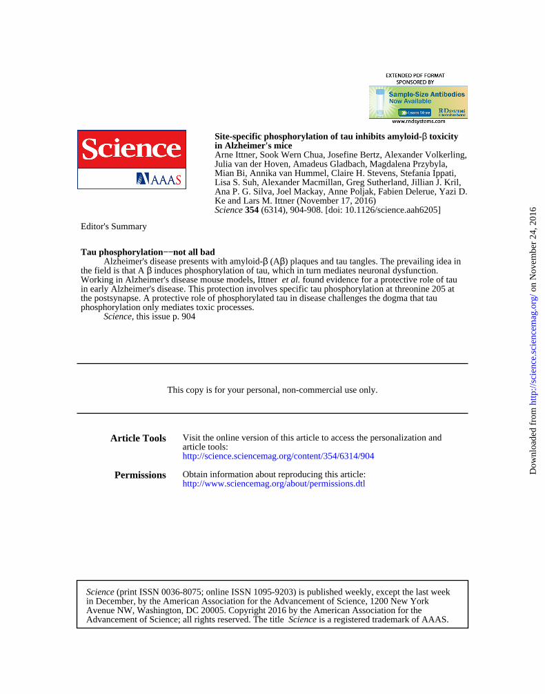
Editor's Summary
, this issue p. 904Sciencephosphorylation only mediates toxic processes.the postsynapse. A protective role of phosphorylated tau in disease challenges the dogma that tauin early Alzheimer's disease. This protection involves specific tau phosphorylation at threonine 205 at
found evidence for a protective role of tauet al.Working in Alzheimer's disease mouse models, Ittner induces phosphorylation of tau, which in turn mediates neuronal dysfunction.βthe field is that A
) plaques and tau tangles. The prevailing idea inβ (AβAlzheimer's disease presents with amyloid-not all bad−−Tau phosphorylation
This copy is for your personal, non-commercial use only.
Article Tools
http://science.sciencemag.org/content/354/6314/904article tools: Visit the online version of this article to access the personalization and
Permissionshttp://www.sciencemag.org/about/permissions.dtlObtain information about reproducing this article:
is a registered trademark of AAAS. ScienceAdvancement of Science; all rights reserved. The title Avenue NW, Washington, DC 20005. Copyright 2016 by the American Association for thein December, by the American Association for the Advancement of Science, 1200 New York
(print ISSN 0036-8075; online ISSN 1095-9203) is published weekly, except the last weekScience
on
Nov
embe
r 24
, 201
6ht
tp://
scie
nce.
scie
ncem
ag.o
rg/
Dow
nloa
ded
from