Embed Size (px)
Citation preview

ANATOMY
Unit 5 Notes:Bone Injury
& Repair

(1) Bone Injury(1) Bone Injury
• Increased weight on bone/joint.
• Irregular movement/manipulation…– Twisting– Rotation– Bending


(2) Comminuted Fractures(2) Comminuted Fractures
- Bone fragments into 3 or more pieces
- Common in aged people, when bones are more brittle

(3) Spiral Fractures(3) Spiral Fractures
- Ragged diagonal break
- Result of excessive twisting forces
- Common sports fracture

(4) Depressed Fractures(4) Depressed Fractures
- Portion of broken bone pressed inward
- Typical skull fracture


(5) Transverse Fractures(5) Transverse Fractures
- Bone breaks straight through bone (perpendicular)
- Clean Break


(6) Oblique Fractures(6) Oblique Fractures
- Bone breaks on diagonal
- Clean break

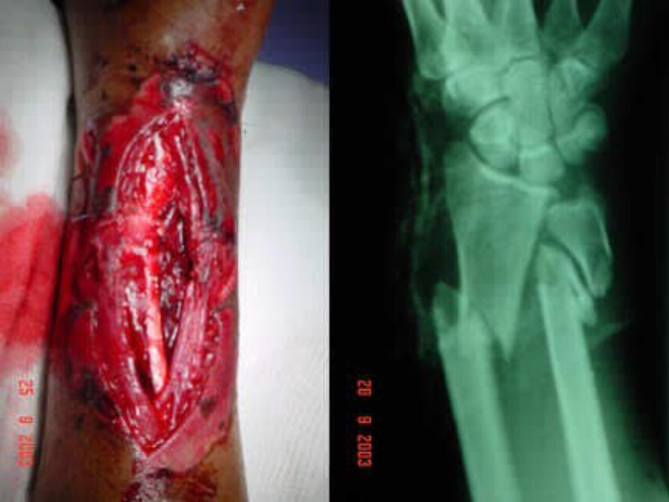
(7) Open Fractures(7) Open Fractures
- Bone breaks and pierces skin
- Common in forearm and shin (leg)

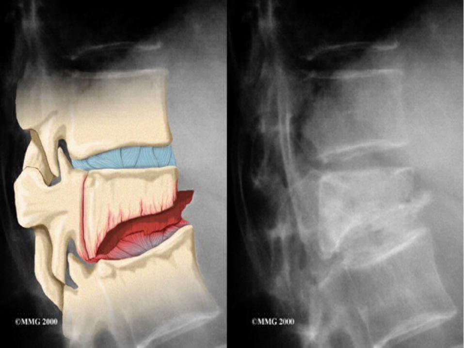
(8) Compression Fractures(8) Compression Fractures
- Bone is crushed
- Common in porous bones (due to osteoporosis)
- Common in vertebral column
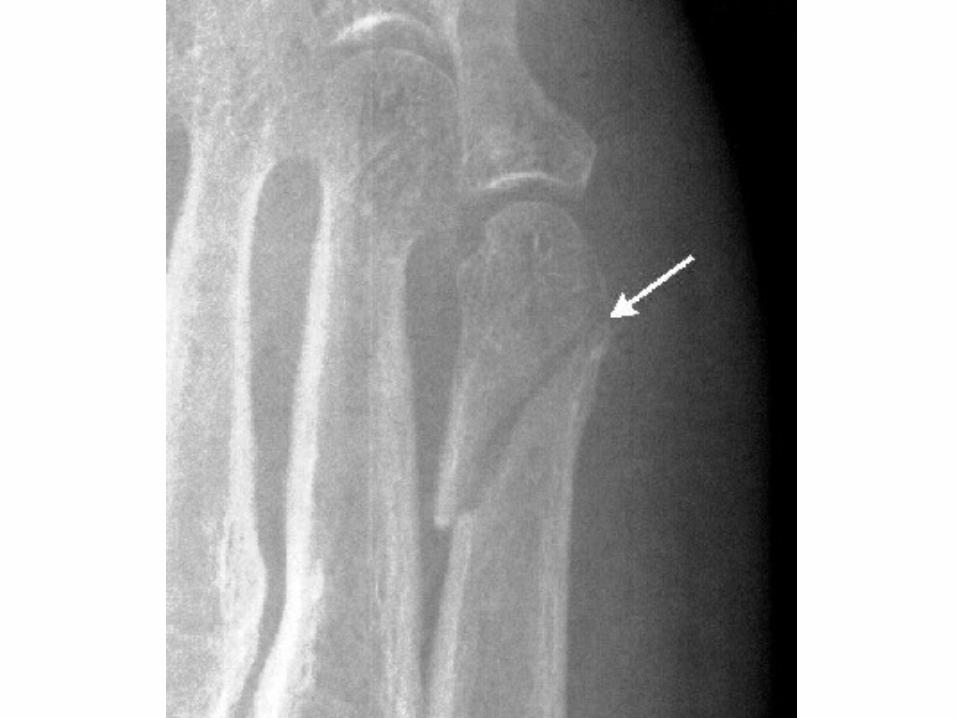
(9) Epiphyseal Fractures(9) Epiphyseal Fractures
- Epiphysis separates from diaphysis along epiphyseal plate
- Usually occurs where cartilage cells are dying and calcifying

(10) Greenstick Fractures(10) Greenstick Fractures
- Bone breaks incompletely (like a twig)
- Only one side of the shaft breaks, other side bends
- Common in children, when bones are more flexible

(11) Bone Repair Stages(11) Bone Repair Stages
1- [Instantly] Hematoma Formation
2- [~2-3days] Fibrocartilaginous Callus Formation
3- [~7days] Bony Callus Formation
4- [~6-8weeks] Bone Remodeling

(12) Hematoma Formation(12) Hematoma Formation
1. Bone breaks, blood vessels are torn and hemorrhage.
2. Mass of clotted blood forms at fracture site.
3. Bone cells are deprived of nutrition and die.
4. Tissue at fracture site becomes swollen, painful and inflamed.

(13) Fibrocartilaginous (13) Fibrocartilaginous Callus Formation Callus Formation
1. Capillaries from surrounding vessels grow into the hematoma.
2. Phagocytic cells (Macrophages + Osteoclasts) invade hematoma and clean up debris.
3. Osteoblasts + Osteocytes invade and begin to rebuild bone.
4. Collagen fibers are secreted to connect ends of bone.

(14) Bony Callus (14) Bony Callus FormationFormation
1. Fibrocartilaginous callus develops and hardens to form a bony callus of spongy bone.
2. Bone cells, collagen fibers and chondrocytes (cartilage cells) are continuously secreted to strengthen matrix.
(15) Final Remodeling(15) Final Remodeling
1. Excess material on outside of diaphysis is removed.
2. Compact bone is laid down to reconstruct shaft walls.