Embed Size (px)
Citation preview
A case of severe autoimmune haemolytic anaemia due to clinically significant auto anti-N antibody
Case Report
A case of severe autoimmune haemolytic anaemiadue to clinically significant auto anti-N antibody
R.N. Makroo a,*, Dhaval Fadadu b, Mohit Chowdhry c, Aakanksha Bhatia d,Prashant Karna e
aDirector & Sr. Consultant, Department of Transfusion Medicine, Molecular Biology and Transplant Immunology,Indraprastha Apollo Hospitals, IndiabDNB Resident, Department of Transfusion Medicine, Molecular Biology and Transplant Immunology,Indraprastha Apollo Hospitals, IndiacConsultant, Department of Transfusion Medicine, Molecular Biology and Transplant Immunology,Indraprastha Apollo Hospitals, Indiad Junior Consultant, Department of Transfusion Medicine, Molecular Biology and Transplant Immunology,Indraprastha Apollo Hospitals, IndiaeDepartment of Transfusion Medicine, Indraprastha Apollo Hospitals, India
1. Introduction
MNS blood group system is the second largest blood groupsystem after ABO system, containing approximately 46antigens. Antibodies of M and N blood group are associatedwith variable clinical significance as both IgG and IgM types ofantibodies are encountered. Frequency of M+N+ phenotype is54.1% in Indian population.1 Among antibodies of MNS bloodgroup system, anti M is a relatively common, naturallyoccurring antibody,2 whereas anti N is quite rare as comparedto anti M.2,3. Generally anti M and anti N are naturallyoccurring, cold reactive IgM saline agglutinins, and are usually
not active at 37 8C; therefore, they are not clinically signifi-cant.4 However, reports of clinically significant antibodies ofMNS blood group are also available in literature.5 Anti N hasbeen implicated as the cause of haemolytic transfusionreactions (HTRs).6 We report a case of autoantibody with antiN specificity which is reactive at 37 8C.
2. Case report
A 45-year-old male presented to Emergency department withthe chief complaint of fever and haematuria for 4 days, sore
a p o l l o m e d i c i n e 1 2 ( 2 0 1 5 ) 1 3 8 – 1 4 0
a r t i c l e i n f o
Article history:
Received 10 April 2015
Accepted 1 May 2015
Available online 15 June 2015
Keywords:
Haemoglobin
Immucor
Galilio
a b s t r a c t
Anti N antibody belongs to the MNS blood group system. Usually, anti N antibodies are
naturally occurring, cold agglutinins. Clinically significant anti N antibodies have also been
reported. We report here a case of autoantibody with anti N specificity presenting with
severe autoimmune haemolytic anaemia.
# 2015 Published by Elsevier B.V. on behalf of Indraprastha Medical Corporation Ltd.
* Corresponding author. Tel.: +91 9810170969.E-mail address: [email protected] (R.N. Makroo).
Available online at www.sciencedirect.com
ScienceDirect
journal homepage: www.elsevier.com/locate/apme
http://dx.doi.org/10.1016/j.apme.2015.05.0040976-0016/# 2015 Published by Elsevier B.V. on behalf of Indraprastha Medical Corporation Ltd.
throat with cough for 2 weeks and urinary hesitancy for 3weeks. He was earlier admitted to a local hospital where hewas found to have elevated blood sugar level and severeanaemia Coombs positive. He was brought to our hospital forfurther management.
On first day, his haemogram revealed, haemoglobin level6.1 g/dl, TLC was 45,300/mm3 and reticulocyte count was0.1%, LDH level 2800 IU/L, total bilirubin 2.7 mg/dl in the labreports. During further work up, both ANA and anti dsDNAwere negative, ruling out SLE. Any significant drug historywhich could lead to haemolysis was elicited. Haemoglobinlevel continued to decline, and at the level of 5.2 g/dl, 2 unitsPRC transfusion was required, and a sample for Group andScreen was received by the department of TransfusionMedicine.
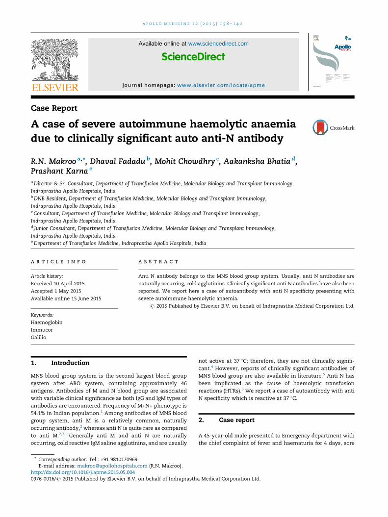
As per the departmental protocol, blood grouping wasperformed on an automated SPRCA platform (Immucor), andwhen a group discrepancy was encountered, blood groupingwasdonewith tube technique, and results are shown inTable 1.
Forward grouping suggested blood group B, Rh (D) positivebut reverse grouping was giving agglutination (4+) with all the3 cells (A1 cells, B cells and O cells). The reverse grouping wasrepeated with incubating the test tubes at different tempera-tures (room temperature, 4 8C and at 37 8C) and the result isdemonstrated in Table 2.
Meanwhile, antibody screening was done on Capture-RReady Screen using SPRCA technique on fully automated IHanalyser (Galilio, Immucor, US) using a 4-cell screen panel.Results are shown in Fig. 1.
Auto control was performed on conventional tube tech-nique with immediate spin (IS) method which gave positivereaction (2+). Direct Coombs test using monospecific (IgG)Coombs sera gave weak (1+) reaction and with monospecificC3d, Coombs sera gave (2+) reaction.
As per the protocol, 4 EDTA blood samples for antibodyidentification were received. Patient did not have anytransfusion history or operative history before. Using 16-cell
(Capture-R Ready-ID) and 10-cell panels (Pano Cell-10; Immu-cor), antibody identification was performed.
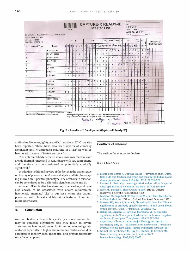
As it is evident that the 16-cell panel points towards apossible anti N, however, the 10-cell panel is pan positive bothat IS as well as at 37 8C (Fig. 2).
In view of positive auto control and pan positive 10-cellpanel, adsorption was attempted. Due to extreme haemolysisin the sample, sufficient intact red cells could not be retrievedfor performance of auto-adsorption. Multiple allo-adsorptionswere done using allogenic cells (R1R1, R2R2 and rr) and antibodyscreening was performed with adsorbed plasma. Antibodyscreen with adsorbed plasma was negative.
According to screening and 16-cell panel, it was concludedthat patient had auto-antibodies reacting at wide thermalrange (4–37 8C) with possible anti-N like specificity.
Antigen typing of the patient revealed N positive pheno-type. The patient had no history of blood transfusion and anyoperative history, which supports the absence of alloantibody.
It was, therefore, concluded that the antibody in questionwas an autoantibody possibly auto anti N.
Approximately 43 units were typed for n antigen, of which,4 units were found as N negative and were compatible withpatient's serum in AHG cross matching.
The patient was transfused 1 unit initially followed by 2units after 3 days. There was no evidence of HTR or aggravatedhaemolysis in the patient. He was also given 2 doses ofrituximab. After 3 units of PRC transfusion his haemoglobinlevel reached 8.9 g/dl.
3. Discussion
Anti N antibody belongs to the MNS blood group system, andhave been reported with variable frequency in literature.Majority of anti-N are naturally occurring cold reactive IgM
Table 1 – Forward grouping.
Anti A Anti B Anti AB Anti D
Reaction – 4+ 4+ 4+
Table 2 – Reverse grouping.
Temperature (8C) A B O Autocontrol
Room temperature 4+ 4+ 4+ 2+4 4+ 4+ 4+ 2+37 4+ 4+ 4+ 2+
[(Fig._1)TD$FIG]
Fig. 1 – Results of screening cell panel (Capture-R Ready Screen).
a p o l l o m e d i c i n e 1 2 ( 2 0 1 5 ) 1 3 8 – 1 4 0 139
antibodies. However, IgG type anti N,7 reactive at 37 8Chas alsobeen reported. There have also been reports of clinicallysignificant anti N antibodies resulting in HTRs8 as well ashaemolytic disease of foetus and new born.
This anti N antibody detected in our case was reactive overa wide thermal range and in AHG phase with IgG component,and therefore can be considered as potentially clinicallysignificant.5
Inaddition to thisand inviewof the fact that thepatient gaveno history of previous transfusions, dialysis and his phenotyp-ing showed an N positive phenotype. The antibody in questioncan be considered to be a clinically significant auto anti N.
Auto anti N antibodies have been reported earlier, and havealso shown to be associated with severe autoimmunehaemolytic anemias,8 like in our case where the patientpresented with clinical and laboratory features of autoim-mune haemolysis.
4. Conclusion
Auto antibodies with anti N specificity are uncommon, butmay be clinically significant; also they result in severeautoimmune haemolytic anaemia. Immunohaematology lab-oratories especially in higher and reference centres should beequipped to identify such antibodies, and provide necessarytransfusion support.
Conflicts of interest
The authors have none to declare.
r e f e r e n c e s
1. Makroo RN, Bhatia A, Gupta R, Phillip J. Prevalence of Rh, Duffy,Kell, Kidd and MNSs blood group antigens in the Indian blooddonor population. Indian J Med Res. 2013;137:521–526.
2. Perrault R. Naturally-occurring anti-M and anti-N with specialcase: IgM anti-N in NN donor. Vox Sang. 1973;24:134–149.
3. Race RR, Sanger R. Blood Groups in Man. 6th ed. Oxford:Blackwell Scientific Publications; 1975.
4. Mollison PL, Engelfriet CP, Contreras M, et al. Blood Transfusionin Clinical Medicine. 10th ed. Oxford: Blackwell Science; 1997.
5. Makroo RN, Arora B, Bhatia A, Chowdhry M, Luka RN. Clinicalsignificance of antibody specificities to M, N and Lewis bloodgroup system. Asian J Transfus Sci. 2014;8:96–99.
6. Ballas SK, Dignam C, Harris M, Marcolina MJ. A clinicallysignificant anti-N in a patient whose red cells were negativefor N and U antigens. Transfusion. 1985;25:377–380.
7. Leger RM, Calhoun L. Other major blood group system. In:Harmening DM, ed. In: Modern Blood Banking and TransfusionPractices 5th ed. New Delhi: Jaypee Publisher; 2008:165–167.
8. Immel CC, McPherson M, Hay SN, Braddy LR, Brecher ME.Severe hemolytic anemia due to auto anti-N.Immunohematology. 2005;21(2):63–65.
[(Fig._2)TD$FIG]
Fig. 2 – Results of 16-cell panel (Capture-R Ready-ID).
a p o l l o m e d i c i n e 1 2 ( 2 0 1 5 ) 1 3 8 – 1 4 0140

Apollo hospitals: http://www.apollohospitals.com/Twitter: https://twitter.com/HospitalsApolloYoutube: http://www.youtube.com/apollohospitalsindiaFacebook: http://www.facebook.com/TheApolloHospitalsSlideshare: http://www.slideshare.net/Apollo_HospitalsLinkedin: http://www.linkedin.com/company/apollo-hospitalsBlog:Blog: http://www.letstalkhealth.in/


![Retrospective study of canine infectious haemolytic …...Haemolytic anaemia develops as a consequence of red blood cell lysis due to infectious or non-infectious causes [1]. Infectious](https://img.pdfslide.us/doc/110x75/5f3a35eedf03db47f4785f1b/retrospective-study-of-canine-infectious-haemolytic-haemolytic-anaemia-develops.jpg)