Embed Size (px)
Citation preview
Until recently, immunohistochemistry was rarely used as a diagnostic aid in gynecological pathology. However, lately there has been a marked expansion of the litera-ture regarding the utility of immunohistochemical markers in the diagnosis of lesions of the female genital tract. In this chapter, the value of these markers is detailed. Markers of prognostic signifi cance are not covered since these are few and, in general, offer no advantage over careful pathological examination. The uses of immunohistochemistry are covered by site within the female genital tract with an emphasis on the most useful panels of markers in various diagnostic situations.
VULVA AND VAGINA
PREINVASIVE VULVAL SQUAMOUS LESIONS
In the vulva, the morphological hallmarks of HPV infec-tion, such as koilocytosis, may not be as well developed as in the cervix and therefore, it may be diffi cult to diagnose a viral-induced lesion. In equivocal cases, in which the diagnosis of condyloma is suspected, immu-nohistochemical staining with the proliferation marker MIB-1 may be of value as low-risk HPV infection of the vulva is associated with the presence of MIB-1-positive nuclei in the middle and upper thirds of the epithelium, which is in contrast to reactive epithelium, in which the
667
17Immunohistochemistry in the Differential Diagnosis of Female Genital Tract PathologyW Glenn McCluggage
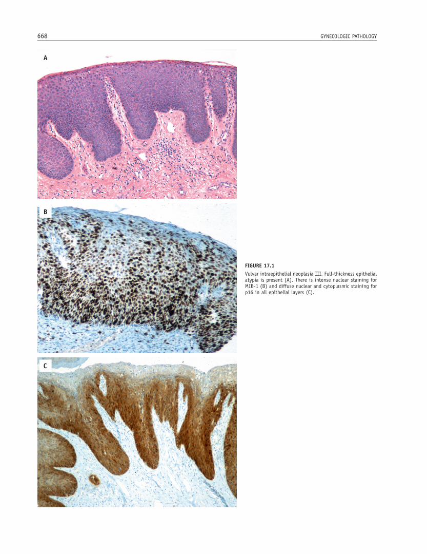
positivity is typically confi ned to the basal and parabasal area. MIB-1 staining is also useful in confi rming a high-grade VIN as the cells of high-grade VIN (VIN II–III) express MIB-1 throughout much of the full epithelial thickness (Figure 17.1) which is useful in the separation of high-grade VIN from atrophic squamous epithelium. Another marker that may be helpful in supporting the diagnosis of VIN associated with high-risk HPV sub-types, i.e., high-grade VIN, is p16 as this marker is typi-cally positive (both in the nucleus and the cytoplasm) in these HPV-related lesions (Figure 17.1). In contrast, HPV-negative VIN, i.e., differentiated VIN, is p16-nega-tive. Immunohistochemistry may also assist in the diag-nosis of differentiated VIN, which may be a diffi cult diagnosis since its morphological features may be subtle. Intense nuclear staining for p53 in the basal and supra-basal cells is characteristic of differentiated VIN and may help distinguish it from nonneoplastic squamous epithelium.
VULVAL PAGET DISEASE
Immunohistochemistry may be of value in the diagnosis of Paget disease involving the vulva and in the distinc-tion of primary Paget disease (which is not usually asso-ciated with an underlying malignancy) from Paget disease secondary to spread from an internal organ such
PREINVASIVE VULVAL SQUAMOUS LESIONS – FACT SHEET
� MIB-1 positivity in upper epithelial layers supports the histologic impression of vulvar condyloma
� High-grade VIN exhibits full-thickness MIB-1 positivity and is useful in its distinction from atrophic epithelium
� p16 positivity supports diagnosis of HPV related high-grade VIN
� Nuclear positivity for p53 in basal and suprabasal squamous cells is characteristic of HPV-negative VIN (“differentiated VIN”)
VULVAL PAGET DISEASE – FACT SHEET
� Primary vulval Paget disease typically CAM 5.2, CK7, G-CDFP-15, and CEA positive
� Pagetoid Bowen disease typically CK 7, CAM 5.2, G-CDFP-15, and CEA negative
� Pagetoid urothelial carcinoma typically uroplakin-III positive; variably CK20 and CEA positive; and CK7 and G-CDFP-15 negative
� Paget disease secondary to spread from a colorectal primary typically CK20 and CEA positive; CK7 and GCDFP negative
668 GYNECOLOGIC PATHOLOGY
FIGURE 17.1Vulvar intraepithelial neoplasia III. Full-thickness epithelial atypia is present (A). There is intense nuclear staining for MIB-1 (B) and diffuse nuclear and cytoplasmic staining for p16 in all epithelial layers (C).
A
B
C
CHAPTER 17 Immunohistochemistry in the Differential Diagnosis of Female Genital Tract Pathology 669
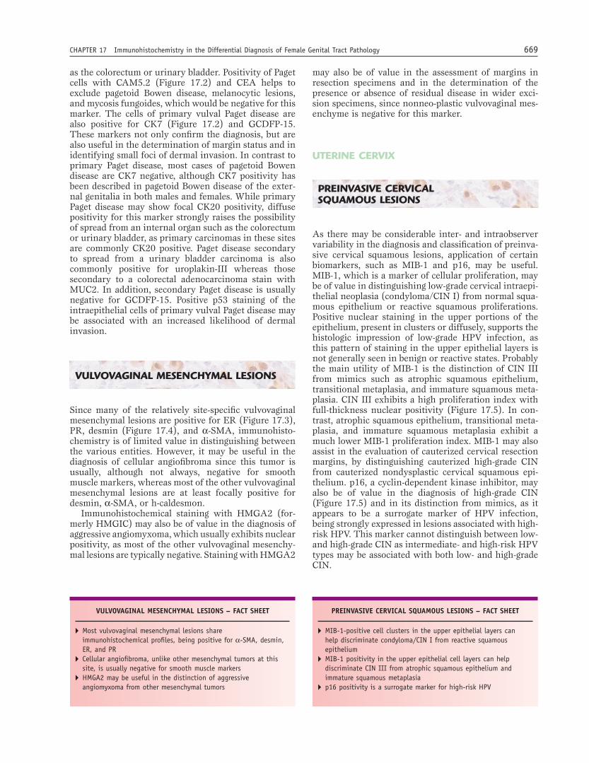
as the colorectum or urinary bladder. Positivity of Paget cells with CAM5.2 (Figure 17.2) and CEA helps to exclude pagetoid Bowen disease, melanocytic lesions, and mycosis fungoides, which would be negative for this marker. The cells of primary vulval Paget disease are also positive for CK7 (Figure 17.2) and GCDFP-15. These markers not only confi rm the diagnosis, but are also useful in the determination of margin status and in identifying small foci of dermal invasion. In contrast to primary Paget disease, most cases of pagetoid Bowen disease are CK7 negative, although CK7 positivity has been described in pagetoid Bowen disease of the exter-nal genitalia in both males and females. While primary Paget disease may show focal CK20 positivity, diffuse positivity for this marker strongly raises the possibility of spread from an internal organ such as the colorectum or urinary bladder, as primary carcinomas in these sites are commonly CK20 positive. Paget disease secondary to spread from a urinary bladder carcinoma is also commonly positive for uroplakin-III whereas those secondary to a colorectal adenocarcinoma stain with MUC2. In addition, secondary Paget disease is usually negative for GCDFP-15. Positive p53 staining of the intraepithelial cells of primary vulval Paget disease may be associated with an increased likelihood of dermal invasion.
VULVOVAGINAL MESENCHYMAL LESIONS
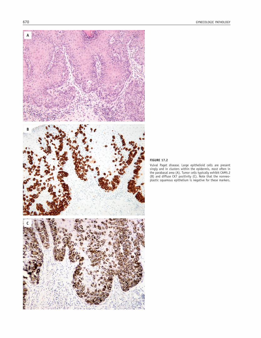
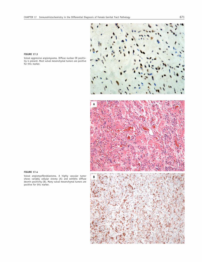
Since many of the relatively site-specifi c vulvovaginal mesenchymal lesions are positive for ER (Figure 17.3), PR, desmin (Figure 17.4), and α-SMA, immunohisto-chemistry is of limited value in distinguishing between the various entities. However, it may be useful in the diagnosis of cellular angiofi broma since this tumor is usually, although not always, negative for smooth muscle markers, whereas most of the other vulvovaginal mesenchymal lesions are at least focally positive for desmin, α-SMA, or h-caldesmon.
Immunohistochemical staining with HMGA2 (for-merly HMGIC) may also be of value in the diagnosis of aggressive angiomyxoma, which usually exhibits nuclear positivity, as most of the other vulvovaginal mesenchy-mal lesions are typically negative. Staining with HMGA2
VULVOVAGINAL MESENCHYMAL LESIONS – FACT SHEET
� Most vulvovaginal mesenchymal lesions share immunohistochemical profi les, being positive for α-SMA, desmin, ER, and PR
� Cellular angiofi broma, unlike other mesenchymal tumors at this site, is usually negative for smooth muscle markers
� HMGA2 may be useful in the distinction of aggressive angiomyxoma from other mesenchymal tumors
may also be of value in the assessment of margins in resection specimens and in the determination of the presence or absence of residual disease in wider exci-sion specimens, since nonneo-plastic vulvovaginal mes-enchyme is negative for this marker.
UTERINE CERVIX
PREINVASIVE CERVICAL SQUAMOUS LESIONS
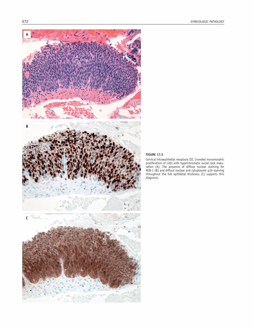
As there may be considerable inter- and intraobserver variability in the diagnosis and classifi cation of preinva-sive cervical squamous lesions, application of certain biomarkers, such as MIB-1 and p16, may be useful. MIB-1, which is a marker of cellular proliferation, may be of value in distinguishing low-grade cervical intraepi-thelial neoplasia (condyloma/CIN I) from normal squa-mous epithelium or reactive squamous proliferations. Positive nuclear staining in the upper portions of the epithelium, present in clusters or diffusely, supports the histologic impression of low-grade HPV infection, as this pattern of staining in the upper epithelial layers is not generally seen in benign or reactive states. Probably the main utility of MIB-1 is the distinction of CIN III from mimics such as atrophic squamous epithelium, transitional metaplasia, and immature squamous meta-plasia. CIN III exhibits a high proliferation index with full-thickness nuclear positivity (Figure 17.5). In con-trast, atrophic squamous epithelium, transitional meta-plasia, and immature squamous metaplasia exhibit a much lower MIB-1 proliferation index. MIB-1 may also assist in the evaluation of cauterized cervical resection margins, by distinguishing cauterized high-grade CIN from cauterized nondysplastic cervical squamous epi-thelium. p16, a cyclin-dependent kinase inhibitor, may also be of value in the diagnosis of high-grade CIN (Figure 17.5) and in its distinction from mimics, as it appears to be a surrogate marker of HPV infection, being strongly expressed in lesions associated with high-risk HPV. This marker cannot distinguish between low- and high-grade CIN as intermediate- and high-risk HPV types may be associated with both low- and high-grade CIN.
PREINVASIVE CERVICAL SQUAMOUS LESIONS – FACT SHEET
� MIB-1-positive cell clusters in the upper epithelial layers can help discriminate condyloma/CIN I from reactive squamous epithelium
� MIB-1 positivity in the upper epithelial cell layers can help discriminate CIN III from atrophic squamous epithelium and immature squamous metaplasia
� p16 positivity is a surrogate marker for high-risk HPV
670 GYNECOLOGIC PATHOLOGY
FIGURE 17.2Vulval Paget disease. Large epithelioid cells are present singly and in clusters within the epidermis, most often in the parabasal area (A). Tumor cells typically exhibit CAM5.2 (B) and diffuse CK7 positivity (C). Note that the nonneo-plastic squamous epithelium is negative for these markers.
A
B
C
CHAPTER 17 Immunohistochemistry in the Differential Diagnosis of Female Genital Tract Pathology 671
FIGURE 17.3Vulval aggressive angiomyxoma. Diffuse nuclear ER positiv-ity is present. Most vulval mesenchymal tumors are positive for this marker.
FIGURE 17.4Vulval angiomyofi broblastoma. A highly vascular tumor shows variably cellular stroma (A) and exhibits diffuse desmin positivity (B). Many vulval mesenchymal tumors are positive for this marker.
A
B
672 GYNECOLOGIC PATHOLOGY
FIGURE 17.5Cervical intraepithelial neoplasia III. Crowded monomorphic proliferation of cells with hyperchromatic nuclei lack matu-ration (A). The presence of diffuse nuclear staining for MIB-1 (B) and diffuse nuclear and cytoplasmic p16 staining throughout the full epithelial thickness (C) supports this diagnosis.
A
B
C
CHAPTER 17 Immunohistochemistry in the Differential Diagnosis of Female Genital Tract Pathology 673
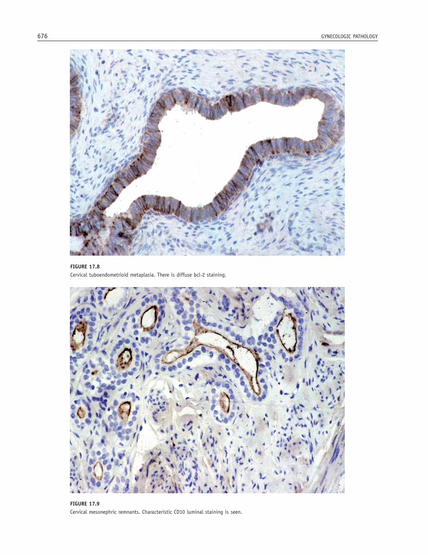
although mesonephric adenocarcinomas are less likely to be positive. CD10 characteristically exhibits luminal positivity in cervical mesonephric remnants (Figure 17.9) and is therefore a good indicator of mesonephric origin. However, some usual endocervical adenocarci-nomas are CD10 positive and thus, this marker is of limited value in confi rmation of a mesonephric origin of an adenocarcinoma. Other markers that may be posi-tive in cervical mesonephric lesions are vimentin, cal-retinin, and AR. These markers are also often positive in benign and malignant mesonephric lesions elsewhere within the female genital tract.
CERVICAL MINIMAL-DEVIATION ADENOCARCINOMA OF MUCINOUS TYPE (ADENOMA MALIGNUM)
The mucinous variant of cervical minimal-deviation adenocarcinoma (adenoma malignum) often exhibits a gastric or pyloric immunophenotype, which may be useful in its distinction from well-differentiated endo-cervical adenocarcinoma, not otherwise specifi ed (NOS), and other benign mimics. In most cases, there is positiv-ity for HIK1083, an antibody directed against pyloric gland mucin, whereas most usual endocervical adeno-carcinomas and benign endocervical glandular lesions are negative. Adenoma malignum is also usually p16 negative, in contrast to the diffuse positivity seen in most usual endocervical adenocarcinomas. The benign endocervical glandular lesion termed lobular endocervi-cal glandular hyperplasia, NOS may also exhibit a pyloric phenotype and be positive for HIK1083; there-fore, one must rely on the differences in morphologic appearance to make the distinction between these two entities. It has been suggested that in some cases this is a precursor lesion of cervical adenoma malignum.
PREINVASIVE CERVICAL GLANDULAR LESIONS AND MIMICS – FACT SHEET
� Adenocarcinoma in situ usually exhibits a high MIB-1 proliferation index, diffuse p16 nuclear and cytoplasmic positivity, shows cytoplasmic positivity for CEA, and is negative for bcl-2 and vimentin
� Tuboendometrioid metaplasia usually exhibits a low MIB-1 proliferation index, is bcl-2 and vimentin positive, but p16 and CEA negative
� Microglandular hyperplasia usually p16 and bcl-2 negative and has a low MIB-1 proliferation index
CERVICAL MESONEPHRIC LESIONS – FACT SHEET
� Mesonephric remnants, hyperplasia, and, to a lesser extent, carcinoma exhibit luminal staining for CD10
� Mesonephric lesions may be positive for vimentin, calretinin, and AR
CERVICAL MINIMAL-DEVIATION ADENOCARCINOMA – FACT SHEET
� Cervical minimal-deviation adenocarcinoma is characteristically HIK1083 positive.
PREINVASIVE CERVICAL GLANDULAR LESIONS
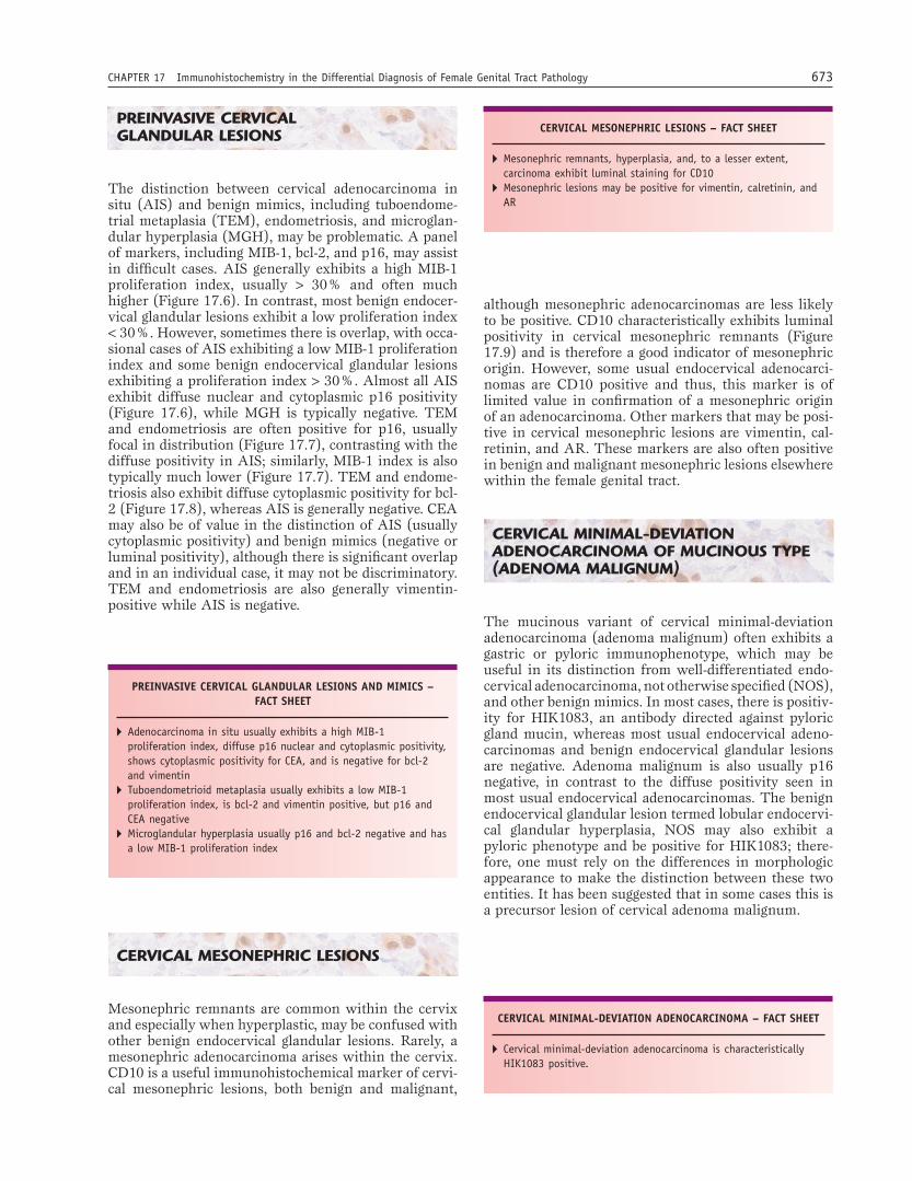
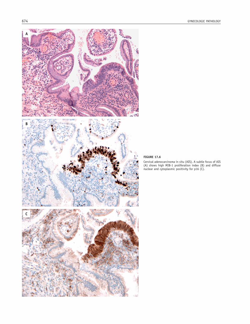
The distinction between cervical adenocarcinoma in situ (AIS) and benign mimics, including tuboendome-trial metaplasia (TEM), endometriosis, and microglan-dular hyperplasia (MGH), may be problematic. A panel of markers, including MIB-1, bcl-2, and p16, may assist in diffi cult cases. AIS generally exhibits a high MIB-1 proliferation index, usually > 30% and often much higher (Figure 17.6). In contrast, most benign endocer-vical glandular lesions exhibit a low proliferation index < 30%. However, sometimes there is overlap, with occa-sional cases of AIS exhibiting a low MIB-1 proliferation index and some benign endocervical glandular lesions exhibiting a proliferation index > 30%. Almost all AIS exhibit diffuse nuclear and cytoplasmic p16 positivity (Figure 17.6), while MGH is typically negative. TEM and endometriosis are often positive for p16, usually focal in distribution (Figure 17.7), contrasting with the diffuse positivity in AIS; similarly, MIB-1 index is also typically much lower (Figure 17.7). TEM and endome-triosis also exhibit diffuse cytoplasmic positivity for bcl-2 (Figure 17.8), whereas AIS is generally negative. CEA may also be of value in the distinction of AIS (usually cytoplasmic positivity) and benign mimics (negative or luminal positivity), although there is signifi cant overlap and in an individual case, it may not be discriminatory. TEM and endometriosis are also generally vimentin-positive while AIS is negative.
CERVICAL MESONEPHRIC LESIONS
Mesonephric remnants are common within the cervix and especially when hyperplastic, may be confused with other benign endocervical glandular lesions. Rarely, a mesonephric adenocarcinoma arises within the cervix. CD10 is a useful immunohistochemical marker of cervi-cal mesonephric lesions, both benign and malignant,
674 GYNECOLOGIC PATHOLOGY
FIGURE 17.6Cervical adenocarcinoma in situ (AIS). A subtle focus of AIS (A) shows high MIB-1 proliferation index (B) and diffuse nuclear and cytoplasmic positivity for p16 (C).
A
B
C

CHAPTER 17 Immunohistochemistry in the Differential Diagnosis of Female Genital Tract Pathology 675
FIGURE 17.7Cervical tuboendometrioid metaplasia. A gland showing tuboendometrioid metaplasia (A) exhibiting weak staining for p16 (B) and a low MIB-1 proliferation index (C).
A
B
C
676 GYNECOLOGIC PATHOLOGY
FIGURE 17.8Cervical tuboendometrioid metaplasia. There is diffuse bcl-2 staining.
FIGURE 17.9Cervical mesonephric remnants. Characteristic CD10 luminal staining is seen.
CHAPTER 17 Immunohistochemistry in the Differential Diagnosis of Female Genital Tract Pathology 677
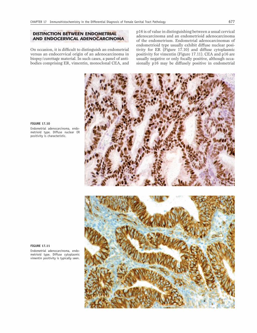
FIGURE 17.10Endometrial adenocarcinoma, endo-metrioid type. Diffuse nuclear ER positivity is characteristic.
FIGURE 17.11Endometrial adenocarcinoma, endo-metrioid type. Diffuse cytoplasmic vimentin positivity is typically seen.
DISTINCTION BETWEEN ENDOMETRIAL AND ENDOCERVICAL ADENOCARCINOMA
On occasion, it is diffi cult to distinguish an endometrial versus an endocervical origin of an adenocarcinoma in biopsy/curettage material. In such cases, a panel of anti-bodies comprising ER, vimentin, monoclonal CEA, and
p16 is of value in distinguishing between a usual cervical adenocarcinoma and an endometrioid adenocarcinoma of the endometrium. Endometrial adenocarcinomas of endometrioid type usually exhibit diffuse nuclear posi-tivity for ER (Figure 17.10) and diffuse cytoplasmic positivity for vimentin (Figure 17.11). CEA and p16 are usually negative or only focally positive, although occa-sionally p16 may be diffusely positive in endometrial
678 GYNECOLOGIC PATHOLOGY
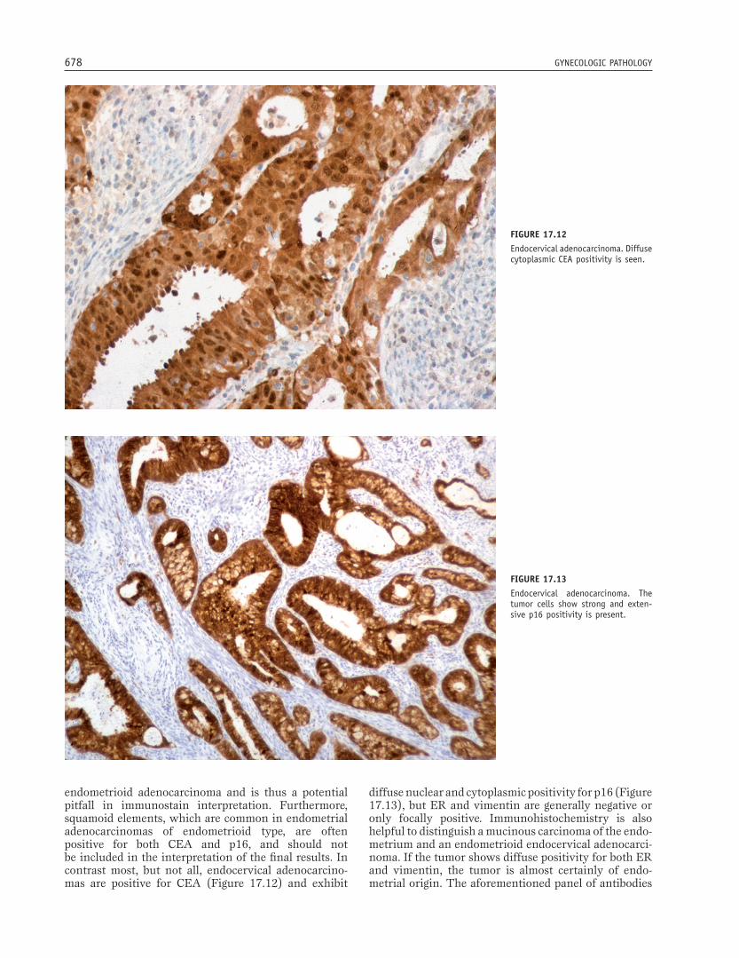
FIGURE 17.12Endocervical adenocarcinoma. Diffuse cytoplasmic CEA positivity is seen.
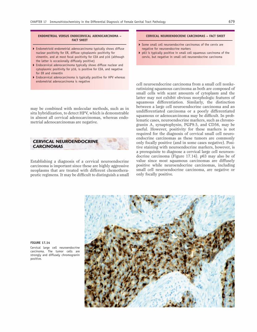
FIGURE 17.13Endocervical adenocarcinoma. The tumor cells show strong and exten-sive p16 positivity is present.
endometrioid adenocarcinoma and is thus a potential pitfall in immunostain interpretation. Furthermore, squamoid elements, which are common in endometrial adenocarcinomas of endometrioid type, are often positive for both CEA and p16, and should not be included in the interpretation of the fi nal results. In contrast most, but not all, endocervical adenocarcino-mas are positive for CEA (Figure 17.12) and exhibit
diffuse nuclear and cytoplasmic positivity for p16 (Figure 17.13), but ER and vimentin are generally negative or only focally positive. Immunohistochemistry is also helpful to distinguish a mucinous carcinoma of the endo-metrium and an endometrioid endocervical adenocarci-noma. If the tumor shows diffuse positivity for both ER and vimentin, the tumor is almost certainly of endo-metrial origin. The aforementioned panel of antibodies
CHAPTER 17 Immunohistochemistry in the Differential Diagnosis of Female Genital Tract Pathology 679
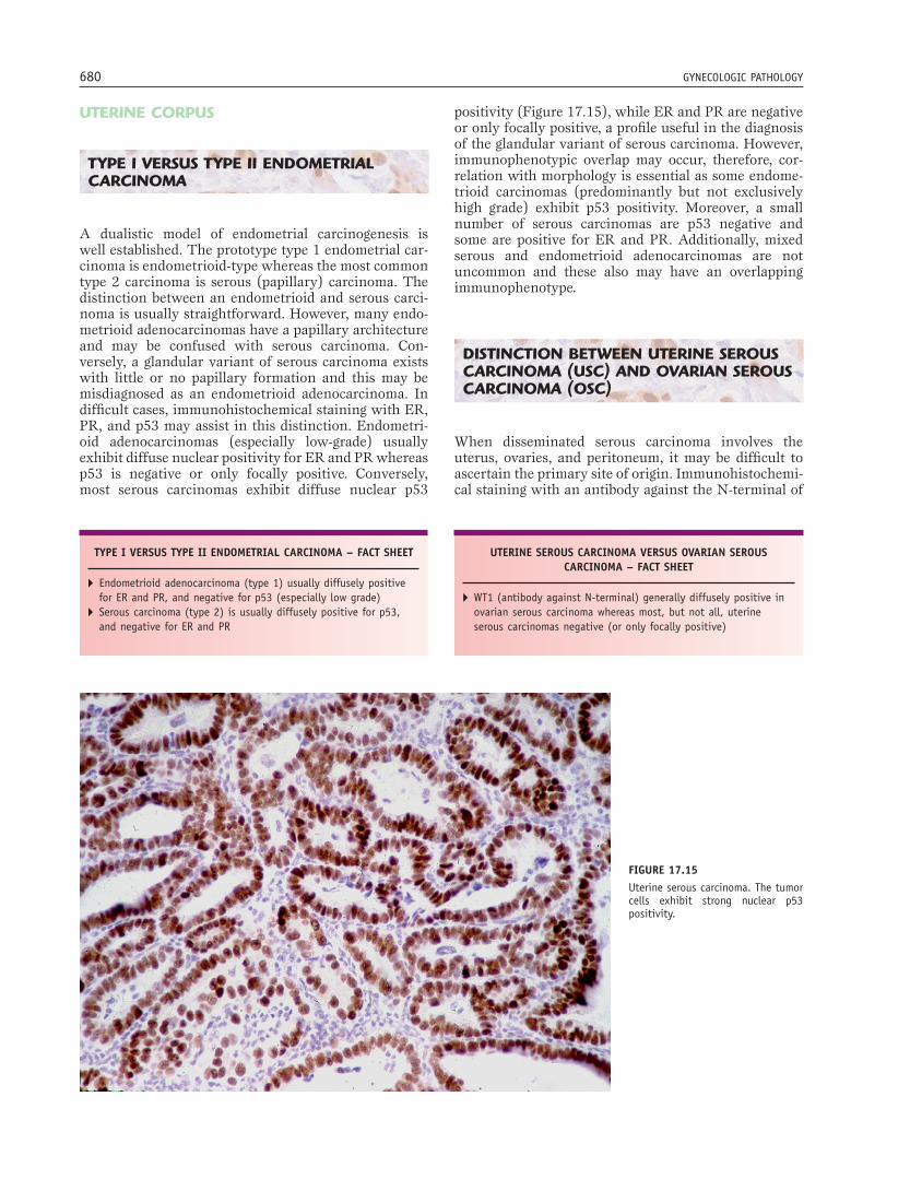
FIGURE 17.14Cervical large cell neuroendocrine carcinoma. The tumor cells are strongly and diffusely chromogranin positive.
CERVICAL NEUROENDOCRINE CARCINOMAS – FACT SHEET
� Some small cell neuroendocrine carcinomas of the cervix are negative for neuroendocrine markers
� p63 is typically positive in small cell squamous carcinoma of the cervix, but negative in small cell neuroendocrine carcinoma
ENDOMETRIAL VERSUS ENDOCERVICAL ADENOCARCINOMA – FACT SHEET
� Endometrioid endometrial adenocarcinoma typically shows diffuse nuclear positivity for ER, diffuse cytoplasmic positivity for vimentin, and at most focal positivity for CEA and p16 (although the latter is occasionally diffusely positive)
� Endocervical adenocarcinoma typically shows diffuse nuclear and cytoplasmic positivity for p16, is positive for CEA, and negative for ER and vimentin
� Endocervical adenocarcinoma is typically positive for HPV whereas endometrial adenocarcinoma is negative
may be combined with molecular methods, such as in situ hybridization, to detect HPV, which is demonstrable in almost all cervical adenocarcinomas, whereas endo-metrial adenocarcinomas are negative.
CERVICAL NEUROENDOCRINE CARCINOMAS
Establishing a diagnosis of a cervical neuroendocrine carcinoma is important since these are highly aggressive neoplasms that are treated with different chemothera-peutic regimens. It may be diffi cult to distinguish a small
cell neuroendocrine carcinoma from a small cell nonke-ratinizing squamous carcinoma as both are composed of small cells with scant amounts of cytoplasm and the latter may not exhibit obvious morphologic features of squamous differentiation. Similarly, the distinction between a large cell neuroendocrine carcinoma and an undifferentiated carcinoma or a poorly differentiated squamous or adenocarcinoma may be diffi cult. In prob-lematic cases, neuroendocrine markers, such as chromo-granin A, synaptophysin, PGP9.5, and CD56, may be useful. However, positivity for these markers is not required for the diagnosis of cervical small cell neuro-endocrine carcinomas as these tumors are commonly only focally positive (and in some cases negative). Posi-tive staining with neuroendocrine markers, however, is a prerequisite to diagnose a cervical large cell neuroen-docrine carcinoma (Figure 17.14). p63 may also be of value since most squamous carcinomas are diffusely positive while neuroendocrine carcinomas, including small cell neuroendocrine carcinoma, are negative or only focally positive.
680 GYNECOLOGIC PATHOLOGY
UTERINE CORPUS
TYPE I VERSUS TYPE II ENDOMETRIAL CARCINOMA
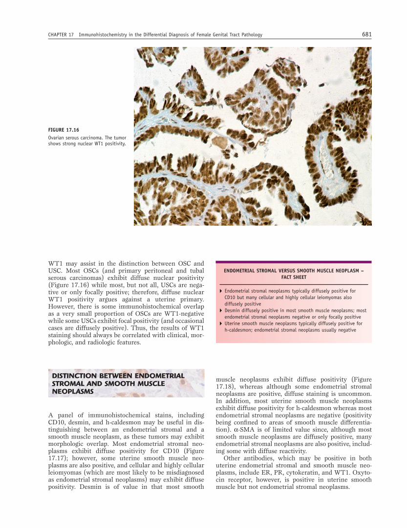
A dualistic model of endometrial carcinogenesis is well established. The prototype type 1 endometrial car-cinoma is endometrioid-type whereas the most common type 2 carcinoma is serous (papillary) carcinoma. The distinction between an endometrioid and serous carci-noma is usually straightforward. However, many endo-metrioid adenocarcinomas have a papillary architecture and may be confused with serous carcinoma. Con-versely, a glandular variant of serous carcinoma exists with little or no papillary formation and this may be misdiagnosed as an endometrioid adenocarcinoma. In diffi cult cases, immunohistochemical staining with ER, PR, and p53 may assist in this distinction. Endometri-oid adenocarcinomas (especially low-grade) usually exhibit diffuse nuclear positivity for ER and PR whereas p53 is negative or only focally positive. Conversely, most serous carcinomas exhibit diffuse nuclear p53
positivity (Figure 17.15), while ER and PR are negative or only focally positive, a profi le useful in the diagnosis of the glandular variant of serous carcinoma. However, immunophenotypic overlap may occur, therefore, cor-relation with morphology is essential as some endome-trioid carcinomas (predominantly but not exclusively high grade) exhibit p53 positivity. Moreover, a small number of serous carcinomas are p53 negative and some are positive for ER and PR. Additionally, mixed serous and endometrioid adenocarcinomas are not uncommon and these also may have an overlapping immunophenotype.
DISTINCTION BETWEEN UTERINE SEROUS CARCINOMA (USC) AND OVARIAN SEROUS CARCINOMA (OSC)
When disseminated serous carcinoma involves the uterus, ovaries, and peritoneum, it may be diffi cult to ascertain the primary site of origin. Immunohistochemi-cal staining with an antibody against the N-terminal of
TYPE I VERSUS TYPE II ENDOMETRIAL CARCINOMA – FACT SHEET
� Endometrioid adenocarcinoma (type 1) usually diffusely positive for ER and PR, and negative for p53 (especially low grade)
� Serous carcinoma (type 2) is usually diffusely positive for p53, and negative for ER and PR
FIGURE 17.15Uterine serous carcinoma. The tumor cells exhibit strong nuclear p53 positivity.
UTERINE SEROUS CARCINOMA VERSUS OVARIAN SEROUS CARCINOMA – FACT SHEET
� WT1 (antibody against N-terminal) generally diffusely positive in ovarian serous carcinoma whereas most, but not all, uterine serous carcinomas negative (or only focally positive)
CHAPTER 17 Immunohistochemistry in the Differential Diagnosis of Female Genital Tract Pathology 681
WT1 may assist in the distinction between OSC and USC. Most OSCs (and primary peritoneal and tubal serous carcinomas) exhibit diffuse nuclear positivity (Figure 17.16) while most, but not all, USCs are nega-tive or only focally positive; therefore, diffuse nuclear WT1 positivity argues against a uterine primary. However, there is some immunohistochemical overlap as a very small proportion of OSCs are WT1-negative while some USCs exhibit focal positivity (and occasional cases are diffusely positive). Thus, the results of WT1 staining should always be correlated with clinical, mor-phologic, and radiologic features.
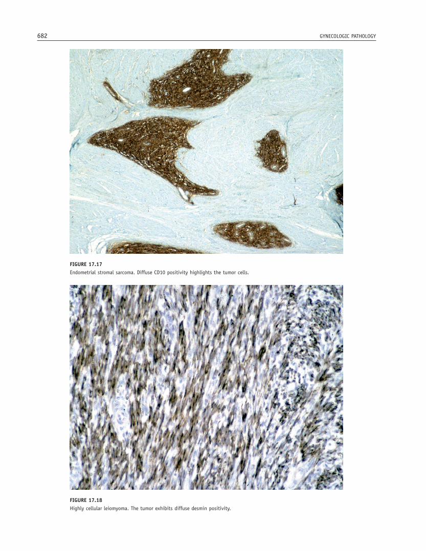
DISTINCTION BETWEEN ENDOMETRIAL STROMAL AND SMOOTH MUSCLE NEOPLASMS
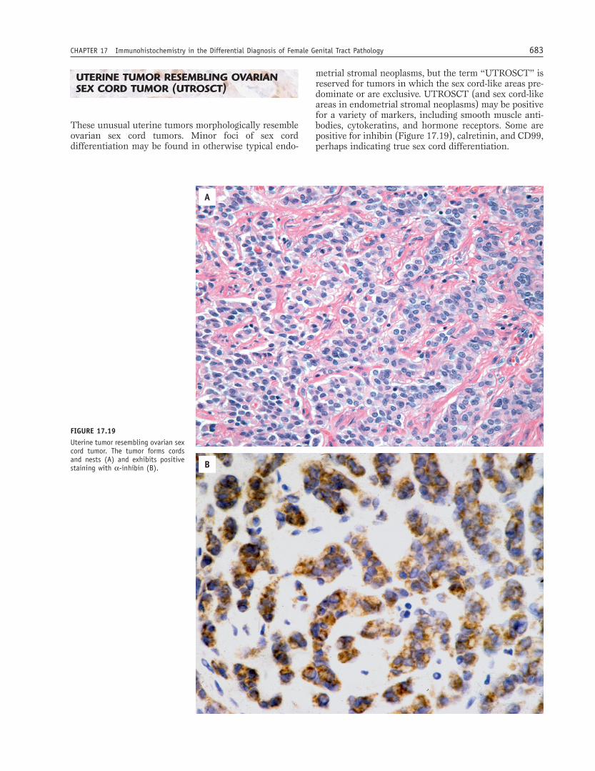
A panel of immunohistochemical stains, including CD10, desmin, and h-caldesmon may be useful in dis-tinguishing between an endometrial stromal and a smooth muscle neoplasm, as these tumors may exhibit morphologic overlap. Most endometrial stromal neo-plasms exhibit diffuse positivity for CD10 (Figure 17.17); however, some uterine smooth muscle neo-plasms are also positive, and cellular and highly cellular leiomyomas (which are most likely to be misdiagnosed as endometrial stromal neoplasms) may exhibit diffuse positivity. Desmin is of value in that most smooth
muscle neoplasms exhibit diffuse positivity (Figure 17.18), whereas although some endometrial stromal neoplasms are positive, diffuse staining is uncommon. In addition, most uterine smooth muscle neoplasms exhibit diffuse positivity for h-caldesmon whereas most endometrial stromal neoplasms are negative (positivity being confi ned to areas of smooth muscle differentia-tion). α-SMA is of limited value since, although most smooth muscle neoplasms are diffusely positive, many endometrial stromal neoplasms are also positive, includ-ing some with diffuse reactivity.
Other antibodies, which may be positive in both uterine endometrial stromal and smooth muscle neo-plasms, include ER, PR, cytokeratin, and WT1. Oxyto-cin receptor, however, is positive in uterine smooth muscle but not endometrial stromal neoplasms.
FIGURE 17.16Ovarian serous carcinoma. The tumor shows strong nuclear WT1 positivity.
ENDOMETRIAL STROMAL VERSUS SMOOTH MUSCLE NEOPLASM – FACT SHEET
� Endometrial stromal neoplasms typically diffusely positive for CD10 but many cellular and highly cellular leiomyomas also diffusely positive
� Desmin diffusely positive in most smooth muscle neoplasms; most endometrial stromal neoplasms negative or only focally positive
� Uterine smooth muscle neoplasms typically diffusely positive for h-caldesmon; endometrial stromal neoplasms usually negative
682 GYNECOLOGIC PATHOLOGY
FIGURE 17.17Endometrial stromal sarcoma. Diffuse CD10 positivity highlights the tumor cells.
FIGURE 17.18Highly cellular leiomyoma. The tumor exhibits diffuse desmin positivity.
CHAPTER 17 Immunohistochemistry in the Differential Diagnosis of Female Genital Tract Pathology 683
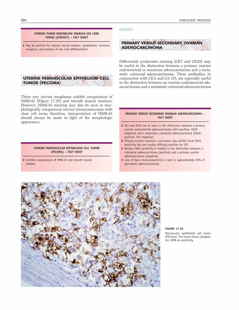
UTERINE TUMOR RESEMBLING OVARIAN SEX CORD TUMOR (UTROSCT)
These unusual uterine tumors morphologically resemble ovarian sex cord tumors. Minor foci of sex cord differentiation may be found in otherwise typical endo-
A
B
FIGURE 17.19Uterine tumor resembling ovarian sex cord tumor. The tumor forms cords and nests (A) and exhibits positive staining with α-inhibin (B).
metrial stromal neoplasms, but the term “UTROSCT” is reserved for tumors in which the sex cord-like areas pre-dominate or are exclusive. UTROSCT (and sex cord-like areas in endometrial stromal neoplasms) may be positive for a variety of markers, including smooth muscle anti-bodies, cytokeratins, and hormone receptors. Some are positive for inhibin (Figure 17.19), calretinin, and CD99, perhaps indicating true sex cord differentiation.
684 GYNECOLOGIC PATHOLOGY
UTERINE TUMOR RESEMBLING OVARIAN SEX CORD TUMOR (UTROSCT) – FACT SHEET
� May be positive for smooth muscle markers, cytokeratins, hormone receptors, and markers of sex cord differentiation
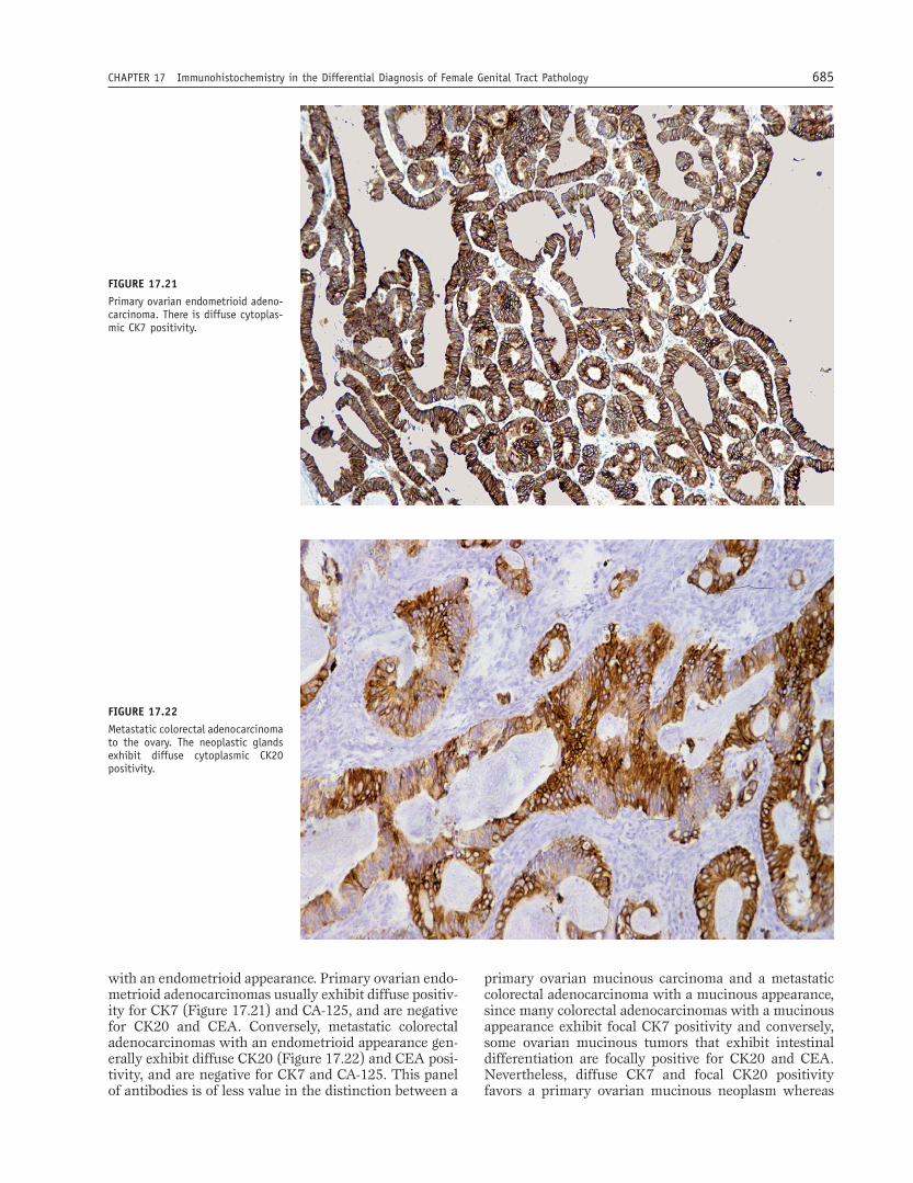
UTERINE PERIVASCULAR EPITHELIOID CELL TUMOR (PECOMA)
These rare uterine neoplasms exhibit coexpression of HMB-45 (Figure 17.20) and smooth muscle markers. However, HMB-45 staining may also be seen in mor-phologically unequivocal uterine leiomyosarcomas with clear cell areas; therefore, interpretation of HMB-45 should always be made in light of the morphologic appearance.
UTERINE PERIVASCULAR EPITHELIOID CELL TUMOR (PECOMA) – FACT SHEET
� Exhibits coexpression of HMB-45 and smooth muscle markers
PRIMARY VERSUS SECONDARY OVARIAN ADENOCARCINOMA – FACT SHEET
� CK7 and CK20 are of value in the distinction between a primary ovarian endometrioid adenocarcinoma (CK7-positive, CK20-negative) and a metastatic colorectal adenocarcinoma (CK20-positive, CK7-negative)
� Primary ovarian mucinous carcinomas may exhibit focal CK20 positivity but are usually diffusely positive for CK7
� Nuclear CDX2 positivity is helpful in the distinction between a colorectal adenocarcinoma (positive) and a primary ovarian adenocarcinoma (negative)
� Loss of Dpc4 immunoreactivity is seen in approximately 50% of pancreatic adenocarcinomas
OVARY
PRIMARY VERSUS SECONDARY OVARIAN ADENOCARCINOMA
Differential cytokeratin staining (CK7 and CK20) may be useful in the distinction between a primary ovarian endometrioid or mucinous adenocarcinoma and a meta-static colorectal adenocarcinoma. These antibodies, in conjunction with CEA and CA-125, are especially useful in the distinction between an ovarian endometrioid ade-nocarcinoma and a metastatic colorectal adenocarcinoma
FIGURE 17.20Perivascular epithelioid cell tumor (PEComa). The tumor shows cytoplas-mic HMB-45 positivity.
CHAPTER 17 Immunohistochemistry in the Differential Diagnosis of Female Genital Tract Pathology 685
with an endometrioid appearance. Primary ovarian endo-metrioid adenocarcinomas usually exhibit diffuse positiv-ity for CK7 (Figure 17.21) and CA-125, and are negative for CK20 and CEA. Conversely, metastatic colorectal adenocarcinomas with an endometrioid appearance gen-erally exhibit diffuse CK20 (Figure 17.22) and CEA posi-tivity, and are negative for CK7 and CA-125. This panel of antibodies is of less value in the distinction between a
primary ovarian mucinous carcinoma and a metastatic colorectal adenocarcinoma with a mucinous appearance, since many colorectal adenocarcinomas with a mucinous appearance exhibit focal CK7 positivity and conversely, some ovarian mucinous tumors that exhibit intestinal differentiation are focally positive for CK20 and CEA. Nevertheless, diffuse CK7 and focal CK20 positivity favors a primary ovarian mucinous neoplasm whereas
FIGURE 17.21Primary ovarian endometrioid adeno-carcinoma. There is diffuse cytoplas-mic CK7 positivity.
FIGURE 17.22Metastatic colorectal adenocarcinoma to the ovary. The neoplastic glands exhibit diffuse cytoplasmic CK20 positivity.
686 GYNECOLOGIC PATHOLOGY
diffuse CK20 and focal CK7 positivity favors a metastatic colorectal adenocarcinoma. As there is considerable immunohistochemical overlap, these results should be interpreted in light of the morphologic fi ndings in the ovary as well as pertinent clinical and radiologic information.
Differential cytokeratin staining is also of value in the setting of coexistent mucinous tumors of the ovary and appendix in association with pseudomyxoma peri-tonei. In most cases, the ovarian and appendiceal lesions and the mucinous epithelium present in the peritoneum are CK20 positive and CK7 negative, supporting an appendiceal origin.
Other antibodies that may be useful in the distinction between a primary ovarian adenocarcinoma and a meta-static colorectal adenocarcinoma include CDX2, β-catenin, villin, MUC2, and MUC5AC. Diffuse nuclear CDX2 positivity is characteristic of metastatic colorectal adenocarcinoma whereas most primary ovarian carcino-mas are negative. β-catenin nuclear positivity is present in approximately half of metastatic colorectal adenocar-cinomas, while most ovarian mucinous neoplasms exhibit no nuclear staining; however, ovarian endometrioid ade-nocarcinomas may exhibit patchy nuclear β-catenin posi-tivity. Villin may also be of value in the confi rmation of a metastatic colorectal adenocarcinoma as it is typically expressed by this tumor. Finally, MUC5AC is usually expressed in ovarian mucinous tumors, but is typically absent in colorectal carcinomas.
Differential cytokeratin staining is of no value in distinguishing between a primary ovarian adenocarci-noma and a metastatic adenocarcinoma from the pan-creas, biliary tree, or stomach. Secondary tumors from the aforementioned organs are usually positive for CK7 whereas CK20 is usually negative (or focally positive). Dpc4 staining, however, may be helpful in diagnosing a metastatic pancreatic adenocarcinoma since primary ovarian mucinous neoplasms are Dpc4-positive while approximately 50% of pancreatic adenocarcinomas are negative.
Other markers, which may be of value in the diagno-sis of rare metastatic tumors in the ovary, include RCC marker and CD10 (positive in metastatic renal cell carcinoma), TTF-1 (positive in metastatic lung carci-noma), uroplakin-III (positive in metastatic urinary transitional cell carcinoma) and S-100, HMB-45, and melan-A (positive in metastatic malignant melanoma). However, it should be noted that ovarian sex cord-stromal tumors may be positive for S-100 and melan-A and rarely for HMB-45.
DISTINCTION BETWEEN OVARIAN SEROUS AND OVARIAN ENDOMETRIOID ADENOCARCINOMA
The distinction between a well-differentiated ovarian serous and endometrioid adenocarcinoma is usually straightforward. However, with poorly differentiated
neoplasms this distinction may be diffi cult. Most ovarian serous carcinomas exhibit diffuse nuclear WT1 positiv-ity whereas almost all ovarian endometrioid adenocar-cinomas are negative. WT1 is also commonly positive in ovarian transitional cell carcinomas, but clear cell and mucinous carcinomas are generally negative.
OVARIAN SEX CORD-STROMAL TUMORS
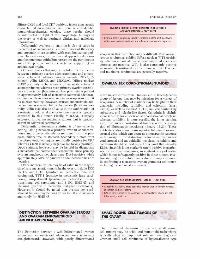
Ovarian sex cord-stromal tumors are a heterogeneous group of lesions that may be mistaken for a variety of neoplasms. A number of markers may be helpful in their diagnosis, including α-inhibin and calretinin (most useful), as well as melan-A, CD99, müllerian-inhibiting substance, and relaxin-like factor. Calretinin is slightly more sensitive for an ovarian sex cord-stromal neoplasm whereas α-inhibin is more specifi c, the latter staining most ovarian sex cord-stromal tumors, with the excep-tion of fi bromatous neoplasms (Figure 17.23). These antibodies also stain nonneoplastic luteinized ovarian stromal cells, which can occur as a nonspecifi c response in the ovary. In the distinction between an ovarian sex cord-stromal and an epithelial neoplasm, α-inhibin and calretinin should be used as part of a panel that includes EMA, since this latter marker is rarely positive in ovarian sex cord-stromal neoplasms, in contrast to cytokeratin, which is not infrequently positive in these tumors. Posi-tive staining for α-inhibin and calretinin may also assist in confi rming a metastatic ovarian granulosa cell tumor, including the sarcomatous variant.
OVARIAN SEROUS VERSUS OVARIAN ENDOMETRIOID ADENOCARCINOMA – FACT SHEET
� Ovarian serous carcinoma usually exhibits nuclear WT1 positivity while most ovarian endometrioid adenocarcinomas are negative
OVARIAN SEX CORD-STROMAL TUMORS – FACT SHEET
� Calretinin is slightly more sensitive marker than α-inhibin whereas α-inhibin is more specifi c
� EMA is rarely positive, in contrast to cytokeratins, which are not infrequently positive
SMALL ROUND CELL TUMORS OF THE OVARY
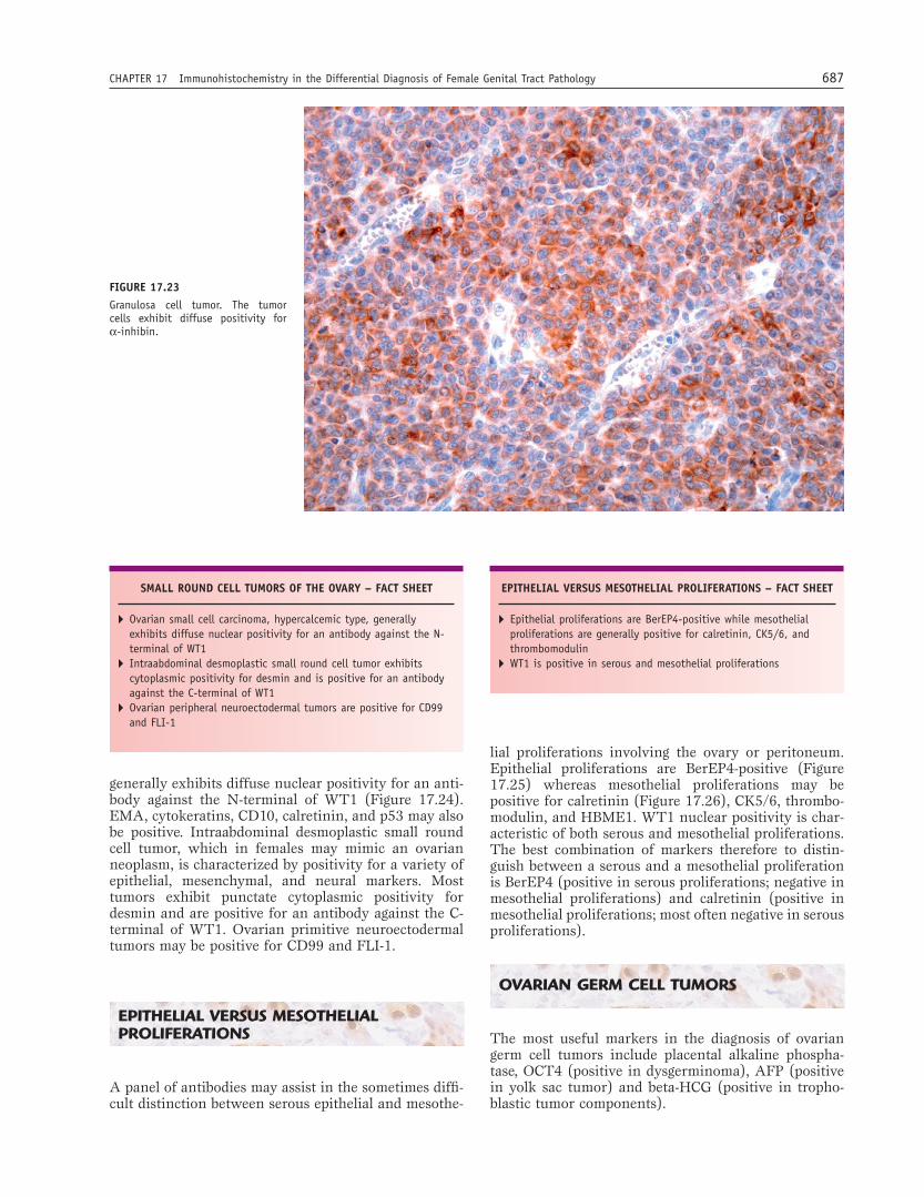
The differential diagnosis of ovarian small round cell tumors may be wide and immunohistochemistry typically plays an important role in their diagnosis. Ovarian small cell carcinoma of hypercalcemic type
CHAPTER 17 Immunohistochemistry in the Differential Diagnosis of Female Genital Tract Pathology 687
SMALL ROUND CELL TUMORS OF THE OVARY – FACT SHEET
� Ovarian small cell carcinoma, hypercalcemic type, generally exhibits diffuse nuclear positivity for an antibody against the N-terminal of WT1
� Intraabdominal desmoplastic small round cell tumor exhibits cytoplasmic positivity for desmin and is positive for an antibody against the C-terminal of WT1
� Ovarian peripheral neuroectodermal tumors are positive for CD99 and FLI-1
FIGURE 17.23Granulosa cell tumor. The tumor cells exhibit diffuse positivity for α-inhibin.
generally exhibits diffuse nuclear positivity for an anti-body against the N-terminal of WT1 (Figure 17.24). EMA, cytokeratins, CD10, calretinin, and p53 may also be positive. Intraabdominal desmoplastic small round cell tumor, which in females may mimic an ovarian neoplasm, is characterized by positivity for a variety of epithelial, mesenchymal, and neural markers. Most tumors exhibit punctate cytoplasmic positivity for desmin and are positive for an antibody against the C-terminal of WT1. Ovarian primitive neuroectodermal tumors may be positive for CD99 and FLI-1.
EPITHELIAL VERSUS MESOTHELIAL PROLIFERATIONS
A panel of antibodies may assist in the sometimes diffi -cult distinction between serous epithelial and mesothe-
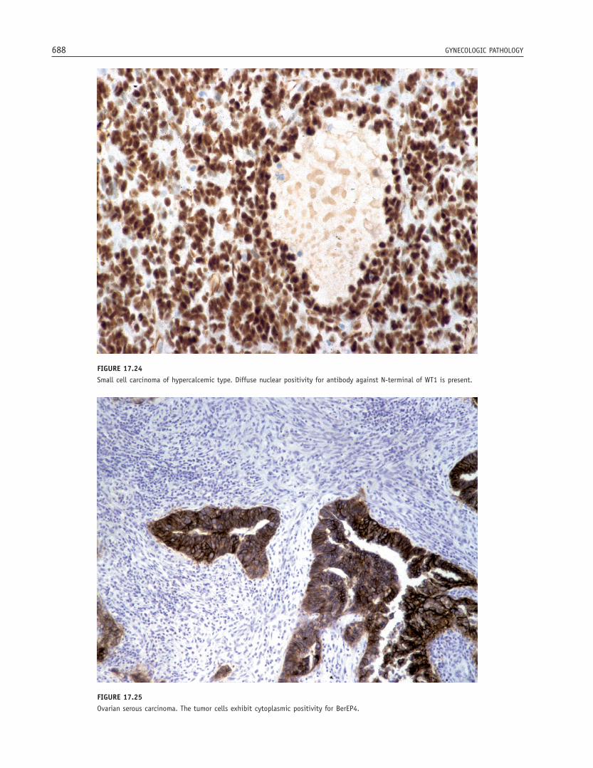
lial proliferations involving the ovary or peritoneum. Epithelial proliferations are BerEP4-positive (Figure 17.25) whereas mesothelial proliferations may be positive for calretinin (Figure 17.26), CK5/6, thrombo-modulin, and HBME1. WT1 nuclear positivity is char-acteristic of both serous and mesothelial proliferations. The best combination of markers therefore to distin-guish between a serous and a mesothelial proliferation is BerEP4 (positive in serous proliferations; negative in mesothelial proliferations) and calretinin (positive in mesothelial proliferations; most often negative in serous proliferations).
OVARIAN GERM CELL TUMORS
The most useful markers in the diagnosis of ovarian germ cell tumors include placental alkaline phospha-tase, OCT4 (positive in dysgerminoma), AFP (positive in yolk sac tumor) and beta-HCG (positive in tropho-blastic tumor components).
EPITHELIAL VERSUS MESOTHELIAL PROLIFERATIONS – FACT SHEET
� Epithelial proliferations are BerEP4-positive while mesothelial proliferations are generally positive for calretinin, CK5/6, and thrombomodulin
� WT1 is positive in serous and mesothelial proliferations
688 GYNECOLOGIC PATHOLOGY
FIGURE 17.24Small cell carcinoma of hypercalcemic type. Diffuse nuclear positivity for antibody against N-terminal of WT1 is present.
FIGURE 17.25Ovarian serous carcinoma. The tumor cells exhibit cytoplasmic positivity for BerEP4.
CHAPTER 17 Immunohistochemistry in the Differential Diagnosis of Female Genital Tract Pathology 689
FIGURE 17.26Mesothelial proliferation. Reactive mesothelial cells exhibit nuclear and cytoplasmic calretinin positivity.
TROPHOBLASTIC DISEASES
RECENTLY CHARACTERIZED MARKERS OF TROPHOBLASTIC CELLS
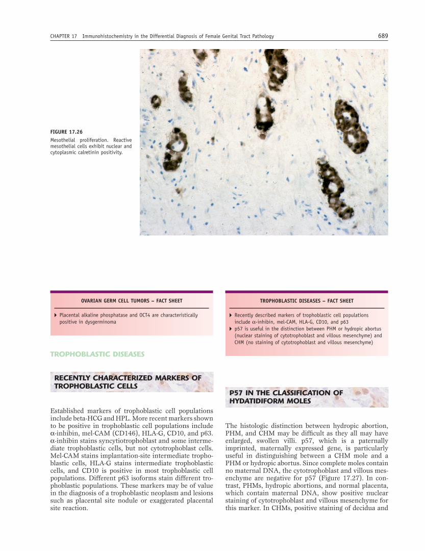
Established markers of trophoblastic cell populations include beta-HCG and HPL. More recent markers shown to be positive in trophoblastic cell populations include α-inhibin, mel-CAM (CD146), HLA-G, CD10, and p63. α-inhibin stains syncytiotrophoblast and some interme-diate trophoblastic cells, but not cytotrophoblast cells. Mel-CAM stains implantation-site intermediate tropho-blastic cells, HLA-G stains intermediate trophoblastic cells, and CD10 is positive in most trophoblastic cell populations. Different p63 isoforms stain different tro-phoblastic populations. These markers may be of value in the diagnosis of a trophoblastic neoplasm and lesions such as placental site nodule or exaggerated placental site reaction.
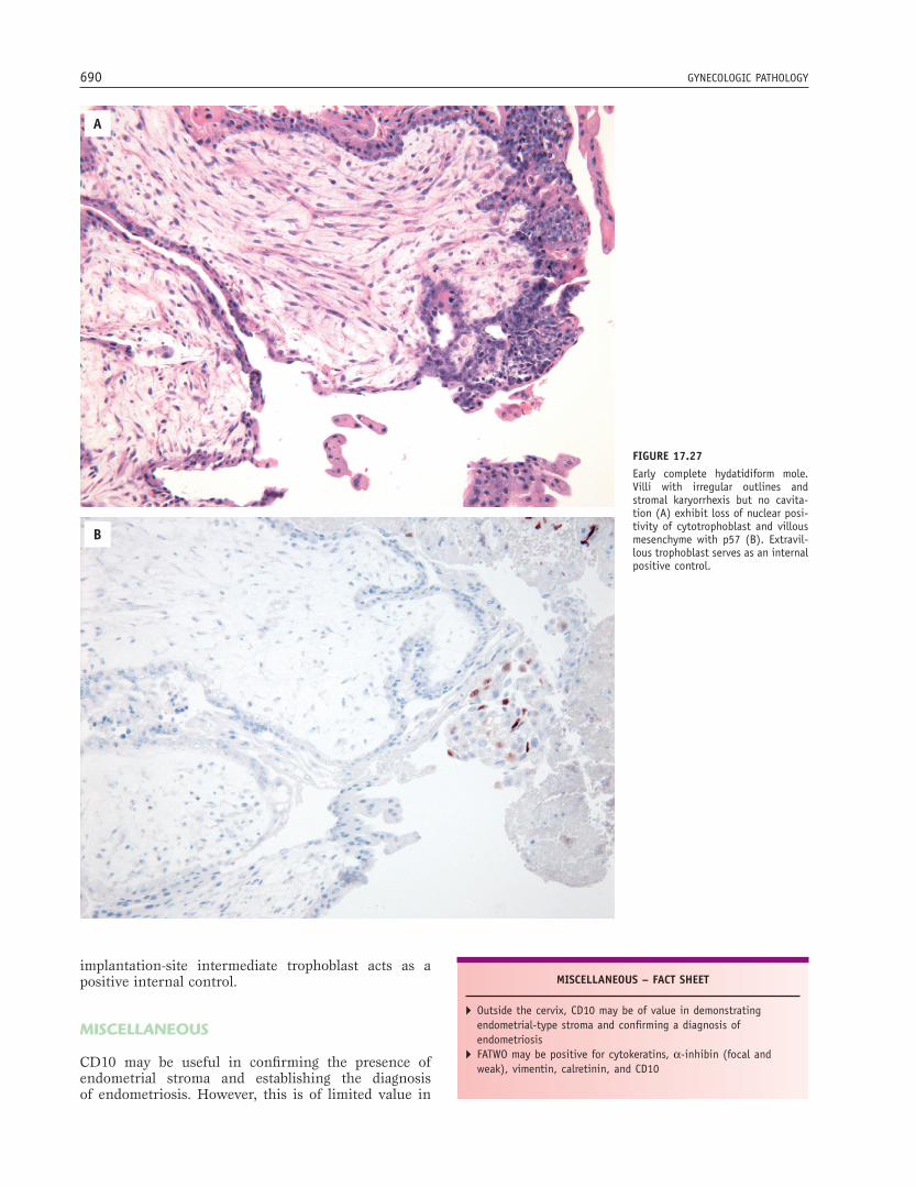
P57 IN THE CLASSIFICATION OF HYDATIDIFORM MOLES
The histologic distinction between hydropic abortion, PHM, and CHM may be diffi cult as they all may have enlarged, swollen villi. p57, which is a paternally imprinted, maternally expressed gene, is particularly useful in distinguishing between a CHM mole and a PHM or hydropic abortus. Since complete moles contain no maternal DNA, the cytotrophoblast and villous mes-enchyme are negative for p57 (Figure 17.27). In con-trast, PHMs, hydropic abortions, and normal placenta, which contain maternal DNA, show positive nuclear staining of cytotrophoblast and villous mesenchyme for this marker. In CHMs, positive staining of decidua and
OVARIAN GERM CELL TUMORS – FACT SHEET
� Placental alkaline phosphatase and OCT4 are characteristically positive in dysgerminoma
TROPHOBLASTIC DISEASES – FACT SHEET
� Recently described markers of trophoblastic cell populations include α-inhibin, mel-CAM, HLA-G, CD10, and p63
� p57 is useful in the distinction between PHM or hydropic abortus (nuclear staining of cytotrophoblast and villous mesenchyme) and CHM (no staining of cytotrophoblast and villous mesenchyme)
690 GYNECOLOGIC PATHOLOGY
A
B
FIGURE 17.27Early complete hydatidiform mole. Villi with irregular outlines and stromal karyorrhexis but no cavita-tion (A) exhibit loss of nuclear posi-tivity of cytotrophoblast and villous mesenchyme with p57 (B). Extravil-lous trophoblast serves as an internal positive control.
implantation-site intermediate trophoblast acts as a positive internal control.
MISCELLANEOUS
CD10 may be useful in confi rming the presence of endometrial stroma and establishing the diagnosis of endometriosis. However, this is of limited value in
MISCELLANEOUS – FACT SHEET
� Outside the cervix, CD10 may be of value in demonstrating endometrial-type stroma and confi rming a diagnosis of endometriosis
� FATWO may be positive for cytokeratins, α-inhibin (focal and weak), vimentin, calretinin, and CD10
CHAPTER 17 Immunohistochemistry in the Differential Diagnosis of Female Genital Tract Pathology 691
the cervix where a rim of CD10 positive stromal cells surrounds normal endocervical glands.
Female adnexal tumor of probable wolffi an origin (FATWO) is often positive for α-inhibin (usually focal and weak), in contrast to the diffuse positivity gener-ally, but not always, seen in ovarian sex cord-stromal neoplasms, which is the main differential diagnostic consideration. FATWO generally expresses cytokera-tins and vimentin, and may be positive for calretinin and CD10. EMA and CEA are generally negative. This immunophenotype is similar to that of mesonephric remnants and provides evidence for a mesonephric origin of FATWO.
Other applications of α-inhibin staining in gyneco-logical pathology include the identifi cation of luteinized stromal cells. Staining with α-inhibin assists in confi rm-ing the presence of granulosa cells in cytological speci-mens and indicates a functional rather than an epithelial lined cyst.
WT1 and calretinin staining may assist in the diag-nosis of an adenomatoid tumor and help confi rm its mesothelial derivation.
SUGGESTED READING
General
Baker PM, Olwa E. Immunohistochemistry as a tool in the differential diagnosis of ovarian tumors: an update. Int J Gynecol Pathol 2005;24:39–55.
Marjoniemi VM. Immunohistochemistry in gynaecological pathology: a review. Pathology 2004;36:109–119.
McCluggage WG. Recent advances in immunohistochemistry in the diagnosis of ovarian neoplasms. J Clin Pathol 2000;53:327–334.
McCluggage WG. Recent advances in immunohistochemistry in gynaeco-logical pathology. Histopathology 2002;40:309–326.
Nucci MR, Castillon DH, Bai H, et al. Biomarkers in diagnostic obstetric and gynecologic pathology: a review. Adv Anat Pathol 2003;10:55–68.
Preinvasive Vulval Squamous Lesions
Logani S, Cu D, Quint WGV, et al. Low-grade vulvar and vaginal intraepi-thelial neoplasia: correlation of histologic features with human papil-lomavirus DNA detection and MIB-1 immunostaining. Mod Pathol 2003;16:735–741.
Pirog EC, Chen Y-T, Isacson C. MIB-1 immunostaining is a benefi cial adjunct test for accurate diagnosis of vulvar condyloma acuminatum. Am J Surg Pathol 2000;24:1393–1399.
Santos M, Montagut C, Mellado B, et al. Immunohistochemical staining for p16 and p53 in premalignant and malignant epithelial lesions of the vulva. Int J Gynecol Pathol 2004;23:206–214.
Yang B, Hart WR. Vulvar intraepithelial neoplasia of the simplex (differ-entiated) type : a clinicopathologic study including analysis of HPV and p53 expression. Am J Surg Pathol 2000;24:429–441.
Vulval Paget Disease
Brown HM, Wilkinson EJ. Uroplakin-III to distinguish primary vulvar Paget disease from Paget disease secondary to urothelial carcinoma. Hum Pathol 2002;33:545–548.
Goldblum JR, Hart WR. Perianal Paget’s disease – a histologic and immunohistochemical study of 11 cases with and without associated rectal adenocarcinoma. Am J Surg Pathol 1998;2:170–171.
Kuan S-F, Montag AG, Hart J, et al. Differential expression of mucin genes in mammary and extramammary Paget’s disease. Am J Surg Pathol 2001;25:1469–1477.
Raju RR, Goldblum JR, Hart WR. Pagetoid squamous cell carcinoma in situ (pagetoid Bowen’s disease) of the external genitalia. Int J Gynecol Pathol 2003;22:127–135.
Zhang C, Zhang P, Sung J, et al. Overexpression of p53 is correlated with stromal invasion in extramammary Paget’s disease of the vulva. Hum Pathol 2003;34:880–885.
Vulvovaginal Mesenchymal Lesions
Iwasa Y, Fletcher CD. Cellular angiofi broma: clinicopathologic and immu-nohistochemical analysis of 51 cases. Am J Surg Pathol 2004;28:1426–1435.
McCluggage WG. A review and update of morphologically bland vulvovagi-nal mesenchymal lesions. Int J Gynecol Pathol 2005;24:26–38.
McCluggage WG, Patterson A, Maxwell P. Aggressive angiomyxoma of pelvic parts exhibits oestrogen and progesterone receptor positivity. J Clin Pathol 2000;53:603–605.
McCluggage WG, Ganesan R, Hirschowitz L, et al. Cellular angiofi broma and related fi bromatous lesions of the vulva: report of a series of cases with a morphological spectrum wider than previously described. Histo-pathology 2004;45:360–368.
Nucci MR, Tallini G, Quade BJ. HMGIC expression as a diagnostic marker for vulvar aggressive angiomyxoma. Mod Pathol 2001;14:829(A).
Nucci MR, Weremonicz S, Neskey DM, et al. Chromosomal translocation t (8, 12) induced aberrant HMGIC expression in aggressive angiomyxoma of the vulva. Genes Chromosomes Cancer 2001;32:172–176.
Preinvasive Cervical Squamous Lesions
Agoff SN, Lin P, Morihara J, et al. p16 INK4A expression correlates with degree of cervical neoplasia: a comparison with Ki-67 expression and detection of high-risk HPV types. Mod Pathol 2003;16:665–673.
Klaes R, Benner A, Freidrich T, et al. p16 (INK4A) immunohistochemistry improves interobserver agreement in the diagnosis of cervical intraepi-thelial neoplasia. Am J Surg Pathol 2002;26:1387–1399.
Klaes R, Friedrich T, Spitkovsky D, et al. Overexpression of p16 (INK4A) as a specifi c marker for dysplastic and neoplastic epithelial cells of the cervix uteri. Int J Cancer 2002;92:276–284.
Kruse A-J, Baak JPA, Helliesen T, et al. Evaluation of MIB-1 positive cell clusters as a diagnostic marker for cervical intraepithelial neoplasia. Am J Surg Pathol 2002;26:1501–1507.
McCluggage WG, Tang L, Maxwell P, et al. Monoclonal antibody MIB-1 in the assessment of cervical squamous intraepithelial lesions. Int J Gynecol Pathol 1996;15:131–136.
Mittal K. Utility of MIB-1 in evaluating cauterized cervical cone biopsy margins. Int J Gynecol Pathol 1999;18:211–214.
Mittal K, Mesia A, Demopoulos RL. MIB-1 expression is useful in distin-guishing dysplasia from atrophy in elderly women. Int J Gynecol Pathol 1999;18:122–124.
Pirog EC, Baergen RN, Soslow RA, et al. Diagnostic accuracy of cervical low-grade squamous intraepithelial lesions is improved with MIB-1 immunostaining. Am J Surg Pathol 2002;26:70–75.
Preinvasive Cervical Glandular Lesions
Cameron RI, Maxwell P, Jenkins, D, et al. Immunohistochemical staining with MIB-1, bcl2 and p16 assists in the distinction of cervical glandular intraepithelial neoplasia from tubo-endometrial metaplasia, endome-triosis and microglandular hyperplasia. Histopathology 2002;41:313–321.
Cina SJ, Richardson MS, Austin RM, et al. Immunohistochemical staining for Ki-67 antigen, carcinoembryonic antigen, and p53 in the differential diagnosis of glandular lesions of the cervix. Mod Pathol 1997;10:176–180.
Ishikawa M, Fujii T, Nasumoto N, et al. Correlation of p16 INK4A overex-pression with human papillomavirus infection in cervical adenocarcino-mas. Int J Gynecol Pathol 2003;22:378–385.
Lee KR, Sun D, Crum CP. Endocervical intraepithelial glandular atypia (dysplasia): a histopathologic, human papillomavirus, and MIB-1 analy-sis of 25 cases. Hum Pathol 2000;31:656–664.
McCluggage WG, Maxwell P, McBride HA, et al. Monoclonal antibodies Ki-67 and MIB-1 in the distinction of tuboendometrial metaplasia from endocervical adenocarcinoma and adenocarcinoma in situ in formalin fi xed material. Int J Gynecol Pathol 1995;14:209–216.
692 GYNECOLOGIC PATHOLOGY
McCluggage WG, Maxwell P. Bcl-2 and p21 staining of cervical tuboendo-metrial metaplasia. Histopathology 2002;40:107.
Negri G, Egarter–Vigi E, Kasal A, et al. p16 (INK4a) is a useful marker for the diagnosis of adenocarcinoma of the cervix uteri and its precursors. Am J Surg Pathol 2003;27:187–193.
Pirog EC, Isacson C, Szabolcs MJ, et al. Proliferative activity of benign and neoplastic endocervical epithelium and correlation with HPV DNA detection. Int J Gynecol Pathol 2002;21:22–26.
Riethdorf L, Riethdorf S, Lee KR, et al. Human papillomaviruses, expres-sion of p16 INK4A, and early endocervical glandular neoplasia. Hum Pathol 2002;33:899–904.
Cervical Mesonephric Lesions
McCluggage WG, Oliva E, Herrington CS, et al. CD10 and calretinin staining of endocervical glandular lesions, endocervical stroma and endometrioid adenocarcinoma of the uterine corpus: CD10 positivity is characteristic of, but not specifi c for, mesonephric lesions and is not specifi c for endometrioid stroma. Histopathology 2003;43:144–150.
Ordi J, Nogales FF, Palacin A, et al. Mesonephric adenocarcinoma of the uterine corpus: CD10 expression as evidence of mesonephric differentia-tion. Am J Surg Pathol 2001;25:1540–1545.
Ordi J, Romagosa C, Tavasson FA, et al. CD10 expression in epithelial tissues and tumors of the gynecologic tract; a useful marker in the diagnosis of mesonephric, trophoblastic and clear cell tumors. Am J Surg Pathol 2003;27:178–186.
Silver SA, Devouassoux-Shisheboran M, Mezetti TP, et al. Mesonephric adenocarcinomas of the uterine cervix: a study of 11 cases with immu-nohistochemical fi ndings. Am J Surg Pathol 2001;25:379–387.
Cervical Minimal Deviation Adenocarcinoma of Mucinous Type (Adenoma Malignum)
Mikami Y, Hata S, Melamed J, et al. Lobular endocervical glandular hyper-plasia is a metaplastic process with a pyloric gland phenotype. Histo-pathology 2001;39:364–372.
Mikami Y, Kiyokawa T, Hata S, et al. Gastrointestinal immunophenotype in adenocarcinomas of the uterine cervix and related glandular lesions: a possible link between lobular endocervical glandular hyperplasia/pyloric gland metaplasia and adenoma malignum. Mod Pathol 2004;17:962–972.
Utsugi K, Hira Y, Takeshima N, et al. Utility of the monoclonal antibody HIK1083 in the diagnosis of adenoma malignum of the uterine cervix. Gynecol Oncol 1999;75:345–348.
Distinction Between Endometrial and Endocervical Adenocarcinoma
Ansari-Lari MA, Staebler A, Zaino RJ, et al. Distinction of endocervical and endometrial adenocarcinomas: immunohistochemical p16 expres-sion correlated with human papillomavirus (HPV) DNA detection. Am J Surg Pathol 2004;28:160–167.
Castrillon DH, Lee KR, Nucci MR. Distinction between endometrial and endocervical adenocarcinoma: an immunohistochemical study. Int J Gynecol Pathol 2002;21:4–10.
Kamoi S, Al Juboury ML, Akin MR, et al. Immunohistochemical staining in the distinction between endometrial and endocervical adenocarcino-mas: another viewpoint. Int J Gynecol Pathol 2002;21:217–223.
McCluggage WG, Jenkins D. Immunohistochemical staining with p16 may assist in the distinction between endometrial and endocervical adeno-carcinoma. Int J Gynecol Pathol 2003;2:231–235.
McCluggage WG, Sumathi VP, McBride HA, et al. A panel of immunohisto-chemical stains, including carcinoembryonic antigen, vimentin and estrogen receptor aids the distinction between primary endometrial and endocervical adenocarcinomas. Int J Gynecol Pathol 2002;21:11–15.
Staebler A, Sherman ME, Zaino RJ, et al. Hormone receptor immunohis-tochemistry and human papillomavirus in situ hybridization are useful for distinguishing endocervical and endometrial adenocarcinomas. Am J Surg Pathol 2002;26:998–1006.
Zaino RJ. The fruits of our labours: distinguishing endometrial from endocervical adenocarcinoma: Int J Gynecol Pathol 2002;21:1–3.
Cervical Neuroendocrine Carcinomas
Gilks CB, Young RH, Gersell DJ, et al. Large cell neuroendocrine carcinoma of the uterine cervix: a clinicopathologic study of 12 cases. Am J Surg Pathol 1997;21:905–914.
Type I Versus Type II Endometrial Adenocarcinoma
Demopoulos RL, Mesia AF, Mittal K, et al. Immunohistochemical compari-son of uterine papillary serous and papillary endometrioid carcinoma: clues to pathogenesis. Int J Gynecol Pathol 1999;18:233–237.
Lax SF, Kendall B, Tashiro H, et al. The frequency of p53, K-ras mutations, and microsatellite instability differs in uterine endometrioid and serous carcinoma: evidence of distinct molecular genetic pathways. Cancer 2000;88:814–825.
Schlosshauer PW, Hedrick Ellenson L, Soslow RA. β-cateinin and E-cad-herin expression patterns in high-grade endometrial carcinoma are associated with histological subtype. Mod Pathol 2002;15:1032–1037.
Vang R, Barner R, Wheeler DT, et al. Immunohistochemical staining for Ki-67 and p53 helps distinguish endometrial Arias–Stella reaction from high grade carcinoma, including clear cell carcinoma. Int J Gynecol Pathol 2004;23:223–233.
Wang TY, Chen BF, Yang YC, et al. Histologic and immnophenotypic clas-sifi cation of cervical carcinomas by expression of the p53 homologue p63: a study of 250 cases. Hum Pathol 2001;32:479–486.
Distinction Between Uterine Serous Carcinoma and Ovarian Serous Carcinoma
Al-Hussaini M, Stockman A, Foster H, et al. WT-1 assists in distinguishing ovarian from uterine serous carcinoma and in distinguishing serous and ovarian endometrioid carcinoma. Histopathology 2004;44:109–115.
Egan JA, Ionescu ML, Eapen E, et al. Differential expression of WT1 and p53 in serous and endometrioid carcinoma of the endometrium. Int J Gynecol Pathol 2004;23:119–122.
Goldstein NS, Uzieblo A. WT-1 immunoreactivity in uterine papillary serous carcinoma is different from ovarian serous carcinomas. Am J Clin Pathol 2002;117:541–545.
Hashi A, Yuminamochi T, Murata S-I, et al. Wilms tumor gene immunore-activity in primary serous carcinomas of the fallopian tube, ovary, endo-metrium and peritoneum. Int J Gynecol Pathol 2003;22:374–377.
Hwang H, Quenneville L, Yaziji H, et al. Wilms tumor gene product. Sensi-tive and contextually specifi c marker of serous carcinomas of ovarian surface epithelial origin. Appl Immunohistochem Mol Morphol 2004;12:122–126.
McCluggage WG. WT1 is of value in ascertaining the site of origin of serous carcinomas within the female genital tract. Int J Gynecol Pathol 2004;23:97–99.
Distinction Between Stromal and Smooth Muscle Neoplasms
Chu PG, Arber PA, Weiss LM, et al. Utility of CD10 in distinguishing between endometrial stromal sarcoma and uterine smooth muscle tumors: an immunohistochemical comparison of 34 cases. Mod Pathol 2001;14:465–471.
Loddenkemper C, Mechsner S, Foss H-D, et al. Use of oxytocin receptor expression in distinguishing between uterine smooth muscle tumors and endometrial stromal sarcoma. Am J Surg Pathol 2003;27:1458–1462.
McCluggage WG, Sumathi VP, Maxwell P. CD10 is a sensitive and diagnosti-cally useful immunohistochemical marker of normal endometrial stroma and of endometrial stromal neoplasms. Histopathology 2001;39:273–278.
Nucci MR, O’Connell JT, Huettner PC, et al. h-caldesmon expression effec-tively distinguishes endometrial stromal tumors from uterine smooth muscle tumors. Am J Surg Pathol 2001;25:253–258.
Oliva E, Young RH, Amin MB, et al. An immunohistochemical analysis of endometrial stromal and smooth muscle tumors of the uterus: a study of 54 cases emphasising the importance of using a panel because of overlap in immunoreactivity for individual antibodies. Am J Surg Pathol 2002;26:403–412.
Rush DS, Tan JY, Baergen RN, et al. h-caldesmon, a novel smooth muscle-specifi c antibody, distinguishes between cellular leiomyoma and endo-metrial stromal sarcoma. Am J Surg Pathol 2001;25:253–258.
CHAPTER 17 Immunohistochemistry in the Differential Diagnosis of Female Genital Tract Pathology 693
Sumathi VP, Al-Hussaini M, Connolly LE, et al. Endometrial stromal neo-plasms are immunoreactive with WT-1 antibody. Int J Gynecol Pathol 2004;23:241–247.
Uterine Tumor Resembling Ovarian Sex Cord Tumor
Baker RJ, Hildebrandt RH, Rouse RV, et al. Inhibin and CD99 (MIC2) expression in uterine stromal neoplasms with sex cord-like elements. Hum Pathol 1999;30:671–679.
Irving JA, Carinelli S, Part J. Uterine tumors resembling ovarian sex cord tumors are polyphenotypic neoplasms with true sex-cord differentia-tion. Mod Pathol 2006;19:17–24.
Krishnamurthy S, Jungbloth AA, Busam KJ, et al. Uterine tumors resem-bling ovarian sex cord tumors have an immunophenotype consistent with true sex cord differentiation. Am J Surg Pathol 1998;22:1078–1082.
McCluggage WG. Uterine tumours resembling ovarian sex cord tumours: immunohistochemical evidence for true sex cord differentiation. Histo-pathology 1999;34:373–380.
Uterine Perivascular Epithelioid Cell Tumor
Silva EG, Deavers MT, Bodurka DC, et al. Uterine epithelioid leiomyosar-comas with clear cells: reactivity with HMB-45 and the concept of PEComa. Am J Surg Pathol 2004;28:244–249.
Sumpson KW, Albores-Saavedra J. HMB-45 reactivity in conventional uterine leiomyosaromas. Am J Surg Pathol 2007;31:95–8.
Vang R, Kempson RL. Perivascular epithelioid cell tumour (PEComa) of the uterus: a subset of HMB-45 positive epithelioid mesenchymal neoplasms with an uncertain relationship to pure smooth muscle tumors. Am J Surg Pathol 2002;26:1–13.
Primary Versus Secondary Ovarian Adenocarcinoma
Albarracin CT, Jafri J, Montag AG, et al. Differential expression of MUC2 and MUC5AC mucin genes in primary ovarian and metastatic colonic carcinoma. Hum Pathol 2000;31:672–677.
Berezowski K, Stasny JF, Kornstein MJ. Cytokeratins 7 and 20 and car-cinoembryonic antigen in ovarian and colonic carcinoma. Mod Pathol 1996;9:426–429.
Cameron RI, Ashe P, O’Rourke DM, et al. A panel of immunohistochemical stains assists in the distinction between ovarian and renal clear cell carcinoma. Int J Gynecol Pathol 2003;22:272–276.
Chou YY, Jeng YM, Kao HL, et al. Differentiation of ovarian mucinous carcinoma and metastatic colorectal adenocarcinoma by immunostain-ing with β-catenin. Histopathology 2003;43:151–156.
Groisman GM, Meir A, Sabo E. The value of cdx2 immunostaining in dif-ferentiating primary ovarian carcinomas from colonic carcinomas meta-static to the ovaries. Int J Gynecol Pathol 2003;23:52–57.
Ji H, Isacson C, Seidman JD, et al. Cytokeratins 7 and 20, Dpc4, and MUC5AC in the distinction of metastatic mucinous carcinomas in the ovary from primary ovarian mucinous tumors : Dpc 4 assists in identify-ing metastatic pancreatic carcinomas. Int J Gynecol Pathol 2002;21:391–400.
Ladendijk JA, Mullink EH, van Diest PJ, et al. Tracing the origin of adeno-carcinomas with unknown primary using immunohistochemistry. Dif-ferential diagnosis between colonic and ovarian carcinomas as primary sites. Hum Pathol 1998;29:491–497.
Logani S, Oliva E, Arnell PM, et al. Use of novel immunohistochemical markers expressed in colonic adenocarcinoma to distinguish primary ovarian tumors from metastatic colorectal carcinoma. Mod Pathol 2005;18:19–25.
McCluggage WG. Recent advances in immunohistochemistry in the diag-nosis of ovarian neoplasms. J Clin Pathol 2000;3:327–334.
Park SY, Kim HS, Hong EK, et al. Expression of cytokeratins 7 and 20 in primary carcinomas of the stomach and colorectum and their value in the differential diagnosis of metastatic carcinomas to the ovary. Hum Pathol 2002;33:1078–1085.
Raspollini MR, Amunni G, Villanucci A, et al. Utility of CDX-2 in distin-guishing between primary and secondary (intestinal) mucinous ovarian carcinoma. Appl Immunohistochem Mol Morphol 2004;12:127–131.
Tornillo L, Moch H, Diener PA, et al. CDX-2 immunostaining in primary and secondary ovarian carcinomas. J Clin Pathol 2004;57:641–643.
Wauters CCAP, Smedts F, Gerrits LGM, et al. Keratins 7 and 20 as diagnostic markers of carcinomas metastatic to the ovary. Hum Pathol 1995;26:852–855.
Werling RW, Yaziji H, Bacchi CE, et al. CDX2, a highly sensitive and specifi c marker of adenocarcinomas of intestinal origin: an immunohistochemi-cal survey of 476 primary and metastatic carcinomas. Am J Surg Pathol 2003;27:303–310.
Distinction Between Ovarian Serous and Ovarian Endometrioid Adenocarcinoma
Al-Hussaini M, Stockman A, Foster H, et al. WT-1 assists in distinguishing ovarian from uterine serous carcinoma and in distinguishing serous and endometrioid ovarian carcinoma. Histopathology 2004;44:109–115.
Shimizu M, Toki T, Takagi Y, et al. Immunohistochemical detection of the Wilms’ tumor gene (WT1) in epithelial ovarian tumors. Int J Gynecol Pathol 2000;19:158–163.
Ovarian Sex Cord-Stromal Tumors
Bamberger AM, Ivell R, Balvers M. Relaxin-like factor (RLF): a new specifi c marker for Leydig cells in the ovary. Int J Gynecol Pathol 1998;18:163–168.
Cao QJ, Jones JG, Li M. Expression of calretinin in human ovary, testis and ovarian sex cord-stromal tumors. Int J Gynecol Pathol 2001;20:346–352.
Costa MJ, Ames PF, Walls J, et al. Inhibin immunohistochemistry applied to ovarian neoplasms: a novel, effective diagnostic tool. Hum Pathol 1997;28:1247–1254.
Deavers MT, Malpica A, Liu J, et al. Ovarian sex cord-stromal tumors: an immunohistochemical study including a comparison of calretinin and inhibin. Mod Pathol 2003;16:584–590.
Guerrieri C, Franlund B, Malmstrom H, et al. Ovarian endometrioid carci-nomas simulating sex cord-stromal tumors: a study using inhibin and cytokeratin 7. Int J Gynecol Pathol 1998;17:266–271.
Kommoss F, Oliva E, Bhan AK, et al. Inhibin expression in ovarian tumors and tumor-like lesions: an immunohistochemical study. Mod Pathol 1998;11:656–664.
Loo KT, Leung AKF, Chan JKC. Immunohistochemical staining of ovarian granulosa cell tumours with MIC2 antibody. Histopathology 1995;27:388–390.
Matias-Guiu X, Pons C, Prat J. Mullerian inhibiting substance, alpha-inhibin, and CD99 expression in sex cord-stromal tumors and endome-trioid ovarian carcinomas resembling sex cord-stromal tumors. Hum Pathol 1998;29:840–845.
McCluggage WG, Maxwell P. Adenocarcinomas of various sites may exhibit immunoreactivity with anti-inhibin antibodies. Histopathology 1999;35:216–220.
McCluggage WG, Maxwell P. Immunohistochemical staining for calretinin is useful in the diagnosis of ovarian sex cord-stromal tumours. Histo-pathology 2001;38:403–408.
McCluggage WG, Maxwell P, Sloan JM. Immunohistochemical staining of ovarian granulosa cell tumors with monoclonal antibody against inhibin. Hum Pathol 1997;28:1034–1038.
Movahedi-Lankarani S, Kurman RJ. Calretinin, a more sensitive but less specifi c marker than α-inhibin for ovarian sex cord-stromal neoplasms. An immunohistochemical study of 215 cases. Am J Surg Pathol 2002;26:1477–1483.
Pelkey TJ, Frierson HF Jr, Mills SE, et al. The diagnostic value of inhibin staining in ovarian neoplasms. Int J Gynecol Pathol 1998;17:97–105.
Riopel MA, Perlman EJ, Seidman JD, et al. Inhibin and epithelial mem-brane antigen immunohistochemistry assist in the diagnosis of sex cord-stromal tumors and provide clues to the histogenesis of hypercal-cemic small cell carcinoma. Int J Gynecol Pathol 1998;17:46–53.
Rishi M, Howard LN, Bratthauer GL, et al. Use of monoclonal antibody against human inhibin as a marker for sex cord-stromal tumors of the ovary. Am J Surg Pathol 1997;21:583–589.
Stewart CJR, Jeffers MD, Kennedy A. Diagnostic value of inhibin immuno-reactivity in ovarian gonadal stromal tumours and their histological mimics. Histopathology 1997;31:67–74.
Stewart CJR, Nandini CL, Richmond JA. Value of A103 (melan-A) immu-nostaining in the differential diagnosis of ovarian sex cord tumours. J Clin Pathol 2000;53:206–211.
694 GYNECOLOGIC PATHOLOGY
Yao DX, Soslow RA, Hedvat CV, et al. Melan-A (A103) and inhibin expres-sion in ovarian neoplasms. Appl Immunohistochem Mol Morphol 2003;11:244–249.
Small Round Cell Tumors of the Ovary
Eichhorn JH, Young RH, Scully RE. Primary ovarian small cell carcinoma of pulmonary type. A clinicopathologic, immunohistologic, and fl ow cytometric analysis of 11 cases. Am J Surg Pathol 1992;16:926–938.
Kawrachi S, Fukuda T, Miyamoto S, et al. Peripheral primitive neuroecto-dermal tumor of the ovary confi rmed by CD99 immunostaining, karyo-typic analysis and RT-PCR for EWS/FLI-1 chimeric mRNA. Am J Surg Pathol 1998;22:1417–1422.
Matias-Guiu X, Prat J, Young RH, et al. Human parathyroid hormone-related protein in ovarian small cell carcinoma. An immunohistochemi-cal study. Cancer 1994;73:1878–1881.
McCluggage WG. Ovarian neoplasms composed of small round cells. A review. Adv Anat Pathol 2004;11:288–296.
McCluggage WG, Oliva E, Connolly LE, et al. An immunohistochemical analysis of ovarian small cell carcinoma of hypercalcemic type. Int J Gynecol Pathol 2004;23:330–336.
Ordoñez NG. Desmoplastic small round cell tumour, II: An ultrastructural and immunohistochemical study with emphasis on new immunohisto-chemical markers. Am J Surg Pathol 1998;22:1314–1327.
Riopel MA, Perlman PJ, Seidman JD, et al. Inhibin and epithelial mem-brane antigen immunohistochemistry assist in the diagnosis of sex cord-stromal tumors and provide clues to the histogenesis of hypercal-cemic small cell carcinomas. Int J Gynecol Pathol 1988;17:46–53.
Epithelial Versus Mesothelial Proliferations
Attanoos RL, Webb R, Dojcinov SD, et al. Value of mesothelial and epi-thelial antibodies in distinguishing diffuse peritoneal mesothelioma in females from serous papillary carcinoma of the ovary and peritoneum. Histopathology 2002;40:237–244.
Khoury N, Raju U, Crissman JD, et al. A comparative immunohistochemical study of peritoneal and ovarian serous tumors and mesotheliomas. Hum Pathol 1990;21:811–819.
Ordoñez NG. Role of immunohistochemistry in distinguishing epithelial peritoneal mesothelioma from peritoneal and ovarian serous carcino-mas. Am J Surg Pathol 1998;22:1203–1214.
Ovarian Germ Cell Tumors
Cheng L, Thomas A, Roth CM, et al. OCT4. A novel biomarker for dysger-minoma of the ovary. Am J Surg Pathol 2004;18:1341–1346.
Trophoblastic Disease
Castrillon DH, Sun DQ, Weremowicz S, et al. Discrimination of complete hydatidiform mole from its mimics by immunohistochemistry of the paternally imprinted gene product p57 (KIP2). Am J Surg Pathol 2001;25:1225–1230.
Crisp H, Burton JL, Stewart R, et al. Refi ning the diagnosis of hydatidiform mole: image ploidy analysis and p57 KIP2 immunohistochemistry. His-topathology 2003;43:363–373.
Fukunaga M. Immunohistochemical characterization of p57 KIP2 expres-sion in early hydatiform moles. Hum Pathol 2002;33:1188–1192.
Genest DR, Dorfman DM, Castrillon DH. Ploidy and imprinting in hydatidi-form moles. Complementary use of fl ow cytometry and immunohisto-chemistry of the imprinted gene product p57 KIP2 to assist molar classifi cation. J Reprod Med 2002;47:342–346.
Jun S-Y, Ro JY, Kim K-R. p57 KIP2 is useful in the classifi cation and dif-ferential diagnosis of complete and partial hydatidiform moles. Histo-pathology 2003;43:17–25.
McCluggage WG, Ashe P, McBride H, et al. Localization of the cellular expression of inhibin in trophoblastic tissue. Histopathology 1998;32:252–256.
Ordi J, Romagosa C, Tavassoli FA, et al. CD10 expression in epithelial tissues and tumors of the gynecologic tract: a useful marker in the diagnosis of mesonephric, trophoblastic and clear cell tumors. Am J Surg Pathol 2003;27:178–186.
Shih IM, Kurman RJ. Ki-67 labelling index in the differential diagnosis of exaggerated placental site, placental site trophoblastic tumor and cho-riocarcinoma. A double immunohistochemical staining technique using Ki-67 and mel CAM antibodies. Hum Pathol 1998;29:27–33.
Shih IM, Kurman RJ. Immunohistochemical localization of inhibin-alpha in the placenta and gestational trophoblastic lesions. Int J Gynecol Pathol 1999;18:144–150.
Shih IM, Kurman RJ. p63 expression is useful in the distinction of epithelioid trophoblastic and placental site trophoblastic tumors by profi ling trophoblastic subpopulations. Am J Surg Pathol 2004;28:1177–1183.
Shih IM, Kurman RJ. The pathology of intermediate trophoblastic tumors and tumor-like lesions. Int J Gynecol Pathol 2001;20:31–47.
Shih IM, Seidman JD, Kurman RJ. Placental site nodule and characteriza-tion of distinctive types of intermediate trophoblast. Hum Pathol 1999;30:687–694.
Singer G, Kurman RJ, McMaster MT, et al. HLA-G immunoreactivity is spe-cifi c for intermediate trophoblast in gestational trophoblastic disease and can serve as a useful marker in differential diagnosis. Am J Surg Pathol 2002;26:914–920.
Xue W-C, Khoo U-S, Ngan HYS, et al. c-mos immunoreactivity aids in the diagnosis of gestational trophoblastic lesions. Int J Gynecol Pathol 2004;23:145–150.
Miscellaneous
Devouassoux-Shisheboran M, Silver SA, Tavassoli FA. Wolffi an adnexal tumor, so-called female adnexal tumor of probable wolffi an origin (FATWO): immunohistochemical evidence in support of a wolffi an origin. Hum Pathol 1999;30:856–863.
Groisman GM, Meir A. CD10 is helpful in detecting occult or inconspicuous endometrial stromal cells in cases of presumptive endometriosis. Arch Pathol Lab Med 2003;127:1003–1006.
McCluggage WG. The value of inhibin staining in gynecological pathology. Int J Gynecol Pathol 2001;20:79–85.
McCluggage WG, Patterson A, White J, et al. Immunocytochemical staining of ovarian cyst aspirates with monoclonal antibody against inhibin. Cytopathology 1998;9:336–342.
Schwartz EJ, Longacre TA. Adenomatoid tumors of the female and male genital tract express WT1. Int J Gynecol Pathol 2004;23:123–128.
Sumathi VP, McCluggage WG. CD10 is useful in demonstrating endometrial stroma at ectopic sites and in confi rming a diagnosis of endometriosis. J Clin Pathol 2002;55:391–392.
Tiltman AJ, Allard U. Female adnexal tumours of probable wolffi an origin: an immunohistochemical study comparing tumours, mesonephric rem-nants and para-mesonephric derivatives. Histopathology 2001;38:237–242.