Embed Size (px)
Citation preview

MOLECULES, CELLS, AND TISSUES OF IMMUNITY
1
# ADHESION MOLECULES
# NATURAL AND ADAPTIVE IMMUNITY
ORGANS AND TISSUES OF THE IMMUNE RESPONSE
ADHESION MOLECULES
Adhesion molecules mediate cell adhesion to their surroundings and to neighboring cells. In the immune system, adhesion molecules are critical to most aspects of leukocyte function, including lymphocyte recirculation through lymphoid organs, leukocyte recruitment into inflammatory sites, antigen-specific recognition and wound healing. There are five principal structural families of adhesion molecules:
• Selectins • Integrins • ImmunoglobuHn superfamily (IgSF) proteins • Cadherins • Mucins
Classification of these major adhesion molecules and their structures and functions are summarized in Table 1.1.
Selectins
Selectins are a group of cell adhesion molecules that are glycoproteins and play an important role in the relationship of circulating cells to the endothelium. The members of this surface molecule family have three separate structural motifs. They have a single N-terminal (extracellular) lectin motif preceding a single epidermal growth factor repeat and various short consensus repeat homology units. They are involved in lymphocyte migration. These carbohydrate-binding proteins facilitate adhesion of leukocytes to endothelial cells. There is a single chain transmembrane glycoprotein in each of the selectin molecules with a similar modular structure, that includes an extracellular calcium-dependent lectin domain. There are three separate groups of selectins:
• L-selectin (CD62L), expressed on leukocytes • P-selectin (CD62P), expressed on platelets and activated
endothelium
• E-selectin (CD62E), expressed on activated endothelium
Under shear forces their characteristic structural motif is comprised of an N-terminal lectin domain, a domain with homology to epidermal growth factor (EGF) and various complement regulatory protein repeat sequences.
Characteristics, receptors/ligands, cellular affinities, distribution, function, and other related data such as the expression and regulation of selectins are summarized in Table 1.2.
Integrins
Integrins are a family of cell membrane glycoproteins that are heterodimers comprised of oc and (3 chain subunits. They serve as extracellular matrix glycoprotein receptors. They identify the RGD sequence of the (3 subunit, which consists of the arginine-glycine-aspartic acid tripeptide that occasionally also includes serine. The RGD sequence serves as a receptor recognition signal. Extracellular matrix glycoproteins, for which integrins serve as receptors, include fibro-nectin, C3, and lymphocyte function-associated antigen 1 (LFA-1), among other proteins. Differences in the (i chain serve as the basis for division of integrins into three categories. Each category has distinctive a chains. The (3 chain provides specificity. The same 95-kD (3 chain is found in one category of integrins that includes lymphocyte function-associated antigen 1 (LFA-1), pi50, 95, and complement receptor 3 (CR3). The same 130-kD (3 chain is shared among VLA-1, VLA-2, VLA-3, VLA-4, VLA-5, VLA-6, and integrins found in chickens. A 110-kD P chain is shared in common by another category that includes the vitronectin receptor and platelet glycoprotein Ilb/IIIa. There are four repeats of 40 amino acid residues in the P chain extracellular domains. There are 45 amino acid residues in the P chain intracellular domains. The principal function of integrins is to link the cytoskeleton to extracel-

Molecules, Cells and Tissues of Immunity
Table 1.1 Major classes of adhesion molecules
Selectin
Integrin
Immunoglobulin superfamily
Cadherin
Mucin
Structure Function
Single transmembrane polypeptide composed of an extracellular lectin-like domain, an EGF motif, 62 amino acid repeats, a transmembrane region and a cytoplasmic tail
Noncovalent ap~heterodimers with 1 oc chain and 1 P chain which are both transmembrane; 16 a chains and 8 p chains identified, resulting in a minimum of 22 different combinations
Cell surface protein of a variable number of related 70-110 amino acid Ig-like domains, transmembrane segment and cytoplasmic tail
Single-pass transmembrane glycoprotein composed of about 700-750 residues, with extracellular domain containing 5 tandem repeats and calcium binding sites
High molecular weight glycoprotein characterized by extensive and dense array of carbohydrates. The carbohydrates linkages are primarily O-linked with sulfated core groups, termed sialyl-Lewis x (sLe")
Slow intravascular leukocytes before transendothelial migration: initiators of leukocyte adhesion to endothelium; serve as signal transducing receptors
Mediate cell adhesion, mediate interactions with extracellular matrix components and with other cells
Engage in homotypic interaction, neurite outgrowth, and myelination; serve as ligands for Pi and p2 integrins to form firm adhesion of leukocytes
Maintaining tissue integrity, cell sorting in development, epithelial integrity
Serve as counter-receptors for selectins
Table 1.2 Selecdns
Molecule Ligands
L-selectin Sulfated:
(CD62L) GlyCAM-1; CD34; MAdCAM-1
E-selecdn Tetrasaccharides: sialyl-Lewis""; sialyl-Lewis^ (CD62E) cutaneous lymphocyte-associated antigen
P-selectin Tetrasaccharides: sialyl-Lewis"" (CD26P) P-selectin glycoprotein ligand-1
IHstrifiiition
Leukocytes (homing receptor)
Endothelial cells
Endothelial cells Platelets
General characteristics:
Expression: Only in vertebrates; in circulatory cells (endothelium and blood cells)
Structure • Single transmembrane polypeptide • N-terminal: Homologous to Ca^"^- dependent lectins • EGF motif • 62 amino acid repeats: Homology to complement
regulatory proteins • Transmembrane region • Cytoplasmic tail
Activation: Induced, then rapidly downregulated
Adhesion • Transient
• Architecture • Binding site: Amino-terminal domain • Connecting arm: Contains EGF-like domain and peptide
repeats • Ca"^~^-dependent • Ligands: Sialated glycans (similar pattern of binding of
sialoadhesins)
Functions • Slow intravascular leukocytes before transendothelial
migration • E-selectin: Mediates initial PMN adhesion to endothehal
cells • Adhesion is rolling, not firm • Firm adhesion via LFA/ICAM-1 and VLA-4/VCAM-1

Immunology Guidebook
lular ligands. They also participate in wound healing, cell migration, kiUing of target cells, and in phagocytosis. Leukocyte adhesion deficiency syndrome occurs when the (3 subunit of LFA-1 and Mac-1 is missing. VLA proteins facilitate binding of cells to collagen (VLA-1, -2, and -3), laminin (VLA-1, -2, and -6), and fibronectin (VLA-3, -4, and -5). The cell to cell contacts formed by integrins are critical for many aspects of the immune response, such as antigen presentation, leukocyte-mediated cytotoxicity and myeloid cell phagocytosis. Integrins comprise an essential part of an adhesion receptor cascade that guides leukocytes from the bloodstream across endothelium and into injured tissue in response to chemotactic signals.
Characteristics, receptors/ligands, cellular affinities, distribution, function, and other related data such as the expression and regulation of integrins are summarized in Table L3.
Immunoglobulin superfamily
The immunoglobulin superfamily is a group of cell surface proteins characterized by the presence of a variable number of related 70-110 amino acid Ig-like domains originally described in the Ig variable and constant regions. Included are CD2, CD3, CD4, CD7, CDS, CD28, T cell receptor (TCR), MHC class I and MHC class II molecules, leukocyte function-associated antigen 3 (LFA-3), the IgG receptor, and a dozen other proteins. These molecules share in common with each other an immunoglobulin-like domain, with a length of approximately 100 amino acid residues and a central disulfide bond that anchors and stabilizes antipar-allel (3 strands into a folded structure resembling immunoglobulin. Immunoglobulin superfamily members may share homology with constant or variable immunoglobuHn domain regions. Various molecules of the cell surface with polypeptide chains whose folded structures are involved in cell to cell interactions belong in this category. Single gene and multigene members are included.
Characteristics, receptors/ligands, cellular affinities, distribution, function, and other related data such as the expression and regulation of the immunoglobuHn super-family are summarized in Table 1.4.
Cadherlns
Cadherins belong to a family of cell adhesion molecules that enable cells to interact with their environment. Cadherins help cells to communicate with other cells in immune surveillance, extravasation, trafficking, tumor metastasis, wound healing, and tissue localization. Cadherins are calcium-dependent. The five different cadherins include N -cadherin, P-cadherin, T-cadherin, V-cadherin, and E-cad-herin. Cytoplasmic domains of cadherins may interact with proteins of the cytoskeleton. They may bind to other recep
tors based on homophilic specificity, but they still depend on intracellular interactions linked to the cytoskeleton.
Characteristics, receptors/ligands, cellular affinities, distribution, function, and other related data such as the expression and regulation of cadherins are summarized in Table 1.5.
Mucins
Mucins are heavily glycosated serine and threonine-rich proteins that serve as ligands for selectins. They contribute to another major group of adhesion molecules.
Other adhesion molecules
Other adhesion molecules that do not fall into these major classes are summarized in Table 1.6.
Endothellal-leukocyte interactions
Table 1.7 summarizes adhesion molecules involved in endothelial-leukocyte interactions. Their ligands, expression, regulation, and functions are listed.
Inflammation
Adhesion molecules play an important role in inflammation. The leukocyte adhesion cascade is a sequence of adhesion and activation events that is mediated by different adhesion molecules in different steps of capture: rolling, slow rolling, firm adhesion, and transmigration. The leukocyte adhesion cascade in inflammation is demonstrated in Figure 1.1. The adhesion molecules involved in different steps of the cascade are summarized in Table 1.8.
NATURAL AND ADAPTIVE IMMUNITY
Natural immunity
Natural or innate immunity comprises the inborn immune mechanisms that do not depend upon previous exposure to an antigen. It is present from birth and is designed to protect the host from injury or infection without previous contact with the infectious agent. It includes the skin, mucous membranes, and other barriers to infection; lysozyme in tears, stomach acid, other antibacterial molecules, and numerous other factors belong to innate immunity. Phagocytes, natural killer cells, complement and cytokines represent key participants in natural innate immunity. Table 1.9 lists the effector mechanisms of natural immunity, including their components and functions. The cells of natural immunity are summarized as Table 1.10. The functions, structure, and membrane markers of these cells are also compared in the table.

Molecules, Cells and Tissues of Immunity
Table 1.3 Integrins
Molecule
oclpl (VLA-1, CD49a/CD29)
Ligands
Laminin; collagen; tenascin, common form
Distrilii;ition
a2pi (VLA-2, CD49b/CD29) Laminin; collagen
a3|3l (VLA-3, CD29c/CD29) Laminin; collagen; fibronectin
a4p l (VLA-4, CD49d/CD29) a4pl;a4|37; fibronectin; VCAM^l;
MAdCAM~I; TSP-1
aSpi (VLA-5, CD49e/CD29) Fibronectin; murine LI
a6pi (VLA^6, CD49f/CD29) Laminin
a7pi
aSpl (CD-/CD29; VLA-8)
a9pl
otvpl
Laminin
Fibronectin; vitronectin; tenascin, common form
Tenascin
Vitronectin; fibronectin; collagen; von Willebrand factor; fibrinogen
aLp2(LFA~la; C D l l a / C D l 8 ) ICAM-1; ICAM-2; ICAM-3
aMP2 (Mac-1, CDl Ib/CD18) ICAM-1; Factor X; iC3b; fibrinogen
aXp2 (P150,95, C D l l c / C D l S ) iC3b; fibrinogen
allbp3(gpllb/ina, CD41/CD61) Fibronectin; vitronectin; von Willebrand
factor; thrombospondin
aVp3(CD51/CD61,Vitronectin-R) Fibronectin; osteoponin; von Willebrand
factor; PE-CAM-1 vitronectin; fibrinogen;
human Ll thrombospondin; collagen
Q^ P4 Laminin
NK, B, and activated T cells; fibroblasts; glial perineurium; Schwann cells; endothelial cells
NK, B, and activated T cells; platelets; endothelial cells; fibroblasts; epithelium astrocytes; Schwann cells; ependymal
Activated T cells; thymocytes; endothelium; fibroblasts; epithelium; astrocytes
NK, B, and T cells; eosinophils; endothelial cells; muscle; fibroblasts; neural-crest derived Function: T cell transendothelial migration
Activated B and T cells; memory T-cells; thymocytes; fibroblasts; epithelium; platelets; endothelial cells; astrocytes a-5 disease: Myopathy in chimeric mouse
Leukocytes; thymocytes; epithelial; T cells (memory and activated); glial; fibroblasts; endothelial cells a-6 disease: Junctional epidermolysis bullosa
Skeletal and cardiac muscle; melanoma a-7 disease: Congenital MD Knockout--^ myopathy
Epithelium; neurons; oligodendroglia
Epithelium (airway); muscle
Oligodendroglia
Leukocytes; thymocytes; macrophages; T cells; microglia
p-2 disease: Leukocyte adhesion deficiency
Myeloid; B cells (activated); NK cells; macrophages; microglia; B leukemic cells
Myeloid; dendritic cells; B cells (activated); macrophages; microglia; B leukemic cells
Platelets
p-3 disease: Glanzmann thrombasthenia, Type B
B cells (activated); T cells (activated and 76); endothelium; monocytes; tumors; glia; Schwann cells; endothehum
Schwann cells; perineum; endothelium; epithelium; fibroblasts Not in immune cells
p-4 disease: Junctional epidermolysis bullosa

Immunology Guidebook
Table 1.3 Integrins
1 Molecule
1 avps (CD51/CD-)
avp6
j avp8
a4p7 (CD49d-)
otIELp? (CD103)
1 ^
{continued)
Ligajids
Vitronectin; fibronectin; fibrinogen
Fibronectin
Fibronectin
Fibronectin; VCAM-1; MAdCAM-1
E-cadherin
General characteristics:
Structure • Heterodimers with 1 oc chain and 1 P chain, 16 oc chains
and 8 P chains, 22 different heterodimers identified • Transmembrane adhesion molecules, both subunits
transmembrane • Non-covalently bound • Usually in low affinity conformation
Activation • Exist in variable activation states
Activated by o Conformational changes: induced by Hgand binding or
intracellular processes o ? Expression at transcriptional level
Adhesion • Binding site: On p subunit
Leukocyte Aitfv^ition
d .Capture Mow HCHL Adhesiciii
Traitsmigratlofi
• Rolling EfiaiHg
,|rffc , \ S Traitsmigratlg
LZI . . J. 1 ^ 1 1 T^I ) SELECTINS
INTEGRINS
0 0 »
0 Chcmo^tractaBts om
mdothdtal cell surface
Otlier €liein@Jittr tMPtjg to
Figure 1.1 Adhesion molecules in leukocyte adhesion cascade of inflammation
DistribBtion
Fibroblasts; monocytes; macrophages; epithelium; oligodendrogHa; tumors
Oligodendroglia; Schwann cells; brain
synapses
NK, B, and T cells Not in neural cells
Intraepithelial T cells (lEL) (intestinal)
Uterus; heart; skeletal muscle; smooth muscle containing tissue
Modified by metal binding to a subunit o oc submit may mediate specificity of ligand binding Binding also influenced by divalent cations Often occurs after selectin binding Extracellular hgands: Binding is low affinity o Often single specific IgCAM o Subset of extracellular matrix molecules: Fibronectin;
laminins Intracellular ligands: Talin; a-actinin Intracellular ligands then linked to o Structural proteins; vinculin; actin microfilaments o Signaling pathways
» Partly via ppl25^^^, a focal adhesion-associated kinase
Effects of extracellular ligand binding o Receptor clustering o Autophosphorylation of tyrosine residues o Loss of integrin interaction may induce apoptosis
Adaptive immunity Adaptive or acquired immuni ty is the protect ion mechanism
from an infectious disease agent as a consequence of clinical
or subclinical infection with that agent or by deliberate
immuniza t ion against that agent with products from it.
Th i s type of immuni ty is mediated by B and T cells follow
ing exposure to a specific antigen. It is characterized by
specificity, immunological memory , and self/nonself recog
nition. T h e response involves clonal selection of l ympho
cytes that respond to a specific antigen. T cells and B cells
are the two major components of adaptive immuni ty .
Compar i son of these two cell types is presented in T a b l e
1.11.
Adaptive immun i ty has features in contrast to innate
immuni ty . T a b l e 1.12 compares the characteristics, cellular
receptors, functions, markers, and other features of these
two limbs of the i m m u n e response.
Molecules, Cells and Tissues of Immunity
Table 1.4 Immunoglobul
1 Molecule
1 Adhesion molecule on glia (AMOG)
LI CAM
j Myelin-associated 1 glycoprotein (MAG)
1 Myelin-oligodendrocyte 1 glycoprotein (MOG)
1 NCAM-1 (CD56)
1 NrCAM
1 OBCAM
1 Po protein
1 PMP-22 protein
j Also neurofascin and 1 NgCAM
1 Molecule
j ALCAM(CD166)
1 Basigin (CD147)
1 BL-CAM(CD22)
1 CD44
1 ICAM-1 (CD54)
1 ICAM-2 (CD102)
1 ICAM-3 (CD50)
1 Lymphocyte function 1 antigen-2 (LFA-2) (CD2)
1 LFA-3 (CD58)
1 Major histocompatibility 1 complex (MHC) 1 molecules
n (Ig) superfamily
NEURAI^SFECIFIC
Ligands
Axonin
MAG
NCAM-1 via polysialic acid; modulated by sialyltransferase X; polysialyltransferase
Ig superfamily
Opioids (^); acidic lipids
Po
PMP-22
Tenascin-R, axonin-1, Fll
IgCAMS . 1
Distributioii . j
Glial neural migration j
Neural j
Myelin j
1 Myelin; oligodendrocytes I
Neural cells j
Neural j
Brain j
Myelin j
Myelin j
Neural i
SYSTEMIC IgCAMS j
Ligands
CD6; CD166; NgCAM; 35 kD protein
Sialylated glycoproteins LCA (CD45)
Hyaluronin; ankyrin; fibronectin; MIPiP osteopontin
aLp2; LFA-1
aLp2 (LFA-1)
aLp2
LFA-3
LFA-2
Distribiidoii 1
Neural; leukocytes j
Leukocytes; RBCs; platelets; endothelial cells j
B cells 1
Lymphocytes; epithelial; WM perivascular astrocytes; j glial tumors (malignant); metastases {CD44v splice | variant) |
Leukocytes; endothelial cells; dendritic cells; fibroblasts; 1 epithelium; synovial cells j Disease: Lys29Met mutation e I Susceptibility to cerebral malaria 1
Endothelial cells; lymphocytes; monocytes 1
Leukocytes I
Lymphocytes; thymocytes 1
Leukocytes; stroma endothelial cells; astrocytoma 1
Immunology Guidebook
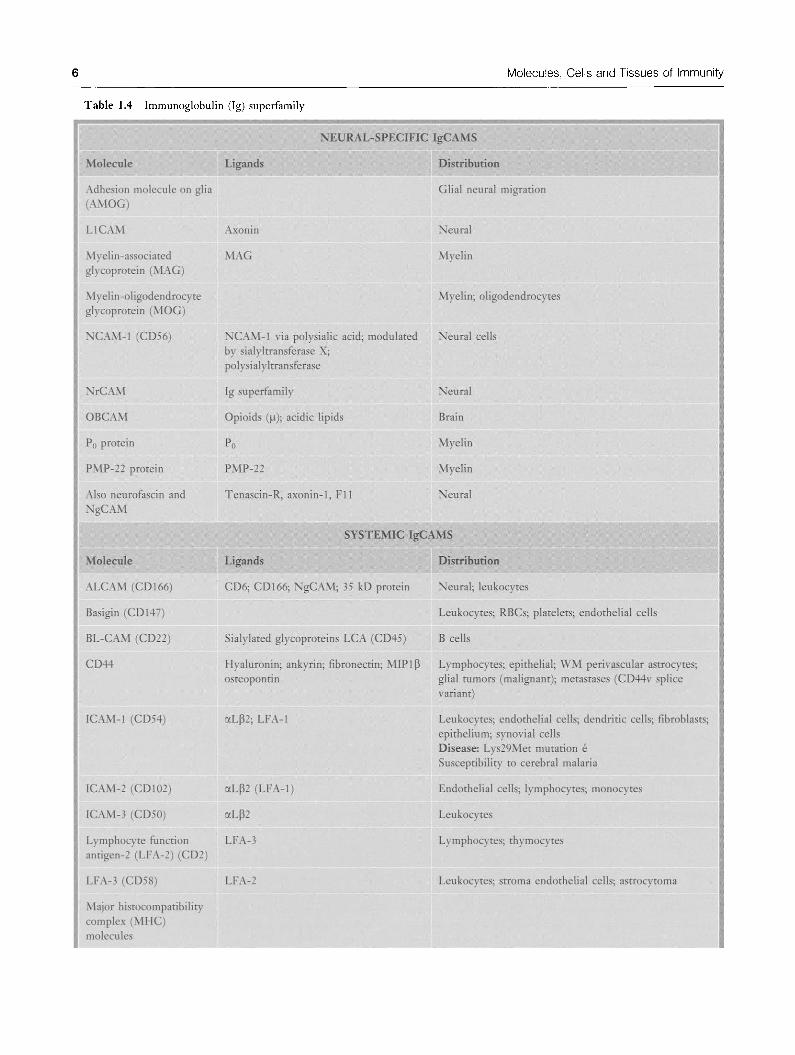
Table 1.4 Immunoglobulin (Ig) superfamily {continued)
1 Molecule Ligand^
1 MAdCAM-1 a4p7; L-selectin
PECAM(CD31) CD31;avp3
1 T cell receptor (C-region)
VCAM-1 a4pi;a4p7
NEURAL^SFECIFIC IgCAMS
Distribution 1
Mucosal endothelial cells 1
Leukocytes; synovial cells; endothelial cells j
Satellite cells; monocytes; synovial cells; activated j endothelial cells j
General characteristics: Expression: Evolutionarily ancient; widely expressed Structure
• 1 or more repeats of Ig fold of 60-100 amino acids: form sites of adhesion
• Ig domain: No somatic hypermutations o Sandwiches of 2 P sheets held together by hydrophobic
interactions • Constitutive or long-term upregulated • Anchor: Transmembrane segment and cytoplasmic tail
Interactions • Homophilic: Neural specific Ig cell adhesion molecules
(IgCAMs)
• Heterophilic: Systemic IgCAMs Adhesion
• Sites: Ig fold(s) domains (distal); fibronectin type III (Fn3) domains
• Inhibited by sialylation • Ca "'"''-independent
Functions • Neurite outgrowth • Myelination • Firm adhesion of leukocytes
« Via LFA/ICAM-1 and VLA-4/VCAM-1
Toll-like receptors (TLBs)
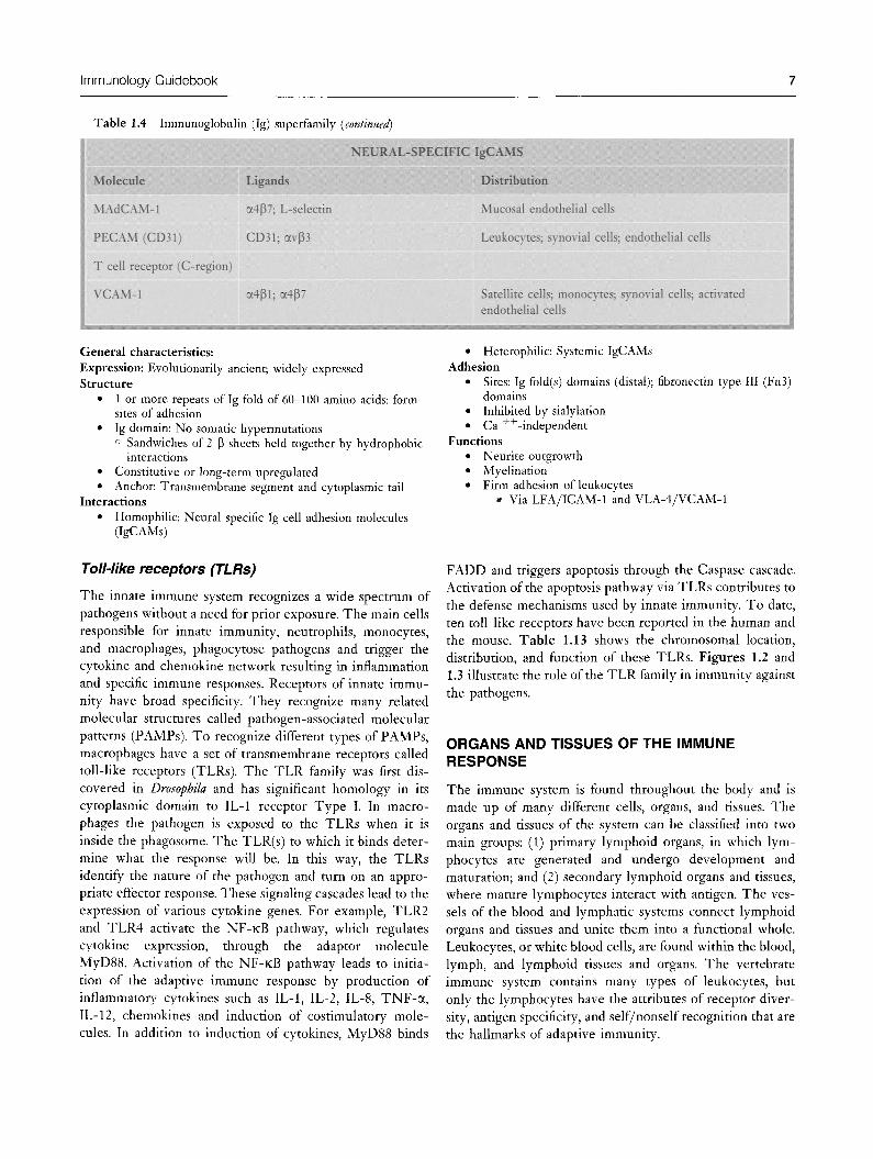
The innate immune system recognizes a wide spectrum of pathogens without a need for prior exposure. The main cells responsible for innate immunity, neutrophils, monocytes, and macrophages, phagocytose pathogens and trigger the cytokine and chemokine network resulting in inflammation and specific immune responses. Receptors of innate immunity have broad specificity. They recognize many related molecular structures called pathogen-associated molecular patterns (PAMPs). To recognize different types of PAMPs, macrophages have a set of transmembrane receptors called toll-like receptors (TLRs). The TLR family was first discovered in Drosophila and has significant homology in its cytoplasmic domain to IL-1 receptor Type I. In macrophages the pathogen is exposed to the TLRs when it is inside the phagosome. The TLR(s) to which it binds determine what the response will be. In this way, the TLRs identify the nature of the pathogen and turn on an appropriate effector response. These signaling cascades lead to the expression of various cytokine genes. For example, TLR2 and TLR4 activate the N F - K B pathway, which regulates cytokine expression, through the adaptor molecule MyD88. Activation of the N F - K B pathway leads to initiation of the adaptive immune response by production of inflammatory cytokines such as IL-1, IL-2, IL-8, TNF-a , IL-12, chemokines and induction of costimulatory molecules. In addition to induction of cytokines, MyD88 binds
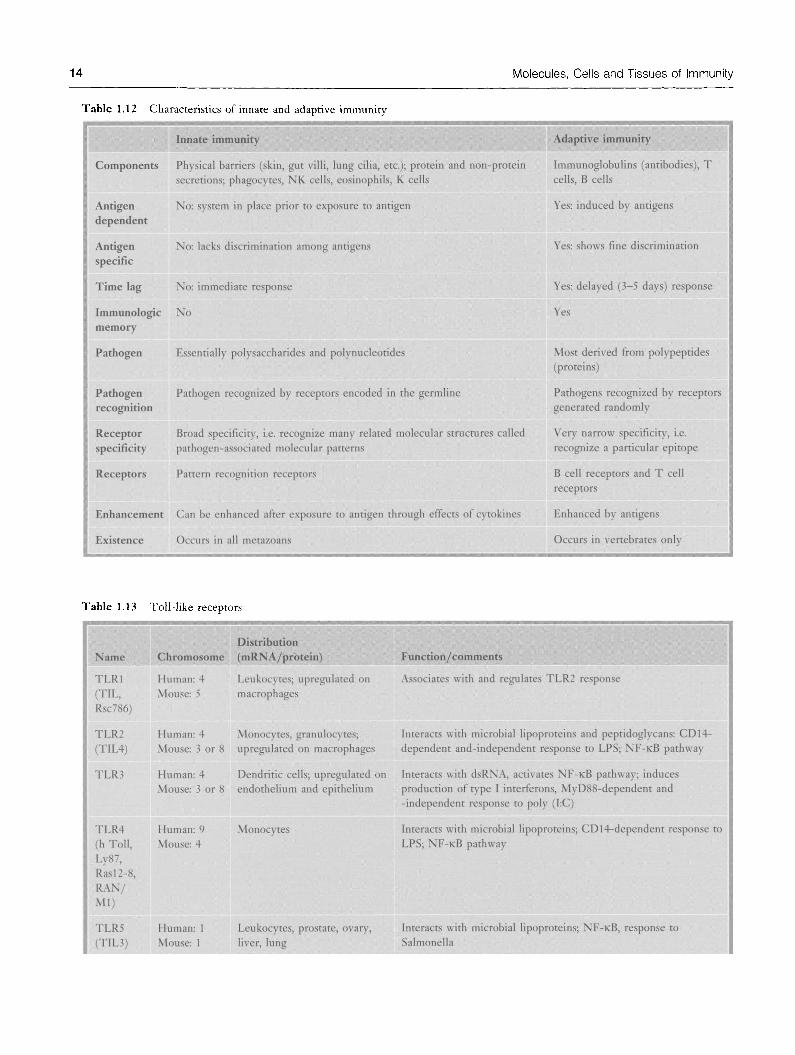
FADD and triggers apoptosis through the Caspase cascade. Activation of the apoptosis pathway via TLRs contributes to the defense mechanisms used by innate immunity. To date, ten toll-like receptors have been reported in the human and the mouse. Table LI3 shows the chromosomal location, distribution, and function of these TLRs. Figures L2 and L3 illustrate the role of the TLR family in immunity against the pathogens.
ORGANS AND TISSUES OF THE IMMUNE RESPONSE
The immune system is found throughout the body and is made up of many different cells, organs, and tissues. The organs and tissues of the system can be classified into two main groups: (1) primary lymphoid organs, in which lymphocytes are generated and undergo development and maturation; and (2) secondary lymphoid organs and tissues, where mature lymphocytes interact with antigen. The vessels of the blood and lymphatic systems connect lymphoid organs and tissues and unite them into a fimctional whole. Leukocytes, or white blood cells, are found within the blood, lymph, and lymphoid tissues and organs. The vertebrate immune system contains many types of leukocytes, but only the lymphocytes have the attributes of receptor diversity, antigen specificity, and self/nonself recognition that are the hallmarks of adaptive immunity.

Molecules, Cells and Tissues of Immunity
Table 1.5 Cadherins
1 Moleci:iles
1 Cadhetin E (1)
1 Cadherin N (2)
1 Cadherin BR (12)
1 Cadherin P (3)
1 Cadherin R (4)
1 Cadherin M (15)
1 Cadherin VE(5) (CD144)
1 Cadherin T and H (13)
1 Cadherin OB (11)
1 Cadherin 7
1 Cadherin 8
1 Cadherin KSP (16)
1 Cadherin LI (17)
1 Cadherin 18
1 Cadherin, fibroblasts 1
1 Cadherin, fibroblasts 2
1 Cadherin, fibroblasts 3
1 Cadherin23
1 Desmocollin 1
1 Desmocollin 2
1 Desmoglein 1
1 Desmoglein 2
1 Desmoglein 3
j Protocadherin 1, 2, 3, '
(19)
(20)
(21)
7 ,8,9
Ligands
H
0
M
O
P
H
I
L
I
C
Distributioii j
Epithelial 1
Neural j
Brain j
Placental j
Retinal |
Muscle 1
Epithelial j
Heart j
Osteoblast 1
Brain; kidney |
Brain |
Kidney 1
GI tract; pancreas |
CNS; small cell lung cancer 1
Fibroblasts p
Fibroblasts p
Fibroblasts J
Ear 1
Skin 1
Epithelium; mucosa; myocardium; lymph nodes |
Epidermis, tongue i
All 1
Epidermis; tongue; antibody target in pemphigus 1
General characteristics: Expression: Evolutionarily ancient; widely expressed Structure
• Extracellular domain: 5 tandem repeats; each comprising sandwich of p sheets
• Ofi:en present as dimers • Anchor: Transmembranse segment; cytoplasmic carboxy-
terminal domain
Adhesion • Homophilic: Via most distal cadherin repeats • Requires: Ca^; specific intracytoplasmic binding • Intracellular
o Cytoplasmic domain binds catenins o Catenins then bind to actin cytoskeleton
• Types

Immunology Guidebook
o Interactive with actin cytoskeleton: Cadherins N, P, R, B, E
o Desmosome-associated: Desmogleins and desmocollins « Interact with intermediate filaments ^ Location: In tight junctions
o Protocadherins '^ Homology to cadherins: Extracellular, but not
intracellular, domains Functions and diseases
• Cadherin E (1): Reduction correlates with tumor malignancy o Gynecologic malignancies
« Point mutations in tumor cells
» Somatic loss of heterozygosity common o Gastric malignancies o Susceptibility to Listeria monocytogenes infection Cadherin N: Role in establishment of left-right asymmetry Cadherin P (3): Congenital hypotrichosis with juvenile muscular dystrophy Cadherin23: Deafness Catenin pi (cadherin-associated protein): Mutations in malignancies o Colon, hepatoblastoma, pilomatricoma, ovarian
(endometrioid) Desmoglein 3: Antibody target in pemphigus
LPSA.BP
dsRJSIA
CpG DNA
Bacterial & Yeast Glycoproteins
i N F - K B
Figure L2 Role of toll-like receptors in immunity against pathogens

10 Molecules, Cells and Tissues of Immunity
Table 1.6 Other adhesion molecules
1 Molecules
1 Agrin (neural)
1 CD34
1 GlyCAM-1
1 OHgodendrocyte-1 (OMGP)
-myelin glycoprotein
Ligands
MuSK; NCAM; laminin; heparin-binding proteins; heparan sulfate proteoglycan
MAdCAM-1; L-selectin
L-selectin
Dlsmbutidn j
Nerve |
Immature lymph/ 1 myeloid j
Lymph nodes j
Myelin; j oligodendrocytes j
Table 1.7 Endothelial-leukocyte adhesion molecules
j Molecy!e(s)
1 aLp2 1 LFA-1 1 C D l l a / C D 1 8
1 aMp2 j Mac-1 1 C D U c / C D l S
1 axp2 1 P 150,95 1 C D l l c / C D l S
a4pl CD49d/CD29
1 a4p7
1 Mole€iile(s)
1 E-selectin CD62E
1 L-selectin j CD62L
Ligaiid{s)
ICAM-1, rCAM-2, ICAM-3
ICAM-l, C3bi unidentified endothelial ligand
C3bi, others?
VCAM-1
MAdCAM-1
L%aiid(s)
Sialyl-Lewis"" glycoproteins, glycoHpids (?) on P M N
GlyCAM-1, CD34 on lymph node HEV; evidence for extralymphatic ligand(s)
INTEGRINS
Dtstrtbiidoii
Lymphocytes > neutrophils; monocytes
Macrophages > monocytes; neutrophils
Macrophages; monocytes
Lymphocytes; monocytes; eosinophils
Lymphocytes
E^nlatioii
Conformational^ activated by chemoattractants
Upregulated by degranulation; activated bf T N F , chemoattractants
Upregulated by degranulation
Constitutively active (?) affinity regulation; higher expression on memory T cells
Constitutive
SELECTINS
Distribution
Activated endothelium
Resting neutrophils; most lymphocytes
E e g a k t i o 0
Upregulated by T N F , IL^l Max. expression at 4-6 hr
Constitutive, shed from surface by chemoattractant
Ftiitctioii j
Lymphocyte, granulocyte adhesion to j
resting endothelium |
Granulocyte, monocyte adhesion to | resting and activated endothelium j transmigration; C3bi binding; leukocyte j activation; phagocytosis j
Complement fixation; phagocytosis? j
Lymphocyte, monocyte adhesion to j endothelium and fibronectin j
Mucosal lymphocyte homing j
FiJ0ctioii 1
Adhesion and rolling of neutrophils; | adhesion of T cells; monocytes; slow | (3 |im/s) leukocyte rolling in vivo ]
Lymphocyte homing to lymph nodes; j sustained leukocyte rolling in vivo; 1 critical for P M N recruitment; mediates j leukocyte capture j

Immunology Guidebook 11
Table 1.7 Endothelial-leukocyte adhesion molecules [continued)
1 P-selectin 1 CD62P
1 Moleeule(s)
1 ICAM-1 1 CD54
ICAM-2 1 CD102
1 ICAM-3 1 CD50
1 VCAM-1
1 PEC AM-1 1 CD-31
1 MAdCAM-1
1 Moleciile(s)
1 PSGL-1 1 CD162
GlyCAM-1
1 CD34
1 ESL-1
1 Unidentified
PSGL-1 on myeloid cells
Ogaiid(s)
LFA-1, Mac-1
LFA-1
LFA-1
VLA-4 (a4pi integrin), a4p7 integrin
PECAM-1 (homotypic) other ligand(s)?
OC4P7 integrin, L-selectin (glycosylation-dependent)
Ligaiid(s)
P-selectin
L-selectin
L-selectin
E-selectin
L-selectin
Activated platelets; thrombin-, histamine-, cytokine-activated endothelium
Surface expressed by degranulation, transcriptional regulation by TNF, IL-i
IMMUNOGLOBUUNS
Distribution
Endothelium; smooth muscle fibroblasts; T cells
Endothelial cells
Resting T cells
Cytokine-stimulated endothelium
High on endothelium, moderate on monocytes, PMN, platelets
Mucosal venules; Peyer's patch HEV; mesenteric lymph node
Regnktioii
Constitutive, increased expression by IL-1, IFN-y
Constitutive
Constitutive
IL~1, IL-4, TNF upregulate expression, max. at 4-24 hr
Constitutive, redistribution to cell borders in confluent EC
Constitutive, different glycosylation in PP and MLN
SELECTIN LIGANDS
Distribudou
All leukocytes
Lymph node HEV; serum
Lymph node HEV; other endothelia
Murine myeloid cells
Activated endothelial cells
Regnlatioii
Constitutive; dimer
Constitutive; secreted; expression depends on lymph flow
Constitutive; expression depends on lymph flow
Constitutive
Induced by TNF in vitro, by tissue trauma in vivo
Platelet-leukocyte adhesion; leukocyte 1 rolling in vivo; critical for PMN 1 recruitment |
Function 1
Cytotoxic T-cell conjugates, leukocyte- 1 endothelial adhesion, transmigration? 1
Leukocyte adhesion to resting 1 endothelium |j
Homotypic lymphocyte aggregation |
Adhesion of monocytes, eosinophils, 1 lymphocytes to activated endothelium; 1 expressed in atherosclerotic plaques j
Critically involved in neutrophil | transmigration; endothelial monolayer j integrity; neutrophil adhesion (?) 1
Lymphocyte homing to mucosal sites; | lymphocyte rolling (contains mucin j domain) |
FBitction 1
Adhesion of myeloid cells to ! P-, L-selectin j
Homing of lymphocytes to lymph node 1 HEV (?); modulation of 1 L-selectin-dependent adhesion? 1
Homing of lymphocyte to lymph node j HEV 1
Function unknown, homologous to | FGF-receptor j
Mediates neutrophil adhesion; j L-selectin-dependent rolling j
12
Central lymphoid organs
Central lymphoid organs are requisite for the development of the lymphoid and, therefore, the immune system. These mclude the thymus, bone marrow, and bursa of Fabricus. Central lymphoid organs are also termed primary lym-
Molecules, Cells and Tissues of Immunity
phoid organs. They are sites where lymphocytes are generated. Both T and B cells originate in the bone marrow but only B cells mature there. Human T cells mature in the thymus.
Protein F \ T I W 4 i
104
••J m-m m % \ M M M ^
J* TRAI^
t MB<K1
''^^
Prolnflammatoi7 Cytokines
Figure 1.3 Pathways of toll-Hke receptors
^Table 1.8 Adhesion molecules involved in leukocyte emigration from vessels during inflammation
Rolling • Sialyl-Lewis'' • L-selectin • P-selectin • E-selectin
Stopping • p2 integrins • VLA-4(a4Pi) • ICAM-1 • VCAM-1 • a4P7 integrins • Made AM-1
^ggrcgmon and shape change • CDl lb /CDl8 (Mac~ l ) • C D l l a / C D l 8 ( L F A - l ) • P-selectin
Migration through vessel wall • p2 integrins • ICAM-1 • VCAM-1 • PECAM-1

Immunology Guidebook 13
Table 1.9 Effector mechanisms of natural immunity
1 ^^ 1 Skin
1 Ey^
1 Nasopharynx
1 Lung
1 GI tract
1 Blood and lymphoid organs
1 Other serous fluids
Component
Squamous cells, sweat
Tears
Mucus, saliva
Tracheal cilia
Columnar cells
K cells, LAK cells, NK cells
Lactoferrin, transferrin; interferons, TNF-a; fibronectin, complement; lysozyme
Functioii • 1
Desquamation; antimicrobial secretions, e.g. fatty acids; 1 flushing 1
Flushing; lysozyme 1
Flushing; lysozyme j
Mucociliary elevator; surfactants j
Stomach acidity, bile salts, fatty acids, peristalsis j
Direct and antibody-dependent cytolysis j
Iron deprivation; phagocyte activation; opsonization; 1 enhanced phagocytosis; peptidoglycan hydrolysis j
Table 1.10 (
j Neutrophils
1 Macrophages
1 Natural killer (NK)
j cells
1 K cells
I ells of natural immunity
Function
Phagocytosis, intracellular killing, inflammation and tissue damage
Phagocytosis, intracellular and extracellular killing, tissue repair, antigen presentation for specific immune response
Kill infected cells and malignant cells; LAK cells kill transformed cells and malignant cells
Recognize antibody-coated targets
Structure characteristics
Characteristic nucleus and cytoplasm
Characteristic nucleus
Also known as large granular lymphocytes (LGL); activated by IL-2 and IFN to become LAK cells
Morphologically undefined
mmummm
Membrane marker j
CD67 1
CD14 1
CD56, CD16 j
Could be NK cells (IgG), I macrophages (IgG), j eosinophils (IgE) or other cells j (IgG) J
Table 1.11 Cells of
1 Origin
1 Site of maturation
1 Antigen receptor
1 Target of binding
1 Branch of immune j response
1 MHC and antigen j presentation
adapt tive immunity
B cells
Bone marrow
Bone marrow
B cell receptor (BCR)
Soluble antigens
Antibody-mediated immune response
Class II MHC molecules
T cells 1
Bone marrow 1
Thymus 1
T cell receptor (TCR) 1
Biomolecular complex displayed at the surface of APC I
Cell-mediated and antibody-mediated immune response 1
1 Class I MHC molecules (CDS"^ T cells) and class II MHC molecules 1 (CD4+ T cells) 1
14 Molecules, Cells and Tissues of Immunity
Table 1.12 Characteristics of innate and adaptive immunity
Innate immunity
Components Physical barriers (skin, gut villi, lung cilia, etc); protein and non-protein secretions; phagocytes, KK cells, eosinophils, K cells
Antigen dependent
Antigen specific
No: system in place prior to exposure to antigen
No: lacks discrimination among antigens
Time lag No: immediate response
Immunologic No memory
Pathogen
Pathogen recognition
Receptor specificity
Essentially polysaccharides and polynucleotides
Pathogen recognized by receptors encoded in the germline
Broad specificity, i.e. recognize many related molecular structures called pathogen-associated molecular patterns
Receptors Pattern recognition receptors
Enhancement Can be enhanced after exposure to antigen through effects of cytokines
Existence Occurs in all metazoans
Adaptive immianity
ImmunoglobuHns (antibodies), T cells, B cells
Yes: induced by antigens
Yes; shows fine discrimination
Yes: delayed (3-5 days) response
Yes
Most derived from polypeptides (proteins)
Pathogens recognized by receptors generated randomly
Very narrow specificity, i.e. recognize a particular epitope
B cell receptors and T cell receptors
Enhanced by antigens
Occurs in vertebrates only
Table 1.13 Toll-like receptors
Name
TLRl (TIL, Rsc786)
TLR2 (TIL4)
TLR3
Distribtition Chromosome (mENA/protein)
Human: 4 Leukocytes; upregulated on Mouse: 5 macrophages
Human: 4 Monocytes, granulocytes; Mouse: 3 or 8 upregulated on macrophages
Function/comments
Associates with and regulates TLR2 response
Interacts with microbial lipoproteins and peptidoglycans: CDH-dependent and-independent response to LPS; NF-KB pathway
Human: 4 Dendritic cells; upregulated on Interacts with dsRNA, activates N F - K B pathway; induces Mouse: 3 or 8 endothelium and epithelium production of type I interferons, MyD88-dependent and
-independent response to poly (I:C)
TLR4 (h Toll, Ly87, Rasl2-8, RAN/ Ml)
TLR5 (TIL3)
Human: 9 Mouse: 4
Human: 1 Mouse: 1
Monocytes Interacts with microbial lipoproteins; CDl4-dependent response to LPS; N F - K B pathway
Leukocytes, prostate, ovary, liver, lung
Interacts with microbial lipoproteins; N F - K B , response to Salmonella

Immunology Guidebook 15
Table 1.13
Name
TLR6
TLR7
TLR8
TLR9
TLRIO
RP105 (CD180, Ly78)
MD-1 (Ly64, Irrp, 14/AlO)
MD-2 (Ly96)
Toll-like receptors {continued)
Chromosome
Human: 4 Mouse: 5
Human: X Mouse: X?
Human: X Mouse: X?
Human; 3 Mouse: 6
p
Human: 5 Mouse: 13
Human: 6
Distdbiitioii (mRNA/protein)
Leukocytes, ovary, lung
Spleen, placenta, lung; upregulated on macrophages
Leukocytes, lung
Leukocytes
Lymphoid tissues
Mature B cells
Mature B cells
Macrophages
Fiiiictioji/commeiits
Interacts with microbial lipoproteins; protein sequence most similar to hTLRl; associates with and regulates TLR2 response
Low similarity to other TLR family members
Receptor for CpG bacterial DNA, weakly similar to TLR3; may mediate protein-protein interaction
Most closely related to TLRl and TLR6
B cell activation; LPS recognition
Associates and regulates surface expression of RP105
Associates and regulates surface expression of TLR4; signals LPS presence
Peripheral lymphoid organs
Peripheral lymphoid organs are not required for onto- the lymph nodes, spleen, tonsils, and mucosal-associated geny of the immune response. They are sites where lymphoid tissues in which immune responses are adaptive immune responses are initiated and where lym- induced. phocytes are maintained. Peripheral lymphoid organs are Table L14 depicts the constituents of the primary and also termed secondary lymphoid organs. They include secondary lymphoid organs.
Table 1.14 Organs and tissues of the immune response :fe:%S8?««"«?^.^»i^«*»<iS«««
Pfimaty lymphoid organs Secondary lymphoid organs
Component Bone marrow, fetal liver, thymus
Proliferation and Antigen-independent differentiation
Product
Event
Immunocompetent cells (B cells and T cells)
Development and maturation of B and T cells
Spleen, lymph nodes, and mucosa-associated lymphoid tissue (MALT) including tonsils, adenoids, respiratory, genitourinary, and gastrointestinal tracts
Antigen-dependent
Effector cells (antibody-secreting plasma cells for humoral immune response and T helper and T cytotoxic cells for cell-mediated immune response)
Induction of immune response: encounter of antigens and antigen-presenting cells (AFC) with mature B and T cells, generation of effector cells, and memory cells