Slow Gating in ClC ClSlow Gating in ClC Cl--
Channels: Normal Mode AnalysisChannels: Normal Mode Analysis
Gennady V. Miloshevsky,Gennady V. Miloshevsky,1,21,2
Ahmed HassaneinAhmed Hassanein22
and Peter C. Jordanand Peter C. Jordan11
11Department of Chemistry, Brandeis University, Waltham, MA, USADepartment of Chemistry, Brandeis University, Waltham, MA, USA22School of Nuclear Engineering, Purdue University, West LafayetteSchool of Nuclear Engineering, Purdue University, West Lafayette, IN, USA, IN, USA
AbstractAbstractAllAll--atom Normal Mode Analysis (NMA) is used to explore possible mechatom Normal Mode Analysis (NMA) is used to explore possible mechanisms for slow anisms for slow gating in ClC Clgating in ClC Cl--
channels. As the channels. As the ““doubledouble--barreledbarreled””
architecture is well established architecture is well established
throughout the ClC family, both channels and transporters [1], wthroughout the ClC family, both channels and transporters [1], we use the highe use the high--resolution resolution (2.5 (2.5 ÅÅ) X) X--ray structure of an ray structure of an E. coliE. coli
ClC transporter (pdb entry 1OTS) [2] as a template, ClC transporter (pdb entry 1OTS) [2] as a template, describe it with the CHARMM22 force field and carry out standarddescribe it with the CHARMM22 force field and carry out standard
allall--mode NMA. The mode NMA. The slowest, intrinsic motions encoded in the structure are determinslowest, intrinsic motions encoded in the structure are determined by protein shape [3]. ed by protein shape [3]. Perturbing the system in either direction along the 7Perturbing the system in either direction along the 7thth
allall--atom normal mode (NM) leads to atom normal mode (NM) leads to slow relative swinging of the subunits, perpendicular to the memslow relative swinging of the subunits, perpendicular to the membrane plane. The inbrane plane. The in--plane plane swivel axis lies at the subunit interface, near the proteinswivel axis lies at the subunit interface, near the protein’’s center. The intracellular s center. The intracellular
interfacial domain is the region most affected. Here the two halinterfacial domain is the region most affected. Here the two halves of the protein oscillate, ves of the protein oscillate, separating and then nearly touching. The R and A helices executeseparating and then nearly touching. The R and A helices execute
large scale swaying, large scale swaying, alternately increasing and decreasing their cytoplasmic endsalternately increasing and decreasing their cytoplasmic ends’’
separations, motion in separations, motion in
agreement with FRET experiments [4]. The ionagreement with FRET experiments [4]. The ion--occupied intracellular pores behave as occupied intracellular pores behave as
almost rigid units. As the subunits separate, the intracellular almost rigid units. As the subunits separate, the intracellular pore tilt relative to the pore tilt relative to the
membrane plane changes notably. In contrast, the extracellular pmembrane plane changes notably. In contrast, the extracellular portion of the subunit ortion of the subunit
interface is significantly less affected, although small interfainterface is significantly less affected, although small interfacial structural changes are cial structural changes are
clearly observable. Those extracellular regions structurally affclearly observable. Those extracellular regions structurally affected by the subunitsected by the subunits’’
slow slow sway are localized at the extracellular Clsway are localized at the extracellular Cl--
pathways. As the subunits separate, these regions pathways. As the subunits separate, these regions compress, shutting the extracellular pores. As they approach, thcompress, shutting the extracellular pores. As they approach, the extracellular regions near e extracellular regions near the Clthe Cl--
conduction pathways relax, opening them.conduction pathways relax, opening them.
IntroductionIntroductionThe channels and transporters of the ClC gene family are crucialThe channels and transporters of the ClC gene family are crucial
in regulating the resting in regulating the resting membrane potential, cell volume and electrical excitability of mmembrane potential, cell volume and electrical excitability of muscle cells. The crystal uscle cells. The crystal structures of bacterial ClC Clstructures of bacterial ClC Cl--/H/H++
exchangers [2] showed a dimer with two pores, each of exchangers [2] showed a dimer with two pores, each of which is entirely contained within a single ClC Clwhich is entirely contained within a single ClC Cl--
subunit. The slow gate closes both ClC subunit. The slow gate closes both ClC pores simultaneously, with an average time on the order of seconpores simultaneously, with an average time on the order of seconds in ClCds in ClC--0, resulting in a 0, resulting in a longlong--lived inactivated state. It is difficult to obtain clues on slowlived inactivated state. It is difficult to obtain clues on slow
gating from the static gating from the static picture provided by the crystal structure of ClCpicture provided by the crystal structure of ClC--ec1. The molecular mechanism of the slow ec1. The molecular mechanism of the slow gate is largely unknown. The complex conformational changes are gate is largely unknown. The complex conformational changes are thought to be associated thought to be associated with the slowwith the slow--gating transition involving the interface between the ClC subunigating transition involving the interface between the ClC subunits. ts.
We hypothesize that the XWe hypothesize that the X--ray structure of an ray structure of an E. coliE. coli
ClCClC--ec1 transporter [2] can be used as ec1 transporter [2] can be used as a template for understanding eukaryotic ClC channels. We use alla template for understanding eukaryotic ClC channels. We use all--atom NMA to identify atom NMA to identify the intrinsic largethe intrinsic large--scale motions of the ClC biomolecule. We find that the most relescale motions of the ClC biomolecule. We find that the most relevant vant collective NM of the bacterial system for the ClCcollective NM of the bacterial system for the ClC--ec1 slow gating is the lowestec1 slow gating is the lowest--frequency frequency NM at ~2.12 cmNM at ~2.12 cm--11. Perturbation in either direction along this NM reveals the sym. Perturbation in either direction along this NM reveals the symmetric metric swinging of the subunits relative to each other. The Cswinging of the subunits relative to each other. The C--termini approach and separate, termini approach and separate,
respectively. These NMA results of ClCrespectively. These NMA results of ClC--ec1 are in agreement with the experimental FRET ec1 are in agreement with the experimental FRET data on slowdata on slow--gating in ClCgating in ClC--0 [4]. The mechanism of the slow gate in physiological 0 [4]. The mechanism of the slow gate in physiological
channels could involvechannels could involve
the strictly conserved GLU, the charge conserved ARG/LYS and the strictly conserved GLU, the charge conserved ARG/LYS and other residues in the extracellular mouth that sterically block other residues in the extracellular mouth that sterically block ClCl--
pathways. pathways.
Computational ModelComputational Model
We use the highWe use the high--resolution (2.5 resolution (2.5 ÅÅ) X) X--ray structure of an ray structure of an E. coliE. coli
ClCClC--ec1 transporter (ec1 transporter (pdbpdb
entry 1OTS) [2] with four entry 1OTS) [2] with four ClCl--
at the binding sites in the pores and 427 crystallographic at the binding sites in the pores and 427 crystallographic waters. Protein hydrogens were added by using our MCICP code, crwaters. Protein hydrogens were added by using our MCICP code, creating 13,524 protein eating 13,524 protein atoms. The total number of atoms in the molecular system is 14,8atoms. The total number of atoms in the molecular system is 14,809.09.
The molecular system was described with the allThe molecular system was described with the all--hydrogen CHARMM22 topology and hydrogen CHARMM22 topology and parameter set, with NMA calculations carried out in vacuum.parameter set, with NMA calculations carried out in vacuum.
To remove To remove stericsteric
clashes and to relax the molecular system, ~2,000 minimization clashes and to relax the molecular system, ~2,000 minimization steps steps were done using steepest descent with a random step length; finawere done using steepest descent with a random step length; finally, the molecular system lly, the molecular system was well tuned via conjugate gradient with guaranteed descent [5was well tuned via conjugate gradient with guaranteed descent [5]. All degrees of freedom ]. All degrees of freedom (bond lengths, bond angles, torsion and improper torsion angles)(bond lengths, bond angles, torsion and improper torsion angles)
in the protein and waters in the protein and waters were variable. were variable.
The geometry of the molecular system with an absolute largest gThe geometry of the molecular system with an absolute largest gradient component of radient component of <5<5··1010--1010
kcal molkcal mol--11
ÅÅ--11
was located. This extremely precise minimum is required when was located. This extremely precise minimum is required when performing NMA on large protein structures where even small resiperforming NMA on large protein structures where even small residual derivatives can lead dual derivatives can lead to serious errors in the calculated to serious errors in the calculated eigendirectionseigendirections. Between crystal and minimized ClC. Between crystal and minimized ClC--ec1 ec1 structures the RMSD for thestructures the RMSD for the
CCαα
is 1.69 is 1.69 ÅÅ; that; that
for all 14,809 atoms is 2.1 for all 14,809 atoms is 2.1 ÅÅ. These are . These are small indicating that the minimized and crystal structures are hsmall indicating that the minimized and crystal structures are highly similar. ighly similar.
Standard allStandard all--mode NMA was carried out using the DSTEVR mode NMA was carried out using the DSTEVR eigensolvereigensolver
from the from the
LAPACK library and highly optimized BLAS routines for performingLAPACK library and highly optimized BLAS routines for performing
basic vector and basic vector and matrix operations; global translational and rotational matrix operations; global translational and rotational NMsNMs
were removed using the were removed using the EckartEckart
conditions. For the bonded and nonconditions. For the bonded and non--bonded energy terms both gradient and Hessian were bonded energy terms both gradient and Hessian were calculated analytically; for other energy terms (angle, dihedralcalculated analytically; for other energy terms (angle, dihedral, improper and , improper and UreyUrey--
Bradley) a fourthBradley) a fourth--order finiteorder finite--difference approximationdifference approximation
was used.was used.
ConclusionsConclusions
When the intracellular parts swing away, the extracellular poreWhen the intracellular parts swing away, the extracellular pore
regions compress, shutting the regions compress, shutting the
extracellular pores. In the FRET experiments [4] the closure of extracellular pores. In the FRET experiments [4] the closure of the slow gate was also accompanied by the slow gate was also accompanied by a physical separation between the Ca physical separation between the C--termini of the two subunits. When the intracellular parts approatermini of the two subunits. When the intracellular parts approach ch each other, the extracellular regions near the each other, the extracellular regions near the ClCl--
conduction pathways relax, opening the extracellular conduction pathways relax, opening the extracellular pores. This is also in agreement with the FRET experiments [4] wpores. This is also in agreement with the FRET experiments [4] where opening of the slow gate was here opening of the slow gate was accompanied by movements decreasing the distance between the C taccompanied by movements decreasing the distance between the C termini. ermini.
AcknowledgementAcknowledgement
Work supported by a grant Work supported by a grant from the National Institutes from the National Institutes of Health, GMof Health, GM--28643. 28643.
ReferencesReferences1. C. 1. C. Miller. Miller. Nature.Nature.
440440, 484, 484--489 (2006).489 (2006).2. 2. R. Dutzler et al., R. Dutzler et al., Science.Science.
300300, 108, 108--112 (2003). 112 (2003). 3. 3. M. Lu & J. Ma. M. Lu & J. Ma. BiophysBiophys. J.. J.
8989, 2395, 2395--2401 (2005). 2401 (2005). 4. 4. E. A. E. A. BykovaBykova
et al., et al., Nat. Nat. StructStruct. Biol.. Biol.
1313, 1115, 1115--1119 (2006).1119 (2006).5. 5. W. W. Hager & H. Zhang, W. W. Hager & H. Zhang, SIAM J. SIAM J. OptimOptim. . 1616, , 170170--192 (2005).192 (2005).6. 6. G. V. Miloshevsky & P. C. Jordan. G. V. Miloshevsky & P. C. Jordan. BiophysBiophys. J.. J.
8686, 825, 825--835 (2004).835 (2004).7. T.7. T.--Y. Chen & T.Y. Chen & T.--C. Hwang. C. Hwang. PhysiolPhysiol. Rev. . Rev. 8888, 351, 351--387 (2008). 387 (2008). 8. G. V. Miloshevsky & P. C. Jordan. 8. G. V. Miloshevsky & P. C. Jordan. Structure.Structure.
1414, 1241, 1241--1249 (2006).1249 (2006).
77--th Normal Mode of the ClCth Normal Mode of the ClC--ec1 systemec1 system
View from within a membrane plane: View from within a membrane plane: ((aa) the minimized ) the minimized system; (system; (bb) and () and (cc) displacement along the 7) displacement along the 7thth
allall--atom NM in atom NM in the the ““positivepositive””
and and ““negativenegative””
directions. The figures are directions. The figures are
generated using our MCICP code.generated using our MCICP code.
Main observations:Main observations:
Perturbation of the ClCPerturbation of the ClC--ec1 system along the 7ec1 system along the 7thth
allall--atom atom NM in either direction (NM in either direction (bb
and and cc) leads to the slow swinging ) leads to the slow swinging motion of the motion of the ClCClC
subunits relative to each other, subunits relative to each other,
perpendicular to a membrane plane. The swivel axis lying in a perpendicular to a membrane plane. The swivel axis lying in a membrane plane is located at the interface between the membrane plane is located at the interface between the ClCClC
subunits close to the proteinsubunits close to the protein’’s center. Black arrows indicate s center. Black arrows indicate the directions of displacement of the the directions of displacement of the ClCClC
domains.domains.
The intracellular portion of the interface between subunits A The intracellular portion of the interface between subunits A and B (highlighted by a gray oval) is the region most affected and B (highlighted by a gray oval) is the region most affected by this sway motion. Here the two protein subunits swing by this sway motion. Here the two protein subunits swing away from each other (away from each other (bb) and then approach closely () and then approach closely (cc). The ). The intracellular ends of the H and I helices and their connecting intracellular ends of the H and I helices and their connecting polypeptide loops separate and move toward each other polypeptide loops separate and move toward each other
(double(double--headed cyan arrow in the intracellular region). The R headed cyan arrow in the intracellular region). The R and A helices undergo large scale swaying, an effect that and A helices undergo large scale swaying, an effect that
increases and decreases the distance between their increases and decreases the distance between their
cytoplasmiccytoplasmic
ends. ends.
The geometry of the intracellular pores is nearly unaffected. The geometry of the intracellular pores is nearly unaffected. The configuration of the R, D and other helices surrounding The configuration of the R, D and other helices surrounding the pore undergoes minor changes. These pores with their the pore undergoes minor changes. These pores with their
content sway in space as almost rigid units. As the content sway in space as almost rigid units. As the ClCClC
subunits swing away, pore tilt with respect to the membrane subunits swing away, pore tilt with respect to the membrane plane changes substantially. Concurrently the intracellular plane changes substantially. Concurrently the intracellular
pore between the two pore between the two ClCl--
ions constricts when perturbed in ions constricts when perturbed in the the ““negativenegative””
direction and expands when perturbed in the direction and expands when perturbed in the ““positivepositive””
direction. direction.
In contrast, the extracellular portion of the interface In contrast, the extracellular portion of the interface
between subunits A and B is significantly less affected, between subunits A and B is significantly less affected,
although small structural changes at the subunitalthough small structural changes at the subunit’’s interface s interface are clearly observed. The extracellular regions (shown as are clearly observed. The extracellular regions (shown as
green triangles) structurally affected by the slow swinging green triangles) structurally affected by the slow swinging
motion of the motion of the ClCClC
subunits are localized in the vicinity of the subunits are localized in the vicinity of the extracellular extracellular ClCl--
pathway. When the pathway. When the ClCClC
subunits swing away subunits swing away ((bb), these regions compress (note rearrangements of the ), these regions compress (note rearrangements of the
extracellular parts of peripheral helices at triangle marks extracellular parts of peripheral helices at triangle marks
relative to the helices I), shutting the extracellular pores. relative to the helices I), shutting the extracellular pores.
When the When the ClCClC
subunits approach each other (subunits approach each other (cc), the ), the
extracellular regions near the extracellular regions near the ClCl--
conduction pathways relax, conduction pathways relax, opening the extracellular pores. opening the extracellular pores.
In summary, the three regions that are mainly affected in the In summary, the three regions that are mainly affected in the slowslow--gating are highlighted by the oval and the triangles. gating are highlighted by the oval and the triangles.
Our NMA data suggest that the structural changes in the vicinitOur NMA data suggest that the structural changes in the vicinity of the extracellular y of the extracellular ClCl--
pathways pathways block the extracellular pores. We find that R147 and E148, hingeblock the extracellular pores. We find that R147 and E148, hinged on strictly conserved G146 and d on strictly conserved G146 and G149, move during slow gating. Side chains of R147 together withG149, move during slow gating. Side chains of R147 together with
Q61, N62 and M65 can Q61, N62 and M65 can stericallysterically
block the extracellular pore and mouth. This finding suggests hoblock the extracellular pore and mouth. This finding suggests how largew large--scale structural changes are scale structural changes are transducedtransduced
to the outer pore vestibule and the associated fast glutamate gto the outer pore vestibule and the associated fast glutamate gate [7]. This conformational ate [7]. This conformational change can explain experimentally observed changes in the accesschange can explain experimentally observed changes in the accessibility of the pore to ibility of the pore to extracellularlyextracellularly
applied methane applied methane thiosulfonatethiosulfonate
reagents during slow gating [7]. Our results also suggest that reagents during slow gating [7]. Our results also suggest that fast and fast and slow gates can be coupled as both involve a strictly conserved gslow gates can be coupled as both involve a strictly conserved glutamate.lutamate.
AllAll--atom NMA only provides the initial direction of the largeatom NMA only provides the initial direction of the large--scale slow transitions in ClCscale slow transitions in ClC--ec1. It ec1. It cannot elucidate any other distinct, stable cannot elucidate any other distinct, stable conformation(sconformation(s) in which the ) in which the ClCClC
protein might be trapped protein might be trapped for seconds. Such longfor seconds. Such long--lived stable lived stable state(sstate(s) can be identified by all) can be identified by all--atom Monte Carlo Normal Mode atom Monte Carlo Normal Mode Following (MCFollowing (MC--NMF) [8] along the lowNMF) [8] along the low--frequency frequency NM(sNM(s). Issues to be investigated are: 1) which ). Issues to be investigated are: 1) which structural rearrangements take place on the gating pathway; 2) hstructural rearrangements take place on the gating pathway; 2) how and why both pores can be locked ow and why both pores can be locked in the closed state for seconds; 3) how and why some specific poin the closed state for seconds; 3) how and why some specific point mutations affect the slow gate.int mutations affect the slow gate.
Minimized ClCMinimized ClC--ec1 structureec1 structureView from within a membrane plane in a View from within a membrane plane in a cylinder representation. Helices H and I cylinder representation. Helices H and I at the interface between at the interface between ClCClC
subunits are subunits are shown in dark magenta (subunit A) and shown in dark magenta (subunit A) and dark cyan (subunit B). Helices R and D dark cyan (subunit B). Helices R and D are shown for both subunits in red and are shown for both subunits in red and green, respectively. The remaining green, respectively. The remaining
helices of subunit A are colored in helices of subunit A are colored in
brown, and those of subunit B are in brown, and those of subunit B are in
blue. Helices A positioned close to blue. Helices A positioned close to
helices R of the other subunit are helices R of the other subunit are
labeled. Polypeptide loops of subunits A labeled. Polypeptide loops of subunits A and B are colored in gray and black, and B are colored in gray and black,
respectively. For clarity, crystallographic respectively. For clarity, crystallographic water molecules are not displayed. Four water molecules are not displayed. Four ClCl--
ions (green spheres) at their binding ions (green spheres) at their binding sites in the curvilinear pores are shown. sites in the curvilinear pores are shown.
Residues forming the extracellular pores viewed from within the Residues forming the extracellular pores viewed from within the membrane: membrane: ((aa) minimized ClC) minimized ClC--ec1; (ec1; (bb) and () and (cc) displacement along the 7) displacement along the 7thth
allall--atom NM in atom NM in ““positivepositive””
and and ““negativenegative””
directions. Onlydirections. Only
the subunit A pore is illustrated. Near the central binding sitethe subunit A pore is illustrated. Near the central binding site
it is bordered by G146, R147, A358, F190 and E148 [6], which blit is bordered by G146, R147, A358, F190 and E148 [6], which blocks it. The side ocks it. The side chains bounding the porechains bounding the pore’’s extracellular mouth are G315, G316, F317, V236 and thes extracellular mouth are G315, G316, F317, V236 and the
guanadiniumguanadinium
group of R147 [6]. R147 and E148 are shown in native colors; thgroup of R147 [6]. R147 and E148 are shown in native colors; their eir backbone segments are effectively hinged to their adjacent stricbackbone segments are effectively hinged to their adjacent strictly conserved residuestly conserved residues
((G146 and G149 (in pink)). The others are shown in different coloG146 and G149 (in pink)). The others are shown in different colors: A358 rs: A358 ––
orange; orange; A189 and F190 A189 and F190 ––
violet; G315, G316, F317, N318 and L319 violet; G315, G316, F317, N318 and L319 ––
brown; E235 and V236 brown; E235 and V236 ––
blue; Q61, N62, M65 and G66 blue; Q61, N62, M65 and G66 ––
green. The pore is indicatedgreen. The pore is indicated
by a thick blue line. by a thick blue line. Main observations:Main observations:
In perturbation to an RMSD of 3.5 In perturbation to an RMSD of 3.5 ÅÅ
along the 7along the 7--th NM in the th NM in the ““positivepositive””
direction, direction, the extracellular pores become the extracellular pores become stericallysterically
blocked by R147, F317 and V236 side chains blocked by R147, F317 and V236 side chains ((bb), which are then almost in direct contact. In perturbation ), which are then almost in direct contact. In perturbation in the in the ““negativenegative””
direction, the extracellular pores open wide (direction, the extracellular pores open wide (cc).).
Near the extracellular mouth the pore is blocked by Q61, N62, NNear the extracellular mouth the pore is blocked by Q61, N62, N318, L319 and E235 side chains (318, L319 and E235 side chains (bb). Side chains Q61, N62 and M65 undergo large scale motion block). Side chains Q61, N62 and M65 undergo large scale motion blocking ing and unblocking the extracellular mouths (and unblocking the extracellular mouths (bb
and and cc). ).
The side chain of R147, its backbone segment effectively hingedThe side chain of R147, its backbone segment effectively hinged
to the neighbouring G146, to the neighbouring G146, alternately approaches and separatesalternately approaches and separates
from thefrom the
opposed F317 and V236 (opposed F317 and V236 (bb
and and cc); ); F317 rotatesF317 rotates
slightly. E148, while similarly hinged to the adjacent G149, is slightly. E148, while similarly hinged to the adjacent G149, is far less affected far less affected ––
its side chain remains hydrogen bonded. In this extracellular rits side chain remains hydrogen bonded. In this extracellular region there areegion there are
numerous glycines, G146, G149, G315, G316 and G66, all promotingnumerous glycines, G146, G149, G315, G316 and G66, all promoting
substantial backbone flexibility. substantial backbone flexibility.
Residues S107, Y445, E148 and R147 lining the pore are displayedResidues S107, Y445, E148 and R147 lining the pore are displayed. The side chain of E148 blocks the pore on the extracellular si. The side chain of E148 blocks the pore on the extracellular side de and side chains of S107 and Y445 constrict the pore on the intraand side chains of S107 and Y445 constrict the pore on the intracellular side [6]. The figure was generated using our MCICP codecellular side [6]. The figure was generated using our MCICP code..
Extracellular Pores of the Perturbed ClCExtracellular Pores of the Perturbed ClC--ec1 system, in displacement along the 7ec1 system, in displacement along the 7--th Normal Modeth Normal Mode
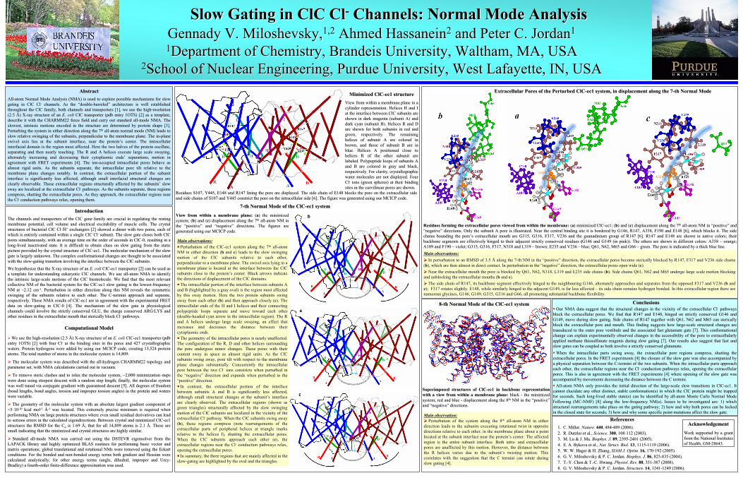
Main observation:Main observation:
Perturbation of the system along the 8Perturbation of the system along the 8thth
allall--atom NM in either atom NM in either
direction leads to the subunits executing rotational twist in opdirection leads to the subunits executing rotational twist in opposite posite directions relative to each other, in the membrane plane about adirections relative to each other, in the membrane plane about a
point point located at the subunitlocated at the subunit
interface nearinterface near
the proteinthe protein’’s center. The affected s center. The affected region is the entire subunitregion is the entire subunit
interface. Both intrainterface. Both intra--
and extracellular and extracellular pores are unaffected by this motion. However, the distance betwepores are unaffected by this motion. However, the distance between en the R helices varies due to thethe R helices varies due to the
subunitsubunit’’s twisting motion. This s twisting motion. This
correlates with the suggestion that the C termini can rotate durcorrelates with the suggestion that the C termini can rotate during ing slow gating [4].slow gating [4].
Superimposed structures of ClCSuperimposed structures of ClC--ec1 in backbone representation ec1 in backbone representation
with a view from within a membrane plane: with a view from within a membrane plane: black black --
the minimized the minimized system; red and blue system; red and blue --
displacement along the 8displacement along the 8thth
NM in the NM in the ““positivepositive””
and and ““negativenegative””
directions. directions.
88--th Normal Mode of the ClCth Normal Mode of the ClC--ec1 systemec1 system
Recommended