Remote Gamma Imaging of High Dose Environments
Copyright © BIL Solutions Ltd 2006. The copyright in this document is wholly vested in BIL Solutions Ltd who reserve all
intellectual property rights. Copying this publication in part or whole without the prior written agreement of BIL Solutions Ltd is
expressly forbidden.
Paul Griffiths

BIL Solutions Ltd
(formerly BNFL Instruments Ltd)
BIL Solutions Ltd have developed and operated the RadScan®:800 over the last 11 years:
• remotely operated• highly collimated• scanning • LRGS detection system• packed with data analysis features.
Specialist Measurement Service offers a comprehensive service
The primary function of the RadScan® :800 is:
• To provide colour contour maps showing the spread and intensity of radiation over an area.

Typical Applications
• In Decommissioning :-– Identifying the gamma hotspots in a plant
– Prioritising removals
– Deciding where shielding should go– Help determine man access routes / dose uptake minimisation
• Incident investigation :-– Extent of spillage / finding “lost” activity
• Operational Plants:-– Identify hold ups (e.g. blocked pipes)– Waste sorting operations
• Used most often :-– Where man access is limited by dose or accessibility– Large areas need to be surveyed
RadScan®:800 Components
Detection head Pan / Tilt UnitControl
Electronics Tripod
Workstation Coupling
Can be constructedby a single individual

RadScan®:800 Detection Head
4o
γ
Laser Range Finder
NaI Detector
CCD Camera

RadScan®:800 Detection Head
LaserRangeFinder
Computer Controlled Zoom Lens
DetachablePerspex
Panel
2 Detachable Collimatoro
Colour CCDVideo Camera
4 Detachable Collimatoro
Cast Aluminium Housing
Lifting Handle
Electrical Connector
9 Collimatoro
CsI Detector
Photodiode and Amplifier
Tungsten Collimator

RadScan®:800 Collimation
– Possible angles of view (of detector) are:
• 4° as standard
• 3° with a screw-on collimator extension
• 2° with a screw-in collimator insert.
– Collimator additions:
• screw-in background correction plug.
– The collimators offer a range of performance as follows:
– The 4° collimator gives standard spatial resolution, maximum sensitivity to a point source and minimum scan duration.
– The 3° collimator gives medium spatial resolution, medium sensitivity to a point source and intermediate scan duration.
– The 2° collimator gives the highest spatial resolution, the lowest sensitivity and hence the longest scan duration.

RadScan®:800 Survey
• Setting up a survey is simple. Identify extent of survey area and select a dwell time.
• The head moves from top LH corner to bottom RH corner of selected scan area in small increments.
• At each dwell position gamma rays are detected. A gamma energy spectrum is formed for each dwell position.

Gamma spectrum acquired
• Powerful function to select an energy region of interest for the radiometric
overlay display – Cs-137, Co-60, Pu, Am-241 etc…
Energy ROI
Scatter
Cs-137 662keV photopeak
Energy
Counts
~30keV ~1500keV

Predominant Isotopes Measured
• 661.6 keV from Cs-137– dominates in decommissioning activities
• 1173 & 1332 keV from Co-60– often dominates in reactor buildings
• 59.6 keV from Am-241– often dominant in Pu bearing facilities
• 186 keV from U-235– 1001 keV from U-238 is very weak

RadScan®:800 Operating Modes
Counts Per Second at the
detector originating from
within the collimators Field
of View
Laser Range Finder
From Inferred Point
Source in isolation at
1m
Interactive or Count Mode

Radiometric overlay image

Case Studies
Presentation of three case studies:
1. Clean Up: RadScan®:800 used to identify remaining Cs-137 hotspots in a high dose environment
2. Blockage Detection: RadScan®:800 used to identify blockages in operational processes
3. Advanced ALARP planning: RadScan®:800
used to identify major radiation sources in a proposed work place at height during the early design stage of a building

Case Study 1: Clean Up
The problem: To identify remaining Cs-137 contamination on a floor area following clean-up operations
Case Study 1- Clean Up
• High dose area (10s of mSv/hr)
– The ability to select an energy ROI allows scatter components to be reduced
• Contaminated environment
– The RadScan®:800 was dressed in its environmental suit
• No access for personnel
– The RadScan®:800 was located on a simple bespoke stand and moved into position using a Remotely Operated Vehicle
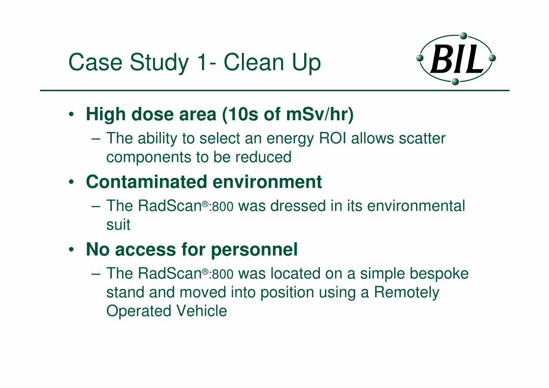
Case Study 1- Clean Up
Composite image showing a number of RadScan®:800 frames

Case Study 1- Clean Up

Case Study 1- Clean Up

Case Study 1- Clean Up

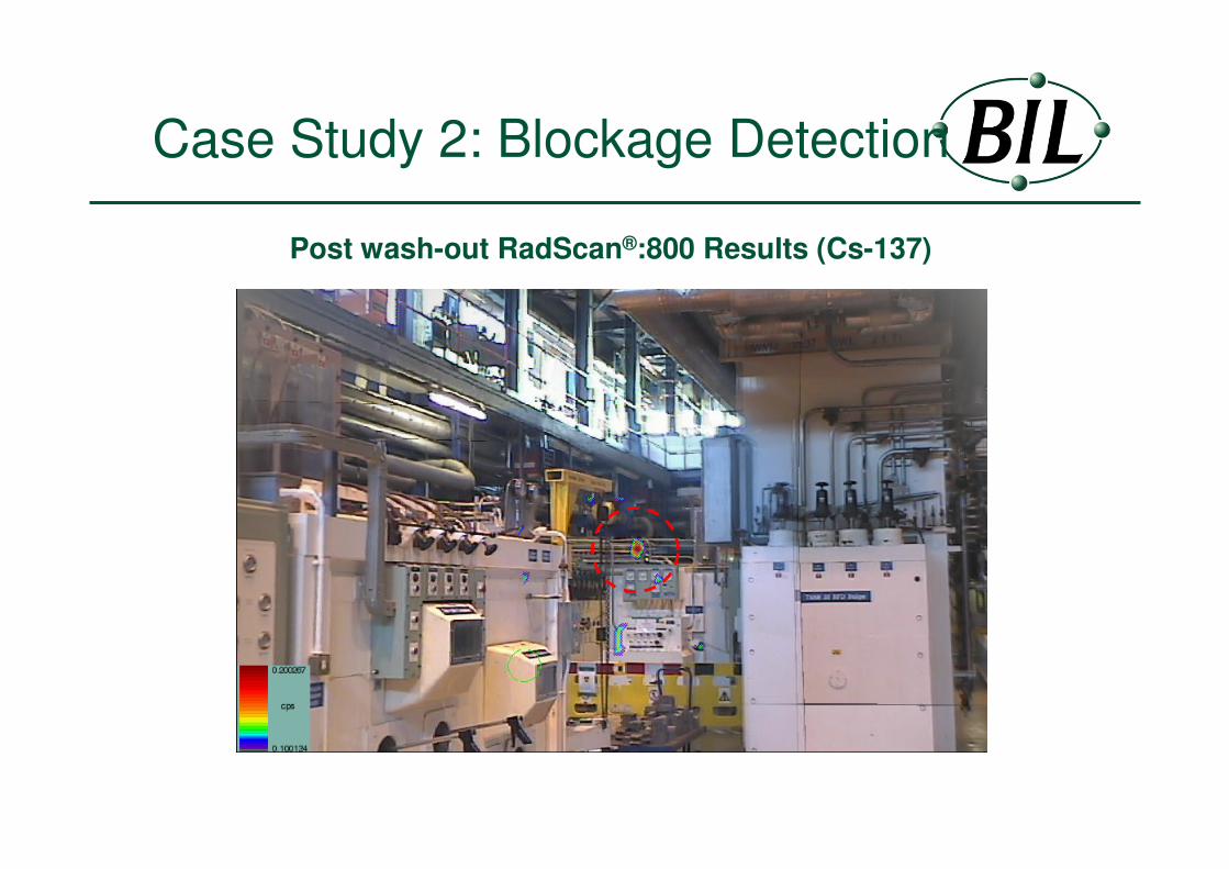
Case Study 2: Blockage Detection
The problem: To identify process material blockage within an operational plant
Assessed HP&S Dose Rate
12mSv/hr

Case Study 2: Blockage Detection
Assessed HP&S Dose Rate
>1Sv/hr

Case Study 2: Blockage Detection
RadScan®:800Count Rate
20cps

Case Study 2: Blockage Detection
RadScan®:800Count Rate
4000cps

Case Study 2: Blockage Detection
Initial RadScan®:800 Results (Cs-137)
Case Study 2: Blockage Detection
Post wash-out RadScan®:800 Results (Cs-137)

Case Study 3:
Advanced ALARP planningThe problem: A structure is to be erected in a high dose environment. The dose to workers at height is to be considered at the early design stage.

Case Study 3 –
Advanced ALARP planning
• Deploying RadScan®:800 at a point in space
– Identify highest dose rate throughout the volume
– Deploy the RadScan®:800 on a Mobile Elevated Work
Platform at the point of highest dose rate
• Dose contributors from buildings
– Perform wide area ~360-degree surveying to identify all
contributors to the dose rate

Case Study 3 –
Advanced ALARP planning
RadScan®:800 located on an Elevated Platform

Case Study 3 –
Advanced ALARP planning
RadScan®:800 survey showing the extent of Cs-137 down the side of a building

RadScan®:800 Performance
Limits of Detection – Gamma Emitters at 1 metre
– < 40 kBq Cs-137– < 100 kBq Co-60– < 100 kBq Am-241– 1 gram U-235– 1.6 kg U-238

RadScan®:800 Summary
The use of RadScan®800 has:
• Increased levels of safety (ALARP planning)
• Identified unknown sources of radiation
• Increased personnel working times
• Help plan decommissioning strategies
• Quantified the success of clean-up projects
• Identified blockages and leaks in process plant

BIL Solutions Ltd
RadScan®800 - A remote gamma surveying tool for the majority of environments
Ingamells W, Preliminary Investigations into the Use of a Gamma Scanner for Safeguards Applications at the Dounreay Nuclear Establishment, Proc. Of the 24th Annual Safeguards Research and Development Association (ESARDA) Symposium on Safeguards and Nuclear Materials Management, Luxemburg, 2002.
Fisher A and Chard P, Use of a gamma-imaging device to optimise measurement of uranium hold-up, Proc. Of the 25th Annual Safeguards Research and Development Association (ESARDA) Symposium on Safeguards and Nuclear Materials Management, Stockholm, 2003.
Hughes K. and Mottershead G, Gamma Imaging as a Complementary Technique to Health Physics Monitoring, The 7th ALARA meeting, Arnhem, Holland, 2003.
Further reading:
Recommended