1046 JACC Vol. 5. No.5May 1985:1046-54
Percutaneous Transluminal Coronary Angioplasty for the Treatmentof Variant Angina
THIERRY CORCOS, MD, PAUL R. DAVID, MD, FACC, MARTIAL G. BOURASSA, MD, FACC,
PERE GUITERAS VAL, MD, JACQUES ROBERT, MD, LUIS A. MATA, MD,
DAVID D. WATERS, MD, FACC
Montreal, Quebec, Canada
Among 268 patients undergoing percutaneous transluminal coronary angioplasty between February 1980andJanuary 1983, a total of 21 patients had variant angina,documented before angioplasty in 14 and after angioplasty in 7. Before angioplasty, all 21 patients had restangina and 17 also had effort angina; single vesselcoronary artery disease with 60 to 95% stenosis was presentin all patients and the left anterior descending coronaryartery was involved in all but 3 patients. Coronary angioplasty was successful in 19 patients (90%). Eight ofthe 19patients remained symptom-free without coronaryrestenosis after successful angioplasty; in the other 11patients, angina reappeared within 4 months, usually inassociation with restenosis. Of the nine patients withcoronary restenosis, six had repeat angioplasty (fivesuccessful procedures and one failure), two received medicaltherapy and one underwent coronary bypass surgery.Patients in whom calcium channel antagonists were discontinued immediately after angioplasty had an exceed-
In 1959, Prinzmetal et al. (I) described a clinical syndromecharacterized by angina at rest with concomitant transientST segment elevation on the electrocardiogram. They suggested that "this variant form of angina pectoris resultedfrom temporary occlusion of a large diseased artery due toa normal increase in the tonus of the vascular wall." A fewyears later, Gensini et al. (2) documented spontaneouslyoccurring coronary spasm associated with chest pain in apatient with angina pectoris and mild coronary artery disease. The mechanism of coronary artery spasm is unknown.Whether it represents a local abnormality of the arterial wall
From the Department of Medicine . Montreal Heart Institute and theUniversity of Montreal Medical School . Montreal . Quebec. Canada . Thiswork was supported in part by the J.-L. Levesque Foundation. Montreal.Quebec. Canada and the Montreal Heart Institute Research Fund. Montreal.Quebec. Canada. Manuscript received August 20. 1984; revised manuscriptreceived November 19. 1984. accepted December 5. 1984.
Address for reprints: Martial G. Bourassa. MD. Montreal Heart Institute. 5000 East Belanger Street. Montreal. Quebec. HIT IC8. Canada.
Ii) 1985 by the American College of Cardiology
ingly high coronary restenosis rate (8 [80%] of 10 successful attempts), but when calcium antagonists werecontinued for an average of 6 ± 4 months after angloplasty, the restenosis rate was low (3 [21%] of 14 successful attempts).
After a mean (± SD) follow-up period of 33 ± 13months, I patient had died and the 20 others (95%) weresymptom-free; among these 20, 15 patients (75%) hadbeen taking no antianginal drugs for more than 1 year,2 still received calcium channel antagonists and 3 hadhad coronary bypass surgery. Repeat coronary arteriography performed 14 ± 7 months after angioplasty inthe 17 patients without angioplasty-related infarction orsurgery showed 50% or less coronary stenosis in 13 patients. Thus, coronary angioplasty appears to be an effective alternative therapy for patients with variant angina and organic coronary stenosis.
(J Am Coil Cardiol 1985;5:1046-54)
or a normal or exaggerated vasomotion in response to various stimuli remains to be established (3).
The coronary anatomy of patients with variant angina isquite variable , ranging from angiographically normal coronary arteries (4) to severe multivessel disease (3,5-7) . However, the majority of patients with variant angina have somedegree of organic stenosis in the coronary artery susceptibleto spasm, and when present, the organic stenosis is the sitein that vessel where the spasm occurs in about 90% of cases(8) .
The respective roles of spasm and fixed obstruction inoccluding a coronary artery may vary from patient to patient(9) . According to MacAlpin (10) , significant coronary obstruction , even complete occlusion, can be dynamicallyproduced by physiologic degrees of vasoconstriction whensuperimposed on even "insignificant" atheromatous stenosis. Although an inappropriate active constriction appearsmore likely than a physiologic one (II), for the same stimulus resulting in a similar muscle contraction, the higher
0735-1097/85/$3 .30
JACC Vol. 5. No.5May 1985:1046--54
CORCOS ET AL.CORONARY ANGIOPLASTY FOR VARIANT ANGINA
1047
the degree of coronary stenosis, the higher the degree ofluminal reduction and impairment of flow. Under these conditions, eliminating the organic stenosis would theoreticallyprevent vasoconstrictive occlusion of the artery and wouldrelieve symptoms in patients with variant angina. Therefore,we considered percutaneous transluminal coronary angioplasty as an alternative treatment for patients with variantangina and significant coronary artery stenosis. In this report, we describe immediate and long-term results of coronary angioplasty and discuss the implications of our findings.
MethodsPatients. Between February 1980 and January 1983, a
total of 268 patients underwent coronary angioplasty at theMontreal Heart Institute. Twenty-one of these patients wereconsidered to have variant angina. This was defined as spontaneous angina at rest with transient ST segment elevationof at least 0.2 mY, relieved by nitroglycerin in less than 5minutes and without subsequent evidence of myocardialnecrosis. It was also diagnosed when these events wereprovoked by ergonovine administration.
The 21 patients were aged 36 to 69 years (mean ± SO49 ± 10), all but 4 were men, all had rest angina for lessthan 8 months (mean ± SO 2 ± 2) and all but 4 had effortangina for I to 48 months (mean ± SO 6 ± II).
In most of the 21 patients, the diagnosis of variant anginawas made after referral and during hospitalization beforecoronary angioplasty. In 7 of the 21 , the diagnosis was madeonly after angioplasty from the demonstration of spontaneous or provoked episodes of variant angina within 4 monthsafter the procedure.
Patient management. Efforts were made to record a 12lead electrocardiogram during spontaneous episodes of angina at rest. Candidates for coronary angioplasty underwenta graded treadmill exercise test with monitoring of ISelectrocardiographic leads with or without thallium-201studies, with the exception of patients thought to have unstable angina. Patients were exercised during midmorningusing a Bruce protocol modified by adding a 3 minute warmup stage at 1.7 mph at a 5% grade (12,13). In patients withsuspected or documented variant angina, an ergonovine testwas performed in the coronary care unit (using a protocoldescribed in detail elsewhere [14]) if the patient had recentlyundergone coronary arteriography, or occasionally duringcoronary arteriography. Administration of ergonovine wasstopped if the test was still negative at a dose of 0.4 mg.Before the angioplasty procedure, all patients were premedicated with platelet function inhibitors (either sulfinpyrazone, 200 mg orally four times daily, or dipyridamole.75 mg orally three times daily, plus aspirin, 650 mg orallyonce daily) and diltiazem (90 or 120 mg orally three timesdaily) on the day before and the morning of coronaryangioplasty.
Angioplasty technique. At the beginning of the procedure, low molecular dextran, 500 ml, was infused intravenously at a rate of ISO ml/h, a bolus of 10,000 IU ofheparin was administered intravenously and a nitroglycerininfusion was started at a rate of 10 to 30 JLg/min. Intracoronary nitroglycerin was administered as required during theprocedure. Selective right and left coronary arteriographywas performed immediately before and after coronary angioplasty by the percutaneous femoral approach using preshaped polyethylene catheters (15). Coronary arteries werefilmed in multiple views, including craniocaudal and caudocranial sagittal-angulated views (16).
Coronary angioplasty was performed according to thetechnique initially described by Gruentzig and Mayer andtheir coworkers (17, 18). AU.S. Catheter and InstrumentCorporation inflation device was used for balloon inflation;the inflation medium was contrast agent warmed and dilutedto 30% of the usual concentration. The number of ballooninflations varied depending on the appearance of the lesionas filmed during control angiography in at least three viewsand on the residual gradient across the stenosis. Angioplastywas termed successful if there was 20% or greater improvement in the luminal diameter of at least one coronary stenosis. Reduction of transstenotic pressure gradient was usedas a guide, but not as a sole criterion of success. For 24hours after coronary angioplasty , all patients underwent continuous electrocardiographic monitoring.
Follow-up. All patients were asked to report to the hospital for clinical evaluation and a graded treadmill exercisetest with 15 electrocardiographic leads at I, 3 and 6 monthsafter coronary angioplasty, including thallium-201 scans atthe I and 6 month exercise tests (13,19). In all patients,coronary arteriography was performed 6 to 8 months afterangioplasty or sooner if restenosis was suspected (20). Several patients had a second postangioplasty angiogram after12 to 25 months. All patients received platelet functioninhibitors for 6 months after angioplasty (21). Medical treatment of variant angina after angioplasty varied from patientto patient and was influenced by the results of treatment inearlier cases. Calcium channel antagonists were not administered after angioplasty in the first few patients or whenvariant angina had not been diagnosed, but subsequent patients with known variant angina received them (usuallydiltiazem, 120 mg three times daily). In most patients withcoronary restenosis, calcium channel antagonists were restarted when angina recurred. Calcium channel antagonistdrugs were discontinued at least 24 hours before the ergonovine tests and whenever possible before the exercise tests.
All angiographic films were interpreted independently byan experienced cardiovascular radiologist. Coronary arterystenoses were defined according to the criteria of the Coronary Artery Surgery Study (22). The estimated percent ofobstruction was derived from the angiographic view showing the greatest reduction in diameter for the vessel in ques-
1048 CORCOS ET AL.CORONARY ANGIOPLASTY FOR VARIANT ANGINA
JACC Vol. 5. NO.5May 1985:1046--54
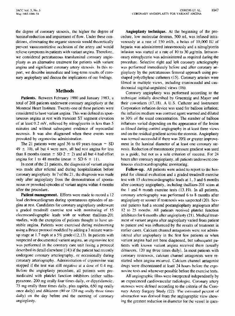
Table 1. Clinical and Angiographic Data of 21 Patients Before Coronary Angioplasty
Duration of Electrocardiogram During Coronary ArteriogramDiagnosis of
Age (yr)Angina (mo)
Rest Ergonovine Exercise Variant Angina VesselCase &Sex Rest Effort Angina Test (rng) Test Before Angioplasty Involved Stenosis (%)
Patients With Primary Failure
I 66F 8 None t ST i ST (0.025) ST Yes Prox. LAD 802 48M I I i ST (0.05) Th pos. Yes Prox. LAD 90
Patients With Coronary Restenosis
3 42M 2 None i ST t ST (0.025) i ST Yes Prox. LAD 704 36M 2 2 J, ST No Prox. LAD 855 51M I I J, ST No Prox. LAD 606 69M I 48 J, ST No Prox. LAD 657 41M 3 3 Neg J, ST No Prox. LAD 608 59F I 7 No Prox. LAD 60
1st diag. 809 47M None i ST J, ST (0.025) ST Yes Mid LAD 80
Mid LAD 6010 61M 3 13 i ST J, ST Yes Mid RCA 85II 36M I I i ST Yes Mid CAD 85
Patients With Late Success
12 62F 4 4 i ST Neg" Yes Mid LAD 7013 52M 4 4 Th pos. No Prox. LAD 9014 SSM I 4 iST(O.i> i ST Yes Prox. LAD 9515 42M I I i ST i ST (0.05) J, ST Yes Prox. LAD 9016 48F I I i ST i ST (0.1) Neg" Yes Prox. LAD 9017 38M I I t ST (0.1) i ST Yes Prox, LAD 8518 38M I None i ST Yes Prox. LAD 8019 47M 2 2 i ST Yes Prox. LAD 8520 38M 5 5 i ST (0.1) Yes Mid RCA 7021 60M I I No Prox. LCx 65
"Exercise test duration < 3 minutes. diag = diagonal artery; F = female; LAD = left anterior descending artery; LCx = left circumflex artery; M= male; pos. = positive; prox. = proximal; RCA = right coronary artery; ST i and ST J, = ST segment elevation and depression, respectively; Th
= thallium scan.
Table 2. Summary of Clinical and Angiographic Data of 21Patients Before Coronary Angioplasty
ginal attack occurred during hospitalization and the diagnosis was made after ergonovine administration. In the 10
tion. All measurements were made after the administrationof nitroglycerin, and all comparisons between pre- and postangioplasty films were made using the same view. Coronaryrestenosis was defined as a 20% or greater decrease in luminal diameter of the stenosis compared with the coronaryangiogram immediately after angioplasty, resulting in acoronary stenosis of more than 50% at repeat angiography.
ResultsTables I to 6 summarize the clinical and angiographic
data before and after coronary angioplasty for the 21 patientsin this study. For clarity, the patients were classified intothree groups according to the results of angioplasty: primaryfailure, restenosis or late success.
Findings before angioplasty (Tables 1 and 2). Variantangina was documented before angioplasty in two-thirds (14of 21) of the patients; in 9 of these 14, transient ST elevationwas present on an electrocardiogram recorded during spontaneous episodes of angina; in the other 5 patients, no an-
Age (yr)
Sex (no.)MaleFemale
Angina (no.)Rest and effortRest only
Diagnosis of variant angina (no.)Before angioplastyAfter angioplasty
Single vessel disease (>50%)Diseasedcoronary artery (no.)
Left anterior descendingRightLeft circumflex
Degree of stenosis (%)
49 :!: 10 (range 36 to 69)
174
174
147
20
182I
77 :!: 12 (range 60 to 95)
JACC Vol. 5. No.5May 19&5:104&-54
CORCOS ET AL.CORONARY ANGIOPLASTY FOR VA RIANT ANGINA
1049
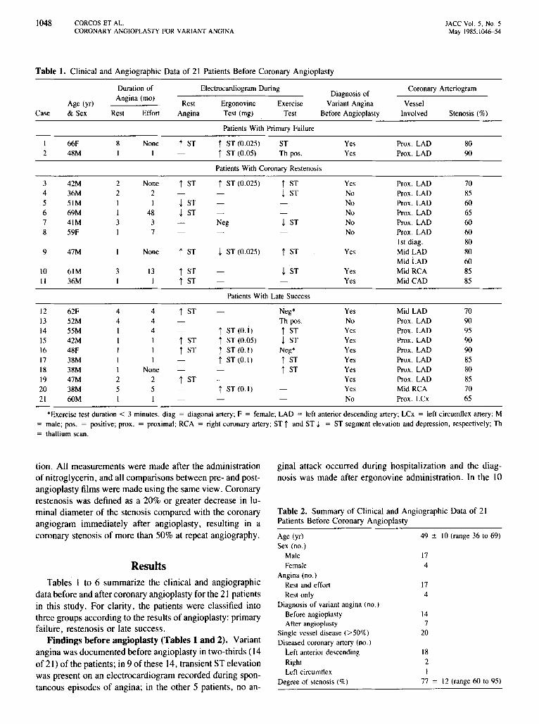
Table 3. Clinica l Follow-up of 2 1 Patient s After Co ronary Angiopl asty
Late Clinical Status
Early Clinical Status Calcium Clinical Status at Time of Restenosis Still
Electrocardiogram DuringChannel
Electrocardiogram DuringTaking
Angina Antagonist Angina Time After CalciumRest Exercise Ergonovine After Rest Ergonovine Exercise Angioplasty Channel
Case Rest Effort Angina Test Test ( rng) Angioplasty Rest Effort Angina Test ( rngj Test (rno) Angina Antagonists
Patients With Primary Failure
Yes No 39 Not No2 No Yes ST 24 Not :j: No
Patients With Coronary Restenosis
3 No No Neg Neg No No Yes t ST (0.2) i ST 40 No No4 No No Neg No Yes* Yes i ST (0.2) i ST 37 No No
5 Yes Yes Neg t ST (0 .05 ) No No Yes i ST 36 NoH No6 No No i ST No Yes No t ST i ST 6 -:j:§
7 No No No Yes' Ye~ i ST 35 No No8 Yes Yes Neg No Yes Yes t ST 31 No No9 No No Neg Yes Yes No t ST t ST (0 .05) Neg 29 No Yes
10 No No Yes Yes No t ST ((l.01) i ST 26 No NoII No No Neg No Yes No i ST 17 No Yes
Patients With Late Success
12 Yes No t ST Neg Yes 36 No No13 No No Neg t ST (0.1) Yes 35 No No14 No No Neg Neg Ye ~ 34 No No15 No No Neg Neg Yes .n No No16 No No Neg Yes 17 No No17 No No Neg Yes 13 No No18 No No Neg Yes 12 No No19 No No Neg Yes 12 No No20 No No Neg Yes 10 No No21 Yes No Neg t ST (0.3) No 10 No:j: No
*In these two patients (Patients 4 and 7). ST segment elevation during spontaneous angina was documented only after the second coronary angioplasty.t These three patients (Patients I. 2 and 5) underwent coronary artery bypass surgery during follow-up after angioplasty. :j:These four patients (Patients2. 5. 6 and 21) had an acute myocardial infarction during follow-up after angioplasty. §This patient (Patient 6) died suddenly approximately I monthafter an acute myocardial infarction. Abbreviations as in Table I.
Table 4. Summary of Clinical Follow-up of 2 1 Patient sAfter An giop1asty
patients who underwent an ergonovine test (5 with suspectedand 5 with documented variant angina) , the test was negativein I patient . induced ST depression in I and was positi ve
Duration (rno)Status (no.)
AliveAlive without myocardial infarction
or surgeryAlive after myocardial infarctionUncomplicated surgery
DeadSymptoms (angina) (no.)Current medical treatment (no.)
NoneCalcium channel antagonists
33 :t 13 (range 18 to 48)
2016
.l1
Io
18
in 8 at a dose of 0.1 mg or less. Fourteen patients had anexercise test that in six patients induced ST elevation in thesame electrocardiographic leads in which ST elevation occurred durin g spontaneous or provoked anginal attacks , andin four patients induced ST depression . In the rema iningfour patients the test produced no ST abnormality; in twoof the four, a reversible perfu sion defect was demonstratedon the thallium exercise scan and in the other two , theexercise test was stopped becau se of fatigue at 3 minutesor less.
All patients had single vessel coronary disease with noaddi tional narrowings of greater than 50% in the other coronary arteries. except for Pat ient 6, who had a 65% stenosisof the posterior descending artery. The coronary stenosiswas graded between 60 and 95% and was located in the leftanterior descending artery in all but 3 patients (in the proximal third in 15 patients) . ST elevation before or after coro nary angioplasty was located in the anterior electrocardio-
1050 CORCOS ET AL.CORONARY ANGIOPLASTY FOR VARIANT ANGINA
lACC Vol. 5. No.5May 1985: 1046-54
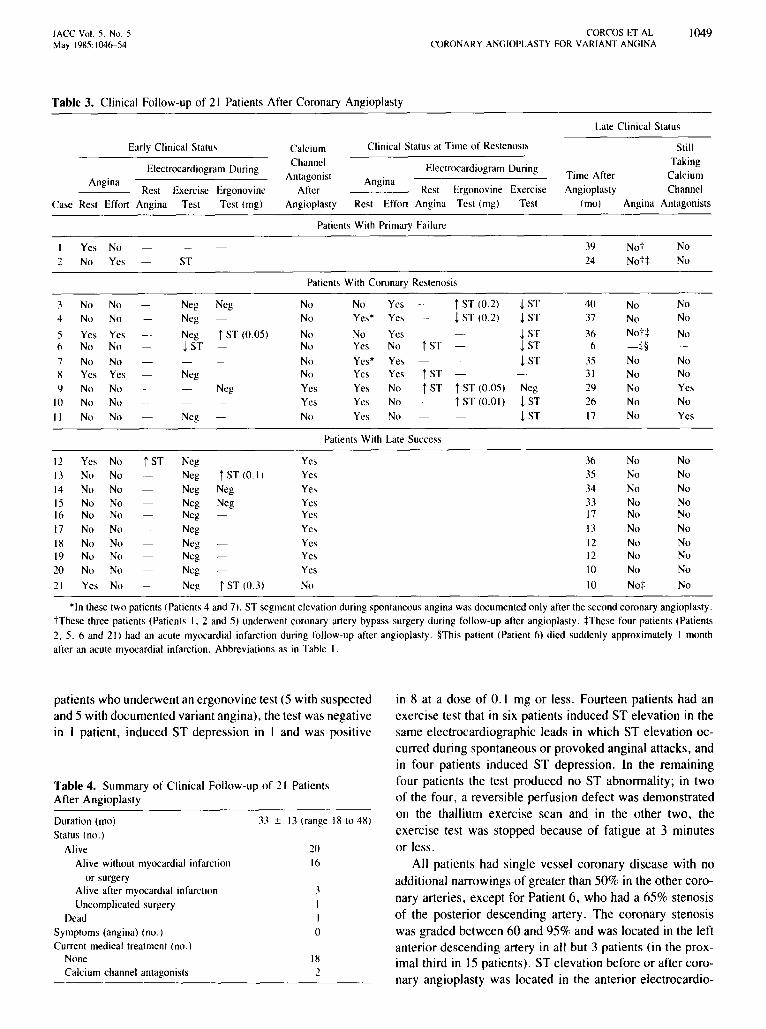
Table 5. Angiographic Follow-up of 21 Patients after Coronary Angioplasty
% Stenosis Calcium % Stenosis Late Angiographic Findings
Immediately Antagonists at6 Months Time AfterAfter First After or at Second Angioplasty Coronary Ergonovine Test;
Case Angioplasty Angioplasty Angioplasty (mo) Stenosis (%) Stenosis (%) (rng)
Patients With Primary Failure
I 80"2 80 80~ HIOtt
Patients With Coronary Stenosis
3 20 No 70~ 25 25 25 ~ 70 (0.4) Clinically neg4 30 No 80~ 30 21 1005 40 No 75":1:6 25 No 75~ 40:1:7 30 No 70~ 40 21 40 Neg8 50 No (60) 15 (55) Neg
60 80 709 30 Yes 70 22 70 ~ 95 (0.2)
35 (40) (40)10 35 Yes 70~ 50 22 20 ~ 100(0.4)II 50 No 70~ 35 7 70
Patients With Late Success
12 30 Yes 10 18 10 Neg13 25 Yes 25 19 20 Diffuse narrowing (0.3)14 50 Yes 40 19 30 Neg15 60 Yes 50 18 50 Neg16 10 Yes 35 6 3517 10 Yes 10 6 10 Neg18 10 Yes 20 6 2019 35 Yes 30 12 3020 30 Yes 20 8 20 ~ 85 (0.4)21 25 No 25:1: 9 25 Neg
"Elective coronary artery bypass surgery; temergency coronary artery bypass surgery; :l:myocardial infarction. Neg = negative.
Table 6. Summary of Angiographic Follow-up of 17 PatientsAfter Angioplasty
graphic leads in all patients with left anterior descendingartery stenosis and in the inferior leads in the other threepatients. Left ventricularejection fraction was 0.50 or greaterin all patients.
Findings after coronary angioplasty (Tables 3 to 6).Angioplasty was successful in all but two patients (Table 5).
Failures. In Patient I, the lesion could not be crossed,variant angina continued despite optimal medical therapyand she underwent coronary bypass surgery associated withplexectomy 12 days later. Surgery was uneventful, but ep-
Duration (mo)Status (no.)
No more stenosis (>50%)Restenosis (>50%)
Ergonovine test (no.)NegativePositive
14 ± 7
134
8
4
isodes of variant angina occurred in the early postoperativeperiod.
In Patient 2, the results of angioplasty were poor andthere was an 80% residual stenosis. Effort angina persisteddespite medical therapy and a second angioplasty was attempted I month later. It resulted in a coronary dissectionwith complete coronary occlusion complicated by a myocardial infarction that was not prevented by emergency coronary bypass surgery associated with plexectomy.
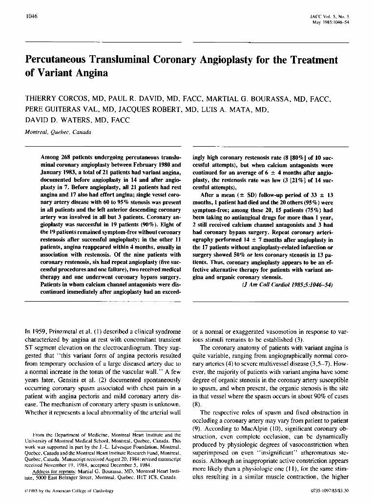
Primary 5ucce55. Coronary angioplasty was successfulin 19 (90%) of the 21 patients (Fig. IA and B, Tables 5and 6). The primary success rate in our first 268 patientswho underwent angioplasty during the same period was68%. None of the first angioplasty attempts in the 21 patientswas complicated by coronary spasm during the procedure.
Soon after successful angioplasty in the 19 patients, allbut 4 patients were symptom-free (Table 3). Patient 5 hadrecurrence of rest and effort angina I month after angioplasty. Coronary angiography showed a 35% residual stenosis with a spontaneous superimposed spasm resulting ina 95% narrowing suppressed by nitroglycerin and reinduced
JACC Vol. 5. No.5May 1985:1046-54
CORCOS ET AL.CORONARY ANGIOPLASTY FOR VARIANT ANGINA
1051
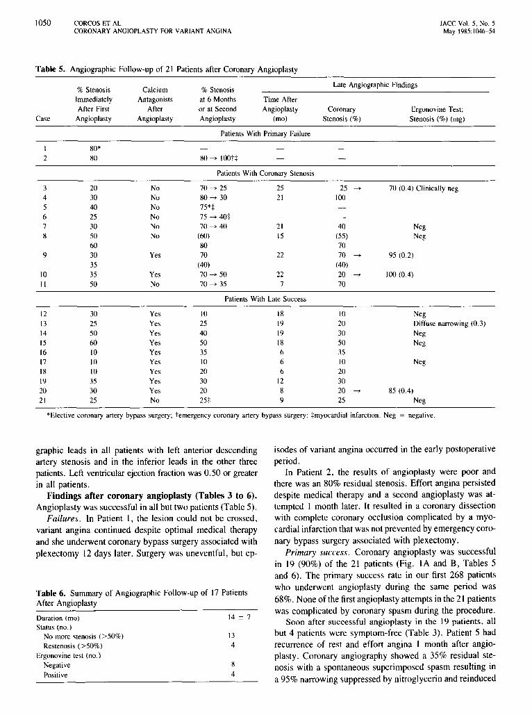
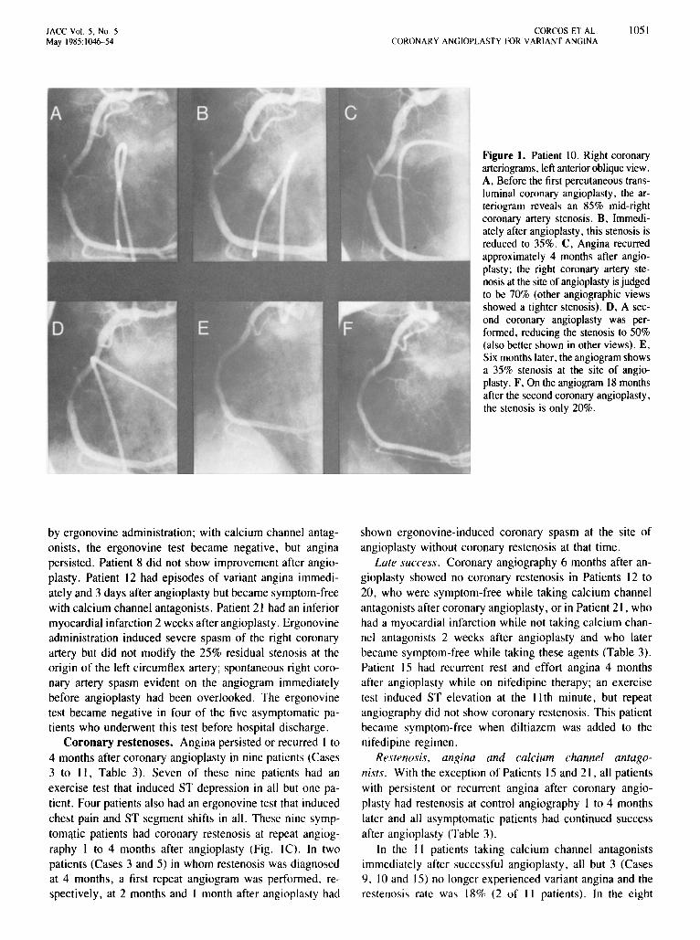
Figure 1. Patient 10. Right coronaryarteriograms, left anterior oblique view.A. Before the first percutaneous transluminal coronary angioplasty, the arteriogram reveals an 85% mid-rightcoronary artery stenosis. B, Immediately after angioplasty, thisstenosis isreduced to 35%. C, Angina recurredapproximately 4 months after angioplasty; the right coronary artery stenosis at the site of angioplasty isjudgedto be 70% (other angiographic viewsshowed a tighter stenosis). D, A second coronary angioplasty was performed, reducing the stenosis to 50%(also better shown in otherviews). E,Sixmonths later, theangiogram showsa 35% stenosis at the site of angioplasty. F, Onthe angiogram 18 monthsafter thesecond coronary angioplasty,the stenosis is only 20%.
by ergonovine administration; with calcium channel antagonists, the ergonovine test became negative, but anginapersisted. Patient 8 did not show improvement after angioplasty. Patient 12 had episodes of variant angina immediately and 3 days after angioplasty but became symptom-freewith calcium channel antagonists. Patient 21 had an inferiormyocardial infarction 2 weeks after angioplasty. Ergonovineadministration induced severe spasm of the right coronaryartery but did not modify the 25% residual stenosis at theorigin of the left circumflex artery; spontaneous right coronary artery spasm evident on the angiogram immediatelybefore angioplasty had been overlooked. The ergonovinetest became negative in four of the five asymptomatic patients who underwent this test before hospital discharge.
Coronary restenoses. Angina persisted or recurred I to4 months after coronary angioplasty in nine patients (Cases3 to II, Table 3). Seven of these nine patients had anexercise test that induced ST depression in all but one patient. Four patients also had an ergonovine test that inducedchest pain and ST segment shifts in all. These nine syrnptomatic patients had coronary restenosis at repeat angiography I to 4 months after angioplasty (Fig. IC). In twopatients (Cases 3 and 5) in whom restenosis was diagnosedat 4 months, a first repeat angiogram was performed. respectively, at 2 months and I month after angioplasty had
shown ergonovine-induced coronary spasm at the site ofangioplasty without coronary restenosis at that time.
Late success. Coronary angiography 6 months after angioplasty showed no coronary restenosis in Patients 12 to20, who were symptom-free while taking calcium channelantagonists after coronary angioplasty, or in Patient 21, whohad a myocardial infarction while not taking calcium channel antagonists 2 weeks after angioplasty and who laterbecame symptom-free while taking these agents (Table 3).Patient 15 had recurrent rest and effort angina 4 monthsafter angioplasty while on nifedipinc therapy; an exercisetest induced ST elevation at the II th minute, but repeatangiography did not show coronary restenosis. This patientbecame symptom-free when diltiazem was added to thenifedipine regimen.
Restenosis, angina and calcium channel antagonists. With the exception of Patients 15 and 21, all patientswith persistent or recurrent angina after coronary angioplasty had restcnosis at control angiography I to 4 monthslater and all asymptomatic patients had continued successafter angioplasty (Table 3).
In the II patients taking calcium channel antagonistsimmediately after successful angioplasty, all but 3 (Cases9. 10 and 15) no longer experienced variant angina and therestenosis rate was 18% (2 of II patients). In the eight
1052 CORCOS ET AI..CORONARY ANGIOPLASTY FOR VARIANT ANGINA
JACC Vol. 5. No.5May 1985:1046-54
patients not taking calcium channel antagonists immediatelyafter successful angioplasty , the restenosis rate was 86%(seven of eight patient s).
Management of restenosis . With the exception of Patients 4 and 7 in whom variant angina was not diagnoseduntil after the second coronary angioplasty , patients withrestenosis were treated with calcium channel antagonists,usually in addition to a second angioplasty attempt. Repeatangioplasty was attempted in six patients; it was successfulin five (Fig . 10, E, F, Table 5), but failed in Patient 6 (whounderwent angioplasty on a coronary stenosis slightly distalto the site of the initial angioplasty). In this patient, coronaryspasm developed during the procedure, completely occluding the left anterior descending artery at the site of the initialangioplasty, and intracoronary nitroglycerin was requiredfor relief of spasm. Six hours later, the patient had an acuteanterior infarction that was compli cated by two episodes ofventricular fibrillation and transient heart failure; this patientdied suddenly I month later . The autopsy revealed a hemorrhage under a plaque associated with a 95% stenosis ofthe left anterior descending artery, an anteroseptal infarctionand a mural thrombus. Patient 5 developed a new 75% leftmain coronary artery stenosis not present at angioplasty orat repeat angiography I month after angioplasty and alsohad a 75% restenosis of the left anterior descending artery ;this patient underwent a double aortocoronary bypass graftprocedure the day after repeat angiography, but surgery wascomplicated by an acute myocardial infarction . Two patients(Cases 8 and 9) were treated with calcium channel antag onists alone .
Of the five patients who had a successful second coronaryangioplasty, three were taking calcium channel antagonistsand two of them had a late success . The remaining twopatient s did not receive calcium channel antagonists becausevariant angina was not diagnosed before the second angioplasty . In Patient 4, rest angina recurred 6 weeks after thesecond angioplasty and at repeat angiography a 60% stenosi swith provoked superimposed coronary spasm was shown atthe site of angioplasty. The patient had become symptomfree while taking calcium channel antagonists, but subsequent angiography revealed a complete occlusion at the siteof angioplasty. In Patient 7, rest angina recurred I weekafter repeat angioplasty; calcium channel antagonist therapywas started I month after angioplasty and the patient becamesymptom-free without coronary restenosi s.
Restenosis and calcium channel antagonists (all angioplasty attempts). In the overall 24 successful coronary angioplasty attempts (19 primary successes and 5 successfulrepeat angioplasties), the restenosis rate was 21% (3 of 14)when patients had received calcium channel antagonists soonafter angioplasty, and 80% (8 of 10) when calcium channelantagonists were not administered soon after angioplasty(Tables 3 and 5). This difference is statistically significant(p < 0.014, Fisher's exact test) .
Late outcome. The mean (± SO) duration of clinicalfollow-up was 33 ± 13 months (range 6 to 48) (Tables 3and 4). One patient died , but all the others remained symptom-free. Three had coronary bypass surgery and no longerrequired antianginal therapy. Two were still taking calciumchannel antagonists . In each of the remaining 15 patients,calcium channel antagonists have been discontinued for morethan 1 year (mean ± SO 25 ± 10 months), and thesepatients remained symptom-free. Calcium channel antagonists were stopped 6 ± 4 months after coronary angioplastyin patients with continued success and 12 ± 3 months afterrepeat angioplasty in patients who had restenosis.
Repeat angiography was available at 14 ± 7 months(range 6 to 25) in all 17 patients who had neither myocardialinfarction associated with angioplasty nor surgery (Tables5 and 6). One had a complete coronary occlusion at the siteof angioplasty and three still had a significant coronarystenosis of greater than 50%, with spontaneous superimposed spasm in one patient (Case II) . Twelve patientsunderwent an ergonovine test which was usually performedduring the last repeat angiographic study. The test was negative in eight patients but induced a 70% spasm in Patient3; it was positive in four patients at a relatively high doselevel of ergonovine (0.2 mg or more).
DiscussionOrganic coronary stenosis in variant angina. Previous
reports (23 ,24) showed that in patients with variant anginathe extent and severity of organic coronary stenoses werethe strongest predictors of survival and survival withoutmyocardial infarction. Patients with variant angina shouldundergo coronary angiography since the clinical or electrocardiographic manifestations do not distinguish patients withangiographically normal coronary arteries from those withsevere proximal obstructive lesions . In fact, the therapeuticchoice may depend, to a great extent, on the presence orabsence of organic atherosclerotic lesions associated withcoronary spasm.
Coronary spasm associated with significant atherosclerotic coronary lesions is relatively frequent; among 162consecutive patients with variant angina undergoing coronary arteriography at our institution, 59% had organic lesions of 70% or more (23) . Until recently, coronary bypasssurgery was the only available therapy for immediate revascularization in these patients . However, coronary bypasssurgery is associated with a higher operative risk, a higherpostoperative myocardial infarction rate and poorer resultsin these patients than in other subsets of patients with fixedcoronary obstructions (6,25-27) .
Coronary angioplasty in variant angina. Until recently, coronary spasm was considered a contraindicationto coronary angioplasty. Indeed, since coronary spasm occurred in 2% of patients undergoing angioplasty and was
lACC Vol. 5, No.5May 1985:1046-54
CORCOS ET AL.CORONARY ANGIOPLASTY FOR VARIANT ANGINA
1053
associated with myocardial infarction (17) or the need foremergency coronary bypass surgery (28) in some patients,angioplasty on an artery already affected by coronary spasmseemed difficult and risky.
This study confirms our preliminary results (29) usingangioplasty for the treatment of variant angina and showsthat angioplasty is technically feasible in patients with thiscondition. Moreover, the primary success rate in these patients was higher than that of the overall population of patients undergoing coronary angioplasty during the same period, This probably occurred because most of our patientsfulfilled the ideal criterion for selection for angioplasty,namely, discrete proximal noncalcified stenosis in a singlemajor coronary artery suitable for coronary artery surgery(17), This pattern is more frequent in patients with variantangina (29) than in the average group of patients undergoingcoronary arteriography (30). Coronary spasm occurred inonly two patients during 28 procedures (during repeat angioplasty for both patients) and was complicated by myocardial infarction in one. In the other patient with angioplasty-related myocardial infarction, this complication wasdue to coronary dissection. Thus, the incidence of complications, particularly those related to coronary spasm, doesnot appear to be much higher in these patients than that inother patients who undergo coronary angioplasty (31) or inpatients with active variant angina (32).
Role of spasm in restenosis after coronary angioplasty. Coronary spasm demonstrated during the first 4months after angioplasty may be a different problem. Variant angina was diagnosed after angioplasty in 7 of our 181patients successfully treated with angioplasty, and Hollmanet al. (33) recently described 5 patients representing 5 oftheir 6 reported cases among 1,000 successful attempts.Interestingly all of our seven patients and three of their fivepatients had rest angina before angioplasty. Three of theirpatients had a coronary stenosis of less than 60% beforeangioplasty (one had an insignificant 37% coronary stenosis2 weeks after a myocardial infarction). Only I of these 13patients had an ergonovine test before angioplasty. Coronaryspasm did not develop immediately after angioplasty-induced balloon injury, but after a symptom-free interval ofseveral days or weeks. In this setting, it appears difficult toassume that coronary spasm diagnosed after angioplasty isdirectly related to it. Variant angina occurring before angioplasty, but discovered only after the procedure, appearsmuch more likely.
In two of our nine patients with coronary restenosis andin four of the patients described by Hollman et al. (33),coronary spasm in hemodynamically insignificant lesionswas demonstrated 2 to 7 months before the diagnosis ofrestenosis. This finding, plus the fact that the rate of restenosis is much higher in patients with variant angina than inpatients with a fixed coronary obstruction and seems to beprevented by calcium channel antagonists, supports the hypothesis that persistence of coronary spasm may playa role
in the pathogenesis of restenosis in patients with variantangina. The possibility of variant angina should, thus, beconsidered in patients with rest angina referred for coronaryangioplasty and in patients with restenosis after angioplastybecause they probably need specific medical managementafter angioplasty similar to that of patients with variantangina undergoing coronary bypass surgery (34).
Long-term clinical results. Our long-term clinical results must be interpreted with caution because spontaneousremission is a frequent outcome of variant angina. However,our proportion of patients currently symptom-free withoutmedical treatment for more than I year may be higher thanthe rate reported in patients who had medical treatment alone(35). Survival rate and survival rate without myocardialinfarction in our series are not different from those of patients with variant angina and single vessel disease who hadmedical or surgical therapy (23,24). In patients with variantangina and multi vessel disease who have been shown tohave the worst prognosis (23) and for whom coronary bypasssurgery has been advised, we have not yet had any experience of successful revascularization using coronary angioplasty. We also have no experience of coronary angioplasty for mild to moderate « 50 to 60%) coronary stenosesin vessels that exhibit coronary spasm; however, because ofthe questionable role of these mild lesions, we doubt thatvariant angina can be better controlled by coronary angioplasty plus calcium channel antagonists than by calciumchannel antagonists alone in most patients with these mildfixed stenoses.
Role of calcium channel antagonists after angioplasty. In patients with variant angina and organic coronarystenoses, coronary angioplasty is usually successful, butrestenoses, which may be induced by recurrent episodes ofcoronary spasm. are frequent and seem to be prevented bycontinuing administration of calcium channel antagonist drugsfor several months after angioplasty. Coronary angioplastycan, thus, be proposed as an alternative therapy for thesepatients. The subset of patients with variant angina andorganic coronary stenoses that should benefit the most fromthis new method of revascularization remains to be determined.
ReferencesI. Prinzmetal M, Kennamer R, Merliss R, Wada T. Bor N. Angina
pectoris. I. A variant form of angina pectoris. Am J Med 1959;27:375-88.
2. Gensini GG. Di Giorgi S, Murad Netto S. Arteriographic demonstration of coronary artery spasm and its release after the use of a vasodilator in a case of angina pectoris and in the experimental animal.Angiology 1962;13:550-3.
3. Theroux P. Waters DD. Latour JG. Clinical manifestations and pathophysiology of myocardial ischemia with special reference to coronaryartery spasm and the role of slow channel calcium blockers. ProgCardiovasc Dis 1982;25: 157-68.
4. Cheng TO. Bashour T. Keiser GA, Weiss L. Bacos L. Variant anginaof Prinzmetal with normal coronary arteriograms. A variant of thevariant. Circulation 1973:47:47&-85.
5. Severi S, Davies G. Maseri A, Marzullo P, L'Abbate A. Long-term
1054 CORCOS ET AL.CORONARY ANGIOPLASTY FOR VARIANT ANGINA
lACC Vol. 5. NO.5May 1985:1046-54
prognosis of "variant" angina with medical treatment. Am 1 Cardiol1980;46:226-32.
6. Shubrooks Sl, Bete 1M, Hutter AM, et al. Variant angina pectoris:clinical and anatomic spectrum and results of coronary bypass surgery.Am 1 Cardiol 1975;36:142-7.
7. Johnson AD. Stroud HA, Viewig WVR, Ross 1 Jr. Variant anginapectoris, clinical presentations, coronary angiographic patterns. andthe results of medical and surgical management in 42 consecutivepatients. Chest 1978;73:786-94.
8. MacAlpin RN. Relation of coronary arterial spasm to site of organicstenosis. Am 1 Cardiol 1980;46:143-53.
9. Muller IE. Prinzrnctal's angina. A model for the role of spasm inischemic heart disease. 1 Cardiovasc Med 1980;5:19-24.
10. MacAlpin RN. Contribution of dynamic .vascular wall thickening toluminal narrowing during coronary arterial constriction. Circulation1980;61:296-30 I.
11. Maseri A. Chierchia S. Coronary artery spasm: demonstration, definition, diagnosis and consequences. Prog Cardiovasc Dis1982;25:169-92.
12. Waters DD, Szlachcic 1, Bourassa MG. Scholl 1M, Theroux P. Exercise testing in patients with variant angina: results. correlation withclinical and angiographic features and prognostic significance. Circulation 1982;65:265-74.
13. Scholl 1M, Chaitman BR, David PR, -et al. Exercise electrocardiography and myocardial scintigraphy in the serial evaluation of the resultsof percutaneous transluminal coronary angioplasty. Circulation1982;66:380-90.
14. Waters DD, Theroux P, Szlachcic 1, et al. Ergonovine testing in acoronary care unit. Am 1 Cardiol 1980;46:922-30.
\5. Bourassa MG, Lesperance 1. Campeau L. Selective coronary arteriography by the percutaneous femoral artery approach. Am 1 Roentgenol 1969;107:377-83.
16. Lesperance 1, Saltiel 1, Petitclerc R, Bourassa MG. Angulated viewsin the sagittal plane for improved accuracy of cinecoronary angiography. Am 1 Roentgenol 1974;12\:565-74.
17. Gruentzig AR, Senning A, Siegenthaler WE. Nonoperative dilatationof coronary artery stenosis. Percutaneous transluminal coronary angioplasty. N Engl 1 Med \979;30\:6\-8.
18. Myler RK, Gruentzig AR, Stertzer SH. Technique and clinical indications for percutaneous transluminal coronary angioplasty. In: MasonDT, Colins JJ Jr, eds. Myocardial Revascularization, New York: YorkeMedical, \981:44\-4.
\9. Hirzel HO, Nuesch K, Gruentzig AR, Horst W, Krayenbuhl HP.Thallium-201 exercise scintigraphy after percutaneous transluminalangioplasty of coronary artery stenoses. Med Clin North Am1980;64:163-76.
20. Dangoisse V, Guiteras Val P, David PR, et al. Recurrence of stenosisafter successful percutaneous transluminal coronary angioplasty (PTCA)(abstr). Circulation 1982;66:(suppl \1):11-331.
21. Thornton MA, Gruentzig AR, Hollman 1. Coumadin or aspirin aftercoronary angioplasty? (abstr). 1 Am Coll Cardiol 1983; I:725
22. Coronary Artery Surgery Study Investigators: National Heart, Lung,and Blood Institute. Coronary Artery Surgery Study. Circulation1981;63:(suppl 1):1-14.
23. Waters DD, Miller DD, Szlachcic 1, et al. Factors influencing thelong-term prognosis of treated patients with variant angina. Circulation1983;68:258-65.
24. Mark DB. Califf RM, Morris KG, et al. Clinical characteristics andlong-term survival of patients with variant angina. Circulation1984;69:880-8.
25. Gaasch WH, Lufschanowski R, Leachman RD, Alexander lK. Surgical managementof Prinzmetal's variant angina. Chest 1974;66:614-2\.
26. Grondin CM, Limet R. Sympathetic denervation in association withcoronary artery grafting in patients with Prinzmetal's variant angina.Ann Thorac Surg \977;23:111-7.
27. Bertrand ME, Lablanche 1M, Rousseau MF, et al. Surgical treatmentof variant angina: use of plexectomy with aortocoronary bypass. Circulation \980;6\ :877-82.
28. Myler RK. Percutaneous transluminal coronary angioplasty. Arch InstCardiol Mex 1980;50:401-5.
29. David PR, Waters DD, Scholl 1M, et al. Percutaneous transluminalcoronary angioplasty in patients with variant angina. Circulation1982;66:695-702.
30. Hamby RI, Katz S. Percutaneous transluminal coronary angioplasty:its potential impact on surgery for coronary artery disease. Am 1Cardiol \980;45:1\61-6.
31. Vliestra RE, Holmes DR, Smith HC, Hartzler GO, Orszulak TA.Percutaneous transluminal coronary angioplasty. Mayo Clin Proc1981;56:287-93.
32. Maseri A, Severi S, DeNes M, et al. "Variant" angina; one aspectof a continuous spectrum of vasospastic myocardial ischemia. Pathogenetic mechanisms, estimated incidence and clinical and coronaryarteriographic findings in 138 patients. Am 1 Cardiol \978;42:10\9-35.
33. Hollman 1, Austin GE, Gruentzig AR, Douglas IS lr, King SB III.Coronary artery spasm at the site of angioplasty in the first 2 monthsafter successful percutaneous transluminal coronary angioplasty. 1 AmColl Cardiol 1983;2:1039-45.
34. Endo M, Kanda I, Hosada S, Hiyashi H, Hirosawa K, Konno S.Prinzmetal's variant form of angina pectoris. Re-evaluation of mechanisms. Circulation \975;52:33-7.
35. Waters DD, Bouchard A, Theroux P. Spontaneous remission is afrequent outcome of variant angina. 1 Am Coil Cardiol 1983;2:195-9.
Recommended