Technische Universität München
Department Chemie
Lehrstuhl II für Organische Chemie
Design and Synthesis of FVIII- and uPAR-Selective Ligands and Cross-
Linked Polymers as Media for NMR-Spectroscopy
Sebastian Knör
Vollständiger Abdruck der von der Fakultät für Chemie
der Technischen Universität München zur Erlangung des akademischen Grades
eines
Doktors der Naturwissenschaften
genehmigten Dissertation.
Vorsitzender: Univ.-Prof. Dr. K. Köhler
Prüfer der Dissertation: 1. Univ.-Prof. Dr. H. Kessler
2. Univ.-Prof. Dr. A. Türler
3. Priv.-Doz. Dr. Viktor Magdolen
Die Dissertation wurde am 23.05.2007 bei der Technischen Universität München
eingereicht und durch die Fakultät für Chemie am 05.07.2007 angenommen.


Die vorliegende Arbeit wurde am Lehrstuhl II für Organische Chemie
der Technischen Universität München in der Zeit von Oktober 2002 bis Mai 2007
unter der Leitung von Prof. Dr. Horst Kessler angefertigt.

Meiner Frau
gewidmet
Σ’αγαπώ πολύ


Publier dans un petit journal ne veut pas dire petit résultat. Ce qui compte c'est que les gens l'essayent pour dire que ça marche!! In einem kleinen Journal zu publizieren heißt nicht, kleine Ergebnisse zu haben. Was zählt ist, dass die Leute es ausprobieren um zu sagen: Es geht!
(Abdellah Benhida, Univ. Leuven)

DANKSAGUNG
Meinem Doktorvater,
Herrn Professor Dr. Horst Kessler,
danke ich besonders für die vielen interessanten Themen, die ich bearbeiten
durfte. Insbesondere bedanke ich mich für die uneingeschränkte Unterstützung in
jeder Hinsicht, für den großen wissenschaftlichen Freiraum, der mir gewährt
wurde, sowie für die zahlreichen fruchtbaren und hilfreichen Diskussionen.
Für die ausgezeichnete Zusammenarbeit auf dem Gebiet der Faktor VIII Liganden
möchte ich mich ganz herzlich bei
• Dr. Alexey Khrenov und Prof. Dr. Evgueni Saenko,
• Dr. Charlotte Hauser,
• Dr. Abdellah Benhida und Prof Dr. Jean-Marie Saint-Remy,
• Dr. Rainer Schwaab und Prof. Dr. Johannes Oldenburg sowie
• Dr. Nathalie Beaufort und PD. Dr. Viktor Magdolen
bedanken. Mit ihrer Hilfe habe ich viel über die Biologie des Faktor VIII und die
Schwierigkeiten in der Behandlung der Hämophilie gelernt. Ihr großartiger Einsatz
hat maßgebliche zum Erfolg des Projektes beigetragen.
Für die hervorragende Zusammenarbeit auf dem Gebieten des Tumor-Imaging und
der Tumor-Therapy und bedanke ich mich bei:
• Dr. Sumito Sato, PD Dr. Viktor Magdolen und Prof. Dr. Manfred Schmitt,
• Dr. Christof Seidl und Prof. Dr. Reingard Senekowitsch-Schmidtke sowie
• Dr. Thorsten Poethko, Dr. Margret Schottelius und Prof. Dr. Hans-Jürgen
Wester
Ohne Ihren unermüdlichen Eifer wäre ein Gelingen dieser Projekte nicht möglich
gewesen.
Burghard Cordes und Helmut Krause bin ich für zahlreiche analytische Messungen
und Maria Kranawetter für das HPLC-Trennen unzähliger Verbindungen zu großem
Dank verpflichtet. Mona Wolff danke ich für das Ausführen zahlreicher Synthesen,
die Organisation unseres Chemikalienhaushaltes und ihre immer hilfreichen
Antworten auf Fragen wie: „Wo ist…?“, „Haben wir…?“, usw. Alexander Frenzel und
Rainer Haeßner danke ich für die Hilfe bei allen Computerproblemen, wie „Internet
geht nicht!“ und Evelyn Bruckmaier für die vielen netten Worte und Aufmunterun-
gen. Der gesamten NMR-Gruppe, insbesondere Rainer Haeßner, Gerd Gemmecker,
Burkhard Luy, Peter Kaden, Grit Kummerlöwe und Andreas Enthart für die
Einführung in das Messen von NMR-Spektren und die sofortige Hilfe bei allen kleinen
und großen Problemen.
Allen derzeitigen und ehemaligen Mitgliedern des Arbeitskreises danke ich für das
gute Klima und die nette Atmosphäre, insbesondere Burkhardt Laufer, Timo Huber,
Jayanter Chatterjee, Eric Biron, Grit Kummerlöwe, Enrique Mann, Armin Modlinger,
Axel Meyer, Dominik Heckmann, Andreas Frank, Jörg Auernheimer, Martin Sukopp,
Ulrich Hersel, Florian Manzenrieder, Florian Opperer, Lukas Doedens, Peter Kaden,
Andreas Enthart und Oliver Demmer.
Mein weiterer Dank gilt:
• Burkhardt Laufer, Jayanter Chatterjee, Timo Huber, Dominik Heckmann und
Grit Kummerlöwe für das Korrekturlesen dieser Arbeit.
• Philipp Gradicsky für zwei Jahre als studentische Hilfskraft im Labor –
jederzeit zur Stelle
• Meinen zahlreichen Forschungsstudenten und Chemielaboranten-Azubis für
ihre selbständige Mitarbeit in meinen Forschungsprojekten und die Hilfe im
Labor, insbesondere Burkhardt Laufer, Philipp Gradicsky, Bianca Ludwig,
Bastian Sauerer, Robert Jäger und Helge Menz.
• Axel Meyer, Jörg Auernhaimer, Armin Modlinger, Thorsten Arndt, Dominik
Heckmann, Burkhardt Laufer, Timo Huber und Eric Biron für anregende
Diskussionen und Hilfestellung jeder Art.
Großer Dank gebührt all meinen Förderern, Lehrer und Tutoren in Schul- und
Studienzeiten, ohne deren Einsatz diese Ausbildung nicht möglich gewesen wäre.
Insbesondere bedanke ich mich bei meinem Chemielehrer Herrn Franz Hetzer, der
seine Begeisterung für die Chemie auf mich hat überspringen lassen, sowie bei
Professor Dr. Wolfgang Steglich und Professor Dr. Paul Knochel für Ihren großen
Einsatz als Hochschullehrer und die intensive und zeitaufwändige Förderung
während meines Studiums.
Meinen Eltern, danke ich für die großzügige Unterstützung während der meiner
gesamten Ausbildung.
Ganz besonderer Dank gilt meiner Frau, für die Erduldung der vielen Entbehrungen
während meiner Ausbildung, die tolle Verpflegung, die uneingeschränkte Unterstüt-
zung in allen Zeiten und alles andere, wofür man einem Menschen sonst noch
danken kann.

Publications
Parts of this work have already been published or are in preparation:
Publications
S. Knör, A. Khrenov, B. Laufer, E. L. Saenko, C. A. E. Hauser, H. Kessler. Develop-
ment of a peptidomimetic ligand for efficient isolation and purification of factor
VIII via affinity chromatography. Journal of Medicinal Chemistry, submitted.
S. Knör, A. Khrenov, B. Laufer, A. Benhida, R. Schwaab, J. Oldenburg, N. Beaufort,
V. Magdolen, J.-M. Saint-Remy, E. L. Saenko, C. A. E. Hauser, H. Kessler. Efficient
FVIII affinity purification using a small synthetic ligand. Journal of Thrombosis and
Haemostasis, submitted.
S. Knör, S. Sato, T. Huber, A. Morgenstern, F. Bruchertseifer, M. Schmitt,
H. Kessler, R. Senekowitsch-Schmidtk, V. Magdolen, C. Seidl. Development and
evaluation of peptidic ligands targeting tumour-associated urokinase plasminogen
activator receptor (uPAR) for use in α-emitter therapy of disseminated ovarian
cancer. European Journal of Nuclear Medicine and Molecular Imaging, submitted.
S. Knör, A. Modlinger, T. Poethko, M. Schottelius, H.J. Wester, H. Kessler. Synthe-
sis of novel DOTA-derivatives for chemoselective attachment to polyfunctionalized
compounds. Chemistry — A European Journal 2007, in press.
S. Knör, B. Laufer, H. Kessler. Efficient enantioselective synthesis of condensed
and aromatic ring-substituted tyrosine derivatives. Journal of Organic Chemistry
2006, 71, 5625-5630.
B. Luy, K. Kobzar, S. Knör, J. Furrer, D. Heckmann, H. Kessler. Orientational
properties of stretched polystyrene gels in organic solvents and the suppression of
their residual 1H NMR signals. Journal of the American Chemical Society 2005, 127,
6459-6465.
J. C. Freudenberger, S. Knör, K. Kobzar, D. Heckmann, T. Paululat, B. Luy.
Stretched poly(vinyl acetate) gels as NMR alignment media fort he measurement of
residual dipolar dipolar couplings in polar organic solvents. Angewandte Chemie
International Edition 2005, 44, 423-426.
Patents
S. Knör, H. Kessler, C. A. E. Hauser, A. Khrenov, E. L. Saenko. Small peptidic and
peptidomimetic affinity ligands for Factor VIII and Factor VIII-like proteins.
International patent application PCT/EP2006/011786.
H. Kessler, C. A. E. Hauser, A. Khrenov, S. Knör, E. L. Saenko. Minimized affinity
ligands for Factor VIII and Factor VIII-like proteins. European patent application No.
04 013 852.6.
S. Knör, A. Modlinger, H.-J. Wester, H. Kessler. Novel DOTA-derivatives for
chemoselective attachment to polyfunctionalized compounds. Great Britain patent
application No. 06 245 87.2.
Abstracts
S. Knör, B. Laufer, A. Khrenov, A. Benhida, S. Grailly, J.-M. Saint-Remy,
E. L. Saenko, C. A. E. Hauser and H. Kessler. Factor VIII affinity purification using
small ligands. Hämostaseologie 2007, 1, A 65.
H. Kessler, B. Luy, K. Kobzar, J. C. Freudenberger, S. Knör, D. Heckmann,
J. Klages. RDC as a new NMR-parameter for peptides. Biopolymers 2005, 80, 500.
S. Knör, D. Heckmann, L. Marinelli, E. L. Saenko, C. A. E. Hauser and H. Kessler.
Linear and cyclic peptides as affinity ligands in Factor VIII purification. Peptides
2004, Proceedings of the 28th European Peptide Symposium, Prague, Czech
Republic & Journal of Peptide Science 2004, 10(S2), 272.
— Table of Contents —
SYNOPSIS 1
CHAPTER 1: DEVELOPMENT OF SMALL MOLECULE LIGANDS FOR AFFINITY PURIFICATION OF FACTOR VIII 9
1.1 Background 9
1.1.1 Blood coagulation and fibrinolysis 10
1.1.1.1 The intrinsic pathway 10 1.1.1.2 The extrinsic pathway 12 1.1.1.3 The common pathway and clot formation 14 1.1.1.4 Temporal and spatial control of clot formation 15 1.1.1.5 Fibrinolysis 17
1.1.2 Hemophilia 18
1.1.2.1 Bleeding disorders: A general overview 18 1.1.2.2 Hemophilia A – The Royal disease 19 1.1.2.3 Treatment of hemophilia A 21
1.1.3 FVIII structure and mechanism of cofactor action 22
1.1.4 FVIII production and purification 26
1.1.5 General synthetic aspects: Solid-phase peptide synthesis 27
1.2 Systematic lead optimization and minimization: From an octa-peptide to an unnatural dipeptide 31
1.2.1 Preliminary studies. Development of the binding assay and selection of the lead structure 31
1.2.2 Optimization of the lead octapeptide EYHSWEYC (3) 32
1.2.3 First approach in minimizing EYHSWEYC (3): Development of hexapeptidic FVIII ligands 36
1.2.4 Second approach in minimizing EYHSWEYC (3): Development of the small molecule FVIII affinity ligand (3-IAA)Eψ[CH2NH]YC (69) 39
1.3 Solution synthesis of (3-IAA)Eψ[CH2NH]YC (69) 42
1.4 Evaluation of biological and biochemical ligand characteristics 46
1.4.1 Verification of the enzymatic stability 46
1.4.2 Evaluation of FVIII binding characteristics of ligand coated resins 47
1.4.2.1 Influence of the ligand loading on FVIII binding 47 1.4.2.2 Binding of recombinant FVIII molecules 48 1.4.2.3 Localization of the binding site in FVIII 49 1.4.2.4 Evaluation of the strength of the FVIII-ligand interaction 50
1.4.3 Purification of pdFVIII using peptidomimetic (69)-coated resin 51
1.4.4 Ligand stability, toxicity and biological properties 53
1.5 Cyclic factor VIII ligands 56
1.6 Discussion and conclusion 58
1.7 Asymmetric synthesis of condensed and aromatic ring-substituted tyrosine derivatives 60
1.7.1 Background 60
1.7.1.1 Intention 60 1.7.1.2 Non-proteinogenic amino acids in drug development 60 1.7.1.3 General strategy and target structures 61
1.7.2 Synthesis of 3-aryl-substituted tyrosine derivatives 62
1.7.3 Synthesis of 4-hydroxy-1-naphthylalanines 66
1.7.3.1 Enantioselective synthesis via catalytic asymmetric hydrogenation 66 1.7.3.2 Alternative procedure via unspecific hydrogenation and enzymatic
resolution 70 1.7.3.3 Discussion and outlook 71
CHAPTER 2: RADIOLIGANDS FOR TUMOR IMAGING AND TUMOR- SPECIFIC RADIONUCLIDE THERAPY 73
2.1 Background 73
2.1.1 Development and spread of cancer 73
2.1.2 Proteolysis in tumor growth and metastasis 76

2.1.3 The uPA/uPAR pathway and pericellular proteolysis 77
2.1.4 Molecular structure of uPA 79
2.1.5 Molecular structure of uPAR 81
2.1.6 New insights into the uPA-uPAR interaction 84
2.1.6.1 The amino-terminal fragment ATF 84 2.1.6.2 The structure of soluble uPAR 84 2.1.6.3 The ATF-suPAR interaction 85
2.1.7 uPAR mediated signal transduction 87
2.1.8 Signaling via integrins and vitronectin 88
2.1.9 Signaling via G protein-coupled receptors 89
2.1.10 Tumor-associated prognostic factors of the uPA/uPAR system: clinical significance of uPA, uPAR, PAI-1 and PAI-2 90
2.1.11 The uPA/uPAR system as potential target in cancer therapy 91
2.1.12 Visualization and treatment of tumors by radiopharmaceuticals 92
2.1.12.1 Tumor imaging and radionuclide therapy 92 2.1.12.2 Receptor-based radionuclide therapy 94 2.1.12.3 Radiolabeling by covalent attachment of the radionuclide 96 2.1.12.4 Non-covalent radiolabeling using chelators 97
2.2 Development of uPAR-selective radioligands 99
2.2.1 Background 99
2.2.1.1 Synthetic peptides targeting uPAR 99 2.2.1.2 Overall aim and target structures 103
2.2.2 Development and evaluation of mono- and dimeric uPAR- selective DOTA-peptide conjugates 104
2.2.2.1 Preparation and evaluation of monomeric DOTA conjugates 104 2.2.2.2 Preparation and evaluation of dimeric DOTA conjugates 106
2.2.3 In vitro and in vivo evaluation of 213Bi-labeled ligands 109
2.2.3.1 Binding of 213Bi-labeled ligands to uPAR expressing cells 109 2.2.3.2 Evaluation of the cytotoxic potential of 213Bi-labeled and non-labeled
ligands 111 2.2.3.3 Tumor accumulation and organ distribution of [213Bi]166 112 2.2.3.4 Discussion of the therapeutic potential 114
2.3 Development of “clickable” DOTA derivatives for chemo- selective attachment to polyfunctionalized compounds 117
2.3.1 Background 117
2.3.1.1 Previous strategies for the synthesis of DOTA-conjugated biomole-cules and their limitations 117
2.3.1.2 Motivation and target structures 118
2.3.2 Synthesis of the keto-functionalized DOTA derivatives (173) and (174) 120
2.3.3 Synthesis of alkynyl-substituted DOTA derivative (175) 122
2.3.4 Application of DOTA derivatives (173) and (175): Chemoselective conjugation with unprotected biomolecules via click reactions 124
2.3.4.1 Conjugation of DOTA-keto derivative (173) and Tyr3-octreotate (190) via oxime ligation 124
2.3.4.2 Conjugation of DOTA-keto derivative (175) and Tyr3-octreotate (196) via Cu(I)-catalyzed azide-alkyne cycloaddition 126
2.3.4.3 Biodistribution of 68Ga-labeled DOTA conjugate 191 129 2.3.4.4 Discussion and outlook 131 2.3.4.5 Conclusion 133
CHAPTER 3: DEVELOPMENT AND OPTIMIZATION OF NOVEL CROSS- LINKED POLYMERS AS MEDIA FOR MEASUREMENT OF RESIDUAL DIPOLAR COUPLINGS 135
3.1 Background 135
3.1.1 Alignment media for measurement of residual dipolar couplings – introduction and motivation 135
3.1.2 The dipolar interaction 137
3.1.3 Structural characteristics and physical properties of polymers 138
3.1.4 Partial alignment in stretched gels: A molecular view 141
3.1.5 Structure and preparation of polymers 143
3.1.5.1 Classification of polymerization reactions 143 3.1.5.2 Free radical polymerization 144 3.1.5.3 The copolymer composition equation 146 3.1.5.4 Methods for preparation of polymers 148

3.2 Synthesis and evaluation of cross-linked polymers 150
3.2.1 Poly(styrene-co-divinylbenzene) sticks (P1) 150
3.2.1.1 Preparation of P1-sticks by bulk polymerization 150 3.2.1.2 Alignment properties 152 3.2.1.3 NMR properties 154
3.2.2 Poly(styrene-d8-co-divinylbenzene) sticks (P2): Intention, preparation and MNR-properties 155
3.2.3 Poly(methyl methacrylate-co-ethylenglycol dimethacrylate) sticks (P3) 157
3.2.3.1 Preparation of P3-sticks by bulk polymerization 157 3.2.3.2 Alignment and NMR properties 158
3.2.4 Poly(vinyl acetate-co-adipic acid divinylester) sticks (P4) 160
3.2.4.1 Preparation of P4-sticks by bulk polymerization 160 3.2.4.2 Alignment and NMR properties 161
3.3 Development of cross-linked polymers bearing chiral side chain moieties 163
3.3.1 Background 163
3.3.2 Selection and preparation of chiral monomers (212)-(215) 164
3.3.3 Preparation of cross-linked polymers P5-P9 165
3.4 Conclusion 166
4 EXPERIMENTAL PROCEDURES 168
4.1 Materials and Methods 168
4.2 General procedures 172
4.3 Synthesis of peptidic and peptidomimetic FVIII ligands 175
4.3.1 Solid-phase synthesis of peptidic ligands 175
4.3.2 Solid-phase synthesis of peptidomimetic ligands 177
4.3.3 Immobilization of ligands to Toyopearl AF-Epoxy-650M resin. 178
4.3.4 Solution synthesis of (3-IAA)EΨ[CH2NH]YC (69) 178
4.4 Synthesis of condensed and aromatic ring-substituted tyrosine derivatives 187
4.4.1 Synthesis of aromatic ring-substituted tyrosine derivatives 187
4.4.2 Synthesis of condensed tyrosine derivatives 198
4.5 Synthesis of mono- and dimeric uPAR ligands 209
4.5.1 Synthesis of monomeric WX360-based ligands 209
4.5.2 Synthesis of monomeric AE105-based ligands 210
4.5.3 Synthesis of dimeric AE105-based ligands 215
4.6 Synthesis and application of keto- and alkynyl-functionalized DOTA derivatives 217
4.6.1 Synthesis of keto-functionalized DOTA derivatives 217
4.6.2 Synthesis of alkynyl-functionalized DOTA derivatives 224
4.6.3 Synthesis of aminooxy- and azido functionalized Tyr3-octreotate derivatives for chemoselective conjugation with keto- and alkynyl-functionalized DOTA derivatives 228
4.6.3.1 Synthesis of azido-functionalized linker (194) 228 4.6.3.2 Synthesis of Tyr3-octreotate derivatives (190) and (196) and sample
peptide (198). 229
4.6.4 Chemoselective conjugation with unprotected biomolecules 232
4.7 Preparation of cross-linked polymer sticks 235
5 APPENDIX 243
6 ABBREVIATIONS AND SYMBOLS 261
7 BIBLIOGRAPHY 271
CURRICULUM VITAE 309
Synopsis
1
— Synopsis —
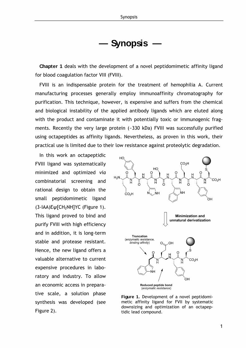
Chapter 1 deals with the development of a novel peptidomimetic affinity ligand
for blood coagulation factor VIII (FVIII).
FVIII is an indispensable protein for the treatment of hemophilia A. Current
manufacturing processes generally employ immunoaffinity chromatography for
purification. This technique, however, is expensive and suffers from the chemical
and biological instability of the applied antibody ligands which are eluted along
with the product and contaminate it with potentially toxic or immunogenic frag-
ments. Recently the very large protein (~330 kDa) FVIII was successfully purified
using octapeptides as affinity ligands. Nevertheless, as proven in this work, their
practical use is limited due to their low resistance against proteolytic degradation.
In this work an octapeptidic
FVIII ligand was systematically
minimized and optimized via
combinatorial screening and
rational design to obtain the
small peptidomimetic ligand
(3-IAA)Eψ[CH2NH]YC (Figure 1).
This ligand proved to bind and
purify FVIII with high efficiency
and in addition, it is long-term
stable and protease resistant.
Hence, the new ligand offers a
valuable alternative to current
expensive procedures in labo-
ratory and industry. To allow
an economic access in prepara-
tive scale, a solution phase
synthesis was developed (see
Figure 2).
Figure 1. Development of a novel peptidomi-metic affinity ligand for FVII by systematic downsizing and optimization of an octapep-tidic lead compound.
Synopsis
2
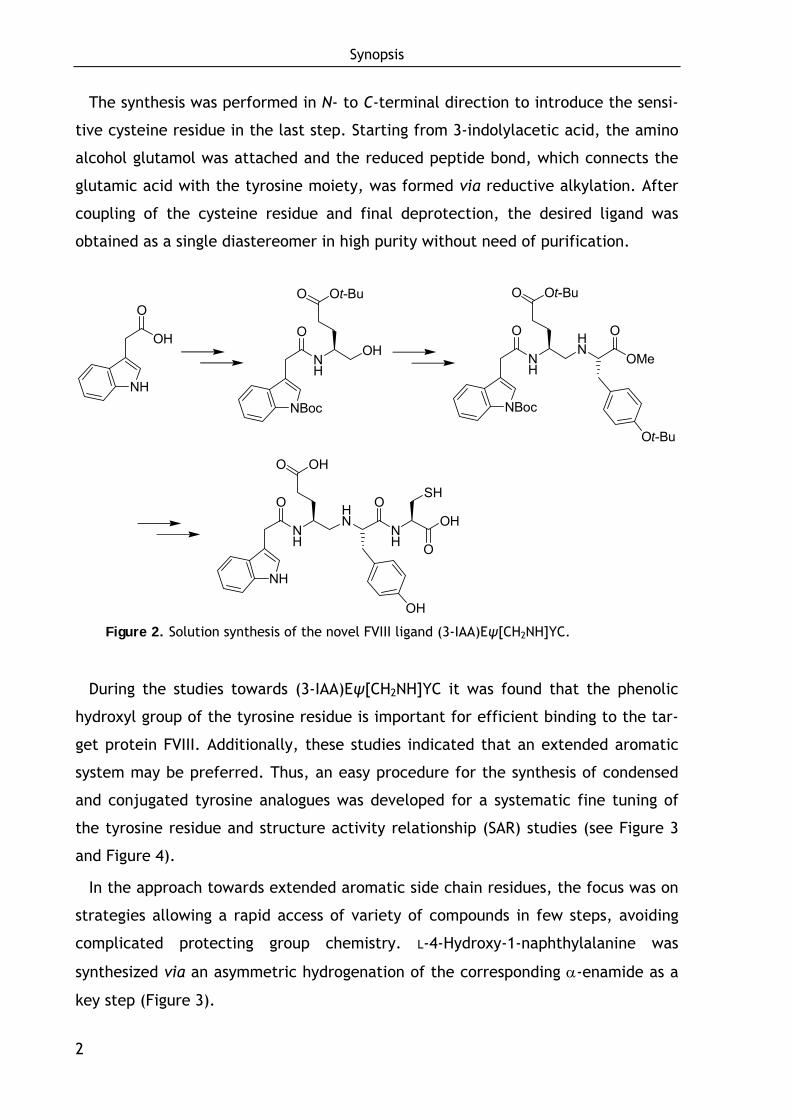
The synthesis was performed in N- to C-terminal direction to introduce the sensi-
tive cysteine residue in the last step. Starting from 3-indolylacetic acid, the amino
alcohol glutamol was attached and the reduced peptide bond, which connects the
glutamic acid with the tyrosine moiety, was formed via reductive alkylation. After
coupling of the cysteine residue and final deprotection, the desired ligand was
obtained as a single diastereomer in high purity without need of purification.
NH
O
NBoc
O Ot-Bu
OHOH
O
NH
NH
O HN
OMe
O
NBoc
O Ot-Bu
Ot-Bu
NH
O HN
NH
OOH
O
NH
O OH
OH
SH
Figure 2. Solution synthesis of the novel FVIII ligand (3-IAA)Eψ[CH2NH]YC.
During the studies towards (3-IAA)Eψ[CH2NH]YC it was found that the phenolic
hydroxyl group of the tyrosine residue is important for efficient binding to the tar-
get protein FVIII. Additionally, these studies indicated that an extended aromatic
system may be preferred. Thus, an easy procedure for the synthesis of condensed
and conjugated tyrosine analogues was developed for a systematic fine tuning of
the tyrosine residue and structure activity relationship (SAR) studies (see Figure 3
and Figure 4).
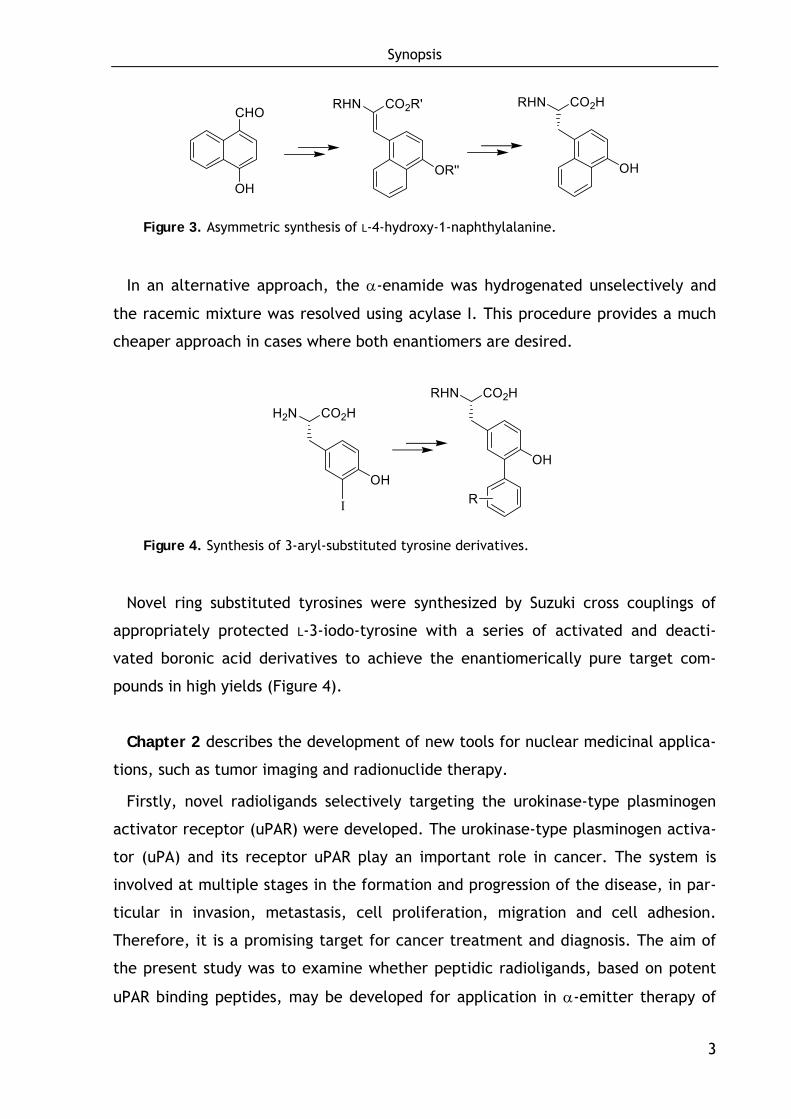
In the approach towards extended aromatic side chain residues, the focus was on
strategies allowing a rapid access of variety of compounds in few steps, avoiding
complicated protecting group chemistry. L-4-Hydroxy-1-naphthylalanine was
synthesized via an asymmetric hydrogenation of the corresponding α-enamide as a
key step (Figure 3).
Synopsis
3
CO2HRHN
OH
CHO
OH
CO2R'RHN
OR''
Figure 3. Asymmetric synthesis of L-4-hydroxy-1-naphthylalanine.
In an alternative approach, the α-enamide was hydrogenated unselectively and
the racemic mixture was resolved using acylase I. This procedure provides a much
cheaper approach in cases where both enantiomers are desired.
CO2HRHN
OH
R
CO2HH2N
OH
I
Figure 4. Synthesis of 3-aryl-substituted tyrosine derivatives.
Novel ring substituted tyrosines were synthesized by Suzuki cross couplings of
appropriately protected L-3-iodo-tyrosine with a series of activated and deacti-
vated boronic acid derivatives to achieve the enantiomerically pure target com-
pounds in high yields (Figure 4).
Chapter 2 describes the development of new tools for nuclear medicinal applica-
tions, such as tumor imaging and radionuclide therapy.
Firstly, novel radioligands selectively targeting the urokinase-type plasminogen
activator receptor (uPAR) were developed. The urokinase-type plasminogen activa-
tor (uPA) and its receptor uPAR play an important role in cancer. The system is
involved at multiple stages in the formation and progression of the disease, in par-
ticular in invasion, metastasis, cell proliferation, migration and cell adhesion.
Therefore, it is a promising target for cancer treatment and diagnosis. The aim of
the present study was to examine whether peptidic radioligands, based on potent
uPAR binding peptides, may be developed for application in α-emitter therapy of

Synopsis
4
disseminated ovarian cancer. Based on the published uPAR inhibitors AE105 and
WX360, mono- and dimeric uPAR-selective ligands equipped with a DOTA chelator
(1,4,7,10-tetraazacyclododecane-1,4,7,10-tetraacetic acid) for radiolabeling were
developed. One compound is shown exemplary in Figure 5.
NH
H2NNH
HN
NH
HN
Ligand
O
O
O
O
O
OH
N
N
N
NHO2C
CO2H
HO2C
O
NH
O
O
H2NNH
HN
NH
HN
Ligand
O
O
O
O
O
OHHN
Ligand = AE105 = Asp-Cha-Phe-(D-Ser)-(D-Arg)-Tyr-Leu-Trp-Ser
Spacer
Figure 5. Structure of a new DOTA-conjugated dimeric uPAR ligand based on the AE105 peptide.
For the synthesis of the dimeric compounds, a facile procedure for N-terminal
cross-linking of solid-phase-bound peptides was worked out, allowing the direct
synthesis of the complete dimeric DOTA-conjugates with high purity (see Figure 6).
Spacer AE105H2N
Spacer AE105H2N
Fmoc-Ser(t-Bu)
Fmoc-Ser(t-Bu)
Spacer Ligand
O
Nε
H
Spacer LigandNε
HO
N
N
N
N
CO2H
Spacer AE105FmocHN
O
Nε
H
Spacer AE105Nε
HO
CO2HOH
OH
HN
O
CO2H
Figure 6. Solid-phase procedure for the synthesis of dimeric DOTA-conjugates.
Synopsis
5
In this procedure, the monomeric peptide strands including the spacer moieties
were built up on solid phase by standard procedures. Thereafter, two single strands
were cross-linked using glutamic acid as bivalent carbonic acid. DOTA was then
attached via the amino terminus of the glutamic acid. The biological evaluation
displayed a specific binding of the new 213Bi-labeled ligands to OV-MZ-6 cancer cells
in vitro as well as in vivo. The latter was demonstrated by specific accumulation of 213Bi-labeled ligands in the tumor tissue of nude mice bearing intraperitoneal OV-
MZ-6-derived tumors. Since kidney uptake of the ligands could be significantly
reduced using gelofusine, the new radiopeptides offer promising options for
therapy of disseminated ovarian cancer.
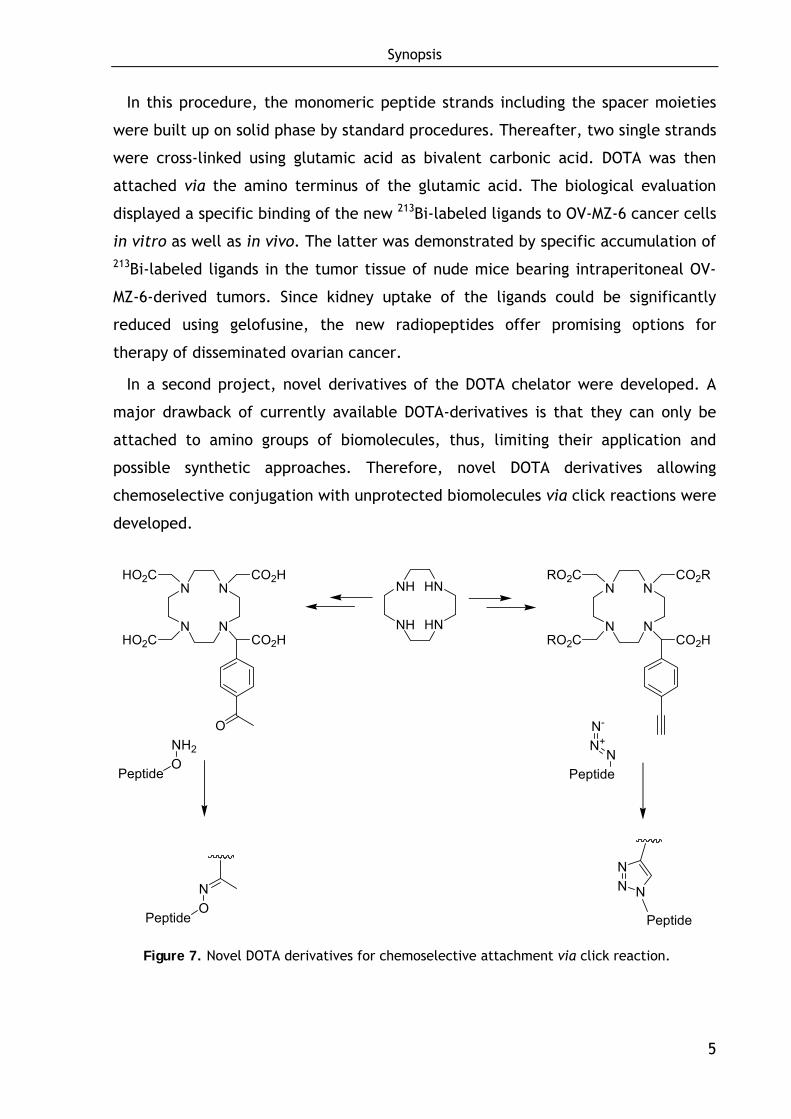
In a second project, novel derivatives of the DOTA chelator were developed. A
major drawback of currently available DOTA-derivatives is that they can only be
attached to amino groups of biomolecules, thus, limiting their application and
possible synthetic approaches. Therefore, novel DOTA derivatives allowing
chemoselective conjugation with unprotected biomolecules via click reactions were
developed.
N
N
N
N
HO2C
HO2C
CO2H
CO2H
O
N
N
N
N
RO2C
RO2C
CO2R
CO2H
NO
Peptide Peptide
NN N
PeptideONH2 N
N+N-
Peptide
NH
NH
HN
HN
Figure 7. Novel DOTA derivatives for chemoselective attachment via click reaction.
Synopsis
6
For the synthesis, 1,4,7,10-tetraazacyclododecane (cyclen) was alkylated with
one equivalent of para-functionalized alkyl 2-bromophenyl-acetate and three
equivalents of tert-butyl 2-bromoacetate (Figure 7). The resulting compounds
having an additional carbonyl or alkyne functionality can be chemoselectively
ligated with appropriately functionalized unprotected biomolecules via oxime
ligation and copper(I)-catalyzed azide-alkyne cycloaddition (Figure 7). This was
demonstrated by the attachment to derivatives of the somatostatin analog Tyr3-
octreotate. Initial biodistribution studies in mice with the radiometalated
compound demonstrated the general applicability of the new chelators.
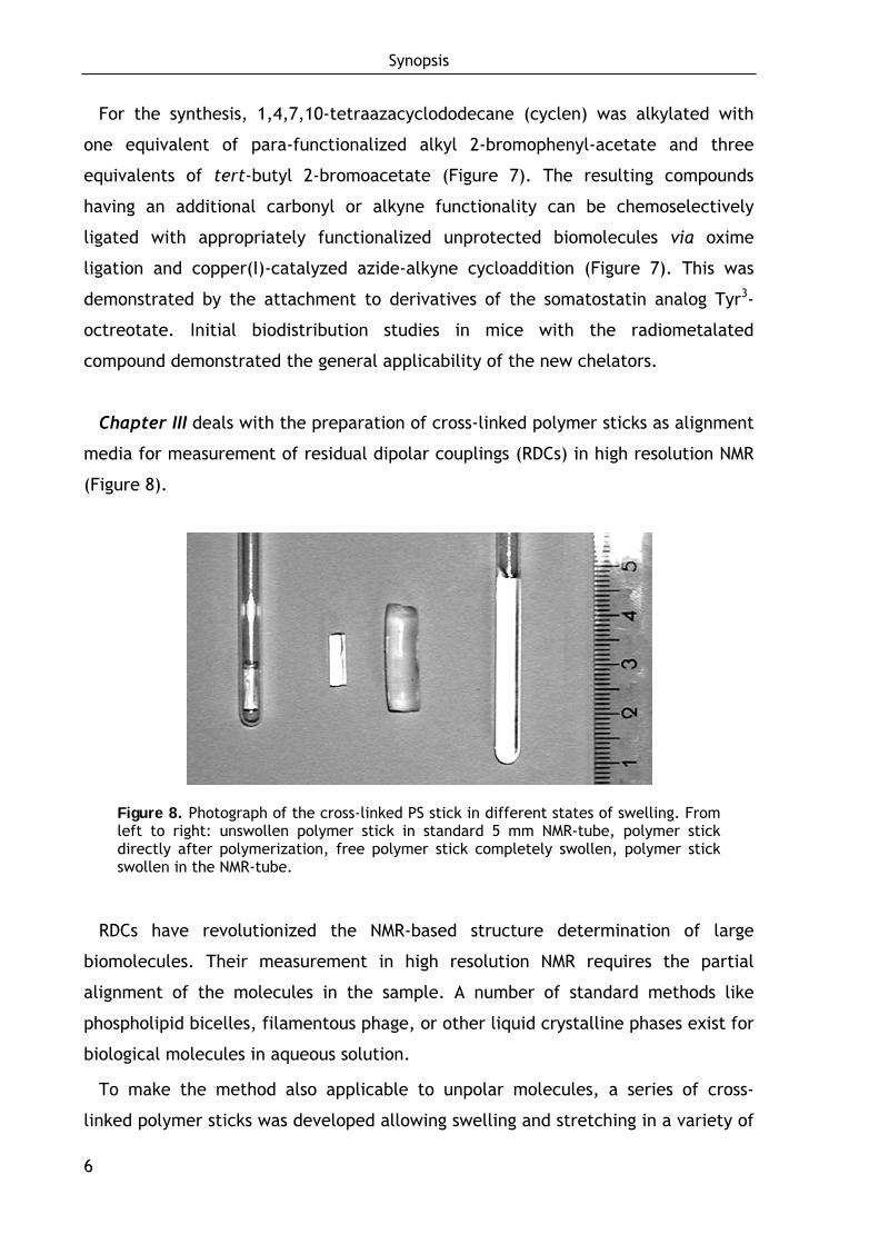
Chapter III deals with the preparation of cross-linked polymer sticks as alignment
media for measurement of residual dipolar couplings (RDCs) in high resolution NMR
(Figure 8).
Figure 8. Photograph of the cross-linked PS stick in different states of swelling. From left to right: unswollen polymer stick in standard 5 mm NMR-tube, polymer stick directly after polymerization, free polymer stick completely swollen, polymer stick swollen in the NMR-tube.
RDCs have revolutionized the NMR-based structure determination of large
biomolecules. Their measurement in high resolution NMR requires the partial
alignment of the molecules in the sample. A number of standard methods like
phospholipid bicelles, filamentous phage, or other liquid crystalline phases exist for
biological molecules in aqueous solution.
To make the method also applicable to unpolar molecules, a series of cross-
linked polymer sticks was developed allowing swelling and stretching in a variety of
Synopsis
7
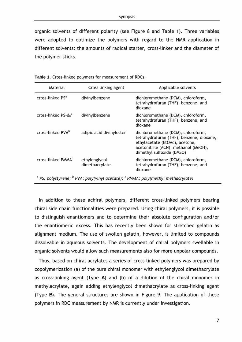
organic solvents of different polarity (see Figure 8 and Table 1). Three variables
were adopted to optimize the polymers with regard to the NMR application in
different solvents: the amounts of radical starter, cross-linker and the diameter of
the polymer sticks.
Table 1. Cross-linked polymers for measurement of RDCs.
Material Cross linking agent Applicable solvents
cross-linked PSa divinylbenzene dichloromethane (DCM), chloroform, tetrahydrofuran (THF), benzene, and dioxane
cross-linked PS-d8a divinylbenzene dichloromethane (DCM), chloroform,
tetrahydrofuran (THF), benzene, and dioxane
cross-linked PVAb adipic acid divinylester dichloromethane (DCM), chloroform, tetrahydrofuran (THF), benzene, dioxane, ethylacetate (EtOAc), acetone, acetonitrile (ACN), methanol (MeOH), dimethyl sulfoxide (DMSO)
cross-linked PMMAc ethylenglycol dimethacrylate
dichloromethane (DCM), chloroform, tetrahydrofuran (THF), benzene, and dioxane
a PS: polystyrene; b PVA: poly(vinyl acetate); c PMMA: poly(methyl methacrylate)
In addition to these achiral polymers, different cross-linked polymers bearing
chiral side chain functionalities were prepared. Using chiral polymers, it is possible
to distinguish enantiomers and to determine their absolute configuration and/or
the enantiomeric excess. This has recently been shown for stretched gelatin as
alignment medium. The use of swollen gelatin, however, is limited to compounds
dissolvable in aqueous solvents. The development of chiral polymers swellable in
organic solvents would allow such measurements also for more unpolar compounds.
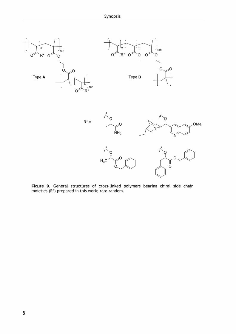
Thus, based on chiral acrylates a series of cross-linked polymers was prepared by
copolymerization (a) of the pure chiral monomer with ethylenglycol dimethacrylate
as cross-linking agent (Type A) and (b) of a dilution of the chiral monomer in
methylacrylate, again adding ethylenglycol dimethacrylate as cross-linking agent
(Type B). The general structures are shown in Figure 9. The application of these
polymers in RDC measurement by NMR is currently under investigation.
Synopsis
8
ran
O
n
n
O
O R*
O R*ran
O
O
rann m
O
O
O R* O O O
O
O
N
OMeN
O
OO
H3CO
O
O
NH2
OO
R* =
Type A Type B
Figure 9. General structures of cross-linked polymers bearing chiral side chain moieties (R*) prepared in this work; ran: random.
Chapter I — Small Molecule FVIII Ligands
9
— Chapter 1 —
1 Development of Small Molecule Ligands
for Affinity Purification of Factor VIII
1.1 Background
The ability of the body to control the flow of blood after vascular injury is indis-
pensable for survival. The process of blood clotting and subsequent dissolution of
the clot, following repair of the injured tissue, is termed hemostasis.[1]
Hemostasis consists of four major events that occur in a certain order after the
loss of vascular integrity:[1]
(1) The initial phase of the process is vascular constriction to limit the flow of
blood to the injured area.
(2) Then, primary hemostasis occurs, wherein platelets become activated by
thrombin and aggregate at the site of injury, forming a loose hemostatic
plug within seconds after injury. Platelets clump by binding to collagen
that becomes exposed following the rupture of the endothelial lining of
vessels.
(3) To insure stability of the initially loose platelet plug, a fibrin mesh forms
and entraps the plug (secondary hemostasis or coagulation). The coagula-
tion pathway involves a complex cascade of coagulation factors, ultimately
resulting in the transformation of fibrinogen into polymerized fibrin,
forming the clot. This process takes several minutes.
Chapter I — Small Molecule FVIII Ligands
10
(4) The attracted clot stimulates the growth of fibroblasts and smooth muscle
cells within the vessel wall and initiates the repair process. Finally the clot
is dissolved in order to allow normal blood flow (fibrinolysis).
1.1.1 Blood coagulation and fibrinolysis
The coagulation is a complex proteolytic cascade which has been proposed for
the first time by Davie et al. in 1964.[2,3] Each enzyme involved is present in the
plasma as a zymogen (inactive form), which is activated by proteolytic cleavage to
release the active factor from the precursor molecule. The coagulation cascade
functions as a series of positive and negative feedback loops which control the
activation process. The ultimate goal is to produce thrombin (factor IIa, FIIa),
which then converts soluble fibrinogen into fibrin, which forms the clot.[2,4-6]
The whole cascade can be divided into three pathways: the intrinsic and extrinsic
pathway, which are initiated by different processes, and which both converge on
the common pathway to finally lead to clot formation (Figure 10). The intrinsic
pathway is initiated by abnormal vessel wall in the absence of tissue injury,
whereas the extrinsic pathway is initiated as a result of tissue injury. Both
pathways are complex and involve numerous different proteins termed clotting
factors.[1,3-5] An overview over the clotting factors and other proteins involved in
coagulation as well as their functions can be found in the appendix (Supplementary
Data 1 and 2 on pages 243-244).
1.1.1.1 The intrinsic pathway
The intrinsic pathway (also termed contact activation pathway) is activated when
blood comes into contact with sub-endothelial connective tissues or with negatively
charged surfaces that are exposed as a result of tissue damage. Quantitatively it is
the more important of the two pathways, but fibrinogen activation proceeds
significantly slower than via the extrinsic pathway. The pathway involves the
clotting factors VIII (FVIII), IX (FIX), X (FX), XI (FXI), and XII (FXII). Also required are
the proteins prekallikrein (PK) and high-molecular-weight kininogen (HMWK), as
well as calcium ions (Ca2+) and phospholipids (PL) secreted from platelets.[2,4-6]
Chapter I — Small Molecule FVIII Ligands
11
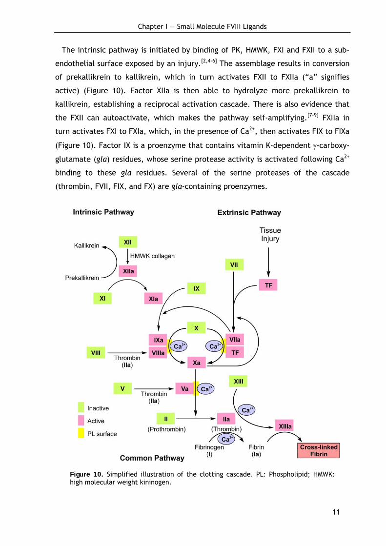
The intrinsic pathway is initiated by binding of PK, HMWK, FXI and FXII to a sub-
endothelial surface exposed by an injury.[2,4-6] The assemblage results in conversion
of prekallikrein to kallikrein, which in turn activates FXII to FXIIa (“a” signifies
active) (Figure 10). Factor XIIa is then able to hydrolyze more prekallikrein to
kallikrein, establishing a reciprocal activation cascade. There is also evidence that
the FXII can autoactivate, which makes the pathway self-amplifying.[7-9] FXIIa in
turn activates FXI to FXIa, which, in the presence of Ca2+, then activates FIX to FIXa
(Figure 10). Factor IX is a proenzyme that contains vitamin K-dependent γ-carboxy-
glutamate (gla) residues, whose serine protease activity is activated following Ca2+
binding to these gla residues. Several of the serine proteases of the cascade
(thrombin, FVII, FIX, and FX) are gla-containing proenzymes.
Figure 10. Simplified illustration of the clotting cascade. PL: Phospholipid; HMWK: high molecular weight kininogen.

Chapter I — Small Molecule FVIII Ligands
12
The intrinsic pathway ultimately activates FX, a process which can also be
triggered by the extrinsic pathway (Figure 10).[2,4-6] The activation of FXa requires
assembly of the intrinsic tenase (also termed Xase) complex (FVIIIa/FIXa/FX) on the
surface of activated platelets which activates FX to FXa in presence of Ca2+. The
role of FVIIIa in this process is to act as a receptor for FIXa and FX and as a cofactor
of FIXa and FX.[10-12] The activation of FVIII to FVIIIa occurs in the presence of
minute quantities of thrombin. As the concentration of thrombin increases, FVIIIa is
ultimately cleaved by thrombin and inactivated. FVIIIa activity is furthermore
controlled by activated protein C (APC) which is an important cofactor inhibitor.[13]
It degrades FVIIIa (and also FVa) and is activated by thrombin in presence of
thrombomodulin (a protein on the surface of endothelial cells) and requires its
coenzyme protein S (PS) to function. The dual action of thrombin (FVIII activation
and deactivation) ensures a limited extent of the tenase complex formation.[11]
1.1.1.2 The extrinsic pathway
The extrinsic pathway (also termed the tissue factor pathway) is an alternative
route for the activation of the clotting cascade.[2,4-6] It provides a very rapid
response to tissue injury, generating activated factor X almost instantly, compared
with the minutes, required for the intrinsic pathway to activate factor X. The main
function of the extrinsic pathway is to augment the activity of the intrinsic
pathway.[14]
There are two components unique to the extrinsic pathway, tissue factor (TF)
and factor VII (FVII). Tissue factor is present in most human cells bound to the cell
membrane and is released at the site of injury. Once activated, circulating FVIIa
binds rapidly to TF which serves as a cofactor in the FVIIa-catalyzed activation of
FX to generate small amounts of FXa (Figure 11a).[15] The TF/FVIIa/FX complex is
termed the extrinsic tenase complex. Factor VIIa, a gla residue containing serine
protease, cleaves FX to FXa in a manner identical to FIXa in the intrinsic pathway.
Factor Xa is the major activator of zymogen FVII (FVII is also activated by FIXa,
FVIIa/TF, and thrombin),[16-22] establishing a reciprocal activation cascade by
catalysis of the formation of more FVIIa and activation of thrombin, which begins
activating the cofactors V and VIII (Figure 11a; compare also Figure 10).[23]
Chapter I — Small Molecule FVIII Ligands
13
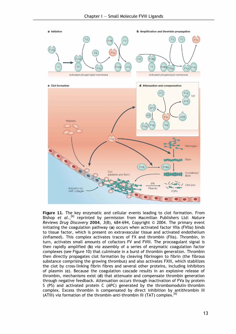
Figure 11. The key enzymatic and cellular events leading to clot formation. From Bishop et al.,[6] reprinted by permission from Macmillan Publishers Ltd: Nature Reviews Drug Discovery 2004, 3(8), 684-694, Copyright © 2004. The primary event initiating the coagulation pathway (a) occurs when activated factor VIIa (FVIIa) binds to tissue factor, which is present on extravascular tissue and activated endothelium (inflamed). This complex activates traces of FX and thrombin (FIIa). Thrombin, in turn, activates small amounts of cofactors FV and FVIII. The procoagulant signal is then rapidly amplified (b) via assembly of a series of enzymatic coagulation factor complexes (see Figure 10) that culminate in a burst of thrombin generation. Thrombin then directly propagates clot formation by cleaving fibrinogen to fibrin (the fibrous substance comprising the growing thrombus) and also activates FXIII, which stabilizes the clot by cross-linking fibrin fibres and several other proteins, including inhibitors of plasmin (c). Because the coagulation cascade results in an explosive release of thrombin, mechanisms exist (d) that attenuate and compensate thrombin generation through negative feedback. Attenuation occurs through inactivation of FVa by protein S (PS) and activated protein C (APC) generated by the thrombomodulin–thrombin complex. Excess thrombin is compensated by direct inhibition by antithrombin III (ATIII) via formation of the thrombin–anti-thrombin III (TAT) complex.[6]

Chapter I — Small Molecule FVIII Ligands
14
The ability of FXa to activate FVII creates a link between the intrinsic and extrin-
sic pathways (see Figure 10). An additional link between the two pathways exists
through the ability of TF and FVIIa to activate FIX (Figure 10).[24] These initial
catalytic events provide the elements for the formation of two essential procoagu-
lant complexes, which sequentially amplify the procoagulant stimulus: FVIIIa/FIXa,
which activates FX, and FXa/FVa, which activates prothrombin to thrombin which
directly propagates clot formation (Figure 11b).[24,25]
A major mechanism for the inhibition of the extrinsic pathway occurs at the acti-
vation of FX by TF/FVIIa. The protein, lipoprotein-associated coagulation inhibitor
(LACI) specifically binds to this complex.[26] LACI is also referred to as extrinsic
pathway inhibitor (EPI) or tissue factor pathway inhibitor (TFPI) and was formerly
named anticonvertin.[2,4-6] The intrinsic and extrinsic systems converge at factor X
to a single common pathway which is responsible for the production of thrombin.[1]
1.1.1.3 The common pathway and clot formation
a) Activation of thrombin and control of thrombin activity:
FXa activates prothrombin (factor II, FII) to thrombin (Figure 11b). Thrombin, in
turn, converts fibrinogen to fibrin (Figure 11c). The activation of thrombin occurs
on the surface of activated platelets and requires the formation of a prothrombi-
nase complex. This complex is composed of the platelet phospholipids, Ca2+, FVa,
FXa, and prothrombin (Figure 11b). FVa functions as cofactor of FXa in the pro-
thrombinase complex, similar to the role of FVIII in the intrinsic tenase complex
formation. Like FVIII activation, FV is activated to FVa by means of minute amounts
of thrombin and is inactivated by increased levels of thrombin. FV activity is
controlled by APC which degrades FVa in presence of cofactor PS (Figure 11d).[13]
In addition to its role in activation of fibrin clot formation, thrombin plays an
important regulatory role in coagulation. Thrombin associates with thrombo-
modulin present on endothelial cell surfaces forming a complex that efficiently
converts PC to APC.[13] The APC/PS complex, in turn, degrades FVa as well as
FVIIIa, thereby limiting the activity of these two factors in the coagulation cascade
and thus thrombin formation. Hence, thrombin is a major regulator of its own
production and of the coagulation cascade.[5,11,15] The activation of thrombin is
Chapter I — Small Molecule FVIII Ligands
15
furthermore regulated by four specific thrombin inhibitors: Antithrombin III (ATIII)
is the most important as it can also inhibit the activities of factors IXa, Xa, XIa and
XIIa.[5,11,15] The activity of ATIII is potentiated in the presence of heparin. This
effect of heparin is the basis for its clinical use as an anticoagulant. Furthermore,
thrombin activity is inhibited by α2-macroglobulin, heparin cofactor II and α1-anti-
trypsin.[5,11,15]
b) Clot formation:
Fibrinogen (factor I, FI) consists of three pairs of polypeptides ([Aα][Bβ][γ])2.[27,28]
The A and B portions of the Aα and Bβ chains comprise the fibrinopeptides, A and
B. The fibrinopeptide regions of fibrinogen are highly negatively charged by several
glutamate and aspartate residues, which improves the solubility of fibrinogen in
plasma.[27,28] Exposure of fibrinogen to thrombin results in rapid proteolysis of
fibrinogen and the release of fibrinopeptide A. The loss of the small peptide A is
not sufficient to render the resulting fibrin molecule insoluble, a process that is re-
quired for clot formation, but it tends to form complexes with adjacent fibrin and
fibrinogen molecules. Then, fibrinopeptide B is cleaved by thrombin, and the fibrin
monomers formed by this second proteolytic cleavage polymerize spontaneously to
form an insoluble gel (Figure 11c). The polymerized fibrin, held together by non-
covalent and electrostatic forces, is stabilized by the transamidating enzyme factor
XIIIa, produced by the action of thrombin on factor XIII (Figure 11c).[27,28] These
insoluble fibrin aggregates together with aggregated platelets, blocks the damaged
blood vessel and prevents further bleeding.[6]
1.1.1.4 Temporal and spatial control of clot formation
As described, the coagulation cascade itself is highly regulated by several positive
and negative feedback loops and inhibitors to ensure blood coagulation in case of
injury and to prevent undesired blood coagulation (e.g. thrombosis). Beside the
temporal control, coagulation is also localized to the site of injury. Recently, this
phenomenon was explained by introduction of the conception of spatial heteroge-
neity into the basic scheme of the coagulation cascade.[13] This conception (as
described below) tries to determine the roles of the different pathways of the
cascade at different temporal and spatial stages of clotting:
Chapter I — Small Molecule FVIII Ligands
16
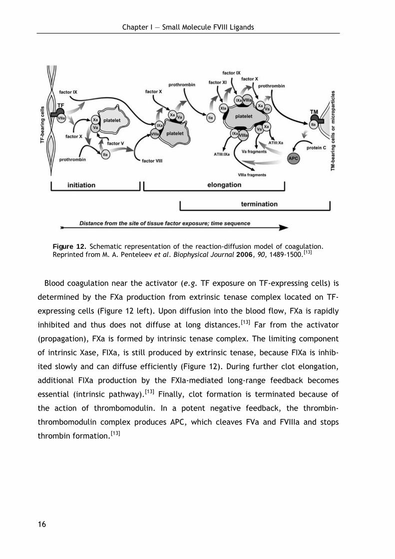
Figure 12. Schematic representation of the reaction-diffusion model of coagulation. Reprinted from M. A. Penteleev et al. Biophysical Journal 2006, 90, 1489-1500.[13]
Blood coagulation near the activator (e.g. TF exposure on TF-expressing cells) is
determined by the FXa production from extrinsic tenase complex located on TF-
expressing cells (Figure 12 left). Upon diffusion into the blood flow, FXa is rapidly
inhibited and thus does not diffuse at long distances.[13] Far from the activator
(propagation), FXa is formed by intrinsic tenase complex. The limiting component
of intrinsic Xase, FIXa, is still produced by extrinsic tenase, because FIXa is inhib-
ited slowly and can diffuse efficiently (Figure 12). During further clot elongation,
additional FIXa production by the FXIa-mediated long-range feedback becomes
essential (intrinsic pathway).[13] Finally, clot formation is terminated because of
the action of thrombomodulin. In a potent negative feedback, the thrombin-
thrombomodulin complex produces APC, which cleaves FVa and FVIIIa and stops
thrombin formation.[13]
Chapter I — Small Molecule FVIII Ligands
17
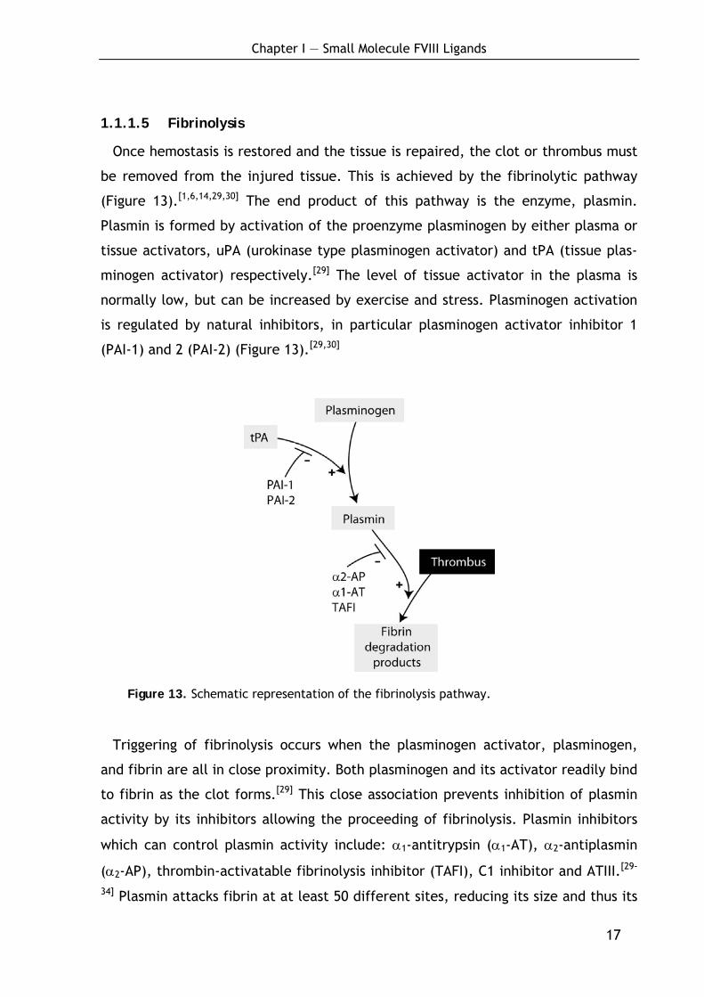
1.1.1.5 Fibrinolysis
Once hemostasis is restored and the tissue is repaired, the clot or thrombus must
be removed from the injured tissue. This is achieved by the fibrinolytic pathway
(Figure 13).[1,6,14,29,30] The end product of this pathway is the enzyme, plasmin.
Plasmin is formed by activation of the proenzyme plasminogen by either plasma or
tissue activators, uPA (urokinase type plasminogen activator) and tPA (tissue plas-
minogen activator) respectively.[29] The level of tissue activator in the plasma is
normally low, but can be increased by exercise and stress. Plasminogen activation
is regulated by natural inhibitors, in particular plasminogen activator inhibitor 1
(PAI-1) and 2 (PAI-2) (Figure 13).[29,30]
Figure 13. Schematic representation of the fibrinolysis pathway.
Triggering of fibrinolysis occurs when the plasminogen activator, plasminogen,
and fibrin are all in close proximity. Both plasminogen and its activator readily bind
to fibrin as the clot forms.[29] This close association prevents inhibition of plasmin
activity by its inhibitors allowing the proceeding of fibrinolysis. Plasmin inhibitors
which can control plasmin activity include: α1-antitrypsin (α1-AT), α2-antiplasmin
(α2-AP), thrombin-activatable fibrinolysis inhibitor (TAFI), C1 inhibitor and ATIII.[29-
34] Plasmin attacks fibrin at at least 50 different sites, reducing its size and thus its
Chapter I — Small Molecule FVIII Ligands
18
hemostatic activity. Many fragments are formed during this process, and some
retain the capacity to polymerize. This may prevent the clot being removed before
the tissue is repaired.[29]
1.1.2 Hemophilia
1.1.2.1 Bleeding disorders: A general overview
It is evident that any defect in the highly controlled cascade can lead to severe
disorders, i.e. to thrombosis or hemophilia. Table 2 summarizes and classifies the
most important bleeding disorders with respect to the kind of dysfunction, severity
and prevalence.
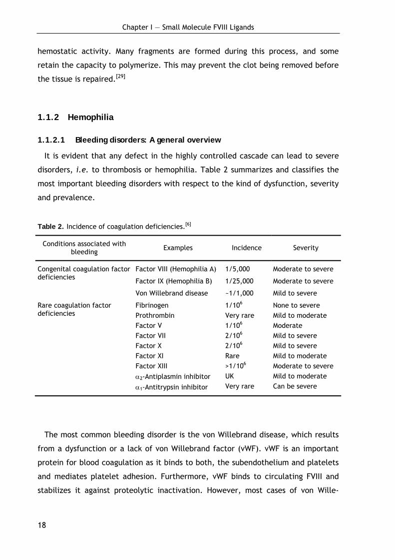
Table 2. Incidence of coagulation deficiencies.[6]
Conditions associated with bleeding Examples Incidence Severity
Congenital coagulation factor deficiencies
Factor VIII (Hemophilia A)
Factor IX (Hemophilia B)
Von Willebrand disease
1/5,000
1/25,000
~1/1,000
Moderate to severe
Moderate to severe
Mild to severe
Rare coagulation factor deficiencies
Fibrinogen Prothrombin Factor V Factor VII Factor X Factor XI Factor XIII
α2-Antiplasmin inhibitor
α1-Antitrypsin inhibitor
1/106 Very rare 1/106 2/106 2/106 Rare >1/106 UK Very rare
None to severe Mild to moderate Moderate Mild to severe Mild to severe Mild to moderate Moderate to severe Mild to moderate Can be severe
The most common bleeding disorder is the von Willebrand disease, which results
from a dysfunction or a lack of von Willebrand factor (vWF). vWF is an important
protein for blood coagulation as it binds to both, the subendothelium and platelets
and mediates platelet adhesion. Furthermore, vWF binds to circulating FVIII and
stabilizes it against proteolytic inactivation. However, most cases of von Wille-
Chapter I — Small Molecule FVIII Ligands
19
brand’s disease are mild; hemarthroses and muscle bleedings occur only in the
most severe type 3 which is uncommon.[6,10]
Hemophilia A results form a lack or dysfunction of FVIII and is one of the most
common and most severe bleeding disorders (Table 2). Incidence and treatment of
hemophilia A will be presented in more detail in the following chapter.
Hemophilia B results from deficiencies in factor IX. The prevalence of hemophilia
B is approximately one-tenth that of hemophilia A (Table 2). All patients with
hemophilia B have prolonged coagulation time and decreased factor IX clotting ac-
tivity. Like hemophilia A, there are severe, moderate and mild forms of hemophilia
B and reflect the factor IX activity in plasma.[6,10] All other bleeding disorders are
rare (see Table 2)[6,10] and shall not be discussed in detail herein.
1.1.2.2 Hemophilia A – The Royal disease
Hemophilia A, also called the Royal disease, is one of the most common and
severe bleeding disorders (Table 2).[35] The name Royal disease arose from the
prevalence of the disease in the royal families of England, Spain, Germany and
Russia, as a result of Queen Victoria of England (1819-1901) being a carrier. The
marriage of Queen Victoria and Prince Albert marked the beginning of hemophilia
in the British royal line. Queen Victoria had nine children and as English royal
family members married into royalty of other countries, the disease eventually
infected most of the royal houses of Europe.[35]
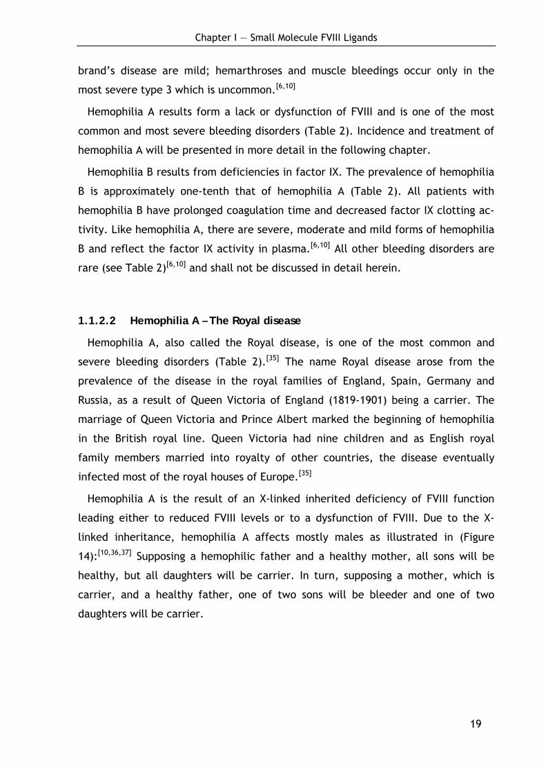
Hemophilia A is the result of an X-linked inherited deficiency of FVIII function
leading either to reduced FVIII levels or to a dysfunction of FVIII. Due to the X-
linked inheritance, hemophilia A affects mostly males as illustrated in (Figure
14):[10,36,37] Supposing a hemophilic father and a healthy mother, all sons will be
healthy, but all daughters will be carrier. In turn, supposing a mother, which is
carrier, and a healthy father, one of two sons will be bleeder and one of two
daughters will be carrier.
Chapter I — Small Molecule FVIII Ligands
20
Figure 14. Inheritance of hemophilia A.
Hemophilia A arises from a variety of mutations. Around 150 different point
mutations have been characterized in the factor VIII gene in hemophilia A.[10,35,38,39]
The incidence of hemophilia A is approximately one in 5000 males, and does not
vary appreciably between populations.[10,35] With respect to the FVIII blood level,
the disease is classified as mild (5-40% of normal), moderate (1-5% of normal) and
severe (<1% of normal) (see also Table 3).
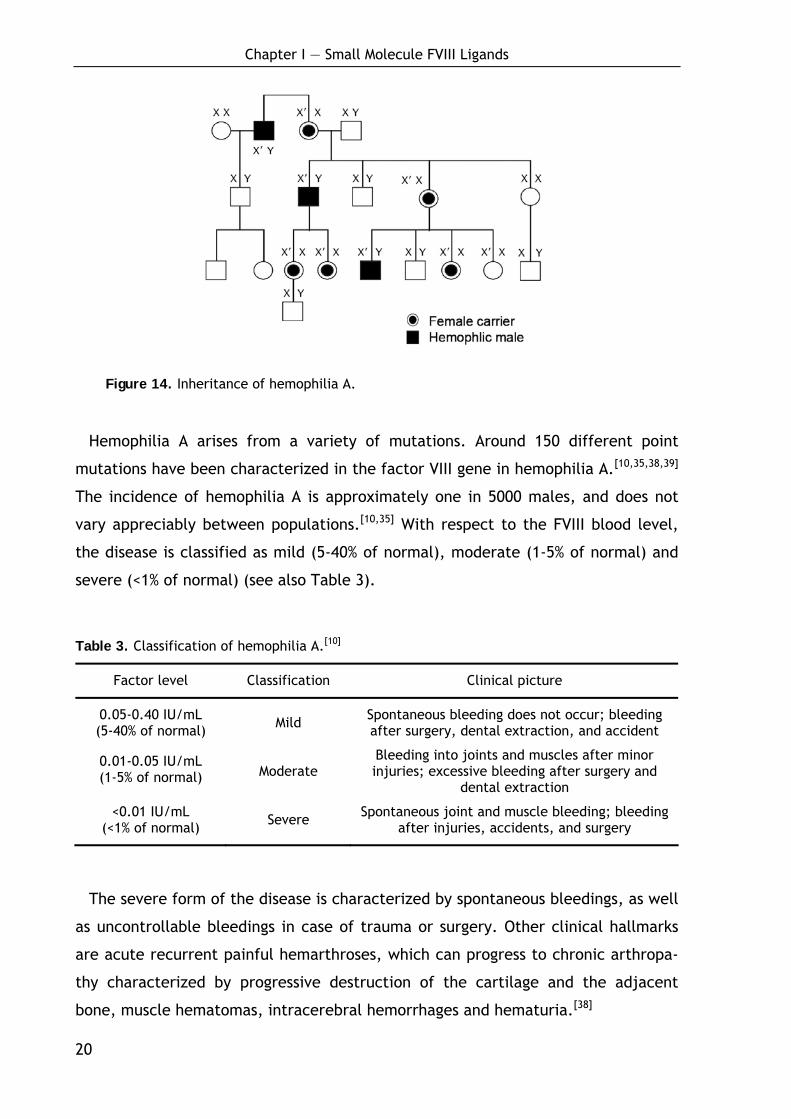
Table 3. Classification of hemophilia A.[10]
Factor level Classification Clinical picture
0.05-0.40 IU/mL (5-40% of normal) Mild Spontaneous bleeding does not occur; bleeding
after surgery, dental extraction, and accident
0.01-0.05 IU/mL (1-5% of normal) Moderate
Bleeding into joints and muscles after minor injuries; excessive bleeding after surgery and
dental extraction
<0.01 IU/mL (<1% of normal) Severe Spontaneous joint and muscle bleeding; bleeding
after injuries, accidents, and surgery
The severe form of the disease is characterized by spontaneous bleedings, as well
as uncontrollable bleedings in case of trauma or surgery. Other clinical hallmarks
are acute recurrent painful hemarthroses, which can progress to chronic arthropa-
thy characterized by progressive destruction of the cartilage and the adjacent
bone, muscle hematomas, intracerebral hemorrhages and hematuria.[38]
Chapter I — Small Molecule FVIII Ligands
21
1.1.2.3 Treatment of hemophilia A
The current treatment for hemophilia A is the infusion of FVIII (replacement
therapy), either purified from human blood plasma (plasma-derived FVIII; pdFVIII)
or expressed in recombinant cells (recombinant FVIII; rFVIII),[6,40-42] which normal-
izes the clotting process and stops or prevents bleeding. Hemophilia A is also an
attractive target for gene-therapy, which is rapidly developing but nevertheless
stands at its beginnings.[10,35,38,43-53]
Hence, the survival and well-being of people with hemophilia depends on the
supply of safe therapeutic products.[54] In the past, numerous hemophilia patients
have been infected with HIV-1 or hepatitis C virus via injections of contaminated
plasma-derived FVIII preparations.[35,40,55] While the safety of pdFVIII products has
been continuously improved during the last decades,[40,56,57] the isolation of the
factor VIII gene in 1984 opened new opportunities for treatment.[58-60] The prepara-
tion of novel recombinant FVIII (rFVIII) molecules significantly improved supply and
product safety.[42,61-64] Nevertheless, rFVIII is a biotechnologically derived product
produced by cell culture and carries a risk of transmitting infectious agents.[65]
Approximately 25% of the first-generation rFVIII concentrates were positive for
transfusion-transmitted viruses from contaminated human serum albumin, which is
added as stabilizer.[66] In contrast, the second-generation rFVIII products like
Kogenate® FS (Bayer) and ReFacto® (Wyeth-Ayerst Pharmacia and Upjohn) the first
licensed B domain deleted rFVIII (BDD-rFVIII) molecule (the FVIII structure will be
discussed in detail in the following chapter),[67] do not have added albumin and
instead use sucrose or other non-human derived material as a stabilizer.[42] This
advancement significantly improved the product safety, so the therapeutic use of
recombinant FVIII has been significantly increased in the past years.[68]
However, as long as human albumin is added to the cell culture media and mono-
clonal antibodies (mAbs) are used as ligands in affinity purification there is still a
risk of transfusing pathogens. Especially transmissible spongiform encephalopathies
and new variants of Creutzfeldt-Jakob disease as well as previously unknown
pathogens, including new murine viruses, may contaminate today’s rFVIII prod-
ucts.[65,69] Hence, all efforts should be made to eliminate all human or animal
proteins from the manufacturing process of recombinant products.[70] Consequently
Chapter I — Small Molecule FVIII Ligands
22
there is a great demand for novel procedures avoiding such components in order to
develop the next generation of recombinant FVIII products.
Besides product safety, financial aspects are major factors in the development of
new FVIII products. While 88% of the safer but more expensive rFVIII is consumed in
Europe and North America, the majority of the world’s population with hemophilia
is reliant on blood products or does not receive any treatment at all due to eco-
nomic reasons.[40,41,54,55,71] Novel techniques reducing production costs can help to
improve the FVIII supply and make treatment accessible for a broader range of the
population.
1.1.3 FVIII structure and mechanism of cofactor action
FVIII is a large secretory glycoprotein containing 2351 amino acids. The first 19
amino acids serve as a signal peptide for translocation of the protein to the
endoplasmatic reticulum and later for the export out of the cell.[60] Without the
signal peptide the FVIII molecule has a molecular weight of ~330 kDa (2332 amino
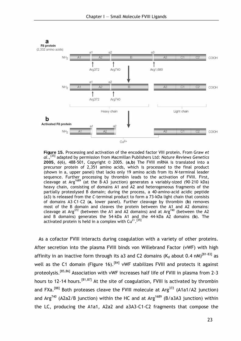
acids) with a multi-domain structure A1-a1-A2-a2-B-a3-A3-C1-C2 (see Figure
15).[12,59,60,72,73] The A and C domains (A1 (FVIII1-329), A2 (FVIII380-711), A3 (FVIII1649-
2019), C1 (FVIII2020-2172), C2 (FVIII2173-2332)) provide 30-40% homology with the A and C
domains of the structurally related proteins ceruloplasmin and factor V, whereas
the B domain and the acidic regions a1, a2 and a3 with a high density of negatively
charged amino acids Asp and Glu are distinctive for FVIII.[72,74,75] The large B domain
is not important for the FVIII procoagulant activity[76] but recent studies demon-
strated its role in protecting the activated form of FVIII from proteolytic inactiva-
tion.[77]
Prior to its secretion into plasma, FVIII is processed intracellularly to a series of
noncovalently associated, Cu2+-linked heterodimers by cleavage at the B-A3 junc-
tion (Figure 15).[12,78,79] The cleavage generates the heavy chain (HC) consisting of
the A1, A2 and B domains and the light chain (LC) composed of the A3, C1 and C2
domains.[12,35] The resulting protein varies in size due to additional cleavages within
the B domain, giving molecules of different length. Thus, preparations of plasma-
derived human FVIII (pdFVIII) contain a heterogeneous mixture of differently sized
FVIII molecules.[72,80]
Chapter I — Small Molecule FVIII Ligands
23
Figure 15. Processing and activation of the encoded factor VIII protein. From Graw et al.,[35] adapted by permission from Macmillan Publishers Ltd: Nature Reviews Genetics 2005, 6(6), 488-501, Copyright © 2005. (a,b) The FVIII mRNA is translated into a precursor protein of 2,351 amino acids, which is processed to the final product (shown in a, upper panel) that lacks only 19 amino acids from its N-terminal leader sequence. Further processing by thrombin leads to the activation of FVIII. First, cleavage at Arg1689 (at the B–A3 junction) generates a variably-sized (90–210 kDa) heavy chain, consisting of domains A1 and A2 and heterogeneous fragments of the partially proteolysed B domain; during the process, a 40-amino-acid acidic peptide (a3) is released from the C-terminal product to form a 73-kDa light chain that consists of domains A3–C1–C2 (a, lower panel). Further cleavage by thrombin (b) removes most of the B domain and cleaves the protein between the A1 and A2 domains: cleavage at Arg372 (between the A1 and A2 domains) and at Arg740 (between the A2 and B domains) generates the 54-kDa A1 and the 44-kDa A2 domains (b). The activated protein is held in a complex with Cu2+.[35]
As a cofactor FVIII interacts during coagulation with a variety of other proteins.
After secretion into the plasma FVIII binds von Willebrand Factor (vWF) with high
affinity in an inactive form through its a3 and C2 domains (Kd about 0.4 nM)[81-83] as
well as the C1 domain (Figure 16).[84] vWF stabilizes FVIII and protects it against
proteolysis.[85,86] Association with vWF increases half life of FVIII in plasma from 2-3
hours to 12-14 hours.[81,87] At the site of coagulation, FVIII is activated by thrombin
and FXa.[88] Both proteases cleave the FVIII molecule at Arg372 (A1a1/A2 junction)
and Arg740 (A2a2/B junction) within the HC and at Arg1689 (B/a3A3 junction) within
the LC, producing the A1a1, A2a2 and a3A3-C1-C2 fragments that compose the

Chapter I — Small Molecule FVIII Ligands
24
heterotrimeric-activated FVIII (see Figure 15 and Figure 16). The cleavage at Arg1689
results in removal of a3 and release of vWF.[83] This cleavage is important for maxi-
mal cofactor activity of FVIIIa.[89] The heterotrimeric FVIIIa binds to negatively
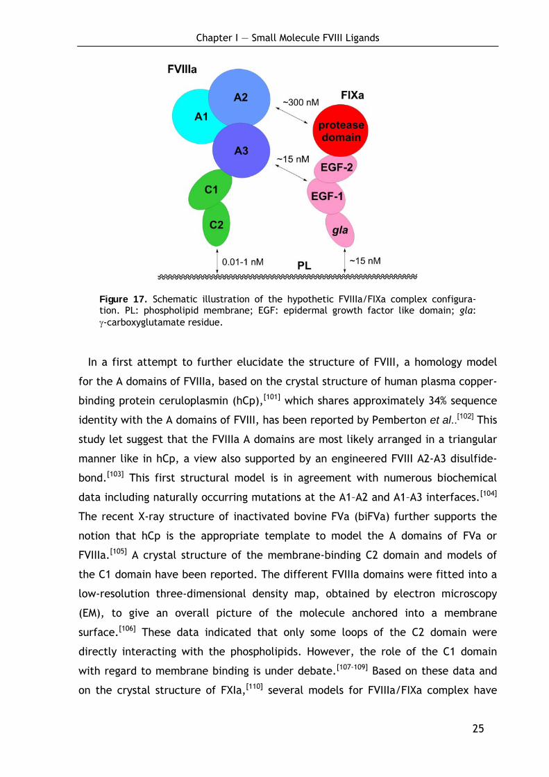
charged phospholipids (PLs) via the C2 domain[90-93] (Figure 16 and Figure 17) and to
factor IXa via the A2 and A3 domains[94-99] to form the intrinsic tenase complex with
FXa by binding via the C2 domain (Figure 16).[100]
Figure 16. Major functional binding sites and epitopes of natural ligands on the factor VIII molecule (cleavage sites are indicated in italics). PL: phospholipid. Modified from Klinge et al..[38]
The exact structure of FVIII and the tenase complex is so far unknown. However,
it is clear that the high affinity interaction of FVIIIa and FIXa (Kd ~15 nM) is mainly
provided by residues FVIII1811-1818 within the A3 domain of the LC of FVIII and the
epidermal growth factor (EGF)-1 like domain of FIXa (Figure 17). The A2 domain is
also involved in FIXa interaction via two regions (FVIII558-565 and FVIII484-508). These
binding sites only become exposed after the cleavage at Arg372 by thrombin of FXa.
Although the affinity of the isolated A2 subunit for FIXa is rather low (Kd ~300 nM),
this interaction defines the cofactor activity of FVIIIa because the A2 domain
amplifies the enzymatic activity of FIXa by modulating its active site.
Chapter I — Small Molecule FVIII Ligands
25
Figure 17. Schematic illustration of the hypothetic FVIIIa/FIXa complex configura-tion. PL: phospholipid membrane; EGF: epidermal growth factor like domain; gla: γ-carboxyglutamate residue.
In a first attempt to further elucidate the structure of FVIII, a homology model
for the A domains of FVIIIa, based on the crystal structure of human plasma copper-
binding protein ceruloplasmin (hCp),[101] which shares approximately 34% sequence
identity with the A domains of FVIII, has been reported by Pemberton et al..[102] This
study let suggest that the FVIIIa A domains are most likely arranged in a triangular
manner like in hCp, a view also supported by an engineered FVIII A2-A3 disulfide-
bond.[103] This first structural model is in agreement with numerous biochemical
data including naturally occurring mutations at the A1–A2 and A1–A3 interfaces.[104]
The recent X-ray structure of inactivated bovine FVa (biFVa) further supports the
notion that hCp is the appropriate template to model the A domains of FVa or
FVIIIa.[105] A crystal structure of the membrane-binding C2 domain and models of
the C1 domain have been reported. The different FVIIIa domains were fitted into a
low-resolution three-dimensional density map, obtained by electron microscopy
(EM), to give an overall picture of the molecule anchored into a membrane
surface.[106] These data indicated that only some loops of the C2 domain were
directly interacting with the phospholipids. However, the role of the C1 domain
with regard to membrane binding is under debate.[107-109] Based on these data and
on the crystal structure of FXIa,[110] several models for FVIIIa/FIXa complex have
Chapter I — Small Molecule FVIII Ligands
26
been reported so far,[79,108,111,112] but the exact structure and the arrangement of
FVIIIa domains remain unclear.[108]
1.1.4 FVIII production and purification
The purification of FVIII remains a challenging task. It involves a complex se-
quence of different purification techniques such as affinity chromatography,[113]
ion-exchange chromatography and virus inactivation.[67,114-120] Today, all recombi-
nant FVIII preparations and many plasma-derived FVIII products are purified via
immunoaffinity chromatography employing monoclonal antibodies (mAbs) as
ligands.[42,121,122] Nevertheless, the use of protein ligands in affinity chromatography
is not only very expensive but also limited by several other factors.[123-126] Antibod-
ies are known to be eluted along with the product contaminating or inactivating it
or even evoking immune responses.[114,118,126] As biologically derived products, they
show lot-to-lot variation and they may be contaminated with e.g. host DNA, viruses
or prions which can be transfused to the product.[125] Their low stability shortens
the column life and they suffer from low binding capacities, limited life cycles and
low scale-up potential.[123,125,126] Moreover, because of the very strong binding of
antibody ligands to FVIII, harsh conditions are required for elution of the protein
which can harm both the target protein and the ligand.[127]
Synthetic ligands like peptides had so far only limited use in affinity separation.
However, the introduction of combinatorial libraries has expanded the repertoire
of peptide-based affinity ligands.[128] Two independent groups recently reported
the development of oligo- and polypeptides as affinity ligands for factor VIII. Kelley
et al.[129,130] described the isolation of a 27 amino acid sequence using phage
display techniques. The cyclic polypeptide is currently used in the manufacturing of
ReFacto AF (Wyeth), a third-generation product, currently in clinical trial.[119]
Another promising result was reported by the group of Jungbauer.[131,132] They
found a series of octapeptides with high affinity towards FVIII, derived from a
combinatorial library using spot technology on cellulose sheets. The immobilized
ligands could be used to purify FVIII from diluted plasma.

Chapter I — Small Molecule FVIII Ligands
27
Nevertheless, the application of oligo- and polypeptides is associated with several
problems restricting their use, above all their high susceptibility towards enzymatic
degradation.[123,126] This significantly limits their application as raw materials such
as serum or cell culture extracts contain proteases. A degradation of the ligands,
covalently bound to the column medium, may rapidly lead to inefficiency and
reduced selectivity and consequently shorten the life of the expensive affinity
column. Furthermore, peptides, although cheaper than antibodies, are still quite
cost-intensive and not trivial to produce in large scale, as they are typically synthe-
sized on solid phase and purified by HPLC.
Even so, peptidic ligands are promising lead structures for the development of
small unnatural molecules. Such compounds have the potential to reduce produc-
tion costs and to improve the safety of current and future FVIII products. The
present study describes the systematic downsizing of an octapeptidic FVIII ligand
into a small peptidomimetic ligand. This novel ligand is protease-resistant and
binds FVIII with high affinity. Using this ligand, FVIII of high purity was isolated
from a complex mixture containing contaminant proteins in vast excess. Moreover,
a stereoselective straightforward solution synthesis was developed allowing cost-
effective production of the ligand in preparative scale.
1.1.5 General synthetic aspects: Solid-phase peptide synthesis
Modern solid-phase peptide synthesis (SPPS) is a powerful tool for lead structure
optimization as it offers the opportunity to synthesize large libraries of compounds
in a short time.[133-137]
Peptide chemistry and synthesis have gone through dramatic changes since
T. Curtius[138] and E. Fischer[139] have synthesized the first simple peptide deriva-
tives. The interest in preparing these chains of amino acids linked by amide bonds
stemmed from the observation made by F. Hofmeister[140] and E. Fischer,[141] that
proteins are polymers consisting of amino acids. Although methods for the coupling
of two amino acids were available, it was due to failures in the development of
applicable protecting groups that substantially delayed further progress in peptide
chemistry.[139,142] A first breakthrough occurred in 1932 when M. Bergmann intro-
duced the benzyloxycarbonyl (Boc) protecting-group.[143] A further milestone was

Chapter I — Small Molecule FVIII Ligands
28
marked by R. B. Merrifield in 1963 with the development of the automated peptide
synthesis on solid support, a copolymer of styrene cross-linked with 1-2% divinyl-
benzene.[133,134]
The SPPS technique substantially facilitates the practical peptide synthesis
procedure, because the anchoring of the C-terminal amino acid to the insoluble
support allows the use of an excess of amino acids and coupling reagents, which
enables almost quantitative yields for the coupling steps of small peptides.
Furthermore, after reaction, coupling reagents and by-products can be removed by
simple filtration.
Scheme 1. General procedure for solid-phase peptide synthesis (SPPS) following
Fmoc-strategy.
NH
R1Fmoc
O
OH
PG
NH
R1
O
PG
OOH
NFmoc
R2PG
NH
R1
O
OHOH
N
RxO
Rn+1
H2N
H2N
R1
O
PG
O
NH
R1Fmoc
O
PG
OLinker+
step 1: resin loading
Linker
Linker
step 2: N-terminal deprotection
step 3: couplingLinker
step 4:
step 5: cleavage from solid support
repeat steps 2 and 3n-times
n-1 linear peptide
The synthesis of peptides longer than two amino acids requires the utilization of
orthogonal temporary and permanent protecting groups.[144] Among others,[145,146]
the Boc-strategy[133,134] and the later developed 9-fluorenylmethoxycarbonyl
(Fmoc)/t-Bu-strategy[135,137,147] are most prominent today. The Fmoc/tBu-strategy,
which has proved to be especially practicable in combination with SPPS (Scheme
Chapter I — Small Molecule FVIII Ligands
29
1), was used for the synthesis of all peptidic ligands described in the following
chapters.
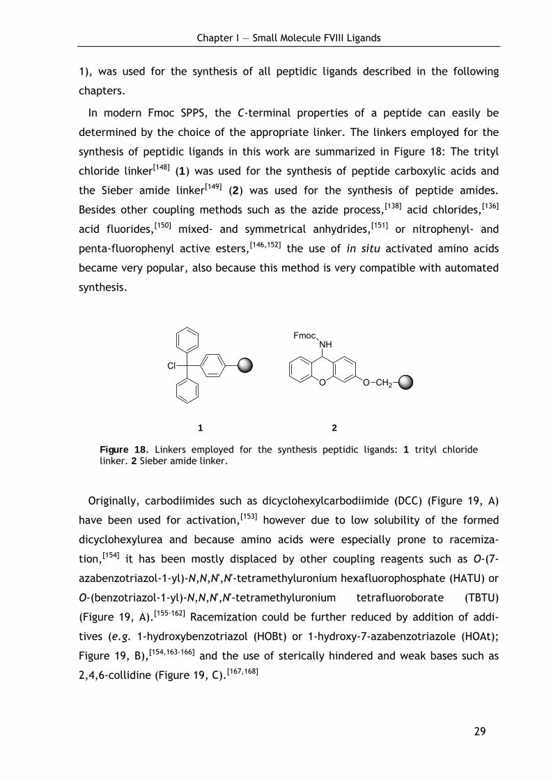
In modern Fmoc SPPS, the C-terminal properties of a peptide can easily be
determined by the choice of the appropriate linker. The linkers employed for the
synthesis of peptidic ligands in this work are summarized in Figure 18: The trityl
chloride linker[148] (1) was used for the synthesis of peptide carboxylic acids and
the Sieber amide linker[149] (2) was used for the synthesis of peptide amides.
Besides other coupling methods such as the azide process,[138] acid chlorides,[136]
acid fluorides,[150] mixed- and symmetrical anhydrides,[151] or nitrophenyl- and
penta-fluorophenyl active esters,[146,152] the use of in situ activated amino acids
became very popular, also because this method is very compatible with automated
synthesis.
O
NHFmoc
O CH2
Cl
1 2
Figure 18. Linkers employed for the synthesis peptidic ligands: 1 trityl chloride linker. 2 Sieber amide linker.
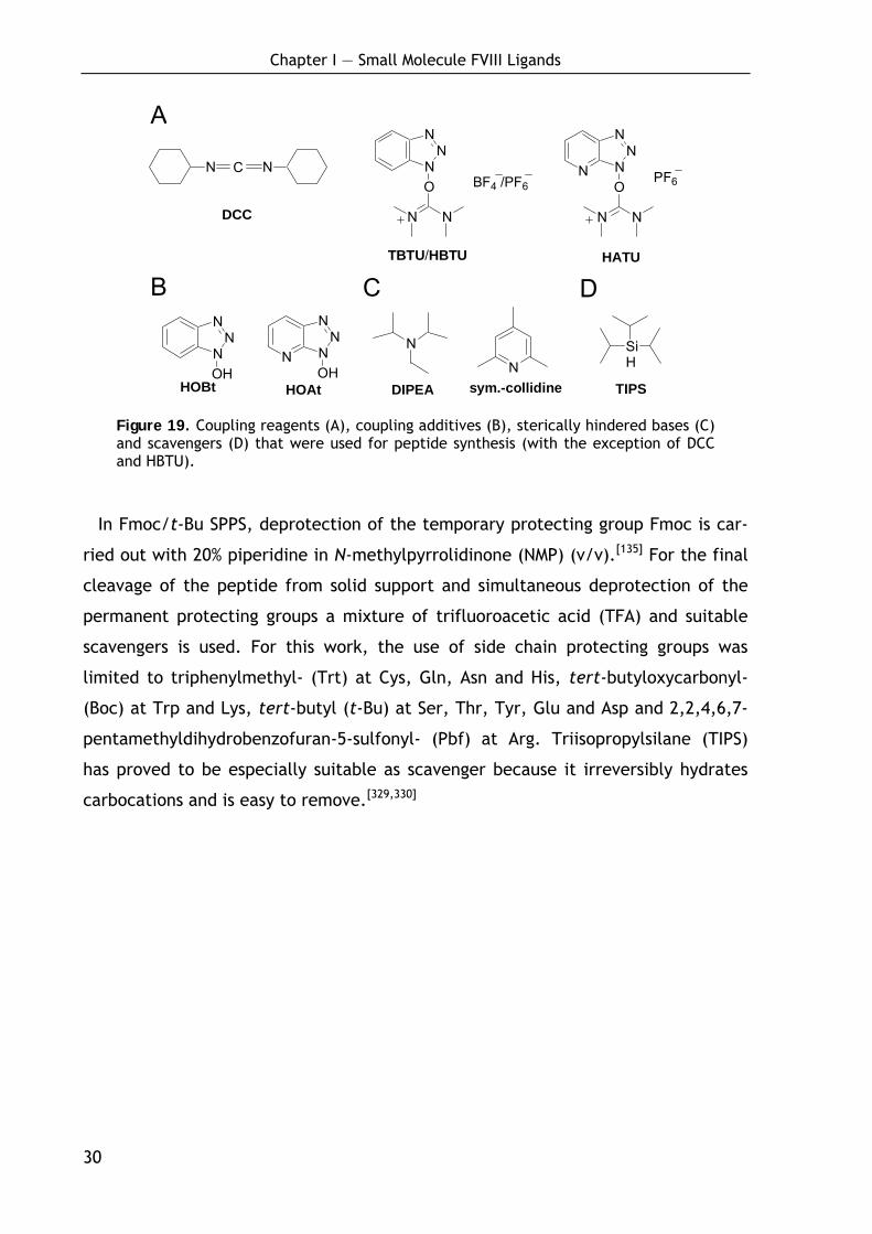
Originally, carbodiimides such as dicyclohexylcarbodiimide (DCC) (Figure 19, A)
have been used for activation,[153] however due to low solubility of the formed
dicyclohexylurea and because amino acids were especially prone to racemiza-
tion,[154] it has been mostly displaced by other coupling reagents such as O-(7-
azabenzotriazol-1-yl)-N,N,N',N'-tetramethyluronium hexafluorophosphate (HATU) or
O-(benzotriazol-1-yl)-N,N,N',N'-tetramethyluronium tetrafluoroborate (TBTU)
(Figure 19, A).[155-162] Racemization could be further reduced by addition of addi-
tives (e.g. 1-hydroxybenzotriazol (HOBt) or 1-hydroxy-7-azabenzotriazole (HOAt);
Figure 19, B),[154,163-166] and the use of sterically hindered and weak bases such as
2,4,6-collidine (Figure 19, C).[167,168]
Chapter I — Small Molecule FVIII Ligands
30
N NC
DCC
NN
N
O
N N
BF4 /PF6N N
NN
O
NN
HATU
PF6
NN
N
OHN N
NN
OHHOBt HOAt
A
B C
N
N
D
SiH
TIPS
TBTU/HBTU
DIPEA sym.-collidine
Figure 19. Coupling reagents (A), coupling additives (B), sterically hindered bases (C) and scavengers (D) that were used for peptide synthesis (with the exception of DCC and HBTU).
In Fmoc/t-Bu SPPS, deprotection of the temporary protecting group Fmoc is car-
ried out with 20% piperidine in N-methylpyrrolidinone (NMP) (v/v).[135] For the final
cleavage of the peptide from solid support and simultaneous deprotection of the
permanent protecting groups a mixture of trifluoroacetic acid (TFA) and suitable
scavengers is used. For this work, the use of side chain protecting groups was
limited to triphenylmethyl- (Trt) at Cys, Gln, Asn and His, tert-butyloxycarbonyl-
(Boc) at Trp and Lys, tert-butyl (t-Bu) at Ser, Thr, Tyr, Glu and Asp and 2,2,4,6,7-
pentamethyldihydrobenzofuran-5-sulfonyl- (Pbf) at Arg. Triisopropylsilane (TIPS)
has proved to be especially suitable as scavenger because it irreversibly hydrates
carbocations and is easy to remove.[329,330]
Chapter I — Small Molecule FVIII Ligands
31
1.2 Systematic lead optimization and minimization: From an
octa-peptide to an unnatural dipeptide
In advance: The biochemical experiments within this project were mainly per-
formed by Dr. A. Khrenov in the laboratories of Prof. E. L. Saenko (University of
Maryland; Baltimore/USA). Further studies were performed by Dr. A. Benhida,
Dr. S. Grailly in the laboratories of Prof. J.-M. Saint-Remy (University of Leuven,
Leuven/Belgium), by Dr. R. Schwaab in the laboratories of Prof. J. Oldenburg
(University of Bonn, Bonn/Germany), by Dr. N. Beaufort in the laboratories of
PD Dr. V. Magdolen as well as by B. Laufer from our group. The quantitative binding
data of all compounds are summarized in the Supplementary Data 4, page 246ff.
1.2.1 Preliminary studies. Development of the binding assay and
selection of the lead structure
For measurement of the FVIII binding ability of the ligands, a microbead assay
based on Jungbauer’s procedures[131,132] was developed in which the binding of 125I-labeled pdFVIII to the immobilized ligands is measured. The affinity resin Toyo-
pearl AF-Epoxy-650M (Tosoh Bioscience, Stuttgart, Germany) was chosen as solid
support giving best binding results and enabling a chemoselective immobilization of
the peptides via a cysteine residue.
Kinetic studies of the immobilization reaction gave that the maximal loading
density is reached after two days of incubation, independent of the peptide
sequence and the amounts of peptide used (see Supplementary Data 3, page 244).
This time frame is also required for the hydrolysis of unoccupied epoxy groups.
After scanning various octapeptides from Jungbauer’s libraries[131,132] the ligand
EYHSWEYC (3) was selected as the most promising lead candidate binding ~50% of
the applied FVIII at a loading density of 10.3 μmol per mL resin. The applied FVIII
concentration of about 0.7 nM let suggest a subnanomolar affinity for FVIII. Beside
its high affinity to pdFVIII, the peptide proved to bind B domain-deleted (BDD)-
Chapter I — Small Molecule FVIII Ligands
32
rFVIII as well as full-length rFVIII (FL-rFVIII) with similar affinity (see Table 7
below). This was an important feature with respect to the goal of developing a
ligand with general applicability. The introduction of an additional spacer molecule
(e.g. 6-amino hexanoic acid, Aha) did not influence the binding ability (see ligand 4
in Figure 21 below), indicating that the peptide does not bind into a deep binding
pocket rather than to the surface of factor VIII.[169]
Therefore, it was decided to stay with the micro-beads binding assay and the
studies were performed with the immobilized ligands, since the binding of the
ligand to FVIII might be significantly affected by the solid support itself or by the
presentation of the ligand on the resin surface. As a consequence, the measured
effects were the combined results of both, the modified sequence itself and
influences coming along with changes of e.g. the loading densities and/or an
altered presentation of the ligands on the resin surface. This made the systematic
combinatorial design more complex but gave directly the practical binding results
of the affinity material, ready to use.
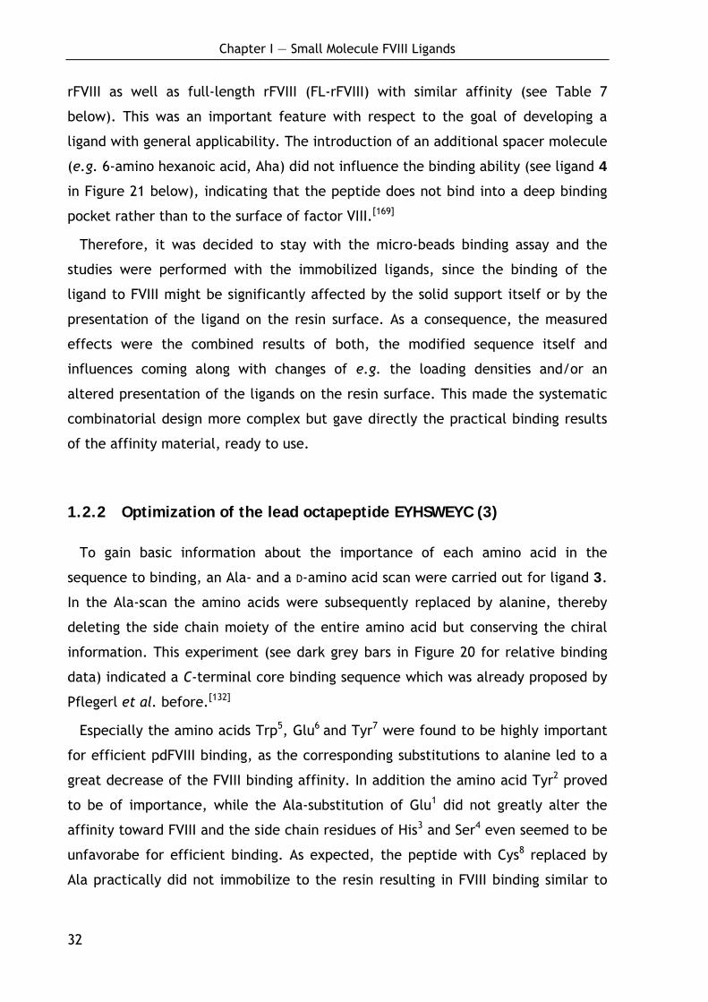
1.2.2 Optimization of the lead octapeptide EYHSWEYC (3)
To gain basic information about the importance of each amino acid in the
sequence to binding, an Ala- and a D-amino acid scan were carried out for ligand 3.
In the Ala-scan the amino acids were subsequently replaced by alanine, thereby
deleting the side chain moiety of the entire amino acid but conserving the chiral
information. This experiment (see dark grey bars in Figure 20 for relative binding
data) indicated a C-terminal core binding sequence which was already proposed by
Pflegerl et al. before.[132]
Especially the amino acids Trp5, Glu6 and Tyr7 were found to be highly important
for efficient pdFVIII binding, as the corresponding substitutions to alanine led to a
great decrease of the FVIII binding affinity. In addition the amino acid Tyr2 proved
to be of importance, while the Ala-substitution of Glu1 did not greatly alter the
affinity toward FVIII and the side chain residues of His3 and Ser4 even seemed to be
unfavorabe for efficient binding. As expected, the peptide with Cys8 replaced by
Ala practically did not immobilize to the resin resulting in FVIII binding similar to
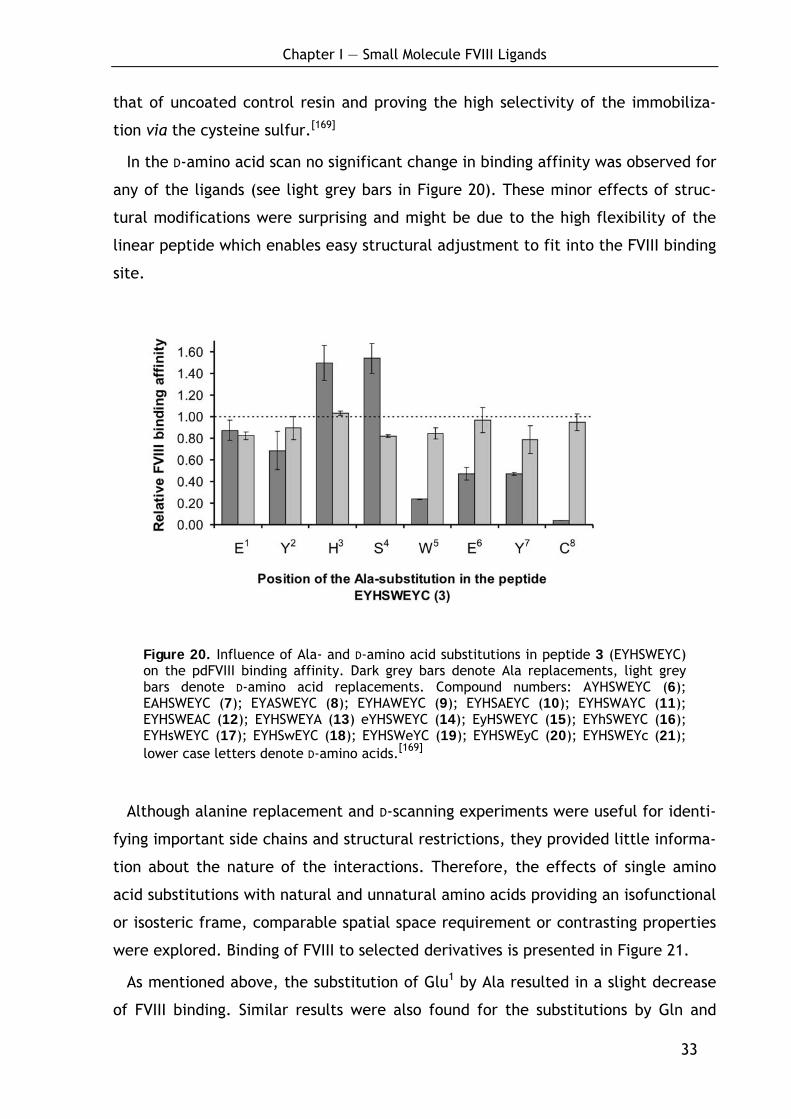
Chapter I — Small Molecule FVIII Ligands
33
that of uncoated control resin and proving the high selectivity of the immobiliza-
tion via the cysteine sulfur.[169]
In the D-amino acid scan no significant change in binding affinity was observed for
any of the ligands (see light grey bars in Figure 20). These minor effects of struc-
tural modifications were surprising and might be due to the high flexibility of the
linear peptide which enables easy structural adjustment to fit into the FVIII binding
site.
Figure 20. Influence of Ala- and D-amino acid substitutions in peptide 3 (EYHSWEYC) on the pdFVIII binding affinity. Dark grey bars denote Ala replacements, light grey bars denote D-amino acid replacements. Compound numbers: AYHSWEYC (6); EAHSWEYC (7); EYASWEYC (8); EYHAWEYC (9); EYHSAEYC (10); EYHSWAYC (11); EYHSWEAC (12); EYHSWEYA (13) eYHSWEYC (14); EyHSWEYC (15); EYhSWEYC (16); EYHsWEYC (17); EYHSwEYC (18); EYHSWeYC (19); EYHSWEyC (20); EYHSWEYc (21); lower case letters denote D-amino acids.[169]
Although alanine replacement and D-scanning experiments were useful for identi-
fying important side chains and structural restrictions, they provided little informa-
tion about the nature of the interactions. Therefore, the effects of single amino
acid substitutions with natural and unnatural amino acids providing an isofunctional
or isosteric frame, comparable spatial space requirement or contrasting properties
were explored. Binding of FVIII to selected derivatives is presented in Figure 21.
As mentioned above, the substitution of Glu1 by Ala resulted in a slight decrease
of FVIII binding. Similar results were also found for the substitutions by Gln and
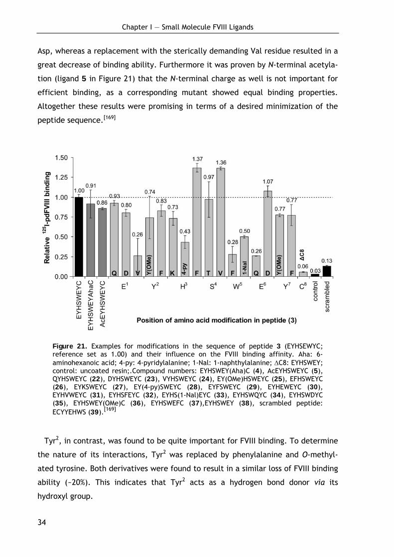
Chapter I — Small Molecule FVIII Ligands
34
Asp, whereas a replacement with the sterically demanding Val residue resulted in a
great decrease of binding ability. Furthermore it was proven by N-terminal acetyla-
tion (ligand 5 in Figure 21) that the N-terminal charge as well is not important for
efficient binding, as a corresponding mutant showed equal binding properties.
Altogether these results were promising in terms of a desired minimization of the
peptide sequence.[169]
Figure 21. Examples for modifications in the sequence of peptide 3 (EYHSEWYC; reference set as 1.00) and their influence on the FVIII binding affinity. Aha: 6-aminohexanoic acid; 4-py: 4-pyridylalanine; 1-Nal: 1-naphthylalanine; ΔC8: EYHSWEY; control: uncoated resin;.Compound numbers: EYHSWEY(Aha)C (4), AcEYHSWEYC (5), QYHSWEYC (22), DYHSWEYC (23), VYHSWEYC (24), EY(OMe)HSWEYC (25), EFHSWEYC (26), EYKSWEYC (27), EY(4-py)SWEYC (28), EYFSWEYC (29), EYHEWEYC (30), EYHVWEYC (31), EYHSFEYC (32), EYHS(1-Nal)EYC (33), EYHSWQYC (34), EYHSWDYC (35), EYHSWEY(OMe)C (36), EYHSWEFC (37),EYHSWEY (38), scrambled peptide: ECYYEHWS (39).[169]
Tyr2, in contrast, was found to be quite important for FVIII binding. To determine
the nature of its interactions, Tyr2 was replaced by phenylalanine and O-methyl-
ated tyrosine. Both derivatives were found to result in a similar loss of FVIII binding
ability (~20%). This indicates that Tyr2 acts as a hydrogen bond donor via its
hydroxyl group.
Chapter I — Small Molecule FVIII Ligands
35
Following the results of the Ala-scan, the positions His3 and Ser4 should allow
further optimization since both side chain residues seemed to be unfavorable for
efficient FVIII binding. In case of the histidine residue, this was probably due to the
positive charge of the side chain residue, as the substitution by other basic amino
acids like 4-pyridylalanine (4-py) and lysine resulted in a further decrease of the
binding affinity. In contrast, replacing His3 by phenylalanine improves the ability to
bind FVIII by almost 40% (see Figure 21).[169]
For the Ser4 residue the reduced binding affinity of 3 to the corresponding Ala-
mutant refers to the polar hydroxyl side chain functionality as the more hydropho-
bic derivatives, especially the Ala and the Val mutant, led to increased binding
properties. Modifications in the C-terminal part of the molecule proved that this
moiety is critical for efficient FVIII binding. All of the mutants at Trp5 showed at
least 50% reduction in FVIII binding ability. Tyr7, like Tyr2, could not be replaced by
phenylalanine and O-methylated tyrosine without loss of binding affinity indicating
an important hydrogen bridge to this residue. Glu6 could only be replaced by
aspartic acid. The negative charge of the carboxylic group was found to be
essential as shown by the reduced binding affinity of the corresponding glutamine
mutant (see Figure 21).[169]
Consequently, numerous other substitutions and combinations of amino acid
replacements, which gave positive results before, were screened, but no significant
improvements of the FVIII binding ability and no additive effects could be gained. A
selection of prepared ligands is summarized in the Supplementary Data 5 (page
248). It should be emphasized, that most data presented in there do only allow a
rough estimation of the ligands binding properties, as the ligand loading has not
been determined for all compounds). Consequently, the attempts to optimize the
octapeptidic sequence were stopped and the further work was focused on the
systematic minimization of the ligand length.
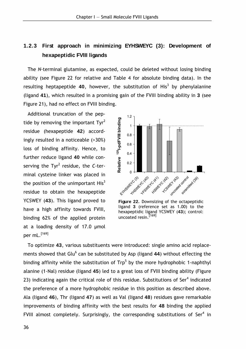
Chapter I — Small Molecule FVIII Ligands
36
1.2.3 First approach in minimizing EYHSWEYC (3): Development of
hexapeptidic FVIII ligands
The N-terminal glutamine, as expected, could be deleted without losing binding
ability (see Figure 22 for relative and Table 4 for absolute binding data). In the
resulting heptapeptide 40, however, the substitution of His3 by phenylalanine
(ligand 41), which resulted in a promising gain of the FVIII binding ability in 3 (see
Figure 21), had no effect on FVIII binding.
Additional truncation of the pep-
tide by removing the important Tyr2
residue (hexapeptide 42) accord-
ingly resulted in a noticeable (>30%)
loss of binding affinity. Hence, to
further reduce ligand 40 while con-
serving the Tyr2 residue, the C-ter-
minal cysteine linker was placed in
the position of the unimportant His3
residue to obtain the hexapeptide
YCSWEY (43). This ligand proved to
have a high affinity towards FVIII,
binding 62% of the applied protein
at a loading density of 17.0 μmol
per mL.[169]
To optimize 43, various substituents were introduced: single amino acid replace-
ments showed that Glu6 can be substituted by Asp (ligand 44) without effecting the
binding affinity while the substitution of Trp5 by the more hydrophobic 1-naphthyl
alanine (1-Nal) residue (ligand 45) led to a great loss of FVIII binding ability (Figure
23) indicating again the critical role of this residue. Substitutions of Ser4 indicated
the preference of a more hydrophobic residue in this position as described above.
Ala (ligand 46), Thr (ligand 47) as well as Val (ligand 48) residues gave remarkable
improvements of binding affinity with the best results for 48 binding the applied
FVIII almost completely. Surprisingly, the corresponding substitutions of Ser4 in
Figure 22. Downsizing of the octapeptidic ligand 3 (reference set as 1.00) to the hexapeptidic ligand YCSWEY (43); control: uncoated resin.[169]

Chapter I — Small Molecule FVIII Ligands
37
combination with an Asp residue at position 5 (ligand 49-51) exhibited lower bind-
ing affinity.[169]
Achieving highly potent FVIII
ligands, an effort was made to
improve the resistance against
proteolytic degradation by the
synthesis of the corresponding
fully retro-inverso derivatives,
which generally provide an en-
hanced enzymatic stability due to
their all-D sequence.[170-174] If the
hydrogen bonding and the secon-
dary structure is mirrored, the
original side chain orientation re-
mains conserved but the N- and
the C-termini and the direction of the peptide bonds are inversed.[175] It was
assumed that neither the peptide bonds nor the termini are involved in target
interactions.
Figure 23. Normalized 125I-pdFVIII binding of mutants of ligand 43 (black bar); dark grey bars represent all-L derivatives, light grey bars represent fully retro-inverso ligands (1-Nal: 1-naphthylalanine; lower case letters denote D-amino acid resi-dues).[169]
Table 4. Absolute FVIII binding data for different ligands: minimization of octapeptide 3.[169]
Sequence no. Ligand loading (μmol/mL)
125I-pdFVIII binding (%)a
EYHSWEYC 3 10.3 ± 0.4 50.2 ± 1.60
YHSWEYC 40 14.0 ± 0.7 61.3 ± 3.20
YFSWEYC 41 15.2 ± 1.1 66.8 ± 7.20
HSWEYC 42 12.7 ± 0.6 39.8 ± 16.8
YCSWEY 43 17.0 ± 0.5 62.0 ± 2.40
controlb - – 01.6 ± 0.10
scrambledc 39 10.3 ± 0.1 06.5 ± 0.50
a % of total applied material; b uncoated resin; c scrambled sequence: ECYYEHWS (39)

Chapter I — Small Molecule FVIII Ligands
38
Indeed, the retro-inverso peptides 52-55 worked fine and showed only a slightly
reduced binding affinity (see Figure 23 for relative and Table 5 for quantitative
binding data), thus providing highly potent factor VIII ligands with improved
enzymatic stability as shown below.
However, with regard to the ultimate goal to develop a novel method for FVIII
purification which is a real improvement in terms of cost efficiency this kind of
compound was not completely satisfying. The goal was to create a novel kind of
ligand which is easy to synthesize in large scale in solution thereby avoiding
expensive techniques like solid phase synthesis and HPLC purification to reduce
production costs. This is practically not possible for the hexapeptidic ligands. In
addition, the unnatural D-amino acid building blocks used for the synthesis of the
fully retro-inverso derivatives make this kind of ligand rather expensive.
Table 5. Optimization of ligand 43: loading density and absolute FVIII binding data for various mutants of ligand 43.[169]
Sequence no. Ligand loading (μmol/mL) 125I-pdFVIII binding (%)a
YCSWEY 43 17.0 ± 0.5 062.0 ± 0.40
YCSWDY 44 16.7 ± 1.0 069.8 ± 12.2
YCS(1-Nal)EY 45 12.7 ± 0.6 035.3 ± 3.00
YCAWEY 46 12.2 ± 0.6 072.1 ± 7.20
YCTWEY 47 14.9 ± 0.7 070.7 ± 2.40
YCVWEY 48 12.9 ± 0.6 094.1 ± 4.70
YCAWDY 49 19.3 ± 1.0 072.0 ± 15.5
YCTWDY 50 17.6 ± 0.0 073.6 ± 0.70
YCVWDY 51 16.5 ± 0.8 072.0 ± 1.20
yewacy 52 12.8 ± 0.6 053.6 ± 2.50
yewvcy 53 14.1 ± 0.7 077.0 ± 4.20
ydwacy 54 18.0 ± 0.9 075.1 ± 0.60
ydwvcy 55 18.5 ± 0.9 073.4 ± 5.60
controlb – 0001.6 ± 0.100
scrambledc 39 10.3 ± 0.1 0006.5 ± 0.500
a FVIII binding is given as percentage of total bindable material; b uncoated resin; c scrambled sequence: ECYYEHWS; 1-Nal is 1-naphthylalanine.

Chapter I — Small Molecule FVIII Ligands
39
1.2.4 Second approach in minimizing EYHSWEYC (3): Development of
the small molecule FVIII affinity ligand (3-IAA)Eψ[CH2NH]YC (69)
The next goal was the reduction of the initial octapeptide EYHSWEYC (3) down to
its C-terminal core binding sequence WEYC (57, Figure 24). As mentioned above,
Glu1 could be deleted without reducing the FVIII affinity but the truncation of the
Glu1-Tyr2 fragment resulted in a 30% loss. However, the larger fragments Glu1-Tyr2-
His3 as well as Glu1-Tyr2-His3-Ser4 could be deleted without additional loss of FVIII
binding (peptides 56 and 57, respectively). Considering the great simplification of
the sequence in 57 the 30% reduced affinity seemed acceptable to proceed with
optimizing this new lead structure.[169]
Figure 24. Relative binding data of minimized mutants of ligand 3. Stepwise minimi-zation of 3 (black bar) to its core binding motive WEYC (57) (light grey bars), side chain optimization of 57 (dark grey bars) and backbone modification of 64 (dashed bars).[169]
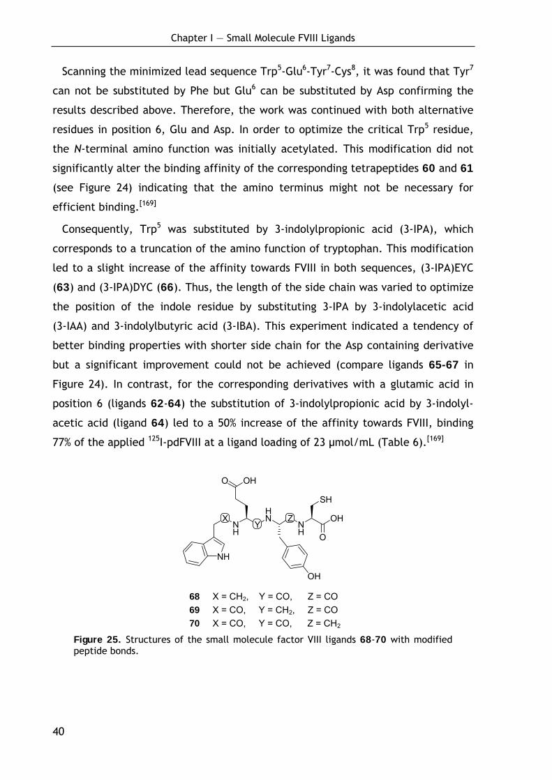
Chapter I — Small Molecule FVIII Ligands
40
Scanning the minimized lead sequence Trp5-Glu6-Tyr7-Cys8, it was found that Tyr7
can not be substituted by Phe but Glu6 can be substituted by Asp confirming the
results described above. Therefore, the work was continued with both alternative
residues in position 6, Glu and Asp. In order to optimize the critical Trp5 residue,
the N-terminal amino function was initially acetylated. This modification did not
significantly alter the binding affinity of the corresponding tetrapeptides 60 and 61
(see Figure 24) indicating that the amino terminus might not be necessary for
efficient binding.[169]
Consequently, Trp5 was substituted by 3-indolylpropionic acid (3-IPA), which
corresponds to a truncation of the amino function of tryptophan. This modification
led to a slight increase of the affinity towards FVIII in both sequences, (3-IPA)EYC
(63) and (3-IPA)DYC (66). Thus, the length of the side chain was varied to optimize
the position of the indole residue by substituting 3-IPA by 3-indolylacetic acid
(3-IAA) and 3-indolylbutyric acid (3-IBA). This experiment indicated a tendency of
better binding properties with shorter side chain for the Asp containing derivative
but a significant improvement could not be achieved (compare ligands 65-67 in
Figure 24). In contrast, for the corresponding derivatives with a glutamic acid in
position 6 (ligands 62-64) the substitution of 3-indolylpropionic acid by 3-indolyl-
acetic acid (ligand 64) led to a 50% increase of the affinity towards FVIII, binding
77% of the applied 125I-pdFVIII at a ligand loading of 23 μmol/mL (Table 6).[169]
XNH
YHN Z
NH
OH
O
NH
O OH
OH
SH
68 X = CH2, Y = CO, Z = CO 69 X = CO, Y = CH2, Z = CO 70 X = CO, Y = CO, Z = CH2
Figure 25. Structures of the small molecule factor VIII ligands 68-70 with modified peptide bonds.
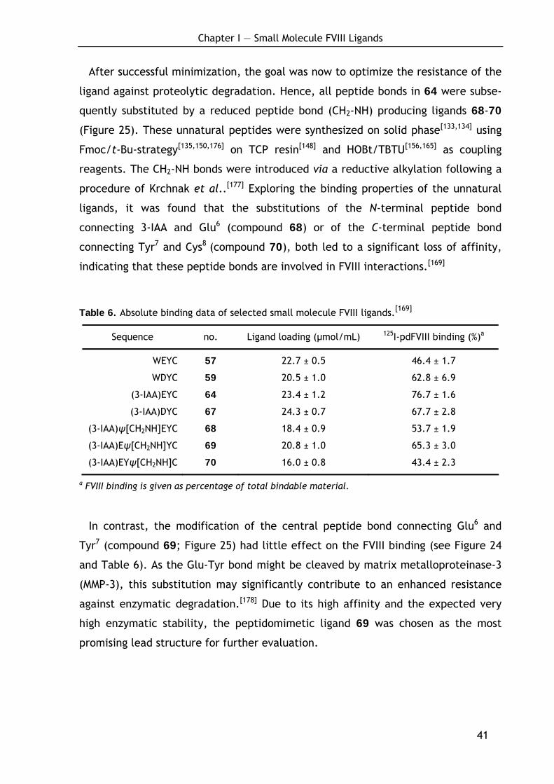
Chapter I — Small Molecule FVIII Ligands
41
After successful minimization, the goal was now to optimize the resistance of the
ligand against proteolytic degradation. Hence, all peptide bonds in 64 were subse-
quently substituted by a reduced peptide bond (CH2-NH) producing ligands 68-70
(Figure 25). These unnatural peptides were synthesized on solid phase[133,134] using
Fmoc/t-Bu-strategy[135,150,176] on TCP resin[148] and HOBt/TBTU[156,165] as coupling
reagents. The CH2-NH bonds were introduced via a reductive alkylation following a
procedure of Krchnak et al..[177] Exploring the binding properties of the unnatural
ligands, it was found that the substitutions of the N-terminal peptide bond
connecting 3-IAA and Glu6 (compound 68) or of the C-terminal peptide bond
connecting Tyr7 and Cys8 (compound 70), both led to a significant loss of affinity,
indicating that these peptide bonds are involved in FVIII interactions.[169]
Table 6. Absolute binding data of selected small molecule FVIII ligands.[169]
Sequence no. Ligand loading (μmol/mL) 125I-pdFVIII binding (%)a
WEYC 57 22.7 ± 0.5 46.4 ± 1.7
WDYC 59 20.5 ± 1.0 62.8 ± 6.9
(3-IAA)EYC 64 23.4 ± 1.2 76.7 ± 1.6
(3-IAA)DYC 67 24.3 ± 0.7 67.7 ± 2.8
(3-IAA)ψ[CH2NH]EYC 68 18.4 ± 0.9 53.7 ± 1.9
(3-IAA)Eψ[CH2NH]YC 69 20.8 ± 1.0 65.3 ± 3.0
(3-IAA)EYψ[CH2NH]C 70 16.0 ± 0.8 43.4 ± 2.3
a FVIII binding is given as percentage of total bindable material.
In contrast, the modification of the central peptide bond connecting Glu6 and
Tyr7 (compound 69; Figure 25) had little effect on the FVIII binding (see Figure 24
and Table 6). As the Glu-Tyr bond might be cleaved by matrix metalloproteinase-3
(MMP-3), this substitution may significantly contribute to an enhanced resistance
against enzymatic degradation.[178] Due to its high affinity and the expected very
high enzymatic stability, the peptidomimetic ligand 69 was chosen as the most
promising lead structure for further evaluation.
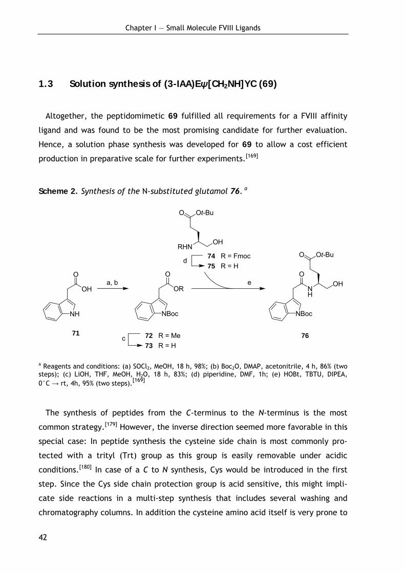
Chapter I — Small Molecule FVIII Ligands
42
NH
O
NBoc
O Ot-Bu
OHOR
O
NBoc
RHN
O Ot-Bu
OH
OH
O
NH
d
ea, b
c
1.3 Solution synthesis of (3-IAA)Eψ[CH2NH]YC (69)
Altogether, the peptidomimetic 69 fulfilled all requirements for a FVIII affinity
ligand and was found to be the most promising candidate for further evaluation.
Hence, a solution phase synthesis was developed for 69 to allow a cost efficient
production in preparative scale for further experiments.[169]
Scheme 2. Synthesis of the N-substituted glutamol 76. a
a Reagents and conditions: (a) SOCl2, MeOH, 18 h, 98%; (b) Boc2O, DMAP, acetonitrile, 4 h, 86% (two steps); (c) LiOH, THF, MeOH, H2O, 18 h, 83%; (d) piperidine, DMF, 1h; (e) HOBt, TBTU, DIPEA, 0°C → rt, 4h, 95% (two steps).[169]
The synthesis of peptides from the C-terminus to the N-terminus is the most
common strategy.[179] However, the inverse direction seemed more favorable in this
special case: In peptide synthesis the cysteine side chain is most commonly pro-
tected with a trityl (Trt) group as this group is easily removable under acidic
conditions.[180] In case of a C to N synthesis, Cys would be introduced in the first
step. Since the Cys side chain protection group is acid sensitive, this might impli-
cate side reactions in a multi-step synthesis that includes several washing and
chromatography columns. In addition the cysteine amino acid itself is very prone to
74 R = Fmoc 75 R = H
72 R = Me 76 73 R = H
71
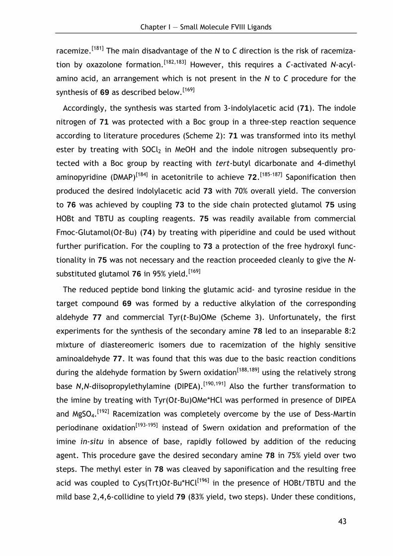
Chapter I — Small Molecule FVIII Ligands
43
racemize.[181] The main disadvantage of the N to C direction is the risk of racemiza-
tion by oxazolone formation.[182,183] However, this requires a C-activated N-acyl-
amino acid, an arrangement which is not present in the N to C procedure for the
synthesis of 69 as described below.[169]
Accordingly, the synthesis was started from 3-indolylacetic acid (71). The indole
nitrogen of 71 was protected with a Boc group in a three-step reaction sequence
according to literature procedures (Scheme 2): 71 was transformed into its methyl
ester by treating with SOCl2 in MeOH and the indole nitrogen subsequently pro-
tected with a Boc group by reacting with tert-butyl dicarbonate and 4-dimethyl
aminopyridine (DMAP)[184] in acetonitrile to achieve 72.[185-187] Saponification then
produced the desired indolylacetic acid 73 with 70% overall yield. The conversion
to 76 was achieved by coupling 73 to the side chain protected glutamol 75 using
HOBt and TBTU as coupling reagents. 75 was readily available from commercial
Fmoc-Glutamol(Ot-Bu) (74) by treating with piperidine and could be used without
further purification. For the coupling to 73 a protection of the free hydroxyl func-
tionality in 75 was not necessary and the reaction proceeded cleanly to give the N-
substituted glutamol 76 in 95% yield.[169]
The reduced peptide bond linking the glutamic acid- and tyrosine residue in the
target compound 69 was formed by a reductive alkylation of the corresponding
aldehyde 77 and commercial Tyr(t-Bu)OMe (Scheme 3). Unfortunately, the first
experiments for the synthesis of the secondary amine 78 led to an inseparable 8:2
mixture of diastereomeric isomers due to racemization of the highly sensitive
aminoaldehyde 77. It was found that this was due to the basic reaction conditions
during the aldehyde formation by Swern oxidation[188,189] using the relatively strong
base N,N-diisopropylethylamine (DIPEA).[190,191] Also the further transformation to
the imine by treating with Tyr(Ot-Bu)OMe*HCl was performed in presence of DIPEA
and MgSO4.[192] Racemization was completely overcome by the use of Dess-Martin
periodinane oxidation[193-195] instead of Swern oxidation and preformation of the
imine in-situ in absence of base, rapidly followed by addition of the reducing
agent. This procedure gave the desired secondary amine 78 in 75% yield over two
steps. The methyl ester in 78 was cleaved by saponification and the resulting free
acid was coupled to Cys(Trt)Ot-Bu*HCl[196] in the presence of HOBt/TBTU and the
mild base 2,4,6-collidine to yield 79 (83% yield, two steps). Under these conditions,
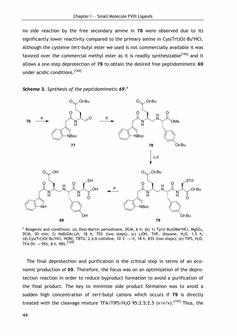
Chapter I — Small Molecule FVIII Ligands
44
NH
O HN
NH
OOH
O
NH
O OH
OH
SH
NH
O HN
OMe
O
NBoc
O Ot-Bu
Ot-Bu
NH
O HN
NH
OOt-Bu
O
NBoc
O Ot-Bu
Ot-Bu
STrt
NH
O
NBoc
O Ot-Bu
Oa b
c,d
e
no side reaction by the free secondary amine in 78 were observed due to its
significantly lower reactivity compared to the primary amine in Cys(Trt)Ot-Bu*HCl.
Although the cysteine tert-butyl ester we used is not commercially available it was
favored over the commercial methyl ester as it is readily synthesizable[196] and it
allows a one-step deprotection of 79 to obtain the desired free peptidomimetic 69
under acidic conditions.[169]
Scheme 3. Synthesis of the peptidomimetic 69.a
69 79
a Reagents and conditions: (a) Dess-Martin periodinane, DCM, 6 h; (b) 1) Tyr(t-Bu)OMe*HCl, MgSO4, DCM, 30 min; 2) NaB(OAc)3H, 18 h, 75% (two steps); (c) LiOH, THF, dioxane, H2O, 1.5 h; (d) Cys(Trt)Ot-Bu*HCl, HOBt, TBTU, 2,4,6-collidine, 10°C → rt, 18 h, 83% (two steps); (e) TIPS, H2O, TFA 0% → 95%, 8 h, 98%.[169]
The final deprotection and purification is the critical step in terms of an eco-
nomic production of 69. Therefore, the focus was on an optimization of the depro-
tection reaction in order to reduce byproduct formation to avoid a purification of
the final product. The key to minimize side product formation was to avoid a
sudden high concentration of tert-butyl cations which occurs if 79 is directly
treated with the cleavage mixture TFA/TIPS/H2O 95:2.5:2.5 (v/v/v).[197] Thus, the
76
77 78
Chapter I — Small Molecule FVIII Ligands
45
problem was solved by suspending 79 in a vigorously stirred mixture of the scaven-
gers water and TIPS (1:1) and slowly adding the TFA over a period of 8 hours to a
final concentration of 95%. By this procedure the byproduct formation (typically
15% as determined by HPLC) was significantly reduced to obtain the final peptido-
mimetic 69 in 98% yield (purity >95%) after precipitation in ether/pentane. To
prove the diastereomeric purity, all possible diastereomers of 69 were synthesized
and analyzed by HPLC. Coinjection with 69 gave baseline separation for all
diastereomers.[169]

Chapter I — Small Molecule FVIII Ligands
46
1.4 Evaluation of biological and biochemical ligand characteristics
1.4.1 Verification of the enzymatic stability
The resistance against enzymatic degradation was determined by treating the
ligands with fresh human serum (single donor). The amounts of intact ligand were
determined by quantitative HPLC and HPLC-MS analysis. For this study the octa-
peptide 3 (EYHSWEYC) as well as the fully retro-inverso peptide 55 (ydwycy), the
tetrapeptide 57 (WEYC) and the peptidomimetic 69 ((3-IAA)Eψ[CH2NH]YC) were
chosen.
As shown in Figure 26, the octapeptide 3 was completely degraded after three
hours of incubation with serum proving its limited applicability as affinity ligand.
Despite the reduced size, the tetrapeptide 57 also turned out to be very unstable
in human serum.[169,198]
Figure 26. Determination of the stability of octapeptide EYHSWEYC (3), fully retro-inverso peptide ydwycy (55), tetrapeptide WEYC (57) and the minimized peptido-mimetic 69 against enzymatic degradation by treating with fresh human serum (single donor). PROK: Proteinase K.[169,198]
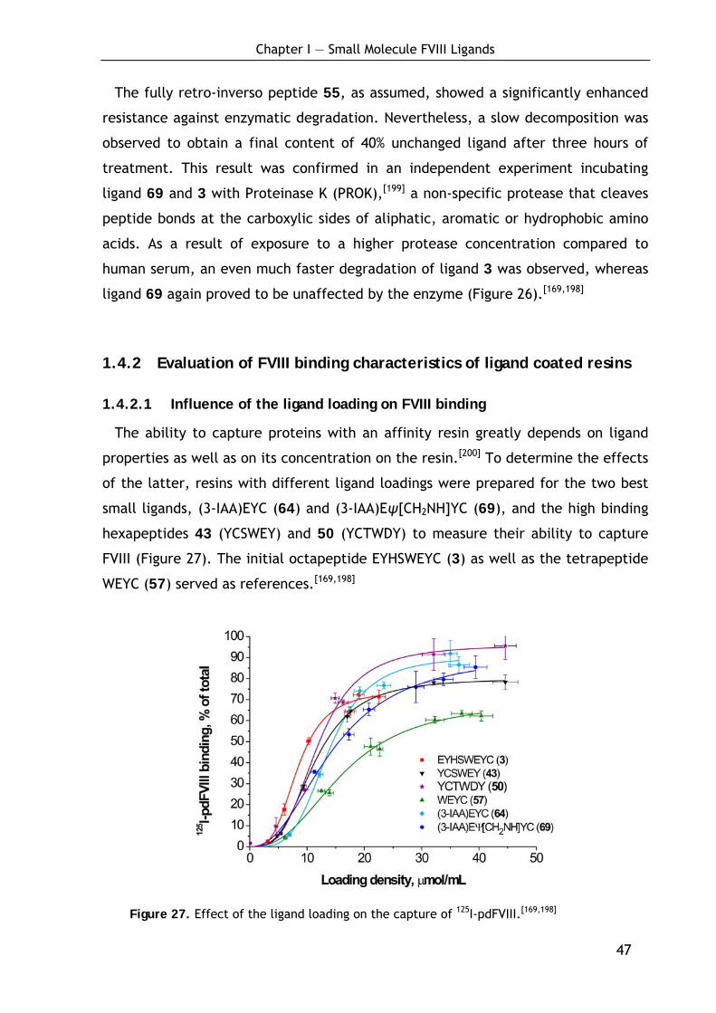
Chapter I — Small Molecule FVIII Ligands
47
The fully retro-inverso peptide 55, as assumed, showed a significantly enhanced
resistance against enzymatic degradation. Nevertheless, a slow decomposition was
observed to obtain a final content of 40% unchanged ligand after three hours of
treatment. This result was confirmed in an independent experiment incubating
ligand 69 and 3 with Proteinase K (PROK),[199] a non-specific protease that cleaves
peptide bonds at the carboxylic sides of aliphatic, aromatic or hydrophobic amino
acids. As a result of exposure to a higher protease concentration compared to
human serum, an even much faster degradation of ligand 3 was observed, whereas
ligand 69 again proved to be unaffected by the enzyme (Figure 26).[169,198]
1.4.2 Evaluation of FVIII binding characteristics of ligand coated resins
1.4.2.1 Influence of the ligand loading on FVIII binding
The ability to capture proteins with an affinity resin greatly depends on ligand
properties as well as on its concentration on the resin.[200] To determine the effects
of the latter, resins with different ligand loadings were prepared for the two best
small ligands, (3-IAA)EYC (64) and (3-IAA)Eψ[CH2NH]YC (69), and the high binding
hexapeptides 43 (YCSWEY) and 50 (YCTWDY) to measure their ability to capture
FVIII (Figure 27). The initial octapeptide EYHSWEYC (3) as well as the tetrapeptide
WEYC (57) served as references.[169,198]
Figure 27. Effect of the ligand loading on the capture of 125I-pdFVIII.[169,198]
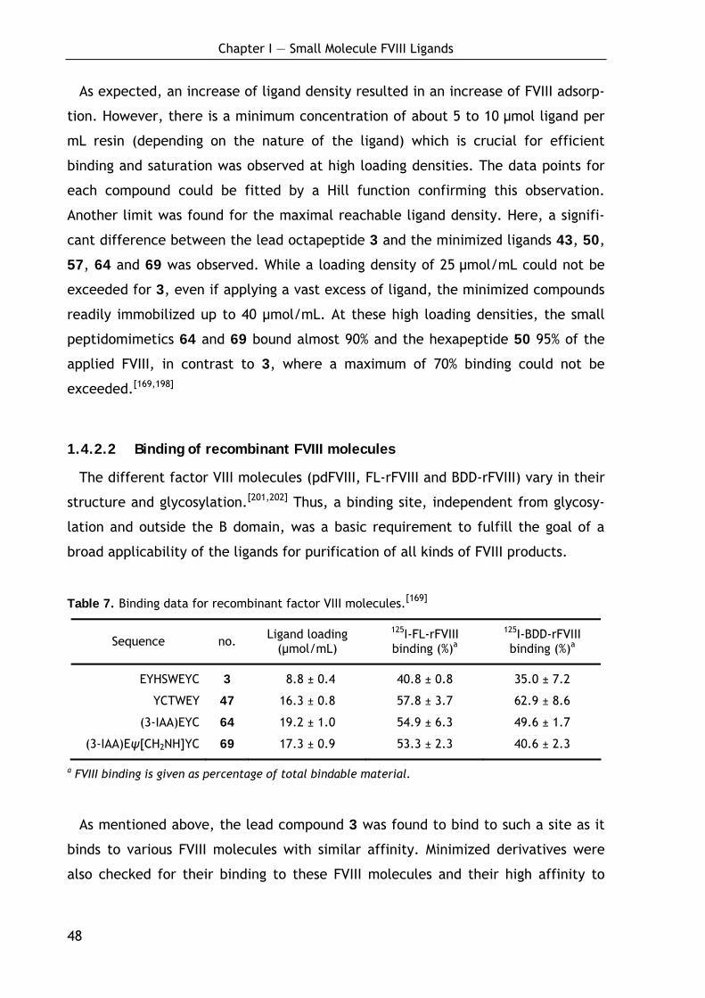
Chapter I — Small Molecule FVIII Ligands
48
As expected, an increase of ligand density resulted in an increase of FVIII adsorp-
tion. However, there is a minimum concentration of about 5 to 10 μmol ligand per
mL resin (depending on the nature of the ligand) which is crucial for efficient
binding and saturation was observed at high loading densities. The data points for
each compound could be fitted by a Hill function confirming this observation.
Another limit was found for the maximal reachable ligand density. Here, a signifi-
cant difference between the lead octapeptide 3 and the minimized ligands 43, 50,
57, 64 and 69 was observed. While a loading density of 25 μmol/mL could not be
exceeded for 3, even if applying a vast excess of ligand, the minimized compounds
readily immobilized up to 40 μmol/mL. At these high loading densities, the small
peptidomimetics 64 and 69 bound almost 90% and the hexapeptide 50 95% of the
applied FVIII, in contrast to 3, where a maximum of 70% binding could not be
exceeded.[169,198]
1.4.2.2 Binding of recombinant FVIII molecules
The different factor VIII molecules (pdFVIII, FL-rFVIII and BDD-rFVIII) vary in their
structure and glycosylation.[201,202] Thus, a binding site, independent from glycosy-
lation and outside the B domain, was a basic requirement to fulfill the goal of a
broad applicability of the ligands for purification of all kinds of FVIII products.
Table 7. Binding data for recombinant factor VIII molecules.[169]
Sequence no. Ligand loading (μmol/mL)
125I-FL-rFVIII binding (%)a
125I-BDD-rFVIII binding (%)a
EYHSWEYC 3 08.8 ± 0.4 40.8 ± 0.8 35.0 ± 7.2
YCTWEY 47 16.3 ± 0.8 57.8 ± 3.7 62.9 ± 8.6
(3-IAA)EYC 64 19.2 ± 1.0 54.9 ± 6.3 49.6 ± 1.7
(3-IAA)Eψ[CH2NH]YC 69 17.3 ± 0.9 53.3 ± 2.3 40.6 ± 2.3
a FVIII binding is given as percentage of total bindable material.
As mentioned above, the lead compound 3 was found to bind to such a site as it
binds to various FVIII molecules with similar affinity. Minimized derivatives were
also checked for their binding to these FVIII molecules and their high affinity to
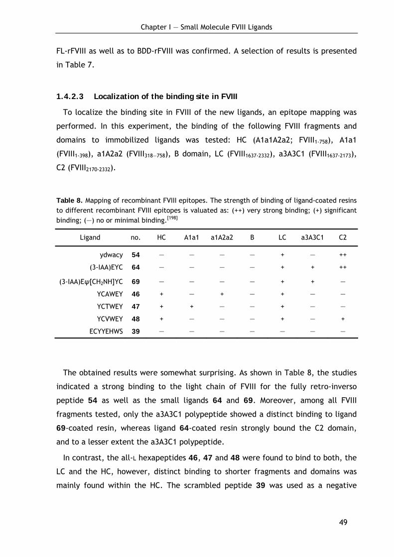
Chapter I — Small Molecule FVIII Ligands
49
FL-rFVIII as well as to BDD-rFVIII was confirmed. A selection of results is presented
in Table 7.
1.4.2.3 Localization of the binding site in FVIII
To localize the binding site in FVIII of the new ligands, an epitope mapping was
performed. In this experiment, the binding of the following FVIII fragments and
domains to immobilized ligands was tested: HC (A1a1A2a2; FVIII1-758), A1a1
(FVIII1-398), a1A2a2 (FVIII318—758), B domain, LC (FVIII1637-2332), a3A3C1 (FVIII1637-2173),
C2 (FVIII2170-2332).
Table 8. Mapping of recombinant FVIII epitopes. The strength of binding of ligand-coated resins to different recombinant FVIII epitopes is valuated as: (++) very strong binding; (+) significant binding; (—) no or minimal binding.[198]
Ligand no. HC A1a1 a1A2a2 B LC a3A3C1 C2
ydwacy 54 — — — — + — ++
(3-IAA)EYC 64 — — — — + + ++
(3-IAA)Eψ[CH2NH]YC 69 — — — — + + —
YCAWEY 46 + — + — + — —
YCTWEY 47 + + — — + — —
YCVWEY 48 + — — — + — +
ECYYEHWS 39 — — — — — — —
The obtained results were somewhat surprising. As shown in Table 8, the studies
indicated a strong binding to the light chain of FVIII for the fully retro-inverso
peptide 54 as well as the small ligands 64 and 69. Moreover, among all FVIII
fragments tested, only the a3A3C1 polypeptide showed a distinct binding to ligand
69-coated resin, whereas ligand 64-coated resin strongly bound the C2 domain,
and to a lesser extent the a3A3C1 polypeptide.
In contrast, the all-L hexapeptides 46, 47 and 48 were found to bind to both, the
LC and the HC, however, distinct binding to shorter fragments and domains was
mainly found within the HC. The scrambled peptide 39 was used as a negative
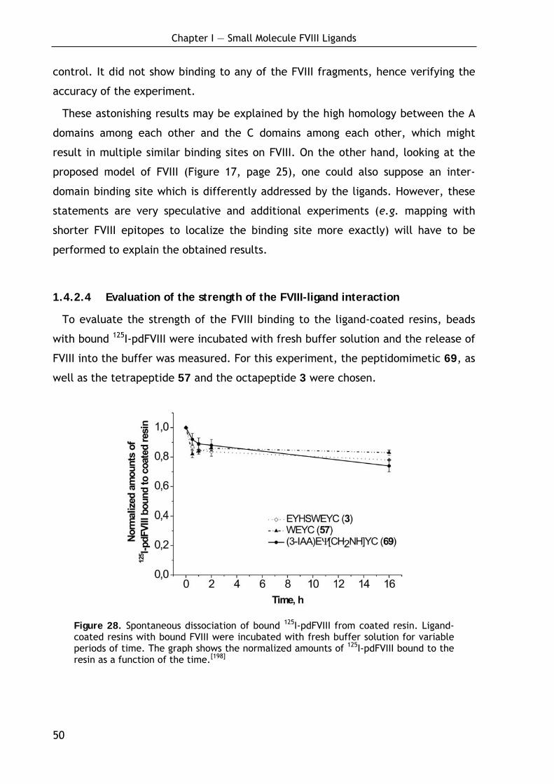
Chapter I — Small Molecule FVIII Ligands
50
control. It did not show binding to any of the FVIII fragments, hence verifying the
accuracy of the experiment.
These astonishing results may be explained by the high homology between the A
domains among each other and the C domains among each other, which might
result in multiple similar binding sites on FVIII. On the other hand, looking at the
proposed model of FVIII (Figure 17, page 25), one could also suppose an inter-
domain binding site which is differently addressed by the ligands. However, these
statements are very speculative and additional experiments (e.g. mapping with
shorter FVIII epitopes to localize the binding site more exactly) will have to be
performed to explain the obtained results.
1.4.2.4 Evaluation of the strength of the FVIII-ligand interaction
To evaluate the strength of the FVIII binding to the ligand-coated resins, beads
with bound 125I-pdFVIII were incubated with fresh buffer solution and the release of
FVIII into the buffer was measured. For this experiment, the peptidomimetic 69, as
well as the tetrapeptide 57 and the octapeptide 3 were chosen.
Figure 28. Spontaneous dissociation of bound 125I-pdFVIII from coated resin. Ligand-coated resins with bound FVIII were incubated with fresh buffer solution for variable periods of time. The graph shows the normalized amounts of 125I-pdFVIII bound to the resin as a function of the time.[198]
Chapter I — Small Molecule FVIII Ligands
51
Figure 28 shows that only minor amounts of FVIII (~10-20%) were released within
16 h of incubation from the coated resins. The main release was observed within
the first two hours which may be due to not optimally bound material or to the
formation of an equilibrium between bound and free FVIII. These experiments show
a high-affinity binding of FVIII to the small ligands and a very low off-rate. This is
an important property for the coated resins, as it provides the opportunity to ex-
tensively wash the resin during affinity purification and thus to elute contaminating
proteins without loss of FVIII.[198]
1.4.3 Purification of pdFVIII using peptidomimetic (69)-coated resin
To demonstrate the potential of peptidomimetic 69 as ligand for FVIII affinity
purification, sample purification from cell-conditioned fetal bovine serum (FBS)-
containing Dulbecco's Modified Eagle Medium (DMEM) was performed (Figure 30).
Affinity purification via ligand 69-coated resin was achieved using two buffers
varying only in their NaCl content. This is an indication that the interaction
between FVIII and the evaluated peptides has predominantly ionic character, as
observed for various antibodies before.[203,204] 0.1 M NaCl was used for FVIII
adsorption and elution was done under very mild conditions applying 0.6 M NaCl at
pH 6.8 to give sharp elution peaks (Figure 29 and Figure 30; left-hand panels). The
pH of the buffers was furthermore adjusted to 6.8 as the slightly acidic condition
were found to give a better elution threshold, and as FVIII is generally more stable
under these conditions. More detailed information about purification conditions are
presented in the Supplementary Data 6.
As a positive control, binding and elution of pure pdFVIII was carry out, resulting
in high FVIII column retention (~90%). Analysis of the flow-through and wash frac-
tions by SDS-PAGE followed by silver staining (Figure 29 middle panel) and Western
blotting (Figure 29 right-hand panel) revealed only traces of FVIII in those fractions
(lanes 3, 8; Figure 29). In the FVIII-rich eluate, the heterogeneous 230-90 kDa
heavy chain bands and the ~80 kDa light chain doublet bands were visually distin-
guished. In addition, traces of proteolytic bands (~73 kDa, ~50 kDa, ~43 kDa) were
found, from which the ~50 kDa band (A1 domain) was strongly reactive against mAb
C5 in the blot.[198]

Chapter I — Small Molecule FVIII Ligands
52
Figure 29. Adsorption and elution of pure pdFVIII. 0.5 mg of pdFVIII were applied to 1 mL of resin. Samples from different purification fractions (see profile in left-hand panel) were analyzed by 10% SDS-PAGE followed by silver staining (middle panel) or Western blotting (right-hand panel) using mAbs C5 and 413 against FVIII. Lanes 1, 6: pure FVIII; Lanes 2, 7: source solution with pure FVIII for the column; Lanes 3, 8: flow-through fraction; Lanes 4, 5, 9, 10: elution fraction with 0.8 M NaCl.[198]
To test the ligand 69-coated resin in a typical application, a purification of
pdFVIII from cell-conditioned FBS-containing DMEM, which was spiked with prepuri-
fied pdFVIII, was performed. Again, a very satisfying retention (89%) was achieved
and the vast majority of contaminant proteins were eluted with the flow-through
containing only traces of FVIII (Figure 30: lanes 3, 8).
Figure 30. Purification of pdFVIII diluted in cell-conditioned FBS-containing DMEM. 0.5 mg of pdFVIII were applied to 1 mL of resin. Samples from different purification fractions (see profile in left-hand panels) were analyzed by 10% SDS-PAGE followed by silver staining (middle panel) or Western blotting (right-hand panel) using mAbs C5 and 413 against FVIII. Lane 1: media; Lanes 2, 7: media with FVIII for column; Lanes 3, 8: flow-through fraction (W1); Lanes 4, 5, 9, 10: wash fractions (W2) with 0.2 M NaCl; Lanes 6, 11: elution fraction with 0.6 M NaCl.[198]
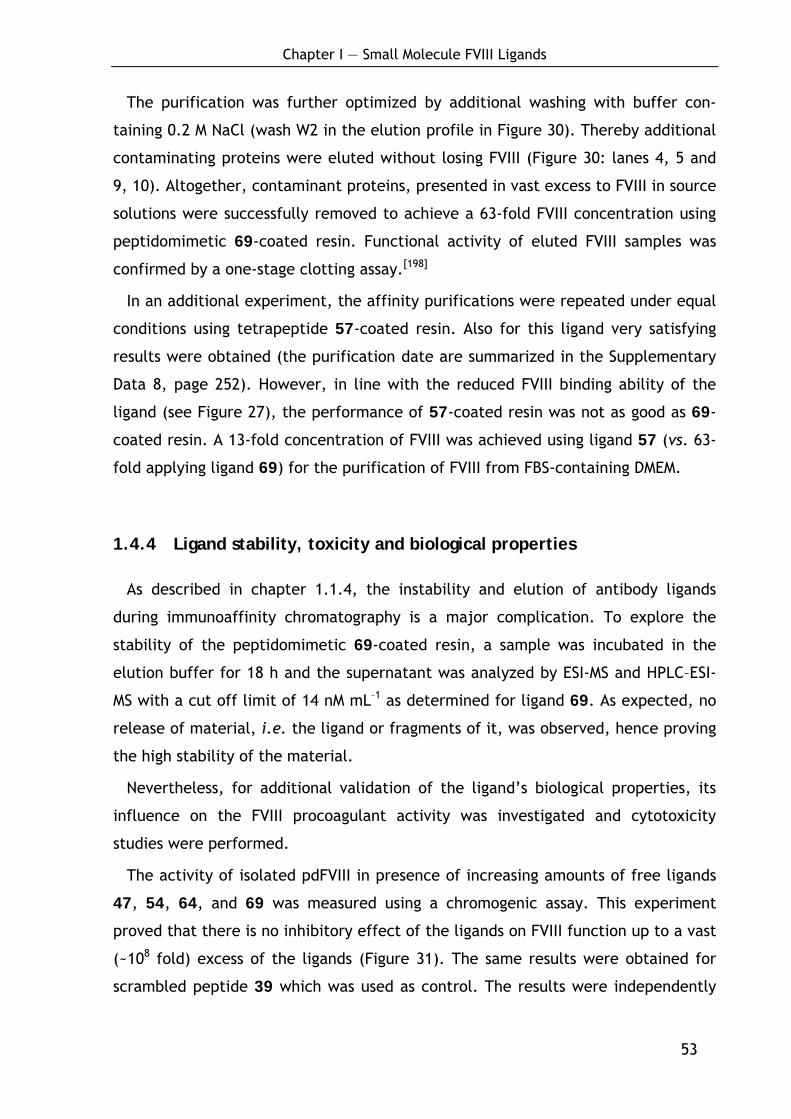
Chapter I — Small Molecule FVIII Ligands
53
The purification was further optimized by additional washing with buffer con-
taining 0.2 M NaCl (wash W2 in the elution profile in Figure 30). Thereby additional
contaminating proteins were eluted without losing FVIII (Figure 30: lanes 4, 5 and
9, 10). Altogether, contaminant proteins, presented in vast excess to FVIII in source
solutions were successfully removed to achieve a 63-fold FVIII concentration using
peptidomimetic 69-coated resin. Functional activity of eluted FVIII samples was
confirmed by a one-stage clotting assay.[198]
In an additional experiment, the affinity purifications were repeated under equal
conditions using tetrapeptide 57-coated resin. Also for this ligand very satisfying
results were obtained (the purification date are summarized in the Supplementary
Data 8, page 252). However, in line with the reduced FVIII binding ability of the
ligand (see Figure 27), the performance of 57-coated resin was not as good as 69-
coated resin. A 13-fold concentration of FVIII was achieved using ligand 57 (vs. 63-
fold applying ligand 69) for the purification of FVIII from FBS-containing DMEM.
1.4.4 Ligand stability, toxicity and biological properties
As described in chapter 1.1.4, the instability and elution of antibody ligands
during immunoaffinity chromatography is a major complication. To explore the
stability of the peptidomimetic 69-coated resin, a sample was incubated in the
elution buffer for 18 h and the supernatant was analyzed by ESI-MS and HPLC–ESI-
MS with a cut off limit of 14 nM mL–1 as determined for ligand 69. As expected, no
release of material, i.e. the ligand or fragments of it, was observed, hence proving
the high stability of the material.
Nevertheless, for additional validation of the ligand’s biological properties, its
influence on the FVIII procoagulant activity was investigated and cytotoxicity
studies were performed.
The activity of isolated pdFVIII in presence of increasing amounts of free ligands
47, 54, 64, and 69 was measured using a chromogenic assay. This experiment
proved that there is no inhibitory effect of the ligands on FVIII function up to a vast
(~108 fold) excess of the ligands (Figure 31). The same results were obtained for
scrambled peptide 39 which was used as control. The results were independently
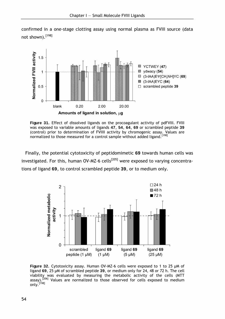
Chapter I — Small Molecule FVIII Ligands
54
confirmed in a one-stage clotting assay using normal plasma as FVIII source (data
not shown).[198]
Figure 31. Effect of dissolved ligands on the procoagulant activity of pdFVIII. FVIII was exposed to variable amounts of ligands 47, 54, 64, 69 or scrambled peptide 39 (control) prior to determination of FVIIII activity by chromogenic assay. Values are normalized to those measured for a control sample without added ligand.[198]
Finally, the potential cytotoxicity of peptidomimetic 69 towards human cells was
investigated. For this, human OV-MZ-6 cells[205] were exposed to varying concentra-
tions of ligand 69, to control scrambled peptide 39, or to medium only.
Figure 32. Cytotoxicity assay. Human OV-MZ-6 cells were exposed to 1 to 25 μM of ligand 69, 25 μM of scrambled peptide 39, or medium only for 24, 48 or 72 h. The cell viability was evaluated by measuring the metabolic activity of the cells (MTT assay).[206] Values are normalized to those observed for cells exposed to medium only.[198]
Chapter I — Small Molecule FVIII Ligands
55
As depicted in Figure 4B, analysis of the cell viability at 24, 48 and 72 h, via
measurement of the metabolic activity of the cells revealed no cytotoxicity of the
ligands, although using concentrations up to 25 µM. In a complementary approach
applying identical experimental conditions, cell proliferation by direct cell count-
ing was analyzed and again no indication for any cytotoxicity of ligand 69 (data not
shown) was found.[198]
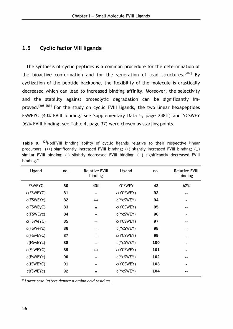
Chapter I — Small Molecule FVIII Ligands
56
1.5 Cyclic factor VIII ligands
The synthesis of cyclic peptides is a common procedure for the determination of
the bioactive conformation and for the generation of lead structures.[207] By
cyclization of the peptide backbone, the flexibility of the molecule is drastically
decreased which can lead to increased binding affinity. Moreover, the selectivity
and the stability against proteolytic degradation can be significantly im-
proved.[208,209] For the study on cyclic FVIII ligands, the two linear hexapeptides
FSWEYC (40% FVIII binding; see Supplementary Data 5, page 248ff) and YCSWEY
(62% FVIII binding; see Table 4, page 37) were chosen as starting points.
Table 9. 125I-pdFVIII binding ability of cyclic ligands relative to their respective linear precursors. (++) significantly increased FVIII binding; (+) slightly increased FVIII binding; (±) similar FVIII binding; (-) slightly decreased FVIII binding; (--) significantly decreased FVIII binding.a
Ligand no. Relative FVIII binding
Ligand no. Relative FVIII binding
FSWEYC 80 40% YCSWEY 43 62%
c(FSWEYC) 81 - c(YCSWEY) 93 -- c(FSWEYc) 82 ++ c(YcSWEY) 94 - c(FSWEyC) 83 ± c(YCSWEY) 95 -- c(FSWEyc) 84 ± c(YcSWEY) 96 - c(FSWeYC) 85 -- c(YCSWEY) 97 -- c(FSWeYc) 86 -- c(YcSWEY) 98 -- c(FSwEYC) 87 + c(YCSWEY) 99 - c(FSwEYc) 88 -- c(YcSWEY) 100 - c(FsWEYC) 89 ++ c(YCSWEY) 101 - c(FsWEYc) 90 + c(YcSWEY) 102 -- c(fSWEYC) 91 + c(YCSWEY) 103 - c(fSWEYc) 92 ± c(YcSWEY) 104 --
a Lower case letters denote D-amino acid residues.
Chapter I — Small Molecule FVIII Ligands
57
Initially, a D-scan was performed to selected appropriate candidates for further
sequence optimization. This was done for both stereochemical configurations of
the cysteine linker (L- and D-cysteine), as the configuration of the linker might
significantly affect the presentation of the ligand on the resin surface. Table 9
summarizes the determined FVIII binding abilities of the diverse cyclic peptide
mutants relative to their respective linear precursor molecule.
As shown in Table 9, all cyclic derivatives of ligand 43 bound worse than the lin-
ear precursor molecule. In contrast, the two ligands c(FSWEYc) (82) and c(FsWEAC)
(89) were found to exhibit a significantly improved FVIII binding ability with re-
spect to the linear precursor 80. The detailed binding data and methods for the
synthesis of the cyclic peptides are presented in Supplementary Data 7 (page 252).
The work on the two ligands is continued in the PhD thesis of D. Heckmann.[210]
Chapter I — Small Molecule FVIII Ligands
58
1.6 Discussion and conclusion
By systematic optimization of the FVIII ligand EYHSWEYC (3) two groups of
affinity ligands, hexapeptidic and small peptidomimetic ligands, each showing
specific advantages, were developed. Their choice for application in FVIII affinity
purification processes will depend on process characteristics and/or the objective.
The best hexapeptides proved to have a superior FVIII binding profile than the
small peptidomimetics, while suffering from lower enzymatic stability and higher
production costs comparing with the small peptidomimetics. However, in specific
applications, in particular if extremely high FVIII binding properties are needed,
these peptide ligands will be the material of choice.
The small peptidomimetic 69 is by far the smallest ligand known to date for the
separation of such a large protein as FVIII. Toyopearl AF-Epoxy-650M resin was
found to be the support material of choice giving the best FVIII binding results in
initial studies. Moreover, ligand 69 can be chemoselectively attached to this resin
via a cysteine residue acting as linkage. The potential of ligand 69-coated resin
was shown in two examples: adsorption and elution of pre-purified FVIII and a
successful purification from cell-conditioned FBS-containing media spiked with
FVIII. Both examples demonstrated the high efficiency of ligand 69-coated resin:
Using a mild buffer for FVIII elution, sharp elution peaks and a high FVIII retention
of 90% were achieved. SDS-PAGE and Western blot analysis confirmed that the
source and the eluted FVIII display the same protein bands and repartition of the
material among protein bands (Figure 29 and Figure 30).
The unnatural structures introduced in ligand 69 not only improved the FVIII
affinity but also significantly enhanced its resistance against enzymatic degrada-
tion. This was shown by treatment with fresh human serum and Proteinase K
(Figure 26). Moreover, the ligand 69-coated resin offers a promising overall
stability: After a long incubation period of 18 hours in elution buffer at a high
concentration of immobilized ligand (1 mL coated resin was suspended in 1 mL of
buffer), no release of material was detected by ESI-MS analysis. In an ongoing study
(data not shown), the ligand 69-coated resin preserved its properties over several

Chapter I — Small Molecule FVIII Ligands
59
months, even if treated with a wide pH range and alcohols for sterilization, condi-
tions under which protein ligands would be severely altered.
The chemical origin is another important feature of the peptidomimetic ligand
69. Ligand synthesis by established solid-phase procedures or by the solution
procedure described herein is straightforward and considerably cheaper than the
biotechnological production of antibodies. Furthermore, it offers opportunities for
easy modification of the ligand’s structure. This can be used e.g. for further
optimizations in terms of stability, or to improve the loading capability or the
elution conditions, which is practically not possible for antibodies. As the synthesis
is performed without use of any materials of human or animal origin, this ligand
does also not carry a risk of transmitting pathogens. Moreover, the ligand shows no
cytotoxicity (Figure 32) and it does not affect the procoagulant activity of FVIII
(Figure 31).
Hence, it was shown that the novel small-molecule FVIII ligand 69 is a valuable
alternative to antibody-based techniques for FVIII purification in laboratory and
industrial production. It has the potential to reduce production costs and to
improve the safety of current and future FVIIII products.

Chapter I — Small Molecule FVIII Ligands
60
1.7 Asymmetric synthesis of condensed and aromatic ring-
substituted tyrosine derivatives
1.7.1 Background
1.7.1.1 Intention
As described above, the phenolic hydroxyl group of the tyrosine residues (Tyr2
and Tyr7) is important for ligand binding affinity as the corresponding phenylalanine
derivatives bound significantly worse (see Figure 21). In addition, the preference of
larger aromatic systems in the position of Tyr2 and Tyr7 was found in several
examples (see Supplementary Data 5, page 248). Thus, the substitution of tyrosine
by analogues bearing both, an expanded aromatic system as well as the phenolic
hydroxyl group, would be interesting in terms of optimizing the binding properties.
1.7.1.2 Non-proteinogenic amino acids in drug development
Non-proteinogenic amino acids play an important role in drug development.
While proteinogenic amino acids provide limited variation in size and shape, the
introduction of unusual substituents allows a systematic study of structure-activity
relationships (SAR). Consequently, non-proteinogenic amino acids have been found
to significantly improve the biological properties of numerous biologically active
peptides and peptido-mimetics, e.g. by limiting conformational flexibility, en-
hancing enzymatic stability, improving pharmaco-dynamics or bioavailability.[211-213]
Especially modified aromatic amino acids are important structural features in
various pharmaceuticals which are currently under development or have already
been introduced into the market. Among the latter are, for example, the broad-
spectrum antibiotics Ampicillin® and Amoxicillin® which contain a D-phenylglycine
and D-4-hydroxyphenylglycine moiety, respectively, and Nafarelin® a luteinising
hormone releasing hormone (LHRH) analogue for treatment of endometrioses
comprising a D-2-naphthylalanine residue.[214]
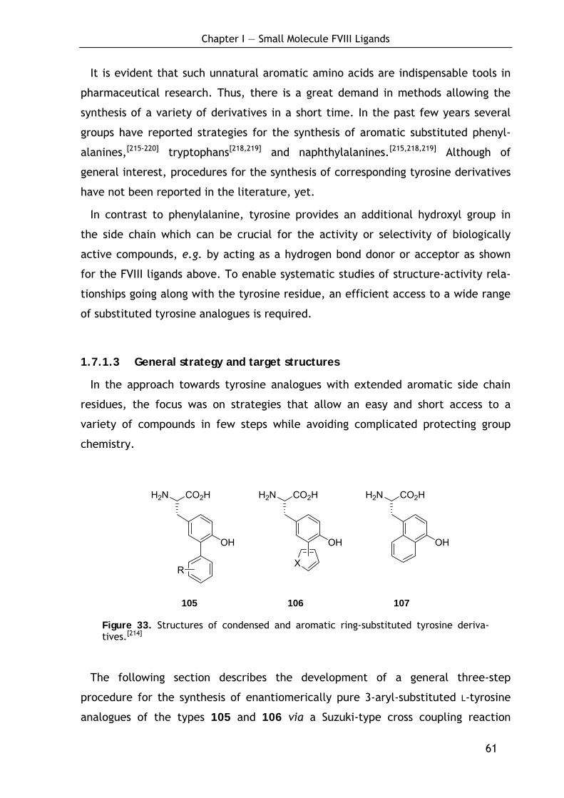
Chapter I — Small Molecule FVIII Ligands
61
It is evident that such unnatural aromatic amino acids are indispensable tools in
pharmaceutical research. Thus, there is a great demand in methods allowing the
synthesis of a variety of derivatives in a short time. In the past few years several
groups have reported strategies for the synthesis of aromatic substituted phenyl-
alanines,[215-220] tryptophans[218,219] and naphthylalanines.[215,218,219] Although of
general interest, procedures for the synthesis of corresponding tyrosine derivatives
have not been reported in the literature, yet.
In contrast to phenylalanine, tyrosine provides an additional hydroxyl group in
the side chain which can be crucial for the activity or selectivity of biologically
active compounds, e.g. by acting as a hydrogen bond donor or acceptor as shown
for the FVIII ligands above. To enable systematic studies of structure-activity rela-
tionships going along with the tyrosine residue, an efficient access to a wide range
of substituted tyrosine analogues is required.
1.7.1.3 General strategy and target structures
In the approach towards tyrosine analogues with extended aromatic side chain
residues, the focus was on strategies that allow an easy and short access to a
variety of compounds in few steps while avoiding complicated protecting group
chemistry.
CO2HH2N
OH
CO2HH2N
OH
R
CO2HH2N
OH
X
105 106 107
Figure 33. Structures of condensed and aromatic ring-substituted tyrosine deriva-tives.[214]
The following section describes the development of a general three-step
procedure for the synthesis of enantiomerically pure 3-aryl-substituted L-tyrosine
analogues of the types 105 and 106 via a Suzuki-type cross coupling reaction
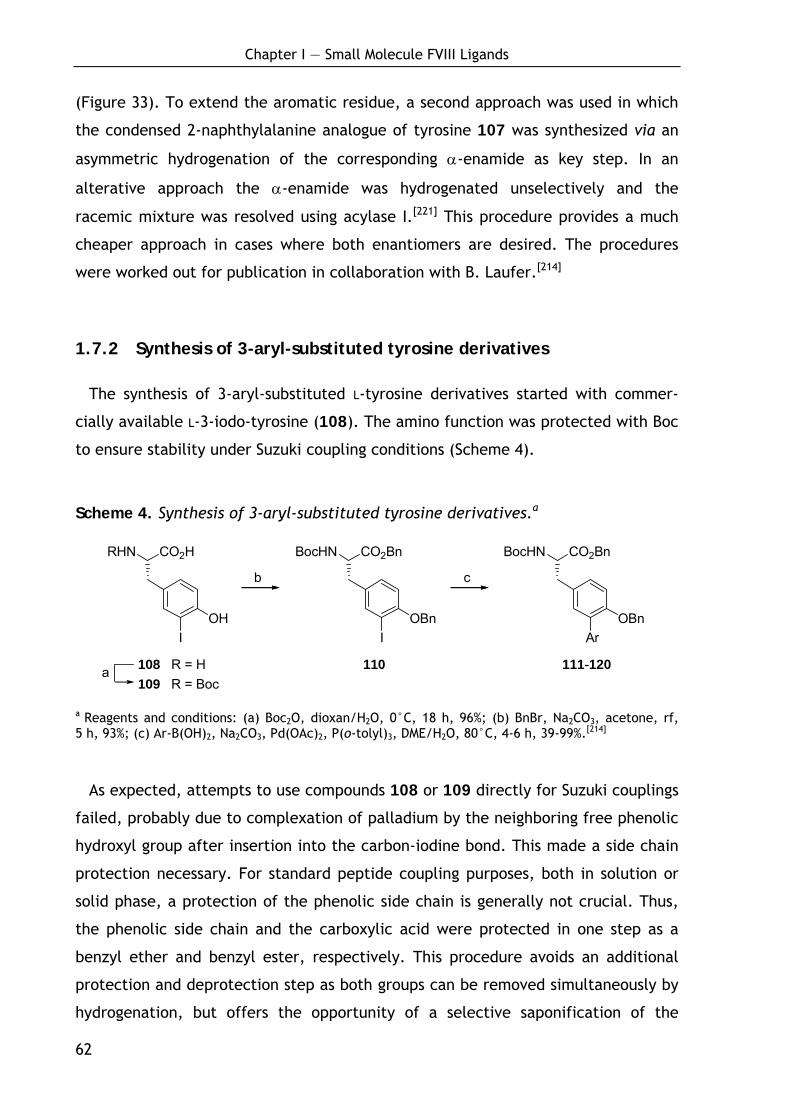
Chapter I — Small Molecule FVIII Ligands
62
CO2HRHN
OHI
CO2BnBocHN
OBnI
CO2BnBocHN
OBnAr
6 74 R1 = H 5 R1 = Boc
b c
a
(Figure 33). To extend the aromatic residue, a second approach was used in which
the condensed 2-naphthylalanine analogue of tyrosine 107 was synthesized via an
asymmetric hydrogenation of the corresponding α-enamide as key step. In an
alterative approach the α-enamide was hydrogenated unselectively and the
racemic mixture was resolved using acylase I.[221] This procedure provides a much
cheaper approach in cases where both enantiomers are desired. The procedures
were worked out for publication in collaboration with B. Laufer.[214]
1.7.2 Synthesis of 3-aryl-substituted tyrosine derivatives
The synthesis of 3-aryl-substituted L-tyrosine derivatives started with commer-
cially available L-3-iodo-tyrosine (108). The amino function was protected with Boc
to ensure stability under Suzuki coupling conditions (Scheme 4).
Scheme 4. Synthesis of 3-aryl-substituted tyrosine derivatives.a
a Reagents and conditions: (a) Boc2O, dioxan/H2O, 0°C, 18 h, 96%; (b) BnBr, Na2CO3, acetone, rf, 5 h, 93%; (c) Ar-B(OH)2, Na2CO3, Pd(OAc)2, P(o-tolyl)3, DME/H2O, 80°C, 4-6 h, 39-99%.[214]
As expected, attempts to use compounds 108 or 109 directly for Suzuki couplings
failed, probably due to complexation of palladium by the neighboring free phenolic
hydroxyl group after insertion into the carbon-iodine bond. This made a side chain
protection necessary. For standard peptide coupling purposes, both in solution or
solid phase, a protection of the phenolic side chain is generally not crucial. Thus,
the phenolic side chain and the carboxylic acid were protected in one step as a
benzyl ether and benzyl ester, respectively. This procedure avoids an additional
protection and deprotection step as both groups can be removed simultaneously by
hydrogenation, but offers the opportunity of a selective saponification of the
108 R = H 110 111-120 109 R = Boc

Chapter I — Small Molecule FVIII Ligands
63
B(OH)2
B(OH)2
HO
B(OH)2
Cl
B(OH)2
O
B(OH)2S
B(OH)2
benzyl ester when a side chain protection is needed. The benzyl protection was
performed under mild conditions using sodium carbonate as base to give the fully
protected amino acid 110 in 93% yield and without loss of enantiomeric purity as
shown below.[214]
Table 10. Suzuki cross coupling of 110 with arylboronic acids.[214]
Compound Ar-B(OH)2 Yield (%)a
111 95
112 93
113 79
114 70
115 80
116 39
117 94
118 92
119 99
120 45
a Yields refer to isolated pure products.
For Suzuki cross couplings, PdOAc2/P(o-tolyl)3 was used as catalyst and sodium
carbonate as base, a system which has proven to give good results in similar sys-
tems.[215-219] Thus, in collaboration with B. Laufer, a series of 3-aryl-substituted
tyrosine derivatives were synthesized in moderate to high yields using a variety of
activated and deactivated phenylboronic acids as well as heteroaromatic boronic
B(OH)2
B(OH)2F
B(OH)2
HO2C B(OH)2

Chapter I — Small Molecule FVIII Ligands
64
acids (Table 10). The formation of the dimeric homo coupling product was gener-
ally less than 2% as measured by HPLC-MS, except for the 3-chloro-phenylboronic
acid where higher amounts of this side product were formed.[214]
To demonstrate the further procedure and to verify the optical purity of the
products, samples of 111 and 114 were deprotected to apply them in solid-phase
peptide synthesis. The benzyl ester in both compounds could be cleaved chemose-
lectively using lithium hydroxide to give the free acids 121 and 122 in 86% and 83%
yield (Scheme 5). Alternatively, simultaneous cleavage of both benzyl groups can
be achieved by hydrogenation with palladium on charcoal, yielding 123 and 124 in
95% and 87%, respectively.[214]
Scheme 5. Regioselective deprotection of 3-aryl-substituted tyrosine derivatives.a
CO2HBocHN
OHR
CO2HBocHN
OBnR
CO2BnBocHN
OBnR
a b
R = phenyl 121 R = phenyl 111 R = phenyl 123 R = o-tolyl 122 R = o-tolyl 114 R = o-tolyl 124
a Reagents and conditions: (a) LiOH, THF/H2O, 0°C → rt, 18 h, 111: 86%, 114: 83%.; (b) H2, Pd/C, MeOH/AcN(Me)2, 6 h, 123: 95%, 124: 87%.[214]
The latter compounds were coupled to the amino free resin-bound L-valine using
1.8 equivalents of the amino acid 123 and 124, respectively, TBTU and HOBt in
NMP for 30 min. After cleavage from the resin and deprotection, the resulting
dipeptides H-m-(phenyl)Tyr-Val-OH (125) and H-m-(o-tolyl)Tyr-Val-OH (126) were
isolated in high purity (Scheme 6). A formation of side products due to the free
phenolic side chain was not observed.[214]
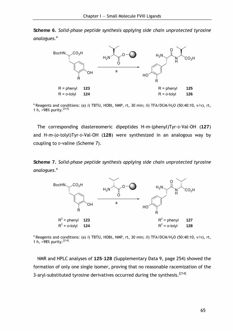
Chapter I — Small Molecule FVIII Ligands
65
Scheme 6. Solid-phase peptide synthesis applying side chain unprotected tyrosine
analogues.a
CO2HBocHN
OHR
H2NO
O H2N
HOR
NH
O
CO2H
a
R = phenyl 123 R = phenyl 125 R = o-tolyl 124 R = o-tolyl 126
a Reagents and conditions: (a) i) TBTU, HOBt, NMP, rt, 30 min; ii) TFA/DCM/H2O (50:40:10, v/v), rt, 1 h, >98% purity.[214]
The corresponding diastereomeric dipeptides H-m-(phenyl)Tyr-D-Val-OH (127)
and H-m-(o-tolyl)Tyr-D-Val-OH (128) were synthesized in an analogous way by
coupling to D-valine (Scheme 7).
Scheme 7. Solid-phase peptide synthesis applying side chain unprotected tyrosine
analogues.a
CO2HBocHN
OHR
H2NO
O H2N
HOR
NH
O
CO2H
a
R2 = phenyl 123 R2 = phenyl 127 R2 = o-tolyl 124 R2 = o-tolyl 128
a Reagents and conditions: (a) i) TBTU, HOBt, NMP, rt, 30 min; ii) TFA/DCM/H2O (50:40:10, v/v), rt, 1 h, >98% purity.[214]
NMR and HPLC analyses of 125-128 (Supplementary Data 9, page 254) showed the
formation of only one single isomer, proving that no reasonable racemization of the
3-aryl-substituted tyrosine derivatives occurred during the synthesis.[214]
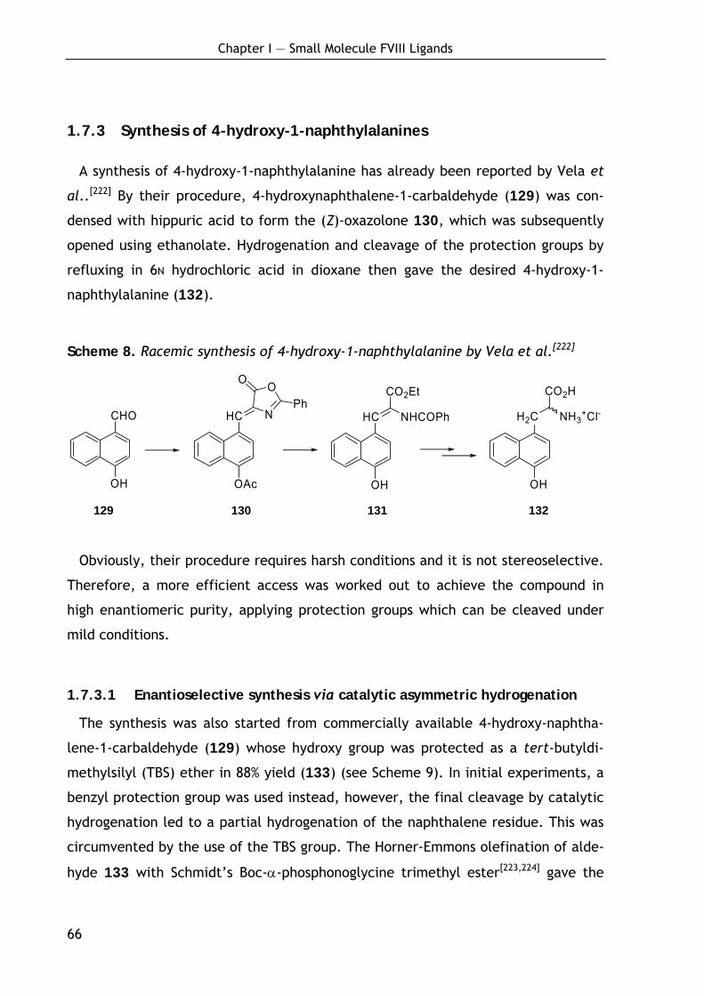
Chapter I — Small Molecule FVIII Ligands
66
1.7.3 Synthesis of 4-hydroxy-1-naphthylalanines
A synthesis of 4-hydroxy-1-naphthylalanine has already been reported by Vela et
al..[222] By their procedure, 4-hydroxynaphthalene-1-carbaldehyde (129) was con-
densed with hippuric acid to form the (Z)-oxazolone 130, which was subsequently
opened using ethanolate. Hydrogenation and cleavage of the protection groups by
refluxing in 6N hydrochloric acid in dioxane then gave the desired 4-hydroxy-1-
naphthylalanine (132).
Scheme 8. Racemic synthesis of 4-hydroxy-1-naphthylalanine by Vela et al.[222]
CHO
OH
HC
OAc
N
OO
PhHC
OH
CO2Et
NHCOPh H2C
OH
CO2H
NH3+Cl-
129 130 131 132
Obviously, their procedure requires harsh conditions and it is not stereoselective.
Therefore, a more efficient access was worked out to achieve the compound in
high enantiomeric purity, applying protection groups which can be cleaved under
mild conditions.
1.7.3.1 Enantioselective synthesis via catalytic asymmetric hydrogenation
The synthesis was also started from commercially available 4-hydroxy-naphtha-
lene-1-carbaldehyde (129) whose hydroxy group was protected as a tert-butyldi-
methylsilyl (TBS) ether in 88% yield (133) (see Scheme 9). In initial experiments, a
benzyl protection group was used instead, however, the final cleavage by catalytic
hydrogenation led to a partial hydrogenation of the naphthalene residue. This was
circumvented by the use of the TBS group. The Horner-Emmons olefination of alde-
hyde 133 with Schmidt’s Boc-α-phosphonoglycine trimethyl ester[223,224] gave the
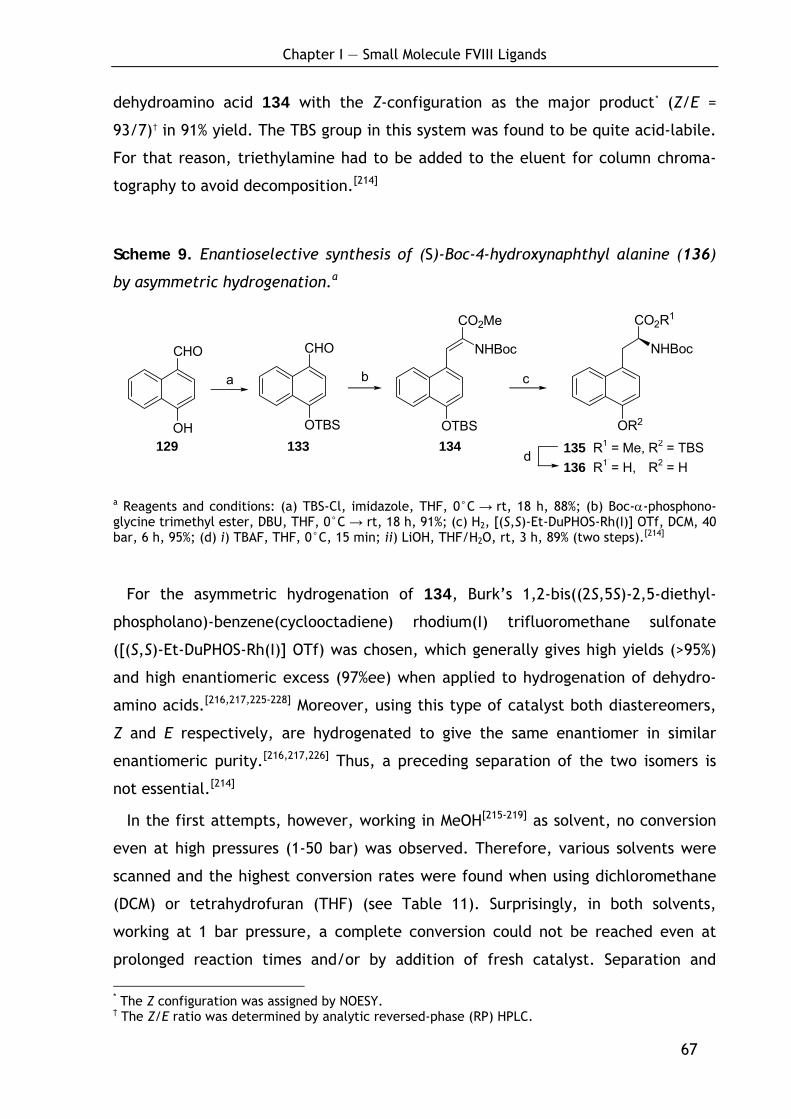
Chapter I — Small Molecule FVIII Ligands
67
CHO
OH
CHO
OTBS OTBS
CO2Me
NHBoc
OR2
CO2R1
NHBoc
14 R3 = Me, R4 = TB15 R3 = H, R4 = H
b ca
d
dehydroamino acid 134 with the Z-configuration as the major product* (Z/E =
93/7)† in 91% yield. The TBS group in this system was found to be quite acid-labile.
For that reason, triethylamine had to be added to the eluent for column chroma-
tography to avoid decomposition.[214]
Scheme 9. Enantioselective synthesis of (S)-Boc-4-hydroxynaphthyl alanine (136)
by asymmetric hydrogenation.a
a Reagents and conditions: (a) TBS-Cl, imidazole, THF, 0°C → rt, 18 h, 88%; (b) Boc-α-phosphono-glycine trimethyl ester, DBU, THF, 0°C → rt, 18 h, 91%; (c) H2, [(S,S)-Et-DuPHOS-Rh(I)] OTf, DCM, 40 bar, 6 h, 95%; (d) i) TBAF, THF, 0°C, 15 min; ii) LiOH, THF/H2O, rt, 3 h, 89% (two steps).[214]
For the asymmetric hydrogenation of 134, Burk’s 1,2-bis((2S,5S)-2,5-diethyl-
phospholano)-benzene(cyclooctadiene) rhodium(I) trifluoromethane sulfonate
([(S,S)-Et-DuPHOS-Rh(I)] OTf) was chosen, which generally gives high yields (>95%)
and high enantiomeric excess (97%ee) when applied to hydrogenation of dehydro-
amino acids.[216,217,225-228] Moreover, using this type of catalyst both diastereomers,
Z and E respectively, are hydrogenated to give the same enantiomer in similar
enantiomeric purity.[216,217,226] Thus, a preceding separation of the two isomers is
not essential.[214]
In the first attempts, however, working in MeOH[215-219] as solvent, no conversion
even at high pressures (1-50 bar) was observed. Therefore, various solvents were
scanned and the highest conversion rates were found when using dichloromethane
(DCM) or tetrahydrofuran (THF) (see Table 11). Surprisingly, in both solvents,
working at 1 bar pressure, a complete conversion could not be reached even at
prolonged reaction times and/or by addition of fresh catalyst. Separation and
* The Z configuration was assigned by NOESY. † The Z/E ratio was determined by analytic reversed-phase (RP) HPLC.
129 133 134 135 R1 = Me, R2 = TBS136 R1 = H, R2 = H
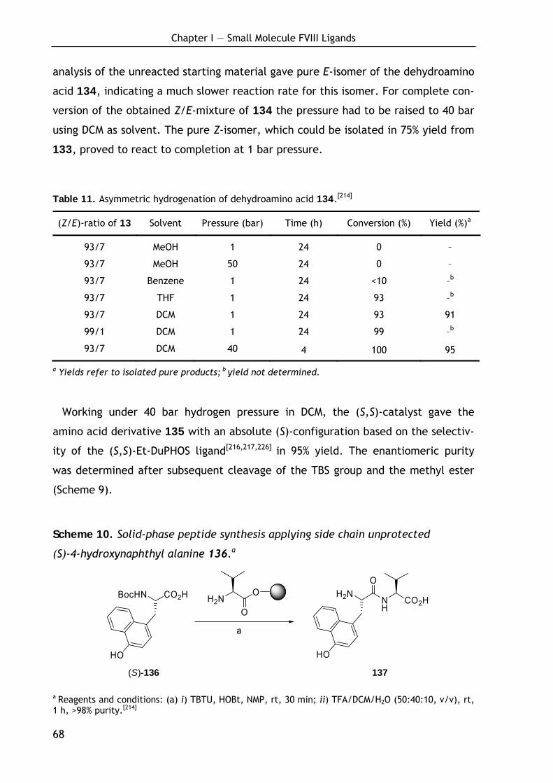
Chapter I — Small Molecule FVIII Ligands
68
analysis of the unreacted starting material gave pure E-isomer of the dehydroamino
acid 134, indicating a much slower reaction rate for this isomer. For complete con-
version of the obtained Z/E-mixture of 134 the pressure had to be raised to 40 bar
using DCM as solvent. The pure Z-isomer, which could be isolated in 75% yield from
133, proved to react to completion at 1 bar pressure.
Table 11. Asymmetric hydrogenation of dehydroamino acid 134.[214]
(Z/E)-ratio of 13 Solvent Pressure (bar) Time (h) Conversion (%) Yield (%)a
93/7 MeOH 1 24 0 –
93/7 MeOH 50 24 0 –
93/7 Benzene 1 24 <10 –b
93/7 THF 1 24 93 –b
93/7 DCM 1 24 93 91
99/1 DCM 1 24 99 –b
93/7 DCM 40 4 100 95
a Yields refer to isolated pure products; b yield not determined.
Working under 40 bar hydrogen pressure in DCM, the (S,S)-catalyst gave the
amino acid derivative 135 with an absolute (S)-configuration based on the selectiv-
ity of the (S,S)-Et-DuPHOS ligand[216,217,226] in 95% yield. The enantiomeric purity
was determined after subsequent cleavage of the TBS group and the methyl ester
(Scheme 9).
Scheme 10. Solid-phase peptide synthesis applying side chain unprotected
(S)-4-hydroxynaphthyl alanine 136.a
H2NO
O H2N
HO
NH
O
CO2H
a
CO2HBocHN
HO (S)-136 137
a Reagents and conditions: (a) i) TBTU, HOBt, NMP, rt, 30 min; ii) TFA/DCM/H2O (50:40:10, v/v), rt, 1 h, >98% purity.[214]
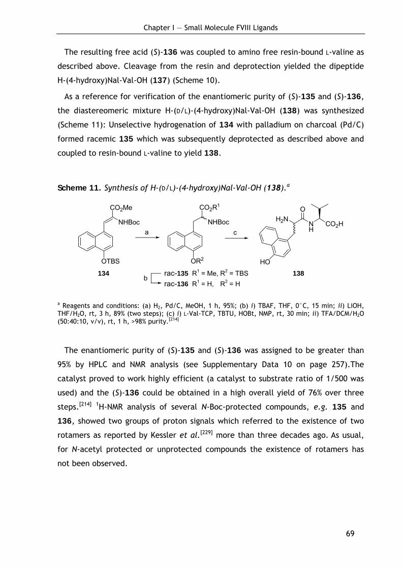
Chapter I — Small Molecule FVIII Ligands
69
OTBS
CO2Me
NHBoc
OR2
CO2R1
NHBoc
14 R3 = Me, R4 = TBS 15 R3 = H, R4 = H
a
b
c
H2NNH
CO2H
O
HO
The resulting free acid (S)-136 was coupled to amino free resin-bound L-valine as
described above. Cleavage from the resin and deprotection yielded the dipeptide
H-(4-hydroxy)Nal-Val-OH (137) (Scheme 10).
As a reference for verification of the enantiomeric purity of (S)-135 and (S)-136,
the diastereomeric mixture H-(D/L)-(4-hydroxy)Nal-Val-OH (138) was synthesized
(Scheme 11): Unselective hydrogenation of 134 with palladium on charcoal (Pd/C)
formed racemic 135 which was subsequently deprotected as described above and
coupled to resin-bound L-valine to yield 138.
Scheme 11. Synthesis of H-(D/L)-(4-hydroxy)Nal-Val-OH (138).a
a Reagents and conditions: (a) H2, Pd/C, MeOH, 1 h, 95%; (b) i) TBAF, THF, 0°C, 15 min; ii) LiOH, THF/H2O, rt, 3 h, 89% (two steps); (c) i) L-Val-TCP, TBTU, HOBt, NMP, rt, 30 min; ii) TFA/DCM/H2O (50:40:10, v/v), rt, 1 h, >98% purity.[214]
The enantiomeric purity of (S)-135 and (S)-136 was assigned to be greater than
95% by HPLC and NMR analysis (see Supplementary Data 10 on page 257).The
catalyst proved to work highly efficient (a catalyst to substrate ratio of 1/500 was
used) and the (S)-136 could be obtained in a high overall yield of 76% over three
steps.[214] 1H-NMR analysis of several N-Boc-protected compounds, e.g. 135 and
136, showed two groups of proton signals which referred to the existence of two
rotamers as reported by Kessler et al.[229] more than three decades ago. As usual,
for N-acetyl protected or unprotected compounds the existence of rotamers has
not been observed.
134 rac-135 R1 = Me, R2 = TBS rac-136 R1 = H, R2 = H
138
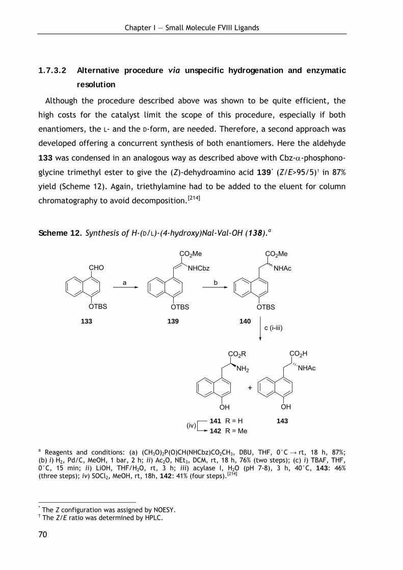
Chapter I — Small Molecule FVIII Ligands
70
CHO
OTBS OTBS
CO2Me
NHCbz
OH
CO2R
NH2
OH
CO2H
NHAc
+
OTBS
CO2Me
NHAc
12 18 19
21
a b
c (i-iii)
(iv)
1.7.3.2 Alternative procedure via unspecific hydrogenation and enzymatic
resolution
Although the procedure described above was shown to be quite efficient, the
high costs for the catalyst limit the scope of this procedure, especially if both
enantiomers, the L- and the D-form, are needed. Therefore, a second approach was
developed offering a concurrent synthesis of both enantiomers. Here the aldehyde
133 was condensed in an analogous way as described above with Cbz-α-phosphono-
glycine trimethyl ester to give the (Z)-dehydroamino acid 139* (Z/E>95/5)† in 87%
yield (Scheme 12). Again, triethylamine had to be added to the eluent for column
chromatography to avoid decomposition.[214]
Scheme 12. Synthesis of H-(D/L)-(4-hydroxy)Nal-Val-OH (138).a
a Reagents and conditions: (a) (CH3O)2P(O)CH(NHCbz)CO2CH3, DBU, THF, 0°C → rt, 18 h, 87%; (b) i) H2, Pd/C, MeOH, 1 bar, 2 h; ii) Ac2O, NEt3, DCM, rt, 18 h, 76% (two steps); (c) i) TBAF, THF, 0°C, 15 min; ii) LiOH, THF/H2O, rt, 3 h; iii) acylase I, H2O (pH 7-8), 3 h, 40°C, 143: 46% (three steps); iv) SOCl2, MeOH, rt, 18h, 142: 41% (four steps).[214]
* The Z configuration was assigned by NOESY. † The Z/E ratio was determined by HPLC.
141 R = H 143 142 R = Me
133 139 140
Chapter I — Small Molecule FVIII Ligands
71
In contrast to the previous procedure, the hydrogenation was carried out using
palladium on charcoal as catalyst leading to a simultaneous hydrogenation of the
double bond and cleavage of the Cbz protection group. The reaction was monitored
by TLC and mass spectroscopy and quenched after complete conversion to the
saturated free amine (4 h). Prolonged reaction times (18 h) led to partial hydro-
genation of the naphthalene residue. The resulting racemic mixture was directly
acetylated by treating with acetic acid anhydride in presence of triethylamine to
yield 140 in 76% yield over two steps.[214]
Subjecting the fully protected compound 140 to enzymatic resolution led to an
unseparatable product mixture due to a partial loss of the TBS group and partial
cleavage of the methyl ester. Thus, both protecting groups were removed prior to
the resolution improving the solubility in aqueous conditions. To avoid additional
separation steps, a one-pot procedure was developed: the TBS ether and the
methyl ester were subsequently cleaved and the racemic mixture was resolved
using acylase I to give the L-isomer 141 as free amine, while the D-isomer 143
remained in the N-acetylated form which was easily separated by extraction in high
purity (93% by HPLC analysis).[221] As the isolation of the free L-amino acid 141 by
crystallization failed, it was converted to the corresponding methyl ester 142 and
also isolated by extraction (90% purity by HPLC analysis). Further purification by
column chromatography gave the pure enantiomer 142[221] and 143 (96%ee)* in high
yields (41% over four steps and 46% over three steps, respectively).[214]
1.7.3.3 Discussion and outlook
The sequences described above provide a variety of tyrosine analogues stereose-
lectively in high yields. A convenient route was developed for the synthesis of
aromatic ring-substituted tyrosine derivatives via a Suzuki cross coupling reaction,
thereby introducing a variety of substituted and unsubstituted aromatic as well as
heteroaromatic residues in high yield. Even heavily deactivated boronic acids could
be coupled in moderate yields. To provide a structural alternative, methods for the
asymmetric synthesis of the ring-condensed tyrosine analogue 4-hydroxy-naphthyl-
alanine were developed. For the synthesis, two alternative methologies were
* The enantiomeric purity of 143 was determined by chiral HPLC analyses after conversion to the corresponding methyl ester.
Chapter I — Small Molecule FVIII Ligands
72
explored to offer the most economical access if only one or both enantiomers are
desired. In case of the latter, the unselective hydrogenation of the corresponding
dehydroamino acid and enzymatic resolution of the racemic mixture is the most
cost efficient procedure, as acylase I is cheap and both enantiomers are obtained
simultaneously. In contrast, if only one single enantiomer is required the racemic
synthesis is associated with 50% loss of substance due to the undesired second
enantiomer. The asymmetric hydrogenation of the dehydroamino acid provides a
high-yielding access to a single enantiomer. Although the catalyst is quite expen-
sive, its great efficiency and the high yields make this procedure the most eco-
nomic in case only one enantiomer is desired. The procedure now provides the
opportunity to systematically scan above-mentioned FVIII ligands to study the
impact of the modified tyrosine analogues.
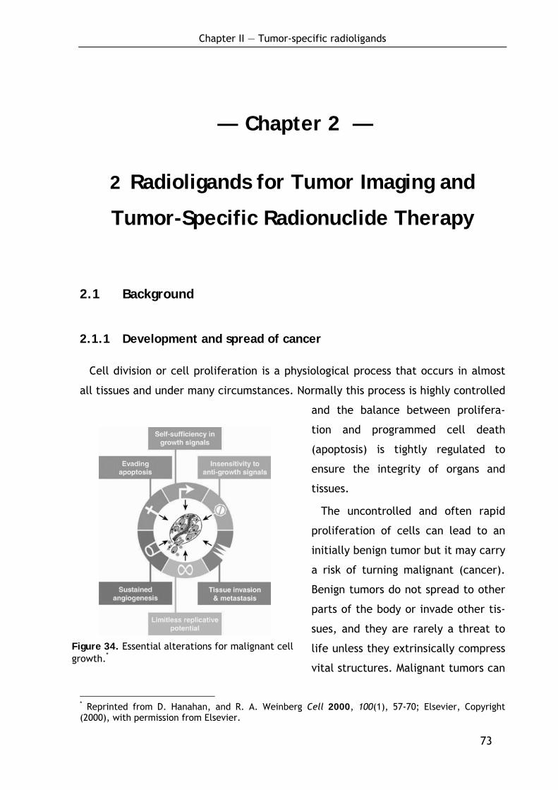
Chapter II — Tumor-specific radioligands
73
— Chapter 2 —
2 Radioligands for Tumor Imaging and
Tumor-Specific Radionuclide Therapy
2.1 Background
2.1.1 Development and spread of cancer
Cell division or cell proliferation is a physiological process that occurs in almost
all tissues and under many circumstances. Normally this process is highly controlled
and the balance between prolifera-
tion and programmed cell death
(apoptosis) is tightly regulated to
ensure the integrity of organs and
tissues.*
The uncontrolled and often rapid
proliferation of cells can lead to an
initially benign tumor but it may carry
a risk of turning malignant (cancer).
Benign tumors do not spread to other
parts of the body or invade other tis-
sues, and they are rarely a threat to
life unless they extrinsically compress
vital structures. Malignant tumors can
* Reprinted from D. Hanahan, and R. A. Weinberg Cell 2000, 100(1), 57-70; Elsevier, Copyright (2000), with permission from Elsevier.
Figure 34. Essential alterations for malignant cell growth.*
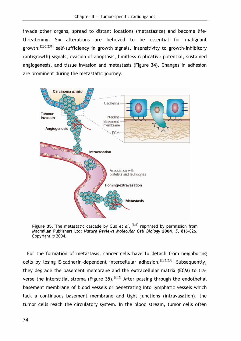
Chapter II — Tumor-specific radioligands
74
invade other organs, spread to distant locations (metastasize) and become life-
threatening. Six alterations are believed to be essential for malignant
growth:[230,231] self-sufficiency in growth signals, insensitivity to growth-inhibitory
(antigrowth) signals, evasion of apoptosis, limitless replicative potential, sustained
angiogenesis, and tissue invasion and metastasis (Figure 34). Changes in adhesion
are prominent during the metastatic journey.
Figure 35. The metastatic cascade by Guo et al.,[232] reprinted by permission from Macmillan Publishers Ltd: Nature Reviews Molecular Cell Biology 2004, 5, 816-826, Copyright © 2004.
For the formation of metastasis, cancer cells have to detach from neighboring
cells by losing E-cadherin-dependent intercellular adhesion.[232,233] Subsequently,
they degrade the basement membrane and the extracellular matrix (ECM) to tra-
verse the interstitial stroma (Figure 35).[232] After passing through the endothelial
basement membrane of blood vessels or penetrating into lymphatic vessels which
lack a continuous basement membrane and tight junctions (intravasation), the
tumor cells reach the circulatory system. In the blood stream, tumor cells often
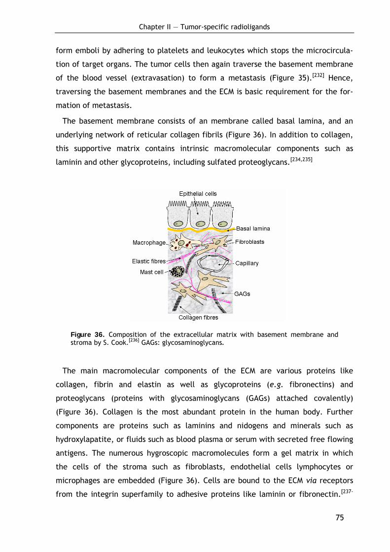
Chapter II — Tumor-specific radioligands
75
form emboli by adhering to platelets and leukocytes which stops the microcircula-
tion of target organs. The tumor cells then again traverse the basement membrane
of the blood vessel (extravasation) to form a metastasis (Figure 35).[232] Hence,
traversing the basement membranes and the ECM is basic requirement for the for-
mation of metastasis.
The basement membrane consists of an membrane called basal lamina, and an
underlying network of reticular collagen fibrils (Figure 36). In addition to collagen,
this supportive matrix contains intrinsic macromolecular components such as
laminin and other glycoproteins, including sulfated proteoglycans.[234,235]
Figure 36. Composition of the extracellular matrix with basement membrane and stroma by S. Cook.[236] GAGs: glycosaminoglycans.
The main macromolecular components of the ECM are various proteins like
collagen, fibrin and elastin as well as glycoproteins (e.g. fibronectins) and
proteoglycans (proteins with glycosaminoglycans (GAGs) attached covalently)
(Figure 36). Collagen is the most abundant protein in the human body. Further
components are proteins such as laminins and nidogens and minerals such as
hydroxylapatite, or fluids such as blood plasma or serum with secreted free flowing
antigens. The numerous hygroscopic macromolecules form a gel matrix in which
the cells of the stroma such as fibroblasts, endothelial cells lymphocytes or
microphages are embedded (Figure 36). Cells are bound to the ECM via receptors
from the integrin superfamily to adhesive proteins like laminin or fibronectin.[237-
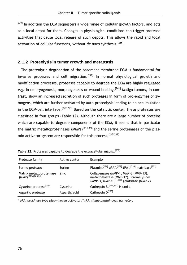
Chapter II — Tumor-specific radioligands
76
239] In addition the ECM sequesters a wide range of cellular growth factors, and acts
as a local depot for them. Changes in physiological conditions can trigger protease
activities that cause local release of such depots. This allows the rapid and local
activation of cellular functions, without de novo synthesis.[236]
2.1.2 Proteolysis in tumor growth and metastasis
The proteolytic degradation of the basement membrane ECM is fundamental for
invasive processes and cell migration.[240] In normal physiological growth and
modification processes, proteases capable to degrade the ECM are highly regulated
e.g. in embryogenesis, morphogenesis or wound healing.[241] Malign tumors, in con-
trast, show an increased secretion of such proteases in form of pro-enzymes or zy-
mogens, which are further activated by auto-proteolysis leading to an accumulation
in the ECM-cell interface.[242,243] Based on the catalytic center, these proteases are
classified in four groups (Table 12). Although there are a large number of proteins
which are capable to degrade components of the ECM, it seems that in particular
the matrix metalloproteinases (MMPs)[244-246]and the serine proteinases of the plas-
min activator system are responsible for this process.[247-249]
Table 12. Proteases capable to degrade the extracellular matrix.[250]
Protease family Active center Example
Serine protease Serine Plasmin,[251] uPAa,[252] tPAb,[234] matripase[253]
Matrix metalloproteinase (MMP)[244,252,254]
Zinc Collagenases (MMP-1, MMP-8, MMP-13), metalloelastase (MMP-12), stromelysines (MMP-3, MMP-10),[255] gelatinase (MMP-2)
Cysteine protease[256] Cysteine Cathepsin B,[252,257] H und L
Aspartic protease Aspartic acid Cathepsin D[258]
a uPA: urokinase type plasminogen activator; b tPA: tissue plasminogen activator.
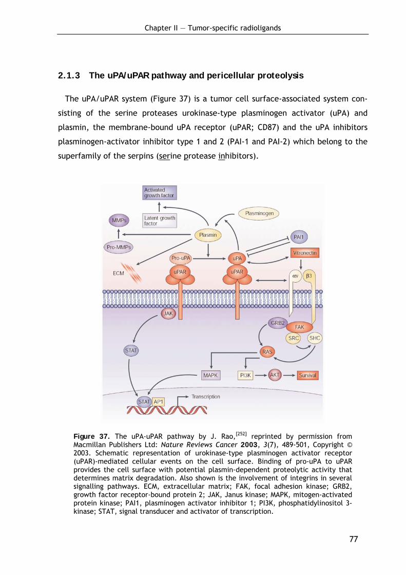
Chapter II — Tumor-specific radioligands
77
2.1.3 The uPA/uPAR pathway and pericellular proteolysis
The uPA/uPAR system (Figure 37) is a tumor cell surface-associated system con-
sisting of the serine proteases urokinase-type plasminogen activator (uPA) and
plasmin, the membrane-bound uPA receptor (uPAR; CD87) and the uPA inhibitors
plasminogen-activator inhibitor type 1 and 2 (PAI-1 and PAI-2) which belong to the
superfamily of the serpins (serine protease inhibitors).
Figure 37. The uPA-uPAR pathway by J. Rao,[252] reprinted by permission from Macmillan Publishers Ltd: Nature Reviews Cancer 2003, 3(7), 489-501, Copyright © 2003. Schematic representation of urokinase-type plasminogen activator receptor (uPAR)-mediated cellular events on the cell surface. Binding of pro-uPA to uPAR provides the cell surface with potential plasmin-dependent proteolytic activity that determines matrix degradation. Also shown is the involvement of integrins in several signalling pathways. ECM, extracellular matrix; FAK, focal adhesion kinase; GRB2, growth factor receptor-bound protein 2; JAK, Janus kinase; MAPK, mitogen-activated protein kinase; PAI1, plasminogen activator inhibitor 1; PI3K, phosphatidylinositol 3-kinase; STAT, signal transducer and activator of transcription.
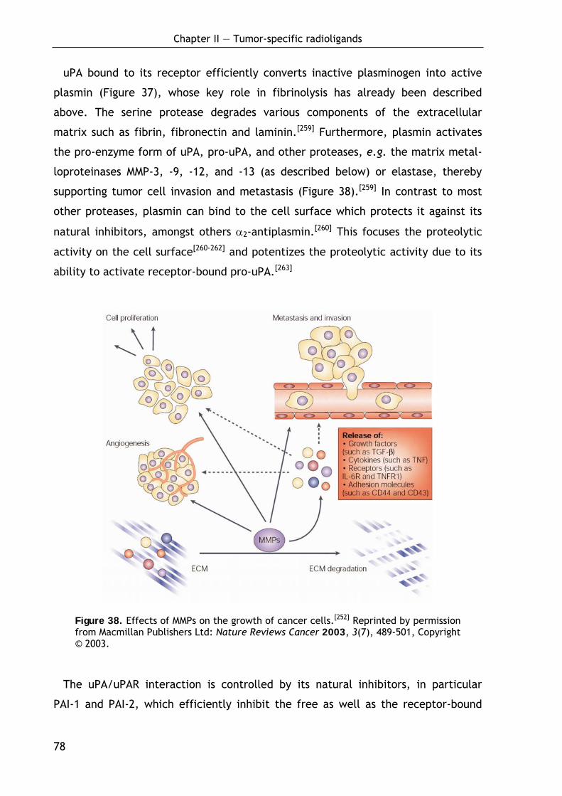
Chapter II — Tumor-specific radioligands
78
uPA bound to its receptor efficiently converts inactive plasminogen into active
plasmin (Figure 37), whose key role in fibrinolysis has already been described
above. The serine protease degrades various components of the extracellular
matrix such as fibrin, fibronectin and laminin.[259] Furthermore, plasmin activates
the pro-enzyme form of uPA, pro-uPA, and other proteases, e.g. the matrix metal-
loproteinases MMP-3, -9, -12, and -13 (as described below) or elastase, thereby
supporting tumor cell invasion and metastasis (Figure 38).[259] In contrast to most
other proteases, plasmin can bind to the cell surface which protects it against its
natural inhibitors, amongst others α2-antiplasmin.[260] This focuses the proteolytic
activity on the cell surface[260-262] and potentizes the proteolytic activity due to its
ability to activate receptor-bound pro-uPA.[263]
Figure 38. Effects of MMPs on the growth of cancer cells.[252] Reprinted by permission from Macmillan Publishers Ltd: Nature Reviews Cancer 2003, 3(7), 489-501, Copyright © 2003.
The uPA/uPAR interaction is controlled by its natural inhibitors, in particular
PAI-1 and PAI-2, which efficiently inhibit the free as well as the receptor-bound
Chapter II — Tumor-specific radioligands
79
form of uPA (Figure 37).[264,265] The stable ternary complexes (uPA/uPAR/PAI-1 and
uPA/uPAR/PAI-2) are subsequently internalized[266-268] in a process that requires the
α2-macroglobulin receptor/low-density lipoprotein (LDL) receptor-related protein
(LRP) (α2MR/LRP).[269-273] New investigations show that tumor cells possess a mecha-
nism to recycle the internalized uPAR back to the cell surface from the cytoplasm
which may help to maintain invasive potential.[271,274-277]
The activation of MMPs plays a fundamental role in cancer progression (Figure
38). MMPs not only directly degrade structural components of the ECM, in particu-
lar laminin-5, hence regulating invasion and migration, but they also promote the
growth of cancer cells by interacting with a variety of ECM molecules, cleaving in-
sulin-like growth factors and shedding transmembrane precursors of growth factors,
including transforming growth factor-α (TGF-α) (Figure 38).[252] In addition, MMPs
interact with integrins and promote angiogenesis by increasing the bioavailability
of pro-angiogenic growth factors.[252]
2.1.4 Molecular structure of uPA
The urokinase-type plasminogen activator is expressed by numerous malign and
normal cells in form of the inactive 52 kDa single-chain pro-enzyme pro-uPA com-
prising 12 disulfide bridges.[278,279] The domain structure is shown in Figure 39. The
N-terminal growth factor-like domain (GFD) (uPA1-44) is part of the amino-terminal
fragment (ATF; uPA1-135) and comprises the uPA receptor binding site.[280] Although
significant sequence homologies to the epidermal growth factor (EGF) and the TGF-
α have been found,[281,282] the binding of uPAR is selective for GFD.[235] uPA45-135 is
called the kringle domain. It is a highly conserved structure motive which is found
in a number of proteins involved in hemostasis or fibrinolysis.[283] In contrast to the
kringle domain of tPA and plasminogen, the kringle domain of uPA does not exhibit
affinity to fibrin but binds to heparin.[284] The C-terminal region uPA136-411 comprises
the enzymatically active region of the serine protease with the catalytic triad,
residues His204, Asp255 und Ser356.[285] X-ray structures of the C-terminal domain in
complex with different inhibitors have been solved by Sperl et al. and Spraggon et
al..[286,287]
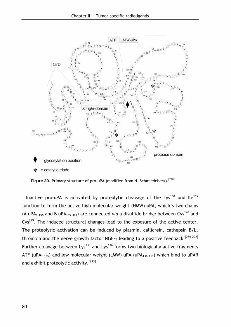
Chapter II — Tumor-specific radioligands
80
SN
ELHQVP
SN C
D CLN
GGTC
VS N K Y
FSNIH
WC
NC
PK
FG G Q
HC
K
E
D K SKT
C
CD
HV
M
YE
GN
GH
FY
RGKAST
DT
M G R P C
C
L P W N S A TV
LQQ
TYHAHRS
QA
LQ
GLGK
HN
YC
L A L G N E E K TH S R I
WP
LF
HS
V R T Y GV P K D KL
ACG
RG
WS
VIGT
M R G QL
S C
C
V LPG
GS
DGQ
CS
DT
KW
QP D A A C L M K T
TV
ES
GYYH
PQQCE
RH
SI
LK V V T M
KL
QE
PYL
YD
TSNE
KG
FG
TIE
STG
FQ
PD
NY
MS
PLC
IT Q
I
RS
PQ
AC
RG
EK
SRIKL
LA
ID
NH
HA
L T DA
SY
DKHLILNE
VEFKMEG
QT
S N L R SR G L
YVIY
DE
KKPYDIFCH
TA
SIV
WC
PS
IL
SG C
GV Y T V S G G
RH
RRY
I
AAF
WP
QNEIT
TFEGGIIKF
RP
RL
TK
QG
CQ
FK
LE
EPPSSPKKGD
A
CEQV
LP
KLG
VQ V Y W P
RR
RNDP
NR
TL
T
N
L
10
1113
19
31
33
30
20
4042
I
50
60
70 71
80
90100
102
110113
120
126
130 131
140
148
150
160
170
180189
190
197
200
205
210
220230
240
250
260
268
270
279280
290
293
300
310
320
325
330
340
341
350
352
360362
370
380390
400411
1
Protease-Domäne
Aktivierung zu HMW-uPA
47
GFD
kringle-Domäne
48
135
136
ATF LMW-uPA
= Glykosylierungsstelle
= katalytische Triade
*
*
*356
204
245
*
protease domain
kringle-domain
= glycosylation position
= catalytic triade
Figure 39. Primary structure of pro-uPA (modified from N. Schmiedeberg).[288]
Inactive pro-uPA is activated by proteolytic cleavage of the Lys158 und Ile159
junction to form the active high molecular weight (HMW)-uPA, which’s two-chains
(A uPA1-158 and B uPA159-411) are connected via a disulfide bridge between Cys148 and
Cys279. The induced structural changes lead to the exposure of the active center.
The proteolytic activation can be induced by plasmin, callicrein, cathepsin B/L,
thrombin and the nerve growth factor NGF-γ leading to a positive feedback.[289-292]
Further cleavage between Lys135 and Lys136 forms two biologically active fragments
ATF (uPA1-135) and low molecular weight (LMW)-uPA (uPA136-411) which bind to uPAR
and exhibit proteolytic activity.[293]
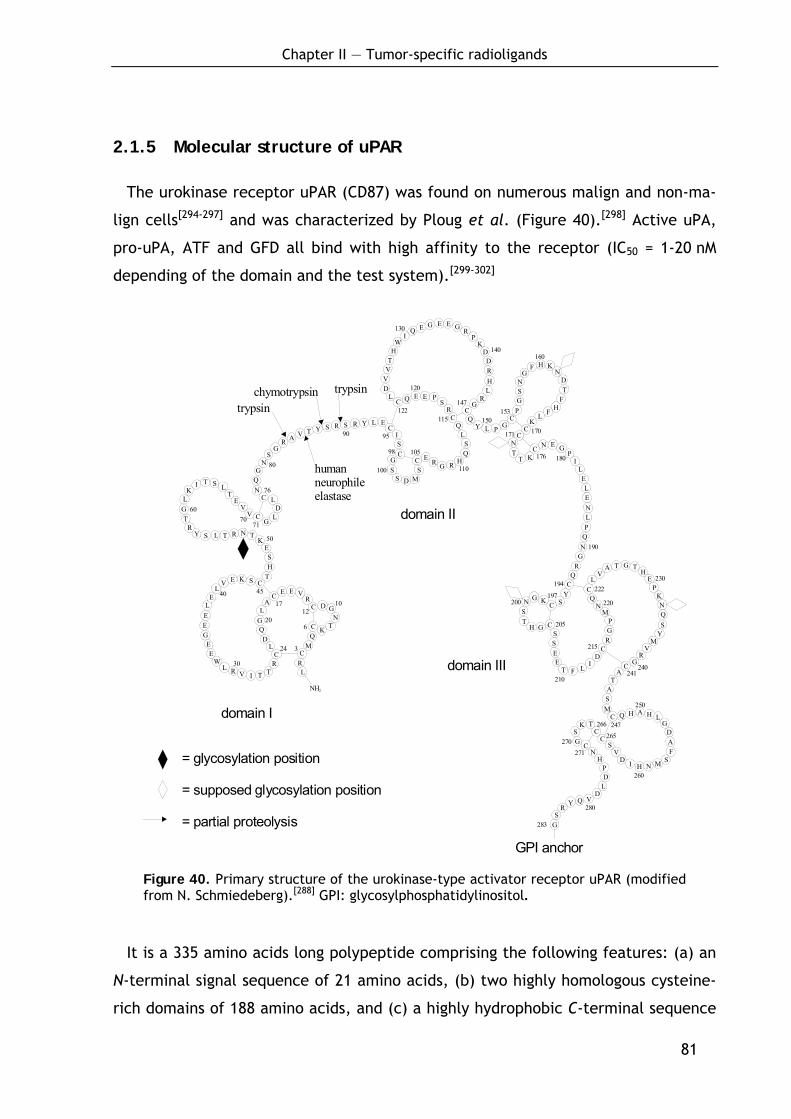
Chapter II — Tumor-specific radioligands
81
2.1.5 Molecular structure of uPAR
The urokinase receptor uPAR (CD87) was found on numerous malign and non-ma-
lign cells[294-297] and was characterized by Ploug et al. (Figure 40).[298] Active uPA,
pro-uPA, ATF and GFD all bind with high affinity to the receptor (IC50 = 1-20 nM
depending of the domain and the test system).[299-302]
= partielle Proteolyse
= Glykosylierungsstelle
= mögliche Glykosylierungsstelle
= glycosylation position
= supposed glycosylation position
= partial proteolysis
100
NH2
LRCC
CC
C
MQ
K TN
GDR
VEEA
LGQD
L
RTTIVRL
WE
EGEEL
EL
V E K S CTHS
EK
TNRTLSYR
TGLK
I T S LT
EV
V CG
LD
LCNQG
NS
GR A V T Y S R S R Y L E
CISC
GS
S D MS
C
CC
C
ER G R H
QS
LQ
RS
PEEQLDVVTH
WI
Q E G E E G RP
KDDRHL
RG
QY L P G
CC
PGSN
GF H K
NDT
FHFL
K
CNT
T KC N E G
PI
LELENLP
QN
GR
QC
YSC
C
KGNST
H GSS
LI
DC
RGP
MN
QC
EE
T F
LV
A T G TH
EP
KNQS
YM
VR
GCA
TASM
C Q H A H LG
DAF
SMNHIDV
SC
CTK
SG C
NH
PD
LD
VQYR
SG
10
3
6
1217
24
30
20
40 45
50
6070
71
76
80
90 95
98 105
110
120
130
140
150115122
147153
160
170171
176 180
190
194
200197
205
210
215
220
222230
240241
247
250
260
265270
266
271
283
280
trypsintrypsinchymotrypsin
humanneurophileelastase
domain I
domain II
domain III
GPI anchor
Figure 40. Primary structure of the urokinase-type activator receptor uPAR (modified from N. Schmiedeberg).[288] GPI: glycosylphosphatidylinositol.
It is a 335 amino acids long polypeptide comprising the following features: (a) an
N-terminal signal sequence of 21 amino acids, (b) two highly homologous cysteine-
rich domains of 188 amino acids, and (c) a highly hydrophobic C-terminal sequence
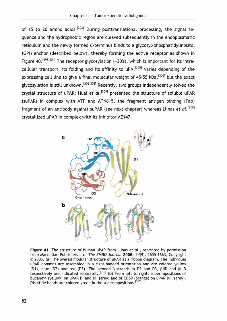
Chapter II — Tumor-specific radioligands
82
of 15 to 20 amino acids.[303] During posttranslational processing, the signal se-
quence and the hydrophobic region are cleaved subsequently in the endoplasmatic
reticulum and the newly formed C-terminus binds to a glycosyl-phosphatidylinositol
(GPI) anchor (described below), thereby forming the active receptor as shown in
Figure 40.[298,304] The receptor glycosylation (~30%), which is important for its intra-
cellular transport, its folding and its affinity to uPA,[305] varies depending of the
expressing cell line to give a final molecular weight of 45-55 kDa,[306] but the exact
glycosylation is still unknown.[306-308] Recently, two groups independently solved the
crystal structure of uPAR: Huai et al.[309] presented the structure of soluble uPAR
(suPAR) in complex with ATF and ATN615, the fragment antigen binding (Fab)
fragment of an antibody against suPAR (see next chapter) whereas Llinas et al.[310]
crystallized uPAR in complex with its inhibitor AE147.
Figure 41. The structure of human uPAR from Llinas et al., reprinted by permission from Macmillan Publishers Ltd: The EMBO Journal 2005, 24(9), 1655-1663, Copyright © 2005. (a) The overall modular structure of uPAR as a ribbon diagram. The individual uPAR domains are assembled in a right-handed orientation and are colored yellow (D1), blue (D2) and red (D3). The bended β-strands in D2 and D3, βIID and βIIID respectively are indicated separately.[310] (b) From left to right, superimpositions of bucandin (yellow) on uPAR DI and DII (grey) and of CD59 (orange) on uPAR DIII (grey). Disulfide bonds are colored green in the superimpositions.[310]
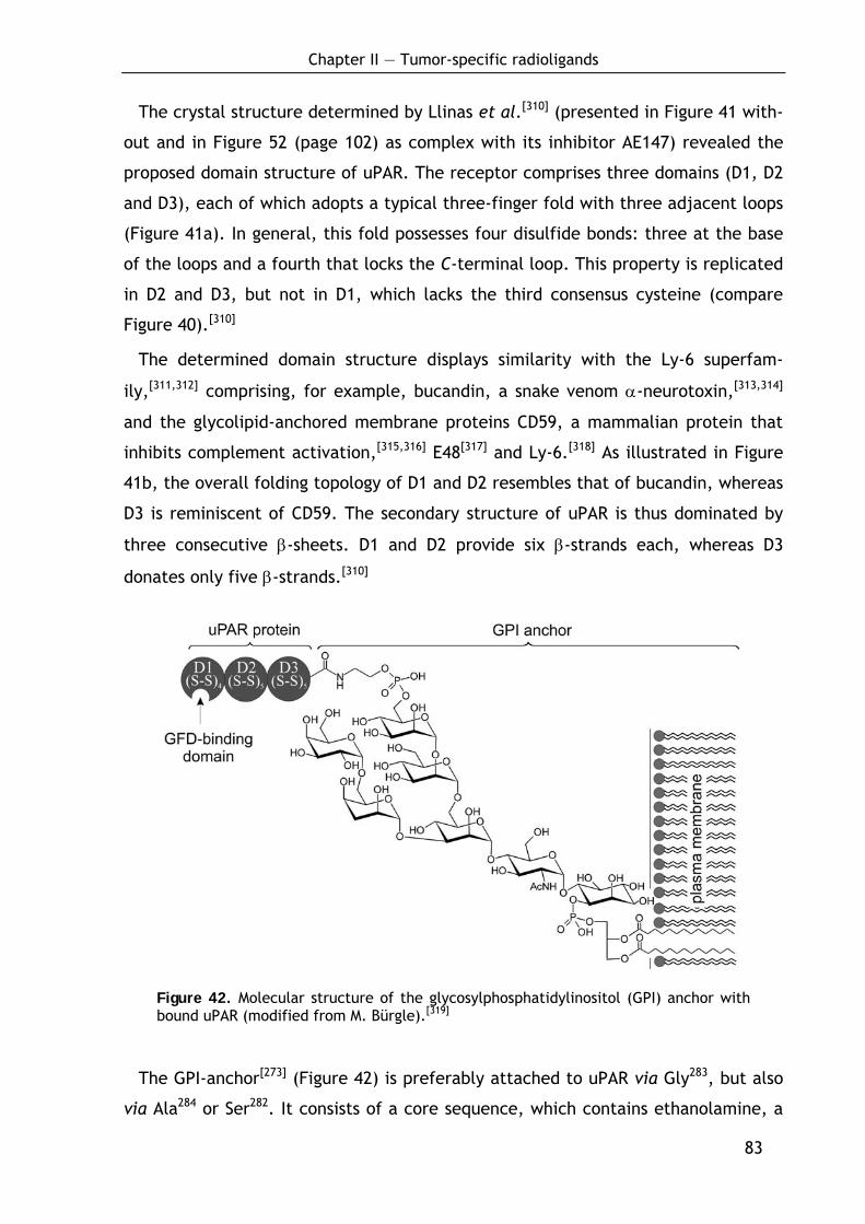
Chapter II — Tumor-specific radioligands
83
The crystal structure determined by Llinas et al.[310] (presented in Figure 41 with-
out and in Figure 52 (page 102) as complex with its inhibitor AE147) revealed the
proposed domain structure of uPAR. The receptor comprises three domains (D1, D2
and D3), each of which adopts a typical three-finger fold with three adjacent loops
(Figure 41a). In general, this fold possesses four disulfide bonds: three at the base
of the loops and a fourth that locks the C-terminal loop. This property is replicated
in D2 and D3, but not in D1, which lacks the third consensus cysteine (compare
Figure 40).[310]
The determined domain structure displays similarity with the Ly-6 superfam-
ily,[311,312] comprising, for example, bucandin, a snake venom α-neurotoxin,[313,314]
and the glycolipid-anchored membrane proteins CD59, a mammalian protein that
inhibits complement activation,[315,316] E48[317] and Ly-6.[318] As illustrated in Figure
41b, the overall folding topology of D1 and D2 resembles that of bucandin, whereas
D3 is reminiscent of CD59. The secondary structure of uPAR is thus dominated by
three consecutive β-sheets. D1 and D2 provide six β-strands each, whereas D3
donates only five β-strands.[310]
Figure 42. Molecular structure of the glycosylphosphatidylinositol (GPI) anchor with bound uPAR (modified from M. Bürgle).[319]
The GPI-anchor[273] (Figure 42) is preferably attached to uPAR via Gly283, but also
via Ala284 or Ser282. It consists of a core sequence, which contains ethanolamine, a
Chapter II — Tumor-specific radioligands
84
variable sequence of diverse mannoses, N-acetyl glucosamine, phosphatidylinositol
and diacetyl glycerol (Figure 42), and lacks transmembrane and cytosolic compo-
nents and therefore intrinsic signal transduction potential. uPAR cleavage from its
anchor by phosphatidylinositol-specific phospholipase C or D (PI-PLC and PI-PLD,
respectively) forms soluble uPAR (suPAR)[320,321] which also binds to uPA and which
can be detected in the body fluids of cancer patients.
2.1.6 New insights into the uPA-uPAR interaction
The X-ray structure of suPAR in complex with ATF and ATN615 obtained by Huai
et al. (Figure 44)[309] recently demonstrated more explicit data about the uPA-uPAR
interaction.
2.1.6.1 The amino-terminal fragment ATF
Looking at the structure of ATF (Figure 43), the authors have shown that ATF is
constrained in the ternary complex, with tow adjacent domains tightly packed and
forming hydrophobic interactions with each other.[309] This is in contrast to previous
results obtained from NMR studies where the GFD and kringle domains of unbound
ATF were found to exhibit a high degree of structural independence involving little
or no interdomain interaction.[280]
2.1.6.2 The structure of soluble uPAR
The X-ray structure furthermore revealed that the D1 and the D3 domains of
suPAR are connected with each other.[309] Key residues for this interaction are
His47, Glu49, Lys50, Arg53, Leu252, Asp254, Asn259, and His260 which maintain the D1D3
domain interface in the suPAR-ATF structure by three hydrogen bonds, His47-Asn259,
Lys50-Asp254, and Arg53-Asp254 between the two domains. Together, the three
domains of suPAR form a concave shape (Figure 45a) with a cone-shaped cavity at
the center. The ATF inserts into the cavity of uPAR, but does not occupy it com-
pletely (Figure 45).[309]
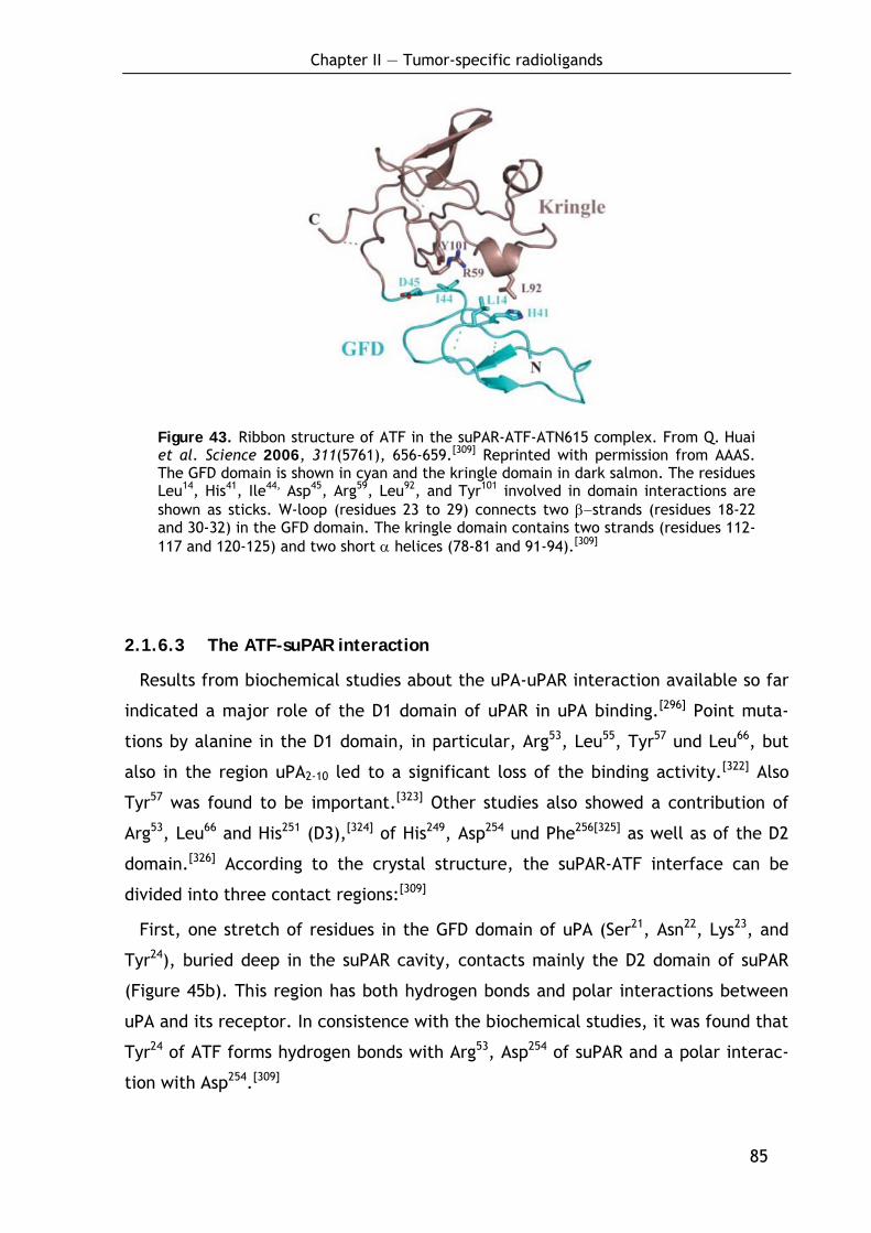
Chapter II — Tumor-specific radioligands
85
Figure 43. Ribbon structure of ATF in the suPAR-ATF-ATN615 complex. From Q. Huai et al. Science 2006, 311(5761), 656-659.[309] Reprinted with permission from AAAS. The GFD domain is shown in cyan and the kringle domain in dark salmon. The residues Leu14, His41, Ile44, Asp45, Arg59, Leu92, and Tyr101 involved in domain interactions are shown as sticks. W-loop (residues 23 to 29) connects two β−strands (residues 18-22 and 30-32) in the GFD domain. The kringle domain contains two strands (residues 112-117 and 120-125) and two short α helices (78-81 and 91-94).[309]
2.1.6.3 The ATF-suPAR interaction
Results from biochemical studies about the uPA-uPAR interaction available so far
indicated a major role of the D1 domain of uPAR in uPA binding.[296] Point muta-
tions by alanine in the D1 domain, in particular, Arg53, Leu55, Tyr57 und Leu66, but
also in the region uPA2-10 led to a significant loss of the binding activity.[322] Also
Tyr57 was found to be important.[323] Other studies also showed a contribution of
Arg53, Leu66 and His251 (D3),[324] of His249, Asp254 und Phe256[325] as well as of the D2
domain.[326] According to the crystal structure, the suPAR-ATF interface can be
divided into three contact regions:[309]
First, one stretch of residues in the GFD domain of uPA (Ser21, Asn22, Lys23, and
Tyr24), buried deep in the suPAR cavity, contacts mainly the D2 domain of suPAR
(Figure 45b). This region has both hydrogen bonds and polar interactions between
uPA and its receptor. In consistence with the biochemical studies, it was found that
Tyr24 of ATF forms hydrogen bonds with Arg53, Asp254 of suPAR and a polar interac-
tion with Asp254.[309]
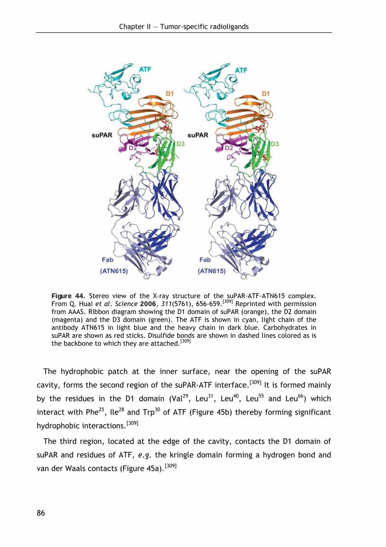
Chapter II — Tumor-specific radioligands
86
Figure 44. Stereo view of the X-ray structure of the suPAR-ATF-ATN615 complex. From Q. Huai et al. Science 2006, 311(5761), 656-659.[309] Reprinted with permission from AAAS. Ribbon diagram showing the D1 domain of suPAR (orange), the D2 domain (magenta) and the D3 domain (green). The ATF is shown in cyan, light chain of the antibody ATN615 in light blue and the heavy chain in dark blue. Carbohydrates in suPAR are shown as red sticks. Disulfide bonds are shown in dashed lines colored as is the backbone to which they are attached.[309]
The hydrophobic patch at the inner surface, near the opening of the suPAR
cavity, forms the second region of the suPAR-ATF interface.[309] It is formed mainly
by the residues in the D1 domain (Val29, Leu31, Leu40, Leu55 and Leu66) which
interact with Phe25, Ile28 and Trp30 of ATF (Figure 45b) thereby forming significant
hydrophobic interactions.[309]
The third region, located at the edge of the cavity, contacts the D1 domain of
suPAR and residues of ATF, e.g. the kringle domain forming a hydrogen bond and
van der Waals contacts (Figure 45a).[309]
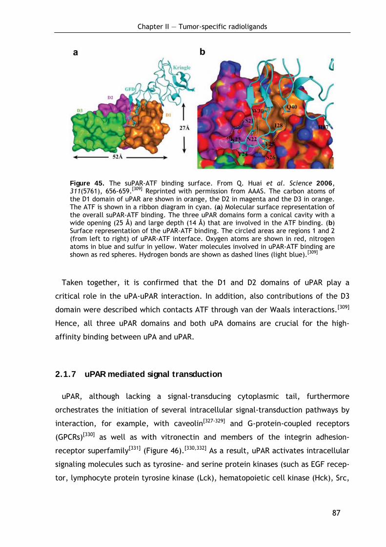
Chapter II — Tumor-specific radioligands
87
Figure 45. The suPAR-ATF binding surface. From Q. Huai et al. Science 2006, 311(5761), 656-659.[309] Reprinted with permission from AAAS. The carbon atoms of the D1 domain of uPAR are shown in orange, the D2 in magenta and the D3 in orange. The ATF is shown in a ribbon diagram in cyan. (a) Molecular surface representation of the overall suPAR-ATF binding. The three uPAR domains form a conical cavity with a wide opening (25 Å) and large depth (14 Å) that are involved in the ATF binding. (b) Surface representation of the uPAR-ATF binding. The circled areas are regions 1 and 2 (from left to right) of uPAR-ATF interface. Oxygen atoms are shown in red, nitrogen atoms in blue and sulfur in yellow. Water molecules involved in uPAR-ATF binding are shown as red spheres. Hydrogen bonds are shown as dashed lines (light blue).[309]
Taken together, it is confirmed that the D1 and D2 domains of uPAR play a
critical role in the uPA-uPAR interaction. In addition, also contributions of the D3
domain were described which contacts ATF through van der Waals interactions.[309]
Hence, all three uPAR domains and both uPA domains are crucial for the high-
affinity binding between uPA and uPAR.
2.1.7 uPAR mediated signal transduction
uPAR, although lacking a signal-transducing cytoplasmic tail, furthermore
orchestrates the initiation of several intracellular signal-transduction pathways by
interaction, for example, with caveolin[327-329] and G-protein-coupled receptors
(GPCRs)[330] as well as with vitronectin and members of the integrin adhesion-
receptor superfamily[331] (Figure 46).[330,332] As a result, uPAR activates intracellular
signaling molecules such as tyrosine- and serine protein kinases (such as EGF recep-
tor, lymphocyte protein tyrosine kinase (Lck), hematopoietic cell kinase (Hck), Src,
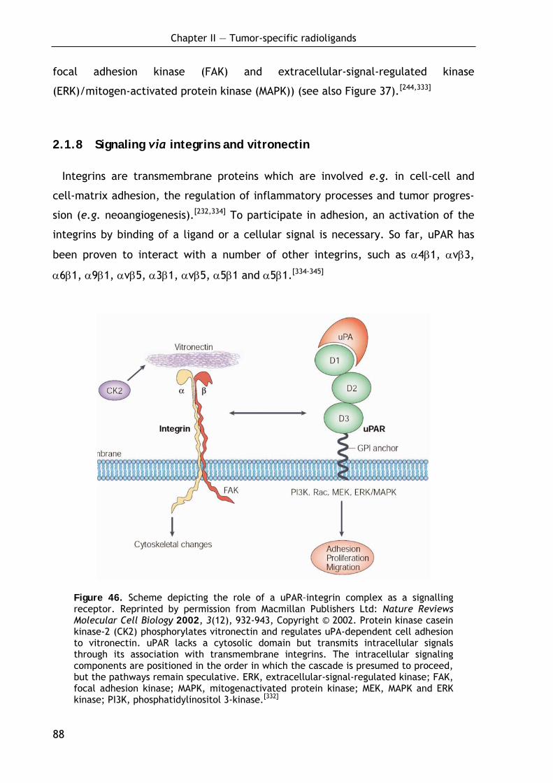
Chapter II — Tumor-specific radioligands
88
focal adhesion kinase (FAK) and extracellular-signal-regulated kinase
(ERK)/mitogen-activated protein kinase (MAPK)) (see also Figure 37).[244,333]
2.1.8 Signaling via integrins and vitronectin
Integrins are transmembrane proteins which are involved e.g. in cell-cell and
cell-matrix adhesion, the regulation of inflammatory processes and tumor progres-
sion (e.g. neoangiogenesis).[232,334] To participate in adhesion, an activation of the
integrins by binding of a ligand or a cellular signal is necessary. So far, uPAR has
been proven to interact with a number of other integrins, such as α4β1, αvβ3,
α6β1, α9β1, αvβ5, α3β1, αvβ5, α5β1 and α5β1.[334-345]
Figure 46. Scheme depicting the role of a uPAR–integrin complex as a signalling receptor. Reprinted by permission from Macmillan Publishers Ltd: Nature Reviews Molecular Cell Biology 2002, 3(12), 932-943, Copyright © 2002. Protein kinase casein kinase-2 (CK2) phosphorylates vitronectin and regulates uPA-dependent cell adhesion to vitronectin. uPAR lacks a cytosolic domain but transmits intracellular signals through its association with transmembrane integrins. The intracellular signaling components are positioned in the order in which the cascade is presumed to proceed, but the pathways remain speculative. ERK, extracellular-signal-regulated kinase; FAK, focal adhesion kinase; MAPK, mitogenactivated protein kinase; MEK, MAPK and ERK kinase; PI3K, phosphatidylinositol 3-kinase.[332]
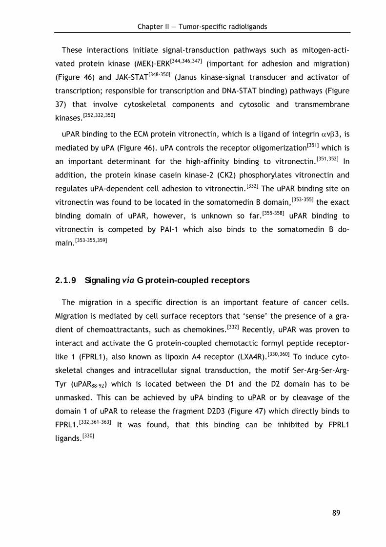
Chapter II — Tumor-specific radioligands
89
These interactions initiate signal-transduction pathways such as mitogen-acti-
vated protein kinase (MEK)–ERK[344,346,347] (important for adhesion and migration)
(Figure 46) and JAK–STAT[348-350] (Janus kinase–signal transducer and activator of
transcription; responsible for transcription and DNA-STAT binding) pathways (Figure
37) that involve cytoskeletal components and cytosolic and transmembrane
kinases.[252,332,350]
uPAR binding to the ECM protein vitronectin, which is a ligand of integrin αvβ3, is
mediated by uPA (Figure 46). uPA controls the receptor oligomerization[351] which is
an important determinant for the high-affinity binding to vitronectin.[351,352] In
addition, the protein kinase casein kinase-2 (CK2) phosphorylates vitronectin and
regulates uPA-dependent cell adhesion to vitronectin.[332] The uPAR binding site on
vitronectin was found to be located in the somatomedin B domain,[353-355] the exact
binding domain of uPAR, however, is unknown so far.[355-358] uPAR binding to
vitronectin is competed by PAI-1 which also binds to the somatomedin B do-
main.[353-355,359]
2.1.9 Signaling via G protein-coupled receptors
The migration in a specific direction is an important feature of cancer cells.
Migration is mediated by cell surface receptors that ‘sense’ the presence of a gra-
dient of chemoattractants, such as chemokines.[332] Recently, uPAR was proven to
interact and activate the G protein-coupled chemotactic formyl peptide receptor-
like 1 (FPRL1), also known as lipoxin A4 receptor (LXA4R).[330,360] To induce cyto-
skeletal changes and intracellular signal transduction, the motif Ser-Arg-Ser-Arg-
Tyr (uPAR88-92) which is located between the D1 and the D2 domain has to be
unmasked. This can be achieved by uPA binding to uPAR or by cleavage of the
domain 1 of uPAR to release the fragment D2D3 (Figure 47) which directly binds to
FPRL1.[332,361-363] It was found, that this binding can be inhibited by FPRL1
ligands.[330]
Chapter II — Tumor-specific radioligands
90
Figure 47. Scheme depicting the role of a uPAR–GPCR complex as a signaling recep-tor. Reprinted by permission from Macmillan Publishers Ltd: Nature Reviews Molecu-lar Cell Biology 2002, 3(12), 932-943, Copyright © 2002. Binding of the D2D3 frag-ment of soluble urokinase (suPAR) to the G-protein-coupled receptor formyl peptide receptor-like 1 (FPRL1) transmits chemotactic signals. The intracellular signaling components are indicated, but the pathways remain speculative. ERK, extracellular-signal-regulated kinase; Hck, haematopoietic cell kinase; MAPK, mitogenactivated protein kinase; MEK, MAPK and ERK kinase; PI3K, phosphatidylinositol 3-kinase.[332]
2.1.10 Tumor-associated prognostic factors of the uPA/uPAR system:
clinical significance of uPA, uPAR, PAI-1 and PAI-2
After the discovery of a casual connection of plasminogen activation and cancer
in the 1960s,[364,365] Åstedt et al. were the first to report the overexpression of uPA
in ovarian carcinoma.[366,367] As early as in the 1980s, an increased concentration of
uPA in tumor tissue was linked with high metastatic potential of tumors and poor
prognosis for the patients.[368] The natural inhibitor PAI-1, however, proved to be
correlated with poor prognosis as well. This is in contrast to the assumption that
PAI-1 as uPA inhibitor should suppress plasminogen activation and metastasis and is
explained by the diverse biological properties[369] of PAI-1 which is not only inhibi-
tor of uPA but also is involved in cell adhesion, proliferation and migration as well
as in signal transduction and apoptosis.[249,368,370-379] Today, the prognostic impact of
Chapter II — Tumor-specific radioligands
91
uPA and PAI-1 is validated at high level of evidence,[248,368,380-382] and also uPAR has
been linked with poor prognosis in numerous studies.[383] In contrast to PAI-1,
increased PAI-2 levels have been linked with low metastatic potential of tumors
and good prognosis for the patients.[368,384-386]
2.1.11 The uPA/uPAR system as potential target in cancer therapy
As presented above, a large body of evidence obtained from both in vitro and in
vivo experiments as well as from clinical studies shows that the uPA-system, espe-
cially the protease uPA, its major inhibitor PAI-1 and its cellular receptor uPAR,
plays an important role in a variety of malignant tumors.[259,374,387,388] uPAR not only
is focusing pericellular proteolysis to the tumor cell, but through direct or indirect
interactions with extracellular matrix proteins or transmembrane receptors such as
the integrins and the EGF receptor, it modulates cell migration, cell-cell and cell-
matrix interactions and signaling pathways. Altogether, the system is involved at
multiple stages in the formation and progression of the disease,[389] in particular in
invasion and metastasis,[252,332] cell proliferation,[390,391] migration[392] and cell adhe-
sion.[252] As overexpression of uPAR (as well as uPA and PAI-1) in tumor tissue is
strongly correlated with poor prognosis for patients afflicted with different types of
cancer, it represents an attractive target for cancer therapy.[393]
Hence, numerous efforts have been made to inhibit the catalytic activity of uPA
or to inhibit uPAR function by biologic and synthetic ligands.[388,393] A selection of
synthetic inhibitors is presented in chapter 2.2.1.1.. Goal of the present study was
now to examine whether peptidic radioligands, based on potent uPAR binding pep-
tides, may be developed for application in receptor-based radionuclide therapy.
Chapter II — Tumor-specific radioligands
92
2.1.12 Visualization and treatment of tumors by radiopharmaceuticals
2.1.12.1 Tumor imaging and radionuclide therapy
Antibodies, peptides or non-peptidic compounds which are capable to selectively
bind to antigens or receptors overexpressed on tumor cells, are promising candi-
dates for tumor imaging or antigen- or receptor-based radiotherapy.[394-397] Over
the past years, great progresses have been made in tumor imaging and several new
methods have been developed: for example, the magnetic resonance imaging
(MRI),[398,399] fluorescence-mediated molecular tomography (FMT),[400] doppler sono-
graphy and szintigraphy methods.[401]
The szintigraphy methods SPECT (single photon emission computed tomography)
and PET (positron emission tomography)[402] use molecular antibodies (mAbs) or
small organic compounds like sugars, amino acids, peptides or peptidomimetics as
ligands which are labeled with a radionuclide. The resulting tracers interact in vivo
with their target structures and allow a non-invasive visualization and even a quan-
tification of the distribution of targeted processes. This offers e.g. the opportunity
to quantify specific targets prior to a therapy and predict its success. Moreover,
the dosage of the chemotherapeutic can be adjusted and optimized and the
success of the therapy can be observed by decreasing target binding. Recently also
hybrid techniques like SPECT-computed tomography (CT) or PET-CT found applica-
tion, which enable the simultaneous detection of a biochemical signal (PET/SPECT)
and morphology (CT).[403] The radionuclide used is dependent on the desired
application (diagnosis, therapy) and the method (e.g. PET, SPECT). Table 13
summarizes important radionuclides for various applications.
Widely used radionuclides in PET scanning are for example 11C, 18F or 68Ga,[404]
whereas 18F turned out to have several advantages over the others:[405]
• Its relatively long half-life permits enough time for the synthesis of the
tracer and for PET and allows kinetic studies.
• In non-fluorine-containing metabolic substrates, the introduction of a fluo-
rine atom (F for H, F for OH) does not cause significant sterical changes.
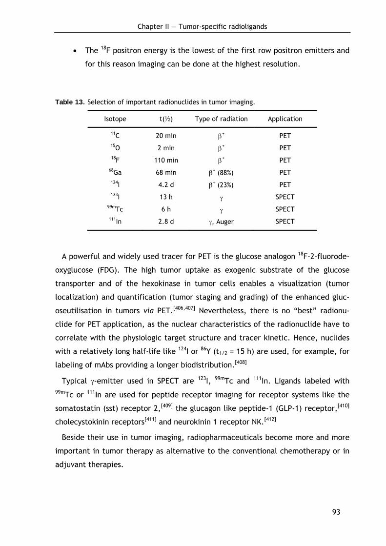
Chapter II — Tumor-specific radioligands
93
• The 18F positron energy is the lowest of the first row positron emitters and
for this reason imaging can be done at the highest resolution.
Table 13. Selection of important radionuclides in tumor imaging.
Isotope t(½) Type of radiation Application
11C 20 min β+ PET 15O 2 min β+ PET 18F 110 min β+ PET
68Ga 68 min β+ (88%) PET 124I 4.2 d β+ (23%) PET 123I 13 h γ SPECT
99mTc 6 h γ SPECT 111In 2.8 d γ, Auger SPECT
A powerful and widely used tracer for PET is the glucose analogon 18F-2-fluorode-
oxyglucose (FDG). The high tumor uptake as exogenic substrate of the glucose
transporter and of the hexokinase in tumor cells enables a visualization (tumor
localization) and quantification (tumor staging and grading) of the enhanced gluc-
oseutilisation in tumors via PET.[406,407] Nevertheless, there is no “best” radionu-
clide for PET application, as the nuclear characteristics of the radionuclide have to
correlate with the physiologic target structure and tracer kinetic. Hence, nuclides
with a relatively long half-life like 124I or 86Y (t1/2 = 15 h) are used, for example, for
labeling of mAbs providing a longer biodistribution.[408]
Typical γ-emitter used in SPECT are 123I, 99mTc and 111In. Ligands labeled with 99mTc or 111In are used for peptide receptor imaging for receptor systems like the
somatostatin (sst) receptor 2,[409] the glucagon like peptide-1 (GLP-1) receptor,[410]
cholecystokinin receptors[411] and neurokinin 1 receptor NK.[412]
Beside their use in tumor imaging, radiopharmaceuticals become more and more
important in tumor therapy as alternative to the conventional chemotherapy or in
adjuvant therapies.
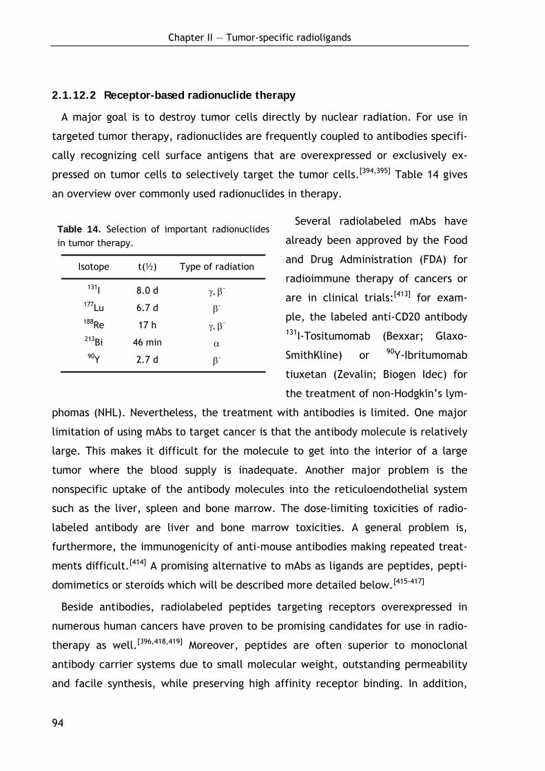
Chapter II — Tumor-specific radioligands
94
2.1.12.2 Receptor-based radionuclide therapy
A major goal is to destroy tumor cells directly by nuclear radiation. For use in
targeted tumor therapy, radionuclides are frequently coupled to antibodies specifi-
cally recognizing cell surface antigens that are overexpressed or exclusively ex-
pressed on tumor cells to selectively target the tumor cells.[394,395] Table 14 gives
an overview over commonly used radionuclides in therapy.
Several radiolabeled mAbs have
already been approved by the Food
and Drug Administration (FDA) for
radioimmune therapy of cancers or
are in clinical trials:[413] for exam-
ple, the labeled anti-CD20 antibody 131I-Tositumomab (Bexxar; Glaxo-
SmithKline) or 90Y-Ibritumomab
tiuxetan (Zevalin; Biogen Idec) for
the treatment of non-Hodgkin’s lym-
phomas (NHL). Nevertheless, the treatment with antibodies is limited. One major
limitation of using mAbs to target cancer is that the antibody molecule is relatively
large. This makes it difficult for the molecule to get into the interior of a large
tumor where the blood supply is inadequate. Another major problem is the
nonspecific uptake of the antibody molecules into the reticuloendothelial system
such as the liver, spleen and bone marrow. The dose-limiting toxicities of radio-
labeled antibody are liver and bone marrow toxicities. A general problem is,
furthermore, the immunogenicity of anti-mouse antibodies making repeated treat-
ments difficult.[414] A promising alternative to mAbs as ligands are peptides, pepti-
domimetics or steroids which will be described more detailed below.[415-417]
Beside antibodies, radiolabeled peptides targeting receptors overexpressed in
numerous human cancers have proven to be promising candidates for use in radio-
therapy as well.[396,418,419] Moreover, peptides are often superior to monoclonal
antibody carrier systems due to small molecular weight, outstanding permeability
and facile synthesis, while preserving high affinity receptor binding. In addition,
Table 14. Selection of important radionuclides in tumor therapy.
Isotope t(½) Type of radiation
131I 8.0 d γ, β- 177Lu 6.7 d β- 188Re 17 h γ, β- 213Bi 46 min α 90Y 2.7 d β-
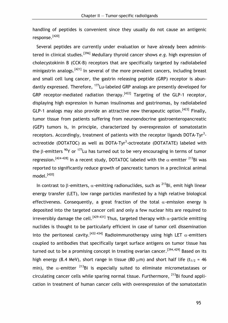
Chapter II — Tumor-specific radioligands
95
handling of peptides is convenient since they usually do not cause an antigenic
response.[420]
Several peptides are currently under evaluation or have already been adminis-
tered in clinical studies.[396] Medullary thyroid cancer shows e.g. high expression of
cholecystokinin B (CCK-B) receptors that are specifically targeted by radiolabeled
minigastrin analogs.[421] In several of the more prevalent cancers, including breast
and small cell lung cancer, the gastrin releasing peptide (GRP) receptor is abun-
dantly expressed. Therefore, 177Lu-labeled GRP analogs are presently developed for
GRP receptor-mediated radiation therapy.[422] Targeting of the GLP-1 receptor,
displaying high expression in human insulinomas and gastrinomas, by radiolabeled
GLP-1 analogs may also provide an attractive new therapeutic option.[423] Finally,
tumor tissue from patients suffering from neuroendocrine gastroenteropancreatic
(GEP) tumors is, in principle, characterized by overexpression of somatostatin
receptors. Accordingly, treatment of patients with the receptor ligands DOTA-Tyr3-
octreotide (DOTATOC) as well as DOTA-Tyr3-octreotate (DOTATATE) labeled with
the β-emitters 90Y or 177Lu has turned out to be very encouraging in terms of tumor
regression.[424-428] In a recent study, DOTATOC labeled with the α-emitter 213Bi was
reported to significantly reduce growth of pancreatic tumors in a preclinical animal
model.[420]
In contrast to β-emitters, α-emitting radionuclides, such as 213Bi, emit high linear
energy transfer (LET), low range particles manifested by a high relative biological
effectiveness. Consequently, a great fraction of the total α-emission energy is
deposited into the targeted cancer cell and only a few nuclear hits are required to
irreversibly damage the cell.[429-431] Thus, targeted therapy with α-particle emitting
nuclides is thought to be particularly efficient in case of tumor cell dissemination
into the peritoneal cavity.[432-434] Radioimmunotherapy using high LET α-emitters
coupled to antibodies that specifically target surface antigens on tumor tissue has
turned out to be a promising concept in treating ovarian cancer.[394,429] Based on its
high energy (8.4 MeV), short range in tissue (80 μm) and short half life (t1/2 = 46
min), the α-emitter 213Bi is especially suited to eliminate micrometastases or
circulating cancer cells while sparing normal tissue. Furthermore, 213Bi found appli-
cation in treatment of human cancer cells with overexpression of the somatostatin
Chapter II — Tumor-specific radioligands
96
receptor, which were efficiently eliminated both in vitro and in vivo using 213Bi-
DOTATOC.[420,435]
In the following, some of the most convenient methods for the preparation of
radiopharmaceuticals will be presented as examples. In principle, radionuclides can
be covalently attached to the carrier molecule or they can be complexed by a
chelator which is covalently attached to the carrier molecule.[436]
2.1.12.3 Radiolabeling by covalent attachment of the radionuclide
Iodine-labeling can be achieved by a electrophilic aromatic substitution in ortho-
position to a phenolic hydroxyl group, e.g. using chloramines-T[437] or Iodogen®
(Figure 48).[438] These reagents lead to a oxidation of iodides to iodine which then
reacts with the activated aromatic residue.
N N
N N
O
O
Cl Cl
ClClS
O O
Na+N-
Cl
144 145
Figure 48. Structures of Iodogen® (144) and chloramine-T (145).
Monoiodotyrosine, however, is enzymatically deiodogenated in vivo. Therefore,
procedures were developed to introduce iodine also in aromatic residues lacking a
hydroxyl group. Successful approaches are reported via ipso-substitution of precur-
sors like silyl[439] or stannyl[440] derivatives.[441,442] In a recent approach, a direct
iodination of non-activated phenylalanine incorporated into peptides or proteins
was reported using bis(pyridine)iodonium (I) tetrafluoroborate in presence of TFA.
This widens iodine labeling opportunities to molecules lacking a tyrosine residue.
11C for PET analysis is often introduced via methylation of e.g. alcohols[443,444] or
amines[445] using [11C]-iodomethane or via nucleophilic substitution by [11C]-cya-
nide.[446] 18F can be introduced by nucleophilic substitution of activated alcohols
using K18F/Kryptofix[447] or via electrophilic aromatic substitution using 18F-acetyl
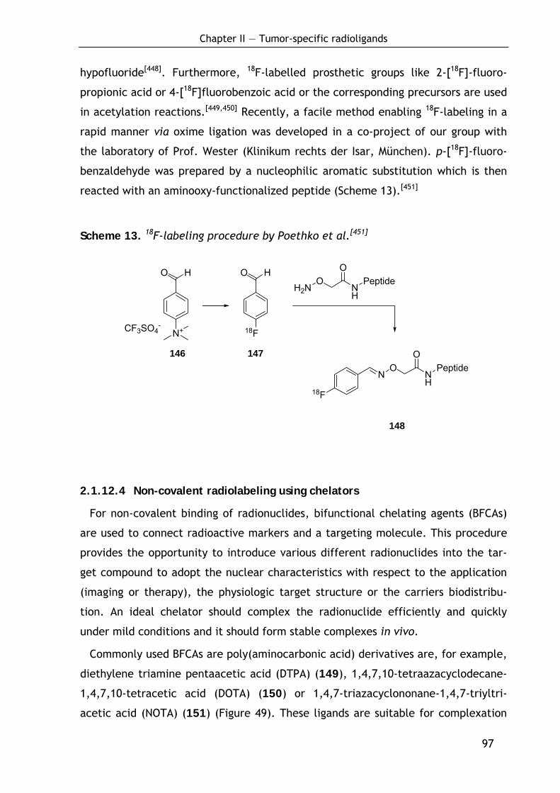
Chapter II — Tumor-specific radioligands
97
O H
N+CF3SO4-
O H
18F
H2NO
NH
OPeptide
NO
NH
OPeptide
18F
hypofluoride[448]. Furthermore, 18F-labelled prosthetic groups like 2-[18F]-fluoro-
propionic acid or 4-[18F]fluorobenzoic acid or the corresponding precursors are used
in acetylation reactions.[449,450] Recently, a facile method enabling 18F-labeling in a
rapid manner via oxime ligation was developed in a co-project of our group with
the laboratory of Prof. Wester (Klinikum rechts der Isar, München). p-[18F]-fluoro-
benzaldehyde was prepared by a nucleophilic aromatic substitution which is then
reacted with an aminooxy-functionalized peptide (Scheme 13).[451]
Scheme 13. 18F-labeling procedure by Poethko et al.[451]
148
2.1.12.4 Non-covalent radiolabeling using chelators
For non-covalent binding of radionuclides, bifunctional chelating agents (BFCAs)
are used to connect radioactive markers and a targeting molecule. This procedure
provides the opportunity to introduce various different radionuclides into the tar-
get compound to adopt the nuclear characteristics with respect to the application
(imaging or therapy), the physiologic target structure or the carriers biodistribu-
tion. An ideal chelator should complex the radionuclide efficiently and quickly
under mild conditions and it should form stable complexes in vivo.
Commonly used BFCAs are poly(aminocarbonic acid) derivatives are, for example,
diethylene triamine pentaacetic acid (DTPA) (149), 1,4,7,10-tetraazacyclodecane-
1,4,7,10-tetracetic acid (DOTA) (150) or 1,4,7-triazacyclononane-1,4,7-triyltri-
acetic acid (NOTA) (151) (Figure 49). These ligands are suitable for complexation
146 147

Chapter II — Tumor-specific radioligands
98
of divalent and trivalent metals by an up to eightfold coordination via N/O-donors,
whereas the cyclic ligands were found to be superior in terms of complex sta-
bility.[452] DOTA complexes are even stable under strong acidic conditions (pH
<2).[453,454] Hence, radiopharmaceuticals containing the DOTA ligand as metal chela-
tor have found widespread use in therapy and diagnostic imaging.[394,418]
N
NN
CO2H
HO2C
CO2HN
NN
HO2C
CO2H
CO2HHO2C
HO2C
N
N
N
N
HO2C
HO2C
CO2H
CO2H
149 150 151
Figure 49. Structures of the BFCAs diethylene triamine pentaacetic acid (DTPA) (149), 1,4,7,10-tetraazacyclodecane-1,4,7,10-tetracetic acid (DOTA) (150) or 1,4,7-triazacyclononane-1,4,7-triacetic acid (NOTA) (151).
Chapter II — Tumor-specific radioligands
99
2.2 Development of uPAR-selective radioligands
2.2.1 Background
Peritoneal spread of tumor cells in ovarian cancer implies poor prognosis owing to
the lack of efficient standard therapies and chemo-resistance. Surgery combined
with chemotherapy can cause a survival benefit, nevertheless the median survival
of patients suffering from peritoneal carcinomatosis is only three to six months.[455]
Hence, while ovarian cancer accounts for only 4% of all cancers in women,[456] it
displays the highest rate of mortality of all gynecologic malignancies. Since
intraperitoneal spread is the most common characteristic of ovarian cancer, in
more than 75% of all cases the disease has already disseminated beyond the ovary
at the time of diagnosis. Accordingly, the major cause of treatment failure is
incomplete debunking of cancer cells from the peritoneal cavity. Following relapse
due to chemo-resistance, there is currently no further therapeutic option avail-
able.[457] Therefore, targeting and efficient elimination of single remaining tumor
cells or small tumor cell clusters could considerably improve prognosis.
2.2.1.1 Synthetic peptides targeting uPAR
As described above, the urokinase-type plasminogen activator (uPA) system is
overexpressed in numerous malignant human cancers. Hence, it represents a
promising target for anti-cancer therapy.[388,393,458-460] 213Bi-PAI-2 conjugates, indi-
rectly targeting tumor cell-associated uPAR, have been successfully applied in
preclinical studies for the control of pancreatic, prostate and ovarian cancer.[461-464]
Synthetic peptides directly binding to uPAR may offer another approach for uPAR-
targeted 213Bi-therapy. In fact, by different approaches, potent uPAR binding
peptides have recently been identified, among them the competitive uPA inhibitors
WX360 (cyclo[21,29][D-Cys21Cys29]-uPA21-30)[465] and AE105 (Asp–Cha–Phe–(D-Ser)–(D-
Arg)–Tyr–Leu–Trp–Ser).[466]
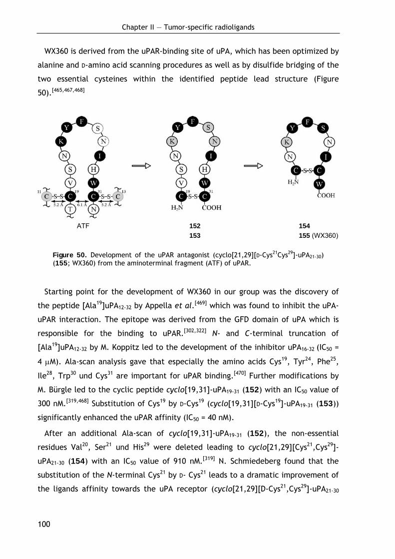
Chapter II — Tumor-specific radioligands
100
WX360 is derived from the uPAR-binding site of uPA, which has been optimized by
alanine and D-amino acid scanning procedures as well as by disulfide bridging of the
two essential cysteines within the identified peptide lead structure (Figure
50).[465,467,468]
ATF 152 154 153 155 (WX360)
Figure 50. Development of the uPAR antagonist (cyclo[21,29][D-Cys21Cys29]-uPA21-30) (155; WX360) from the aminoterminal fragment (ATF) of uPAR.
Starting point for the development of WX360 in our group was the discovery of
the peptide [Ala19]uPA12-32 by Appella et al.[469] which was found to inhibit the uPA-
uPAR interaction. The epitope was derived from the GFD domain of uPA which is
responsible for the binding to uPAR.[302,322] N- and C-terminal truncation of
[Ala19]uPA12-32 by M. Koppitz led to the development of the inhibitor uPA16-32 (IC50 =
4 μM). Ala-scan analysis gave that especially the amino acids Cys19, Tyr24, Phe25,
Ile28, Trp30 und Cys31 are important for uPAR binding.[470] Further modifications by
M. Bürgle led to the cyclic peptide cyclo[19,31]-uPA19-31 (152) with an IC50 value of
300 nM.[319,468] Substitution of Cys19 by D-Cys19 (cyclo[19,31][D-Cys19]-uPA19-31 (153))
significantly enhanced the uPAR affinity (IC50 = 40 nM).
After an additional Ala-scan of cyclo[19,31]-uPA19-31 (152), the non-essential
residues Val20, Ser21 und His29 were deleted leading to cyclo[21,29][Cys21,Cys29]-
uPA21-30 (154) with an IC50 value of 910 nM.[319] N. Schmiedeberg found that the
substitution of the N-terminal Cys21 by D- Cys21 leads to a dramatic improvement of
the ligands affinity towards the uPA receptor (cyclo[21,29][D-Cys21,Cys29]-uPA21-30
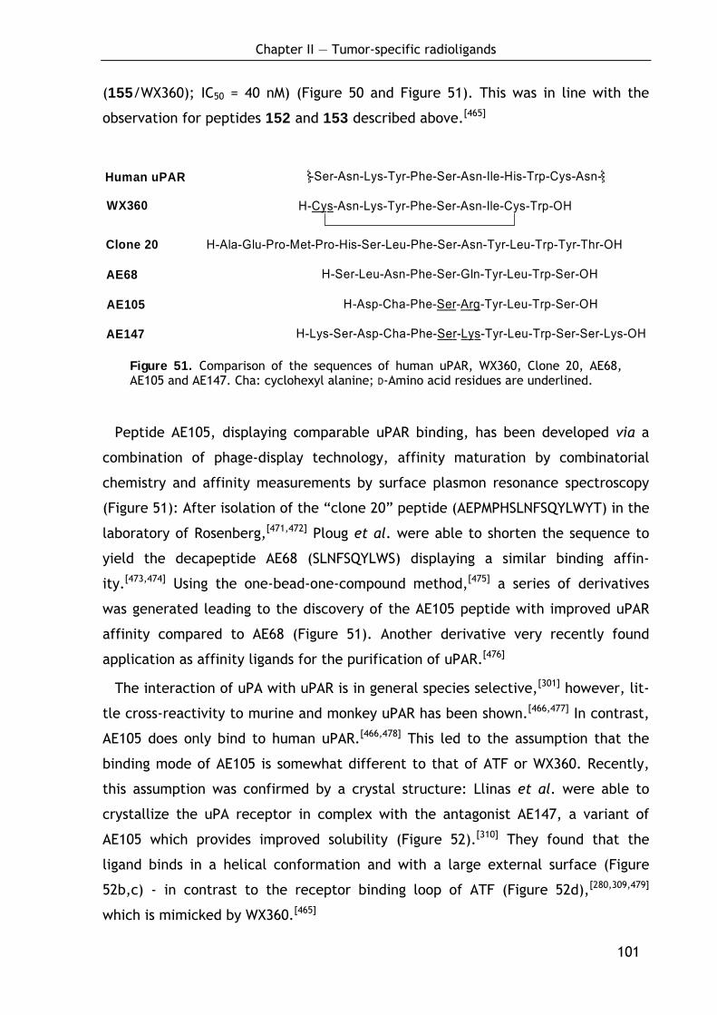
Chapter II — Tumor-specific radioligands
101
(155/WX360); IC50 = 40 nM) (Figure 50 and Figure 51). This was in line with the
observation for peptides 152 and 153 described above.[465]
H-Asp-Cha-Phe-Ser-Arg-Tyr-Leu-Trp-Ser-OH
H-Ser-Leu-Asn-Phe-Ser-Gln-Tyr-Leu-Trp-Ser-OH
H-Ala-Glu-Pro-Met-Pro-His-Ser-Leu-Phe-Ser-Asn-Tyr-Leu-Trp-Tyr-Thr-OH
-Ser-Asn-Lys-Tyr-Phe-Ser-Asn-Ile-His-Trp-Cys-Asn-
H-Cys-Asn-Lys-Tyr-Phe-Ser-Asn-Ile-Cys-Trp-OH
Human uPAR
WX360
Clone 20
AE68
AE105
AE147 H-Lys-Ser-Asp-Cha-Phe-Ser-Lys-Tyr-Leu-Trp-Ser-Ser-Lys-OH
Figure 51. Comparison of the sequences of human uPAR, WX360, Clone 20, AE68, AE105 and AE147. Cha: cyclohexyl alanine; D-Amino acid residues are underlined.
Peptide AE105, displaying comparable uPAR binding, has been developed via a
combination of phage-display technology, affinity maturation by combinatorial
chemistry and affinity measurements by surface plasmon resonance spectroscopy
(Figure 51): After isolation of the “clone 20” peptide (AEPMPHSLNFSQYLWYT) in the
laboratory of Rosenberg,[471,472] Ploug et al. were able to shorten the sequence to
yield the decapeptide AE68 (SLNFSQYLWS) displaying a similar binding affin-
ity.[473,474] Using the one-bead-one-compound method,[475] a series of derivatives
was generated leading to the discovery of the AE105 peptide with improved uPAR
affinity compared to AE68 (Figure 51). Another derivative very recently found
application as affinity ligands for the purification of uPAR.[476]
The interaction of uPA with uPAR is in general species selective,[301] however, lit-
tle cross-reactivity to murine and monkey uPAR has been shown.[466,477] In contrast,
AE105 does only bind to human uPAR.[466,478] This led to the assumption that the
binding mode of AE105 is somewhat different to that of ATF or WX360. Recently,
this assumption was confirmed by a crystal structure: Llinas et al. were able to
crystallize the uPA receptor in complex with the antagonist AE147, a variant of
AE105 which provides improved solubility (Figure 52).[310] They found that the
ligand binds in a helical conformation and with a large external surface (Figure
52b,c) - in contrast to the receptor binding loop of ATF (Figure 52d),[280,309,479]
which is mimicked by WX360.[465]

Chapter II — Tumor-specific radioligands
102
Figure 52. The structure of human uPAR from Llinas et al.,[310] reprinted by permission from Macmillan Publishers Ltd: The EMBO Journal 2005, 24(9), 1655-1663, Copyright © 2005. (a) The overall modular structure of uPAR as a ribbon diagram. The individual uPAR domains are assembled in a right-handed orientation and are colored yellow (DI), blue (DII) and red (DIII). The bended β-strands in DII and DIII are indicated separately.[310] (b) Molecular surface representation of the uPAR–peptide complex. The surface of uPAR is shown in grey, whereas the bound antagonist peptide (AE147) is shown as a ribbon diagram in dark blue. Receptor residues interacting with AE147 are coloured orange (hydrophobic) or cyan (polar) and the asparagine-linked glycosylation sites coloured light green. (c) Enlarged uPAR binding cavity of AE147 (illustrated by a combined ribbon and stick representation in dark blue). The indole side chain of W10 engages the deep and prominent hydrophobic hole of the central cavity of uPAR and the side chain of L9 is located proximate to its entrance. (d) Model for the structure of the human GFD–uPAR complex. The uPAR molecule is shown as a molecular surface representation. The receptor-binding module of uPA is shown in red as a ribbon diagram with the side chains of Y24, F25, I28 and W30, which are highlighted by sticks. Note that W10 and L9 of AE147 exhibit a spatial superimposition with Y24 and F25 in uPA. A schematic representation of the glycolipid (GPI) anchor is shown to connect the C-terminus of uPAR to a model of the cell membrane.[310]
Chapter II — Tumor-specific radioligands
103
Although direct comparison of uPAR/ATF and uPAR/AE147 complexes confirms a
spatial overlap between the GFD and AE147 in the binding cavity of uPAR, pro-
nounced differences exist in the positioning of the individual side-chains and in
their interactions with the receptor.[480] The residues W10 and L9 of AE147, for
example, exhibit a spatial superimposition with Y24 and F25 of ATF (compare Figure
52b and d).[310] These differences could be responsible for observed species
selectivity of the inhibitory peptides towards human uPAR.
2.2.1.2 Overall aim and target structures
The aim of the present study was to examine whether peptidic radioligands,
based on potent uPAR binding peptides (i.e. the 10-mer peptide cyclo[21,29]-
[D-Cys21Cys29]-uPA21-30 (WX360)[465] and the 9-mer peptide AE105[466]), may be devel-
oped for application in α-emitter therapy of disseminated ovarian cancer. For this,
the chelator DOTA, which stably chelates 213Bi, was coupled to the uPAR-binding
peptides via different types of spacers. For the synthesis of N-terminally cross-
linked dimeric ligands, a straight-forward procedure was worked out allowing the
synthesis of the complete DOTA-conjugate with high purity on solid phase. The
synthetic work in this project was performed in collaboration with T. Huber in our
group.
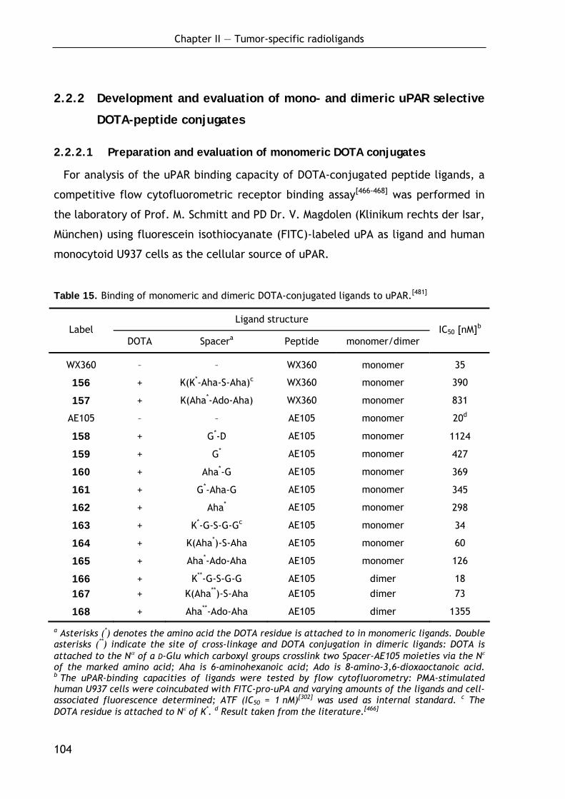
Chapter II — Tumor-specific radioligands
104
2.2.2 Development and evaluation of mono- and dimeric uPAR selective
DOTA-peptide conjugates
2.2.2.1 Preparation and evaluation of monomeric DOTA conjugates
For analysis of the uPAR binding capacity of DOTA-conjugated peptide ligands, a
competitive flow cytofluorometric receptor binding assay[466-468] was performed in
the laboratory of Prof. M. Schmitt and PD Dr. V. Magdolen (Klinikum rechts der Isar,
München) using fluorescein isothiocyanate (FITC)-labeled uPA as ligand and human
monocytoid U937 cells as the cellular source of uPAR.
Table 15. Binding of monomeric and dimeric DOTA-conjugated ligands to uPAR.[481]
Ligand structure Label
DOTA Spacera Peptide monomer/dimer IC50 [nM]b
WX360 – – WX360 monomer 35
156 + K(K*-Aha-S-Aha)c WX360 monomer 390
157 + K(Aha*-Ado-Aha) WX360 monomer 831
AE105 – – AE105 monomer 20d
158 + G*-D AE105 monomer 1124
159 + G* AE105 monomer 427
160 + Aha*-G AE105 monomer 369
161 + G*-Aha-G AE105 monomer 345
162 + Aha* AE105 monomer 298
163 + K*-G-S-G-Gc AE105 monomer 34
164 + K(Aha*)-S-Aha AE105 monomer 60
165 + Aha*-Ado-Aha AE105 monomer 126
166 + K**-G-S-G-G AE105 dimer 18
167 + K(Aha**)-S-Aha AE105 dimer 73
168 + Aha**-Ado-Aha AE105 dimer 1355
a Asterisks (*) denotes the amino acid the DOTA residue is attached to in monomeric ligands. Double asterisks (**) indicate the site of cross-linkage and DOTA conjugation in dimeric ligands: DOTA is attached to the Nα of a D-Glu which carboxyl groups crosslink two Spacer-AE105 moieties via the Nε of the marked amino acid; Aha is 6-aminohexanoic acid; Ado is 8-amino-3,6-dioxaoctanoic acid. b The uPAR-binding capacities of ligands were tested by flow cytofluorometry: PMA-stimulated human U937 cells were coincubated with FITC-pro-uPA and varying amounts of the ligands and cell-associated fluorescence determined; ATF (IC50 = 1 nM)[302] was used as internal standard. c The DOTA residue is attached to Nε of K*. d Result taken from the literature.[466]
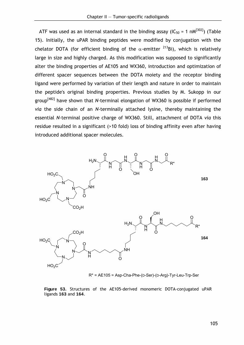
Chapter II — Tumor-specific radioligands
105
NN
NN
HO2C
CO2H
HO2C
O
NH
H2NNH
HN
NH
HN
R*
O
O
O
O
O
OH
NN
NN
HO2C
CO2H
HO2C
O
H2NNH
HN
R*
O
NH O
NH
O
OHO
R* = AE105 = Asp-Cha-Phe-(D-Ser)-(D-Arg)-Tyr-Leu-Trp-Ser
ATF was used as an internal standard in the binding assay (IC50 = 1 nM[302]) (Table
15). Initially, the uPAR binding peptides were modified by conjugation with the
chelator DOTA (for efficient binding of the α-emitter 213Bi), which is relatively
large in size and highly charged. As this modification was supposed to significantly
alter the binding properties of AE105 and WX360, introduction and optimization of
different spacer sequences between the DOTA moiety and the receptor binding
ligand were performed by variation of their length and nature in order to maintain
the peptide's original binding properties. Previous studies by M. Sukopp in our
group[482] have shown that N-terminal elongation of WX360 is possible if performed
via the side chain of an N-terminally attached lysine, thereby maintaining the
essential N-terminal positive charge of WX360. Still, attachment of DOTA via this
residue resulted in a significant (>10 fold) loss of binding affinity even after having
introduced additional spacer molecules.
Figure 53. Structures of the AE105-derived monomeric DOTA-conjugated uPAR ligands 163 and 164.
164
163
Chapter II — Tumor-specific radioligands
106
In contrast to that, after optimization of the spacer sequence monomeric AE105-
based DOTA-conjugates preserved their high affinity towards uPAR. Compared to
unsubstituted AE105 (IC50 = 20 nM[466]), the AE105-DOTA conjugate 159 equipped
with a single glycine residue as spacer, showed an approximately 20-fold reduction
of the binding affinity (IC50 = 427 nM), whereas the binding capacity of the ligand
163 (see Figure 53) with the five amino acid spacer Lys-Gly-Ser-Gly-Gly[466] was
only slightly reduced (IC50 = 34 nM). Using spacers containing the more hydrophobic
aminohexanoic acid (Aha) building block (160, 164), peptide binding was found to
be less effective compared to 163 (Table 15).[481]
2.2.2.2 Preparation and evaluation of dimeric DOTA conjugates
Since multimeric ligands often show superior binding affinities compared to
monomeric ligands[483-486], the monomeric ligands 163, 164 (structures shown in
Figure 53) and 165 were dimerized by cross linking via the spacer moiety. For this,
a novel straightforward procedure was developed:
The AE105 sequence was built up on solid phase and the different spacer moieties
were attached. Then, the deprotected Nε-termini of two resin-bound monomers
were cross-linked via side chain unprotected Fmoc-D-Glu as bivalent carbonic acid
(Scheme 14). The amino group of glutamic acid was finally used to attach the DOTA
chelator by coupling of DOTA-tris (tert-butyl ester).[487] The coupling of DOTA-
tris(tert-butyl) ester (169)[487] to the solid-phase bound mono- and dimeric ligands
using the standard coupling reagents HOBt and TBTU proceeded smoothly and in
case of the dimeric ligands not to completion. Therefore, HOAt and HATU[166] were
used as significantly stronger reagents leading to a complete conversion within 1 h.
Cleavage and deprotection resulted in crude dimers 166, 167 (structures shown in
Figure 54, page 108) and 168 with already high purity (~60-80% by HPLC) and good
overall yields after HPLC purification (~25%).[481]
The strategy for the synthesis arose from experiences in directed intramolecular
reactions of solid phase-bound peptides. The realization of selective intramolecular
reactions (e.g. cyclisation) on solid-phase is often limited by concurring intermo-
lecular reactions. In the attempt to synthesize N-terminally cross-linked peptide
dimers, this normally undesired effect was switched into a highly selective inter-
molecular reaction between the single strands. The key event in the cross linking
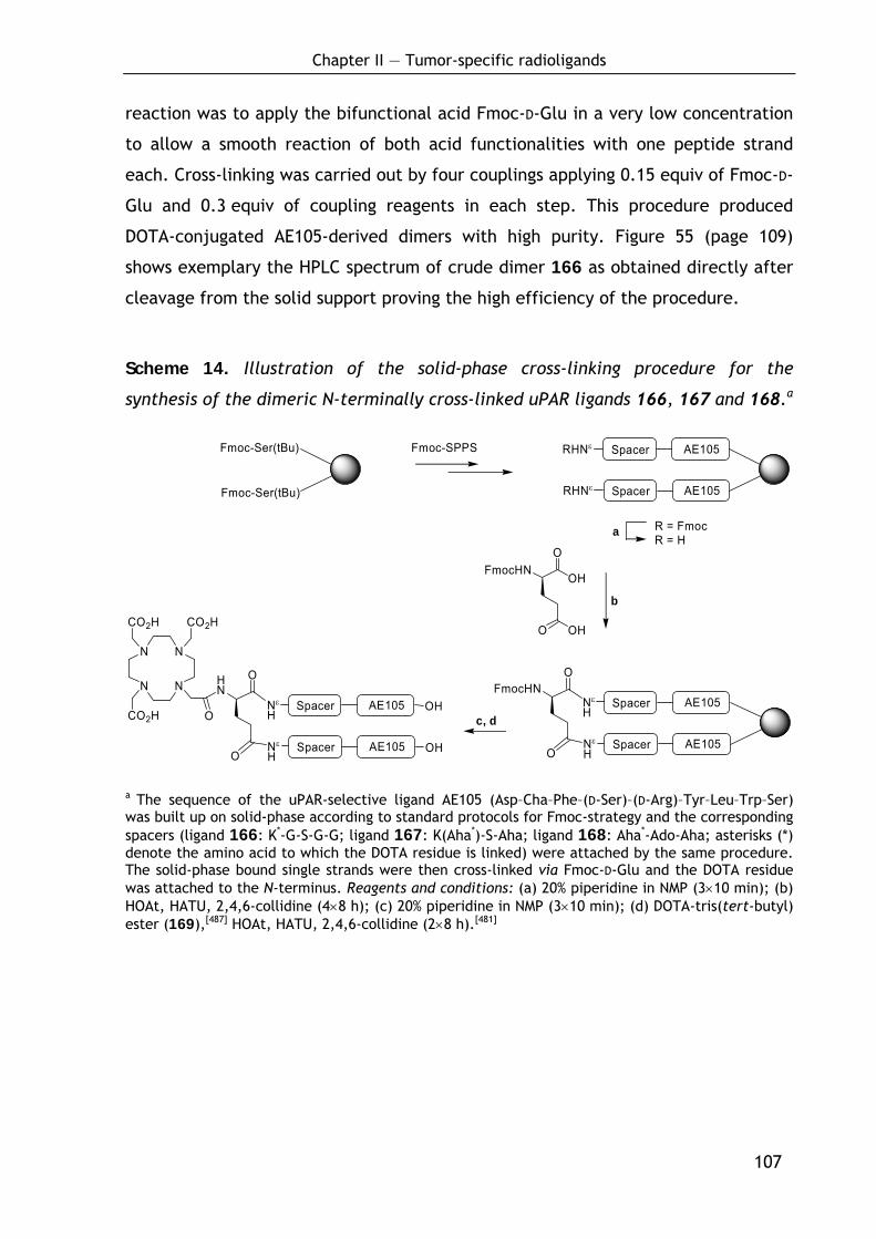
Chapter II — Tumor-specific radioligands
107
reaction was to apply the bifunctional acid Fmoc-D-Glu in a very low concentration
to allow a smooth reaction of both acid functionalities with one peptide strand
each. Cross-linking was carried out by four couplings applying 0.15 equiv of Fmoc-D-
Glu and 0.3 equiv of coupling reagents in each step. This procedure produced
DOTA-conjugated AE105-derived dimers with high purity. Figure 55 (page 109)
shows exemplary the HPLC spectrum of crude dimer 166 as obtained directly after
cleavage from the solid support proving the high efficiency of the procedure.
Scheme 14. Illustration of the solid-phase cross-linking procedure for the
synthesis of the dimeric N-terminally cross-linked uPAR ligands 166, 167 and 168.a
Spacer AE105
FmocHNO
OH
O OH
Fmoc-SPPS
R = Fmoc R = H
b
a
c, d
RHNε
Spacer AE105RHNε
Fmoc-Ser(tBu)
Fmoc-Ser(tBu)
Spacer AE105
O
Nε
H
Spacer AE105Nε
HO
N
N
N
N
CO2H
Spacer AE105FmocHN
O
Nε
H
Spacer AE105Nε
HO
CO2HOH
OH
HN
O
CO2H
a The sequence of the uPAR-selective ligand AE105 (Asp–Cha–Phe–(D-Ser)–(D-Arg)–Tyr–Leu–Trp–Ser) was built up on solid-phase according to standard protocols for Fmoc-strategy and the corresponding spacers (ligand 166: K*-G-S-G-G; ligand 167: K(Aha*)-S-Aha; ligand 168: Aha*-Ado-Aha; asterisks (*) denote the amino acid to which the DOTA residue is linked) were attached by the same procedure. The solid-phase bound single strands were then cross-linked via Fmoc-D-Glu and the DOTA residue was attached to the N-terminus. Reagents and conditions: (a) 20% piperidine in NMP (3×10 min); (b) HOAt, HATU, 2,4,6-collidine (4×8 h); (c) 20% piperidine in NMP (3×10 min); (d) DOTA-tris(tert-butyl) ester (169),[487] HOAt, HATU, 2,4,6-collidine (2×8 h).[481]
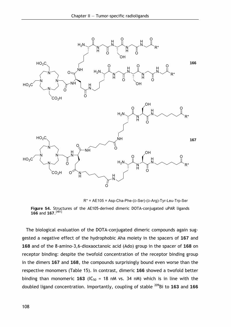
Chapter II — Tumor-specific radioligands
108
NH
H2NNH
HN
NH
HN
R*
O
O
O
O
O
OH
H2NNH
HN
R*
O
NH O
NH
O
OHO
N
N
N
NHO2C
CO2H
HO2C
O
NH
O
O
H2NNH
HN
NH
HN
R*
O
O
O
O
O
OHHN
N
N
N
NHO2C
CO2H
HO2C
O
HN
O
O
H2NNH
HN
R*
O
NH
O
HN
O
OHO
R* = AE105 = Asp-Cha-Phe-(D-Ser)-(D-Arg)-Tyr-Leu-Trp-Ser Figure 54. Structures of the AE105-derived dimeric DOTA-conjugated uPAR ligands 166 and 167.[481]
The biological evaluation of the DOTA-conjugated dimeric compounds again sug-
gested a negative effect of the hydrophobic Aha moiety in the spacers of 167 and
168 and of the 8-amino-3,6-dioxaoctanoic acid (Ado) group in the spacer of 168 on
receptor binding: despite the twofold concentration of the receptor binding group
in the dimers 167 and 168, the compounds surprisingly bound even worse than the
respective monomers (Table 15). In contrast, dimeric 166 showed a twofold better
binding than monomeric 163 (IC50 = 18 nM vs. 34 nM) which is in line with the
doubled ligand concentration. Importantly, coupling of stable 209Bi to 163 and 166
166
167
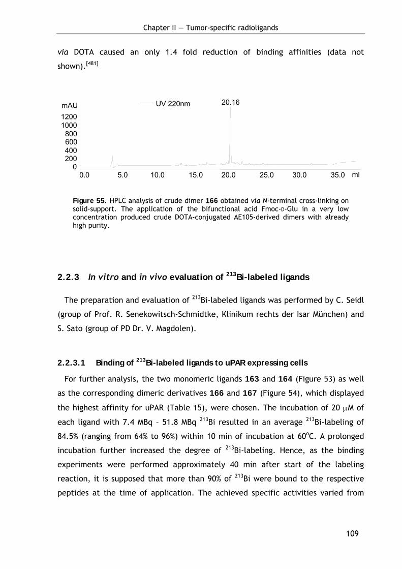
Chapter II — Tumor-specific radioligands
109
via DOTA caused an only 1.4 fold reduction of binding affinities (data not
shown).[481]
UV 220nm
0200 400 600800
1000 1200mAU
0.0 5.0 10.0 15.0 20.0 25.0 30.0 35.0 ml
20.16
Figure 55. HPLC analysis of crude dimer 166 obtained via N-terminal cross-linking on solid-support. The application of the bifunctional acid Fmoc-D-Glu in a very low concentration produced crude DOTA-conjugated AE105-derived dimers with already high purity.
2.2.3 In vitro and in vivo evaluation of 213Bi-labeled ligands
The preparation and evaluation of 213Bi-labeled ligands was performed by C. Seidl
(group of Prof. R. Senekowitsch-Schmidtke, Klinikum rechts der Isar München) and
S. Sato (group of PD Dr. V. Magdolen).
2.2.3.1 Binding of 213Bi-labeled ligands to uPAR expressing cells
For further analysis, the two monomeric ligands 163 and 164 (Figure 53) as well
as the corresponding dimeric derivatives 166 and 167 (Figure 54), which displayed
the highest affinity for uPAR (Table 15), were chosen. The incubation of 20 μM of
each ligand with 7.4 MBq – 51.8 MBq 213Bi resulted in an average 213Bi-labeling of
84.5% (ranging from 64% to 96%) within 10 min of incubation at 60oC. A prolonged
incubation further increased the degree of 213Bi-labeling. Hence, as the binding
experiments were performed approximately 40 min after start of the labeling
reaction, it is supposed that more than 90% of 213Bi were bound to the respective
peptides at the time of application. The achieved specific activities varied from
Chapter II — Tumor-specific radioligands
110
0.12 MBq/μg to 2.4 MBq/μg. This corresponds to a 213Bi-labeling of approximately
1/280,000 to 1/26,000 peptide molecules.[481]
U937 cells were used for the initial characterization of binding of radiolabeled
peptides to cells. Subsequently, the cells were treated with acid, to remove uPA
bound to uPAR on the cell surface and 213Bi-labeled peptide (20 nM, 10 – 54 kBq)
was added to 3 × 10⁶ cells. The binding characteristics were evaluated by quanti-
fying 213Bi-activities in cell sediments and supernatants after 30 min incubation on
ice. It turned out, that the binding of 213Bi-labeled dimeric peptides 166 and 167
to the uPAR-expressing cells was distinctly better than that of the monomeric pep-
tides 163 and 164 (Figure 56 and Supplementary Data 12 on page 260). To estimate
the specificity of the ligands, the U937 cells were pre-incubated with both pro-uPA
12 μg and the soluble form of uPAR (suPAR) (1.6 – 4.8 μg), each leading to a signifi-
cant reduction of the binding of the monomeric as well as the dimeric ligands
(Figure 56 and Supplementary Data 12 on page 260). Since suPAR competes with
membrane-anchored uPAR and also high levels of pro-uPA block the uPAR binding
site of the peptides, these results demonstrate that 213Bi-labled peptides bind
specifically to uPAR expressed by U937 cells.[481]
The U937 cells used for this initial study, are well-suited for analysis of the
binding characteristics of uPAR-directed peptides[488] but do not correspond to the
potential target cells in treatment of disseminated ovarian cancer. Therefore, the
ability of monomeric (163, 164) and dimeric ligands (166, 167) to bind also to OV-
MZ-6 ovarian cancer cells was explored. These cells produce and secrete different
members of the plasminogen activation system including uPAR, uPA and PAI-1.[488]
Considering that OV-MZ-6 cells intrinsically produce and secrete the uPAR ligand
(pro-)uPA, the cells were not acid-treated in this experiment to test the ligands
under more realistic conditions.[481]
As shown in Figure 56b, [213Bi]166 distinctly binds to OV-MZ-6 cells (19.3±3.3%)
and binding was almost completely blocked in the presence of suPAR (3.2 μg)
(3.0±0.5%). Both binding of [213Bi]167 to OV-MZ-6 cells (16.5±2.0%) and blockage of
binding with suPAR (3.7±0.1%) were somewhat reduced compared to [213Bi]166.[481]
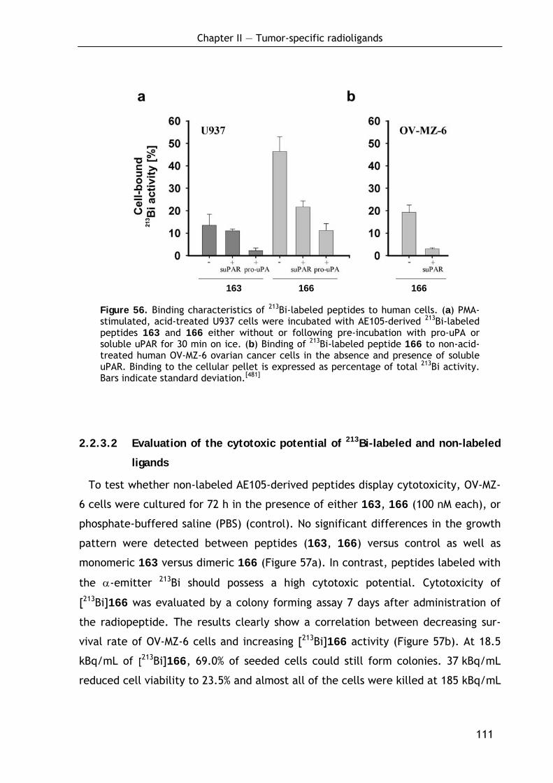
Chapter II — Tumor-specific radioligands
111
163 166 166
Figure 56. Binding characteristics of 213Bi-labeled peptides to human cells. (a) PMA-stimulated, acid-treated U937 cells were incubated with AE105-derived 213Bi-labeled peptides 163 and 166 either without or following pre-incubation with pro-uPA or soluble uPAR for 30 min on ice. (b) Binding of 213Bi-labeled peptide 166 to non-acid-treated human OV-MZ-6 ovarian cancer cells in the absence and presence of soluble uPAR. Binding to the cellular pellet is expressed as percentage of total 213Bi activity. Bars indicate standard deviation.[481]
2.2.3.2 Evaluation of the cytotoxic potential of 213Bi-labeled and non-labeled
ligands
To test whether non-labeled AE105-derived peptides display cytotoxicity, OV-MZ-
6 cells were cultured for 72 h in the presence of either 163, 166 (100 nM each), or
phosphate-buffered saline (PBS) (control). No significant differences in the growth
pattern were detected between peptides (163, 166) versus control as well as
monomeric 163 versus dimeric 166 (Figure 57a). In contrast, peptides labeled with
the α-emitter 213Bi should possess a high cytotoxic potential. Cytotoxicity of
[213Bi]166 was evaluated by a colony forming assay 7 days after administration of
the radiopeptide. The results clearly show a correlation between decreasing sur-
vival rate of OV-MZ-6 cells and increasing [213Bi]166 activity (Figure 57b). At 18.5
kBq/mL of [213Bi]166, 69.0% of seeded cells could still form colonies. 37 kBq/mL
reduced cell viability to 23.5% and almost all of the cells were killed at 185 kBq/mL
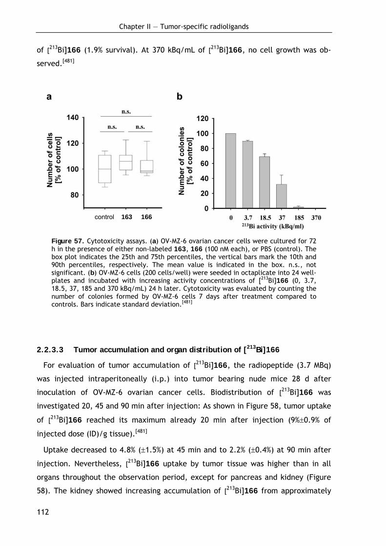
Chapter II — Tumor-specific radioligands
112
of [213Bi]166 (1.9% survival). At 370 kBq/mL of [213Bi]166, no cell growth was ob-
served.[481]
Figure 57. Cytotoxicity assays. (a) OV-MZ-6 ovarian cancer cells were cultured for 72 h in the presence of either non-labeled 163, 166 (100 nM each), or PBS (control). The box plot indicates the 25th and 75th percentiles, the vertical bars mark the 10th and 90th percentiles, respectively. The mean value is indicated in the box. n.s., not significant. (b) OV-MZ-6 cells (200 cells/well) were seeded in octaplicate into 24 well-plates and incubated with increasing activity concentrations of [213Bi]166 (0, 3.7, 18.5, 37, 185 and 370 kBq/mL) 24 h later. Cytotoxicity was evaluated by counting the number of colonies formed by OV-MZ-6 cells 7 days after treatment compared to controls. Bars indicate standard deviation.[481]
2.2.3.3 Tumor accumulation and organ distribution of [213Bi]166
For evaluation of tumor accumulation of [213Bi]166, the radiopeptide (3.7 MBq)
was injected intraperitoneally (i.p.) into tumor bearing nude mice 28 d after
inoculation of OV-MZ-6 ovarian cancer cells. Biodistribution of [213Bi]166 was
investigated 20, 45 and 90 min after injection: As shown in Figure 58, tumor uptake
of [213Bi]166 reached its maximum already 20 min after injection (9%±0.9% of
injected dose (ID)/g tissue).[481]
Uptake decreased to 4.8% (±1.5%) at 45 min and to 2.2% (±0.4%) at 90 min after
injection. Nevertheless, [213Bi]166 uptake by tumor tissue was higher than in all
organs throughout the observation period, except for pancreas and kidney (Figure
58). The kidney showed increasing accumulation of [213Bi]166 from approximately
control 163 166

Chapter II — Tumor-specific radioligands
113
18.9% (±3.6%) of ID/g tissue after 20 min to approximately 31.9% (±8.8%) of ID/g
tissue after 90 min, while in all other organs 213Bi-activity decreased over time
(Figure 58).[481]
Figure 58. Biodistribution of [213Bi]166 in nude mice bearing intraperitoneal ovarian tumors. Nude mice carrying OV-MZ-6-derived tumors were analyzed at 20 min (a, b), 45 min (c, d) and 90 min (e, f) after injection of the radiopeptide in the absence (black bars) or presence (grey bars) of the plasma expander gelofusine. High-uptake organs (a, c, e) and low-uptake organs (b, d, f) as deduced from uptake values 20 min after injection. Tumor uptake of [213Bi]166 proved to be superior over organ uptake throughout the observation period except for pancreas and kidney. Intravenous injection of gelofusine 5 min before i.p. injection of [213Bi]166 reduced kidney accumulation roughly by half (grey bars) compared to the animals which had not received gelofusine (black bars).[481]
Chapter II — Tumor-specific radioligands
114
Half of the animals were injected intravenal (i.v.) with the plasma expander
gelofusine[489,490] 5 min before i.p. injection of [213Bi]166 to reduce kidney uptake
of [213Bi]166. Gelofusine caused a mean reduction of [213Bi]166 kidney uptake to
55.8% (±11%) of the uptake without gelofusine: to 46.6% (±3.6%) at 20 min, to
63.5% (±16.5%) at 45 min and to 57.3% (±5.3%) at 90 min. In contrast, [213Bi]166
uptake in almost all of the other organs and in tumor tissue was slightly elevated in
the presence of gelofusine.[481]
In tumor-free mice, kidney uptake of [213Bi]166 was 1.4 fold higher than in mice
presenting tumor, indicating that [213Bi]166 efficiently binds to tumor tissue. Gelo-
fusine reduced kidney uptake of [213Bi]166 by half also in tumor free mice (data not
shown).
2.2.3.4 Discussion of the therapeutic potential
The goal of the present study was to test whether peptidic radioligands, based on
potent uPAR binding peptides (i.e. the 10-mer peptide cyclo[21,29][D-Cys21Cys29]-
uPA21-30 (WX360)[465] and the 9-mer peptide AE105[466]), may be developed for
application in α−emitter therapy of disseminated ovarian cancer overexpressing the
uPA receptor. For this, the uPAR binding peptides were modified by conjugation
with the chelator DOTA (for efficient binding of the α-emitter 213Bi). It turned out
that the WX360 ligand is not suitable for this purpose, as the modification with
DOTA led to a great decrease of the uPAR affinity even after introduction of spacer
sequences. In contrast, after optimization of the spacer moiety, the DOTA chelator
could by attached to the AE105 peptide thereby only minimally affecting the
binding affinity.
Evaluation of the AE105-based ligands demonstrated that the newly developed
monomeric and dimeric peptides, labeled with the α-emitter 213Bi, specifically rec-
ognized uPAR on the surface of both the human monocytoid U937 cells and OV-MZ-6
ovarian cancer cells. While the extent of binding of peptides to U937 cells clearly
improved with removal of receptor-bound uPA via acid wash, OV-MZ-6 cells showed
excellent binding of dimeric 213Bi-peptides irrespective of this pre-treatment. This
strongly suggests that OV-MZ-6 ovarian cancer cells, not only expressing uPAR but
also its ligand uPA and other members of the plasminogen activation system,[488]
Chapter II — Tumor-specific radioligands
115
present a considerable number of cell surface-associated uPARs that are not
occupied by uPA and therefore can be targeted by appropriate peptide conjugates.
Excellent binding of dimeric [213Bi]166 to OV-MZ-6 cells was also confirmed in vivo
following intraperitoneal injection into nude mice bearing intraperitoneal tumor
nodules. Tumor binding was approximately 9-10%, 5-10% and 2.5% of injected
dose/g (ID/g) of tissue at 20, 45 and 90 min after administration of [213Bi]166,
respectively. In contrast, tumor binding of 213Bi-DOTATOC, targeting the soma-
tostatin receptor, was reported to be only 0.75% ID/g tissue 1 h after injection.[420]
Also, tumor binding of 111In-minigastrin targeting the CCK-B receptor was described
to be comparatively low, amounting to 2% ID/g tissue at 10 min and 0.45% ID/g
tissue at 1 h after injection of the radiopeptide.[421] 177Lu-AMBA targeting GRP
receptor with high affinity was reported to show approximately 5% ID/g tissue
tumor binding 1 h after administration, which is comparable to the presented
results.
Although peptides have distinct advantages compared to monoclonal antibodies
as carriers for radionuclides,[491] potential kidney toxicity of radiopeptides might
impair their efficacy in targeted radiotherapy of tumors. The kidney is a major site
in the catabolism of peptides. Due to their small size, radiopeptides can traverse
the glomerular basement membrane of the kidney leading to kidney damage.[492]
Therefore, efforts are made in order to reduce kidney uptake of low-molecular-
weight radiopeptides. Promising results in terms of prevention of renal damage
following administration of radiopeptides have been obtained previously via co-in-
fusion of polyglutamic acids[421] or the basic amino acids arginine and lysine.[493,494]
Nevertheless, co-infusion of amino acids also involves a number of drawbacks.
Since large volumes of the amino acid solutions have to be administered, generally,
hyperkalemia arises, which may lead to cardiac arrhythmia.[492] Therefore, novel
compounds for reduction of renal uptake of radiopeptides are currently being
explored. Among them are the aldosterone receptor antagonist spironolactone,[495]
probenecid[496] and the plasma expander gelofusine.[489] For instance, it was demon-
strated that infusion of gelofusine significantly reduced renal uptake of radio-
labeled octreotide by 45% without any side effects.[489] In line with that, it was
demonstrated that gelofusine injection also significantly reduced kidney uptake of
[213Bi]166, approximately by half.
Chapter II — Tumor-specific radioligands
116
The extent of radiation induced kidney damage following therapy with low
molecular weight radiopeptides also has turned out to be dependent on the physi-
cal properties of the applied radionuclide. Administration of DOTATOC labeled with
short-lived, short-ranged 213Bi caused only minimal to mild nephrotoxicity com-
pared to moderate to severe nephrotoxicity following DOTATOC therapy with the
longer-lived, longer-ranged β-emitter 90Y.[420]
In summary, the synthetic DOTA-modified, 213Bi-labeled peptide 166, which effi-
ciently targets uPAR expressing tumor cells in vitro and in vivo, is a very promising
new candidate for peptide receptor radiotherapy. The project is continued in the
thesis of T. Huber.
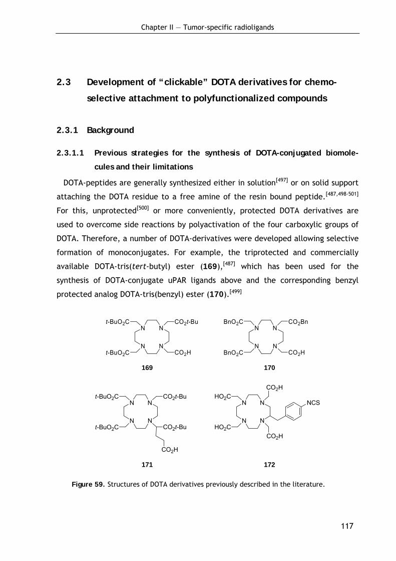
Chapter II — Tumor-specific radioligands
117
2.3 Development of “clickable” DOTA derivatives for chemo-
selective attachment to polyfunctionalized compounds
2.3.1 Background
2.3.1.1 Previous strategies for the synthesis of DOTA-conjugated biomole-
cules and their limitations
DOTA-peptides are generally synthesized either in solution[497] or on solid support
attaching the DOTA residue to a free amine of the resin bound peptide.[487,498-501]
For this, unprotected[500] or more conveniently, protected DOTA derivatives are
used to overcome side reactions by polyactivation of the four carboxylic groups of
DOTA. Therefore, a number of DOTA-derivatives were developed allowing selective
formation of monoconjugates. For example, the triprotected and commercially
available DOTA-tris(tert-butyl) ester (169),[487] which has been used for the
synthesis of DOTA-conjugate uPAR ligands above and the corresponding benzyl
protected analog DOTA-tris(benzyl) ester (170).[499]
N
N
N
N
t-BuO2C
t-BuO2C
CO2t-Bu
CO2H
N
N
N
N
BnO2C
BnO2C
CO2Bn
CO2H
169 170
N
N
N
N
t-BuO2C
t-BuO2C
CO2t-Bu
CO2t-Bu
CO2H
N
N
N
N
HO2C
HO2C
CO2H
CO2H
NCS
171 172
Figure 59. Structures of DOTA derivatives previously described in the literature.
Chapter II — Tumor-specific radioligands
118
Further derivatives are DOTAGA(tBu)4 (171), offering an additional unprotected
carboxylic group,[498] and the isothiocyanate-functionalized p-NCS-Bz-DOTA
(172).[501] Furthermore, derivatized amino acids containing a DOTA moiety were
used[502] or DOTA was synthesized stepwise on a resin bound peptide.[503,504]
The main limitation of all these methods is the introduction of the DOTA residue
into the bioconjugate via an electrophilic reaction. This procedure tolerates no
other N- or S-nucleophilic groups for a selective reaction which greatly limits the
application of above derivatives. Chemoselective approaches allowing a site-
selective functionalization of unprotected polyfunctionalized compounds remained
in demand. Such a method would be a powerful tool for the synthesis of DOTA-
linked radiopharmaceuticals enabling new synthetic strategies and the synthesis of
complex structures. For example, Thumshirn et al. recently developed multimeric
aminooxy-functionalized RGD-derivatives[505] which were 18F-labeled via oxime
ligation using 4-[18F]fluorobenzaldehyde.[506] A carbonyl functionalized DOTA
derivative would allow a selective conjugation with DOTA and subsequent labeling
with various radiometals. Very recently, Lin and coworkers have demonstrated a
site-specific protein modification through Cu(I)-catalyzed 1,2,3-triazole forma-
tion.[507] Applying this methodology and using an alkyne functionalized DOTA
derivative would enable site specific DOTA labeling of proteins. In a recent
publication, J. Hovinen reported the synthesis of an aminooxy-functionalized
chelate by derivatization of the tris tert-butyl ester of DOTA which was conjugated
with naltrexone and 2-deoxy-D-ribose.[508] However, the reported procedure
requires expensive starting material and the applicability to polyfunctionalized
compounds like peptides remains unclear.
2.3.1.2 Motivation and target structures
Considering above mentioned limitations, the idea was to design novel DOTA-
derivatives enabling the chemoselective attachment in presence of a wide range of
functional groups. Thereby, the focus was on structures readily accessible in few
synthetic steps from cheap commercial materials to make the method convenient
and even a general alternative to the widely used but quite expensive DOTA-tris-
(tert-butyl) ester. Furthermore, the accessibility of the correspondingly polyfunc-
tionalized compounds was an important aspect in the consideration.
Chapter II — Tumor-specific radioligands
119
Click reactions,[509] in particular the oxime ligation[510,511] as well as the Cu(I)-
catalyzed azide-alkyne cycloaddition,[512,513] are well-studied and powerful reac-
tions for chemoselective couplings which fulfill all requirements for this purpose.
The oxime ligation denotes the highly selective reaction between an aminooxy
component and aldehydes or methylketones[514] under formation of an oxime bond,
which is known to be stable both in vitro and in vivo.[515] The reaction was shown
to tolerate every free amino acid side chain except an N-terminal cysteine and
found widespread use, e.g. in the synthesis of template assembled synthetic
proteins,[516,517] radioactive labeled peptide conjugates,[515,518] cyclic peptides[519]
and protein analogs.[520,521] The Cu(I)-catalyzed azide-alkyne cycloaddition is a
catalyzed variant of the chemoselective Huisgen 1,3-dipolar cycloaddition[522-525] of
an azide and an alkyne for the formation of a triazole which has found application
in various developments in medicinal chemistry.[526-531] Thus, to provide users with
different methodologies for the synthesis of DOTA-conjugates, the focus was on
appropriate DOTA-derivatives for both types of reaction described above.
The following chapters describe the synthesis of 2-[1-(1,4,7,10-tetraazacyclode-
cane)-4,7,10-tris(tert-butylacetate)]-(4-acetylphenyl) acetic acid tert-butyl ester
(173) and 2-[1-(1,4,7,10-tetraazacyclodecane)-4,7,10-tris(tert-butylace-tate)]-(4-
acetylphenyl) acetic acid (174) as carbonyl components for oxime ligations with
aminooxy-functionalized compounds and 2-[1-(1,4,7,10-tetraaza-cyclodecane)-
4,7,10-tris(tert-butylacetate)]acetic acid methyl ester (175) as alkynyl component
for Cu(I)-catalyzed azide-alkyne cycloaddition with azide functionalized compounds
(Figure 60).[532] 174, bearing one free acid functionality and the orthogonally pro-
tected alkynyl derivative 175 were designed to enable a selective further transfor-
mation as described below. Initial steps of the synthesis have been worked out by
A. Modlinger.[533] In a proof-of-principal experiment, the applicability of the novel
derivatives was proven by reacting both types of DOTA derivatives with appropri-
ately functionalized Tyr3-octreotate, a somatostatin analog which is a well known
targeting molecule for tumor diagnostics and endoradiotherapeutic purposes.[428,534-
538] Tyr3-octreotate was chosen as a highly functionalized model compound provid-
ing a broad range of functionalities. In addition, one of the radioligands was
labeled with 68Ga and an initial biodistribution study in AR42J tumor bearing nude
mice was performed proving the applicability of the modified linkage.
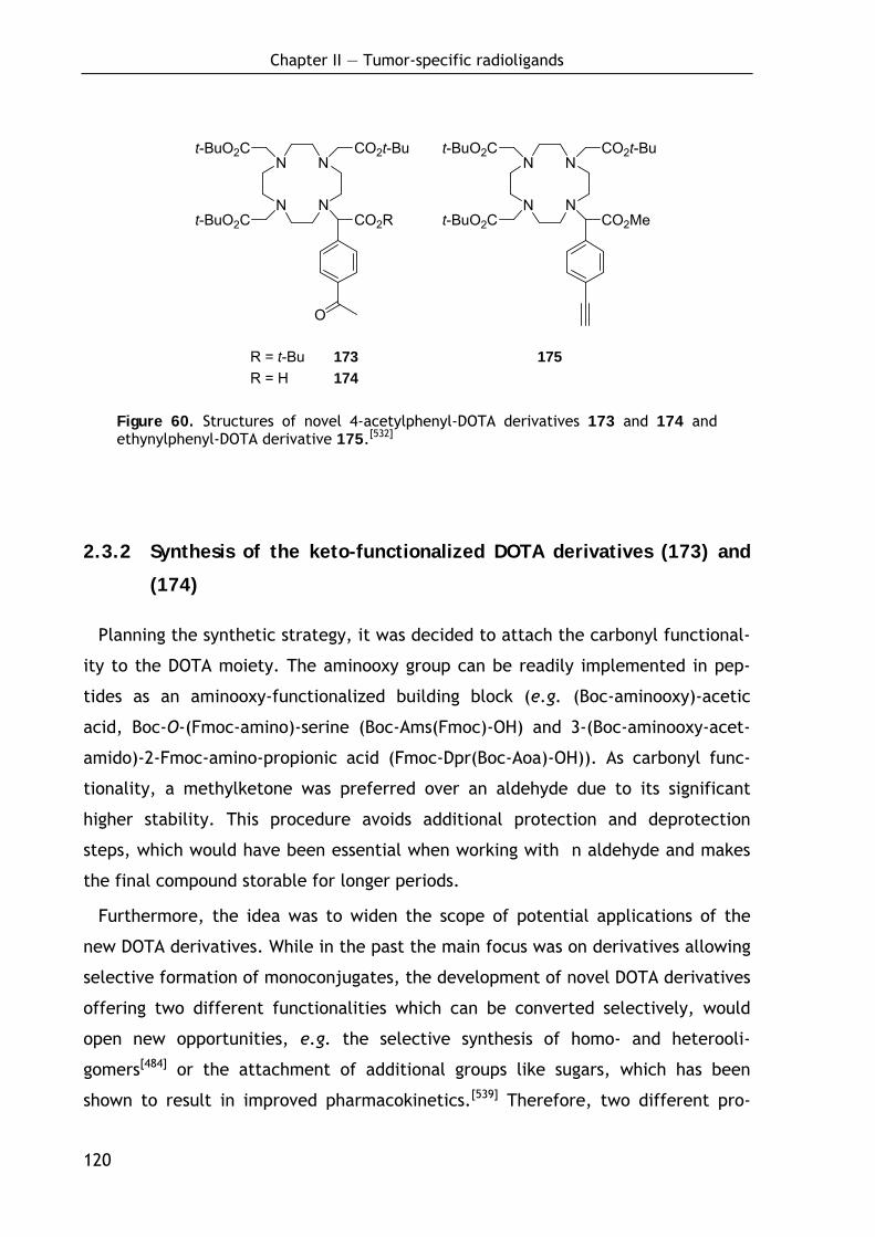
Chapter II — Tumor-specific radioligands
120
N
N
N
N
t-BuO2C
t-BuO2C
CO2t-Bu
CO2R
O
N
N
N
N
t-BuO2C
t-BuO2C
CO2t-Bu
CO2Me
R = t-Bu 173 175 R = H 174
Figure 60. Structures of novel 4-acetylphenyl-DOTA derivatives 173 and 174 and ethynylphenyl-DOTA derivative 175.[532]
2.3.2 Synthesis of the keto-functionalized DOTA derivatives (173) and
(174)
Planning the synthetic strategy, it was decided to attach the carbonyl functional-
ity to the DOTA moiety. The aminooxy group can be readily implemented in pep-
tides as an aminooxy-functionalized building block (e.g. (Boc-aminooxy)-acetic
acid, Boc-O-(Fmoc-amino)-serine (Boc-Ams(Fmoc)-OH) and 3-(Boc-aminooxy-acet-
amido)-2-Fmoc-amino-propionic acid (Fmoc-Dpr(Boc-Aoa)-OH)). As carbonyl func-
tionality, a methylketone was preferred over an aldehyde due to its significant
higher stability. This procedure avoids additional protection and deprotection
steps, which would have been essential when working with n aldehyde and makes
the final compound storable for longer periods.
Furthermore, the idea was to widen the scope of potential applications of the
new DOTA derivatives. While in the past the main focus was on derivatives allowing
selective formation of monoconjugates, the development of novel DOTA derivatives
offering two different functionalities which can be converted selectively, would
open new opportunities, e.g. the selective synthesis of homo- and heterooli-
gomers[484] or the attachment of additional groups like sugars, which has been
shown to result in improved pharmacokinetics.[539] Therefore, two different pro-
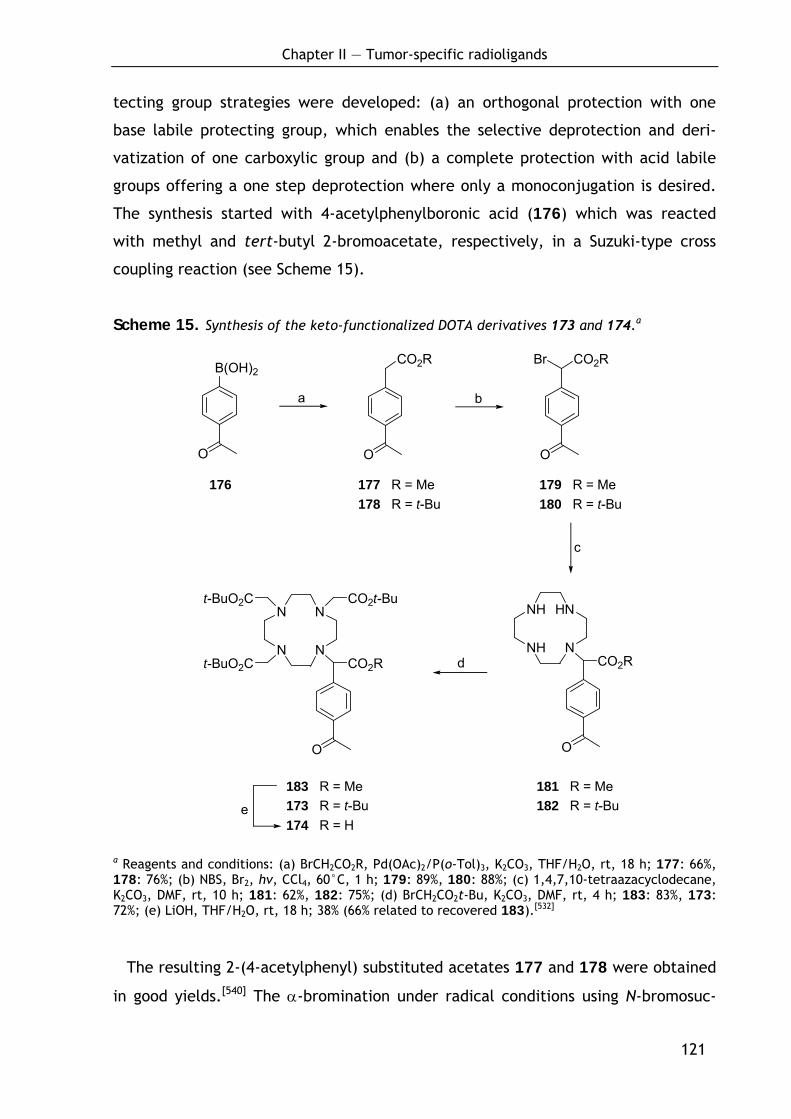
Chapter II — Tumor-specific radioligands
121
O O
CO2R
O
CO2RBr
NH
NH
HN
NCO2R
O
N
N
N
N
t-BuO2C
t-BuO2C
CO2t-Bu
CO2R
O
B(OH)2
a b
c
d
e
tecting group strategies were developed: (a) an orthogonal protection with one
base labile protecting group, which enables the selective deprotection and deri-
vatization of one carboxylic group and (b) a complete protection with acid labile
groups offering a one step deprotection where only a monoconjugation is desired.
The synthesis started with 4-acetylphenylboronic acid (176) which was reacted
with methyl and tert-butyl 2-bromoacetate, respectively, in a Suzuki-type cross
coupling reaction (see Scheme 15).
Scheme 15. Synthesis of the keto-functionalized DOTA derivatives 173 and 174.a
a Reagents and conditions: (a) BrCH2CO2R, Pd(OAc)2/P(o-Tol)3, K2CO3, THF/H2O, rt, 18 h; 177: 66%, 178: 76%; (b) NBS, Br2, hν, CCl4, 60°C, 1 h; 179: 89%, 180: 88%; (c) 1,4,7,10-tetraazacyclodecane, K2CO3, DMF, rt, 10 h; 181: 62%, 182: 75%; (d) BrCH2CO2t-Bu, K2CO3, DMF, rt, 4 h; 183: 83%, 173: 72%; (e) LiOH, THF/H2O, rt, 18 h; 38% (66% related to recovered 183).[532]
The resulting 2-(4-acetylphenyl) substituted acetates 177 and 178 were obtained
in good yields.[540] The α-bromination under radical conditions using N-bromosuc-
176 177 R = Me 179 R = Me 178 R = t-Bu 180 R = t-Bu
183 R = Me 181 R = Me 173 R = t-Bu 182 R = t-Bu 174 R = H
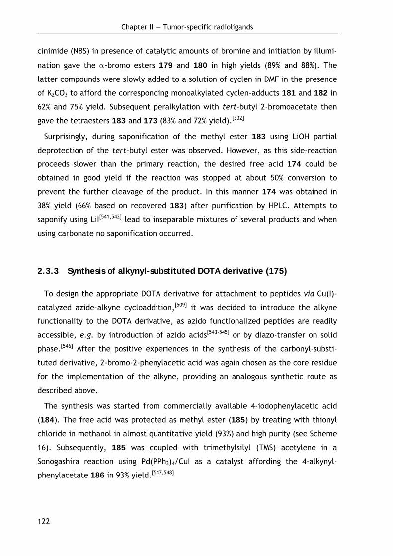
Chapter II — Tumor-specific radioligands
122
cinimide (NBS) in presence of catalytic amounts of bromine and initiation by illumi-
nation gave the α-bromo esters 179 and 180 in high yields (89% and 88%). The
latter compounds were slowly added to a solution of cyclen in DMF in the presence
of K2CO3 to afford the corresponding monoalkylated cyclen-adducts 181 and 182 in
62% and 75% yield. Subsequent peralkylation with tert-butyl 2-bromoacetate then
gave the tetraesters 183 and 173 (83% and 72% yield).[532]
Surprisingly, during saponification of the methyl ester 183 using LiOH partial
deprotection of the tert-butyl ester was observed. However, as this side-reaction
proceeds slower than the primary reaction, the desired free acid 174 could be
obtained in good yield if the reaction was stopped at about 50% conversion to
prevent the further cleavage of the product. In this manner 174 was obtained in
38% yield (66% based on recovered 183) after purification by HPLC. Attempts to
saponify using LiI[541,542] lead to inseparable mixtures of several products and when
using carbonate no saponification occurred.
2.3.3 Synthesis of alkynyl-substituted DOTA derivative (175)
To design the appropriate DOTA derivative for attachment to peptides via Cu(I)-
catalyzed azide-alkyne cycloaddition,[509] it was decided to introduce the alkyne
functionality to the DOTA derivative, as azido functionalized peptides are readily
accessible, e.g. by introduction of azido acids[543-545] or by diazo-transfer on solid
phase.[546] After the positive experiences in the synthesis of the carbonyl-substi-
tuted derivative, 2-bromo-2-phenylacetic acid was again chosen as the core residue
for the implementation of the alkyne, providing an analogous synthetic route as
described above.
The synthesis was started from commercially available 4-iodophenylacetic acid
(184). The free acid was protected as methyl ester (185) by treating with thionyl
chloride in methanol in almost quantitative yield (93%) and high purity (see Scheme
16). Subsequently, 185 was coupled with trimethylsilyl (TMS) acetylene in a
Sonogashira reaction using Pd(PPh3)4/CuI as a catalyst affording the 4-alkynyl-
phenylacetate 186 in 93% yield.[547,548]
Chapter II — Tumor-specific radioligands
123
CO2Me
NH
NH
HN
NCO2Me
16
14 1512 R = H13 R = Me
CO2R
I
TMS
CO2Me
TMS
Br
TMS
N
N
N
N
t-BuO2C
t-BuO2C
CO2t-Bu
CO2Me
3
a
b c
d
e
Scheme 16. Synthesis of alkyne-functionalized DOTA derivatives.a
a Reagents and conditions: (a) SOCl2, MeOH, 0°C → rt, 1 h; 93%; (b) HC≡C–TMS, Pd(PPh3)4/CuI, NEt3, CH3CN, 0°C → rt, 3 h; 93%; (c) 1) LDA, THF, -78°C, 1 h; 2) NBS, THF, -78°C → rt, 18 h; 47% (92% related to recovered 186); (d) 1,4,7,10-tetraazacyclodecane, K2CO3, DMF, rt, 10 h; 73%; (e) 1) BrCH2CO2t-Bu, K2CO3, DMF, rt, 4 h; 2) TBAF, THF, rt, 15 min; 85%.[532]
However, the α-bromination of 186 with NBS failed with both, Br2/hν and
2,2’-azobisisobutyronitrile (AIBN) as initiators. This is most likely due to side reac-
tions caused by the presence of a triple bond. This problem was circumvent by
introducing the bromide in an electrophilic manner. For this purpose, the ester 186
was transformed into the boron enolate in an analogous manner as described by
Evans et al.,[549] treating subsequently with lithium diisopropyl amide (LDA) and
Bu2BOTf. After addition of N-bromosuccinimide as electrophilic brominating
reagent, the α-bromo ester 187 was isolated in rather poor yield. In an attempt to
further optimize the reaction, it was found that the conversion proceeds cleanly if
186 187
175 188
184 R = H 185 R = Me
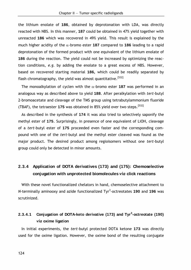
Chapter II — Tumor-specific radioligands
124
the lithium enolate of 186, obtained by deprotonation with LDA, was directly
reacted with NBS. In this manner, 187 could be obtained in 47% yield together with
unreacted 186 which was recovered in 49% yield. This result is explained by the
much higher acidity of the α-bromo ester 187 compared to 186 leading to a rapid
deprotonation of the formed product with one equivalent of the lithium enolate of
186 during the reaction. The yield could not be increased by optimizing the reac-
tion conditions, e.g. by adding the enolate to a great excess of NBS. However,
based on recovered starting material 186, which could be readily separated by
flash chromatography, the yield was almost quantitative.[532]
The monoalkylation of cyclen with the α-bromo ester 187 was performed in an
analogous way as described above to yield 188. After peralkylation with tert-butyl
2-bromoacetate and cleavage of the TMS group using tetrabutylammonium fluoride
(TBAF), the tetraester 175 was obtained in 85% yield over two steps.[532]
As described in the synthesis of 174 it was also tried to selectively saponify the
methyl ester of 175. Surprisingly, in presence of one equivalent of LiOH, cleavage
of a tert-butyl ester of 175 proceeded even faster and the corresponding com-
pound with one of the tert-butyl and the methyl ester cleaved was found as the
major product. The desired product among regioisomers without one tert-butyl
group could only be detected in minor amounts.
2.3.4 Application of DOTA derivatives (173) and (175): Chemoselective
conjugation with unprotected biomolecules via click reactions
With these novel functionalized chelators in hand, chemoselective attachment to
N-terminally aminooxy and azide functionalized Tyr3-octreotates 190 and 196 was
scrutinized.
2.3.4.1 Conjugation of DOTA-keto derivative (173) and Tyr3-octreotate (190)
via oxime ligation
In initial experiments, the tert-butyl protected DOTA ketone 173 was directly
used for the oxime ligation. However, the oxime bond of the resulting conjugate
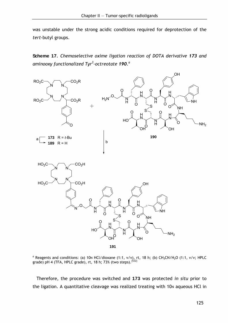
Chapter II — Tumor-specific radioligands
125
N
N
N
N
RO2C
RO2C
CO2R
CO2R
O
HN
O
NH
HN
NH
HN
O
NHOS
S O
OHNH2
NH
HN
OH
HO
O
ONH
O
OH
OH2N
O
O
N
N
N
N
HO2C
HO2C
CO2H
CO2H
HN
O
NH
HN
NH
HN
O
NHOS
S
O
O
OHNH2
NH
HN
OH
HO
O
ONH
O
OH
ON
O
01 R = t-Bu 17 R = H
18a
b
was unstable under the strong acidic conditions required for deprotection of the
tert-butyl groups.
Scheme 17. Chemoselective oxime ligation reaction of DOTA derivative 173 and
aminooxy functionalized Tyr3-octreotate 190.a
191
a Reagents and conditions: (a) 10N HCl/dioxane (1:1, v/v), rt, 18 h; (b) CH3CN/H2O (1:1, v/v; HPLC grade) pH 4 (TFA, HPLC grade), rt, 18 h; 73% (two steps).[532]
Therefore, the procedure was switched and 173 was protected in situ prior to
the ligation. A quantitative cleavage was realized treating with 10N aqueous HCl in
190
173 R = t-Bu 189 R = H
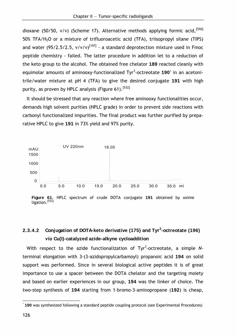
Chapter II — Tumor-specific radioligands
126
dioxane (50/50, v/v) (Scheme 17). Alternative methods applying formic acid,[550]
50% TFA/H2O or a mixture of trifluoroacetic acid (TFA), triisopropyl silane (TIPS)
and water (95/2.5/2.5, v/v/v)[197] - a standard deprotection mixture used in Fmoc
peptide chemistry - failed. The latter procedure in addition let to a reduction of
the keto group to the alcohol. The obtained free chelator 189 reacted cleanly with
equimolar amounts of aminooxy-functionalized Tyr3-octreotate 190* in an acetoni-
trile/water mixture at pH 4 (TFA) to give the desired conjugate 191 with high
purity, as proven by HPLC analysis (Figure 61).[532]
It should be stressed that any reaction where free aminooxy functionalities occur,
demands high solvent purities (HPLC grade) in order to prevent side reactions with
carbonyl functionalized impurities. The final product was further purified by prepa-
rative HPLC to give 191 in 73% yield and 97% purity.
UV 220nm
0
500
1000
1500 mAU
0.0 5.0 10.0 15.0 20.0 25.0 30.0 35.0 ml
18.05
Figure 61. HPLC spectrum of crude DOTA conjugate 191 obtained by oxime ligation.[532]
2.3.4.2 Conjugation of DOTA-keto derivative (175) and Tyr3-octreotate (196)
via Cu(I)-catalyzed azide-alkyne cycloaddition
With respect to the azide functionalization of Tyr3-octreotate, a simple N-
terminal elongation with 3-(3-azidopropylcarbamoyl) propanoic acid 194 on solid
support was performed. Since in several biological active peptides it is of great
importance to use a spacer between the DOTA chelator and the targeting moiety
and based on earlier experiences in our group, 194 was the linker of choice. The
two-step synthesis of 194 starting from 1-bromo-3-aminopropane (192) is cheap,
* 190 was synthesized following a standard peptide coupling protocol (see Experimental Procedures)
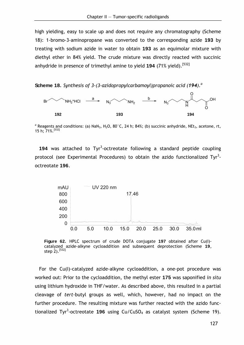
Chapter II — Tumor-specific radioligands
127
high yielding, easy to scale up and does not require any chromatography (Scheme
18): 1-bromo-3-aminopropane was converted to the corresponding azide 193 by
treating with sodium azide in water to obtain 193 as an equimolar mixture with
diethyl ether in 84% yield. The crude mixture was directly reacted with succinic
anhydride in presence of trimethyl amine to yield 194 (71% yield).[532]
Scheme 18. Synthesis of 3-(3-azidopropylcarbamoyl)propanoic acid (194).a
Br NH2*HCl N3 NH2 N3 NH
O
O
OHa b
192 193 194
a Reagents and conditions: (a) NaN3, H2O, 80°C, 24 h; 84%; (b) succinic anhydride, NEt3, acetone, rt, 15 h; 71%.[532]
194 was attached to Tyr3-octreotate following a standard peptide coupling
protocol (see Experimental Procedures) to obtain the azido functionalized Tyr3-
octreotate 196.
UV 220 nm
0 200 400 600 800 mAU
0.0 5.0 10.0 15.0 20.0 25.0 30.0 35.0 ml
17.46
Figure 62. HPLC spectrum of crude DOTA conjugate 197 obtained after Cu(I)-catalyzed azide-alkyne cycloaddition and subsequent deprotection (Scheme 19, step 2).[532]
For the Cu(I)-catalyzed azide-alkyne cycloaddition, a one-pot procedure was
worked out: Prior to the cycloaddition, the methyl ester 175 was saponified in situ
using lithium hydroxide in THF/water. As described above, this resulted in a partial
cleavage of tert-butyl groups as well, which, however, had no impact on the
further procedure. The resulting mixture was further reacted with the azido func-
tionalized Tyr3-octreotate 196 using Cu/CuSO4 as catalyst system (Scheme 19).
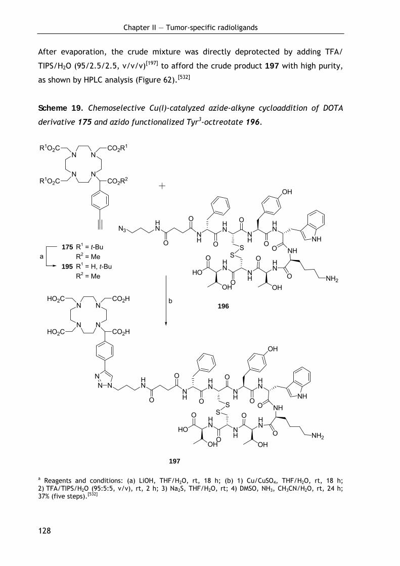
Chapter II — Tumor-specific radioligands
128
N3HN
O
OHN
O
NH
HN
NH
O HN
O
NHOS
S
O
O
OHNH2
NH
HN
OH
HO
O
O
OH
NH
N
N
N
N
R1O2C
R1O2C
CO2R1
CO2R2
N
N
N
N
HO2C
HO2C
CO2H
CO2H
NHN
O
OHN
O
NH
HN
NH
O HN
O
NHOS
S
O
O
OHNH2
NH
HN
OH
HO
O
O
OH
NH
NN
23
a
b
After evaporation, the crude mixture was directly deprotected by adding TFA/
TIPS/H2O (95/2.5/2.5, v/v/v)[197] to afford the crude product 197 with high purity,
as shown by HPLC analysis (Figure 62).[532]
Scheme 19. Chemoselective Cu(I)-catalyzed azide-alkyne cycloaddition of DOTA
derivative 175 and azido functionalized Tyr3-octreotate 196.
197
a Reagents and conditions: (a) LiOH, THF/H2O, rt, 18 h; (b) 1) Cu/CuSO4, THF/H2O, rt, 18 h; 2) TFA/TIPS/H2O (95:5:5, v/v), rt, 2 h; 3) Na2S, THF/H2O, rt; 4) DMSO, NH3, CH3CN/H2O, rt, 24 h; 37% (five steps).[532]
196
175 R1 = t-Bu R2 = Me 195 R1 = H, t-Bu R2 = Me
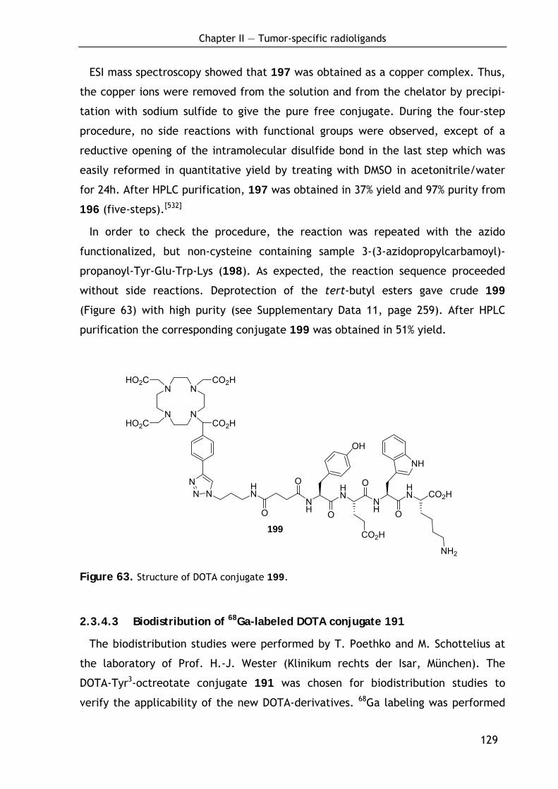
Chapter II — Tumor-specific radioligands
129
N
N
N
N
HO2C
HO2C
CO2H
CO2H
NHN
O
OHN
O
NH
HN
OONH
NN CO2H
OH
NH
CO2H
NH2
ESI mass spectroscopy showed that 197 was obtained as a copper complex. Thus,
the copper ions were removed from the solution and from the chelator by precipi-
tation with sodium sulfide to give the pure free conjugate. During the four-step
procedure, no side reactions with functional groups were observed, except of a
reductive opening of the intramolecular disulfide bond in the last step which was
easily reformed in quantitative yield by treating with DMSO in acetonitrile/water
for 24h. After HPLC purification, 197 was obtained in 37% yield and 97% purity from
196 (five-steps).[532]
In order to check the procedure, the reaction was repeated with the azido
functionalized, but non-cysteine containing sample 3-(3-azidopropylcarbamoyl)-
propanoyl-Tyr-Glu-Trp-Lys (198). As expected, the reaction sequence proceeded
without side reactions. Deprotection of the tert-butyl esters gave crude 199
(Figure 63) with high purity (see Supplementary Data 11, page 259). After HPLC
purification the corresponding conjugate 199 was obtained in 51% yield.
Figure 63. Structure of DOTA conjugate 199.
2.3.4.3 Biodistribution of 68Ga-labeled DOTA conjugate 191
The biodistribution studies were performed by T. Poethko and M. Schottelius at
the laboratory of Prof. H.-J. Wester (Klinikum rechts der Isar, München). The
DOTA-Tyr3-octreotate conjugate 191 was chosen for biodistribution studies to
verify the applicability of the new DOTA-derivatives. 68Ga labeling was performed
199
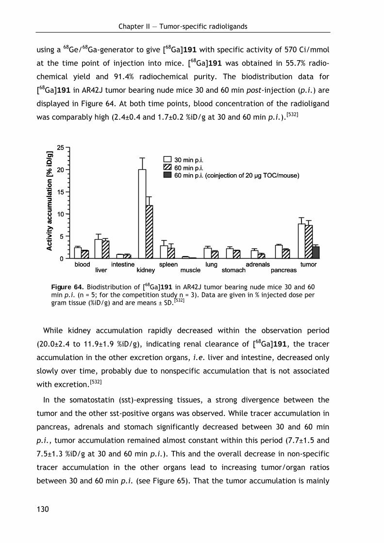
Chapter II — Tumor-specific radioligands
130
using a 68Ge/68Ga-generator to give [68Ga]191 with specific activity of 570 Ci/mmol
at the time point of injection into mice. [68Ga]191 was obtained in 55.7% radio-
chemical yield and 91.4% radiochemical purity. The biodistribution data for
[68Ga]191 in AR42J tumor bearing nude mice 30 and 60 min post-injection (p.i.) are
displayed in Figure 64. At both time points, blood concentration of the radioligand
was comparably high (2.4±0.4 and 1.7±0.2 %iD/g at 30 and 60 min p.i.).[532]
bloodliver
intestinekidney
spleenmuscle
lungstomach
adrenalspancreas
tumor0
5
10
15
20
25
Act
ivity
accu
mul
atio
n[%
iD/g
]
30 min p.i.60 min p.i.60 min p.i. (coinjection of 20 µg TOC/mouse)
bloodliver
intestinekidney
spleenmuscle
lungstomach
adrenalspancreas
tumor0
5
10
15
20
25
Act
ivity
accu
mul
atio
n[%
iD/g
]
30 min p.i.60 min p.i.60 min p.i. (coinjection of 20 µg TOC/mouse)
Figure 64. Biodistribution of [68Ga]191 in AR42J tumor bearing nude mice 30 and 60 min p.i. (n = 5; for the competition study n = 3). Data are given in % injected dose per gram tissue (%iD/g) and are means ± SD.[532]
While kidney accumulation rapidly decreased within the observation period
(20.0±2.4 to 11.9±1.9 %iD/g), indicating renal clearance of [68Ga]191, the tracer
accumulation in the other excretion organs, i.e. liver and intestine, decreased only
slowly over time, probably due to nonspecific accumulation that is not associated
with excretion.[532]
In the somatostatin (sst)-expressing tissues, a strong divergence between the
tumor and the other sst-positive organs was observed. While tracer accumulation in
pancreas, adrenals and stomach significantly decreased between 30 and 60 min
p.i., tumor accumulation remained almost constant within this period (7.7±1.5 and
7.5±1.3 %iD/g at 30 and 60 min p.i.). This and the overall decrease in non-specific
tracer accumulation in the other organs lead to increasing tumor/organ ratios
between 30 and 60 min p.i. (see Figure 65). That the tumor accumulation is mainly
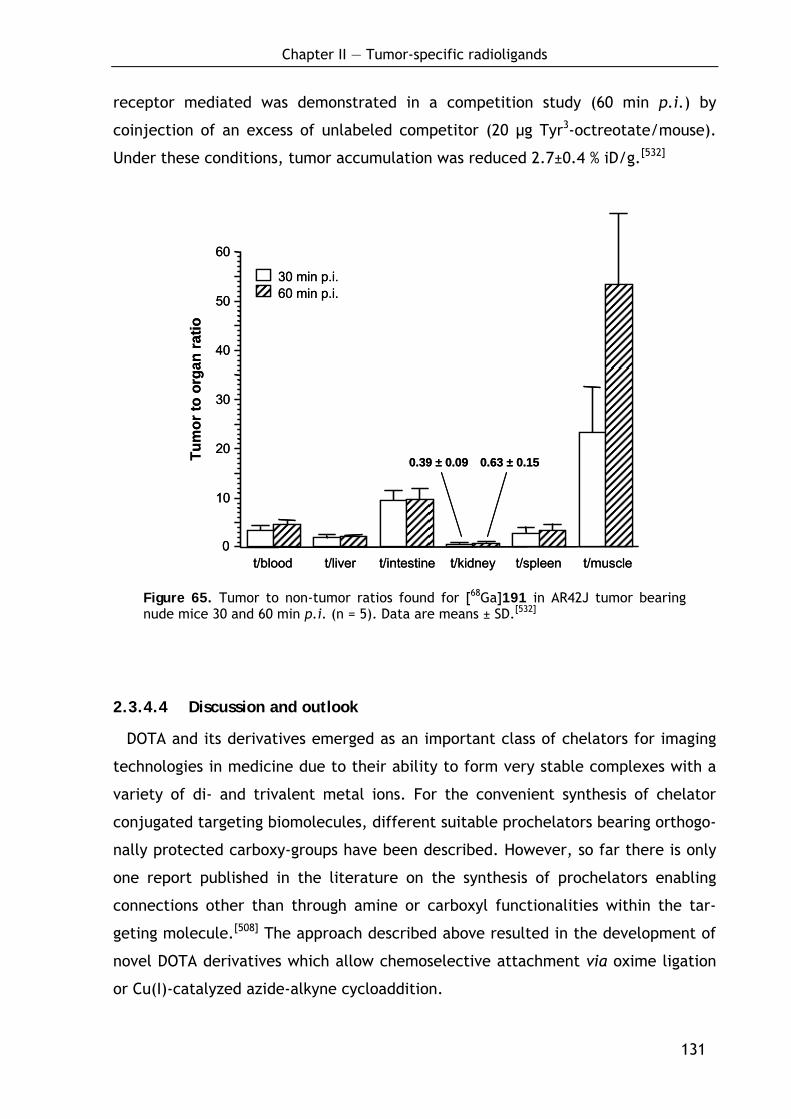
Chapter II — Tumor-specific radioligands
131
receptor mediated was demonstrated in a competition study (60 min p.i.) by
coinjection of an excess of unlabeled competitor (20 µg Tyr3-octreotate/mouse).
Under these conditions, tumor accumulation was reduced 2.7±0.4 % iD/g.[532]
t/blood t/liver t/intestine t/kidney t/spleen t/muscle0
10
20
30
40
50
60
30 min p.i.60 min p.i.
Tum
or to
org
anra
tio
0.39 ± 0.09 0.63 ± 0.15
t/blood t/liver t/intestine t/kidney t/spleen t/muscle0
10
20
30
40
50
60
30 min p.i.60 min p.i.
Tum
or to
org
anra
tio
0.39 ± 0.09 0.63 ± 0.15
Figure 65. Tumor to non-tumor ratios found for [68Ga]191 in AR42J tumor bearing nude mice 30 and 60 min p.i. (n = 5). Data are means ± SD.[532]
2.3.4.4 Discussion and outlook
DOTA and its derivatives emerged as an important class of chelators for imaging
technologies in medicine due to their ability to form very stable complexes with a
variety of di- and trivalent metal ions. For the convenient synthesis of chelator
conjugated targeting biomolecules, different suitable prochelators bearing orthogo-
nally protected carboxy-groups have been described. However, so far there is only
one report published in the literature on the synthesis of prochelators enabling
connections other than through amine or carboxyl functionalities within the tar-
geting molecule.[508] The approach described above resulted in the development of
novel DOTA derivatives which allow chemoselective attachment via oxime ligation
or Cu(I)-catalyzed azide-alkyne cycloaddition.
Chapter II — Tumor-specific radioligands
132
For the synthesis of the new compounds, a monoalkylation of cyclen had to be
accomplished to obtain 181, 182 and 188. In the literature, there are several
examples of similar monoalkylations where high yields of monoalkylated product
were achieved by applying cyclen in great excess (e.g. 2 equiv[487] or 5 equiv[499])
and where the yields are calculated based on the amount of alkylating agents. As a
result of cyclen being the most expensive reagent in the sequence, almost equi-
molar amounts of the alkylating agent were used in the synthesis to obtain satisfy-
ing yields of approximately 70% for the three compounds. Although the crude
product could be directly further alkylated, purification of this intermediate was
carried out via flash chromatography at this stage, as it was easier to separate the
monoalkylated product from higher alkylated products and unreacted cyclen,
rather than separation after peralkylation, where the resulting compounds only
differ in the nature of the alkyl substituent. It should also be noted that in general
separation of cyclen derivatives on silica is sometimes quite laborious due to
diffuse bands and that purification by preparative RP-HPLC is a good alternative.
The syntheses of the α-bromo esters 179, 180 and 187 were straightforward and
the products were obtained in good yields.
For the Cu(I)-catalyzed azide-alkyne cycloaddition[509,512,526-531] 1.2 equivalents of
copper sulfate together with an excess of copper metal were used, which leads to
in situ generation of the catalytically active Cu(I) species. Although there are
reports[551] where only 1 mol% of Cu(II) salts are used, an excess had to be used,
given the fact that one equivalent of the Cu(II) is immediately complexed with the
DOTA residue. However, in the literature there is a plethora of different proce-
dures[551] and for this purposes, optimization of the catalyst loading was not essen-
tial as dissolved copper ions could be precipitated and filtered off by addition of
sodium sulfide. Using Tyr3-octreotate as a model compound, the conjugation by
Cu(I)-catalyzed azide-alkyne cycloaddition was shown to be applicable with a broad
range of functionalities. As the only side reaction, an opening of disulfide bridge
was found. However, this causes no problem, as the reduced DOTA conjugate is
smoothly recyclized in quantitative yield. Nevertheless, this side reaction should be
kept in mind when planning to synthesize a disulfide bridged conjugate. Of course,
it can be avoided if the linear precursor peptide is used for conjugation and the
disulfide bridge is formed afterwards. Hence, the alkyne derivatized chelator offers
Chapter II — Tumor-specific radioligands
133
a promising alternative for attachment of BFCAs in a highly chemoselective manner
as demonstrated by reaction with model peptides 197 and 199. It is worth
mentioning that Prasuhn et al. concurrently reports an alternative approach for
DOTA conjugation via azide-alkyne cycloaddition.[552]
The oxime ligation of the new methyl ketone functionalized chelator 189 with
unprotected N-terminally aminooxy-functionalized Tyr3-octreotate proceeded
smoothly and without byproducts, thereby adding another example of the
chemoselective condensation of a methyl ketone and a hydroxylamine. In compari-
son to the recently presented aminooxy-functionalized DOTA-derivatives,[508] the
inverse approach presented here allows the introduction of the chelator at any
position in a peptide sequence, since various appropriate aminooxy functionalized
building blocks for SPPS are commercially available (e.g. (Boc-aminooxy)-acetic
acid, Boc-Ams(Fmoc)-OH and Fmoc-Dpr(Boc-Aoa)-OH).
As a first proof of principle, the 68Ga-labeled derivative of the oxime linked DOTA
conjugate 191 was used in an in vivo experiment. AR42J tumor bearing nude mice
were treated with 20 µg [68Ga]191 per mouse and investigated for tumor accumula-
tion at 30 and 60 min p.i.. The results exhibit good contrast and high accumulation
in the tumor (see Figure 64).
2.3.4.5 Conclusion
The novel alkyne and keto-functionalized DOTA derivatives described above allow
a facile and chemoselective conjugation with polyfunctionalized compounds. With
regard to the synthesis of the new modified chelators, economical straightforward
procedures were developed avoiding complicated protection group chemistry
considering the final application of the BFCA. This allows easy access to labeled
compounds in few synthetic steps. Both the alkyne as well as the methylketone-
functionalized DOTA derivatives proved to react selectively in the corresponding
conjugation with N-terminally modified Tyr3-octreotate. Furthermore, the pharma-
cokinetics of [68Ga]191 in AR42J tumor bearing nude mice demonstrate the suit-
ability of the chemoselective BFC-conjugation approach for the synthesis of new
radiometallated peptide ligands for diagnostic and therapeutic in vivo applications.

Chapter III — Cross-linked polymers as media for partial alignment
135
— Chapter 3 —
3 Development and Optimization of Novel
Cross-Linked Polymers as Media for
Measurement of Residual Dipolar Couplings
3.1 Background
3.1.1 Alignment media for measurement of residual dipolar couplings –
introduction and motivation
Magnetically active nuclei are magnetic dipols and hence interact in molecules
through space. These dipolar couplings contain important structural information
about the distance between two atoms and the angle between this pair of atoms
and a static magnetic field. In solid state NMR, where molecules are highly ori-
ented, such couplings are in the order of several kHz and cause immense numbers
of splittings. In solution, in contrast, the Brownian motion averages out all dipolar
couplings, resulting in the narrow line width of high resolution NMR-spectroscopy
but also in a significant loss in potential structural information. Therefore, an
intermediate state between solid and liquid is desirable in which the narrow line
width is maintained and anisotropic interactions are measurable, but significantly
reduced in size compared to solid state NMR. This can be achieved by a so-called
alignment medium in which the molecules are just partially aligned but show
almost free rotation. In such media the huge dipolar couplings observed in solid-
Chapter III — Cross-linked polymers as media for partial alignment
136
state are reduced to only a few Hz and therefore called residual dipolar couplings
(RDCs).[553-559] This has already advanced to a standard technique in high resolution
NMR of biomacromolecules and numerous aqueous alignment media have been de-
scribed, such as lipid bicelles,[558,560] stretched polyacrylamide gels,[561-564] filamen-
tous phage[565] and other liquid crystalline phases[566,567] are available. However,
quite recently this technique started to find application in the field of small
molecule NMR.[568-580]
A limiting step for the application of RDCs to small molecules is still the avail-
ability of alignment media for organic solvents. Liquid crystalline phases such as
poly-γ-benzyl-L-glutamate (PBLG)[573,574,581-584] are known to align organic molecules
in CDCl3 and similar apolar organic solvents. Liquid crystals, however, have the
disadvantage that for the phase transition a minimum concentration is needed and
therefore a minimum anisotropy is induced in the sample. The development of
specially designed crystalline phases with lower minimum alignment, as shown in
the case of 4-n-pentyl-4’-cyanobiphenyl (PCBP)[585] and poly-γ-ethyl-L-glutamate
(PELG),[572] improves the applicability of such systems.
A second approach for partial alignment is strain induced alignment in a gel (SAG)
as shown by the pioneering work of DeLoche and Samulski[586] and many follow up
applications in polymer NMR spectroscopy.[587-589] This technique is not limited by a
minimum anisotropy and it was extensively used to study the properties of polymer
gels by means of high resolution deuterium NMR.[590] Nevertheless, gel alignment
was only lately used to induce RDCs in molecules dissolved in the gel.[561,562] SAG
allows the unrestricted scaling of alignment over a wide range and can be used for
aqueous as well as organic solvents, depending on the polymer used.[563,579,580,591,592]
As a first example in organic solvents, RDC measurements in stretched cross-linked
polystyrene (PS) gels swollen in CDCl3 were reported by B. Luy from our group as a
promising alignment method.[579]
After the proof of principle, the aim of this study was to optimize the procedure
to control the alignment and to widen the scope of applicable solvents by devel-
opment of novel polymeric media. The focus here was on materials available by
radical polymerization. A polymerization technique was worked out allowing the
production of long sticks which can be cut into peaces suitable for the NMR appli-
cation and whose diameter can be easily controlled.[593] Moreover, the amounts of

Chapter III — Cross-linked polymers as media for partial alignment
137
cross-linking agent and radical starter were varied to adopt the swelling properties
of the polymers. As PS-sticks are mainly swellable in relatively unpolar organic
solvents like CHCl3 or benzene, a major goal was to find suitable polymers for polar
organic solvent like CH3CN, MeOH or DMSO. This was successfully achieved with
cross-linked poly(vinyl acetate) (PVA).[594]
3.1.2 The dipolar interaction
A simplistic picture for the dipolar interaction is described by Luy et al.:[595] Spins
can be illustrated as magnets with an inherent rotation at the Larmor frequency.
Although spins are not oriented directly along the static magnetic field B0, the av-
eraging over time of the fast rotating magnets yields a resulting magnetic moment
parallel or antiparallel to B0 (Figure 66a, b).
Figure 66. Illustration of the dipolar interaction. (a, b) The magnetic field induced by spin I adds up to the static magnetic field B0 and leads to a shift of the resonance frequency of the close-by spin S. Since spins parallel and antiparallel to the magnetic field are almost equally distributed, this leads to a splitting with the dipolar coupling DIS (c). The (3 cos2 Θ-1) angular dependence is shown in (d).[595]
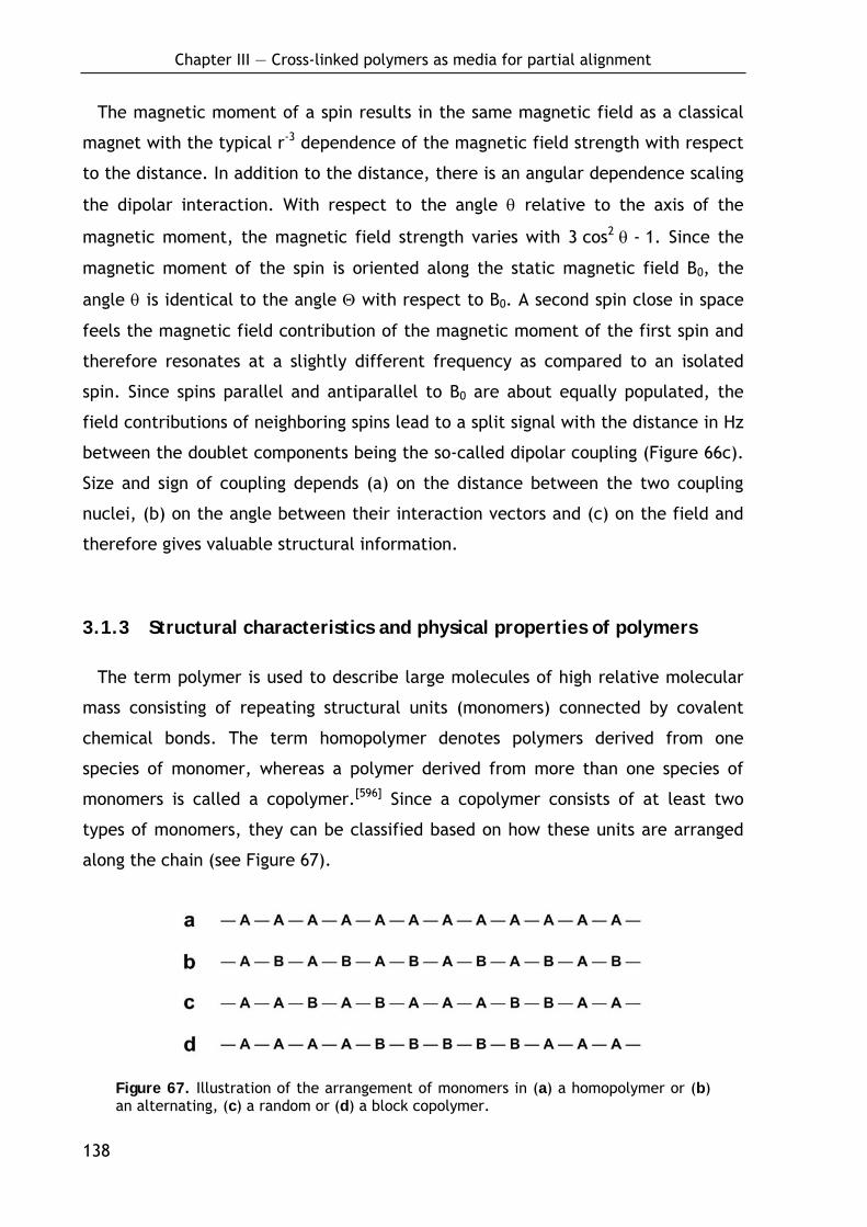
Chapter III — Cross-linked polymers as media for partial alignment
138
The magnetic moment of a spin results in the same magnetic field as a classical
magnet with the typical r-3 dependence of the magnetic field strength with respect
to the distance. In addition to the distance, there is an angular dependence scaling
the dipolar interaction. With respect to the angle θ relative to the axis of the
magnetic moment, the magnetic field strength varies with 3 cos2 θ - 1. Since the
magnetic moment of the spin is oriented along the static magnetic field B0, the
angle θ is identical to the angle Θ with respect to B0. A second spin close in space
feels the magnetic field contribution of the magnetic moment of the first spin and
therefore resonates at a slightly different frequency as compared to an isolated
spin. Since spins parallel and antiparallel to B0 are about equally populated, the
field contributions of neighboring spins lead to a split signal with the distance in Hz
between the doublet components being the so-called dipolar coupling (Figure 66c).
Size and sign of coupling depends (a) on the distance between the two coupling
nuclei, (b) on the angle between their interaction vectors and (c) on the field and
therefore gives valuable structural information.
3.1.3 Structural characteristics and physical properties of polymers
The term polymer is used to describe large molecules of high relative molecular
mass consisting of repeating structural units (monomers) connected by covalent
chemical bonds. The term homopolymer denotes polymers derived from one
species of monomer, whereas a polymer derived from more than one species of
monomers is called a copolymer.[596] Since a copolymer consists of at least two
types of monomers, they can be classified based on how these units are arranged
along the chain (see Figure 67).
Figure 67. Illustration of the arrangement of monomers in (a) a homopolymer or (b) an alternating, (c) a random or (d) a block copolymer.
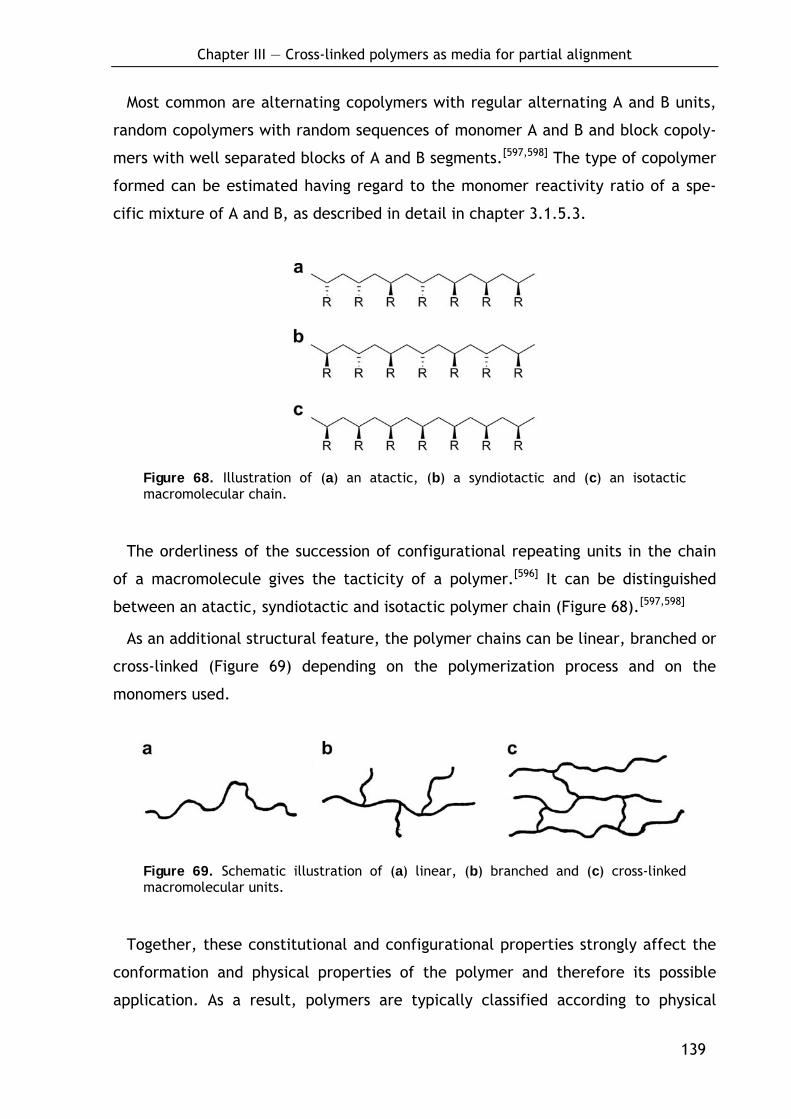
Chapter III — Cross-linked polymers as media for partial alignment
139
Most common are alternating copolymers with regular alternating A and B units,
random copolymers with random sequences of monomer A and B and block copoly-
mers with well separated blocks of A and B segments.[597,598] The type of copolymer
formed can be estimated having regard to the monomer reactivity ratio of a spe-
cific mixture of A and B, as described in detail in chapter 3.1.5.3.
Figure 68. Illustration of (a) an atactic, (b) a syndiotactic and (c) an isotactic macromolecular chain.
The orderliness of the succession of configurational repeating units in the chain
of a macromolecule gives the tacticity of a polymer.[596] It can be distinguished
between an atactic, syndiotactic and isotactic polymer chain (Figure 68).[597,598]
As an additional structural feature, the polymer chains can be linear, branched or
cross-linked (Figure 69) depending on the polymerization process and on the
monomers used.
Figure 69. Schematic illustration of (a) linear, (b) branched and (c) cross-linked macromolecular units.
Together, these constitutional and configurational properties strongly affect the
conformation and physical properties of the polymer and therefore its possible
application. As a result, polymers are typically classified according to physical

Chapter III — Cross-linked polymers as media for partial alignment
140
behavior into three main groups: thermoplastics, thermosetting polymers (thermo-
sets) and elastomers.
Thermoplastics are plastic or deformable materials which consist of linear or
branched macromolecular chains. The chains are associated through van der Waals
forces (e.g. polyethylene or polypropylene), dipole-dipole interactions and hydro-
gen bonding (nylon) or stacking of aromatic rings (polystyrene). Therefore, such
materials normally melt to a liquid when heated and dissolve readily in appropriate
solvents (e.g. toluene for polystyrene).[597,598]
Thermosetting polymers are heavily cross-linked materials and therefore gener-
ally stronger than thermoplastic materials due the 3-D network of bonds. Thermo-
sets, once formed and cured, can never be remelted and remolded again and are
insoluble.[597,598]
Elastomers are slightly cross-linked thermoplastics or thermosets that can be
stretched and then return to its original shape without permanent deformation.
This is due to the loose network in which the macromolecular chains are hold
together by single cross-linkages: If a stress is applied, the polymer chains are
stretched out and the covalent cross-linkages ensure that the elastomer will return
to its original configuration when the stress is removed (Figure 70). Without the
cross-linkages the applied stress would result in a permanent deformation.
Generally, such materials swell in appropriate solvents but do not dissolve.[597,598]
Figure 70. Schematic drawing of an elastomer. (a) Unstressed polymer; the dots denote cross-links. (b) The same polymer under stress. When the stress is removed, it will return to the (a) configuration.
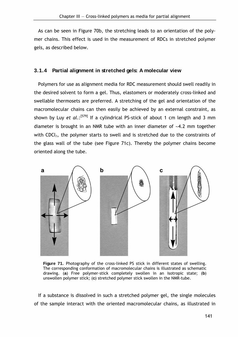
Chapter III — Cross-linked polymers as media for partial alignment
141
As can be seen in Figure 70b, the stretching leads to an orientation of the poly-
mer chains. This effect is used in the measurement of RDCs in stretched polymer
gels, as described below.
3.1.4 Partial alignment in stretched gels: A molecular view
Polymers for use as alignment media for RDC measurement should swell readily in
the desired solvent to form a gel. Thus, elastomers or moderately cross-linked and
swellable thermosets are preferred. A stretching of the gel and orientation of the
macromolecular chains can then easily be achieved by an external constraint, as
shown by Luy et al.:[579] If a cylindrical PS-stick of about 1 cm length and 3 mm
diameter is brought in an NMR tube with an inner diameter of ∼4.2 mm together
with CDCl3, the polymer starts to swell and is stretched due to the constraints of
the glass wall of the tube (see Figure 71c). Thereby the polymer chains become
oriented along the tube.
Figure 71. Photography of the cross-linked PS stick in different states of swelling. The corresponding conformation of macromolecular chains is illustrated as schematic drawing. (a) Free polymer-stick completely swollen in an isotropic state; (b) unswollen polymer stick; (c) stretched polymer stick swollen in the NMR-tube.
If a substance is dissolved in such a stretched polymer gel, the single molecules
of the sample interact with the oriented macromolecular chains, as illustrated in
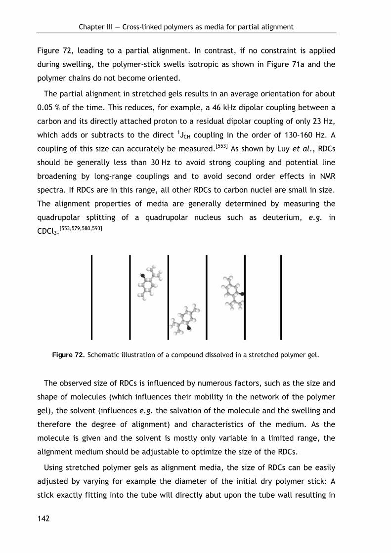
Chapter III — Cross-linked polymers as media for partial alignment
142
Figure 72, leading to a partial alignment. In contrast, if no constraint is applied
during swelling, the polymer-stick swells isotropic as shown in Figure 71a and the
polymer chains do not become oriented.
The partial alignment in stretched gels results in an average orientation for about
0.05 % of the time. This reduces, for example, a 46 kHz dipolar coupling between a
carbon and its directly attached proton to a residual dipolar coupling of only 23 Hz,
which adds or subtracts to the direct 1JCH coupling in the order of 130-160 Hz. A
coupling of this size can accurately be measured.[553] As shown by Luy et al., RDCs
should be generally less than 30 Hz to avoid strong coupling and potential line
broadening by long-range couplings and to avoid second order effects in NMR
spectra. If RDCs are in this range, all other RDCs to carbon nuclei are small in size.
The alignment properties of media are generally determined by measuring the
quadrupolar splitting of a quadrupolar nucleus such as deuterium, e.g. in
CDCl3.[553,579,580,593]
Figure 72. Schematic illustration of a compound dissolved in a stretched polymer gel.
The observed size of RDCs is influenced by numerous factors, such as the size and
shape of molecules (which influences their mobility in the network of the polymer
gel), the solvent (influences e.g. the salvation of the molecule and the swelling and
therefore the degree of alignment) and characteristics of the medium. As the
molecule is given and the solvent is mostly only variable in a limited range, the
alignment medium should be adjustable to optimize the size of the RDCs.
Using stretched polymer gels as alignment media, the size of RDCs can be easily
adjusted by varying for example the diameter of the initial dry polymer stick: A
stick exactly fitting into the tube will directly abut upon the tube wall resulting in
Chapter III — Cross-linked polymers as media for partial alignment
143
a strong stretching during swelling. In contrast, a stick of equal composition but
smaller in diameter will swell isotropic until it reaches the glass wall. As a result,
the degree of orientation will be less. Hence, the degree of orientation signifi-
cantly depends on the ratio of stick-diameter and the inner diameter of the NMR
tube. Moreover, the polymerization conditions and the composition of the polymer
itself influence the degree of alignment. It is evident, that the higher the cross-
linking and therefore the cohesion of polymer chains, the less the swelling and
orientation of the polymer chains will be. However, a tighter network causes also
stronger interactions between the polymer chains and the embedded sample mole-
cules which increases the degree of orientation. The polymerization conditions, in
particular the reaction temperature, additionally affect the polymer’s characteris-
tics by influencing its constitution (lower reaction temperatures lead to the forma-
tion of longer polymer chains, as described below).
Beside the described facile procedure to stretch gels, there are a variety of other
techniques reported in the literature, ranging from drying on a glass capillary
before reswelling,[562] gel compression with a Shigemi plunger,[599] to apparatuses
for squeezing a readily swollen gel into an NMR-tube[564] or stretching gelatin inside
a rubber tube.[600] The focus of this work, however, was on the controlled produc-
tion of sticks for alignment by swelling in NMR tubes, as this is a very simplistic and
easily reproducible method requiring no additional tools such as a teflon-funnel.[564]
3.1.5 Structure and preparation of polymers
3.1.5.1 Classification of polymerization reactions
Polymerization reactions are usually classified in two main groups according to
their mechanism: the step-growth polymerizations and the chain-growth polymeri-
zations.
The step-growth polymerization occurs by consecutive reactions in which the
degree of polymerization and average molecular weight of the polymer increase as
the reaction proceeds.[596] As a major characteristic, in a step-growth polymeriza-
tion the growth of polymer chains proceeds by reactions between molecules of all
degrees of polymerization. Examples for this kind of polyreactions are polyaddi-
tions (e.g. polyurethanes formed from isocyanates and alcohols as bifunctional
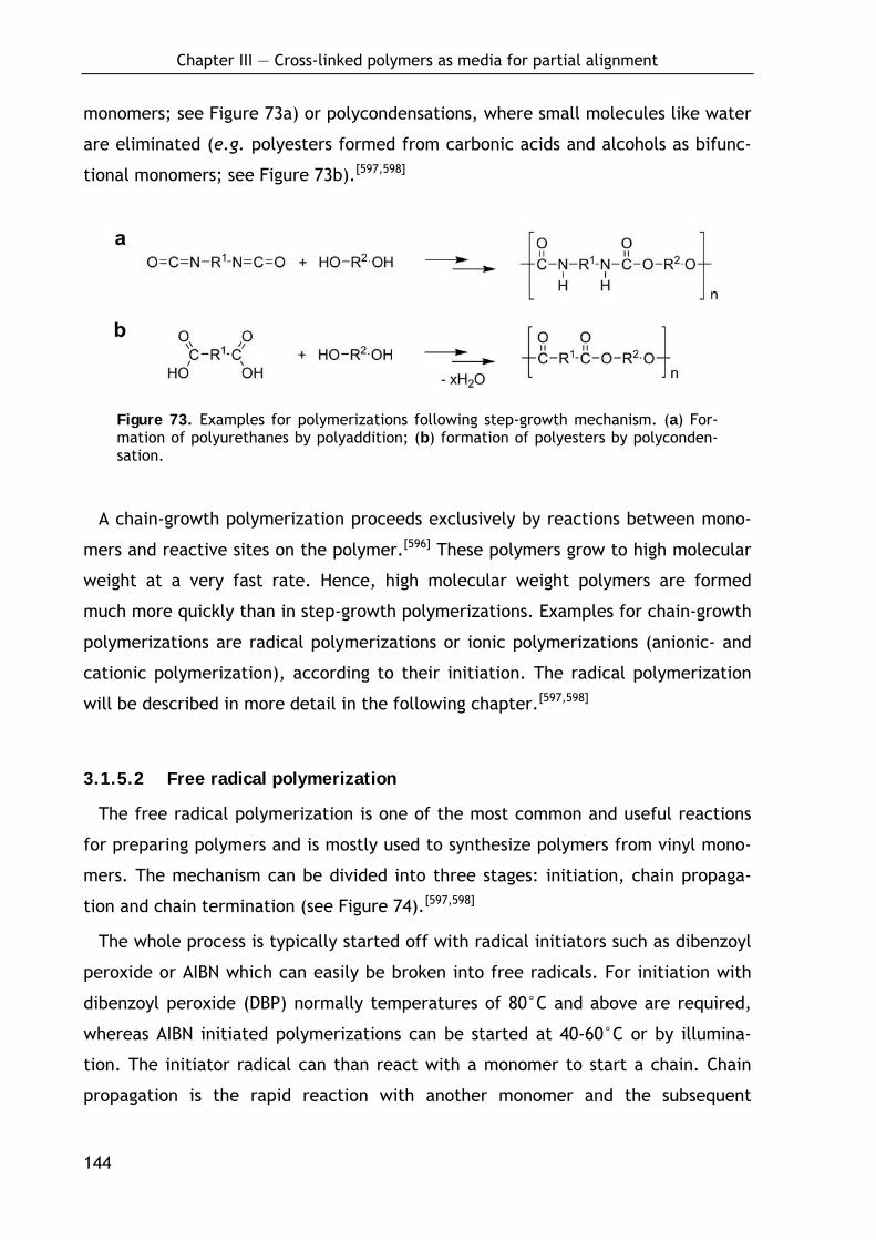
Chapter III — Cross-linked polymers as media for partial alignment
144
monomers; see Figure 73a) or polycondensations, where small molecules like water
are eliminated (e.g. polyesters formed from carbonic acids and alcohols as bifunc-
tional monomers; see Figure 73b).[597,598]
Figure 73. Examples for polymerizations following step-growth mechanism. (a) For-mation of polyurethanes by polyaddition; (b) formation of polyesters by polyconden-sation.
A chain-growth polymerization proceeds exclusively by reactions between mono-
mers and reactive sites on the polymer.[596] These polymers grow to high molecular
weight at a very fast rate. Hence, high molecular weight polymers are formed
much more quickly than in step-growth polymerizations. Examples for chain-growth
polymerizations are radical polymerizations or ionic polymerizations (anionic- and
cationic polymerization), according to their initiation. The radical polymerization
will be described in more detail in the following chapter.[597,598]
3.1.5.2 Free radical polymerization
The free radical polymerization is one of the most common and useful reactions
for preparing polymers and is mostly used to synthesize polymers from vinyl mono-
mers. The mechanism can be divided into three stages: initiation, chain propaga-
tion and chain termination (see Figure 74).[597,598]
The whole process is typically started off with radical initiators such as dibenzoyl
peroxide or AIBN which can easily be broken into free radicals. For initiation with
dibenzoyl peroxide (DBP) normally temperatures of 80°C and above are required,
whereas AIBN initiated polymerizations can be started at 40-60°C or by illumina-
tion. The initiator radical can than react with a monomer to start a chain. Chain
propagation is the rapid reaction with another monomer and the subsequent
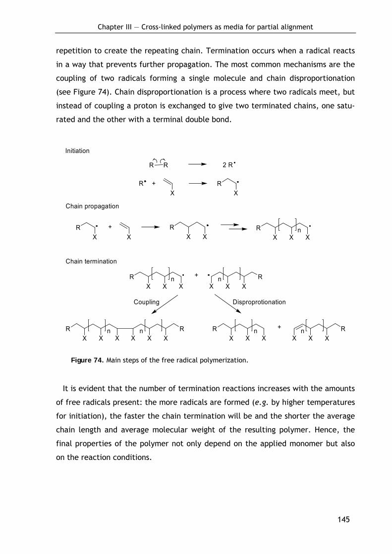
Chapter III — Cross-linked polymers as media for partial alignment
145
repetition to create the repeating chain. Termination occurs when a radical reacts
in a way that prevents further propagation. The most common mechanisms are the
coupling of two radicals forming a single molecule and chain disproportionation
(see Figure 74). Chain disproportionation is a process where two radicals meet, but
instead of coupling a proton is exchanged to give two terminated chains, one satu-
rated and the other with a terminal double bond.
R R 2 R
R +X X
R
XR +
X XR
X XR
X Xn
XR
X Xn
XR
XXn+
XR
X Xn
XR
XXn
XR
X Xn
XR
XXn+
DisproprotionationCoupling
Initiation
Chain propagation
Chain termination
Figure 74. Main steps of the free radical polymerization.
It is evident that the number of termination reactions increases with the amounts
of free radicals present: the more radicals are formed (e.g. by higher temperatures
for initiation), the faster the chain termination will be and the shorter the average
chain length and average molecular weight of the resulting polymer. Hence, the
final properties of the polymer not only depend on the applied monomer but also
on the reaction conditions.
Chapter III — Cross-linked polymers as media for partial alignment
146
3.1.5.3 The copolymer composition equation
Staudinger 1930 found that the composition of a copolymer alters during poly-
merization. Starting with an equimolar mixture of vinyl chloride and vinyl acetate,
the ratio of chloride to acetate in the copolymer changed from 9:3 to 5:7 in the
course of the reaction.[598] This shift in the composition is a result of the diverse
reactivity of different types of monomer with each other which leads to a changing
composition of the comonomer feed from which the copolymer is produced.
The reaction is a typical example for a copolymerization of two monomers A
(vinyl chloride) and B (vinyl acetate), whereby A reacts preferably with radicals A˙
at the propagating chain ending (see below) and also B reacts preferable with
radicals A˙ at the propagating chain ending. As a consequence, at the beginning of
the reaction larger amounts of A are incorporated in the growing copolymer, while
the source solution gets enriched in B. When a specific excess of B is reached, the
introduction of B is favored as a result of lacking A. The mutual reactivity of the
monomers finally defines whether a random, alternating or block polymer is
formed.
In case of copolymerization of two monomers A and B, two types of propagating
species are possible, one with A at the propagating end and the other with B.
Assuming furthermore that the reactivity of the propagating species is dependent
only on the monomer unit at the end of the chain,[597,601-603] four propagation
reactions are possible:
A
A
B
B
+ A
+ B
+ A
+ B
kAA
kAB
kBA
kBB
A
A
B
B
Monomers A and B can each add either to a propagating chain ending in A or to one
ending in B.[597] kAA is the rate constant for a propagating chain ending in A adding
to monomer A, kAB that for a propagating chain ending in A adding to monomer B
and so on. The parameters rA and rB, which are defined by
Chapter III — Cross-linked polymers as media for partial alignment
147
rA = kAA/kAB and rB = kBB/kBA
are termed the monomer reactivity ratios. Each r is the ratio of the rate constant
for a reactive propagating species adding the own type of monomer to the rate
constant for its addition of the other monomer. The tendency of two monomers to
copolymerize is noted by r values between zero and unity. A rA value greater than
unity means that A• preferentially adds A instead of B while an rA value less than
unity means that A• preferentially adds B. An rA value of zero would mean that A is
incapable of undergoing homopolymerization.[597,598] The so-called copolymerization
equation or also termed the copolymer composition equation is given as:
d[A]/d[B] = (1+rA[A]/[B])/(1+rB[B]/[A]).
The copolymer composition, d[A]/d[B], is the molar ratio of the two monomer units
in the copolymer and is expressed by above equation as being related to the con-
centrations of the two monomers in the feed, [A] and [B] and the parameters rA
and rB.
Different types of copolymerization behavior are observed depending on the
values[604] of the monomer reactivity ratios. Copolymerizations can be classified
into three types based on whether the product of the two monomer reactivity
ratios rArB is 1, < 1 or > 1:
• rArB = 1:
If the product rArB is 1, the reaction is termed ideal copolymerization. In
the special case rA ≈ rB ≈ 1, the two monomers show equal reactivities
towards both propagating species leading to a random copolymer. If the
two monomer reactivity ratios are different (rA > 1 and rB < 1 or rA < 1 and
rB > 1), one of the monomers is more reactive than the other towards both
propagating species. In such cases, the copolymer will contain a larger
proportion of the more reactive monomer in random placement.[597,598]
• rArB < 1:
If rArB < 1, in each case the reaction of the propagating species with a
monomer of the other species is preferred leading to short segments of A
and B. In the special case rA = rB = 0, an alternating copolymer is formed.
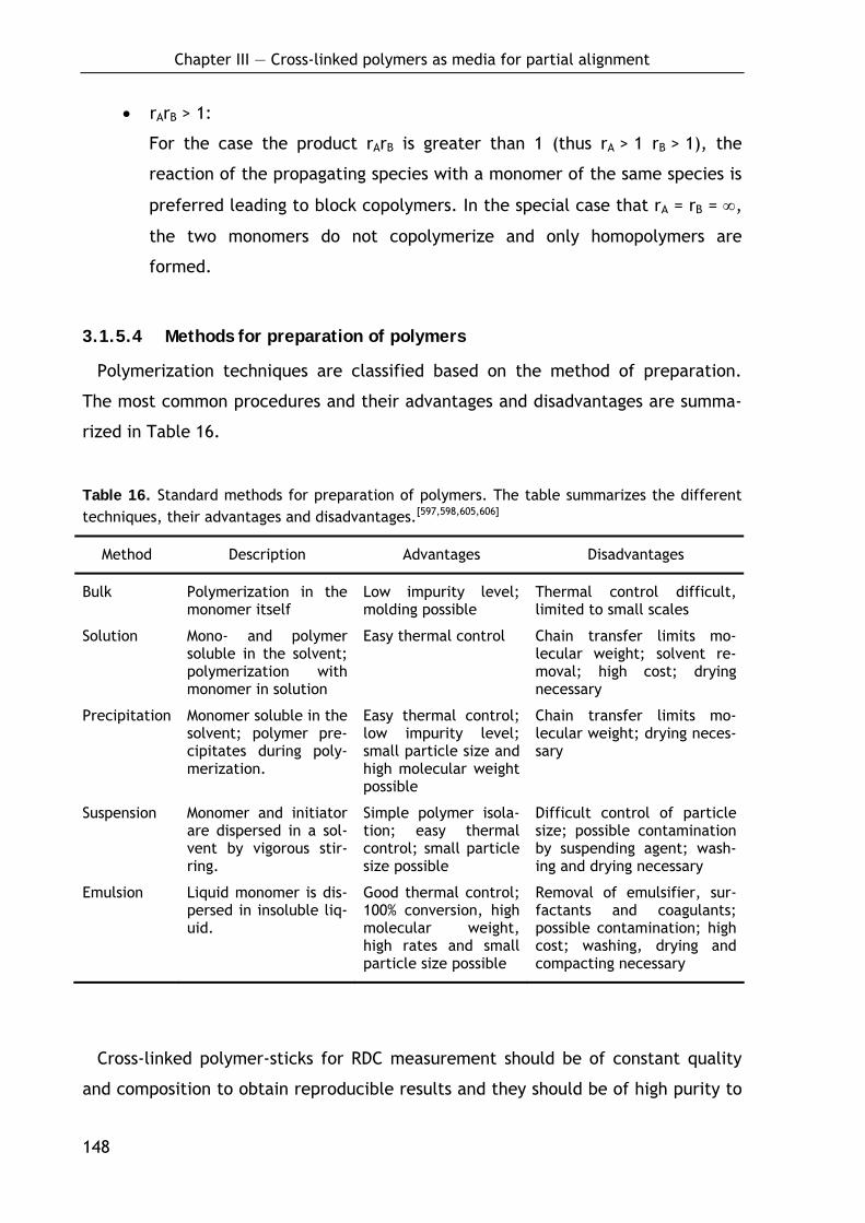
Chapter III — Cross-linked polymers as media for partial alignment
148
• rArB > 1:
For the case the product rArB is greater than 1 (thus rA > 1 rB > 1), the
reaction of the propagating species with a monomer of the same species is
preferred leading to block copolymers. In the special case that rA = rB = ∞,
the two monomers do not copolymerize and only homopolymers are
formed.
3.1.5.4 Methods for preparation of polymers
Polymerization techniques are classified based on the method of preparation.
The most common procedures and their advantages and disadvantages are summa-
rized in Table 16.
Table 16. Standard methods for preparation of polymers. The table summarizes the different techniques, their advantages and disadvantages.[597,598,605,606]
Method Description Advantages Disadvantages
Bulk Polymerization in the monomer itself
Low impurity level; molding possible
Thermal control difficult, limited to small scales
Solution Mono- and polymer soluble in the solvent; polymerization with monomer in solution
Easy thermal control Chain transfer limits mo-lecular weight; solvent re-moval; high cost; drying necessary
Precipitation Monomer soluble in the solvent; polymer pre-cipitates during poly-merization.
Easy thermal control; low impurity level; small particle size and high molecular weight possible
Chain transfer limits mo-lecular weight; drying neces-sary
Suspension Monomer and initiator are dispersed in a sol-vent by vigorous stir-ring.
Simple polymer isola-tion; easy thermal control; small particle size possible
Difficult control of particle size; possible contamination by suspending agent; wash-ing and drying necessary
Emulsion Liquid monomer is dis-persed in insoluble liq-uid.
Good thermal control; 100% conversion, high molecular weight, high rates and small particle size possible
Removal of emulsifier, sur-factants and coagulants; possible contamination; high cost; washing, drying and compacting necessary
Cross-linked polymer-sticks for RDC measurement should be of constant quality
and composition to obtain reproducible results and they should be of high purity to
Chapter III — Cross-linked polymers as media for partial alignment
149
reduce background signals. As described above, the diameter of the sticks influ-
ences the degree of orientation of the polymer chains after swelling and therefore
the size of the RDCs. Hence, it is desirable to obtain sticks of defined and control-
lable diameter to easily adopt the degree of alignment. Therefore, the bulk polym-
erization was considered as the method of choice as it provides the opportunity to
control the diameter. Furthermore, no additives and solvents are necessary, hence,
reducing possible contaminants and avoiding drying after polymerization.
Chapter III — Cross-linked polymers as media for partial alignment
150
3.2 Synthesis and evaluation of cross-linked polymers
3.2.1 Poly(styrene-co-divinylbenzene) sticks (P1)
Styrene-divinylbenzene copolymer P1 (see Scheme 20) is widely used, e.g. as
basis for ion-exchange resins or resins for solid-phase peptide synthesis. As both
monomers are quite similar in their chemical properties with respect to radical
polymerization, the mixture shows an almost ideal copolymerization. The r values
for styrene (200) and divinylbenzene (201) are rA = 0.65 and rB = 1.1 (rArB = 0.72),
meaning that there is a slight preference of divinylbenzene to be introduced into
the growing chains. Initial experiments for the preparation of cross-linked PS-sticks
as media for measurement of RDCs have been reported by Luy et al.[579] In their
experiments styrene was copolymerized with technical divinylbenzene (DVB) to
achieve cross linking and DBP was used as radical starter at 80-120°C.
3.2.1.1 Preparation of P1-sticks by bulk polymerization
To obtain polymer sticks of reproducible composition and properties, the
procedure described above was optimized in several respects: The sticks were
prepared in glass tubes, one end of which was sealed by melting. The tubes were
carefully dried and the inside surface was silylated with a 1:1 mixture of chloro-
trimethylsilane and dichlorodimethylsilane to ensure apolar surfaces. This was done
to prevent the polymer from sticking to the glass wall which can cause fractures of
the stick. Furthermore, the monomers were purified before use to remove
contaminants. Hence, the monomers styrene 200 and divinylbenzene 201 were
filtered through basic aluminum oxides (pH 10) and distilled under reduced
pressure to remove stabilizer. Immediately before polymerization the monomers
were degassed for 15 minutes by ultrasound in vacuo and ventilated in an argon
atmosphere to remove oxygen. In addition, the radical starter dibenzoylperoxide
was substituted by AIBN. This allowed a controlled polymerization at much lower
temperatures (45°C). In this manner, a series of poly(styrene-co-divinylbenzene)
(P1) sticks with varying the amounts of radical starter (from 0.005 to 0.5%, w/v)

Chapter III — Cross-linked polymers as media for partial alignment
151
and cross linking agent (from 0.05 to 5%, v/v) were prepared in diameters of 1.6
mm, 2.4 mm, 3.4 mm and 4.0 mm (Scheme 20).[593]
Scheme 20. Preparation of poly(styrene-co-divinylbenzene) (P1).a
n
+
AIBN(0.005-0.5%)
0.05%-5.00%
99.95%-95.00%
n
ran
ran
200 201 P1
a Conditions: 45°C (5 d) then 60°C (2 d); ran: random.[593]
Table 17 shows a summary of prepared sticks. A clear tendency for the consis-
tency of the formed polymers was observed. At very low concentrations of radical
starter (0.05% AIBN, v/v) and cross-linking agent (0.005 and 0.01% DVB, w/v) the
material did not become solid. Rising the amounts starter or cross-linker resulted
first in a gummy then solid consistency. Surprisingly, at low to mediate concentra-
tions of cross-linker (in particular from 0.2-0.75% DVB, v/v) a formation of variable
amounts of bubbles was observed (see Table 17) which could not be overcome by
optimizing the reaction conditions, i.e. polymerization temperature, reaction time
or degasing. The combination of low amounts of cross-linking agent with low
amounts of AIBN (0.005-0.025%, w/v) even resulted in the formation of foams. In
contrast, sticks with DVB content of 1.0 % and greater were clear, transparent and
very hard.
From the prepared materials, the gummy sticks, which completely dissolved in
solvents and the foamy sticks, which are highly inhomogeneous, were unusable for
RDC measurement. From the sticks containing bubbles, only bubble-free parts
could be used. Nevertheless, the heavy formation of bubbles was found to be a
special issue for the production of P1-sticks, as similar problems did not occur for
any other material as described below.

Chapter III — Cross-linked polymers as media for partial alignment
152
Table 17. Composition and physical consistency of prepared poly(styrene-co-divinylbenzene) (P1) sticks (c.s. = clear solid).
Amounts of AIBN (%, w/v)
0.005 0.01 0.025 0.05 0.075 0.10 0.50
5.0 c.s. c.s. c.s. c.s. c.s. c.s. c.s.
3.0 c.s. c.s. c.s. c.s. c.s. c.s. c.s.
2.0 c.s. c.s. c.s. c.s. c.s. c.s. c.s.
1.5 c.s. c.s. c.s. c.s. c.s. c.s. c.s.
1.0 bubbles foamy c.s. c.s. c.s. c.s. c.s.
0.75 foamy c.s. bubbles bubbles c.s. bubbles c.s.
0.5 foamy foamy foamy bubbles bubbles bubbles c.s.
0.2 foamy bubbles foamy bubbles bubbles cloudy cloudy
0.1 gummy gummy c.s. cloudy c.s. cloudy cloudy Am
ount
s of
div
inyl
benz
ene
(%,
v/v)
0.05 liquid liquid gummy c.s. c.s. cloudy cloudy
3.2.1.2 Alignment properties
A point of major interest in the applicability of P1-gels is the range of solvents in
which molecules can be aligned. In line with the very low solubility of non-cross-
linked PS,[607] no swelling could be observed for very apolar solvents like octane
and relatively polar solvents like acetone or acetonitrile. However, CHCl3, DCM,
THF, benzene and dioxane showed significant swelling and were used for further
experiments. The NMR studies were performed by B. Luy and K. Kobzar.[593]
As it was shown previously,[579] the anisotropy induced by the gel is dependent on
the amount of cross linking agent used for polymerization. To repeat the study,
five samples per concentration were swollen in CDCl3 and quadrupolar deuterium
splittings were recorded in 5.0 mm NMR-tubes with an inner diameter of ~4.2 mm.
The resulting graph is shown in Figure 75a. Anisotropies corresponding to quadru-
polar deuterium splittings in the range of 0-540 Hz for P1-sticks with initial diame-
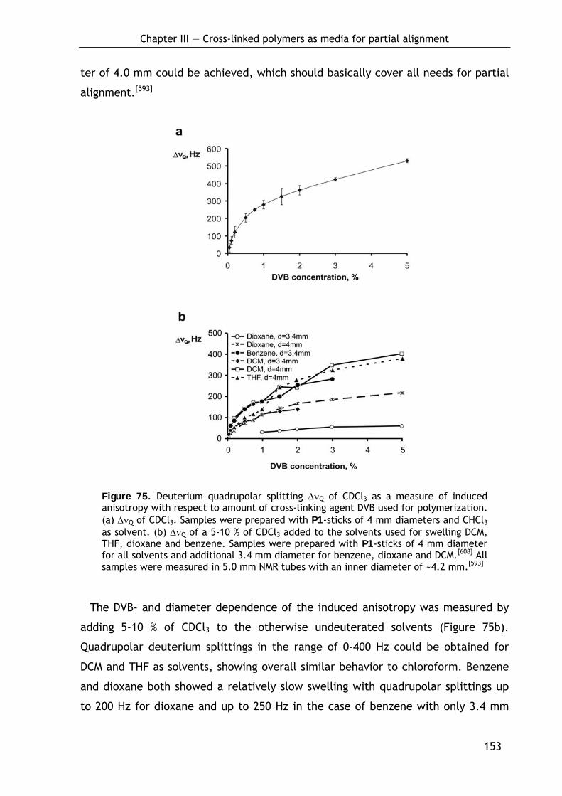
Chapter III — Cross-linked polymers as media for partial alignment
153
ter of 4.0 mm could be achieved, which should basically cover all needs for partial
alignment.[593]
Figure 75. Deuterium quadrupolar splitting ΔνQ of CDCl3 as a measure of induced anisotropy with respect to amount of cross-linking agent DVB used for polymerization. (a) ΔνQ of CDCl3. Samples were prepared with P1-sticks of 4 mm diameters and CHCl3 as solvent. (b) ΔνQ of a 5-10 % of CDCl3 added to the solvents used for swelling DCM, THF, dioxane and benzene. Samples were prepared with P1-sticks of 4 mm diameter for all solvents and additional 3.4 mm diameter for benzene, dioxane and DCM.[608] All samples were measured in 5.0 mm NMR tubes with an inner diameter of ~4.2 mm.[593]
The DVB- and diameter dependence of the induced anisotropy was measured by
adding 5-10 % of CDCl3 to the otherwise undeuterated solvents (Figure 75b).
Quadrupolar deuterium splittings in the range of 0-400 Hz could be obtained for
DCM and THF as solvents, showing overall similar behavior to chloroform. Benzene
and dioxane both showed a relatively slow swelling with quadrupolar splittings up
to 200 Hz for dioxane and up to 250 Hz in the case of benzene with only 3.4 mm
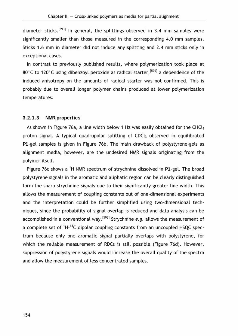
Chapter III — Cross-linked polymers as media for partial alignment
154
diameter sticks.[593] In general, the splittings observed in 3.4 mm samples were
significantly smaller than those measured in the corresponding 4.0 mm samples.
Sticks 1.6 mm in diameter did not induce any splitting and 2.4 mm sticks only in
exceptional cases.
In contrast to previously published results, where polymerization took place at
80°C to 120°C using dibenzoyl peroxide as radical starter,[579] a dependence of the
induced anisotropy on the amounts of radical starter was not confirmed. This is
probably due to overall longer polymer chains produced at lower polymerization
temperatures.
3.2.1.3 NMR properties
As shown in Figure 76a, a line width below 1 Hz was easily obtained for the CHCl3
proton signal. A typical quadrupolar splitting of CDCl3 observed in equilibrated
P1-gel samples is given in Figure 76b. The main drawback of polystyrene-gels as
alignment media, however, are the undesired NMR signals originating from the
polymer itself.
Figure 76c shows a 1H NMR spectrum of strychnine dissolved in P1-gel. The broad
polystyrene signals in the aromatic and aliphatic region can be clearly distinguished
form the sharp strychnine signals due to their significantly greater line width. This
allows the measurement of coupling constants out of one-dimensional experiments
and the interpretation could be further simplified using two-dimensional tech-
niques, since the probability of signal overlap is reduced and data analysis can be
accomplished in a conventional way.[593] Strychnine e.g. allows the measurement of
a complete set of 1H-13C dipolar coupling constants from an uncoupled HSQC spec-
trum because only one aromatic signal partially overlaps with polystyrene, for
which the reliable measurement of RDCs is still possible (Figure 76d). However,
suppression of polystyrene signals would increase the overall quality of the spectra
and allow the measurement of less concentrated samples.
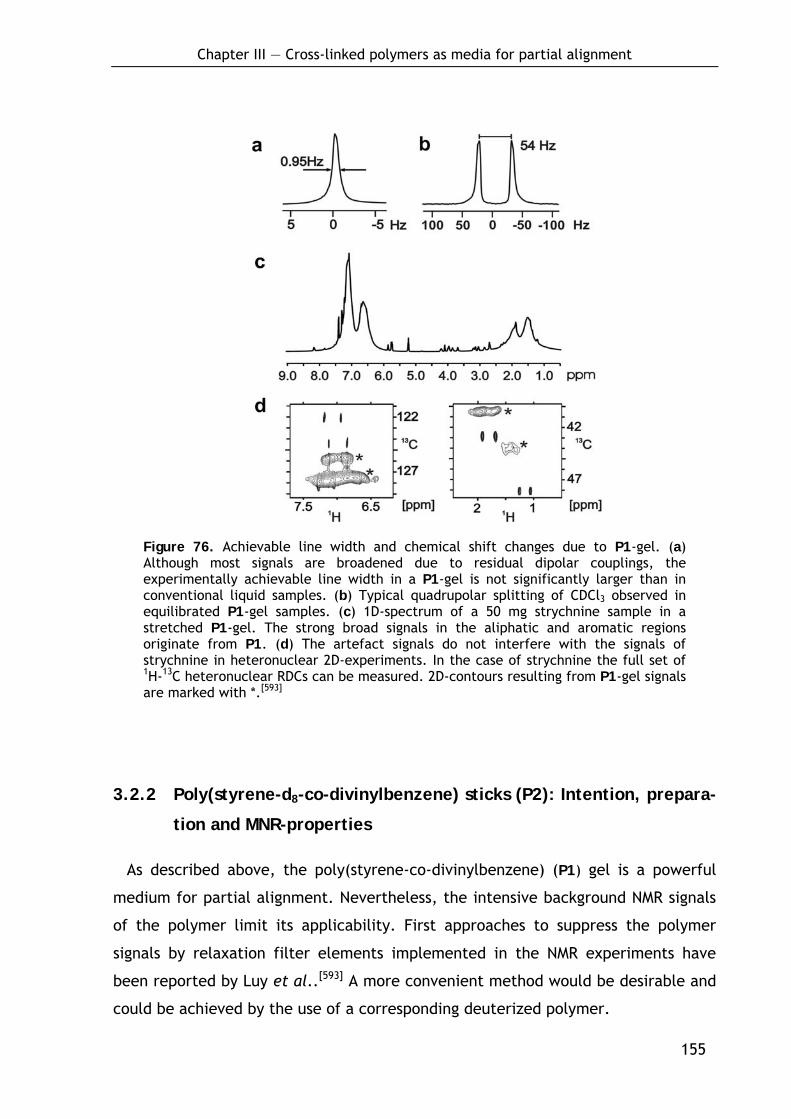
Chapter III — Cross-linked polymers as media for partial alignment
155
Figure 76. Achievable line width and chemical shift changes due to P1-gel. (a) Although most signals are broadened due to residual dipolar couplings, the experimentally achievable line width in a P1-gel is not significantly larger than in conventional liquid samples. (b) Typical quadrupolar splitting of CDCl3 observed in equilibrated P1-gel samples. (c) 1D-spectrum of a 50 mg strychnine sample in a stretched P1-gel. The strong broad signals in the aliphatic and aromatic regions originate from P1. (d) The artefact signals do not interfere with the signals of strychnine in heteronuclear 2D-experiments. In the case of strychnine the full set of 1H-13C heteronuclear RDCs can be measured. 2D-contours resulting from P1-gel signals are marked with *.[593]
3.2.2 Poly(styrene-d8-co-divinylbenzene) sticks (P2): Intention, prepara-
tion and MNR-properties
As described above, the poly(styrene-co-divinylbenzene) (P1) gel is a powerful
medium for partial alignment. Nevertheless, the intensive background NMR signals
of the polymer limit its applicability. First approaches to suppress the polymer
signals by relaxation filter elements implemented in the NMR experiments have
been reported by Luy et al..[593] A more convenient method would be desirable and
could be achieved by the use of a corresponding deuterized polymer.
Chapter III — Cross-linked polymers as media for partial alignment
156
Scheme 21. Preparation of styrene-d8-co-divinylbenzene (P2).a
ran
n
+AIBN (0.1%)
0.02%-1.00%
99.98%-99.00%
DD
D
D
DD
D
D
D
DD
D
DD
DD
n
D
DD
D
DD
DD
ran
202 201 P2
a Conditions: 45°C (5 d) then 60°C (2 d); ran: random.
Obviously, a major limitation of this approach could be the availability and the
prize of the starting materials. Styrene-d8 (202), however, is commercially avail-
able at a reasonable cost. Although deuterized divinylbenzene (201) is not avail-
able, this causes no problem as the cross-linking agent is merely added in the range
of approximately one percent with respect to styrene-d8 (202) which corresponds
to the isotopic purity of styrene-d8 (>98 atom% D).
For preparation of styrene-d8-co-divinylbenzene (P2) sticks, above described
procedure was optimized in one step with regard to the significantly smaller
reaction scale: to reduce loss of substance during separation from stabilizers, the
distillation step was substituted by a condensation allowing the purification of 5 mL
of styrene-d8 in approximately 90% yield. In this manner a series of sticks of 3.4
mm diameter was prepared with constant amounts of AIBN (0.1%, w/v) and variable
amounts of DVB (0.2, 0.5 and 1.0%, v/v) (Scheme 21). All mixtures gave clear solids
with only sporadic occurring bubbles.
Initial NMR studies performed by G. Kummerlöwe in our group showed a dramatic
reduction of polymer signals in the deuterated P2 polymer (see Figure 77, lower
spectra). Figure 77a shows a superposition of 1H-NMR spectra of undeuterated
polymer P1 (upper spectrum) and the corresponding deuterated polymer P2 (lower
spectrum). NMR signals arising from the polymer are almost invisible in P2.

Chapter III — Cross-linked polymers as media for partial alignment
157
Moreover, 1H-NMR spectra of strychnine were recorded in both polymer gels (Figure
77b). This experiment convincingly demonstrated the potential of P2 polymer in
the application. Accordingly, the promising material is currently evaluated in more
detail by G. Kummerlöwe.
Figure 77. Superposition of 1H NMR spectra of poly(styrene-co-divinylbenzene) (P1) (upper spectra) and poly(styrene-d8-co-divinylbenzene) (P2) (lower spectra) recorded under equal conditions (receiver gain, number of scans, etc.). (a) 1D-spectra of pure polymers P1 and P2 swollen in CDCl3. (b) 1D-spectra of 50 mg strychnine in P1 and P2 gels (solvent CDCl3).
3.2.3 Poly(methyl methacrylate-co-ethylenglycol dimethacrylate) sticks
(P3)
Poly(methyl methacrylate) (PMMA) is a common polymer showing solubility simi-
lar to PS.[604] Nevertheless, its application as alignment medium is quite interesting
as the expected 1H NMR signals of the polymer are different compared to PS which
makes this material an interesting alternative in cases when substance and polymer
signals overlap.
3.2.3.1 Preparation of P3-sticks by bulk polymerization
Ethylenglycol dimethacrylate (204) was chosen for cross-linking of methyl meth-
acrylate (203) polymer chains as it provides similar structure and properties as the
main monomer to give a preferred statistical incorporation into the growing poly-

Chapter III — Cross-linked polymers as media for partial alignment
158
mer chain. Cross-linked PMMA (P3)-sticks (Scheme 22) were prepared in a broad
composition range in collaboration with D. Heckmann according to the procedure
developed for cross-linked PS (P1).
In this manner methyl methacrylate (203) was copolymerized with variable
amounts of ethylenglycol dimethacrylate (204) (0.05, 0.1, 0.2, 0.5, 0.75, 1.0, 1.5,
2.0, 3.0 and 5.0%, v/v) in silylated glass tubes of diameters 1.6, 2.4, 3.4 and
4.0 mm thereby varying the amounts of AIBN (0.025, 0.05, 0.075, 0.1, 0.5%, w/v).
All possible combinations were prepared. In contrast to the cross-linked PS sticks,
clear solids with only sporadic occurring bubbles were obtained in all cases.
Scheme 22. Preparation of poly(methyl methacrylate-co-ethylenglycol dimetha-crylate) (P3).a
ran
+
AIBN(0.025-0.5%, v/v)
0.05%-5.00%
99.95%-95.00%
OO
O O
H3CO O O O
O OO OCH3
O OCH3
n ran
n
203 204 P3
a Conditions: 45°C (5 d) then 60°C (2 d); ran: random.
3.2.3.2 Alignment and NMR properties
The P3-sticks are currently under acute investigation by P. Kaden in our group.
Applicable solvents are DCM, CHCl3, THF, benzene and dioxane. So far it was found
that only sticks of 3.4 and 4.0 mm diameter show sufficient swelling to give a
reasonable deuterium splitting and that swelling decreases at higher cross-linking.
The absence of aromatic signals in 1H NMR spectra of P3 (see Figure 78) is the main
advantage over P1 as medium for partial alignment.
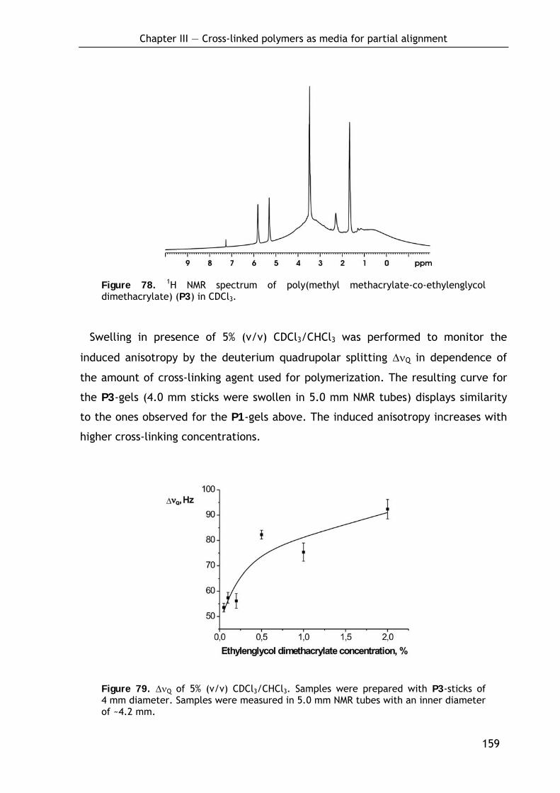
Chapter III — Cross-linked polymers as media for partial alignment
159
Figure 78. 1H NMR spectrum of poly(methyl methacrylate-co-ethylenglycol dimethacrylate) (P3) in CDCl3.
Swelling in presence of 5% (v/v) CDCl3/CHCl3 was performed to monitor the
induced anisotropy by the deuterium quadrupolar splitting ΔνQ in dependence of
the amount of cross-linking agent used for polymerization. The resulting curve for
the P3-gels (4.0 mm sticks were swollen in 5.0 mm NMR tubes) displays similarity
to the ones observed for the P1-gels above. The induced anisotropy increases with
higher cross-linking concentrations.
Figure 79. ΔνQ of 5% (v/v) CDCl3/CHCl3. Samples were prepared with P3-sticks of 4 mm diameter. Samples were measured in 5.0 mm NMR tubes with an inner diameter of ~4.2 mm.
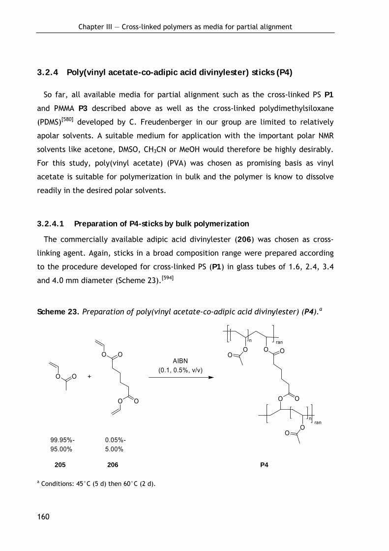
Chapter III — Cross-linked polymers as media for partial alignment
160
3.2.4 Poly(vinyl acetate-co-adipic acid divinylester) sticks (P4)
So far, all available media for partial alignment such as the cross-linked PS P1
and PMMA P3 described above as well as the cross-linked polydimethylsiloxane
(PDMS)[580] developed by C. Freudenberger in our group are limited to relatively
apolar solvents. A suitable medium for application with the important polar NMR
solvents like acetone, DMSO, CH3CN or MeOH would therefore be highly desirably.
For this study, poly(vinyl acetate) (PVA) was chosen as promising basis as vinyl
acetate is suitable for polymerization in bulk and the polymer is know to dissolve
readily in the desired polar solvents.
3.2.4.1 Preparation of P4-sticks by bulk polymerization
The commercially available adipic acid divinylester (206) was chosen as cross-
linking agent. Again, sticks in a broad composition range were prepared according
to the procedure developed for cross-linked PS (P1) in glass tubes of 1.6, 2.4, 3.4
and 4.0 mm diameter (Scheme 23).[594]
Scheme 23. Preparation of poly(vinyl acetate-co-adipic acid divinylester) (P4).a
ran
+
AIBN(0.1, 0.5%, v/v)
0.05%-5.00%
99.95%-95.00%
O
O On
O
O
O
O O
O O O
On
O
ran
O
205 206 P4
a Conditions: 45°C (5 d) then 60°C (2 d).
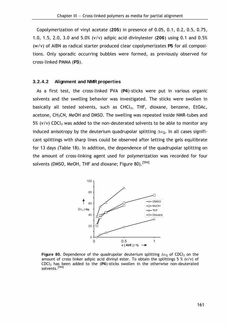
Chapter III — Cross-linked polymers as media for partial alignment
161
Copolymerization of vinyl acetate (205) in presence of 0.05, 0.1, 0.2, 0.5, 0.75,
1.0, 1.5, 2.0, 3.0 and 5.0% (v/v) adipic acid divinylester (206) using 0.1 and 0.5%
(w/v) of AIBN as radical starter produced clear copolymerizates P5 for all composi-
tions. Only sporadic occurring bubbles were formed, as previously observed for
cross-linked PMMA (P3).
3.2.4.2 Alignment and NMR properties
As a first test, the cross-linked PVA (P4)-sticks were put in various organic
solvents and the swelling behavior was investigated. The sticks were swollen in
basically all tested solvents, such as CHCl3, THF, dioxane, benzene, EtOAc,
acetone, CH3CN, MeOH and DMSO. The swelling was repeated inside NMR-tubes and
5% (v/v) CDCl3 was added to the non-deuterated solvents to be able to monitor any
induced anisotropy by the deuterium quadrupolar splitting ΔνQ. In all cases signifi-
cant splittings with sharp lines could be observed after letting the gels equilibrate
for 13 days (Table 18). In addition, the dependence of the quadrupolar splitting on
the amount of cross-linking agent used for polymerization was recorded for four
solvents (DMSO, MeOH, THF and dioxane; Figure 80).[594]
Figure 80. Dependence of the quadrupolar deuterium splitting ΔνQ of CDCl3 on the amount of cross linker adipic acid divinyl ester. To obtain the splittings 5 % (v/v) of CDCl3 has been added to the (P4)-sticks swollen in the otherwise non-deuterated solvents.[594]
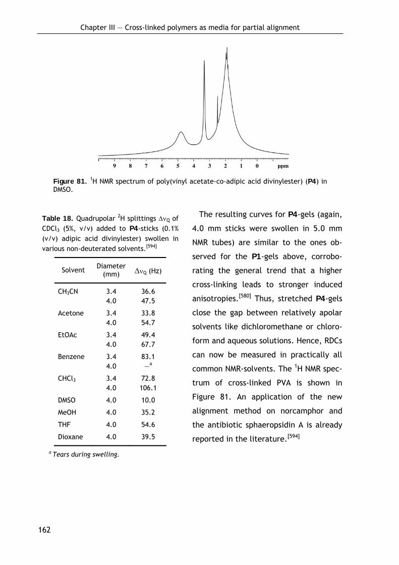
Chapter III — Cross-linked polymers as media for partial alignment
162
Figure 81. 1H NMR spectrum of poly(vinyl acetate-co-adipic acid divinylester) (P4) in DMSO.
The resulting curves for P4-gels (again,
4.0 mm sticks were swollen in 5.0 mm
NMR tubes) are similar to the ones ob-
served for the P1-gels above, corrobo-
rating the general trend that a higher
cross-linking leads to stronger induced
anisotropies.[580] Thus, stretched P4-gels
close the gap between relatively apolar
solvents like dichloromethane or chloro-
form and aqueous solutions. Hence, RDCs
can now be measured in practically all
common NMR-solvents. The 1H NMR spec-
trum of cross-linked PVA is shown in
Figure 81. An application of the new
alignment method on norcamphor and
the antibiotic sphaeropsidin A is already
reported in the literature.[594]
Table 18. Quadrupolar 2H splittings ΔνQ of CDCl3 (5%, v/v) added to P4-sticks (0.1% (v/v) adipic acid divinylester) swollen in various non-deuterated solvents.[594]
Solvent Diameter (mm) ΔνQ (Hz)
CH3CN 3.4 36.6 4.0 47.5
Acetone 3.4 33.8 4.0 54.7
EtOAc 3.4 49.4 4.0 67.7
Benzene 3.4 83.1 4.0 —a
CHCl3 3.4 72.8 4.0 106.1
DMSO 4.0 10.0
MeOH 4.0 35.2
THF 4.0 54.6
Dioxane 4.0 39.5
a Tears during swelling.
Chapter III — Cross-linked polymers as media for partial alignment
163
3.3 Development of cross-linked polymers bearing chiral side
chain moieties
3.3.1 Background
In modern organic chemistry, the determination of enantiomeric excess and
verification of the enantiomeric purity of a product is a common problem. Classic
procedures utilize chiral auxiliaries such as chiral derivatizing agents, lanthanide
chiral-shift reagents and chiral solvating agents for discriminating enantiomers via
NMR.[609,610] However, these methods are limited to molecules that can interact
with the auxiliaries to build detectable diastereomeric compounds or adducts.
Chiral orienting media, in contrast, allow a discrimination of enantiomers through
the differential ordering effect of enantiomers within a chiral phase.[611,612] With
this method, also saturated chiral hydrocarbons lacking polar groups[613] and
prochiral elements in symmetrical molecules[614,615] have been distinguished by NMR
spectroscopy.
Media, such as surfactant bilayers,[616-618] chiral liquid crystalline media,[619-623]
and achiral liquid crystals with chiral cages[624] have the disadvantage to be usually
difficult to prepare and to allow only limited temperature ranges. Moreover, their
induced orientation is dependent on the strength of the magnetic field. Stretched-
polymer gels, in contrast, are relatively easy to handle and provide field-independ-
ent orientation and the alignment strength can easily be scaled. Based on these
experiences, Kobzar et al.[625] demonstrated the first application of a chiral poly-
mer gel for partial alignment and successfully distinguished the two enantiomers L-
and D-alanine using stretched gelatin as medium. This novel procedure combines
the possibility of enantiomeric resolution with the advantages of partial alignment.
Nevertheless, the procedure is limited to aqueous solvents. Hence, the goal of this
work was to find suitable media for organic solvents on basis of the cross-linked
polymers described above. This study was performed in collaboration with
D. Heckmann.
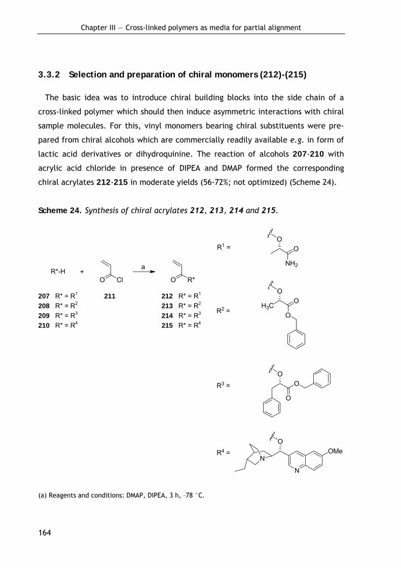
Chapter III — Cross-linked polymers as media for partial alignment
164
O
N
OMeN
R2 = O
OO
H3C
O
O
O
O R*O ClR*-H +
R3 =
R4 =
NH2
OO
R1 =
a
3.3.2 Selection and preparation of chiral monomers (212)-(215)
The basic idea was to introduce chiral building blocks into the side chain of a
cross-linked polymer which should then induce asymmetric interactions with chiral
sample molecules. For this, vinyl monomers bearing chiral substituents were pre-
pared from chiral alcohols which are commercially readily available e.g. in form of
lactic acid derivatives or dihydroquinine. The reaction of alcohols 207-210 with
acrylic acid chloride in presence of DIPEA and DMAP formed the corresponding
chiral acrylates 212-215 in moderate yields (56-72%; not optimized) (Scheme 24).
Scheme 24. Synthesis of chiral acrylates 212, 213, 214 and 215.
(a) Reagents and conditions: DMAP, DIPEA, 3 h, –78 °C.
207 R* = R1 211 212 R* = R1
208 R* = R2 213 R* = R2
209 R* = R3 214 R* = R3 210 R* = R4 215 R* = R4

Chapter III — Cross-linked polymers as media for partial alignment
165
3.3.3 Preparation of cross-linked polymers P5-P9
The cross-linked polymer sticks P5 and P6 from acrylates 213 and 214 respec-
tively, were prepared by copolymerization with 0.5 and 1.0% ethylenglycol di-
methacrylate (204) as cross-linking agent using 0.1% AIBN as radical starter. The
polymerization was carried out in glass tubes with an inner diameter of 4.0 mm
according to the procedure developed for cross-linked PS (P1). Clear solid sticks
were obtained which are currently investigated in NMR for their alignment proper-
ties and their ability to discriminating enantiomers.
Scheme 25. Preparation of cross-linked polyacrylates P5 and P6.a
ran
+O R*
OO
O O
AIBN
O
n
n
O
O R*
O R*ran
O
O
213 R* = R2 204 P5 R* = R2 214 R* = R3 P6 R* = R3
a R2 and R3 are as defined in Scheme 24; conditions: 45°C (5 d) then 60°C (2 d); ran: random.
In a second approach the acrylates 212, 214 and 215 were copolymerized with
methyl acrylate (216) and ethylenglycol dimethacrylate (204). As shown by Green
et al., the copolymerization of minor amounts of a chiral “sergeant” with a non-
chiral “soldier” can induce a helical preference and thus a strong chiral effect
(“Sergeant-Soldier-Effect”).[626,627] This would allow a significant cheaper produc-
tion of chiral media.
Hence, the following sticks were prepared according to above procedure: 212
was copolymerized with 33% (v/v) methyl acrylate (216) and 1.0% (v/v) ethylengly-
col dimethacrylate (204), 214 was copolymerized with 50% methyl acrylate (216)
and 1.0% (v/v) ethylenglycol dimethacrylate (204) and 215 was copolymerized with
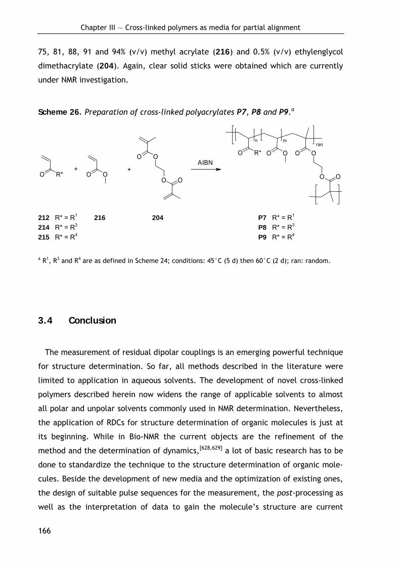
Chapter III — Cross-linked polymers as media for partial alignment
166
75, 81, 88, 91 and 94% (v/v) methyl acrylate (216) and 0.5% (v/v) ethylenglycol
dimethacrylate (204). Again, clear solid sticks were obtained which are currently
under NMR investigation.
Scheme 26. Preparation of cross-linked polyacrylates P7, P8 and P9.a
rann m
O
OO O+
O R*
OO
O O+
AIBNO R* O O O
O
212 R* = R1 216 204 P7 R* = R1 214 R* = R3 P8 R* = R3 215 R* = R4 P9 R* = R4
a R1, R3 and R4 are as defined in Scheme 24; conditions: 45°C (5 d) then 60°C (2 d); ran: random.
3.4 Conclusion
The measurement of residual dipolar couplings is an emerging powerful technique
for structure determination. So far, all methods described in the literature were
limited to application in aqueous solvents. The development of novel cross-linked
polymers described herein now widens the range of applicable solvents to almost
all polar and unpolar solvents commonly used in NMR determination. Nevertheless,
the application of RDCs for structure determination of organic molecules is just at
its beginning. While in Bio-NMR the current objects are the refinement of the
method and the determination of dynamics,[628,629] a lot of basic research has to be
done to standardize the technique to the structure determination of organic mole-
cules. Beside the development of new media and the optimization of existing ones,
the design of suitable pulse sequences for the measurement, the post-processing as
well as the interpretation of data to gain the molecule’s structure are current

Chapter III — Cross-linked polymers as media for partial alignment
167
issues in this field. However, first great strides have been made, other will hope-
fully follow.

4 Experimental Procedures
168
4 Experimental Procedures
4.1 Materials and Methods
Solvents for synthesis were either obtained ‘technical grade’ and distilled prior
to use or purchased (‘absolute’ or ‘for synthesis’) from Fluka (Seelze, Germany),
Acros Organics (Geel; Belgium) or Merck (Darmstadt, Germany). NMP (distilled) was
a gift from BASF (Ludwigshafen, Germany). Solvents for column chromatography
were obtained ‘technical grade’ and distilled prior to use except hexane, which
usually was used undistilled. The HPLC-solvents acetonitrile (solvent B) and TFA
were purchased ‘gradient grade’ from Merck, water (solvent A) was deionized and
further purified using a Milli-Q system from Millipore (Molsheim, France).
Fmoc-protected amino acids were purchased from Novabiochem (Darmstadt,
Germany), Advanced ChemTech (Louisville, USA) or MultiSynTech (Witten,
Germany), Sieber Amide resin was purchased from Novabiochem. Tritylchloride
polystyrole resin (TCP resin) was obtained from PepChem (Tübingen, Germany),
HATU, TBTU and HOBt from Iris Biotech (Marktredwitz, Germany) and HOAt from
GL Biochem (Shanghai, China), TFA from Solvay (Hannover, Germany). Toyopearl
AF-Epoxy-650M resin was obtained from Tosoh (Stuttgart, Germany), Fmoc-
Glutamol(Ot-Bu) from ChemPep Inc. (Miami, USA) and DOTA-tris (tBu ester) from
Macrocyclics (Dallas, TX, USA). All other chemicals were purchased from Aldrich
(Deisenhofen, Germany), Merck (Darmstadt, Germany), Lancaster (Mühlheim,
Germany), Acros Organics (Geel; Belgium) or Fluka (Seelze, Germany).
Peptide Synthesis was carried out manually using PE-syringes (2 mL, 5 mL or
20 mL) from Becton-Dickinson (Fraga, Spain) or Braun (Melsungen) equipped with
4 Experimental Procedures
169
PE-frits from Roland Vetter Laborbedarf (Ammerbuch). To ensure optimal mixing
conditions, the syringes were rotating at about 30 rpm. Loading of the resin was
carried out in glassware equipped with glass frits.
The following protected amino acid building blocks were used for Fmoc-SPPS:
Fmoc-Ado, Fmoc-Ala, Fmoc-Aha, Fmoc-Arg(Pbf), Fmoc-Asn(Trt), Fmoc-Asp(t-Bu),
Fmoc-Bip, Fmoc-Bpa, Fmoc-Bta, Fmoc-Cha, Fmoc-Cys(Trt), Fmoc-Gln(Trt), Fmoc-
Glu(t-Bu), Fmoc-Gly, Fmoc-His(Trt), Fmoc-Ile, Fmoc-Leu, Fmoc-Lys(Boc), Boc-
Lys(Fmoc), Fmoc-1Nal, Fmoc-2Nal, Fmoc-Phe, Fmoc-(2,4-Cl2-Phe), Fmoc-4-py,
Fmoc-Ser(t-Bu), Fmoc-Thr(t-Bu), Fmoc-Trp(Boc), Fmoc-Tyr(OMe), Fmoc-Tyr(t-Bu),
Fmoc-Val.
Air or moisture sensitive reactions were carried out in dry glassware and under
argon (99.996%) atmosphere. Moisture sensitive and/or absoluted solvents were
transferred in syringes under argon.
For HPLC purification, compounds were dissolved in DMSO, acetonitrile or
methanol (‘gradient grade’) and filtered using syringe filters RC 15 or RC 25 (RC-
membrane, 0.45 μm) from Sartorius (Göttingen). Purification and analytical purity
determination was carried out on RP-HPLC systems from Amersham Pharmacia Bio-
tech (analytical: Äkta Basic 10F with pump system P-900, detector UV-900 and
Autosampler A-900; preparative: Äkta Basic 100F with pump system P-900, detector
UV-900) or Beckman (System Gold with solvent module 125 and detector module
166; pump system 110B, control unit 420 and detector Knauer Uvicord) were used
equipped with columns from Omnicrom YMC (analytical: ODS-A C18, 250 mm × 4.6
mm, 5 μm, flow rate: 1 mL/min; semi-preparative: ODS-A C18, 250 mm × 20 mm,
5 μm or 10 μm, flow rate: 8 mL/min; preparative: ODS-A C18, 250 mm × 30 mm,
10 μm, flow rate: 25 mL/min) or Macherey-Nagel (preparative: Nucleosil C18,
250 mm × 40 mm, 7 μm, flow rate: 25 mL/min). Compounds were eluted with
linear gradients (30 min) of acetonitrile (solvent B) in water (solvent A) and 0.1 %
(v/v) TFA. For analytical purity determination of the compounds after semi-
preparative or preparative HPLC purification the peak integrals of the analytical
4 Experimental Procedures
170
HPLC chromatogram at the detector wavelength of 220 nm or 254 nm were evalu-
ated.
For column chromatography Silica Gel 60 (230-400 mesh ASTM, Korngröße 0.040-
0.063 mm) from Merck (Darmstadt) was used in 50-100 fold excess to the raw
material, for flash chromatography pressure of 1-1.2 bar was applied.
Thin layer chromatography (TLC) and the determination of the Rf-value was
carried out using TLC aluminium sheets covered with Silica Gel 60 F254 from Merck
(Darmstadt). For detection, TLC-plates were examined under UV-light (λ = 254
nm). If enhanced visualization was necessary, the plate was treated with aqueous
molybdophosphoric acid/cer-(IV)-sulfate solution (6.25 g molybdophosphoric acid
hydrate, 2.5 g cer-(IV)-sulfate, 15 mL sulfuric acid and 235 mL water) and heated.
Melting points were determined on a Büchi 510 melting point apparatus and are
uncorrected.
Optical rotation was measured on a Perkin-Elmer 241 MC.
All 1H-NMR and 13C-NMR spectra were recorded on Bruker AC-250, AV-360, AV-
500 and DMX500 instruments at 300 K and processed running MestRe-C software.
Spectra were calibrated to their respective solvent signals (CDCl3: 1H 7.26 ppm, 13C
77.0 ppm; MeOH-d4: 1H 3.31 ppm, 13C 49.05 ppm; DMSO-d6: 1H 2.50 ppm, 13C
39.43 ppm; acetone-d6: 1H 2.05 ppm, 13C 30.83 ppm). Chemical shifts (δ) are
reported in parts per million (ppm) and coupling constants (J values) are given in
Hertz (Hz). The following abbreviations were used to explain the multiplicities: s,
singulet; d, doublet; t, triplet; q, quartet; dd, doublet of doublets; dt, doublet of
triplets; m, multiplet; br, broad.
4 Experimental Procedures
171
Electrospray ionisation (ESI)-mass spectra were recorded on a Finnigan LCQ
mass spectrometer in combination with a Hewlett Packard 1100 HPLC-system
equipped with a ODS-A C18 (125 mm × 2 mm, 3 μm, flow rate: 0.2 mL/min) column
from Omnicrom YMC. Compounds were eluted with linear gradients (15 min) of
water (solvent A) and acetonitrile (solvent B) in 0.1% (v/v) formic acid. High-
resolution mass spectra (HRMS-EI) were recorded on a Finnigan MAT 8200.
Lyophilization was carried out using an Alpha 2-4 lyophilizer from Christ
(Osterode).
4 Experimental Procedures
172
4.2 General procedures
General procedure I. Attachment of N-Fmoc-amino acids to TCP resin
After swelling in dry DCM (10 mL) for 20 min, the TCP resin (theoretical loading
0.96 mmol/g, 1.00 g, 0.96 mmol, 1.0 equiv) was treated with a solution of Fmoc-
protected amino acids (1.2 mmol, 1.2 equiv) in dry DCM (8 mL) and DIPEA (490 μL,
2.9 mmol, 3.0 equiv) at room temperature (rt) for 2 h. MeOH (1 mL) and DIPEA (0.2
mL) were added to cap unreacted sites on the resin and the reaction mixture was
shaken for 15 min. The resin was washed with NMP (3×10 mL), DCM (5×10 mL) and
NMP (3×10 mL) and dried under vacuo to give resin bound N-Fmoc-amino acids.
General procedure II. Fmoc deprotection
The Fmoc-protected resin was treated with 10 ml of 20% piperidine in NMP (3×10
min) and washed with NMP (5×10 ml).
General procedure III. Coupling with TBTU/HOBt
Fmoc-amino acid (2.0 equiv), TBTU (2.0 equiv), HOBt (2.0 equiv) were dissolved in
NMP to achieve a 0.2 mM solution of the single components. Then, DIPEA (5.6
equiv) was added and the solution given to the resin. The reaction mixture was
shaken at rt for 60 min and washed with NMP (5×10 mL).
General procedure IV. Coupling with HATU/HOAt
Fmoc-amino acid (2.0 equiv), HATU (2.0 equiv), HOAt (2.0 equiv) were dissolved in
NMP to give a 0.2 mM solution of the single components. Then, 2,4,6-collidine
(15 equiv) was added and the solution given to the resin. The reaction mixture was
shaken at rt for 60 min and washed with NMP (5×10 ml).
4 Experimental Procedures
173
General procedure V. Capping
The resin was treated with 10 ml of a solution of acetic anhydride and DIPEA in
NMP (7/2/91, v/v/v; 15 min) and washed with NMP (5×10 mL).
General procedure VI. N-Terminal dimerization on solid phase
Fmoc-D-glutamic acid (0.17 equiv), HATU (0.33 equiv), HOAt (0.33 equiv) and 2,4,6-
collidine (10 equiv) were dissolved in NMP to give a 0.04 mM solution of the amino
acid, which was added to the resin. The reaction mixture was shaken at rt for 8 h.
The resin was removed by filtration and the procedure repeated three times.
Finally, the resin was washed with NMP (5×10 mL).
General procedure VII. Cleavage and deprotection with trifluoroacetic acid
The resin was washed with DCM (3×10 mL) and treated with TFA/H2O/triisopropyl-
silane (95:2.5:2.5, v/v/v; 20 mL) for 3×10 min. After removal of the resin by filtra-
tion, the filtrates were combined and stirred for another 90 min. The solvent was
concentrated under reduced pressure to precipitate the peptide with diethyl ether.
General procedure VIII: Disulfide cyclization of the linear peptide using H2O2.
The linear peptide was dissolved in high dilution (c = 1 mM) in water and the pH
was adjusted to 7-8 by addition of concentrated aqueous NaHCO3. Then, 30% aque-
ous H2O2 (3 equiv) was added drop wise under vigorous stirring. The reaction was
stopped after 30 min and the solvent removed by lyophilization.
General procedure IX: Disulfide cyclization of the linear peptide using DMSO.
The linear peptide was dissolved in high dilution (c = 1 mM) in water and the pH
was adjusted to 7-8 by addition of concentrated aqueous NaHCO3. Then, DMSO (2%,
v/v) was added under vigorous stirring. After complete conversion (usually 2-4 d;
ESI-MS or HPLC control) the reaction was stopped and the solvent removed by
lyophilization.
4 Experimental Procedures
174
General procedure X: Cleavage of side-chain-protected peptides from TCP-
resin.
The resin was swollen in DCM and then treated with a solution of DCM, acetic acid
and TFE (6 / 3 / 1, v/v/v). After shaking for 1 h, the procedure was repeated and
finally the resin washed once with the cleavage solution. The collected solutions
were diluted with toluene and concentrated in vacuo. The dilution with toluene
and evaporation was repeated twice (no smell of acetic acid). The peptide was
obtained as acetate.
General procedure XI: Backbone cyclization of linear peptides.[630]
The linear, side-chain protected peptide was diluted with DMF to 10-3 - 10-4 M. After
addition of DPPA (3 equiv) and NaHCO3 (5 equiv), the mixture was stirred until all
starting material was consumed (HPLC / LC-MS monitoring), usually 12 h. The solu-
tion was concentrated under reduced pressure and the cyclic peptide precipitated
by addition of water. In case of an improper precipitation, water was substituted
with brine. The peptide was spun down in a centrifuge, washed twice with water
and dried under vacuum.
General procedure XII: Synthesis of N-Fmoc-amino aldehydes.
N-Fmoc-amino aldehydes were synthesized from the corresponding N-Fmoc-amino
alcohols using Dess-Martin periodinane[194,195] as described.[193]
General procedure XIII: Reductive alkylation on solid phase.
The reaction was carried out following a slightly modified procedure of Krchnak et
al.[177] The resin was washed with trimethyl orthoformate (3×10 mL) and the N-Fmoc-
amino aldehyde (5.0 equiv) was added as a 0.1 M solution in trimethyl orthoformate.
The mixture was shaken for 18 h, after which the solution was ejected and a 0.1M
suspension of sodium triacetoxyborohydride (5.0 equiv) in dry DCM was added. The
mixture was shaken for 16 h and the resin was washed with NMP (5×10 mL).
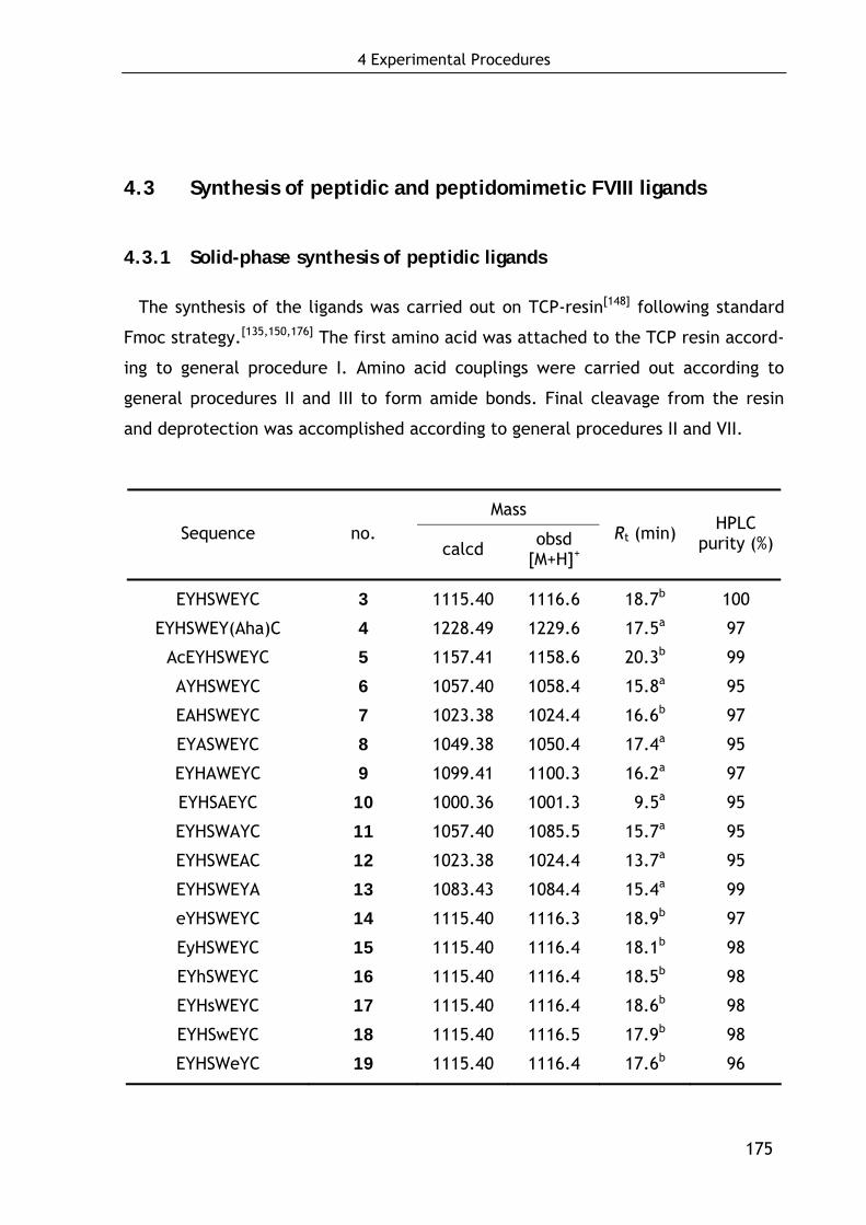
4 Experimental Procedures
175
4.3 Synthesis of peptidic and peptidomimetic FVIII ligands
4.3.1 Solid-phase synthesis of peptidic ligands
The synthesis of the ligands was carried out on TCP-resin[148] following standard
Fmoc strategy.[135,150,176] The first amino acid was attached to the TCP resin accord-
ing to general procedure I. Amino acid couplings were carried out according to
general procedures II and III to form amide bonds. Final cleavage from the resin
and deprotection was accomplished according to general procedures II and VII.
Mass Sequence no.
calcd obsd [M+H]+
Rt (min) HPLC purity (%)
EYHSWEYC 3 1115.40 1116.6 18.7b 100
EYHSWEY(Aha)C 4 1228.49 1229.6 17.5a 97
AcEYHSWEYC 5 1157.41 1158.6 20.3b 99
AYHSWEYC 6 1057.40 1058.4 15.8a 95
EAHSWEYC 7 1023.38 1024.4 16.6b 97
EYASWEYC 8 1049.38 1050.4 17.4a 95
EYHAWEYC 9 1099.41 1100.3 16.2a 97
EYHSAEYC 10 1000.36 1001.3 09.5a 95
EYHSWAYC 11 1057.40 1085.5 15.7a 95
EYHSWEAC 12 1023.38 1024.4 13.7a 95
EYHSWEYA 13 1083.43 1084.4 15.4a 99
eYHSWEYC 14 1115.40 1116.3 18.9b 97
EyHSWEYC 15 1115.40 1116.4 18.1b 98
EYhSWEYC 16 1115.40 1116.4 18.5b 98
EYHsWEYC 17 1115.40 1116.4 18.6b 98
EYHSwEYC 18 1115.40 1116.5 17.9b 98
EYHSWeYC 19 1115.40 1116.4 17.6b 96
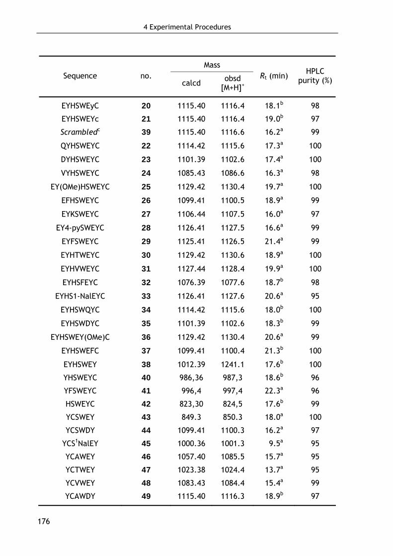
4 Experimental Procedures
176
Mass Sequence no.
calcd obsd [M+H]+
Rt (min) HPLC purity (%)
EYHSWEyC 20 1115.40 1116.4 18.1b 98
EYHSWEYc 21 1115.40 1116.4 19.0b 97
Scrambledc 39 1115.40 1116.6 16.2a 99
QYHSWEYC 22 1114.42 1115.6 17.3a 100
DYHSWEYC 23 1101.39 1102.6 17.4a 100
VYHSWEYC 24 1085.43 1086.6 16.3a 98
EY(OMe)HSWEYC 25 1129.42 1130.4 19.7a 100
EFHSWEYC 26 1099.41 1100.5 18.9a 99
EYKSWEYC 27 1106.44 1107.5 16.0a 97
EY4-pySWEYC 28 1126.41 1127.5 16.6a 99
EYFSWEYC 29 1125.41 1126.5 21.4a 99
EYHTWEYC 30 1129.42 1130.6 18.9a 100
EYHVWEYC 31 1127.44 1128.4 19.9a 100
EYHSFEYC 32 1076.39 1077.6 18.7b 98
EYHS1-NalEYC 33 1126.41 1127.6 20.6a 95
EYHSWQYC 34 1114.42 1115.6 18.0b 100
EYHSWDYC 35 1101.39 1102.6 18.3b 99
EYHSWEY(OMe)C 36 1129.42 1130.4 20.6a 99
EYHSWEFC 37 1099.41 1100.4 21.3b 100
EYHSWEY 38 1012.39 1241.1 17.6b 100
YHSWEYC 40 986,36 987,3 18.6b 96
YFSWEYC 41 996,4 997,4 22.3a 96
HSWEYC 42 823,30 824,5 17.6b 99
YCSWEY 43 849.3 850.3 18.0a 100
YCSWDY 44 1099.41 1100.3 16.2a 97
YCS1NalEY 45 1000.36 1001.3 09.5a 95
YCAWEY 46 1057.40 1085.5 15.7a 95
YCTWEY 47 1023.38 1024.4 13.7a 95
YCVWEY 48 1083.43 1084.4 15.4a 99
YCAWDY 49 1115.40 1116.3 18.9b 97
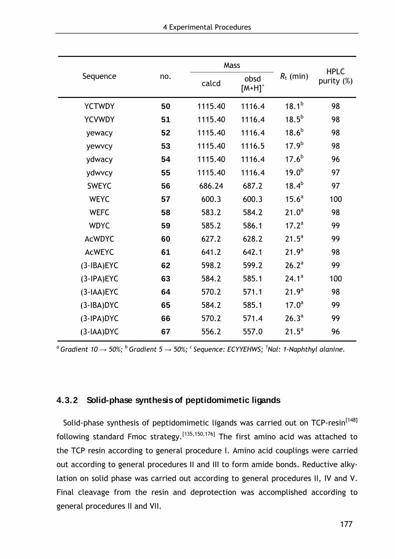
4 Experimental Procedures
177
Mass Sequence no.
calcd obsd [M+H]+
Rt (min) HPLC purity (%)
YCTWDY 50 1115.40 1116.4 18.1b 98
YCVWDY 51 1115.40 1116.4 18.5b 98
yewacy 52 1115.40 1116.4 18.6b 98
yewvcy 53 1115.40 1116.5 17.9b 98
ydwacy 54 1115.40 1116.4 17.6b 96
ydwvcy 55 1115.40 1116.4 19.0b 97
SWEYC 56 686.24 687.2 18.4b 97
WEYC 57 600.3 600.3 15.6a 100
WEFC 58 583.2 584.2 21.0a 98
WDYC 59 585.2 586.1 17.2a 99
AcWDYC 60 627.2 628.2 21.5a 99
AcWEYC 61 641.2 642.1 21.9a 98
(3-IBA)EYC 62 598.2 599.2 26.2a 99
(3-IPA)EYC 63 584.2 585.1 24.1a 100
(3-IAA)EYC 64 570.2 571.1 21.9a 98
(3-IBA)DYC 65 584.2 585.1 17.0a 99
(3-IPA)DYC 66 570.2 571.4 26.3a 99
(3-IAA)DYC 67 556.2 557.0 21.5a 96
a Gradient 10 → 50%; b Gradient 5 → 50%; c Sequence: ECYYEHWS; 1Nal: 1-Naphthyl alanine.
4.3.2 Solid-phase synthesis of peptidomimetic ligands
Solid-phase synthesis of peptidomimetic ligands was carried out on TCP-resin[148]
following standard Fmoc strategy.[135,150,176] The first amino acid was attached to
the TCP resin according to general procedure I. Amino acid couplings were carried
out according to general procedures II and III to form amide bonds. Reductive alky-
lation on solid phase was carried out according to general procedures II, IV and V.
Final cleavage from the resin and deprotection was accomplished according to
general procedures II and VII.
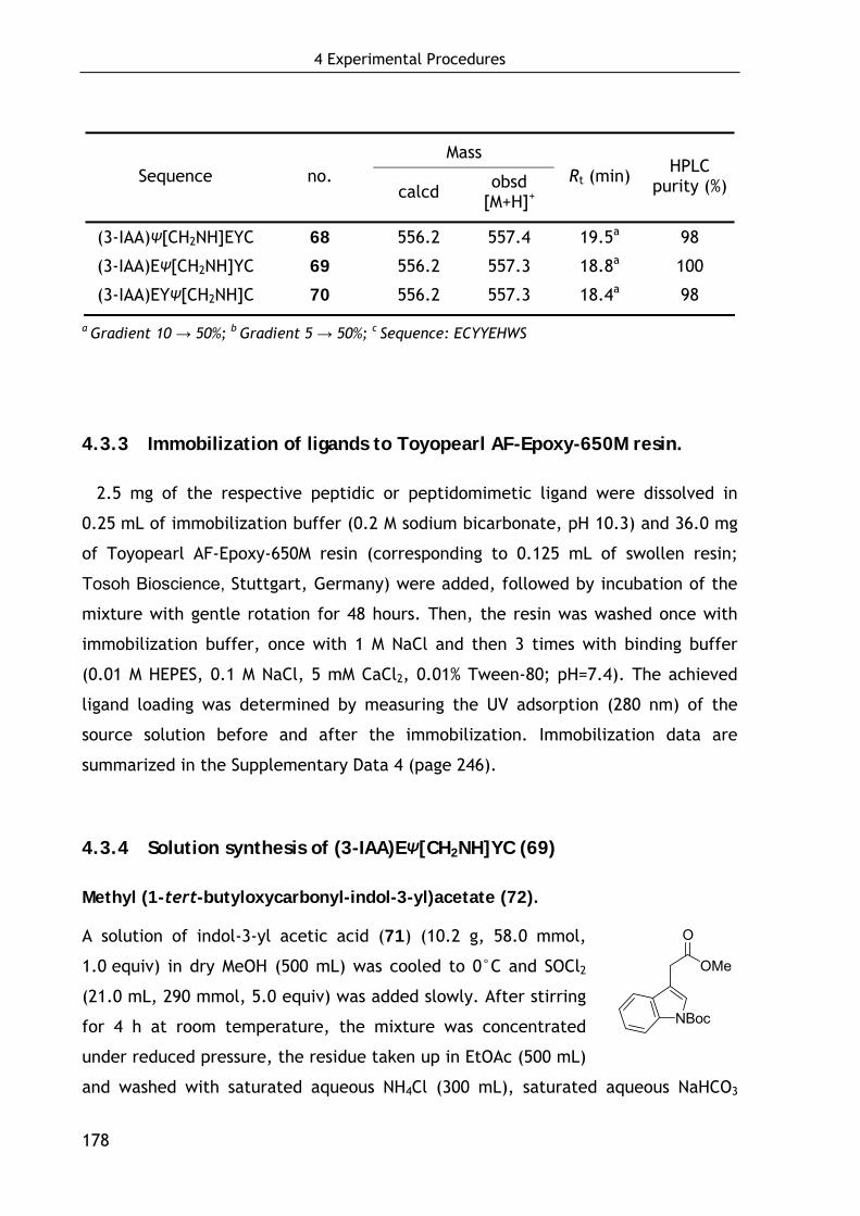
4 Experimental Procedures
178
Mass Sequence no.
calcd obsd [M+H]+
Rt (min) HPLC purity (%)
(3-IAA)Ψ[CH2NH]EYC 68 556.2 557.4 19.5a 98
(3-IAA)EΨ[CH2NH]YC 69 556.2 557.3 18.8a 100
(3-IAA)EYΨ[CH2NH]C 70 556.2 557.3 18.4a 98
a Gradient 10 → 50%; b Gradient 5 → 50%; c Sequence: ECYYEHWS
4.3.3 Immobilization of ligands to Toyopearl AF-Epoxy-650M resin.
2.5 mg of the respective peptidic or peptidomimetic ligand were dissolved in
0.25 mL of immobilization buffer (0.2 M sodium bicarbonate, pH 10.3) and 36.0 mg
of Toyopearl AF-Epoxy-650M resin (corresponding to 0.125 mL of swollen resin;
Tosoh Bioscience, Stuttgart, Germany) were added, followed by incubation of the
mixture with gentle rotation for 48 hours. Then, the resin was washed once with
immobilization buffer, once with 1 M NaCl and then 3 times with binding buffer
(0.01 M HEPES, 0.1 M NaCl, 5 mM CaCl2, 0.01% Tween-80; pH=7.4). The achieved
ligand loading was determined by measuring the UV adsorption (280 nm) of the
source solution before and after the immobilization. Immobilization data are
summarized in the Supplementary Data 4 (page 246).
4.3.4 Solution synthesis of (3-IAA)EΨ[CH2NH]YC (69)
Methyl (1-tert-butyloxycarbonyl-indol-3-yl)acetate (72).
A solution of indol-3-yl acetic acid (71) (10.2 g, 58.0 mmol,
1.0 equiv) in dry MeOH (500 mL) was cooled to 0°C and SOCl2
(21.0 mL, 290 mmol, 5.0 equiv) was added slowly. After stirring
for 4 h at room temperature, the mixture was concentrated
under reduced pressure, the residue taken up in EtOAc (500 mL)
and washed with saturated aqueous NH4Cl (300 mL), saturated aqueous NaHCO3
OMe
O
NBoc
4 Experimental Procedures
179
(300 mL) and brine (150 mL). After drying over MgSO4, the mixture was
concentrated to dryness to give the pure methyl ester as brown oil (10.8 g, 98%).
Spectroscopic data (see below) agree with the literature.[631]
The methyl ester (10.6 g, 56.0 mmol, 1.0 equiv) was dissolved in acetonitrile
(45 mL) and di-tert-butyl dicarbonate (37.4 g, 171 mmol, 3.0 equiv) and DMAP
(1.34 g, 11.0 mmol, 0.2 equiv) were added.[186] The mixture was stirred for 2 h
after which the solvent was removed under reduced pressure. The residue was
dissolved in EtOAc (500 mL) and subsequently washed with saturated aqueous
NH4Cl (300 mL), saturated aqueous NaHCO3 (300 mL) and brine (150 mL). After
drying over MgSO4, the solvent was removed in vacuo and the crude product
purified by flash chromatography on silica gel (EtOAc/hexane; gradient 1:10 → 1:2)
to obtain 72 (14.0 g, 86 %; 2 steps) as a colorless solid. Spectroscopic data agree
with the literature.[632]
Methyl (indol-3-yl)acetate
Rf = 0.3 (EtOAc/hexane 1:2)
1H NMR (360 MHz, CDCl3): δ = 8.17 (s, 1H, Harom), 7.65 (d, J = 7.7 Hz, 1H, NH), 7.29
(d, J = 7.8 Hz, 1H, Harom), 7.26-7.14 (m, 2H, Harom), 7.02 (s, 1H, Harom), 3.82 (s, 2H,
CH2), 3.74 (s, 3H, CH3).
13C NMR (91 MHz, CDCl3): δ = 172.7, 136.0, 127.1, 123.2, 122.0, 119.5, 118.6,
111.2, 108.0, 51.9, 31.0.
HRMS (EI) calcd for C11H11NO2 189.07898; found 189.07883.
Methyl (1-tert-butyloxycarbonyl-indol-3-yl)acetate (72).
RP-HPLC (10 → 100%) Rt = 27.4; 96% purity.
Rf = 0.5 (EtOAc/hexane 1:2)
mp 53°C
4 Experimental Procedures
180
1H NMR (500 MHz, CDCl3): δ = 8.15 (d, J = 6.4 Hz, 1H, Harom), 7.57 (s, 1H, Harom),
7.52 (m, J = 7.8 Hz, 1H, Harom), 7.32 (dd, J = 7.6, 7.6 Hz, 1H, Harom), 7.24 (dd,
J = 7.5, 7.5 Hz, 1H, Harom), 3.71 (s, 2H, CH2), 3.71 (s, 3H, CH3), 1.66 (s, 9H,
C(CH3)3).
13C NMR (91 MHz, CDCl3): δ = 171.5, 149.6, 135.5, 130.1, 124.6, 124.5, 122.7,
119.0, 115.3, 113.1, 83.6, 52.1, 30.9, 28.2.
HRMS (EI) calcd for C16H19NO4 289.13141; found 289.13145.
(1-tert-Butyloxycarbonyl-indol-3-yl)acetic acid (73).
A solution of lithium hydroxide (LiOH*H2O, 6.00 g, 143 mmol,
3 equiv) in H2O (250 mL) was added to a solution of 72 (13.8 g,
47.7 mmol, 1.0 equiv) in THF (350 mL) and MeOH (150 mL). After
stirring for 18 h, the mixture was concentrated in vacuo and a
10% aqueous solution of citric acid (300 mL) was added. The
aqueous layer was extracted with EtOAc (3×200 mL) and the combined organic
layers were washed with water (200 mL) and brine (200 mL) and dried over MgSO4.
The solvent was removed under reduced pressure and the residue purified by flash
chromatography on silica gel (EtOAc/hexane gradient 1:10 → 1:1, 1% AcOH) to
yield 73 (10.9 g, 83%) as a colorless solid. Spectroscopic data agree with
literature.[633]
RP-HPLC (10 → 90%) Rt = 24.5; 98% purity.
Rf = 0.6 (MeOH/CHCl3 1:9, 1% AcOH)
mp 117-120°C
1H NMR (500 MHz, CDCl3): δ = 8.15 (s, 1H, Harom), 7.59 (s, 1H, Harom), 7.53 (dd,
J = 7.8, 0.6 Hz, 1H, Harom), 7.34 (dd, J = 8.2, 7.3 Hz, 1H, Harom), 7.30-7.23 (m, 2H,
Harom), 3.76 (s, 2H, CH2), 1.67 (s, 9H, C(CH3)3).
13C NMR (125 MHz, CDCl3): δ = 176.2, 149.5, 129.8, 124.8, 124.6, 122.6, 118.9,
115.3, 112.3, 83.7, 30.6, 28.1.
HRMS (EI) calcd for C15H17NO4 275.11576; found 275.11531.
OH
O
NBoc
4 Experimental Procedures
181
(S)-tert-Butyl 4-(2-(1-tert-butyloxycarbonyl-indol-3-yl)-5-hydroxypentanoate
(76).
Fmoc-Glutamol(Ot-Bu) (74) (1.83 g, 4.45 mmol) was dis-
solved in piperidine/DMF (20/80, v/v; 100 mL) and after
stirring for 1 h the mixture was evaporated to dryness. The
resulting crude amine 75 was taken up in DMF (250 mL)
and 73 (1.23 g, 4.45 mmol, 1.0 equiv), HOBt (681 mg, 4.45
mmol, 1.0 equiv) and TBTU (1.43 g, 4.45 mmol, 1.0 equiv)
were added. Then, the mixture was cooled to 0°C and
DIPEA (2.25 mL, 13.2 mmol, 3.0 equiv) was added. After stirring for 18 h at room
temperature, the solvent was removed in vacuo and the residue taken up in EtOAc
(300 mL). The mixture was washed with saturated aqueous NH4Cl (2×150 mL),
saturated aqueous NaHCO3 (2×150 mL) and brine (150 mL). After drying over
Na2SO4, the solvent was removed under reduced pressure and the crude product
purified by flash chromatography on silica gel (MeOH/CHCl3; gradient 1:50 → 1:20)
to obtain 76 (1.88 g, 95%) as a hygroscopic colorless oil.
RP-HPLC (30 → 100%) Rt = 20.3; 96% purity.
Rf = 0.1 (MeOH/CHCl3 1:30), Rf = 0.3 (EtOAc/hexane 2:1)
[α]D23 –13.2 (c 7.0, MeOH)
1H NMR (500 MHz, CDCl3): δ = 8.14 (s, 1H, Harom), 7.55 (s, 1H, Harom), 7.49 (d,
J = 7.8 Hz, 1H, Harom), 7.32 (dd, J = 7.3, 7.3 Hz, 1H, Harom), 7.23 (dd, J = 7.5,
7.5 Hz, 1H, Harom), 6.36 (d, J = 8.1 Hz, 1H, NH), 5.38 (s, 1H, br, OH), 3.96-3.87 (m,
1H, NHCH), 3.67 (s, 2H, ArCH2CO), 3.56 (dd, J = 11.5, 3.7 Hz, 1H, CHCHHOH), 3.49
(dd, J = 11.4, 5.0 Hz, 1H, CHCHHOH), 2.19 (t, J = 6.9, 2H, CHCH2CH2CO), 1.75 (dt,
J = 14.0, 7.3 Hz, 1H, CHCHHCH2CO), 1.66 (s, 9H, C(CH3)3), 1.65-1.59 (m, 1H,
CHCHHCH2CO), 1.40 (s, 9H, C(CH3)3).
13C NMR (63 MHz, CDCl3): δ = 173.1, 170.9, 149.4, 135.5, 129.7, 124.8, 122.8,
118.8, 115.3, 113.6, 83.8, 80.8, 64.5, 51.6, 33.3, 31.7, 28.1, 27.9, 25.4.
HRMS (EI) calcd for C24H34N2O6 446.24169; found 446.24164.
NH
O
NBoc
O Ot-Bu
OH

4 Experimental Procedures
182
(S)-tert-Butyl 4-(2-(1-tert-butyloxycarbonyl-indol-3-yl)acetylamino)-4-formyl-
butanoate (77).
To a solution of 76 (1.2 g, 2.68 mmol, 1.0 equiv) in dry
DCM (30 mL) was added Dess-Martin periodinane (3.41 g,
8.04 mmol, 3.0 equiv) in three portions over 1.5 h. The
mixture was allowed to stir for another 1 h after which
the suspension was diluted with DCM (30 mL) and subse-
quently washed with a mixture of 10% aqueous Na2S2O3
and saturated aqueous NaHCO3 (1:1, v/v; 2×30 mL), water (30 mL) and brine (30
mL). Drying over Na2SO4 and evaporation gave the crude aldehyde 77 (1.18 g) as a
pale yellow solid which was directly used without further purification.
Rf = 0.2 (MeOH/CHCl3 1:30), Rf = 0.5 (EtOAc/hexane 2:1)
1H NMR (360 MHz, CDCl3): δ = 9.44 (s, 1H, CHO), 8.11 (d, J = 7.9 Hz, 1H, Harom),
7.55 (s, 1H, Harom), 7.48 (d, J = 7.7 Hz, 1H, Harom), 7.30 (dd, J = 7.9, 7.9, 1H, Harom),
7.24-7.17 (m, 1H, Harom), 6.49 (d, J = 6.8 Hz, 1H, NH), 4.42 (m, 1H, NHCH), 3.67 (s,
2H, ArCH2CO), 2.24-2.04 (m, 3H, CHCH2CHHCO), 1.76 (m, 1H, CHCH2CHHCO), 1.63
(s, 9H, C(CH3)3), 1.34 (s, 9H, C(CH3)3).
13C NMR (91 MHz, CDCl3): δ = 198.5, 172.1, 170.6, 149.4, 135.5, 129.6, 124.8,
122.8, 118.8, 115.3, 113.3, 83.8, 80.9, 58.2, 33.0, 30.8, 28.1, 27.9, 23.6.
HRMS (EI) calcd for C24H32N2O6 444.22604; found 444.22634.
(S)-tert-Butyl 5-[(S)-1-methoxycarbonyl-2-(4-tert-butoxyphenyl)ethylamino]-4-
(2-(1-tert-butyloxycarbonyl-indol-3-yl)acetylamino)-pentanoate (78).
The crude aldehyde 77 (1.18 g) was dissolved in
dry DCM (60 mL) and Tyr(OtBu)OMe * HCl (925
mg, 3.21 mmol, 1.2 equiv) and MgSO4 (1.61 g,
13.4 mmol, 5.0 equiv) were added. After react-
ing for 30 min, NaB(OAc)3H (3.18 g, 15.0 mmol,
5.6 equiv) was added and stirring continued for
additional 18 h. Then, the mixture was diluted
NH
O
NBoc
O Ot-Bu
O
NH
O HN
OMe
O
NBoc
O Ot-Bu
Ot-Bu
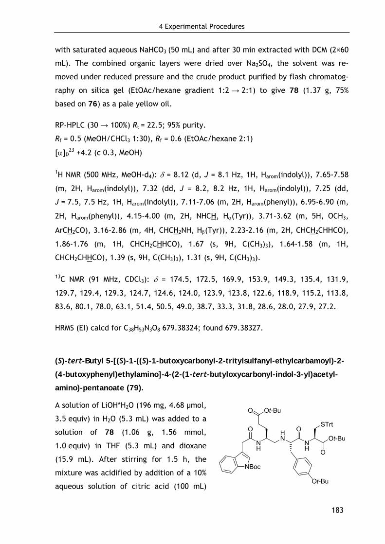
4 Experimental Procedures
183
with saturated aqueous NaHCO3 (50 mL) and after 30 min extracted with DCM (2×60
mL). The combined organic layers were dried over Na2SO4, the solvent was re-
moved under reduced pressure and the crude product purified by flash chromatog-
raphy on silica gel (EtOAc/hexane gradient 1:2 → 2:1) to give 78 (1.37 g, 75%
based on 76) as a pale yellow oil.
RP-HPLC (30 → 100%) Rt = 22.5; 95% purity.
Rf = 0.5 (MeOH/CHCl3 1:30), Rf = 0.6 (EtOAc/hexane 2:1)
[α]D23 +4.2 (c 0.3, MeOH)
1H NMR (500 MHz, MeOH-d4): δ = 8.12 (d, J = 8.1 Hz, 1H, Harom(indolyl)), 7.65-7.58
(m, 2H, Harom(indolyl)), 7.32 (dd, J = 8.2, 8.2 Hz, 1H, Harom(indolyl)), 7.25 (dd,
J = 7.5, 7.5 Hz, 1H, Harom(indolyl)), 7.11-7.06 (m, 2H, Harom(phenyl)), 6.95-6.90 (m,
2H, Harom(phenyl)), 4.15-4.00 (m, 2H, NHCH, Hα(Tyr)), 3.71-3.62 (m, 5H, OCH3,
ArCH2CO), 3.16-2.86 (m, 4H, CHCH2NH, Hβ(Tyr)), 2.23-2.16 (m, 2H, CHCH2CHHCO),
1.86-1.76 (m, 1H, CHCH2CHHCO), 1.67 (s, 9H, C(CH3)3), 1.64-1.58 (m, 1H,
CHCH2CHHCO), 1.39 (s, 9H, C(CH3)3), 1.31 (s, 9H, C(CH3)3).
13C NMR (91 MHz, CDCl3): δ = 174.5, 172.5, 169.9, 153.9, 149.3, 135.4, 131.9,
129.7, 129.4, 129.3, 124.7, 124.6, 124.0, 123.9, 123.8, 122.6, 118.9, 115.2, 113.8,
83.6, 80.1, 78.0, 63.1, 51.4, 50.5, 49.0, 38.7, 33.3, 31.8, 28.6, 28.0, 27.9, 27.2.
HRMS (EI) calcd for C38H53N3O8 679.38324; found 679.38327.
(S)-tert-Butyl 5-[(S)-1-((S)-1-butoxycarbonyl-2-tritylsulfanyl-ethylcarbamoyl)-2-
(4-butoxyphenyl)ethylamino]-4-(2-(1-tert-butyloxycarbonyl-indol-3-yl)acetyl-
amino)-pentanoate (79).
A solution of LiOH*H2O (196 mg, 4.68 μmol,
3.5 equiv) in H2O (5.3 mL) was added to a
solution of 78 (1.06 g, 1.56 mmol,
1.0 equiv) in THF (5.3 mL) and dioxane
(15.9 mL). After stirring for 1.5 h, the
mixture was acidified by addition of a 10%
aqueous solution of citric acid (100 mL)
NH
O HN
NH
OOt-Bu
O
NBoc
O Ot-Bu
Ot-Bu
STrt

4 Experimental Procedures
184
and the aqueous layer was extracted with EtOAc (3×100 mL). The combined organic
layers were washed with water (100 mL) and brine (50 mL), dried (Na2SO4) and
evaporated to give the crude acid (986 mg; 93% purity; RP-HPLC (20 → 100%)
Rt = 23.0) as a pale yellow solid which was redissolved in DMF (55 mL). Then,
Cys(Trt)OtBu*HCl[196] (1.06 g, 2.34 mmol, 1.5 equiv), HOBt (232 mg, 1.72 mmol,
1.1 equiv) and TBTU (552 mg, 1.72 mmol, 1.1 equiv) were added and the mixture
was cooled to 10°C. 2,4,6-Collidine (1.03 mL, 7.80 mmol, 5.0 equiv) was added and
the solution was stirred for 18 h at room temperature. After the solvent was
evaporated, the residue was taken up in EtOAc (200 mL) and subsequently washed
with saturated aqueous NH4Cl (2×150 mL), saturated aqueous NaHCO3 (2×150 mL)
and brine (150 mL). After drying over Na2SO4, the solvent was removed under
reduced pressure and the crude product purified by flash chromatography on silica
gel (EtOAc/hexane; gradient 1:4 → 2:1) to give 79 (1.38 g, 83%) as a hygroscopic,
pale yellow oil.
RP-HPLC (20 → 100%) Rt = 31.2; 97% purity.
Rf = 0.6 (EtOAc/hexane 2:1)
[α]D23 +3.6 (c 0.6, ACN)
1H NMR (500 MHz, MeOH-d4): δ = 8.12 (d, J = 8.1 Hz, 1H, Harom(indolyl)), 7.65-7.58
(m, 2H, Harom(indolyl)), 7.32 (dd, J = 8.2, 8.2 Hz, 1H, Harom(indolyl)), 7.25 (dd,
J = 7.5, 7.5 Hz, 1H, Harom(indolyl)), 7.11-7.06 (m, 2H, Harom(phenyl)), 6.95-6.90 (m,
2H, Harom(phenyl)), 4.15-4.00 (m, 2H, NHCH, Hα(Tyr)), 3.71-3.62 (m, 5H, OCH3,
ArCH2CO), 3.16-2.86 (m, 4H, CHCH2NH, Hβ(Tyr)), 2.23-2.16 (m, 2H, CHCH2CHHCO),
1.86-1.76 (m, 1H, CHCH2CHHCO), 1.67 (s, 9H, C(CH3)3), 1.64-1.58 (m, 1H,
CHCH2CHHCO), 1.39 (s, 9H, C(CH3)3), 1.31 (s, 9H, C(CH3)3)
1H NMR (500 MHz, DMSO-d6): δ = 8.95 (d, J = 7.7 Hz, 1H, NH), 8.09 (d, J = 8.0 Hz,
1H, NH), 8.05 (d, J = 8.2 Hz, 1H, NH), 7.60 (d, J = 7.8 Hz, 1H, Harom(indolyl)), 7.57
(s, 1H, Harom(indolyl)), 7.38-7.27 (m, 14H, Harom(Trt) , Harom(indolyl)), 7.27-7.20 (m,
4H, Harom(Trt), Harom(indolyl)), 7.10 (d, J = 8.3 Hz, 2H,Harom(Tyrphenyl)), 6.83 (d,
J = 8.3 Hz, 2H, Harom(Tyr)), 4.08-4.02 (m, 1H, NHCH), 4.00-3.92 (m, 1H, NHCH),
3.91-3.85 (m, 1H, NHCH), 3.59 (d, J = 15.5 Hz, 1H, ArCHHCO), 3.49 (d, J = 15.4 Hz,
21H, ArCHHCO), 3.09-3.01 (m, 2H, CHCH2NH), 2.93-2.86 (m, 1H, Hβ(Tyr)), 2.84-2.80

4 Experimental Procedures
185
(m, 1H, Hβ(Tyr)), 2.57-2.52 (m, 1H, Hβ(Cys)), 2.39 (dd, J = 12.6, 5.2 Hz, 1H,
Hβ(Cys)), 2.14-1.98 (m, 2H, CHCH2CHHCO), 1.62 (s, 10H, C(CH3)3, CHCH2CHHCO),
1.50-1.44 (m, 1H, CHCH2CHHCO), 1.32 (s, 9H, C(CH3)3), 1.29 (s, 9H, C(CH3)3), 1.25
(s, 9H, C(CH3)3).
13C NMR (125 MHz, DMSO-d6): δ = 171.3, 170.4, 168.1, 166.7, 154.1, 148.9, 143.8,
134.5, 130.0, 129.8, 128.9, 128.6, 128.0, 126.7, 124.3, 124.0, 123.4, 122.3, 119.4,
114.8, 114.5, 83.4, 81.5, 79.5, 77.6, 66.4, 60.8, 52.5, 49.7, 45.7, 34.9, 32.7, 31.9,
30.6, 28.4, 27.6, 27.5, 27.3.
MS (ESI) calcd for C63H78N4O9S 1066.55; found 1067.6 [M+H]+, 1089.3 [M+Na]+.
5-[1-(1-Carboxy-2-mercapto-ethylcarbamoyl)-2-(4-hydroxy-phenyl)-ethylami-
no]-4-(2-1H-indol-3-yl-acetylamino)-pentanoic acid (69).
To a vigorously stirred mixture of 79 (500
mg, 0.47 mmol) in triisopropylethylsilane
(250 μL; 2.5%) and water (250 μL; 2.5%) was
added trifluoroacetic acid (9.50 mL; 95%)
slowly over 8h. The mixture was stirred for
additional 30 min, after which the solvent
was removed in vacuo. The residue was
dissolved in a minimum volume of acetic acid and precipitated in ice cold
ether/pentane (2×20 mL) to give pure 69 (242 mg, 93%).
96% purity; RP-HPLC (10 → 50%) Rt = 14.4
[α]D23 –0.4 (c 0.2, MeOH/H2O 2:1)
mp 135°C
Two conformers (ca. 4:1 ratio)
1H NMR (500 MHz, MeOH-d4): δ = 7.58 (d, J = 7.9 Hz, 1H, 20%, Hindolyl), 7.56 (d,
J = 8.0 Hz, 1H, 80%, Hindolyl), 7.38-7.35 (m, 1H, Hindolyl), 7.22 (s, 1H, 20%, Hindolyl),
7.20 (s, 1H, 80%, Hindolyl), 7.15-7.09 (m, 1H, Hindolyl), 7.07 (d, J = 8.5 Hz, 2H,Hphenyl),
NH
O HN
NH
OOH
O
NH
O OH
OH
SH
4 Experimental Procedures
186
7.05-7.02 (m, 1H, Hindolyl), 6.75 (d, J = 8.46 Hz, 2H, Hphenyl), 4.54-4.46 (m, 1H,
Hα(Cys)), 4.20-4.14 (m, 1H, 20%, Hα(Tyr)), 4.12 (t, J = 7.0, 1H, 20%, CH(CH2)2), 4.05
(m, 1H, 80%, Hα(Tyr)), 3.98 (t, J = 6.9, 1H, 80%, CH(CH2)2), 3.77-3.63 (m, 2H,
CH2Ar), 3.11-3.03 (m, 2H, Hβ(Cys)), 3.03-2.96 (m, 2H, Hβ(Tyr)), 2.94-2.81 (m, 2H,
CH2NH), 2.33-2.20 (m, 2H, CHCH2CH2), 1.87-1.76 (m, 1H, CHCHHCH2), 1.72-1.60
(m, 1H, CHCHHCH2).
13C NMR (125 MHz, MeOH-d4): δ = 176.4, 176.3, 172.5, 170.4, 158.1, 138.2, 131.5,
131.5, 125.2, 125.1, 122.6, 120.0, 119.2, 116.8, 112.4, 109.0, 64.1 (80%), 63.2
(20%), 56.5 (20%), 56.3 (80%), 52.9 (80%), 52.5 (20%), 48.1, 37.5, 34.0 (20%), 33.9
(80%), 31.0 (80%), 30.9 (20%), 28.3 (20%), 28.0 (80%), 26.8.
HRMS (ESI) calcd for C27H33N4O7S 557.20700 [M+H]+; found 557.20590 [M+H]+, 1089.3
[M+Na]+.
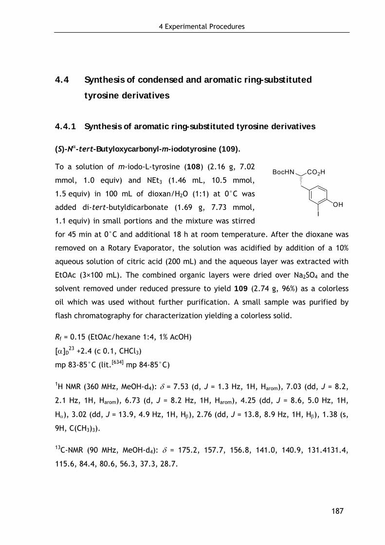
4 Experimental Procedures
187
4.4 Synthesis of condensed and aromatic ring-substituted
tyrosine derivatives
4.4.1 Synthesis of aromatic ring-substituted tyrosine derivatives
(S)-Nα-tert-Butyloxycarbonyl-m-iodotyrosine (109).
To a solution of m-iodo-L-tyrosine (108) (2.16 g, 7.02
mmol, 1.0 equiv) and NEt3 (1.46 mL, 10.5 mmol,
1.5 equiv) in 100 mL of dioxan/H2O (1:1) at 0°C was
added di-tert-butyldicarbonate (1.69 g, 7.73 mmol,
1.1 equiv) in small portions and the mixture was stirred
for 45 min at 0°C and additional 18 h at room temperature. After the dioxane was
removed on a Rotary Evaporator, the solution was acidified by addition of a 10%
aqueous solution of citric acid (200 mL) and the aqueous layer was extracted with
EtOAc (3×100 mL). The combined organic layers were dried over Na2SO4 and the
solvent removed under reduced pressure to yield 109 (2.74 g, 96%) as a colorless
oil which was used without further purification. A small sample was purified by
flash chromatography for characterization yielding a colorless solid.
Rf = 0.15 (EtOAc/hexane 1:4, 1% AcOH)
[α]D23 +2.4 (c 0.1, CHCl3)
mp 83-85°C (lit.[634] mp 84-85°C)
1H NMR (360 MHz, MeOH-d4): δ = 7.53 (d, J = 1.3 Hz, 1H, Harom), 7.03 (dd, J = 8.2,
2.1 Hz, 1H, Harom), 6.73 (d, J = 8.2 Hz, 1H, Harom), 4.25 (dd, J = 8.6, 5.0 Hz, 1H,
Hα), 3.02 (dd, J = 13.9, 4.9 Hz, 1H, Hβ), 2.76 (dd, J = 13.8, 8.9 Hz, 1H, Hβ), 1.38 (s,
9H, C(CH3)3).
13C-NMR (90 MHz, MeOH-d4): δ = 175.2, 157.7, 156.8, 141.0, 140.9, 131.4131.4,
115.6, 84.4, 80.6, 56.3, 37.3, 28.7.
CO2HBocHN
OHI
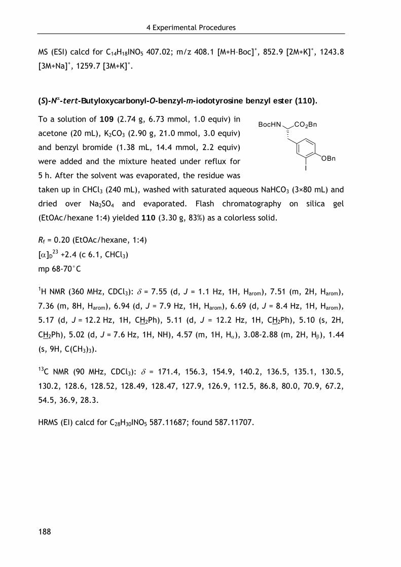
4 Experimental Procedures
188
MS (ESI) calcd for C14H18INO5 407.02; m/z 408.1 [M+H–Boc]+, 852.9 [2M+K]+, 1243.8
[3M+Na]+, 1259.7 [3M+K]+.
(S)-Nα-tert-Butyloxycarbonyl-O-benzyl-m-iodotyrosine benzyl ester (110).
To a solution of 109 (2.74 g, 6.73 mmol, 1.0 equiv) in
acetone (20 mL), K2CO3 (2.90 g, 21.0 mmol, 3.0 equiv)
and benzyl bromide (1.38 mL, 14.4 mmol, 2.2 equiv)
were added and the mixture heated under reflux for
5 h. After the solvent was evaporated, the residue was
taken up in CHCl3 (240 mL), washed with saturated aqueous NaHCO3 (3×80 mL) and
dried over Na2SO4 and evaporated. Flash chromatography on silica gel
(EtOAc/hexane 1:4) yielded 110 (3.30 g, 83%) as a colorless solid.
Rf = 0.20 (EtOAc/hexane, 1:4)
[α]D23 +2.4 (c 6.1, CHCl3)
mp 68-70°C
1H NMR (360 MHz, CDCl3): δ = 7.55 (d, J = 1.1 Hz, 1H, Harom), 7.51 (m, 2H, Harom),
7.36 (m, 8H, Harom), 6.94 (d, J = 7.9 Hz, 1H, Harom), 6.69 (d, J = 8.4 Hz, 1H, Harom),
5.17 (d, J = 12.2 Hz, 1H, CH2Ph), 5.11 (d, J = 12.2 Hz, 1H, CH2Ph), 5.10 (s, 2H,
CH2Ph), 5.02 (d, J = 7.6 Hz, 1H, NH), 4.57 (m, 1H, Hα), 3.08-2.88 (m, 2H, Hβ), 1.44
(s, 9H, C(CH3)3).
13C NMR (90 MHz, CDCl3): δ = 171.4, 156.3, 154.9, 140.2, 136.5, 135.1, 130.5,
130.2, 128.6, 128.52, 128.49, 128.47, 127.9, 126.9, 112.5, 86.8, 80.0, 70.9, 67.2,
54.5, 36.9, 28.3.
HRMS (EI) calcd for C28H30INO5 587.11687; found 587.11707.
CO2BnBocHN
OBnI
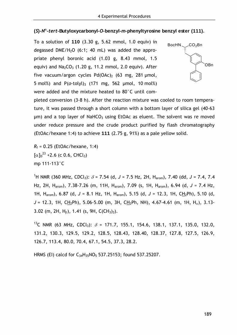
4 Experimental Procedures
189
(S)-Nα-tert-Butyloxycarbonyl-O-benzyl-m-phenyltyrosine benzyl ester (111).
To a solution of 110 (3.30 g, 5.62 mmol, 1.0 equiv) in
degassed DME/H2O (6:1; 40 mL) was added the appro-
priate phenyl boronic acid (1.03 g, 8.43 mmol, 1.5
equiv) and Na2CO3 (1.20 g, 11.2 mmol, 2.0 equiv). After
five vacuum/argon cycles Pd(OAc)2 (63 mg, 281 μmol,
5 mol%) and P(o-tolyl)3 (171 mg, 562 μmol, 10 mol%)
were added and the mixture heated to 80°C until com-
pleted conversion (3-8 h). After the reaction mixture was cooled to room tempera-
ture, it was passed through a short column with a bottom layer of silica gel (40-63
μm) and a top layer of NaHCO3 using EtOAc as eluent. The solvent was re moved
under reduce pressure and the crude product purified by flash chromatography
(EtOAc/hexane 1:4) to achieve 111 (2.75 g, 91%) as a pale yellow solid.
Rf = 0.25 (EtOAc/hexane, 1:4)
[α]D23 +2.6 (c 0.6, CHCl3)
mp 111-113°C
1H NMR (360 MHz, CDCl3): δ = 7.54 (d, J = 7.5 Hz, 2H, Harom), 7.40 (dd, J = 7.4, 7.4
Hz, 2H, Harom), 7.38-7.26 (m, 11H, Harom), 7.09 (s, 1H, Harom), 6.94 (d, J = 7.4 Hz,
1H, Harom), 6.87 (d, J = 8.1 Hz, 1H, Harom), 5.15 (d, J = 12.3, 1H, CH2Ph), 5.10 (d,
J = 12.3, 1H, CH2Ph), 5.06-5.00 (m, 3H, CH2Ph, NH), 4.67-4.61 (m, 1H, Hα), 3.13-
3.02 (m, 2H, Hβ), 1.41 (s, 9H, C(CH3)3).
13C NMR (63 MHz, CDCl3): δ = 171.7, 155.1, 154.6, 138.1, 137.1, 135.0, 132.0,
131.2, 130.3, 129.5, 129.2, 128.5, 128.43, 128.40, 128.37, 127.8, 127.5, 126.9,
126.7, 113.4, 80.0, 70.4, 67.1, 54.5, 37.3, 28.2.
HRMS (EI) calcd for C34H35NO5 537.25153; found 537.25207.
CO2BnBocHN
OBn
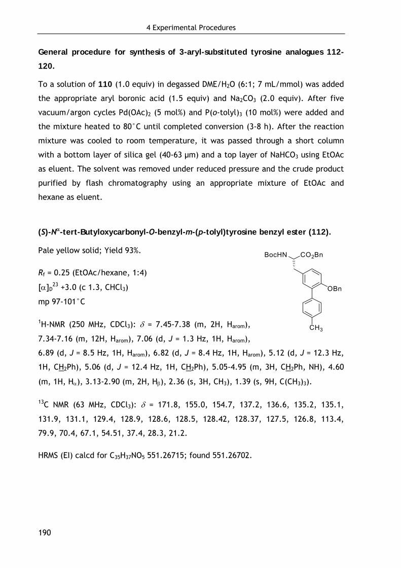
4 Experimental Procedures
190
General procedure for synthesis of 3-aryl-substituted tyrosine analogues 112-
120.
To a solution of 110 (1.0 equiv) in degassed DME/H2O (6:1; 7 mL/mmol) was added
the appropriate aryl boronic acid (1.5 equiv) and Na2CO3 (2.0 equiv). After five
vacuum/argon cycles Pd(OAc)2 (5 mol%) and P(o-tolyl)3 (10 mol%) were added and
the mixture heated to 80°C until completed conversion (3-8 h). After the reaction
mixture was cooled to room temperature, it was passed through a short column
with a bottom layer of silica gel (40-63 μm) and a top layer of NaHCO3 using EtOAc
as eluent. The solvent was removed under reduced pressure and the crude product
purified by flash chromatography using an appropriate mixture of EtOAc and
hexane as eluent.
(S)-Nα-tert-Butyloxycarbonyl-O-benzyl-m-(p-tolyl)tyrosine benzyl ester (112).
Pale yellow solid; Yield 93%.
Rf = 0.25 (EtOAc/hexane, 1:4)
[α]D23 +3.0 (c 1.3, CHCl3)
mp 97-101°C
1H-NMR (250 MHz, CDCl3): δ = 7.45-7.38 (m, 2H, Harom),
7.34-7.16 (m, 12H, Harom), 7.06 (d, J = 1.3 Hz, 1H, Harom),
6.89 (d, J = 8.5 Hz, 1H, Harom), 6.82 (d, J = 8.4 Hz, 1H, Harom), 5.12 (d, J = 12.3 Hz,
1H, CH2Ph), 5.06 (d, J = 12.4 Hz, 1H, CH2Ph), 5.05-4.95 (m, 3H, CH2Ph, NH), 4.60
(m, 1H, Hα), 3.13-2.90 (m, 2H, Hβ), 2.36 (s, 3H, CH3), 1.39 (s, 9H, C(CH3)3).
13C NMR (63 MHz, CDCl3): δ = 171.8, 155.0, 154.7, 137.2, 136.6, 135.2, 135.1,
131.9, 131.1, 129.4, 128.9, 128.6, 128.5, 128.42, 128.37, 127.5, 126.8, 113.4,
79.9, 70.4, 67.1, 54.51, 37.4, 28.3, 21.2.
HRMS (EI) calcd for C35H37NO5 551.26715; found 551.26702.
CO2BnBocHN
OBn
CH3
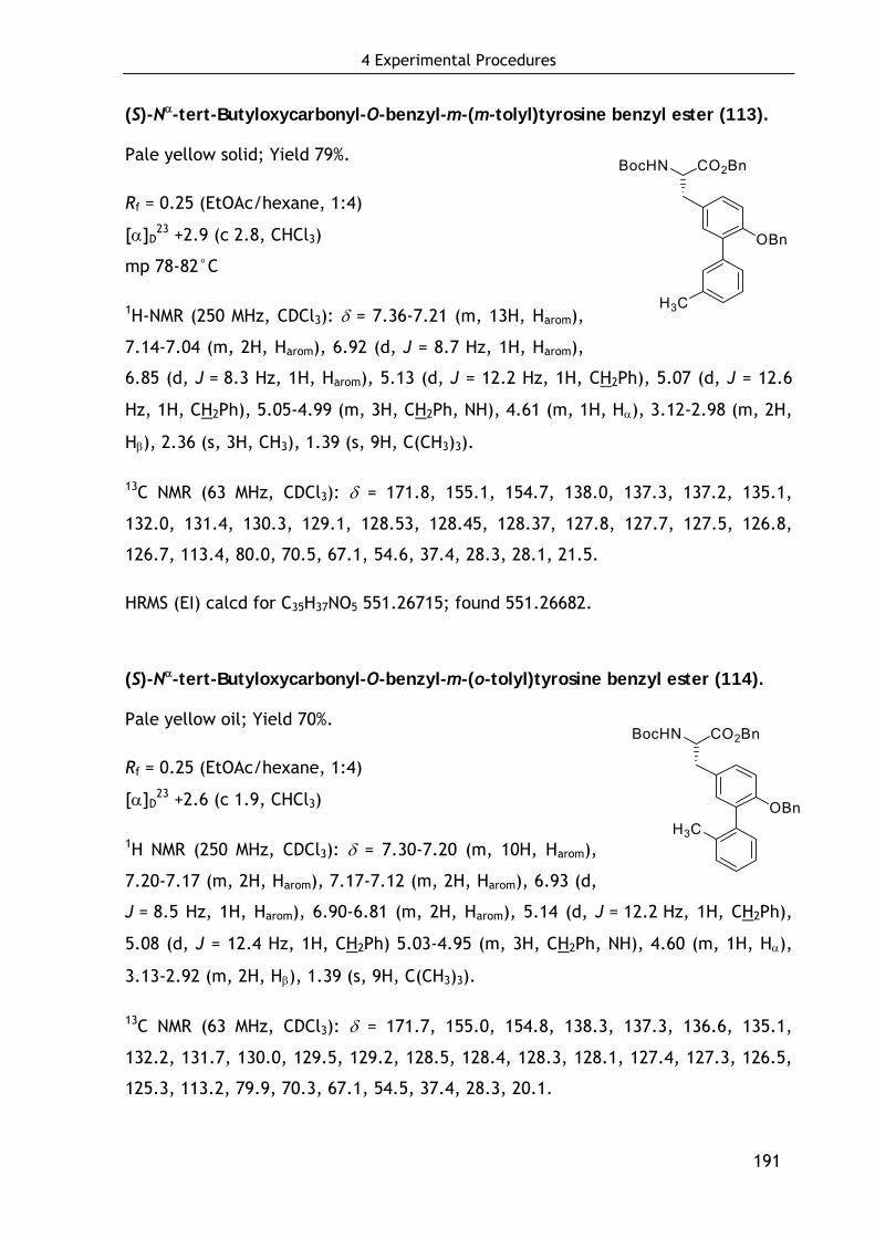
4 Experimental Procedures
191
(S)-Nα-tert-Butyloxycarbonyl-O-benzyl-m-(m-tolyl)tyrosine benzyl ester (113).
Pale yellow solid; Yield 79%.
Rf = 0.25 (EtOAc/hexane, 1:4)
[α]D23 +2.9 (c 2.8, CHCl3)
mp 78-82°C
1H-NMR (250 MHz, CDCl3): δ = 7.36-7.21 (m, 13H, Harom),
7.14-7.04 (m, 2H, Harom), 6.92 (d, J = 8.7 Hz, 1H, Harom),
6.85 (d, J = 8.3 Hz, 1H, Harom), 5.13 (d, J = 12.2 Hz, 1H, CH2Ph), 5.07 (d, J = 12.6
Hz, 1H, CH2Ph), 5.05-4.99 (m, 3H, CH2Ph, NH), 4.61 (m, 1H, Hα), 3.12-2.98 (m, 2H,
Hβ), 2.36 (s, 3H, CH3), 1.39 (s, 9H, C(CH3)3).
13C NMR (63 MHz, CDCl3): δ = 171.8, 155.1, 154.7, 138.0, 137.3, 137.2, 135.1,
132.0, 131.4, 130.3, 129.1, 128.53, 128.45, 128.37, 127.8, 127.7, 127.5, 126.8,
126.7, 113.4, 80.0, 70.5, 67.1, 54.6, 37.4, 28.3, 28.1, 21.5.
HRMS (EI) calcd for C35H37NO5 551.26715; found 551.26682.
(S)-Nα-tert-Butyloxycarbonyl-O-benzyl-m-(o-tolyl)tyrosine benzyl ester (114).
Pale yellow oil; Yield 70%.
Rf = 0.25 (EtOAc/hexane, 1:4)
[α]D23 +2.6 (c 1.9, CHCl3)
1H NMR (250 MHz, CDCl3): δ = 7.30-7.20 (m, 10H, Harom),
7.20-7.17 (m, 2H, Harom), 7.17-7.12 (m, 2H, Harom), 6.93 (d,
J = 8.5 Hz, 1H, Harom), 6.90-6.81 (m, 2H, Harom), 5.14 (d, J = 12.2 Hz, 1H, CH2Ph),
5.08 (d, J = 12.4 Hz, 1H, CH2Ph) 5.03-4.95 (m, 3H, CH2Ph, NH), 4.60 (m, 1H, Hα),
3.13-2.92 (m, 2H, Hβ), 1.39 (s, 9H, C(CH3)3).
13C NMR (63 MHz, CDCl3): δ = 171.7, 155.0, 154.8, 138.3, 137.3, 136.6, 135.1,
132.2, 131.7, 130.0, 129.5, 129.2, 128.5, 128.4, 128.3, 128.1, 127.4, 127.3, 126.5,
125.3, 113.2, 79.9, 70.3, 67.1, 54.5, 37.4, 28.3, 20.1.
CO2BnBocHN
OBn
H3C
CO2BnBocHN
OBnH3C
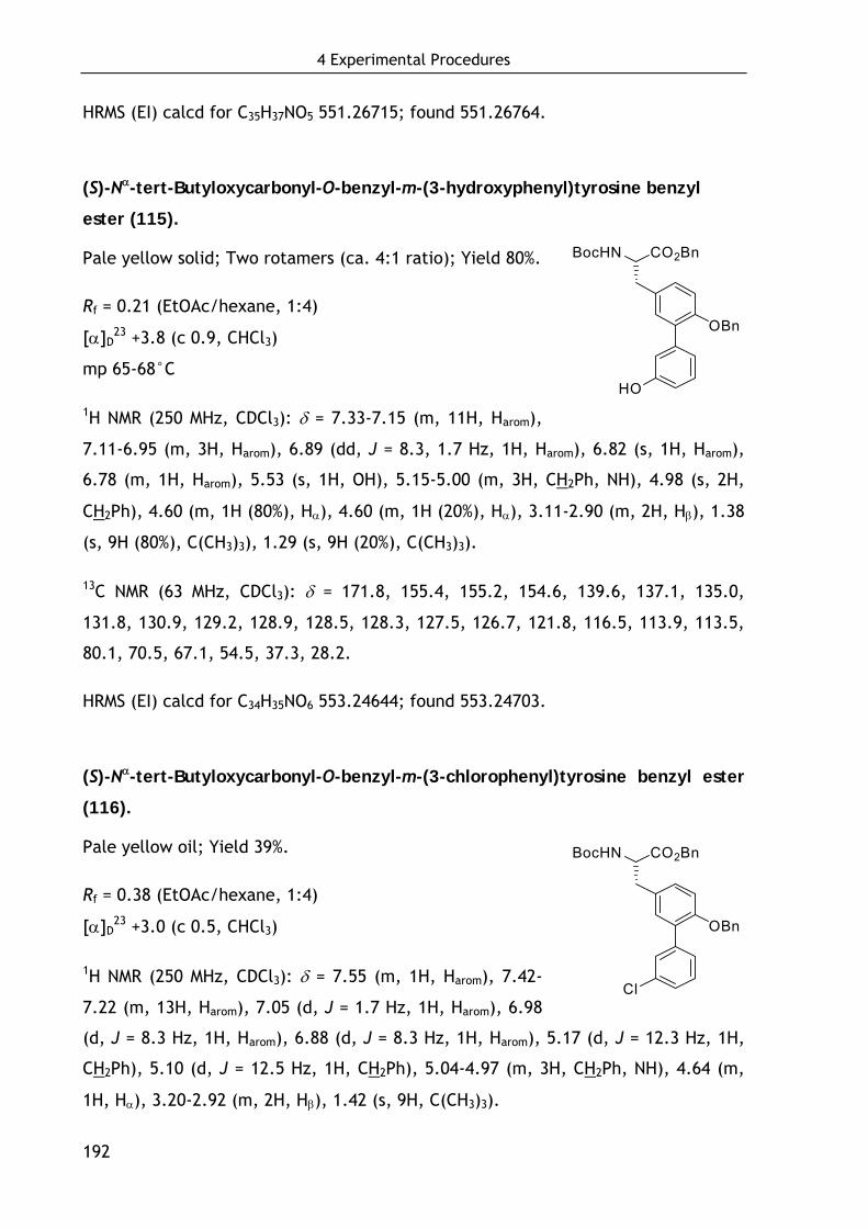
4 Experimental Procedures
192
HRMS (EI) calcd for C35H37NO5 551.26715; found 551.26764.
(S)-Nα-tert-Butyloxycarbonyl-O-benzyl-m-(3-hydroxyphenyl)tyrosine benzyl
ester (115).
Pale yellow solid; Two rotamers (ca. 4:1 ratio); Yield 80%.
Rf = 0.21 (EtOAc/hexane, 1:4)
[α]D23 +3.8 (c 0.9, CHCl3)
mp 65-68°C
1H NMR (250 MHz, CDCl3): δ = 7.33-7.15 (m, 11H, Harom),
7.11-6.95 (m, 3H, Harom), 6.89 (dd, J = 8.3, 1.7 Hz, 1H, Harom), 6.82 (s, 1H, Harom),
6.78 (m, 1H, Harom), 5.53 (s, 1H, OH), 5.15-5.00 (m, 3H, CH2Ph, NH), 4.98 (s, 2H,
CH2Ph), 4.60 (m, 1H (80%), Hα), 4.60 (m, 1H (20%), Hα), 3.11-2.90 (m, 2H, Hβ), 1.38
(s, 9H (80%), C(CH3)3), 1.29 (s, 9H (20%), C(CH3)3).
13C NMR (63 MHz, CDCl3): δ = 171.8, 155.4, 155.2, 154.6, 139.6, 137.1, 135.0,
131.8, 130.9, 129.2, 128.9, 128.5, 128.3, 127.5, 126.7, 121.8, 116.5, 113.9, 113.5,
80.1, 70.5, 67.1, 54.5, 37.3, 28.2.
HRMS (EI) calcd for C34H35NO6 553.24644; found 553.24703.
(S)-Nα-tert-Butyloxycarbonyl-O-benzyl-m-(3-chlorophenyl)tyrosine benzyl ester
(116).
Pale yellow oil; Yield 39%.
Rf = 0.38 (EtOAc/hexane, 1:4)
[α]D23 +3.0 (c 0.5, CHCl3)
1H NMR (250 MHz, CDCl3): δ = 7.55 (m, 1H, Harom), 7.42-
7.22 (m, 13H, Harom), 7.05 (d, J = 1.7 Hz, 1H, Harom), 6.98
(d, J = 8.3 Hz, 1H, Harom), 6.88 (d, J = 8.3 Hz, 1H, Harom), 5.17 (d, J = 12.3 Hz, 1H,
CH2Ph), 5.10 (d, J = 12.5 Hz, 1H, CH2Ph), 5.04-4.97 (m, 3H, CH2Ph, NH), 4.64 (m,
1H, Hα), 3.20-2.92 (m, 2H, Hβ), 1.42 (s, 9H, C(CH3)3).
CO2BnBocHN
OBn
HO
CO2BnBocHN
OBn
Cl

4 Experimental Procedures
193
13C NMR (63 MHz, CDCl3): δ = 171.7, 155.0, 154.5, 139.9, 136.9, 135.1, 133.7,
131.8, 129.9, 129.7, 129.1, 128.6, 128.54, 128.46, 128.4, 127.73, 127.68, 127.0,
126.8, 113.3, 80.0, 70.5, 67.1, 54.5, 37.4, 28.3.
HRMS (EI) calcd for C34H34ClNO5 571.21255; found 571.21109.
(S)-Nα-tert-Butyloxycarbonyl-O-benzyl-m-(4-fluorophenyl)tyrosine benzyl ester
(117).
Pale yellow solid; Yield 94%.
Rf = 0.25 (EtOAc/hexane, 1:4)
[α]D23 +1.3 (c 1.7, CHCl3)
mp 108-110°C
1H NMR (250 MHz, CDCl3): δ = 7.54-7.45 (m, 2H, Harom),
7.36-7.25 (m, 10H, Harom), 7.12-7.03 (m, 3H, Harom), 6.96
(d, J = 8.4Hz, 1H, Harom), 6.88 (d, J = 8.4 Hz, 1H, Harom), 5.17 (d, J = 12.2 Hz, 1H,
CH2Ph), 5.10 (d, J = 12.4 Hz, 1H, CH2Ph), 5.04 (m, 3H, CH2Ph, NH), 4.64 (m, 1H,
Hα), 3.20-2.95 (m, 2H, Hβ), 1.42 (s, 9H, C(CH3)3).
13C NMR (63 MHz, CDCl3): δ = 171.7, 163.9, 160.0, 155.0, 154.6, 137.0, 135.1,
134.11, 134.06, 131.9, 131.2, 131.1, 130.2, 129.4, 128.5, 128.43, 128.37, 127.7,
126.8, 114.9, 114.6, 113.3, 79.9, 70.5, 67.1, 54.5, 37.4, 28.3.
HRMS (EI) calcd for C34H34FNO5 555.24210; found 555.24184.
(S)-Nα-tert-Butyloxycarbonyl-O-benzyl-m-(4-carboxyphenyl)tyrosine benzyl
ester (118).
Pale yellow solid; Two rotamers (ca. 7:3 ratio); Yield
92%.
Rf = 0.35 (EtOAc/hexane 1:4, 1% AcOH)
[α]D23 –10.5 (c 0.02, CHCl3)
CO2BnBocHN
OBn
F
CO2BnBocHN
OBn
CO2H
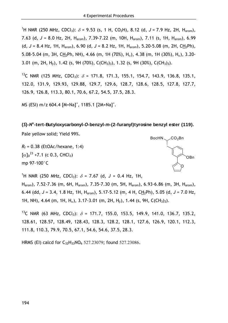
4 Experimental Procedures
194
1H NMR (250 MHz, CDCl3): δ = 9.53 (s, 1 H, CO2H), 8.12 (d, J = 7.9 Hz, 2H, Harom),
7.63 (d, J = 8.0 Hz, 2H, Harom), 7.39-7.22 (m, 10H, Harom), 7.11 (s, 1H, Harom), 6.99
(d, J = 8.4 Hz, 1H, Harom), 6.90 (d, J = 8.2 Hz, 1H, Harom), 5.20-5.08 (m, 2H, CH2Ph),
5.08-5.04 (m, 3H, CH2Ph, NH), 4.66 (m, 1H (70%), Hα), 4.38 (m, 1H (30%), Hα), 3.20-
3.01 (m, 2H, Hβ), 1.42 (s, 9H (70%), C(CH3)3), 1.32 (s, 9H (30%), C(CH3)3).
13C NMR (125 MHz, CDCl3): δ = 171.8, 171.3, 155.1, 154.7, 143.9, 136.8, 135.1,
132.0, 131.9, 129.93, 129.88, 129.7, 129.6, 128.7, 128.6, 128.5, 127.8, 127.7,
126.9, 126.8, 113.3, 80.1, 70.6, 67.2, 54.5, 37.5, 28.3.
MS (ESI) m/z 604.4 [M+Na]+, 1185.1 [2M+Na]+.
(S)-Nα-tert-Butyloxycarbonyl-O-benzyl-m-(2-furanyl)tyrosine benzyl ester (119).
Pale yellow solid; Yield 99%.
Rf = 0.38 (EtOAc/hexane, 1:4)
[α]D23 +7.1 (c 0.3, CHCl3)
mp 97-100°C
1H NMR (250 MHz, CDCl3): δ = 7.67 (d, J = 0.4 Hz, 1H,
Harom), 7.52-7.36 (m, 6H, Harom), 7.35-7.30 (m, 5H, Harom), 6.93-6.86 (m, 3H, Harom),
6.44 (dd, J = 3.4, 1.8 Hz, 1H, Harom), 5.17-5.12 (m, 4 H, CH2Ph), 5.05 (d, J = 7.0 Hz,
1H, NH), 4.64 (m, 1H, Hα), 3.17-3.01 (m, 2H, Hβ), 1.44 (s, 9H, C(CH3)3).
13C NMR (63 MHz, CDCl3): δ = 171.7, 155.0, 153.5, 149.9, 141.0, 136.7, 135.2,
128.61, 128.57, 128.49, 128.43, 128.3, 128.2, 128.1, 127.6, 126.9, 120.1, 112.3,
111.8, 110.3, 79.9, 70.5, 67.1, 54.6, 54.6, 37.5, 28.3.
HRMS (EI) calcd for C32H33NO6 527.23079; found 527.23086.
CO2BnBocHN
OBn
O
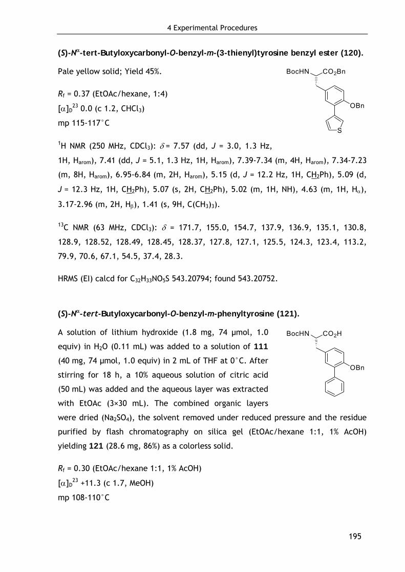
4 Experimental Procedures
195
(S)-Nα-tert-Butyloxycarbonyl-O-benzyl-m-(3-thienyl)tyrosine benzyl ester (120).
Pale yellow solid; Yield 45%.
Rf = 0.37 (EtOAc/hexane, 1:4)
[α]D23 0.0 (c 1.2, CHCl3)
mp 115-117°C
1H NMR (250 MHz, CDCl3): δ = 7.57 (dd, J = 3.0, 1.3 Hz,
1H, Harom), 7.41 (dd, J = 5.1, 1.3 Hz, 1H, Harom), 7.39-7.34 (m, 4H, Harom), 7.34-7.23
(m, 8H, Harom), 6.95-6.84 (m, 2H, Harom), 5.15 (d, J = 12.2 Hz, 1H, CH2Ph), 5.09 (d,
J = 12.3 Hz, 1H, CH2Ph), 5.07 (s, 2H, CH2Ph), 5.02 (m, 1H, NH), 4.63 (m, 1H, Hα),
3.17-2.96 (m, 2H, Hβ), 1.41 (s, 9H, C(CH3)3).
13C NMR (63 MHz, CDCl3): δ = 171.7, 155.0, 154.7, 137.9, 136.9, 135.1, 130.8,
128.9, 128.52, 128.49, 128.45, 128.37, 127.8, 127.1, 125.5, 124.3, 123.4, 113.2,
79.9, 70.6, 67.1, 54.5, 37.4, 28.3.
HRMS (EI) calcd for C32H33NO5S 543.20794; found 543.20752.
(S)-Nα-tert-Butyloxycarbonyl-O-benzyl-m-phenyltyrosine (121).
A solution of lithium hydroxide (1.8 mg, 74 μmol, 1.0
equiv) in H2O (0.11 mL) was added to a solution of 111
(40 mg, 74 μmol, 1.0 equiv) in 2 mL of THF at 0°C. After
stirring for 18 h, a 10% aqueous solution of citric acid
(50 mL) was added and the aqueous layer was extracted
with EtOAc (3×30 mL). The combined organic layers
were dried (Na2SO4), the solvent removed under reduced pressure and the residue
purified by flash chromatography on silica gel (EtOAc/hexane 1:1, 1% AcOH)
yielding 121 (28.6 mg, 86%) as a colorless solid.
Rf = 0.30 (EtOAc/hexane 1:1, 1% AcOH)
[α]D23 +11.3 (c 1.7, MeOH)
mp 108-110°C
CO2HBocHN
OBn
CO2BnBocHN
OBn
S
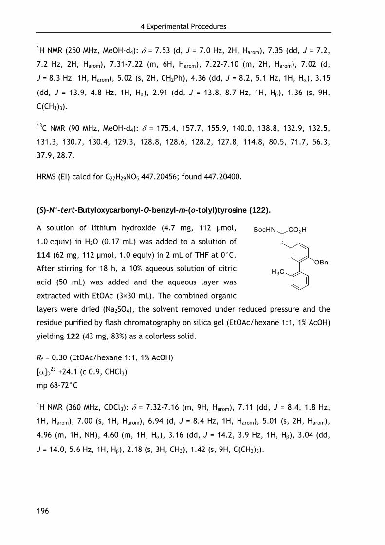
4 Experimental Procedures
196
1H NMR (250 MHz, MeOH-d4): δ = 7.53 (d, J = 7.0 Hz, 2H, Harom), 7.35 (dd, J = 7.2,
7.2 Hz, 2H, Harom), 7.31-7.22 (m, 6H, Harom), 7.22-7.10 (m, 2H, Harom), 7.02 (d,
J = 8.3 Hz, 1H, Harom), 5.02 (s, 2H, CH2Ph), 4.36 (dd, J = 8.2, 5.1 Hz, 1H, Hα), 3.15
(dd, J = 13.9, 4.8 Hz, 1H, Hβ), 2.91 (dd, J = 13.8, 8.7 Hz, 1H, Hβ), 1.36 (s, 9H,
C(CH3)3).
13C NMR (90 MHz, MeOH-d4): δ = 175.4, 157.7, 155.9, 140.0, 138.8, 132.9, 132.5,
131.3, 130.7, 130.4, 129.3, 128.8, 128.6, 128.2, 127.8, 114.8, 80.5, 71.7, 56.3,
37.9, 28.7.
HRMS (EI) calcd for C27H29NO5 447.20456; found 447.20400.
(S)-Nα-tert-Butyloxycarbonyl-O-benzyl-m-(o-tolyl)tyrosine (122).
A solution of lithium hydroxide (4.7 mg, 112 μmol,
1.0 equiv) in H2O (0.17 mL) was added to a solution of
114 (62 mg, 112 μmol, 1.0 equiv) in 2 mL of THF at 0°C.
After stirring for 18 h, a 10% aqueous solution of citric
acid (50 mL) was added and the aqueous layer was
extracted with EtOAc (3×30 mL). The combined organic
layers were dried (Na2SO4), the solvent removed under reduced pressure and the
residue purified by flash chromatography on silica gel (EtOAc/hexane 1:1, 1% AcOH)
yielding 122 (43 mg, 83%) as a colorless solid.
Rf = 0.30 (EtOAc/hexane 1:1, 1% AcOH)
[α]D23 +24.1 (c 0.9, CHCl3)
mp 68-72°C
1H NMR (360 MHz, CDCl3): δ = 7.32-7.16 (m, 9H, Harom), 7.11 (dd, J = 8.4, 1.8 Hz,
1H, Harom), 7.00 (s, 1H, Harom), 6.94 (d, J = 8.4 Hz, 1H, Harom), 5.01 (s, 2H, Harom),
4.96 (m, 1H, NH), 4.60 (m, 1H, Hα), 3.16 (dd, J = 14.2, 3.9 Hz, 1H, Hβ), 3.04 (dd,
J = 14.0, 5.6 Hz, 1H, Hβ), 2.18 (s, 3H, CH3), 1.42 (s, 9H, C(CH3)3).
CO2HBocHN
OBnH3C
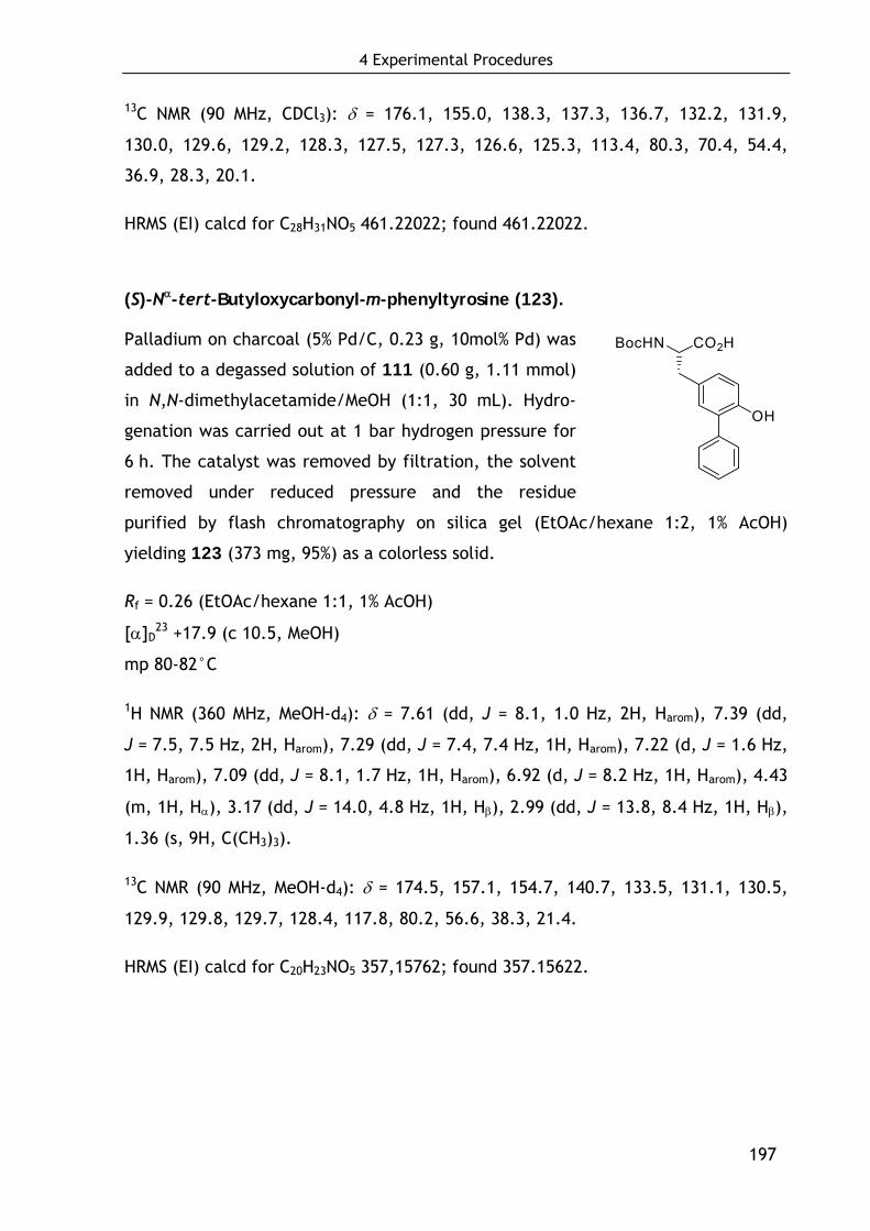
4 Experimental Procedures
197
13C NMR (90 MHz, CDCl3): δ = 176.1, 155.0, 138.3, 137.3, 136.7, 132.2, 131.9,
130.0, 129.6, 129.2, 128.3, 127.5, 127.3, 126.6, 125.3, 113.4, 80.3, 70.4, 54.4,
36.9, 28.3, 20.1.
HRMS (EI) calcd for C28H31NO5 461.22022; found 461.22022.
(S)-Nα-tert-Butyloxycarbonyl-m-phenyltyrosine (123).
Palladium on charcoal (5% Pd/C, 0.23 g, 10mol% Pd) was
added to a degassed solution of 111 (0.60 g, 1.11 mmol)
in N,N-dimethylacetamide/MeOH (1:1, 30 mL). Hydro-
genation was carried out at 1 bar hydrogen pressure for
6 h. The catalyst was removed by filtration, the solvent
removed under reduced pressure and the residue
purified by flash chromatography on silica gel (EtOAc/hexane 1:2, 1% AcOH)
yielding 123 (373 mg, 95%) as a colorless solid.
Rf = 0.26 (EtOAc/hexane 1:1, 1% AcOH)
[α]D23 +17.9 (c 10.5, MeOH)
mp 80-82°C
1H NMR (360 MHz, MeOH-d4): δ = 7.61 (dd, J = 8.1, 1.0 Hz, 2H, Harom), 7.39 (dd,
J = 7.5, 7.5 Hz, 2H, Harom), 7.29 (dd, J = 7.4, 7.4 Hz, 1H, Harom), 7.22 (d, J = 1.6 Hz,
1H, Harom), 7.09 (dd, J = 8.1, 1.7 Hz, 1H, Harom), 6.92 (d, J = 8.2 Hz, 1H, Harom), 4.43
(m, 1H, Hα), 3.17 (dd, J = 14.0, 4.8 Hz, 1H, Hβ), 2.99 (dd, J = 13.8, 8.4 Hz, 1H, Hβ),
1.36 (s, 9H, C(CH3)3).
13C NMR (90 MHz, MeOH-d4): δ = 174.5, 157.1, 154.7, 140.7, 133.5, 131.1, 130.5,
129.9, 129.8, 129.7, 128.4, 117.8, 80.2, 56.6, 38.3, 21.4.
HRMS (EI) calcd for C20H23NO5 357,15762; found 357.15622.
CO2HBocHN
OH
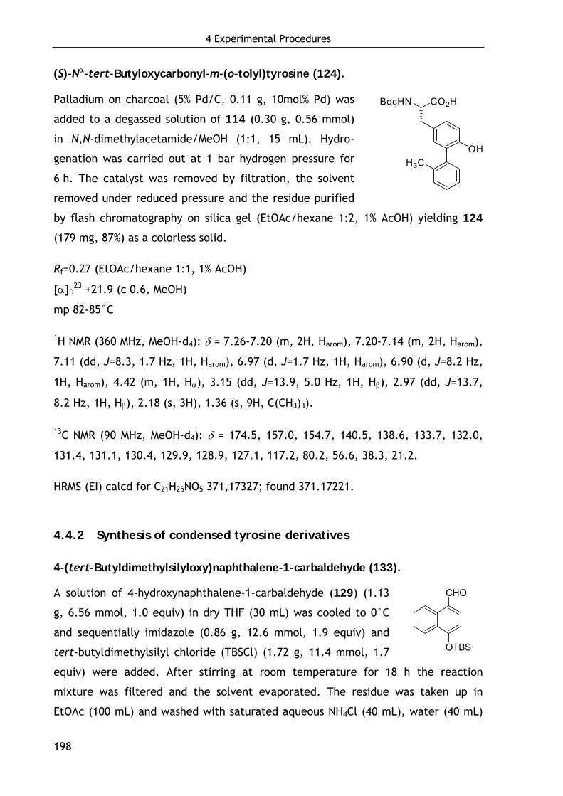
4 Experimental Procedures
198
CHO
OTBS
(S)-Nα-tert-Butyloxycarbonyl-m-(o-tolyl)tyrosine (124).
Palladium on charcoal (5% Pd/C, 0.11 g, 10mol% Pd) was
added to a degassed solution of 114 (0.30 g, 0.56 mmol)
in N,N-dimethylacetamide/MeOH (1:1, 15 mL). Hydro-
genation was carried out at 1 bar hydrogen pressure for
6 h. The catalyst was removed by filtration, the solvent
removed under reduced pressure and the residue purified
by flash chromatography on silica gel (EtOAc/hexane 1:2, 1% AcOH) yielding 124
(179 mg, 87%) as a colorless solid.
Rf=0.27 (EtOAc/hexane 1:1, 1% AcOH)
[α]D23 +21.9 (c 0.6, MeOH)
mp 82-85°C
1H NMR (360 MHz, MeOH-d4): δ = 7.26-7.20 (m, 2H, Harom), 7.20-7.14 (m, 2H, Harom),
7.11 (dd, J=8.3, 1.7 Hz, 1H, Harom), 6.97 (d, J=1.7 Hz, 1H, Harom), 6.90 (d, J=8.2 Hz,
1H, Harom), 4.42 (m, 1H, Hα), 3.15 (dd, J=13.9, 5.0 Hz, 1H, Hβ), 2.97 (dd, J=13.7,
8.2 Hz, 1H, Hβ), 2.18 (s, 3H), 1.36 (s, 9H, C(CH3)3).
13C NMR (90 MHz, MeOH-d4): δ = 174.5, 157.0, 154.7, 140.5, 138.6, 133.7, 132.0,
131.4, 131.1, 130.4, 129.9, 128.9, 127.1, 117.2, 80.2, 56.6, 38.3, 21.2.
HRMS (EI) calcd for C21H25NO5 371,17327; found 371.17221.
4.4.2 Synthesis of condensed tyrosine derivatives
4-(tert-Butyldimethylsilyloxy)naphthalene-1-carbaldehyde (133).
A solution of 4-hydroxynaphthalene-1-carbaldehyde (129) (1.13
g, 6.56 mmol, 1.0 equiv) in dry THF (30 mL) was cooled to 0°C
and sequentially imidazole (0.86 g, 12.6 mmol, 1.9 equiv) and
tert-butyldimethylsilyl chloride (TBSCl) (1.72 g, 11.4 mmol, 1.7
equiv) were added. After stirring at room temperature for 18 h the reaction
mixture was filtered and the solvent evaporated. The residue was taken up in
EtOAc (100 mL) and washed with saturated aqueous NH4Cl (40 mL), water (40 mL)
CO2HBocHN
OHH3C
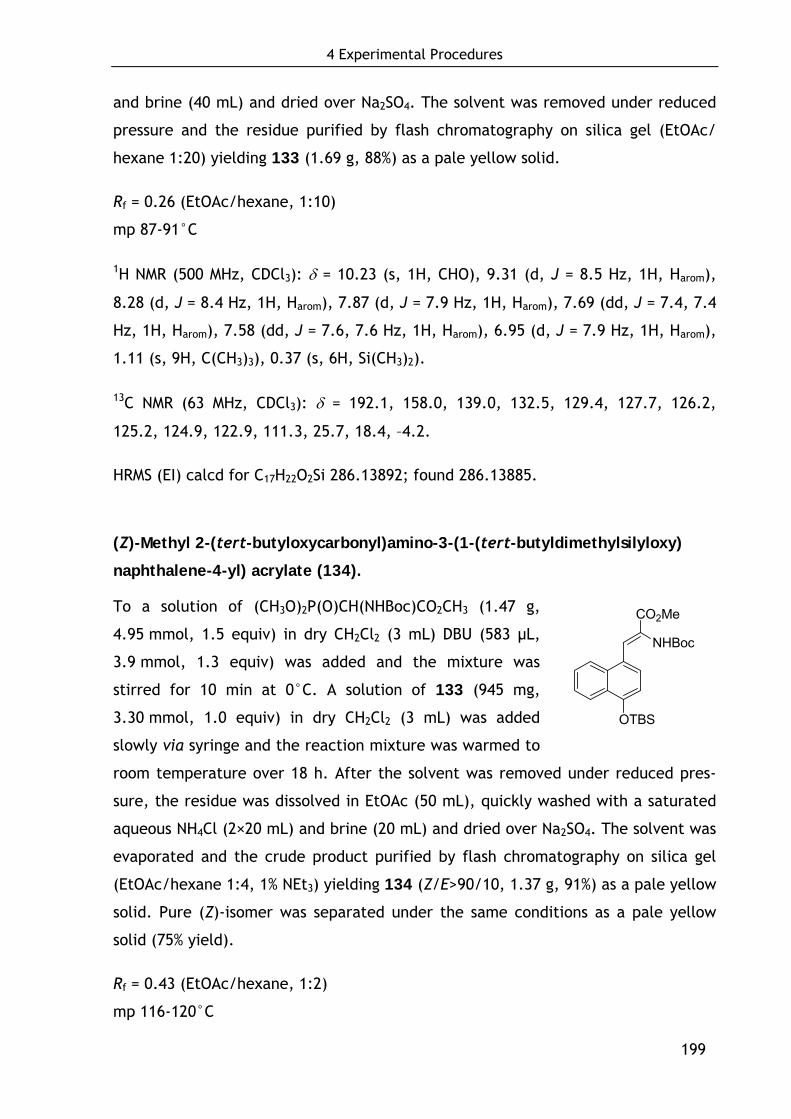
4 Experimental Procedures
199
OTBS
CO2Me
NHBoc
and brine (40 mL) and dried over Na2SO4. The solvent was removed under reduced
pressure and the residue purified by flash chromatography on silica gel (EtOAc/
hexane 1:20) yielding 133 (1.69 g, 88%) as a pale yellow solid.
Rf = 0.26 (EtOAc/hexane, 1:10)
mp 87-91°C
1H NMR (500 MHz, CDCl3): δ = 10.23 (s, 1H, CHO), 9.31 (d, J = 8.5 Hz, 1H, Harom),
8.28 (d, J = 8.4 Hz, 1H, Harom), 7.87 (d, J = 7.9 Hz, 1H, Harom), 7.69 (dd, J = 7.4, 7.4
Hz, 1H, Harom), 7.58 (dd, J = 7.6, 7.6 Hz, 1H, Harom), 6.95 (d, J = 7.9 Hz, 1H, Harom),
1.11 (s, 9H, C(CH3)3), 0.37 (s, 6H, Si(CH3)2).
13C NMR (63 MHz, CDCl3): δ = 192.1, 158.0, 139.0, 132.5, 129.4, 127.7, 126.2,
125.2, 124.9, 122.9, 111.3, 25.7, 18.4, –4.2.
HRMS (EI) calcd for C17H22O2Si 286.13892; found 286.13885.
(Z)-Methyl 2-(tert-butyloxycarbonyl)amino-3-(1-(tert-butyldimethylsilyloxy)
naphthalene-4-yl) acrylate (134).
To a solution of (CH3O)2P(O)CH(NHBoc)CO2CH3 (1.47 g,
4.95 mmol, 1.5 equiv) in dry CH2Cl2 (3 mL) DBU (583 μL,
3.9 mmol, 1.3 equiv) was added and the mixture was
stirred for 10 min at 0°C. A solution of 133 (945 mg,
3.30 mmol, 1.0 equiv) in dry CH2Cl2 (3 mL) was added
slowly via syringe and the reaction mixture was warmed to
room temperature over 18 h. After the solvent was removed under reduced pres-
sure, the residue was dissolved in EtOAc (50 mL), quickly washed with a saturated
aqueous NH4Cl (2×20 mL) and brine (20 mL) and dried over Na2SO4. The solvent was
evaporated and the crude product purified by flash chromatography on silica gel
(EtOAc/hexane 1:4, 1% NEt3) yielding 134 (Z/E>90/10, 1.37 g, 91%) as a pale yellow
solid. Pure (Z)-isomer was separated under the same conditions as a pale yellow
solid (75% yield).
Rf = 0.43 (EtOAc/hexane, 1:2)
mp 116-120°C
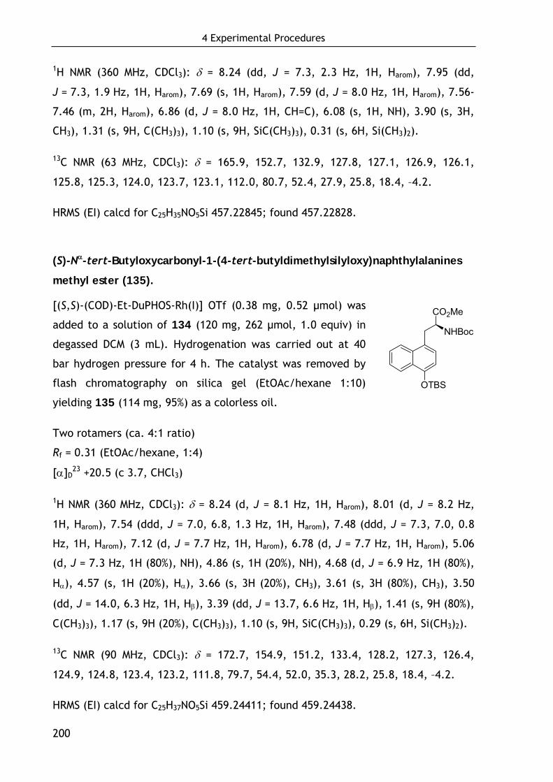
4 Experimental Procedures
200
1H NMR (360 MHz, CDCl3): δ = 8.24 (dd, J = 7.3, 2.3 Hz, 1H, Harom), 7.95 (dd,
J = 7.3, 1.9 Hz, 1H, Harom), 7.69 (s, 1H, Harom), 7.59 (d, J = 8.0 Hz, 1H, Harom), 7.56-
7.46 (m, 2H, Harom), 6.86 (d, J = 8.0 Hz, 1H, CH=C), 6.08 (s, 1H, NH), 3.90 (s, 3H,
CH3), 1.31 (s, 9H, C(CH3)3), 1.10 (s, 9H, SiC(CH3)3), 0.31 (s, 6H, Si(CH3)2).
13C NMR (63 MHz, CDCl3): δ = 165.9, 152.7, 132.9, 127.8, 127.1, 126.9, 126.1,
125.8, 125.3, 124.0, 123.7, 123.1, 112.0, 80.7, 52.4, 27.9, 25.8, 18.4, –4.2.
HRMS (EI) calcd for C25H35NO5Si 457.22845; found 457.22828.
(S)-Nα-tert-Butyloxycarbonyl-1-(4-tert-butyldimethylsilyloxy)naphthylalanines
methyl ester (135).
[(S,S)-(COD)-Et-DuPHOS-Rh(I)] OTf (0.38 mg, 0.52 μmol) was
added to a solution of 134 (120 mg, 262 μmol, 1.0 equiv) in
degassed DCM (3 mL). Hydrogenation was carried out at 40
bar hydrogen pressure for 4 h. The catalyst was removed by
flash chromatography on silica gel (EtOAc/hexane 1:10)
yielding 135 (114 mg, 95%) as a colorless oil.
Two rotamers (ca. 4:1 ratio)
Rf = 0.31 (EtOAc/hexane, 1:4)
[α]D23 +20.5 (c 3.7, CHCl3)
1H NMR (360 MHz, CDCl3): δ = 8.24 (d, J = 8.1 Hz, 1H, Harom), 8.01 (d, J = 8.2 Hz,
1H, Harom), 7.54 (ddd, J = 7.0, 6.8, 1.3 Hz, 1H, Harom), 7.48 (ddd, J = 7.3, 7.0, 0.8
Hz, 1H, Harom), 7.12 (d, J = 7.7 Hz, 1H, Harom), 6.78 (d, J = 7.7 Hz, 1H, Harom), 5.06
(d, J = 7.3 Hz, 1H (80%), NH), 4.86 (s, 1H (20%), NH), 4.68 (d, J = 6.9 Hz, 1H (80%),
Hα), 4.57 (s, 1H (20%), Hα), 3.66 (s, 3H (20%), CH3), 3.61 (s, 3H (80%), CH3), 3.50
(dd, J = 14.0, 6.3 Hz, 1H, Hβ), 3.39 (dd, J = 13.7, 6.6 Hz, 1H, Hβ), 1.41 (s, 9H (80%),
C(CH3)3), 1.17 (s, 9H (20%), C(CH3)3), 1.10 (s, 9H, SiC(CH3)3), 0.29 (s, 6H, Si(CH3)2).
13C NMR (90 MHz, CDCl3): δ = 172.7, 154.9, 151.2, 133.4, 128.2, 127.3, 126.4,
124.9, 124.8, 123.4, 123.2, 111.8, 79.7, 54.4, 52.0, 35.3, 28.2, 25.8, 18.4, –4.2.
HRMS (EI) calcd for C25H37NO5Si 459.24411; found 459.24438.
OTBS
CO2Me
NHBoc
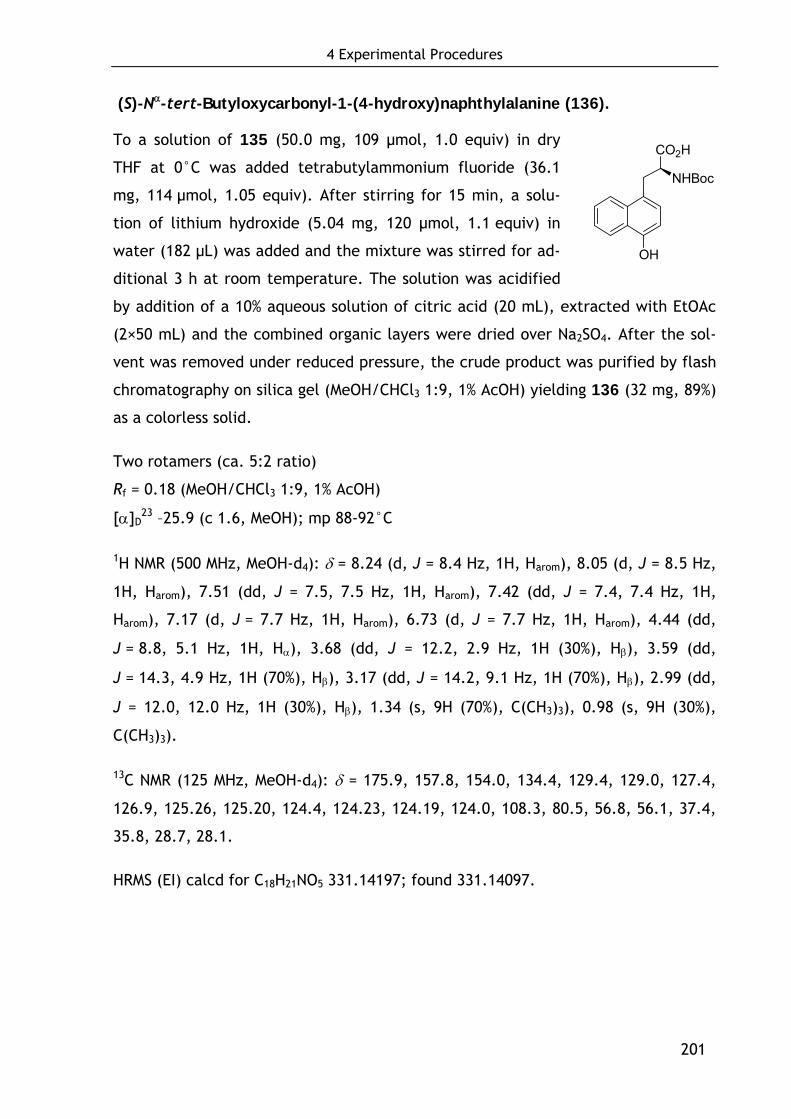
4 Experimental Procedures
201
(S)-Nα-tert-Butyloxycarbonyl-1-(4-hydroxy)naphthylalanine (136).
To a solution of 135 (50.0 mg, 109 μmol, 1.0 equiv) in dry
THF at 0°C was added tetrabutylammonium fluoride (36.1
mg, 114 μmol, 1.05 equiv). After stirring for 15 min, a solu-
tion of lithium hydroxide (5.04 mg, 120 μmol, 1.1 equiv) in
water (182 μL) was added and the mixture was stirred for ad-
ditional 3 h at room temperature. The solution was acidified
by addition of a 10% aqueous solution of citric acid (20 mL), extracted with EtOAc
(2×50 mL) and the combined organic layers were dried over Na2SO4. After the sol-
vent was removed under reduced pressure, the crude product was purified by flash
chromatography on silica gel (MeOH/CHCl3 1:9, 1% AcOH) yielding 136 (32 mg, 89%)
as a colorless solid.
Two rotamers (ca. 5:2 ratio)
Rf = 0.18 (MeOH/CHCl3 1:9, 1% AcOH)
[α]D23 –25.9 (c 1.6, MeOH); mp 88-92°C
1H NMR (500 MHz, MeOH-d4): δ = 8.24 (d, J = 8.4 Hz, 1H, Harom), 8.05 (d, J = 8.5 Hz,
1H, Harom), 7.51 (dd, J = 7.5, 7.5 Hz, 1H, Harom), 7.42 (dd, J = 7.4, 7.4 Hz, 1H,
Harom), 7.17 (d, J = 7.7 Hz, 1H, Harom), 6.73 (d, J = 7.7 Hz, 1H, Harom), 4.44 (dd,
J = 8.8, 5.1 Hz, 1H, Hα), 3.68 (dd, J = 12.2, 2.9 Hz, 1H (30%), Hβ), 3.59 (dd,
J = 14.3, 4.9 Hz, 1H (70%), Hβ), 3.17 (dd, J = 14.2, 9.1 Hz, 1H (70%), Hβ), 2.99 (dd,
J = 12.0, 12.0 Hz, 1H (30%), Hβ), 1.34 (s, 9H (70%), C(CH3)3), 0.98 (s, 9H (30%),
C(CH3)3).
13C NMR (125 MHz, MeOH-d4): δ = 175.9, 157.8, 154.0, 134.4, 129.4, 129.0, 127.4,
126.9, 125.26, 125.20, 124.4, 124.23, 124.19, 124.0, 108.3, 80.5, 56.8, 56.1, 37.4,
35.8, 28.7, 28.1.
HRMS (EI) calcd for C18H21NO5 331.14197; found 331.14097.
OH
CO2H
NHBoc
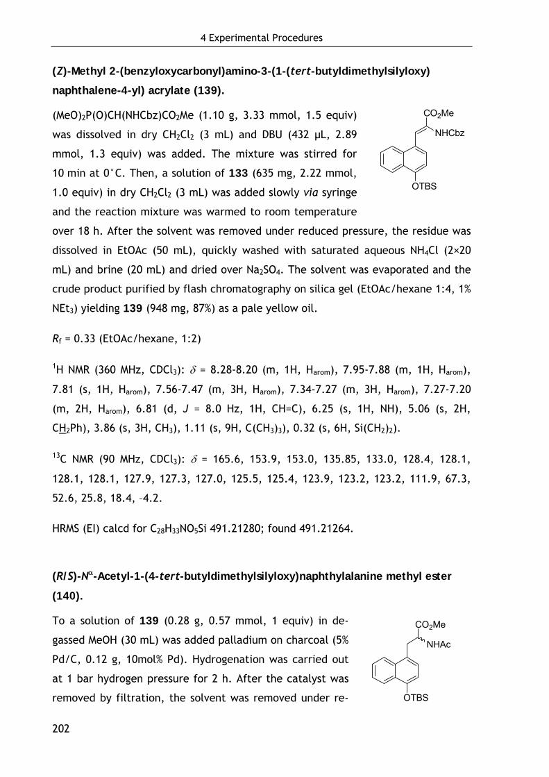
4 Experimental Procedures
202
OTBS
CO2Me
NHCbz
OTBS
CO2Me
NHAc
(Z)-Methyl 2-(benzyloxycarbonyl)amino-3-(1-(tert-butyldimethylsilyloxy)
naphthalene-4-yl) acrylate (139).
(MeO)2P(O)CH(NHCbz)CO2Me (1.10 g, 3.33 mmol, 1.5 equiv)
was dissolved in dry CH2Cl2 (3 mL) and DBU (432 μL, 2.89
mmol, 1.3 equiv) was added. The mixture was stirred for
10 min at 0°C. Then, a solution of 133 (635 mg, 2.22 mmol,
1.0 equiv) in dry CH2Cl2 (3 mL) was added slowly via syringe
and the reaction mixture was warmed to room temperature
over 18 h. After the solvent was removed under reduced pressure, the residue was
dissolved in EtOAc (50 mL), quickly washed with saturated aqueous NH4Cl (2×20
mL) and brine (20 mL) and dried over Na2SO4. The solvent was evaporated and the
crude product purified by flash chromatography on silica gel (EtOAc/hexane 1:4, 1%
NEt3) yielding 139 (948 mg, 87%) as a pale yellow oil.
Rf = 0.33 (EtOAc/hexane, 1:2)
1H NMR (360 MHz, CDCl3): δ = 8.28-8.20 (m, 1H, Harom), 7.95-7.88 (m, 1H, Harom),
7.81 (s, 1H, Harom), 7.56-7.47 (m, 3H, Harom), 7.34-7.27 (m, 3H, Harom), 7.27-7.20
(m, 2H, Harom), 6.81 (d, J = 8.0 Hz, 1H, CH=C), 6.25 (s, 1H, NH), 5.06 (s, 2H,
CH2Ph), 3.86 (s, 3H, CH3), 1.11 (s, 9H, C(CH3)3), 0.32 (s, 6H, Si(CH2)2).
13C NMR (90 MHz, CDCl3): δ = 165.6, 153.9, 153.0, 135.85, 133.0, 128.4, 128.1,
128.1, 128.1, 127.9, 127.3, 127.0, 125.5, 125.4, 123.9, 123.2, 123.2, 111.9, 67.3,
52.6, 25.8, 18.4, –4.2.
HRMS (EI) calcd for C28H33NO5Si 491.21280; found 491.21264.
(R/S)-Nα-Acetyl-1-(4-tert-butyldimethylsilyloxy)naphthylalanine methyl ester
(140).
To a solution of 139 (0.28 g, 0.57 mmol, 1 equiv) in de-
gassed MeOH (30 mL) was added palladium on charcoal (5%
Pd/C, 0.12 g, 10mol% Pd). Hydrogenation was carried out
at 1 bar hydrogen pressure for 2 h. After the catalyst was
removed by filtration, the solvent was removed under re-
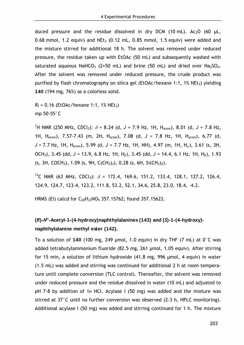
4 Experimental Procedures
203
duced pressure and the residue dissolved in dry DCM (10 mL). Ac2O (60 μL,
0.68 mmol, 1.2 equiv) and NEt3 (0.12 mL, 0.85 mmol, 1.5 equiv) were added and
the mixture stirred for additional 18 h. The solvent was removed under reduced
pressure, the residue taken up with EtOAc (50 mL) and subsequently washed with
saturated aqueous NaHCO3 (2×50 mL) and brine (50 mL) and dried over Na2SO4.
After the solvent was removed under reduced pressure, the crude product was
purified by flash chromatography on silica gel (EtOAc/hexane 1:1, 1% NEt3) yielding
140 (194 mg, 76%) as a colorless solid.
Rf = 0.16 (EtOAc/hexane 1:1, 1% NEt3)
mp 50-55°C
1H NMR (250 MHz, CDCl3): δ = 8.24 (d, J = 7.9 Hz, 1H, Harom), 8.01 (d, J = 7.8 Hz,
1H, Harom), 7.57-7.43 (m, 2H, Harom), 7.08 (d, J = 7.8 Hz, 1H, Harom), 6.77 (d,
J = 7.7 Hz, 1H, Harom), 5.99 (d, J = 7.7 Hz, 1H, NH), 4.97 (m, 1H, Hα), 3.61 (s, 3H,
OCH3), 3.45 (dd, J = 13.9, 6.8 Hz, 1H, Hβ), 3.45 (dd, J = 14.4, 6.1 Hz, 1H, Hβ), 1.93
(s, 3H, COCH3), 1.09 (s, 9H, C(CH3)3), 0.28 (s, 6H, Si(CH3)2).
13C NMR (63 MHz, CDCl3): δ = 172.4, 169.6, 151.2, 133.4, 128.1, 127.2, 126.4,
124.9, 124.7, 123.4, 123.2, 111.8, 53.2, 52.1, 34.6, 25.8, 23.0, 18.4, –4.2.
HRMS (EI) calcd for C20H23NO5 357.15762; found 357.15622.
(R)-Nα-Acetyl-1-(4-hydroxy)naphthylalanines (143) and (S)-1-(4-hydroxy)-
naphthylalanine methyl ester (142).
To a solution of 140 (100 mg, 249 μmol, 1.0 equiv) in dry THF (7 mL) at 0°C was
added tetrabutylammonium fluoride (82.5 mg, 261 μmol, 1.05 equiv). After stirring
for 15 min, a solution of lithium hydroxide (41.8 mg, 996 μmol, 4 equiv) in water
(1.5 mL) was added and stirring was continued for additional 2 h at room tempera-
ture until complete conversion (TLC control). Thereafter, the solvent was removed
under reduced pressure and the residue dissolved in water (10 mL) and adjusted to
pH 7-8 by addition of 1N HCl. Acylase I (50 mg) was added and the mixture was
stirred at 37°C until no further conversion was observed (2-3 h, HPLC monitoring).
Additional acylase I (50 mg) was added and stirring continued for 1 h. The mixture
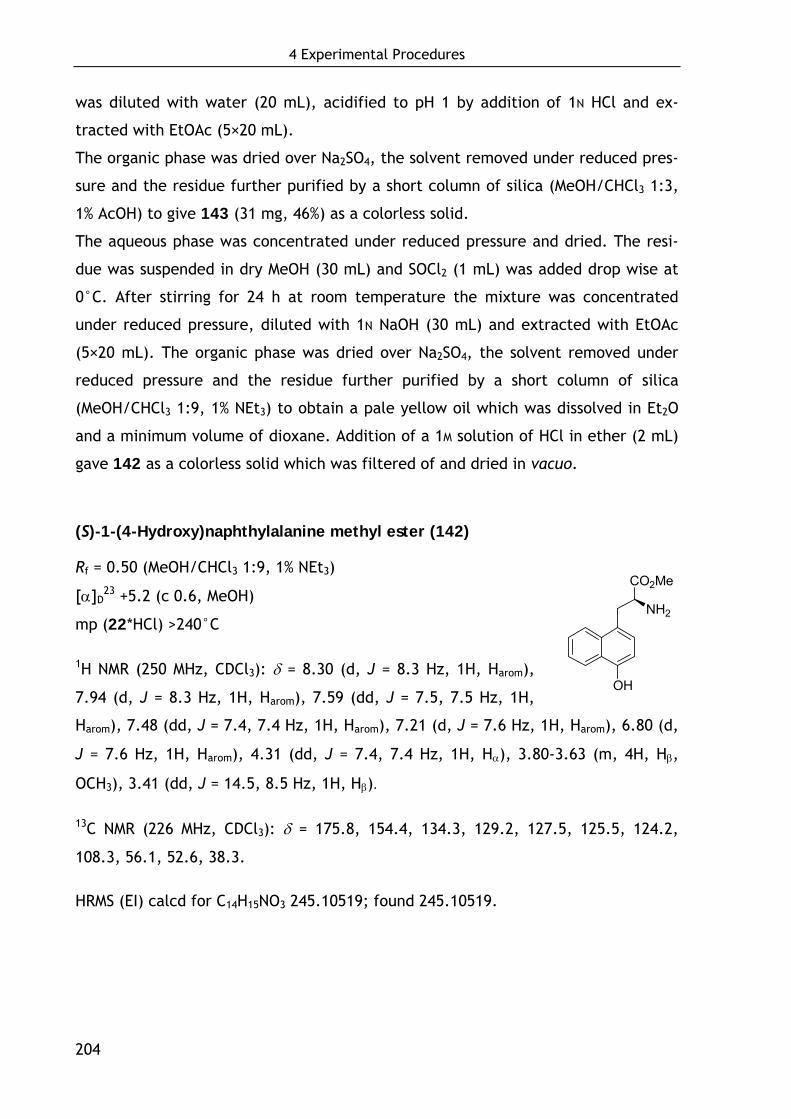
4 Experimental Procedures
204
was diluted with water (20 mL), acidified to pH 1 by addition of 1N HCl and ex-
tracted with EtOAc (5×20 mL).
The organic phase was dried over Na2SO4, the solvent removed under reduced pres-
sure and the residue further purified by a short column of silica (MeOH/CHCl3 1:3,
1% AcOH) to give 143 (31 mg, 46%) as a colorless solid.
The aqueous phase was concentrated under reduced pressure and dried. The resi-
due was suspended in dry MeOH (30 mL) and SOCl2 (1 mL) was added drop wise at
0°C. After stirring for 24 h at room temperature the mixture was concentrated
under reduced pressure, diluted with 1N NaOH (30 mL) and extracted with EtOAc
(5×20 mL). The organic phase was dried over Na2SO4, the solvent removed under
reduced pressure and the residue further purified by a short column of silica
(MeOH/CHCl3 1:9, 1% NEt3) to obtain a pale yellow oil which was dissolved in Et2O
and a minimum volume of dioxane. Addition of a 1M solution of HCl in ether (2 mL)
gave 142 as a colorless solid which was filtered of and dried in vacuo.
(S)-1-(4-Hydroxy)naphthylalanine methyl ester (142)
Rf = 0.50 (MeOH/CHCl3 1:9, 1% NEt3)
[α]D23 +5.2 (c 0.6, MeOH)
mp (22*HCl) >240°C
1H NMR (250 MHz, CDCl3): δ = 8.30 (d, J = 8.3 Hz, 1H, Harom),
7.94 (d, J = 8.3 Hz, 1H, Harom), 7.59 (dd, J = 7.5, 7.5 Hz, 1H,
Harom), 7.48 (dd, J = 7.4, 7.4 Hz, 1H, Harom), 7.21 (d, J = 7.6 Hz, 1H, Harom), 6.80 (d,
J = 7.6 Hz, 1H, Harom), 4.31 (dd, J = 7.4, 7.4 Hz, 1H, Hα), 3.80-3.63 (m, 4H, Hβ,
OCH3), 3.41 (dd, J = 14.5, 8.5 Hz, 1H, Hβ).
13C NMR (226 MHz, CDCl3): δ = 175.8, 154.4, 134.3, 129.2, 127.5, 125.5, 124.2,
108.3, 56.1, 52.6, 38.3.
HRMS (EI) calcd for C14H15NO3 245.10519; found 245.10519.
OH
CO2Me
NH2
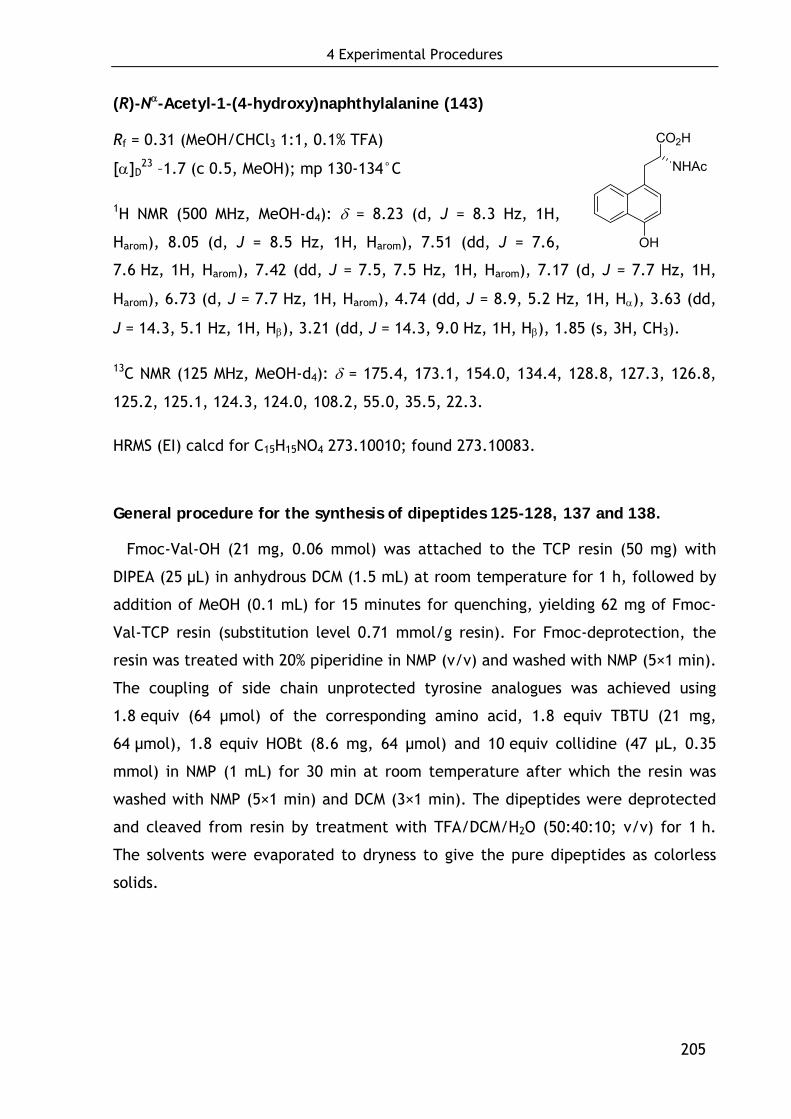
4 Experimental Procedures
205
(R)-Nα-Acetyl-1-(4-hydroxy)naphthylalanine (143)
Rf = 0.31 (MeOH/CHCl3 1:1, 0.1% TFA)
[α]D23 –1.7 (c 0.5, MeOH); mp 130-134°C
1H NMR (500 MHz, MeOH-d4): δ = 8.23 (d, J = 8.3 Hz, 1H,
Harom), 8.05 (d, J = 8.5 Hz, 1H, Harom), 7.51 (dd, J = 7.6,
7.6 Hz, 1H, Harom), 7.42 (dd, J = 7.5, 7.5 Hz, 1H, Harom), 7.17 (d, J = 7.7 Hz, 1H,
Harom), 6.73 (d, J = 7.7 Hz, 1H, Harom), 4.74 (dd, J = 8.9, 5.2 Hz, 1H, Hα), 3.63 (dd,
J = 14.3, 5.1 Hz, 1H, Hβ), 3.21 (dd, J = 14.3, 9.0 Hz, 1H, Hβ), 1.85 (s, 3H, CH3).
13C NMR (125 MHz, MeOH-d4): δ = 175.4, 173.1, 154.0, 134.4, 128.8, 127.3, 126.8,
125.2, 125.1, 124.3, 124.0, 108.2, 55.0, 35.5, 22.3.
HRMS (EI) calcd for C15H15NO4 273.10010; found 273.10083.
General procedure for the synthesis of dipeptides 125-128, 137 and 138.
Fmoc-Val-OH (21 mg, 0.06 mmol) was attached to the TCP resin (50 mg) with
DIPEA (25 μL) in anhydrous DCM (1.5 mL) at room temperature for 1 h, followed by
addition of MeOH (0.1 mL) for 15 minutes for quenching, yielding 62 mg of Fmoc-
Val-TCP resin (substitution level 0.71 mmol/g resin). For Fmoc-deprotection, the
resin was treated with 20% piperidine in NMP (v/v) and washed with NMP (5×1 min).
The coupling of side chain unprotected tyrosine analogues was achieved using
1.8 equiv (64 μmol) of the corresponding amino acid, 1.8 equiv TBTU (21 mg,
64 μmol), 1.8 equiv HOBt (8.6 mg, 64 μmol) and 10 equiv collidine (47 μL, 0.35
mmol) in NMP (1 mL) for 30 min at room temperature after which the resin was
washed with NMP (5×1 min) and DCM (3×1 min). The dipeptides were deprotected
and cleaved from resin by treatment with TFA/DCM/H2O (50:40:10; v/v) for 1 h.
The solvents were evaporated to dryness to give the pure dipeptides as colorless
solids.
OH
CO2H
NHAc
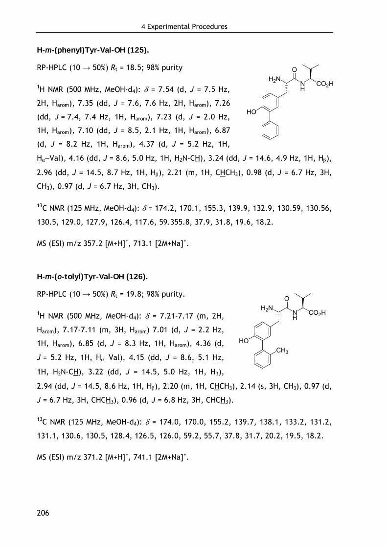
4 Experimental Procedures
206
H-m-(phenyl)Tyr-Val-OH (125).
RP-HPLC (10 → 50%) Rt = 18.5; 98% purity
1H NMR (500 MHz, MeOH-d4): δ = 7.54 (d, J = 7.5 Hz,
2H, Harom), 7.35 (dd, J = 7.6, 7.6 Hz, 2H, Harom), 7.26
(dd, J = 7.4, 7.4 Hz, 1H, Harom), 7.23 (d, J = 2.0 Hz,
1H, Harom), 7.10 (dd, J = 8.5, 2.1 Hz, 1H, Harom), 6.87
(d, J = 8.2 Hz, 1H, Harom), 4.37 (d, J = 5.2 Hz, 1H,
Hα−Val), 4.16 (dd, J = 8.6, 5.0 Hz, 1H, H2N-CH), 3.24 (dd, J = 14.6, 4.9 Hz, 1H, Hβ),
2.96 (dd, J = 14.5, 8.7 Hz, 1H, Hβ), 2.21 (m, 1H, CHCH3), 0.98 (d, J = 6.7 Hz, 3H,
CH3), 0.97 (d, J = 6.7 Hz, 3H, CH3).
13C NMR (125 MHz, MeOH-d4): δ = 174.2, 170.1, 155.3, 139.9, 132.9, 130.59, 130.56,
130.5, 129.0, 127.9, 126.4, 117.6, 59.355.8, 37.9, 31.8, 19.6, 18.2.
MS (ESI) m/z 357.2 [M+H]+, 713.1 [2M+Na]+.
H-m-(o-tolyl)Tyr-Val-OH (126).
RP-HPLC (10 → 50%) Rt = 19.8; 98% purity.
1H NMR (500 MHz, MeOH-d4): δ = 7.21-7.17 (m, 2H,
Harom), 7.17-7.11 (m, 3H, Harom) 7.01 (d, J = 2.2 Hz,
1H, Harom), 6.85 (d, J = 8.3 Hz, 1H, Harom), 4.36 (d,
J = 5.2 Hz, 1H, Hα−Val), 4.15 (dd, J = 8.6, 5.1 Hz,
1H, H2N-CH), 3.22 (dd, J = 14.5, 5.0 Hz, 1H, Hβ),
2.94 (dd, J = 14.5, 8.6 Hz, 1H, Hβ), 2.20 (m, 1H, CHCH3), 2.14 (s, 3H, CH3), 0.97 (d,
J = 6.7 Hz, 3H, CHCH3), 0.96 (d, J = 6.8 Hz, 3H, CHCH3).
13C NMR (125 MHz, MeOH-d4): δ = 174.0, 170.0, 155.2, 139.7, 138.1, 133.2, 131.2,
131.1, 130.6, 130.5, 128.4, 126.5, 126.0, 59.2, 55.7, 37.8, 31.7, 20.2, 19.5, 18.2.
MS (ESI) m/z 371.2 [M+H]+, 741.1 [2M+Na]+.
H2N
HO
NH
O
CO2H
H2N
HO
NH
O
CO2H
CH3
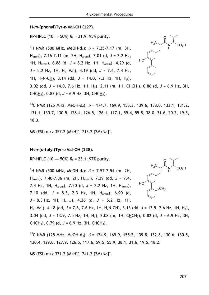
4 Experimental Procedures
207
H-m-(phenyl)Tyr-D-Val-OH (127).
RP-HPLC (10 → 50%) Rt = 21.9: 95% purity.
1H NMR (500 MHz, MeOH-d4): δ = 7.25-7.17 (m, 3H,
Harom), 7.16-7.11 (m, 2H, Harom), 7.01 (d, J = 2.2 Hz,
1H, Harom), 6.88 (d, J = 8.2 Hz, 1H, Harom), 4.29 (d,
J = 5.2 Hz, 1H, Hα−Val), 4.19 (dd, J = 7.4, 7.4 Hz,
1H, H2N-CH), 3.14 (dd, J = 14.0, 7.2 Hz, 1H, Hβ),
3.02 (dd, J = 14.0, 7.6 Hz, 1H, Hβ), 2.11 (m, 1H, CHCH3), 0.86 (d, J = 6.9 Hz, 3H,
CHCH3), 0.83 (d, J = 6.9 Hz, 3H, CHCH3).
13C NMR (125 MHz, MeOH-d4): δ = 174.7, 169.9, 155.3, 139.6, 138.0, 133.1, 131.2,
131.1, 130.7, 130.5, 128.4, 126.5, 126.1, 117.1, 59.4, 55.8, 38.0, 31.6, 20.2, 19.5,
18.3.
MS (ESI) m/z 357.2 [M+H]+, 713.2 [2M+Na]+.
H-m-(o-tolyl)Tyr-D Val-OH (128).
RP-HPLC (10 → 50%) Rt = 23.1; 97% purity.
1H NMR (500 MHz, MeOH-d4): δ = 7.57-7.54 (m, 2H,
Harom), 7.40-7.36 (m, 2H, Harom), 7.29 (dd, J = 7.4,
7.4 Hz, 1H, Harom), 7.20 (d, J = 2.2 Hz, 1H, Harom),
7.10 (dd, J = 8.3, 2.3 Hz, 1H, Harom), 6.90 (d,
J = 8.3 Hz, 1H, Harom), 4.26 (d, J = 5.2 Hz, 1H,
Hα−Val), 4.18 (dd, J = 7.6, 7.6 Hz, 1H, H2N-CH), 3.13 (dd, J = 13.9, 7.6 Hz, 1H, Hβ),
3.04 (dd, J = 13.9, 7.5 Hz, 1H, Hβ), 2.08 (m, 1H, CHCH3), 0.82 (d, J = 6.9 Hz, 3H,
CHCH3), 0.79 (d, J = 6.9 Hz, 3H, CHCH3).
13C NMR (125 MHz, MeOH-d4): δ = 174.9, 169.9, 155.2, 139.8, 132.8, 130.6, 130.5,
130.4, 129.0, 127.9, 126.5, 117.6, 59.5, 55.9, 38.1, 31.6, 19.5, 18.2.
MS (ESI) m/z 371.2 [M+H]+, 741.2 [2M+Na]+.
H2N
HO
NH
O
CO2H
H2N
HO
NH
O
CO2H
CH3

4 Experimental Procedures
208
H-1-(4-hydroxy)Nal-Val-OH (137).
RP-HPLC (10 → 80%) Rt = 14.7; 98% purity.
1H NMR (500 MHz, MeOH-d4): δ = 8.29 (d, J = 8.4 Hz,
1H, Harom), 8.11 (d, J = 8.5 Hz, 1H, Harom), 7.57 (dd,
J = 7.6, 7.6 Hz, 1H, Harom), 7.47 (dd, J = 7.6, 7.6 Hz,
1H, Harom), 7.26 (d, J = 7.7 Hz, 1H, Harom), 6.80 (d,
J = 7.70 Hz, 1H, Harom), 4.32 (d, J = 5.6 Hz, 1H, Hα−Val), 4.28 (dd, J = 8.2, 6.6 Hz,
1H, H2N-CH), 3.64 (dd, J = 14.6, 6.5 Hz, 1H, Hβ), 3.37 (dd, J = 14.6, 8.3 Hz, 1H,
Hβ), 2.17 (m, 1H, CHCH3), 0.98 (d, J = 6.9 Hz, 6H, CH(CH3)2).
13C NMR (125 MHz, MeOH-d4): δ = 173.8, 170.1, 155.0, 134.4, 130.1, 127.9, 127.1,
125.7, 124.3, 124.1, 121.5, 108.5, 59.5, 54.8, 35.4, 31.8, 19.4, 18.5.
MS (ESI) m/z 331.1 [M+H]+.
H-(D/L)-1-(4-hydroxy)Nal-Val-OH (138).
RP-HPLC (10 → 80%) Rt = 14.6, 18.5
1H NMR (500 MHz, MeOH-d4): δ = 8.28 (dd, J = 8.0,
5.4 Hz, 2H, Harom), 8.10 (d, J = 8.5 Hz, 1H, Harom),
8.04 (d, J = 8.5 Hz, 1H, Harom), 7.57 (dd, J = 7.7,
7.7 Hz, 2H, Harom), 7.47 (dd, J = 7.7, 7.7 Hz, 2H,
Harom), 7.26 (d, J = 7.7 Hz, 1H, Harom), 7.22 (d, J = 7.7 Hz, 1H, Harom), 6.80 (d,
J = 7.4 Hz, 1H, Harom), 6.79 (d, J = 7.5 Hz, 1H, Harom), 4.34-4.23 (m, 3H, 2×Hα−Val,
H2N-CH), 4.07 (d, J = 5.6 Hz, 1H, H2N-CH), 3.64 (dd, J = 14.6, 6.5 Hz, 1H, Hβ), 3.54
(dd, J = 13.9, 9.2 Hz, 1H, Hβ), 3.44 (dd, J = 13.9, 6.6 Hz, 1H, Hβ), 3.37 (dd,
J = 14.6, 8.3 Hz, 1H, Hβ), 2.22-2.11 (m, 1H, CHCH3), 1.88-1.78 (m, 1H, CHCH3),
0.98 (d, J = 6.9 Hz, 6H, 2×CHCH3), 0.62 (d, J = 6.9 Hz, 3H, CHCH3), 0.59 (d,
J = 6.9 Hz, 3H, CHCH3).
H2N
HO
NH
O
CO2H
H2N
HO
NH
O
CO2H
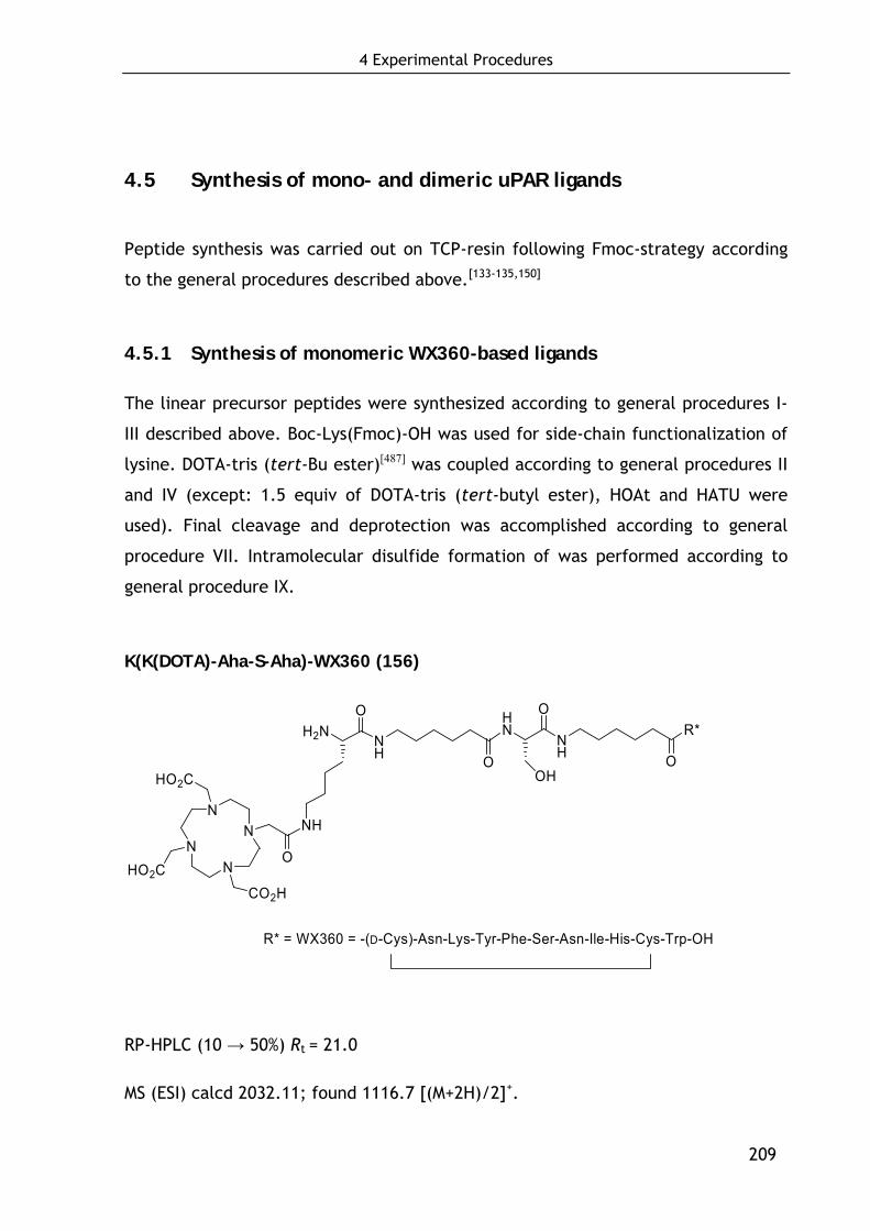
4 Experimental Procedures
209
4.5 Synthesis of mono- and dimeric uPAR ligands
Peptide synthesis was carried out on TCP-resin following Fmoc-strategy according
to the general procedures described above.[133-135,150]
4.5.1 Synthesis of monomeric WX360-based ligands
The linear precursor peptides were synthesized according to general procedures I-
III described above. Boc-Lys(Fmoc)-OH was used for side-chain functionalization of
lysine. DOTA-tris (tert-Bu ester)[487] was coupled according to general procedures II
and IV (except: 1.5 equiv of DOTA-tris (tert-butyl ester), HOAt and HATU were
used). Final cleavage and deprotection was accomplished according to general
procedure VII. Intramolecular disulfide formation of was performed according to
general procedure IX.
K(K(DOTA)-Aha-S-Aha)-WX360 (156)
NN
NN
HO2CCO2H
HO2C
O
H2NO
NH
NH O
HN
NH
R*O
OHO
R* = WX360 = -(D-Cys)-Asn-Lys-Tyr-Phe-Ser-Asn-Ile-His-Cys-Trp-OH
RP-HPLC (10 → 50%) Rt = 21.0
MS (ESI) calcd 2032.11; found 1116.7 [(M+2H)/2]+.
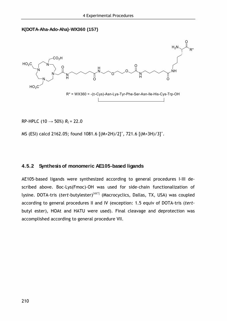
4 Experimental Procedures
210
K(DOTA-Aha-Ado-Aha)-WX360 (157)
R* = WX360 = -(D-Cys)-Asn-Lys-Tyr-Phe-Ser-Asn-Ile-His-Cys-Trp-OH
NN
NN
HO2CCO2H
HO2C
O HN
OO
NH
NHO
O
H2NO
R*
ONH
RP-HPLC (10 → 50%) Rt = 22.0
MS (ESI) calcd 2162.05; found 1081.6 [(M+2H)/2]+, 721.6 [(M+3H)/3]+.
4.5.2 Synthesis of monomeric AE105-based ligands
AE105-based ligands were synthesized according to general procedures I-III de-
scribed above. Boc-Lys(Fmoc)-OH was used for side-chain functionalization of
lysine. DOTA-tris (tert-butylester)[487] (Macrocyclics, Dallas, TX, USA) was coupled
according to general procedures II and IV (exception: 1.5 equiv of DOTA-tris (tert-
butyl ester), HOAt and HATU were used). Final cleavage and deprotection was
accomplished according to general procedure VII.
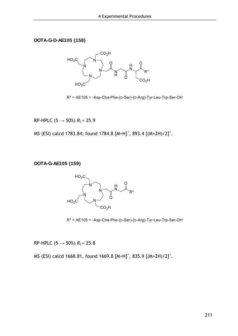
4 Experimental Procedures
211
DOTA-G-D-AE105 (158)
NN
NN
HO2CCO2H
HO2C
O
NH
HN
R* = AE105 = -Asp-Cha-Phe-(D-Ser)-(D-Arg)-Tyr-Leu-Trp-Ser-OH
O
O
R*
CO2H
RP-HPLC (5 → 50%) Rt = 25.9
MS (ESI) calcd 1783.84; found 1784.8 [M+H]+, 893.4 [(M+2H)/2]+.
DOTA-G-AE105 (159)
NN
NN
HO2CCO2H
HO2C
O
HN
R*
R* = AE105 = -Asp-Cha-Phe-(D-Ser)-(D-Arg)-Tyr-Leu-Trp-Ser-OH
O
RP-HPLC (5 → 50%) Rt = 25.8
MS (ESI) calcd 1668.81, found 1669.8 [M+H]+, 835.9 [(M+2H)/2]+.
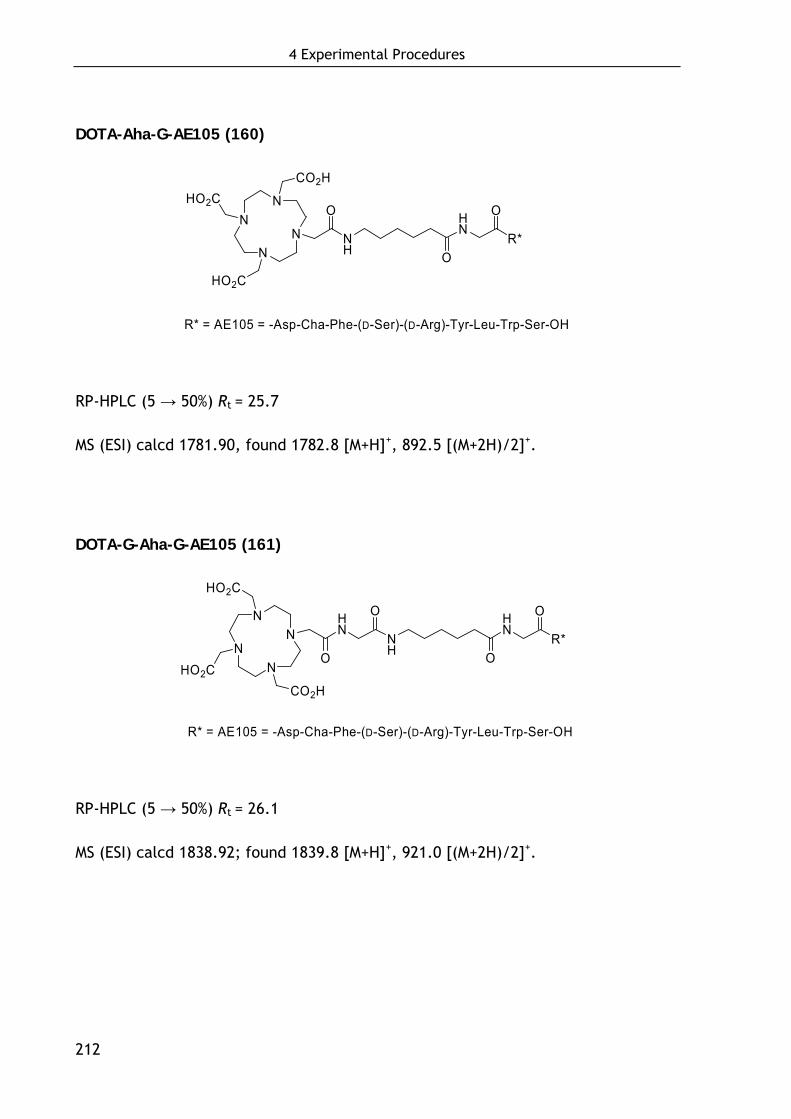
4 Experimental Procedures
212
DOTA-Aha-G-AE105 (160)
NN
NN
HO2CCO2H
HO2C
O
NH
R* = AE105 = -Asp-Cha-Phe-(D-Ser)-(D-Arg)-Tyr-Leu-Trp-Ser-OH
O
HN
O
R*
RP-HPLC (5 → 50%) Rt = 25.7
MS (ESI) calcd 1781.90, found 1782.8 [M+H]+, 892.5 [(M+2H)/2]+.
DOTA-G-Aha-G-AE105 (161)
NN
NN
HO2CCO2H
HO2C
O
HN
NH
R* = AE105 = -Asp-Cha-Phe-(D-Ser)-(D-Arg)-Tyr-Leu-Trp-Ser-OH
HN
O
OR*
O
RP-HPLC (5 → 50%) Rt = 26.1
MS (ESI) calcd 1838.92; found 1839.8 [M+H]+, 921.0 [(M+2H)/2]+.
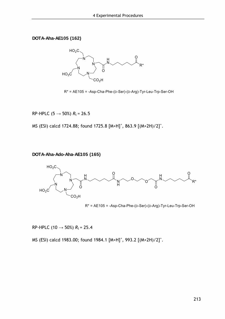
4 Experimental Procedures
213
DOTA-Aha-AE105 (162)
NN
NN
HO2CCO2H
HO2C
O
HN
R* = AE105 = -Asp-Cha-Phe-(D-Ser)-(D-Arg)-Tyr-Leu-Trp-Ser-OH
R*
O
RP-HPLC (5 → 50%) Rt = 26.5
MS (ESI) calcd 1724.88; found 1725.8 [M+H]+, 863.9 [(M+2H)/2]+.
DOTA-Aha-Ado-Aha-AE105 (165)
NN
NN
HO2CCO2H
HO2C
O
HN
R* = AE105 = -Asp-Cha-Phe-(D-Ser)-(D-Arg)-Tyr-Leu-Trp-Ser-OH
NH
OO
HN
O
OR*
O
RP-HPLC (10 → 50%) Rt = 25.4
MS (ESI) calcd 1983.00; found 1984.1 [M+H]+, 993.2 [(M+2H)/2]+.
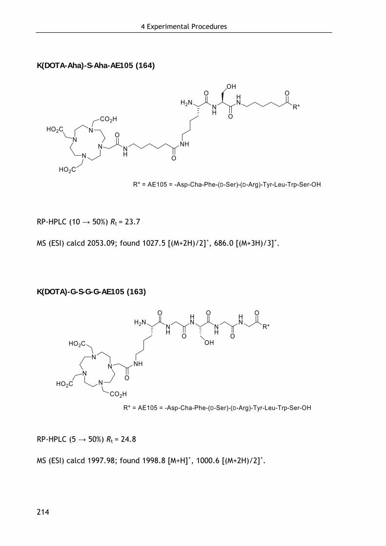
4 Experimental Procedures
214
K(DOTA-Aha)-S-Aha-AE105 (164)
NN
NN
HO2CCO2H
HO2C
O
H2NNH
HN
R*
O
NH O
NH
O
OHO
R* = AE105 = -Asp-Cha-Phe-(D-Ser)-(D-Arg)-Tyr-Leu-Trp-Ser-OH
RP-HPLC (10 → 50%) Rt = 23.7
MS (ESI) calcd 2053.09; found 1027.5 [(M+2H)/2]+, 686.0 [(M+3H)/3]+.
K(DOTA)-G-S-G-G-AE105 (163)
R* = AE105 = -Asp-Cha-Phe-(D-Ser)-(D-Arg)-Tyr-Leu-Trp-Ser-OH
NN
NN
HO2CCO2H
HO2C
O
NH
H2NNH
HN
NH
HN
R*
O
O
O
O
O
OH
RP-HPLC (5 → 50%) Rt = 24.8
MS (ESI) calcd 1997.98; found 1998.8 [M+H]+, 1000.6 [(M+2H)/2]+.
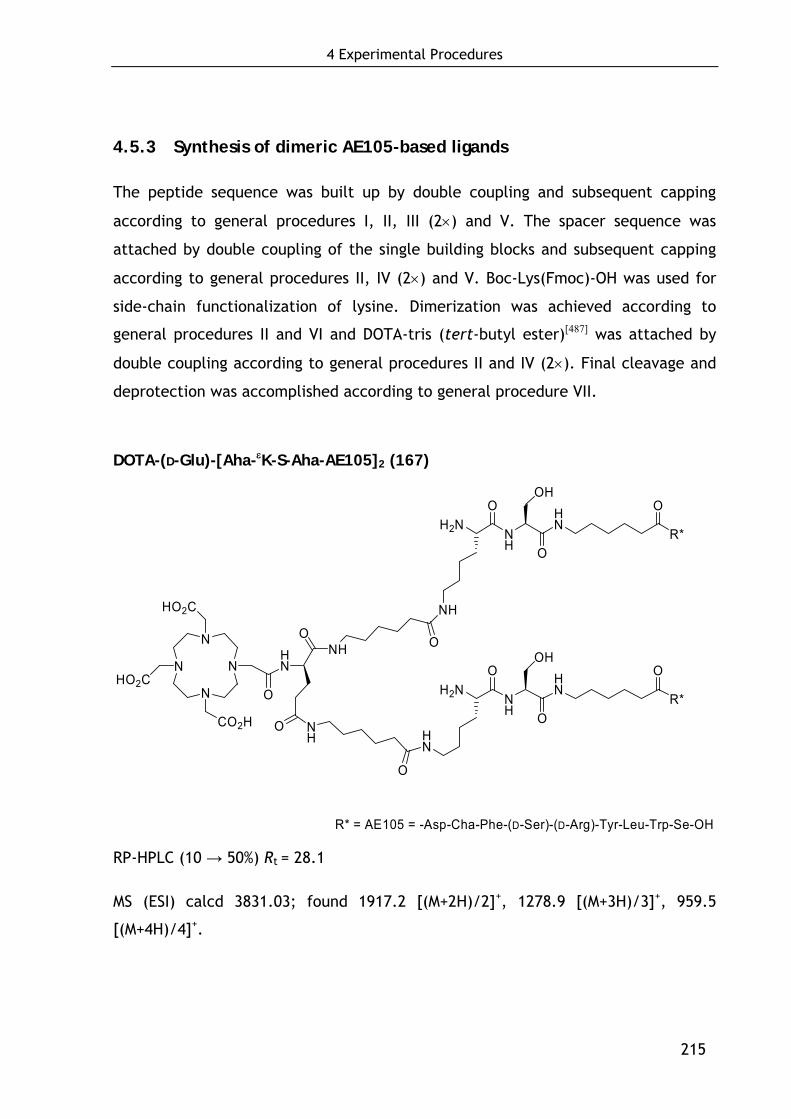
4 Experimental Procedures
215
4.5.3 Synthesis of dimeric AE105-based ligands
The peptide sequence was built up by double coupling and subsequent capping
according to general procedures I, II, III (2×) and V. The spacer sequence was
attached by double coupling of the single building blocks and subsequent capping
according to general procedures II, IV (2×) and V. Boc-Lys(Fmoc)-OH was used for
side-chain functionalization of lysine. Dimerization was achieved according to
general procedures II and VI and DOTA-tris (tert-butyl ester)[487] was attached by
double coupling according to general procedures II and IV (2×). Final cleavage and
deprotection was accomplished according to general procedure VII.
DOTA-(D-Glu)-[Aha-εK-S-Aha-AE105]2 (167)
H2NNH
HN
R*
O
NH O
NH
O
OHO
N
N
N
NHO2C
CO2H
HO2C
O
HN
O
O
H2NNH
HN
R*
O
NH
O
HN
O
OHO
R* = AE105 = -Asp-Cha-Phe-(D-Ser)-(D-Arg)-Tyr-Leu-Trp-Se-OH
RP-HPLC (10 → 50%) Rt = 28.1
MS (ESI) calcd 3831.03; found 1917.2 [(M+2H)/2]+, 1278.9 [(M+3H)/3]+, 959.5
[(M+4H)/4]+.
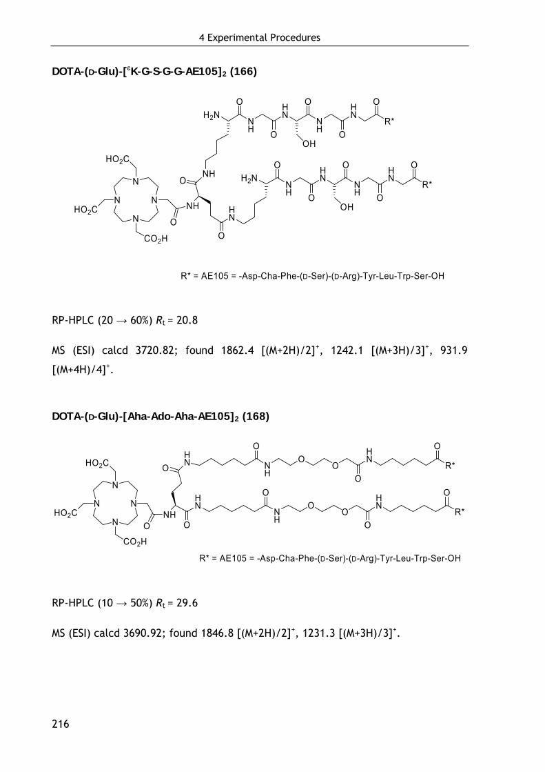
4 Experimental Procedures
216
DOTA-(D-Glu)-[εK-G-S-G-G-AE105]2 (166)
NH
H2NNH
HN
NH
HN
R*
O
O
O
O
O
OH
N
N
N
NHO2C
CO2H
HO2C
O
NH
O
O
H2NNH
HN
NH
HN
R*
O
O
O
O
O
OHHN
R* = AE105 = -Asp-Cha-Phe-(D-Ser)-(D-Arg)-Tyr-Leu-Trp-Ser-OH
RP-HPLC (20 → 60%) Rt = 20.8
MS (ESI) calcd 3720.82; found 1862.4 [(M+2H)/2]+, 1242.1 [(M+3H)/3]+, 931.9
[(M+4H)/4]+.
DOTA-(D-Glu)-[Aha-Ado-Aha-AE105]2 (168)
N
N
N
NHO2C
HO2C
ONH
R* = AE105 = -Asp-Cha-Phe-(D-Ser)-(D-Arg)-Tyr-Leu-Trp-Ser-OH
O
HN
HNO
NH
OO
HN
R*
NH
OO
HN
R*
CO2H
O
OO
O
O
O
RP-HPLC (10 → 50%) Rt = 29.6
MS (ESI) calcd 3690.92; found 1846.8 [(M+2H)/2]+, 1231.3 [(M+3H)/3]+.
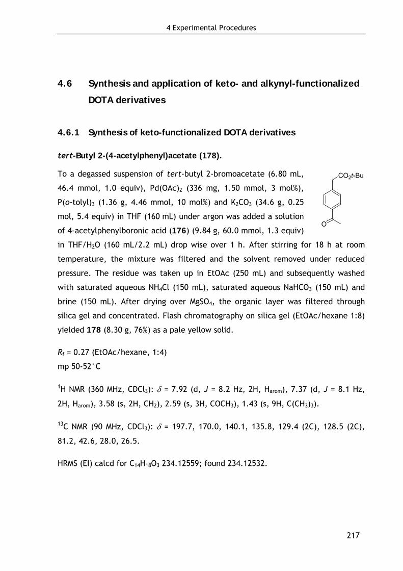
4 Experimental Procedures
217
4.6 Synthesis and application of keto- and alkynyl-functionalized
DOTA derivatives
4.6.1 Synthesis of keto-functionalized DOTA derivatives
tert-Butyl 2-(4-acetylphenyl)acetate (178).
To a degassed suspension of tert-butyl 2-bromoacetate (6.80 mL,
46.4 mmol, 1.0 equiv), Pd(OAc)2 (336 mg, 1.50 mmol, 3 mol%),
P(o-tolyl)3 (1.36 g, 4.46 mmol, 10 mol%) and K2CO3 (34.6 g, 0.25
mol, 5.4 equiv) in THF (160 mL) under argon was added a solution
of 4-acetylphenylboronic acid (176) (9.84 g, 60.0 mmol, 1.3 equiv)
in THF/H2O (160 mL/2.2 mL) drop wise over 1 h. After stirring for 18 h at room
temperature, the mixture was filtered and the solvent removed under reduced
pressure. The residue was taken up in EtOAc (250 mL) and subsequently washed
with saturated aqueous NH4Cl (150 mL), saturated aqueous NaHCO3 (150 mL) and
brine (150 mL). After drying over MgSO4, the organic layer was filtered through
silica gel and concentrated. Flash chromatography on silica gel (EtOAc/hexane 1:8)
yielded 178 (8.30 g, 76%) as a pale yellow solid.
Rf = 0.27 (EtOAc/hexane, 1:4)
mp 50-52°C
1H NMR (360 MHz, CDCl3): δ = 7.92 (d, J = 8.2 Hz, 2H, Harom), 7.37 (d, J = 8.1 Hz,
2H, Harom), 3.58 (s, 2H, CH2), 2.59 (s, 3H, COCH3), 1.43 (s, 9H, C(CH3)3).
13C NMR (90 MHz, CDCl3): δ = 197.7, 170.0, 140.1, 135.8, 129.4 (2C), 128.5 (2C),
81.2, 42.6, 28.0, 26.5.
HRMS (EI) calcd for C14H18O3 234.12559; found 234.12532.
O
CO2t-Bu
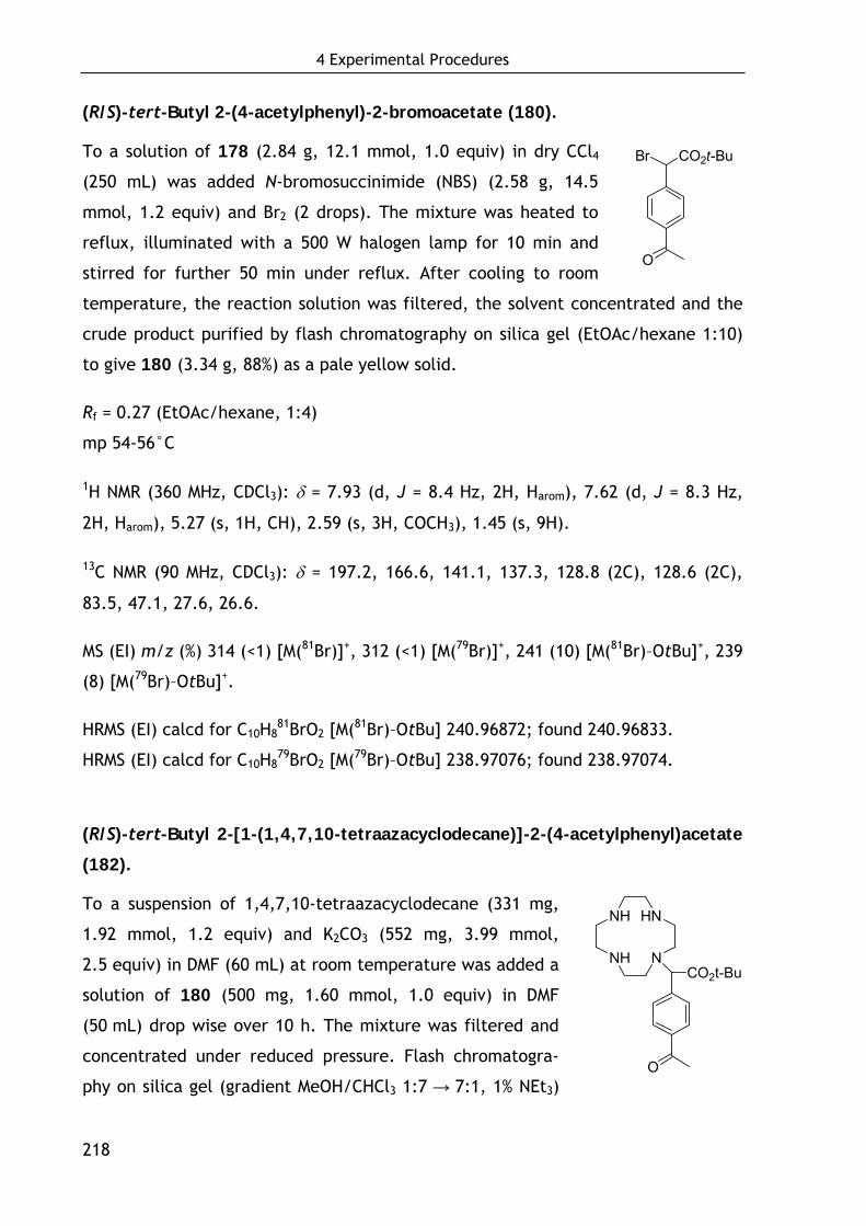
4 Experimental Procedures
218
(R/S)-tert-Butyl 2-(4-acetylphenyl)-2-bromoacetate (180).
To a solution of 178 (2.84 g, 12.1 mmol, 1.0 equiv) in dry CCl4
(250 mL) was added N-bromosuccinimide (NBS) (2.58 g, 14.5
mmol, 1.2 equiv) and Br2 (2 drops). The mixture was heated to
reflux, illuminated with a 500 W halogen lamp for 10 min and
stirred for further 50 min under reflux. After cooling to room
temperature, the reaction solution was filtered, the solvent concentrated and the
crude product purified by flash chromatography on silica gel (EtOAc/hexane 1:10)
to give 180 (3.34 g, 88%) as a pale yellow solid.
Rf = 0.27 (EtOAc/hexane, 1:4)
mp 54-56°C
1H NMR (360 MHz, CDCl3): δ = 7.93 (d, J = 8.4 Hz, 2H, Harom), 7.62 (d, J = 8.3 Hz,
2H, Harom), 5.27 (s, 1H, CH), 2.59 (s, 3H, COCH3), 1.45 (s, 9H).
13C NMR (90 MHz, CDCl3): δ = 197.2, 166.6, 141.1, 137.3, 128.8 (2C), 128.6 (2C),
83.5, 47.1, 27.6, 26.6.
MS (EI) m/z (%) 314 (<1) [M(81Br)]+, 312 (<1) [M(79Br)]+, 241 (10) [M(81Br)–OtBu]+, 239
(8) [M(79Br)–OtBu]+.
HRMS (EI) calcd for C10H881BrO2 [M(81Br)–OtBu] 240.96872; found 240.96833.
HRMS (EI) calcd for C10H879BrO2 [M(79Br)–OtBu] 238.97076; found 238.97074.
(R/S)-tert-Butyl 2-[1-(1,4,7,10-tetraazacyclodecane)]-2-(4-acetylphenyl)acetate
(182).
To a suspension of 1,4,7,10-tetraazacyclodecane (331 mg,
1.92 mmol, 1.2 equiv) and K2CO3 (552 mg, 3.99 mmol,
2.5 equiv) in DMF (60 mL) at room temperature was added a
solution of 180 (500 mg, 1.60 mmol, 1.0 equiv) in DMF
(50 mL) drop wise over 10 h. The mixture was filtered and
concentrated under reduced pressure. Flash chromatogra-
phy on silica gel (gradient MeOH/CHCl3 1:7 → 7:1, 1% NEt3)
O
CO2t-BuBr
NH
NH
HN
NCO2t-Bu
O
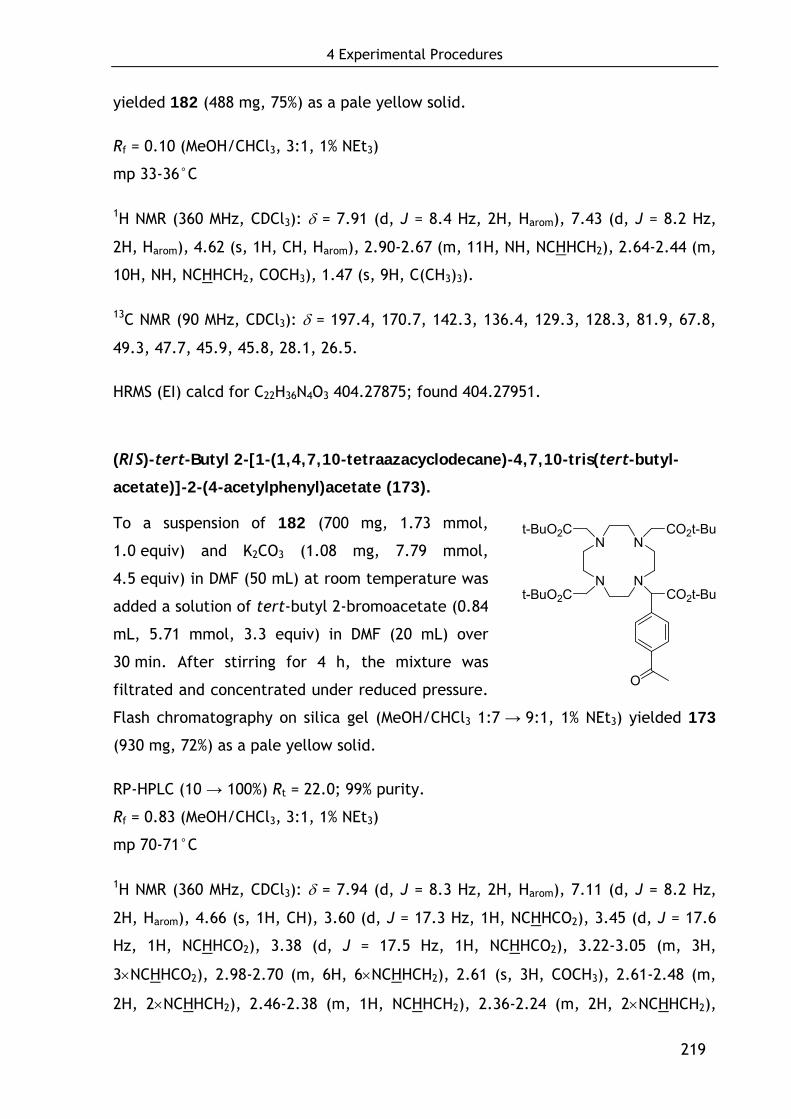
4 Experimental Procedures
219
yielded 182 (488 mg, 75%) as a pale yellow solid.
Rf = 0.10 (MeOH/CHCl3, 3:1, 1% NEt3)
mp 33-36°C
1H NMR (360 MHz, CDCl3): δ = 7.91 (d, J = 8.4 Hz, 2H, Harom), 7.43 (d, J = 8.2 Hz,
2H, Harom), 4.62 (s, 1H, CH, Harom), 2.90-2.67 (m, 11H, NH, NCHHCH2), 2.64-2.44 (m,
10H, NH, NCHHCH2, COCH3), 1.47 (s, 9H, C(CH3)3).
13C NMR (90 MHz, CDCl3): δ = 197.4, 170.7, 142.3, 136.4, 129.3, 128.3, 81.9, 67.8,
49.3, 47.7, 45.9, 45.8, 28.1, 26.5.
HRMS (EI) calcd for C22H36N4O3 404.27875; found 404.27951.
(R/S)-tert-Butyl 2-[1-(1,4,7,10-tetraazacyclodecane)-4,7,10-tris(tert-butyl-
acetate)]-2-(4-acetylphenyl)acetate (173).
To a suspension of 182 (700 mg, 1.73 mmol,
1.0 equiv) and K2CO3 (1.08 mg, 7.79 mmol,
4.5 equiv) in DMF (50 mL) at room temperature was
added a solution of tert-butyl 2-bromoacetate (0.84
mL, 5.71 mmol, 3.3 equiv) in DMF (20 mL) over
30 min. After stirring for 4 h, the mixture was
filtrated and concentrated under reduced pressure.
Flash chromatography on silica gel (MeOH/CHCl3 1:7 → 9:1, 1% NEt3) yielded 173
(930 mg, 72%) as a pale yellow solid.
RP-HPLC (10 → 100%) Rt = 22.0; 99% purity.
Rf = 0.83 (MeOH/CHCl3, 3:1, 1% NEt3)
mp 70-71°C
1H NMR (360 MHz, CDCl3): δ = 7.94 (d, J = 8.3 Hz, 2H, Harom), 7.11 (d, J = 8.2 Hz,
2H, Harom), 4.66 (s, 1H, CH), 3.60 (d, J = 17.3 Hz, 1H, NCHHCO2), 3.45 (d, J = 17.6
Hz, 1H, NCHHCO2), 3.38 (d, J = 17.5 Hz, 1H, NCHHCO2), 3.22-3.05 (m, 3H,
3×NCHHCO2), 2.98-2.70 (m, 6H, 6×NCHHCH2), 2.61 (s, 3H, COCH3), 2.61-2.48 (m,
2H, 2×NCHHCH2), 2.46-2.38 (m, 1H, NCHHCH2), 2.36-2.24 (m, 2H, 2×NCHHCH2),
N
N
N
N
t-BuO2C
t-BuO2C
CO2t-Bu
CO2t-Bu
O
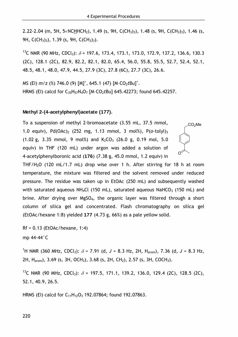
4 Experimental Procedures
220
2.22-2.04 (m, 5H, 5×NCHHCH2), 1.49 (s, 9H, C(CH3)3), 1.48 (s, 9H, C(CH3)3), 1.46 (s,
9H, C(CH3)3), 1.39 (s, 9H, C(CH3)3).
13C NMR (90 MHz, CDCl3): δ = 197.6, 173.4, 173.1, 173.0, 172.9, 137.2, 136.6, 130.3
(2C), 128.1 (2C), 82.9, 82.2, 82.1, 82.0, 65.4, 56.0, 55.8, 55.5, 52.7, 52.4, 52.1,
48.5, 48.1, 48.0, 47.9, 44.5, 27.9 (3C), 27.8 (6C), 27.7 (3C), 26.6.
MS (EI) m/z (%) 746.0 (9) [M]+, 645.1 (47) [M–CO2tBu]+.
HRMS (EI) calcd for C35H57N4O7 [M–CO2tBu] 645.42273; found 645.42257.
Methyl 2-(4-acetylphenyl)acetate (177).
To a suspension of methyl 2-bromoacetate (3.55 mL, 37.5 mmol,
1.0 equiv), Pd(OAc)2 (252 mg, 1.13 mmol, 3 mol%), P(o-tolyl)3
(1.02 g, 3.35 mmol, 9 mol%) and K2CO3 (26.0 g, 0.19 mol, 5.0
equiv) in THF (120 mL) under argon was added a solution of
4-acetylphenylboronic acid (176) (7.38 g, 45.0 mmol, 1.2 equiv) in
THF/H2O (120 mL/1.7 mL) drop wise over 1 h. After stirring for 18 h at room
temperature, the mixture was filtered and the solvent removed under reduced
pressure. The residue was taken up in EtOAc (250 mL) and subsequently washed
with saturated aqueous NH4Cl (150 mL), saturated aqueous NaHCO3 (150 mL) and
brine. After drying over MgSO4, the organic layer was filtered through a short
column of silica gel and concentrated. Flash chromatography on silica gel
(EtOAc/hexane 1:8) yielded 177 (4.73 g, 66%) as a pale yellow solid.
Rf = 0.13 (EtOAc/hexane, 1:4)
mp 44-44°C
1H NMR (360 MHz, CDCl3): δ = 7.91 (d, J = 8.3 Hz, 2H, Harom), 7.36 (d, J = 8.3 Hz,
2H, Harom), 3.69 (s, 3H, OCH3), 3.68 (s, 2H, CH2), 2.57 (s, 3H, COCH3).
13C NMR (90 MHz, CDCl3): δ = 197.5, 171.1, 139.2, 136.0, 129.4 (2C), 128.5 (2C),
52.1, 40.9, 26.5.
HRMS (EI) calcd for C11H12O3 192.07864; found 192.07863.
O
CO2Me
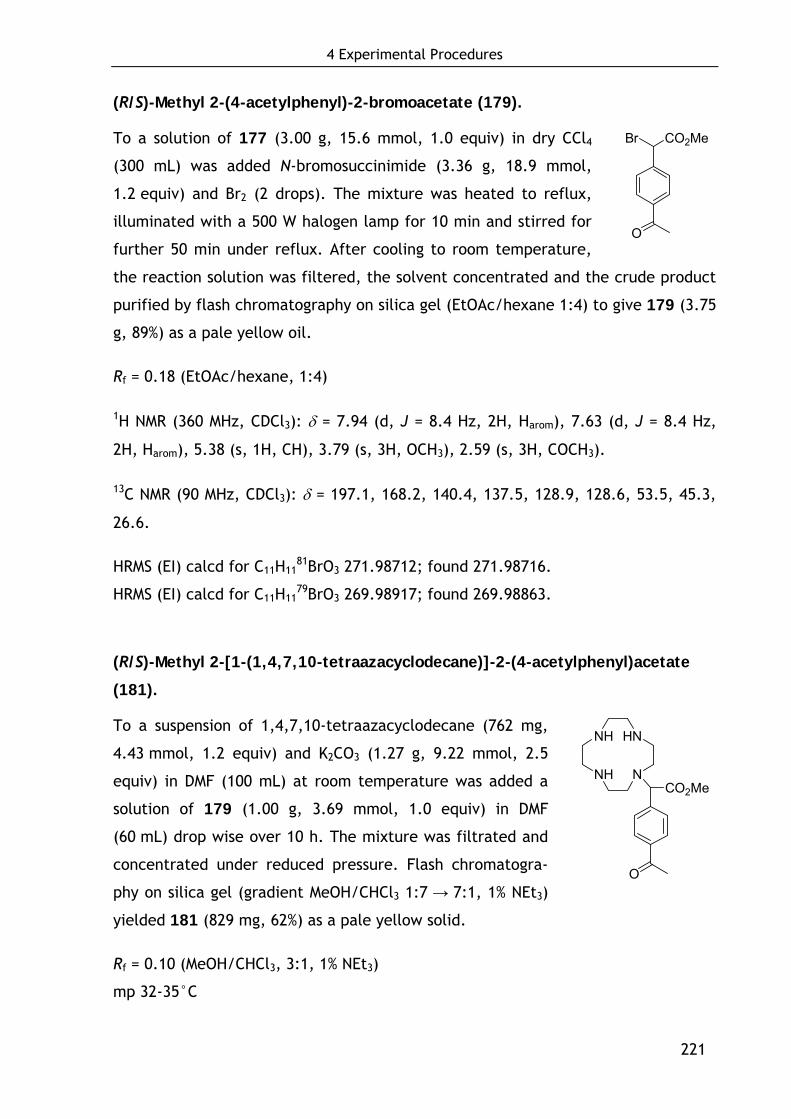
4 Experimental Procedures
221
(R/S)-Methyl 2-(4-acetylphenyl)-2-bromoacetate (179).
To a solution of 177 (3.00 g, 15.6 mmol, 1.0 equiv) in dry CCl4
(300 mL) was added N-bromosuccinimide (3.36 g, 18.9 mmol,
1.2 equiv) and Br2 (2 drops). The mixture was heated to reflux,
illuminated with a 500 W halogen lamp for 10 min and stirred for
further 50 min under reflux. After cooling to room temperature,
the reaction solution was filtered, the solvent concentrated and the crude product
purified by flash chromatography on silica gel (EtOAc/hexane 1:4) to give 179 (3.75
g, 89%) as a pale yellow oil.
Rf = 0.18 (EtOAc/hexane, 1:4)
1H NMR (360 MHz, CDCl3): δ = 7.94 (d, J = 8.4 Hz, 2H, Harom), 7.63 (d, J = 8.4 Hz,
2H, Harom), 5.38 (s, 1H, CH), 3.79 (s, 3H, OCH3), 2.59 (s, 3H, COCH3).
13C NMR (90 MHz, CDCl3): δ = 197.1, 168.2, 140.4, 137.5, 128.9, 128.6, 53.5, 45.3,
26.6.
HRMS (EI) calcd for C11H1181BrO3 271.98712; found 271.98716.
HRMS (EI) calcd for C11H1179BrO3 269.98917; found 269.98863.
(R/S)-Methyl 2-[1-(1,4,7,10-tetraazacyclodecane)]-2-(4-acetylphenyl)acetate
(181).
To a suspension of 1,4,7,10-tetraazacyclodecane (762 mg,
4.43 mmol, 1.2 equiv) and K2CO3 (1.27 g, 9.22 mmol, 2.5
equiv) in DMF (100 mL) at room temperature was added a
solution of 179 (1.00 g, 3.69 mmol, 1.0 equiv) in DMF
(60 mL) drop wise over 10 h. The mixture was filtrated and
concentrated under reduced pressure. Flash chromatogra-
phy on silica gel (gradient MeOH/CHCl3 1:7 → 7:1, 1% NEt3)
yielded 181 (829 mg, 62%) as a pale yellow solid.
Rf = 0.10 (MeOH/CHCl3, 3:1, 1% NEt3)
mp 32-35°C
O
CO2MeBr
NH
NH
HN
NCO2Me
O
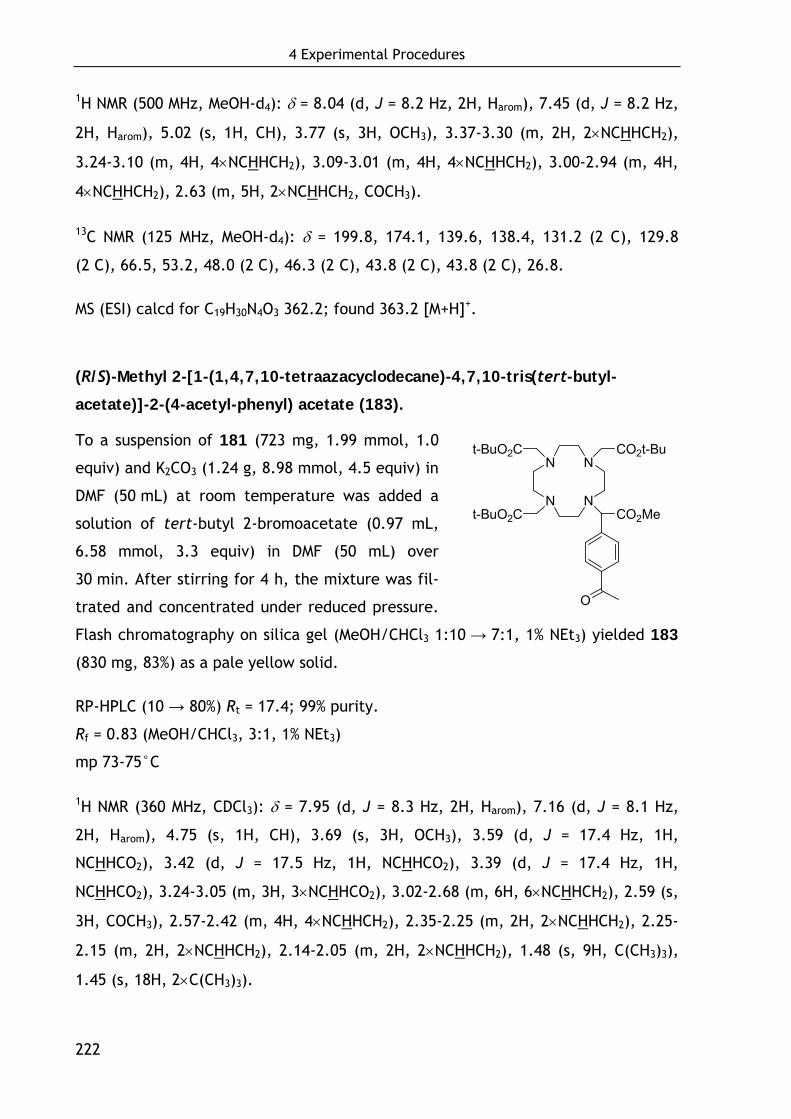
4 Experimental Procedures
222
1H NMR (500 MHz, MeOH-d4): δ = 8.04 (d, J = 8.2 Hz, 2H, Harom), 7.45 (d, J = 8.2 Hz,
2H, Harom), 5.02 (s, 1H, CH), 3.77 (s, 3H, OCH3), 3.37-3.30 (m, 2H, 2×NCHHCH2),
3.24-3.10 (m, 4H, 4×NCHHCH2), 3.09-3.01 (m, 4H, 4×NCHHCH2), 3.00-2.94 (m, 4H,
4×NCHHCH2), 2.63 (m, 5H, 2×NCHHCH2, COCH3).
13C NMR (125 MHz, MeOH-d4): δ = 199.8, 174.1, 139.6, 138.4, 131.2 (2 C), 129.8
(2 C), 66.5, 53.2, 48.0 (2 C), 46.3 (2 C), 43.8 (2 C), 43.8 (2 C), 26.8.
MS (ESI) calcd for C19H30N4O3 362.2; found 363.2 [M+H]+.
(R/S)-Methyl 2-[1-(1,4,7,10-tetraazacyclodecane)-4,7,10-tris(tert-butyl-
acetate)]-2-(4-acetyl-phenyl) acetate (183).
To a suspension of 181 (723 mg, 1.99 mmol, 1.0
equiv) and K2CO3 (1.24 g, 8.98 mmol, 4.5 equiv) in
DMF (50 mL) at room temperature was added a
solution of tert-butyl 2-bromoacetate (0.97 mL,
6.58 mmol, 3.3 equiv) in DMF (50 mL) over
30 min. After stirring for 4 h, the mixture was fil-
trated and concentrated under reduced pressure.
Flash chromatography on silica gel (MeOH/CHCl3 1:10 → 7:1, 1% NEt3) yielded 183
(830 mg, 83%) as a pale yellow solid.
RP-HPLC (10 → 80%) Rt = 17.4; 99% purity.
Rf = 0.83 (MeOH/CHCl3, 3:1, 1% NEt3)
mp 73-75°C
1H NMR (360 MHz, CDCl3): δ = 7.95 (d, J = 8.3 Hz, 2H, Harom), 7.16 (d, J = 8.1 Hz,
2H, Harom), 4.75 (s, 1H, CH), 3.69 (s, 3H, OCH3), 3.59 (d, J = 17.4 Hz, 1H,
NCHHCO2), 3.42 (d, J = 17.5 Hz, 1H, NCHHCO2), 3.39 (d, J = 17.4 Hz, 1H,
NCHHCO2), 3.24-3.05 (m, 3H, 3×NCHHCO2), 3.02-2.68 (m, 6H, 6×NCHHCH2), 2.59 (s,
3H, COCH3), 2.57-2.42 (m, 4H, 4×NCHHCH2), 2.35-2.25 (m, 2H, 2×NCHHCH2), 2.25-
2.15 (m, 2H, 2×NCHHCH2), 2.14-2.05 (m, 2H, 2×NCHHCH2), 1.48 (s, 9H, C(CH3)3),
1.45 (s, 18H, 2×C(CH3)3).
N
N
N
N
t-BuO2C
t-BuO2C
CO2t-Bu
CO2Me
O
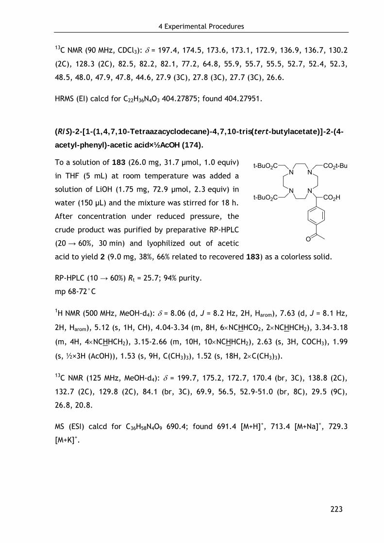
4 Experimental Procedures
223
13C NMR (90 MHz, CDCl3): δ = 197.4, 174.5, 173.6, 173.1, 172.9, 136.9, 136.7, 130.2
(2C), 128.3 (2C), 82.5, 82.2, 82.1, 77.2, 64.8, 55.9, 55.7, 55.5, 52.7, 52.4, 52.3,
48.5, 48.0, 47.9, 47.8, 44.6, 27.9 (3C), 27.8 (3C), 27.7 (3C), 26.6.
HRMS (EI) calcd for C22H36N4O3 404.27875; found 404.27951.
(R/S)-2-[1-(1,4,7,10-Tetraazacyclodecane)-4,7,10-tris(tert-butylacetate)]-2-(4-
acetyl-phenyl)-acetic acid×½AcOH (174).
To a solution of 183 (26.0 mg, 31.7 μmol, 1.0 equiv)
in THF (5 mL) at room temperature was added a
solution of LiOH (1.75 mg, 72.9 μmol, 2.3 equiv) in
water (150 μL) and the mixture was stirred for 18 h.
After concentration under reduced pressure, the
crude product was purified by preparative RP-HPLC
(20 → 60%, 30 min) and lyophilized out of acetic
acid to yield 2 (9.0 mg, 38%, 66% related to recovered 183) as a colorless solid.
RP-HPLC (10 → 60%) Rt = 25.7; 94% purity.
mp 68-72°C
1H NMR (500 MHz, MeOH-d4): δ = 8.06 (d, J = 8.2 Hz, 2H, Harom), 7.63 (d, J = 8.1 Hz,
2H, Harom), 5.12 (s, 1H, CH), 4.04-3.34 (m, 8H, 6×NCHHCO2, 2×NCHHCH2), 3.34-3.18
(m, 4H, 4×NCHHCH2), 3.15-2.66 (m, 10H, 10×NCHHCH2), 2.63 (s, 3H, COCH3), 1.99
(s, ½×3H (AcOH)), 1.53 (s, 9H, C(CH3)3), 1.52 (s, 18H, 2×C(CH3)3).
13C NMR (125 MHz, MeOH-d4): δ = 199.7, 175.2, 172.7, 170.4 (br, 3C), 138.8 (2C),
132.7 (2C), 129.8 (2C), 84.1 (br, 3C), 69.9, 56.5, 52.9-51.0 (br, 8C), 29.5 (9C),
26.8, 20.8.
MS (ESI) calcd for C36H58N4O9 690.4; found 691.4 [M+H]+, 713.4 [M+Na]+, 729.3
[M+K]+.
N
N
N
N
t-BuO2C
t-BuO2C
CO2t-Bu
CO2H
O
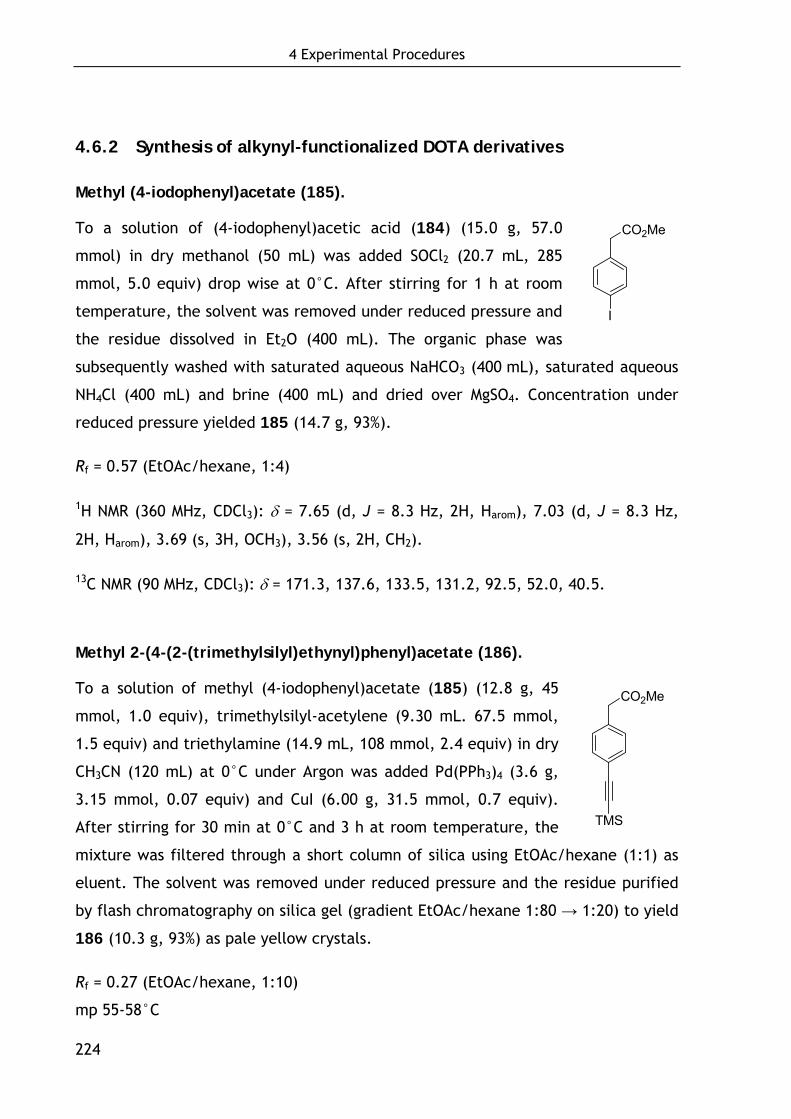
4 Experimental Procedures
224
4.6.2 Synthesis of alkynyl-functionalized DOTA derivatives
Methyl (4-iodophenyl)acetate (185).
To a solution of (4-iodophenyl)acetic acid (184) (15.0 g, 57.0
mmol) in dry methanol (50 mL) was added SOCl2 (20.7 mL, 285
mmol, 5.0 equiv) drop wise at 0°C. After stirring for 1 h at room
temperature, the solvent was removed under reduced pressure and
the residue dissolved in Et2O (400 mL). The organic phase was
subsequently washed with saturated aqueous NaHCO3 (400 mL), saturated aqueous
NH4Cl (400 mL) and brine (400 mL) and dried over MgSO4. Concentration under
reduced pressure yielded 185 (14.7 g, 93%).
Rf = 0.57 (EtOAc/hexane, 1:4)
1H NMR (360 MHz, CDCl3): δ = 7.65 (d, J = 8.3 Hz, 2H, Harom), 7.03 (d, J = 8.3 Hz,
2H, Harom), 3.69 (s, 3H, OCH3), 3.56 (s, 2H, CH2).
13C NMR (90 MHz, CDCl3): δ = 171.3, 137.6, 133.5, 131.2, 92.5, 52.0, 40.5.
Methyl 2-(4-(2-(trimethylsilyl)ethynyl)phenyl)acetate (186).
To a solution of methyl (4-iodophenyl)acetate (185) (12.8 g, 45
mmol, 1.0 equiv), trimethylsilyl-acetylene (9.30 mL. 67.5 mmol,
1.5 equiv) and triethylamine (14.9 mL, 108 mmol, 2.4 equiv) in dry
CH3CN (120 mL) at 0°C under Argon was added Pd(PPh3)4 (3.6 g,
3.15 mmol, 0.07 equiv) and CuI (6.00 g, 31.5 mmol, 0.7 equiv).
After stirring for 30 min at 0°C and 3 h at room temperature, the
mixture was filtered through a short column of silica using EtOAc/hexane (1:1) as
eluent. The solvent was removed under reduced pressure and the residue purified
by flash chromatography on silica gel (gradient EtOAc/hexane 1:80 → 1:20) to yield
186 (10.3 g, 93%) as pale yellow crystals.
Rf = 0.27 (EtOAc/hexane, 1:10)
mp 55-58°C
CO2Me
TMS
I
CO2Me
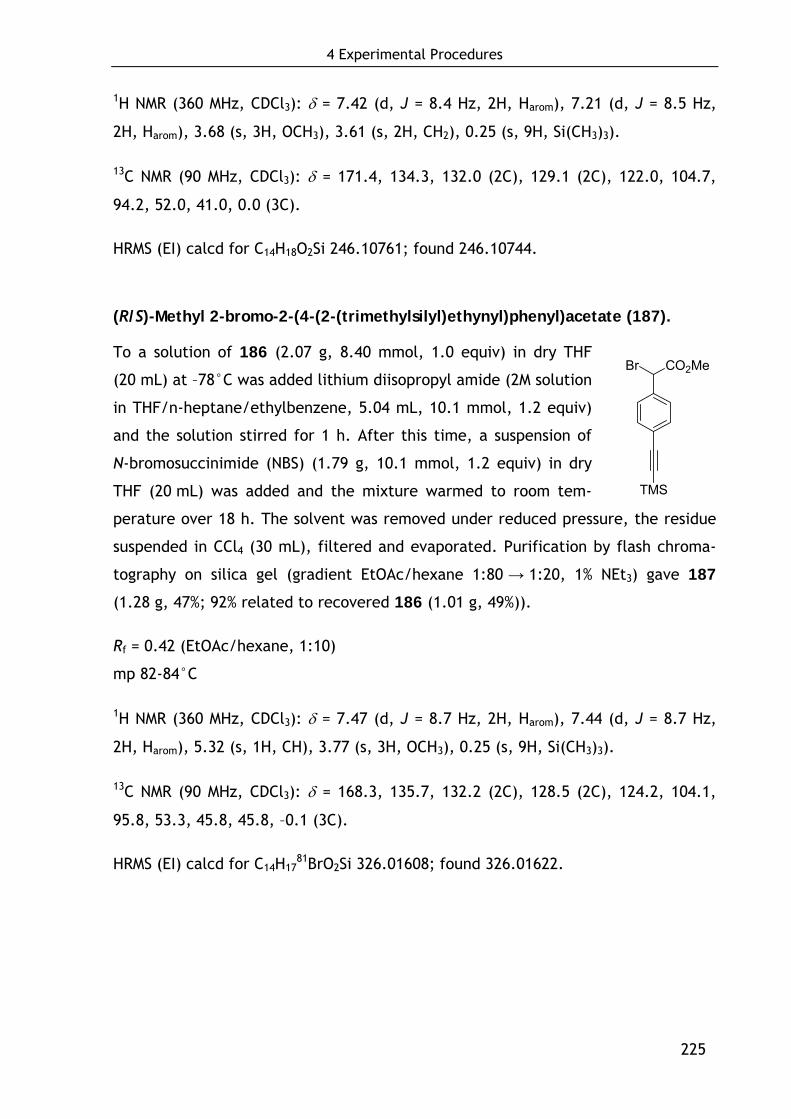
4 Experimental Procedures
225
1H NMR (360 MHz, CDCl3): δ = 7.42 (d, J = 8.4 Hz, 2H, Harom), 7.21 (d, J = 8.5 Hz,
2H, Harom), 3.68 (s, 3H, OCH3), 3.61 (s, 2H, CH2), 0.25 (s, 9H, Si(CH3)3).
13C NMR (90 MHz, CDCl3): δ = 171.4, 134.3, 132.0 (2C), 129.1 (2C), 122.0, 104.7,
94.2, 52.0, 41.0, 0.0 (3C).
HRMS (EI) calcd for C14H18O2Si 246.10761; found 246.10744.
(R/S)-Methyl 2-bromo-2-(4-(2-(trimethylsilyl)ethynyl)phenyl)acetate (187).
To a solution of 186 (2.07 g, 8.40 mmol, 1.0 equiv) in dry THF
(20 mL) at –78°C was added lithium diisopropyl amide (2M solution
in THF/n-heptane/ethylbenzene, 5.04 mL, 10.1 mmol, 1.2 equiv)
and the solution stirred for 1 h. After this time, a suspension of
N-bromosuccinimide (NBS) (1.79 g, 10.1 mmol, 1.2 equiv) in dry
THF (20 mL) was added and the mixture warmed to room tem-
perature over 18 h. The solvent was removed under reduced pressure, the residue
suspended in CCl4 (30 mL), filtered and evaporated. Purification by flash chroma-
tography on silica gel (gradient EtOAc/hexane 1:80 → 1:20, 1% NEt3) gave 187
(1.28 g, 47%; 92% related to recovered 186 (1.01 g, 49%)).
Rf = 0.42 (EtOAc/hexane, 1:10)
mp 82-84°C
1H NMR (360 MHz, CDCl3): δ = 7.47 (d, J = 8.7 Hz, 2H, Harom), 7.44 (d, J = 8.7 Hz,
2H, Harom), 5.32 (s, 1H, CH), 3.77 (s, 3H, OCH3), 0.25 (s, 9H, Si(CH3)3).
13C NMR (90 MHz, CDCl3): δ = 168.3, 135.7, 132.2 (2C), 128.5 (2C), 124.2, 104.1,
95.8, 53.3, 45.8, 45.8, –0.1 (3C).
HRMS (EI) calcd for C14H1781BrO2Si 326.01608; found 326.01622.
CO2Me
TMS
Br
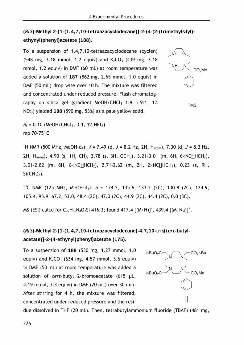
4 Experimental Procedures
226
(R/S)-Methyl 2-[1-(1,4,7,10-tetraazacyclodecane)]-2-(4-(2-(trimethylsilyl)-
ethynyl)phenyl)acetate (188).
To a suspension of 1,4,7,10-tetraazacyclodecane (cyclen)
(548 mg, 3.18 mmol, 1.2 equiv) and K2CO3 (439 mg, 3.18
mmol, 1.2 equiv) in DMF (60 mL) at room temperature was
added a solution of 187 (862 mg, 2.65 mmol, 1.0 equiv) in
DMF (50 mL) drop wise over 10 h. The mixture was filtered
and concentrated under reduced pressure. Flash chromatog-
raphy on silica gel (gradient MeOH/CHCl3 1:9 → 9:1, 1%
NEt3) yielded 188 (590 mg, 53%) as a pale yellow solid.
Rf = 0.10 (MeOH/CHCl3, 3:1, 1% NEt3)
mp 70-75°C
1H NMR (500 MHz, MeOH-d4): δ = 7.49 (d, J = 8.2 Hz, 2H, Harom), 7.30 (d, J = 8.3 Hz,
2H, Harom), 4.90 (s, 1H, CH), 3.78 (s, 3H, OCH3), 3.21-3.01 (m, 6H, 6×NCHHCH2),
3.01-2.82 (m, 8H, 8×NCHHCH2), 2.71-2.62 (m, 2H, 2×NCHHCH2), 0.23 (s, 9H,
Si(CH3)3).
13C NMR (125 MHz, MeOH-d4): δ = 174.2, 135.6, 133.2 (2C), 130.8 (2C), 124.9,
105.4, 95.9, 67.2, 53.0, 48.4 (2C), 47.0 (2C), 44.9 (2C), 44.4 (2C), 0.0 (3C).
MS (ESI) calcd for C22H36N4O2Si 416.3; found 417.4 [(M+H)]+, 439.4 [(M+Na)]+.
(R/S)-Methyl 2-[1-(1,4,7,10-tetraazacyclodecane)-4,7,10-tris(tert-butyl-
acetate)]-2-(4-ethynyl)phenyl)acetate (175).
To a suspension of 188 (530 mg, 1.27 mmol, 1.0
equiv) and K2CO3 (634 mg, 4.57 mmol, 3.6 equiv)
in DMF (50 mL) at room temperature was added a
solution of tert-butyl 2-bromoacetate (615 μL,
4.19 mmol, 3.3 equiv) in DMF (20 mL) over 30 min.
After stirring for 4 h, the mixture was filtered,
concentrated under reduced pressure and the resi-
due dissolved in THF (20 mL). Then, tetrabutylammonium fluoride (TBAF) (481 mg,
NH
NH
HN
NCO2Me
TMS
N
N
N
N
t-BuO2C
t-BuO2C
CO2t-Bu
CO2Me

4 Experimental Procedures
227
1.52 mmol, 1.2 equiv) was added and after stirring for 15 min, the solvent was
removed and the crude product purified by flash chromatography on silica gel
(MeOH/CHCl3 1:10, 1% NEt3) to yield 175 (508 mg, 85%) as a pale yellow solid.
RP-HPLC (10 → 100%) Rt = 20.0; 99% purity.
Rf = 0.28 (MeOH/CHCl3, 1:9, 1% NEt3)
mp 63-68°C
1H NMR (500 MHz, MeOH-d4): δ = 7.48 (d, J = 8.2 Hz, 2H, Harom), 7.20 (d, J = 8.0 Hz,
2H, Harom), 4.83 (s, 1H, CH), 3.75 (d, J = 17.2 Hz, 1H, NCHHCO2), 3.74 (s, 3H,
OCH3), 3.55 (s, 1H, C≡CH), 3.54 (d, J = 17.5 Hz, 1H, NCHHCO2), 3.51 (d, J = 17.6
Hz, 1H, NCHHCO2), 3.26-3.21 (m, 1H, NCHHCO2), 3.18-3.05 (m, 4H, 2×NCHHCO2,
2×NCHHCH2), 2.99-2.92 (m, 3H, 3×NCHHCH2), 2.89 (d, J = 17.6 Hz, 1H, NCHHCH2),
2.87 (d, J = 17.6 Hz, 1H, NCHHCH2), 2.74-2.63 (m, 2H, 2×NCHHCH2), 2.33 (d, J =
11.5 Hz, 1H, NCHHCH2), 2.28-2.12 (m, 4H, 4×NCHHCH2), 2.12-2.05 (m, 2H,
2×NCHHCH2), 1.54 (s, 9H, C(CH3)3), 1.52 (s, 18H, 2×C(CH3)3).
13C NMR (125 MHz, MeOH-d4): δ = 176.3, 175.4, 175.1, 174.7, 134.1, 132.8, 131.7,
123.8, 83.8, 83.5, 83.1, 79.7, 66.4, 59.61, 59.59, 59.57, 57.1, 56.8, 56.7, 54.2,
53.9, 53.7, 53.2, 45.9, 28.5, 28.4, 28.3, 24.8.
HRMS (EI) calcd for C37H58N4O8 686.42546; found 686.42532.

4 Experimental Procedures
228
4.6.3 Synthesis of aminooxy- and azido functionalized Tyr3-octreotate
derivatives for chemoselective conjugation with keto- and alkynyl-
functionalized DOTA derivatives
4.6.3.1 Synthesis of azido-functionalized linker (194)
1-Azido-3-aminopropane (193).
The literature procedure[635] was slightly modified: A solution of
1-bromo-3-aminopropane hydrobromide (5.47 g, 25.0 mmol,
1.0 equiv) and sodium azide (3.25 g, 50.0 mmol, 2.0 equiv) in
20 mL water was heated at 80°C for 24 h. The reaction mixture was cooled in an
ice bath followed by addition of diethyl ether (30 mL). The pH was adjusted to
14 by addition of KOH pellets keeping the temperature below 10°C. After separa-
tion of the organic phase the aqueous layer was further extracted with diethyl
ether. The combined organic layers were dried over MgSO4 and carefully concen-
trated in vacuo. The remaining oil contained 193 (21.0 mmol; 84%) and about equi-
molar amounts of diethyl ether (based on integration of proton NMR) and was used
without further purification. Spectroscopic data were identical to literature.[635]
3-(3-Azidopropylcarbamoyl)propanoic acid (194).
To a solution of 193 (501 mg, 5.00 mmol, 1.1 equiv)
and NEt3 (693 μL, 5.00 mmol, 1.1 equiv) in 10 mL
acetone was added a solution of succinic anhydride
(460 mg, 4.55 mmol, 1.0 equiv) in 5 mL acetone over a period of 15 min at room
temperature. The reaction mixture was stirred for 15 h at room temperature. After
concentration in vacuo the residue was partitioned between diluted aqueous HCl
(20 mL) and ethyl acetate (20mL), separated and the aqueous layer was further
extracted with ethyl acetate (2×10 mL). After concentration of the combined
organic layers 194 (653 mg, 3.27 mmol, 71 %) was obtained as a white solid.
N3 NH2
N3 NH
O
O
OH
4 Experimental Procedures
229
1H NMR (360 MHz, MeOH-d4): δ = 3.40-3.20 (m, 4H, N3CH2CH2CH2NH), 2.59 (dt, J =
6.5 Hz, 1.1 Hz, 2H, NHCOCH2CH2CO2H), 2.44 (dt, J = 6.7 Hz, 1.3 Hz, 2H, NHCO-
CH2CH2CO2H), 1.74 (q, J = 6.8 Hz, 2H, CH2CH2CH2).
13C NMR (90 MHz, MeOH-d4): δ = 176.2, 174.7, 50.0, 37.7, 31.5, 30.2, 29.7.
MS (ESI) calcd for C11H12O3 200.1; found 201.1 (M+H+).
4.6.3.2 Synthesis of Tyr3-octreotate derivatives (190) and (196) and sample
peptide (198).
Peptide synthesis was carried out on TCP-resin following Fmoc-strategy according
to the general procedures described above.[133-135,150]
cyclo[2,7]-AoxAc-(D-Phe)-Cys-Tyr-(D-Trp)-Lys-Thr-Cys-Thr-OH (190) and
cyclo[2,7]-3-(3-azidopropylcarbamoyl)propanoyl-(D-Phe)-Cys-Tyr-(D-Trp)-Lys-
Thr-Cys-Thr-OH (196).
The linear peptide sequence H-(D-Phe)-Cys-Tyr-(D-Trp)-Lys-Thr-Cys-Thr-OH was
synthesized on solid phase according to the general procedures I-III. Double-
couplings were performed in every step. A sample was cleaved from the resin
following procedure VII and the peptide sequence was confirmed by MALDI-TOF
peptide sequence analysis. For the further procedure the resin was parted into two
portions.
MS (MALDI) calcd for C49H66N10O12S2 1050.43; found 1051.40 [M+H]+.

4 Experimental Procedures
230
cyclo[2,7]-AoxAc-(D-Phe)-Cys-Tyr-(D-Trp)-Lys-Thr-Cys-Thr-OH (190):
HN
O
NH
HN
NH
O HN
O
NHOS
S
O
O
OHNH2
NH
HN
OH
HO
O
ONH
O
OH
OH2N
(Boc-aminoxy)acetic acid was coupled to one portion of above resin bound peptide
in a similar manner to the general procedures I-III and the linear peptide was
cleaved from resin following procedure VII. The disulfide cyclization was accom-
plished according to general procedure VIII. Great care must be taken to the qual-
ity of all solvents used in steps VII and VIII (HPLC quality). A small batch of the
crude product was purified to obtain the aminooxy-functionalized Tyr3-octreotate
190 as a colorless solid in 58% yield after purification by semipreparative RP-HPLC
(20 → 50%, 30 min).
RP-HPLC (10 → 60%) Rt = 17.3; 97% purity.
MS (ESI) calcd for C51H67N11O14S2 1121.4; found 1122.5 [M+H]+, 562.1 [(2M+2H)/2]2+.
cyclo[2,7]-3-(3-azidopropylcarbamoyl)propanoyl-(D-Phe)-Cys-Tyr-(D-Trp)-Lys-
Thr-Cys-Thr-OH (196):
N3HN
O
OHN
O
NH
HN
NH
O HN
O
NHOS
S
O
O
OHNH2
NH
HN
OH
HO
O
O
OH
NH

4 Experimental Procedures
231
194 was coupled to one portion of above resin bound peptide in a similar manner
to the general procedures I-III and the linear peptide was cleaved from resin
following procedure VII. The disulfide cyclization was accomplished according to
general procedure VIII. 196 was obtained as a colorless solid in 54% yield after
purification by semipreparative RP-HPLC (20 → 60%, 30 min).
RP-HPLC (10 → 50%) Rt = 25.0; 96% purity.
MS (ESI) calcd for C56H74N14O14S2 1230.5; found 1231.6 [M+H]+, 1253.6 [M+Na]+,
1269.3 [M+K]+.
Synthesis of 3-(3-azidopropylcarbamoyl)propanoyl-Tyr-Glu-Trp-Lys (198).
HN
NH
HN
HN CO2H
NH2
O
O
O
CO2H
NH
NH
OH
O
O
N3
The linear peptide was synthesized on solid phase in a similar manner to the gen-
eral procedures I-III and cleaved from resin following procedure VII. Peptide 198
was obtained as a colorless solid in 78% yield after purification by semipreparative
RP-HPLC (20 → 50%, 30 min).
RP-HPLC (10 → 60%, 30 min) Rt = 16.8; 97% purity.
MS (ESI) calcd for C38H50N10O10 806.37; found 807.6 [M+H]+, 829.5 [M+Na]+.
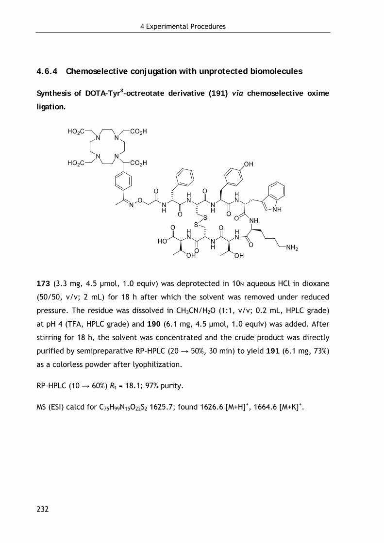
4 Experimental Procedures
232
4.6.4 Chemoselective conjugation with unprotected biomolecules
Synthesis of DOTA-Tyr3-octreotate derivative (191) via chemoselective oxime
ligation.
N
N
N
N
HO2C
HO2C
CO2H
CO2H
HN
O
NH
HN
NH
O HN
O
NHOS
S
O
O
OHNH2
NH
HN
OH
HO
O
ONH
O
OH
ON
173 (3.3 mg, 4.5 μmol, 1.0 equiv) was deprotected in 10N aqueous HCl in dioxane
(50/50, v/v; 2 mL) for 18 h after which the solvent was removed under reduced
pressure. The residue was dissolved in CH3CN/H2O (1:1, v/v; 0.2 mL, HPLC grade)
at pH 4 (TFA, HPLC grade) and 190 (6.1 mg, 4.5 μmol, 1.0 equiv) was added. After
stirring for 18 h, the solvent was concentrated and the crude product was directly
purified by semipreparative RP-HPLC (20 → 50%, 30 min) to yield 191 (6.1 mg, 73%)
as a colorless powder after lyophilization.
RP-HPLC (10 → 60%) Rt = 18.1; 97% purity.
MS (ESI) calcd for C75H99N15O22S2 1625.7; found 1626.6 [M+H]+, 1664.6 [M+K]+.
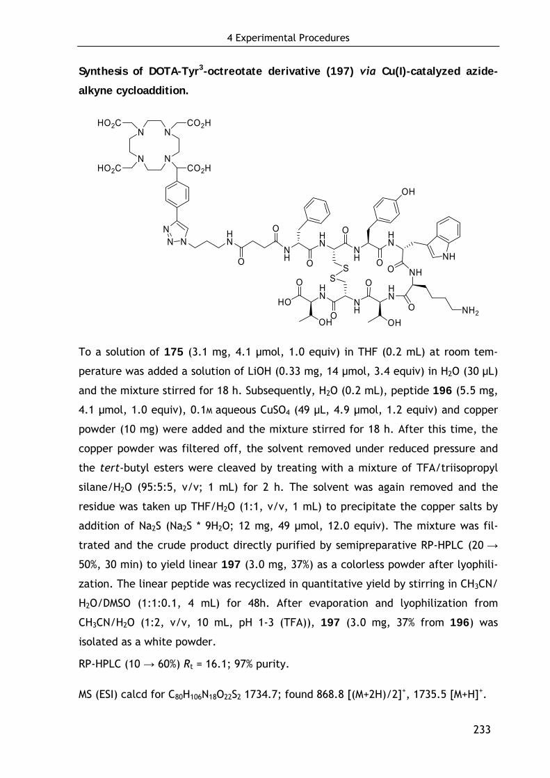
4 Experimental Procedures
233
Synthesis of DOTA-Tyr3-octreotate derivative (197) via Cu(I)-catalyzed azide-
alkyne cycloaddition.
N
N
N
N
HO2C
HO2C
CO2H
CO2H
NHN
O
OHN
O
NH
HN
NH
O HN
O
NHOS
S
O
O
OHNH2
NH
HN
OH
HO
O
O
OH
NH
NN
To a solution of 175 (3.1 mg, 4.1 μmol, 1.0 equiv) in THF (0.2 mL) at room tem-
perature was added a solution of LiOH (0.33 mg, 14 μmol, 3.4 equiv) in H2O (30 μL)
and the mixture stirred for 18 h. Subsequently, H2O (0.2 mL), peptide 196 (5.5 mg,
4.1 μmol, 1.0 equiv), 0.1M aqueous CuSO4 (49 μL, 4.9 μmol, 1.2 equiv) and copper
powder (10 mg) were added and the mixture stirred for 18 h. After this time, the
copper powder was filtered off, the solvent removed under reduced pressure and
the tert-butyl esters were cleaved by treating with a mixture of TFA/triisopropyl
silane/H2O (95:5:5, v/v; 1 mL) for 2 h. The solvent was again removed and the
residue was taken up THF/H2O (1:1, v/v, 1 mL) to precipitate the copper salts by
addition of Na2S (Na2S * 9H2O; 12 mg, 49 μmol, 12.0 equiv). The mixture was fil-
trated and the crude product directly purified by semipreparative RP-HPLC (20 →
50%, 30 min) to yield linear 197 (3.0 mg, 37%) as a colorless powder after lyophili-
zation. The linear peptide was recyclized in quantitative yield by stirring in CH3CN/
H2O/DMSO (1:1:0.1, 4 mL) for 48h. After evaporation and lyophilization from
CH3CN/H2O (1:2, v/v, 10 mL, pH 1-3 (TFA)), 197 (3.0 mg, 37% from 196) was
isolated as a white powder.
RP-HPLC (10 → 60%) Rt = 16.1; 97% purity.
MS (ESI) calcd for C80H106N18O22S2 1734.7; found 868.8 [(M+2H)/2]+, 1735.5 [M+H]+.
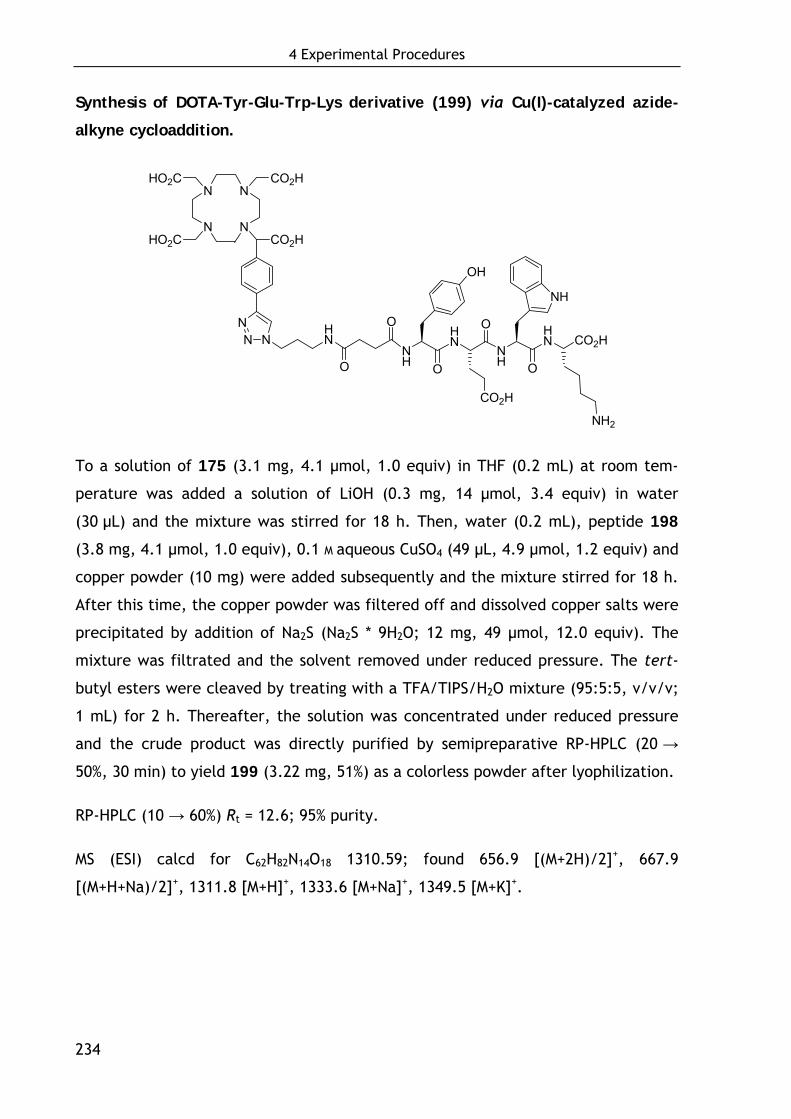
4 Experimental Procedures
234
Synthesis of DOTA-Tyr-Glu-Trp-Lys derivative (199) via Cu(I)-catalyzed azide-
alkyne cycloaddition.
N
N
N
N
HO2C
HO2C
CO2H
CO2H
NHN
O
OHN
O
NH
HN
OONH
NN CO2H
OH
NH
CO2H
NH2
To a solution of 175 (3.1 mg, 4.1 μmol, 1.0 equiv) in THF (0.2 mL) at room tem-
perature was added a solution of LiOH (0.3 mg, 14 μmol, 3.4 equiv) in water
(30 μL) and the mixture was stirred for 18 h. Then, water (0.2 mL), peptide 198
(3.8 mg, 4.1 μmol, 1.0 equiv), 0.1 M aqueous CuSO4 (49 μL, 4.9 μmol, 1.2 equiv) and
copper powder (10 mg) were added subsequently and the mixture stirred for 18 h.
After this time, the copper powder was filtered off and dissolved copper salts were
precipitated by addition of Na2S (Na2S * 9H2O; 12 mg, 49 μmol, 12.0 equiv). The
mixture was filtrated and the solvent removed under reduced pressure. The tert-
butyl esters were cleaved by treating with a TFA/TIPS/H2O mixture (95:5:5, v/v/v;
1 mL) for 2 h. Thereafter, the solution was concentrated under reduced pressure
and the crude product was directly purified by semipreparative RP-HPLC (20 →
50%, 30 min) to yield 199 (3.22 mg, 51%) as a colorless powder after lyophilization.
RP-HPLC (10 → 60%) Rt = 12.6; 95% purity.
MS (ESI) calcd for C62H82N14O18 1310.59; found 656.9 [(M+2H)/2]+, 667.9
[(M+H+Na)/2]+, 1311.8 [M+H]+, 1333.6 [M+Na]+, 1349.5 [M+K]+.
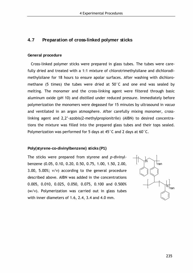
4 Experimental Procedures
235
4.7 Preparation of cross-linked polymer sticks
General procedure
Cross-linked polymer sticks were prepared in glass tubes. The tubes were care-
fully dried and treated with a 1:1 mixture of chlorotrimethylsilane and dichlorodi-
methylsilane for 18 hours to ensure apolar surfaces. After washing with dichloro-
methane (5 times) the tubes were dried at 50°C and one end was sealed by
melting. The monomer and the cross-linking agent were filtered through basic
aluminum oxide (pH 10) and distilled under reduced pressure. Immediately before
polymerization the monomers were degassed for 15 minutes by ultrasound in vacuo
and ventilated in an argon atmosphere. After carefully mixing monomer, cross-
linking agent and 2,2’-azobis(2-methylpropionitrile) (AIBN) to desired concentra-
tions the mixture was filled into the prepared glass tubes and their tops sealed.
Polymerization was performed for 5 days at 45°C and 2 days at 60°C.
Poly(styrene-co-divinylbenzene) sticks (P1)
The sticks were prepared from styrene and p-divinyl-
benzene (0.05, 0.10, 0.20, 0.50, 0.75, 1.00, 1.50, 2.00,
3.00, 5.00%; v/v) according to the general procedure
described above. AIBN was added in the concentrations
0.005, 0.010, 0.025, 0.050, 0.075, 0.100 and 0.500%
(w/v). Polymerization was carried out in glass tubes
with inner diameters of 1.6, 2.4, 3.4 and 4.0 mm.
n
n ran
ran
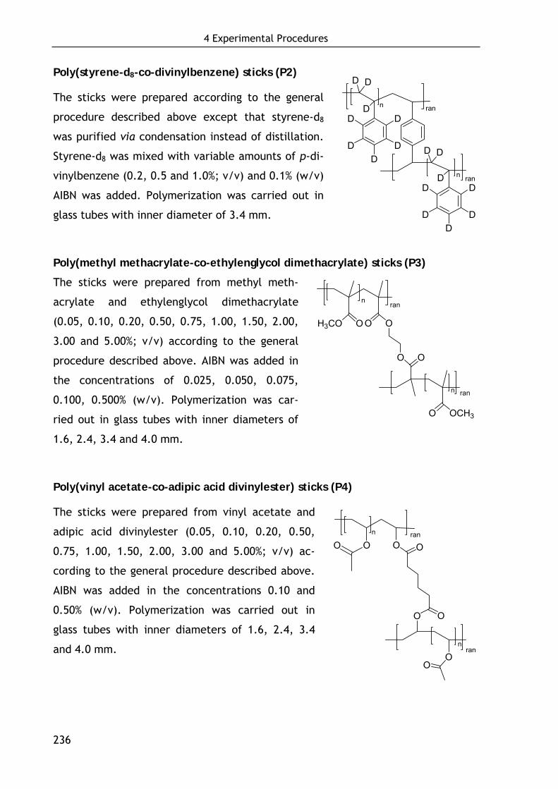
4 Experimental Procedures
236
Poly(styrene-d8-co-divinylbenzene) sticks (P2)
The sticks were prepared according to the general
procedure described above except that styrene-d8
was purified via condensation instead of distillation.
Styrene-d8 was mixed with variable amounts of p-di-
vinylbenzene (0.2, 0.5 and 1.0%; v/v) and 0.1% (w/v)
AIBN was added. Polymerization was carried out in
glass tubes with inner diameter of 3.4 mm.
Poly(methyl methacrylate-co-ethylenglycol dimethacrylate) sticks (P3)
The sticks were prepared from methyl meth-
acrylate and ethylenglycol dimethacrylate
(0.05, 0.10, 0.20, 0.50, 0.75, 1.00, 1.50, 2.00,
3.00 and 5.00%; v/v) according to the general
procedure described above. AIBN was added in
the concentrations of 0.025, 0.050, 0.075,
0.100, 0.500% (w/v). Polymerization was car-
ried out in glass tubes with inner diameters of
1.6, 2.4, 3.4 and 4.0 mm.
Poly(vinyl acetate-co-adipic acid divinylester) sticks (P4)
The sticks were prepared from vinyl acetate and
adipic acid divinylester (0.05, 0.10, 0.20, 0.50,
0.75, 1.00, 1.50, 2.00, 3.00 and 5.00%; v/v) ac-
cording to the general procedure described above.
AIBN was added in the concentrations 0.10 and
0.50% (w/v). Polymerization was carried out in
glass tubes with inner diameters of 1.6, 2.4, 3.4
and 4.0 mm.
rann
D
DD
D
DD
DD
n
D
DD
D
DD
DD
ran
ran
H3CO O O O
O O
O OCH3
n ran
n
ranO O
n
O
O
O
On
O
O
ran

4 Experimental Procedures
237
(S)-1-Benzyloxycarbonylethyl acrylate (213)
(S)-2-Hydroxypropionic acid benzylester (3.60 g, 20.0 mmol,
1.0 equiv) and DMAP (0.22 g, 2.00 mmol, 0.1 equiv) were dis-
solved in dry DCM (100 mL). After cooling to –78°C, DIPEA
(10.6 mL, 62.0 mmol, 3.1 equiv) and acryloyl chloride (211)
(2.20 mL, 25.0 mmol, 1.3 equiv) were added subsequently and
the mixture was stirred for 3 h. Thereafter, DCM (100 mL) and
saturated aqueous NaCl (100 mL) were added and the aqueous
layer was extracted with DCM (2×50 mL). The combined organic layers were dried
over MgSO4 and the solvent was removed in vacuo. Purification by flash chromatog-
raphy on silica gel (EtOAc/hexane 1:10) yielded 213 (4.2 g, 58%) as a colorless oil.
1H NMR (250 MHz, CDCl3): δ = 7.44-7.24 (m, 5H, Harom), 6.30 (dd, J = 17.5, 1.6 Hz,
1H, CHcisHCH), 6.28-6.08 (m, 1H, CH2CH), 5.87 (dd, J = 10.0, 1.6 Hz, 1H,
CHtransHCH), 5.30-5.10 (m, 3H, CH2Ph, CHCH3), 1.5 (d, J = 2.7 Hz, 3H, CHCH3).
Poly((S)-1-benzyloxycarbonylethyl acrylate-co-ethylenglycol dimethacrylate)
sticks (P5)
The sticks were prepared from (S)-1-benzy-
loxycarbonyl ethyl acrylate (213), ethylen-
glycol dimethacrylate (204) (0.50 and
1.00%; v/v) and 0.10% (w/v) AIBN accord-
ing to the general procedure described
above. Polymerization was carried out in
glass tubes with an inner diameter of 4.0
mm.
O
OOO
O O
n
OO
O
O
n
O
O
O
O
O
O
ran
ran
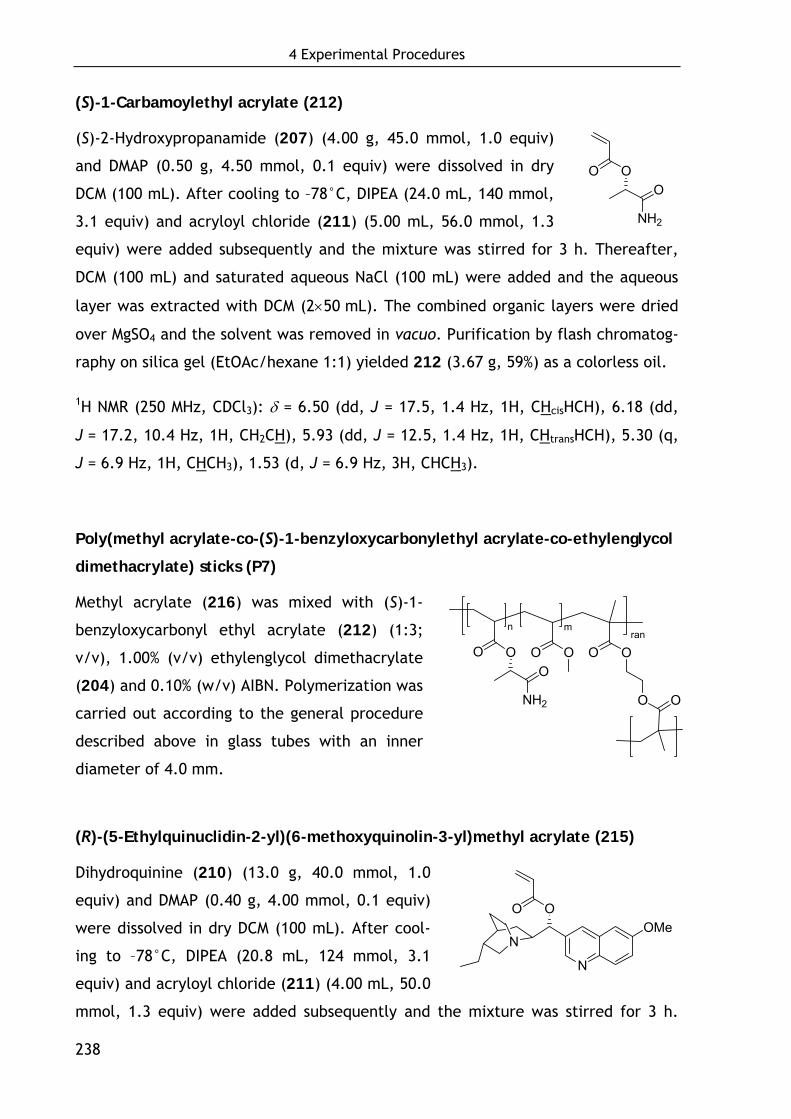
4 Experimental Procedures
238
(S)-1-Carbamoylethyl acrylate (212)
(S)-2-Hydroxypropanamide (207) (4.00 g, 45.0 mmol, 1.0 equiv)
and DMAP (0.50 g, 4.50 mmol, 0.1 equiv) were dissolved in dry
DCM (100 mL). After cooling to –78°C, DIPEA (24.0 mL, 140 mmol,
3.1 equiv) and acryloyl chloride (211) (5.00 mL, 56.0 mmol, 1.3
equiv) were added subsequently and the mixture was stirred for 3 h. Thereafter,
DCM (100 mL) and saturated aqueous NaCl (100 mL) were added and the aqueous
layer was extracted with DCM (2×50 mL). The combined organic layers were dried
over MgSO4 and the solvent was removed in vacuo. Purification by flash chromatog-
raphy on silica gel (EtOAc/hexane 1:1) yielded 212 (3.67 g, 59%) as a colorless oil.
1H NMR (250 MHz, CDCl3): δ = 6.50 (dd, J = 17.5, 1.4 Hz, 1H, CHcisHCH), 6.18 (dd,
J = 17.2, 10.4 Hz, 1H, CH2CH), 5.93 (dd, J = 12.5, 1.4 Hz, 1H, CHtransHCH), 5.30 (q,
J = 6.9 Hz, 1H, CHCH3), 1.53 (d, J = 6.9 Hz, 3H, CHCH3).
Poly(methyl acrylate-co-(S)-1-benzyloxycarbonylethyl acrylate-co-ethylenglycol
dimethacrylate) sticks (P7)
Methyl acrylate (216) was mixed with (S)-1-
benzyloxycarbonyl ethyl acrylate (212) (1:3;
v/v), 1.00% (v/v) ethylenglycol dimethacrylate
(204) and 0.10% (w/v) AIBN. Polymerization was
carried out according to the general procedure
described above in glass tubes with an inner
diameter of 4.0 mm.
(R)-(5-Ethylquinuclidin-2-yl)(6-methoxyquinolin-3-yl)methyl acrylate (215)
Dihydroquinine (210) (13.0 g, 40.0 mmol, 1.0
equiv) and DMAP (0.40 g, 4.00 mmol, 0.1 equiv)
were dissolved in dry DCM (100 mL). After cool-
ing to –78°C, DIPEA (20.8 mL, 124 mmol, 3.1
equiv) and acryloyl chloride (211) (4.00 mL, 50.0
mmol, 1.3 equiv) were added subsequently and the mixture was stirred for 3 h.
NH2
OOO
OO
N
OMeN
rann m
O
O
O O O O
O
O
NH2
O

4 Experimental Procedures
239
Thereafter, DCM (100 mL) and saturated aqueous NaCl (100 mL) were added and
the aqueous layer was extracted with DCM (2×50 mL). The combined organic layers
were dried over MgSO4 and the solvent was removed in vacuo. Purification by flash
chromatography on silica gel (MeOH/DCM 1:100, 1% NEt3) yielded 215 (8.50 g, 56%)
as a yellow oil.
1H NMR (250 MHz, CDCl3): δ = 8.68 (d, J = 4.7 Hz, 1H, Harom), 7.95 (d, J = 9.2 Hz,
1H, Harom), 7.52-7.38 (m, 1H, Harom), 7.37-7.24 (m, 2H, Harom), 6.61-6.41 (m, 2H,
CHcisHCH), 6.23-6.03 (m, 1H, CHtransHCH), 5.83 (d, J = 10.7 Hz, 1H, OCHCH), 4.01-
3.81 (m, 3H, OCH3), 3.44-3.24 (m, 1H, OCHCH), 3.10-2.90 (m, 2H, NCH2), 2.64-2.44
(m, 2H, NCH2), 1.44-1.24 (m, 6H, CH2CH(CH2)CH), 1.08-0.88 (m, 2H, CH2CH3), 0.90-
0.70 (m, 3H, CH2CH3).
Poly(methyl acrylate-co-(R)-(5-ethylquinuclidin-2-yl)(6-methoxyquinolin-3-
yl)methyl acrylate-co-ethylenglycol dimethacrylate) sticks (P9)
Methyl acrylate (216) was mixed
with (R)-(5-ethylquinuclidin-2-yl)
(6-methoxyquinolin-3-yl) methyl
acrylate (215) in the ratios: 4:1,
6:1, 8:1, 12:1, and 16:1 (v/v).
0.50% (v/v) ethyleneglycol dime-
thacrylate (204) and 0.10% (w/v)
AIBN were added and polymerization was carried out according to the general
procedure described above in glass tubes with an inner diameter of 4.0 mm.
rann m
O
O
O O O O
ON
MeON
O
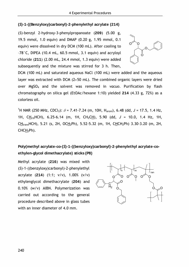
4 Experimental Procedures
240
(S)-1-((Benzyloxy)carbonyl)-2-phenylethyl acrylate (214)
(S)-benzyl 2-hydroxy-3-phenylpropanoate (209) (5.00 g,
19.5 mmol, 1.0 equiv) and DMAP (0.20 g, 1.95 mmol, 0.1
equiv) were dissolved in dry DCM (100 mL). After cooling to
–78°C, DIPEA (10.4 mL, 60.5 mmol, 3.1 equiv) and acryloyl
chloride (211) (2.00 mL, 24.4 mmol, 1.3 equiv) were added
subsequently and the mixture was stirred for 3 h. Then,
DCM (100 mL) and saturated aqueous NaCl (100 mL) were added and the aqueous
layer was extracted with DCM (2×50 mL). The combined organic layers were dried
over MgSO4 and the solvent was removed in vacuo. Purification by flash
chromatography on silica gel (EtOAc/hexane 1:10) yielded 214 (4.33 g, 72%) as a
colorless oil.
1H NMR (250 MHz, CDCl3): δ = 7.41-7.24 (m, 10H, Harom), 6.48 (dd, J = 17.5, 1.4 Hz,
1H, CHcisHCH), 6.25-6.14 (m, 1H, CH2CH), 5.90 (dd, J = 10.0, 1.4 Hz, 1H,
CHtransHCH), 5.21 (s, 2H, OCH2Ph), 5.52-5.32 (m, 1H, CHCH2Ph) 3.30-3.20 (m, 2H,
CHCH2Ph).
Poly(methyl acrylate-co-(S)-1-((benzyloxy)carbonyl)-2-phenylethyl acrylate-co-
ethylen-glycol dimethacrylate) sticks (P8)
Methyl acrylate (216) was mixed with
(S)-1-((benzyloxy)carbonyl)-2-phenylethyl
acrylate (214) (1:1; v/v), 1.00% (v/v)
ethylenglycol dimethacrylate (204) and
0.10% (w/v) AIBN. Polymerization was
carried out according to the general
procedure described above in glass tubes
with an inner diameter of 4.0 mm.
O
O
OO
rann m
O
O
O O O O
OO
OO
CH2
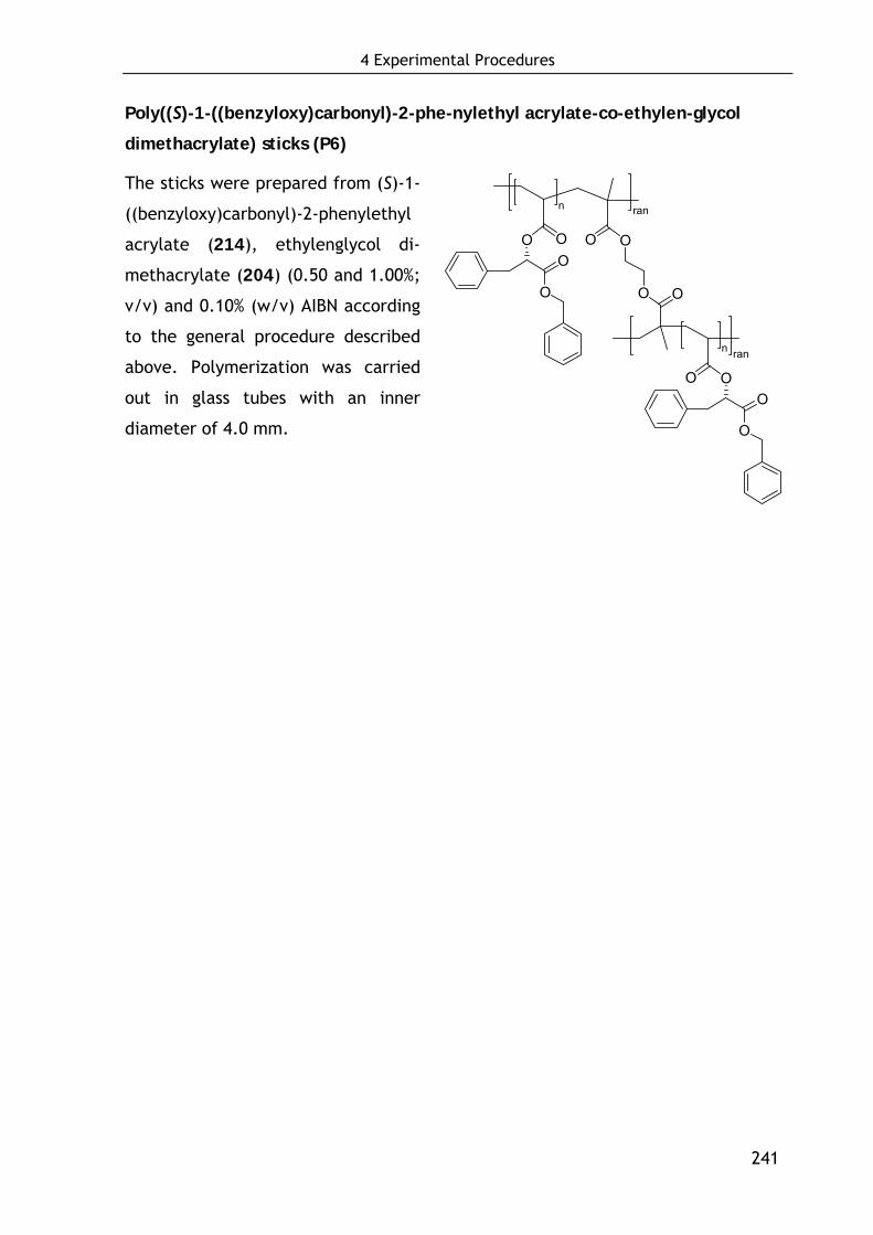
4 Experimental Procedures
241
Poly((S)-1-((benzyloxy)carbonyl)-2-phe-nylethyl acrylate-co-ethylen-glycol
dimethacrylate) sticks (P6)
The sticks were prepared from (S)-1-
((benzyloxy)carbonyl)-2-phenylethyl
acrylate (214), ethylenglycol di-
methacrylate (204) (0.50 and 1.00%;
v/v) and 0.10% (w/v) AIBN according
to the general procedure described
above. Polymerization was carried
out in glass tubes with an inner
diameter of 4.0 mm.
O O
n
OO
O
O
n
O
O
O
O
O
O
ran
ran


5 Appendix
243
5 Appendix
Supplementary Data 1. Summary of clotting factors and characteristics
Factor Trivial Name(s) Pathway Characteristic
Prekallikrein (PK)
Fletcher factor Intrinsic Functions with HMWK and FXII
High molecular weight kininogen
(HMWK)
contact activation cofactor; Fitzgerald,
Flaujeac Williams factor
Intrinsic Cofactor in kallikrein and factor XII activation,
necessary in FXIIa activation of FXI,
I Fibrinogen Both -
II Prothrombin Both Contains N-term. gla segment
III Tissue Factor (TF) Extrinsic subendothelial cell-surface receptor of and cofactor for
FVII -
IV Calcium Both -
V Proaccelerin Both
VI (same as Va) Accelerin Both Protein cofactor
VII Proconvertin Extrinsic Endopeptidase with gla residues
VIII Antihemophiliac factor A, Intrinsic Protein cofactor
IX Christmas Factor, antihemophilic factor B
Intrinsic Endopeptidase with gla residues
X Stuart-Prower Factor Both Endopeptidase with gla residues
XI Plasma thromboplastin antecedent (PTA)
Intrinsic Endopeptidase
XII Hageman Factor Intrinsic Endopeptidase
XIII fibrin stabilizing factor (FSF), fibrinoligase
Both Transpeptidase

5 Appendix
244
Supplementary Data 2. Regulatory and other proteins involving clot formation.
Protein Function
von Willebrand factor (vWF)
Associated with subendothelial connective tissue; serves as a bridge between platelet glycoprotein GPIb/IX and collagen
Protein C (PC) Activated to protein Ca (APC) by thrombin bound to thrombomodulin; then degrades factors VIIIa and Va
Protein S (PS) Acts as a cofactor of protein C; both proteins contain gla residues
Thrombomodulin Protein on the surface of endothelial cells; binds thrombin, which then activates protein C
Antithrombin III Most important coagulation inhibitor, controls activities of thrombin and factors IXa, Xa, XIa and XIIa
Supplementary Data 3. Kinetic analysis of ligand immobilization
Immobilization kinetics of ligand 3 (EYHSWEYC) binding to Toyopearl AF-Epoxy-
650M resin at different concentrations expressed as % of free peptides in solution.
0 10 20 30 40 500
20
40
60
80
100 5 mg/mL 10 mg/mL 20 mg/mL 40 mg/mL 60 mg/mL
Pep
tide
in s
olut
ion,
%
Time, h
Immobilization kinetics of EYHSWEYC (1)
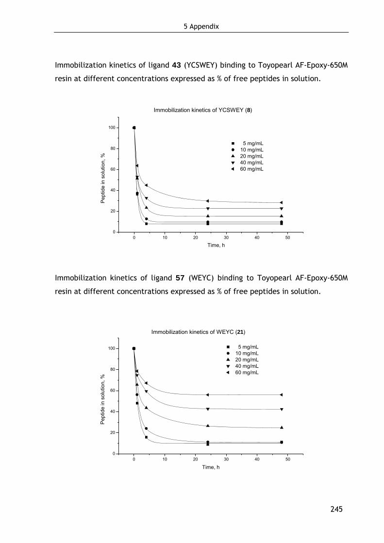
5 Appendix
245
Immobilization kinetics of ligand 43 (YCSWEY) binding to Toyopearl AF-Epoxy-650M
resin at different concentrations expressed as % of free peptides in solution.
0 10 20 30 40 500
20
40
60
80
100
Immobilization kinetics of YCSWEY (8)
5 mg/mL 10 mg/mL 20 mg/mL 40 mg/mL 60 mg/mL
Pep
tide
in s
olut
ion,
%
Time, h
Immobilization kinetics of ligand 57 (WEYC) binding to Toyopearl AF-Epoxy-650M
resin at different concentrations expressed as % of free peptides in solution.
0 10 20 30 40 500
20
40
60
80
100
Immobilization kinetics of WEYC (21)
5 mg/mL 10 mg/mL 20 mg/mL 40 mg/mL 60 mg/mL
Pep
tide
in s
olut
ion,
%
Time, h
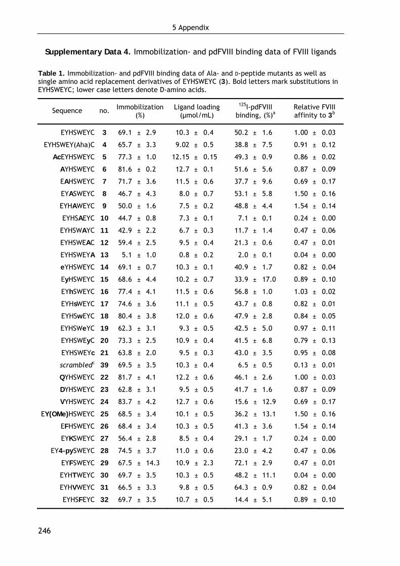
5 Appendix
246
Supplementary Data 4. Immobilization- and pdFVIII binding data of FVIII ligands
Table 1. Immobilization- and pdFVIII binding data of Ala- and D-peptide mutants as well as single amino acid replacement derivatives of EYHSWEYC (3). Bold letters mark substitutions in EYHSWEYC; lower case letters denote D-amino acids.
Sequence no. Immobilization (%)
Ligand loading (μmol/mL)
125I-pdFVIII binding, (%)a
Relative FVIII affinity to 3b
EYHSWEYC 3 69.1 ± 2.9 10.3 ± 0.4 50.2 ± 1.6 1.00 ± 0.03
EYHSWEY(Aha)C 4 65.7 ± 3.3 9.02 ± 0.5 38.8 ± 7.5 0.91 ± 0.12
AcEYHSWEYC 5 77.3 ± 1.0 12.15 ± 0.15 49.3 ± 0.9 0.86 ± 0.02
AYHSWEYC 6 81.6 ± 0.2 12.7 ± 0.1 51.6 ± 5.6 0.87 ± 0.09
EAHSWEYC 7 71.7 ± 3.6 11.5 ± 0.6 37.7 ± 9.6 0.69 ± 0.17
EYASWEYC 8 46.7 ± 4.3 8.0 ± 0.7 53.1 ± 5.8 1.50 ± 0.16
EYHAWEYC 9 50.0 ± 1.6 7.5 ± 0.2 48.8 ± 4.4 1.54 ± 0.14
EYHSAEYC 10 44.7 ± 0.8 7.3 ± 0.1 7.1 ± 0.1 0.24 ± 0.00
EYHSWAYC 11 42.9 ± 2.2 6.7 ± 0.3 11.7 ± 1.4 0.47 ± 0.06
EYHSWEAC 12 59.4 ± 2.5 9.5 ± 0.4 21.3 ± 0.6 0.47 ± 0.01
EYHSWEYA 13 5.1 ± 1.0 0.8 ± 0.2 2.0 ± 0.1 0.04 ± 0.00
eYHSWEYC 14 69.1 ± 0.7 10.3 ± 0.1 40.9 ± 1.7 0.82 ± 0.04
EyHSWEYC 15 68.6 ± 4.4 10.2 ± 0.7 33.9 ± 17.0 0.89 ± 0.10
EYhSWEYC 16 77.4 ± 4.1 11.5 ± 0.6 56.8 ± 1.0 1.03 ± 0.02
EYHsWEYC 17 74.6 ± 3.6 11.1 ± 0.5 43.7 ± 0.8 0.82 ± 0.01
EYHSwEYC 18 80.4 ± 3.8 12.0 ± 0.6 47.9 ± 2.8 0.84 ± 0.05
EYHSWeYC 19 62.3 ± 3.1 9.3 ± 0.5 42.5 ± 5.0 0.97 ± 0.11
EYHSWEyC 20 73.3 ± 2.5 10.9 ± 0.4 41.5 ± 6.8 0.79 ± 0.13
EYHSWEYc 21 63.8 ± 2.0 9.5 ± 0.3 43.0 ± 3.5 0.95 ± 0.08
scrambledc 39 69.5 ± 3.5 10.3 ± 0.4 6.5 ± 0.5 0.13 ± 0.01
QYHSWEYC 22 81.7 ± 4.1 12.2 ± 0.6 46.1 ± 2.6 1.00 ± 0.03
DYHSWEYC 23 62.8 ± 3.1 9.5 ± 0.5 41.7 ± 1.6 0.87 ± 0.09
VYHSWEYC 24 83.7 ± 4.2 12.7 ± 0.6 15.6 ± 12.9 0.69 ± 0.17
EY(OMe)HSWEYC 25 68.5 ± 3.4 10.1 ± 0.5 36.2 ± 13.1 1.50 ± 0.16
EFHSWEYC 26 68.4 ± 3.4 10.3 ± 0.5 41.3 ± 3.6 1.54 ± 0.14
EYKSWEYC 27 56.4 ± 2.8 8.5 ± 0.4 29.1 ± 1.7 0.24 ± 0.00
EY4-pySWEYC 28 74.5 ± 3.7 11.0 ± 0.6 23.0 ± 4.2 0.47 ± 0.06
EYFSWEYC 29 67.5 ± 14.3 10.9 ± 2.3 72.1 ± 2.9 0.47 ± 0.01
EYHTWEYC 30 69.7 ± 3.5 10.3 ± 0.5 48.2 ± 11.1 0.04 ± 0.00
EYHVWEYC 31 66.5 ± 3.3 9.8 ± 0.5 64.3 ± 0.9 0.82 ± 0.04
EYHSFEYC 32 69.7 ± 3.5 10.7 ± 0.5 14.4 ± 5.1 0.89 ± 0.10
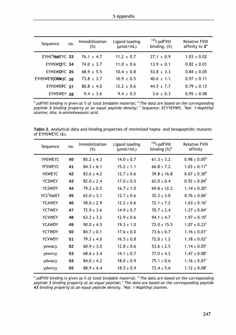
5 Appendix
247
Sequence no. Immobilization (%)
Ligand loading (μmol/mL)
125I-pdFVIII binding, (%)
Relative FVIII affinity to 3a
EYHS1NalEYC 33 76.1 ± 4.7 11.2 ± 0.7 27.1 ± 0.9 1.03 ± 0.02
EYHSWQYC 34 74.0 ± 3.7 11.0 ± 0.6 13.9 ± 0.1 0.82 ± 0.01
EYHSWDYC 35 68.9 ± 5.5 10.4 ± 0.8 53.8 ± 3.3 0.84 ± 0.05
EYHSWEY(OMe)C 36 73.8 ± 3.7 10.9 ± 0.5 40.6 ± 1.1 0.97 ± 0.11
EYHSWEFC 37 80.8 ± 4.0 12.2 ± 0.6 44.5 ± 7.7 0.79 ± 0.13
EYHSWEY 38 9.4 ± 3.6 9.4 ± 0.5 3.6 ± 0.3 0.95 ± 0.08
a pdFVIII binding is given as % of total bindable material; b The data are based on the corresponding peptide 3 binding property at an equal peptide density; c Sequence: ECYYEHWS. 1Nal: 1-Naphthyl alanine; Aha: 6-aminohexanoic acid.
Table 2. Analytical data and binding properties of minimized hepta- and hexapeptidic mutants of EYHSWEYC (1).
Sequence no. Immobilization (%)
Ligand loading (μmol/mL)
125I-pdFVIII binding (%)a
Relative FVIII affinity
YHSWEYC 40 85.2 ± 4.3 14.0 ± 0.7 61.3 ± 3.20 0.98 ± 0.05b
YFSWEYC 41 84.3 ± 6.1 15.2 ± 1.1 66.8 ± 7.20 1.03 ± 0.11b
HSWEYC 42 83.6 ± 4.2 12.7 ± 0.6 39.8 ± 16.8 0.67 ± 0.30b
YCSWEY 43 82.0 ± 2.4 17.0 ± 0.5 62.0 ± 0.40 0.92 ± 0.04b
YCSWDY 44 79.2 ± 0.5 16.7 ± 1.0 69.8 ± 12.2 1.14 ± 0.20c
YCS1NalEY 45 62.0 ± 3.1 12.7 ± 0.6 35.3 ± 3.00 0.76 ± 0.06c
YCAWEY 46 58.0 ± 2.9 12.2 ± 0.6 72.1 ± 7.20 1.63 ± 0.16c
YCTWEY 47 72.9 ± 3.6 14.9 ± 0.7 70.7 ± 2.40 1.27 ± 0.04c
YCVWEY 48 63.2 ± 3.2 12.9 ± 0.6 94.1 ± 4.70 1.97 ± 0.10c
YCAWDY 49 90.0 ± 4.5 19.3 ± 1.0 72.0 ± 15.5 1.07 ± 0.23c
YCTWDY 50 84.7 ± 0.1 17.6 ± 0.0 73.6 ± 0.70 1.16 ± 0.01c
YCVWDY 51 79.3 ± 4.0 16.5 ± 0.8 72.0 ± 1.20 1.18 ± 0.02c
yewacy 52 60.9 ± 3.0 12.8 ± 0.6 53.6 ± 2.50 1.14 ± 0.05c
yewvcy 53 68.6 ± 3.4 14.1 ± 0.7 77.0 ± 4.20 1.47 ± 0.08c
ydwacy 54 84.0 ± 4.2 18.0 ± 0.9 75.1 ± 0.60 1.16 ± 0.01c
ydwvcy 55 88.9 ± 4.4 18.5 ± 0.9 73.4 ± 5.60 1.12 ± 0.08c
a pdFVIII binding is given as % of total bindable material, b The data are based on the corresponding peptide 3 binding property at an equal peptide; c The data are based on the corresponding peptide 43 binding property at an equal peptide density. 1Nal: 1-Naphthyl alanine.
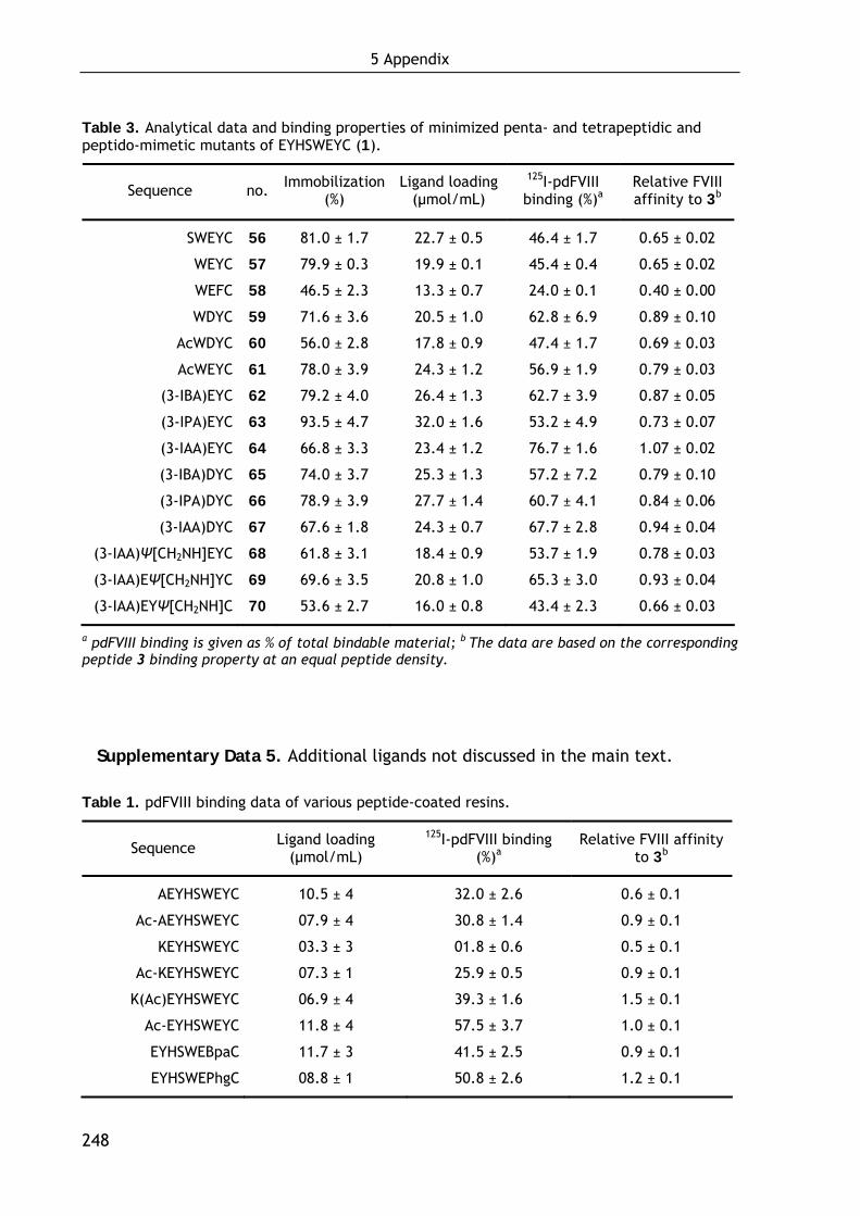
5 Appendix
248
Table 3. Analytical data and binding properties of minimized penta- and tetrapeptidic and peptido-mimetic mutants of EYHSWEYC (1).
Sequence no. Immobilization (%)
Ligand loading (μmol/mL)
125I-pdFVIII binding (%)a
Relative FVIII affinity to 3b
SWEYC 56 81.0 ± 1.7 22.7 ± 0.5 46.4 ± 1.7 0.65 ± 0.02
WEYC 57 79.9 ± 0.3 19.9 ± 0.1 45.4 ± 0.4 0.65 ± 0.02
WEFC 58 46.5 ± 2.3 13.3 ± 0.7 24.0 ± 0.1 0.40 ± 0.00
WDYC 59 71.6 ± 3.6 20.5 ± 1.0 62.8 ± 6.9 0.89 ± 0.10
AcWDYC 60 56.0 ± 2.8 17.8 ± 0.9 47.4 ± 1.7 0.69 ± 0.03
AcWEYC 61 78.0 ± 3.9 24.3 ± 1.2 56.9 ± 1.9 0.79 ± 0.03
(3-IBA)EYC 62 79.2 ± 4.0 26.4 ± 1.3 62.7 ± 3.9 0.87 ± 0.05
(3-IPA)EYC 63 93.5 ± 4.7 32.0 ± 1.6 53.2 ± 4.9 0.73 ± 0.07
(3-IAA)EYC 64 66.8 ± 3.3 23.4 ± 1.2 76.7 ± 1.6 1.07 ± 0.02
(3-IBA)DYC 65 74.0 ± 3.7 25.3 ± 1.3 57.2 ± 7.2 0.79 ± 0.10
(3-IPA)DYC 66 78.9 ± 3.9 27.7 ± 1.4 60.7 ± 4.1 0.84 ± 0.06
(3-IAA)DYC 67 67.6 ± 1.8 24.3 ± 0.7 67.7 ± 2.8 0.94 ± 0.04
(3-IAA)Ψ[CH2NH]EYC 68 61.8 ± 3.1 18.4 ± 0.9 53.7 ± 1.9 0.78 ± 0.03
(3-IAA)EΨ[CH2NH]YC 69 69.6 ± 3.5 20.8 ± 1.0 65.3 ± 3.0 0.93 ± 0.04
(3-IAA)EYΨ[CH2NH]C 70 53.6 ± 2.7 16.0 ± 0.8 43.4 ± 2.3 0.66 ± 0.03
a pdFVIII binding is given as % of total bindable material; b The data are based on the corresponding peptide 3 binding property at an equal peptide density.
Supplementary Data 5. Additional ligands not discussed in the main text.
Table 1. pdFVIII binding data of various peptide-coated resins.
Sequence Ligand loading (μmol/mL)
125I-pdFVIII binding (%)a
Relative FVIII affinity to 3b
AEYHSWEYC 10.5 ± 4 32.0 ± 2.6 0.6 ± 0.1
Ac-AEYHSWEYC 07.9 ± 4 30.8 ± 1.4 0.9 ± 0.1
KEYHSWEYC 03.3 ± 3 01.8 ± 0.6 0.5 ± 0.1
Ac-KEYHSWEYC 07.3 ± 1 25.9 ± 0.5 0.9 ± 0.1
K(Ac)EYHSWEYC 06.9 ± 4 39.3 ± 1.6 1.5 ± 0.1
Ac-EYHSWEYC 11.8 ± 4 57.5 ± 3.7 1.0 ± 0.1
EYHSWEBpaC 11.7 ± 3 41.5 ± 2.5 0.9 ± 0.1
EYHSWEPhgC 08.8 ± 1 50.8 ± 2.6 1.2 ± 0.1
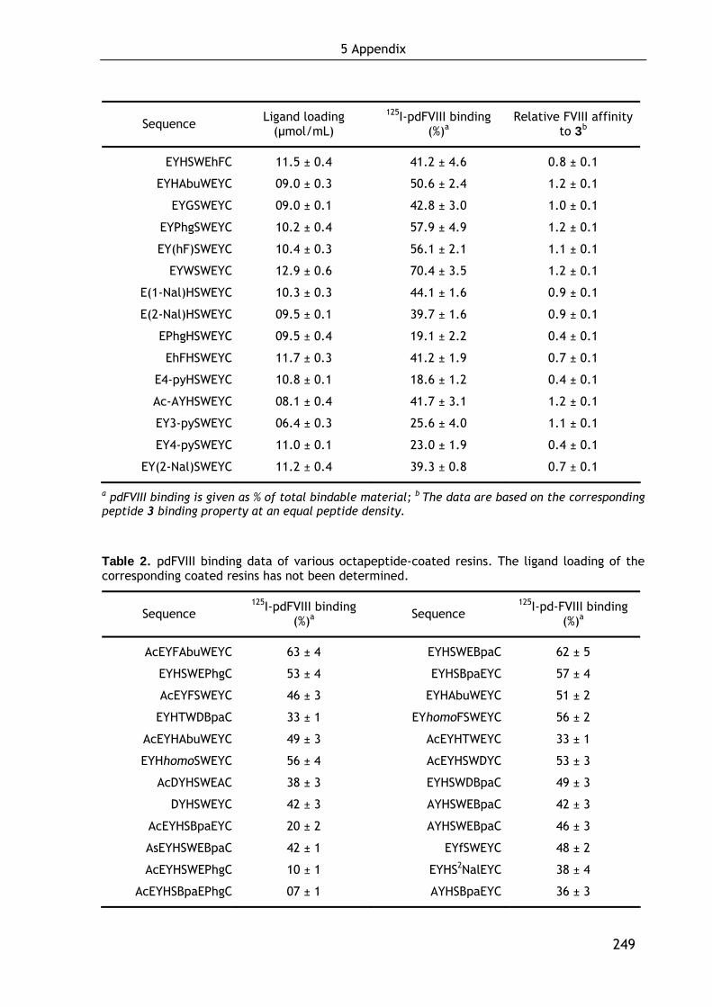
5 Appendix
249
Sequence Ligand loading (μmol/mL)
125I-pdFVIII binding (%)a
Relative FVIII affinity to 3b
EYHSWEhFC 11.5 ± 0.4 41.2 ± 4.6 0.8 ± 0.1
EYHAbuWEYC 09.0 ± 0.3 50.6 ± 2.4 1.2 ± 0.1
EYGSWEYC 09.0 ± 0.1 42.8 ± 3.0 1.0 ± 0.1
EYPhgSWEYC 10.2 ± 0.4 57.9 ± 4.9 1.2 ± 0.1
EY(hF)SWEYC 10.4 ± 0.3 56.1 ± 2.1 1.1 ± 0.1
EYWSWEYC 12.9 ± 0.6 70.4 ± 3.5 1.2 ± 0.1
E(1-Nal)HSWEYC 10.3 ± 0.3 44.1 ± 1.6 0.9 ± 0.1
E(2-Nal)HSWEYC 09.5 ± 0.1 39.7 ± 1.6 0.9 ± 0.1
EPhgHSWEYC 09.5 ± 0.4 19.1 ± 2.2 0.4 ± 0.1
EhFHSWEYC 11.7 ± 0.3 41.2 ± 1.9 0.7 ± 0.1
E4-pyHSWEYC 10.8 ± 0.1 18.6 ± 1.2 0.4 ± 0.1
Ac-AYHSWEYC 08.1 ± 0.4 41.7 ± 3.1 1.2 ± 0.1
EY3-pySWEYC 06.4 ± 0.3 25.6 ± 4.0 1.1 ± 0.1
EY4-pySWEYC 11.0 ± 0.1 23.0 ± 1.9 0.4 ± 0.1
EY(2-Nal)SWEYC 11.2 ± 0.4 39.3 ± 0.8 0.7 ± 0.1
a pdFVIII binding is given as % of total bindable material; b The data are based on the corresponding peptide 3 binding property at an equal peptide density.
Table 2. pdFVIII binding data of various octapeptide-coated resins. The ligand loading of the corresponding coated resins has not been determined.
Sequence 125I-pdFVIII binding
(%)a Sequence 125I-pd-FVIII binding
(%)a
AcEYFAbuWEYC 63 ± 4 EYHSWEBpaC 62 ± 5
EYHSWEPhgC 53 ± 4 EYHSBpaEYC 57 ± 4
AcEYFSWEYC 46 ± 3 EYHAbuWEYC 51 ± 2
EYHTWDBpaC 33 ± 1 EYhomoFSWEYC 56 ± 2
AcEYHAbuWEYC 49 ± 3 AcEYHTWEYC 33 ± 1
EYHhomoSWEYC 56 ± 4 AcEYHSWDYC 53 ± 3
AcDYHSWEAC 38 ± 3 EYHSWDBpaC 49 ± 3
DYHSWEYC 42 ± 3 AYHSWEBpaC 42 ± 3
AcEYHSBpaEYC 20 ± 2 AYHSWEBpaC 46 ± 3
AsEYHSWEBpaC 42 ± 1 EYfSWEYC 48 ± 2
AcEYHSWEPhgC 10 ± 1 EYHS2NalEYC 38 ± 4
AcEYHSBpaEPhgC 07 ± 1 AYHSBpaEYC 36 ± 3
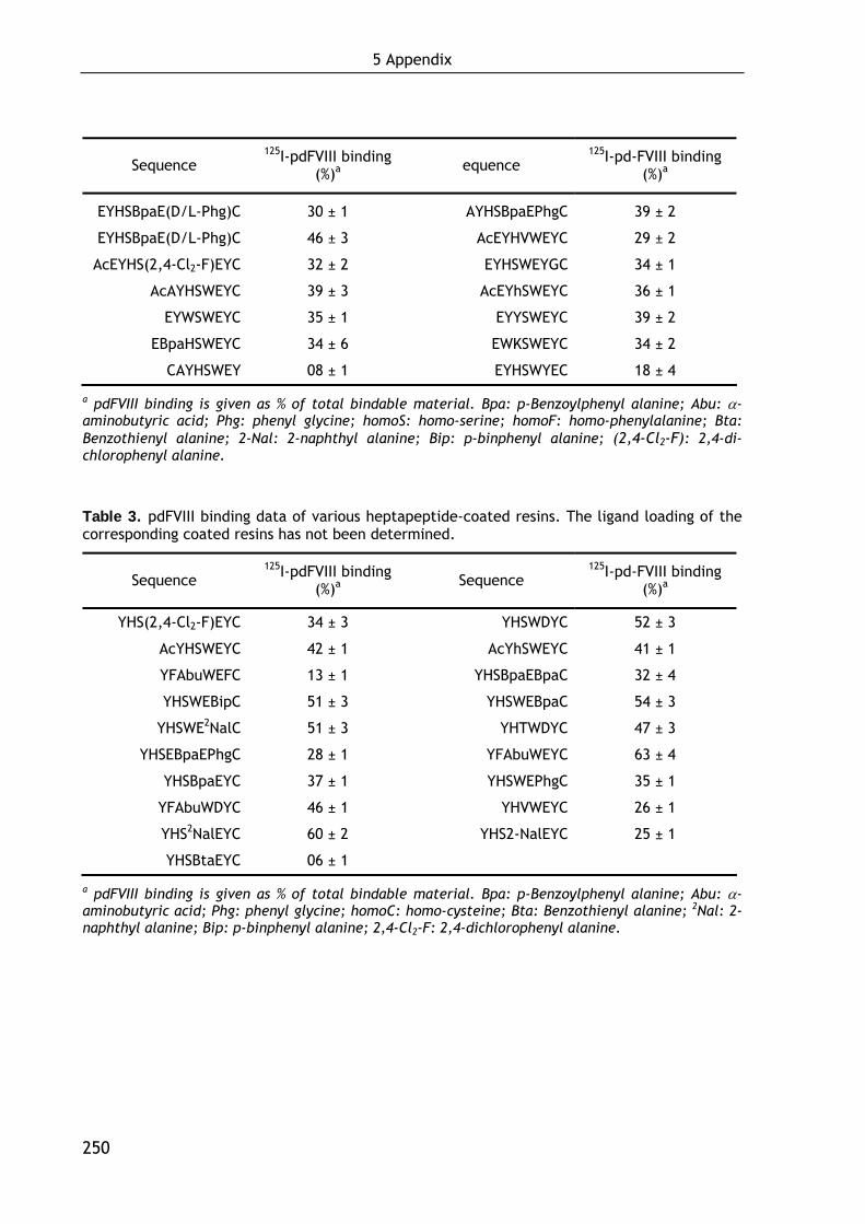
5 Appendix
250
Sequence 125I-pdFVIII binding
(%)a equence 125I-pd-FVIII binding
(%)a
EYHSBpaE(D/L-Phg)C 30 ± 1 AYHSBpaEPhgC 39 ± 2
EYHSBpaE(D/L-Phg)C 46 ± 3 AcEYHVWEYC 29 ± 2
AcEYHS(2,4-Cl2-F)EYC 32 ± 2 EYHSWEYGC 34 ± 1
AcAYHSWEYC 39 ± 3 AcEYhSWEYC 36 ± 1
EYWSWEYC 35 ± 1 EYYSWEYC 39 ± 2
EBpaHSWEYC 34 ± 6 EWKSWEYC 34 ± 2
CAYHSWEY 08 ± 1 EYHSWYEC 18 ± 4
a pdFVIII binding is given as % of total bindable material. Bpa: p-Benzoylphenyl alanine; Abu: α-aminobutyric acid; Phg: phenyl glycine; homoS: homo-serine; homoF: homo-phenylalanine; Bta: Benzothienyl alanine; 2-Nal: 2-naphthyl alanine; Bip: p-binphenyl alanine; (2,4-Cl2-F): 2,4-di-chlorophenyl alanine.
Table 3. pdFVIII binding data of various heptapeptide-coated resins. The ligand loading of the corresponding coated resins has not been determined.
Sequence 125I-pdFVIII binding
(%)a Sequence 125I-pd-FVIII binding
(%)a
YHS(2,4-Cl2-F)EYC 34 ± 3 YHSWDYC 52 ± 3
AcYHSWEYC 42 ± 1 AcYhSWEYC 41 ± 1
YFAbuWEFC 13 ± 1 YHSBpaEBpaC 32 ± 4
YHSWEBipC 51 ± 3 YHSWEBpaC 54 ± 3
YHSWE2NalC 51 ± 3 YHTWDYC 47 ± 3
YHSEBpaEPhgC 28 ± 1 YFAbuWEYC 63 ± 4
YHSBpaEYC 37 ± 1 YHSWEPhgC 35 ± 1
YFAbuWDYC 46 ± 1 YHVWEYC 26 ± 1
YHS2NalEYC 60 ± 2 YHS2-NalEYC 25 ± 1
YHSBtaEYC 06 ± 1
a pdFVIII binding is given as % of total bindable material. Bpa: p-Benzoylphenyl alanine; Abu: α-aminobutyric acid; Phg: phenyl glycine; homoC: homo-cysteine; Bta: Benzothienyl alanine; 2Nal: 2-naphthyl alanine; Bip: p-binphenyl alanine; 2,4-Cl2-F: 2,4-dichlorophenyl alanine.
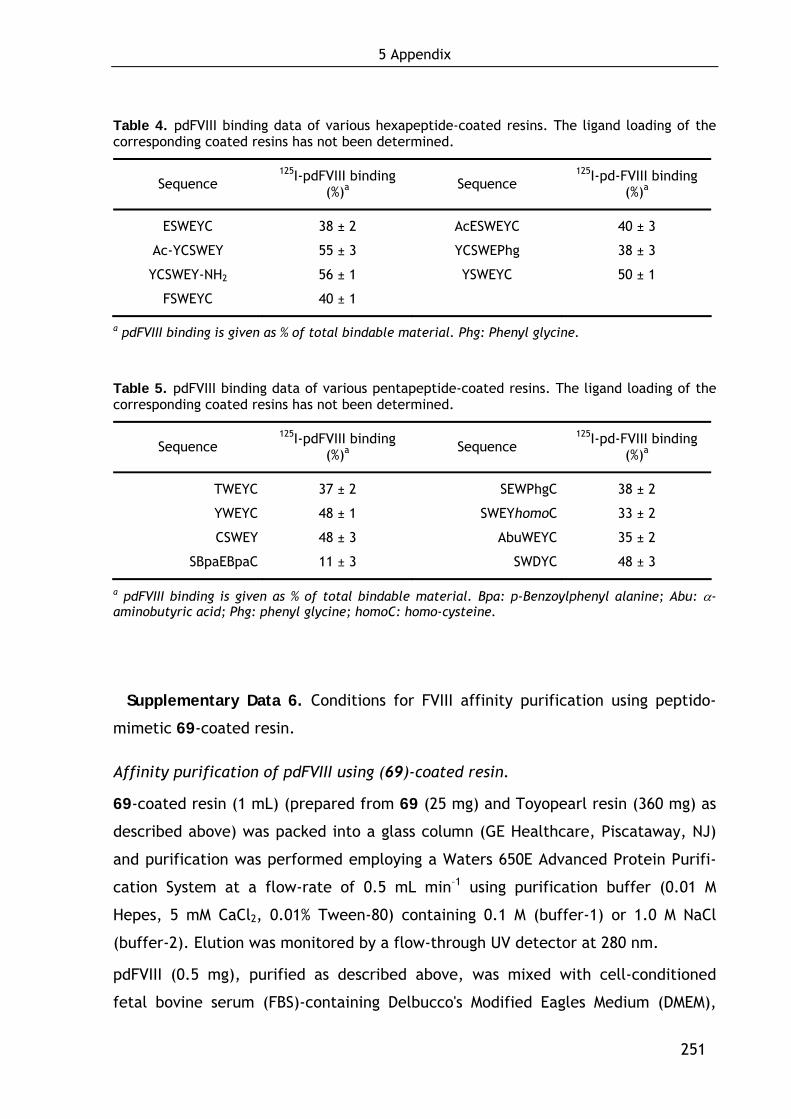
5 Appendix
251
Table 4. pdFVIII binding data of various hexapeptide-coated resins. The ligand loading of the corresponding coated resins has not been determined.
Sequence 125I-pdFVIII binding
(%)a Sequence 125I-pd-FVIII binding
(%)a
ESWEYC 38 ± 2 AcESWEYC 40 ± 3
Ac-YCSWEY 55 ± 3 YCSWEPhg 38 ± 3
YCSWEY-NH2 56 ± 1 YSWEYC 50 ± 1
FSWEYC 40 ± 1
a pdFVIII binding is given as % of total bindable material. Phg: Phenyl glycine.
Table 5. pdFVIII binding data of various pentapeptide-coated resins. The ligand loading of the corresponding coated resins has not been determined.
Sequence 125I-pdFVIII binding
(%)a Sequence 125I-pd-FVIII binding
(%)a
TWEYC 37 ± 2 SEWPhgC 38 ± 2
YWEYC 48 ± 1 SWEYhomoC 33 ± 2
CSWEY 48 ± 3 AbuWEYC 35 ± 2
SBpaEBpaC 11 ± 3 SWDYC 48 ± 3
a pdFVIII binding is given as % of total bindable material. Bpa: p-Benzoylphenyl alanine; Abu: α-aminobutyric acid; Phg: phenyl glycine; homoC: homo-cysteine.
Supplementary Data 6. Conditions for FVIII affinity purification using peptido-
mimetic 69-coated resin.
Affinity purification of pdFVIII using (69)-coated resin.
69-coated resin (1 mL) (prepared from 69 (25 mg) and Toyopearl resin (360 mg) as
described above) was packed into a glass column (GE Healthcare, Piscataway, NJ)
and purification was performed employing a Waters 650E Advanced Protein Purifi-
cation System at a flow-rate of 0.5 mL min–1 using purification buffer (0.01 M
Hepes, 5 mM CaCl2, 0.01% Tween-80) containing 0.1 M (buffer-1) or 1.0 M NaCl
(buffer-2). Elution was monitored by a flow-through UV detector at 280 nm.
pdFVIII (0.5 mg), purified as described above, was mixed with cell-conditioned
fetal bovine serum (FBS)-containing Delbucco's Modified Eagles Medium (DMEM),
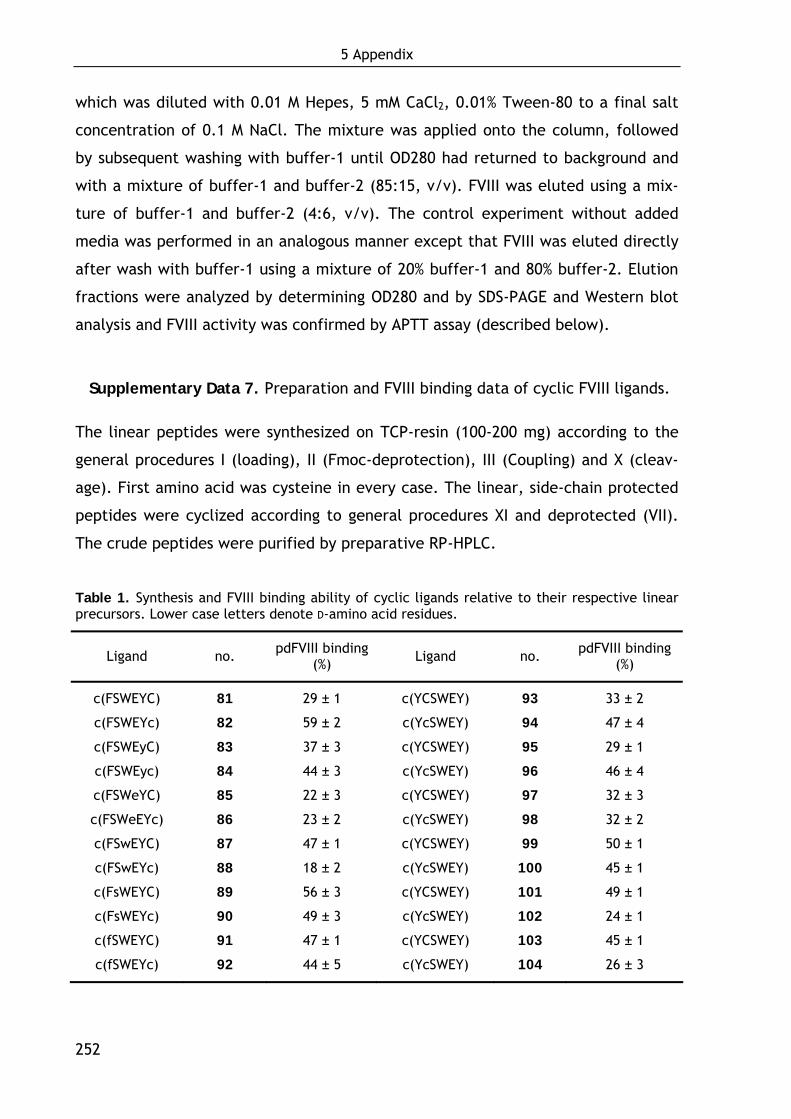
5 Appendix
252
which was diluted with 0.01 M Hepes, 5 mM CaCl2, 0.01% Tween-80 to a final salt
concentration of 0.1 M NaCl. The mixture was applied onto the column, followed
by subsequent washing with buffer-1 until OD280 had returned to background and
with a mixture of buffer-1 and buffer-2 (85:15, v/v). FVIII was eluted using a mix-
ture of buffer-1 and buffer-2 (4:6, v/v). The control experiment without added
media was performed in an analogous manner except that FVIII was eluted directly
after wash with buffer-1 using a mixture of 20% buffer-1 and 80% buffer-2. Elution
fractions were analyzed by determining OD280 and by SDS-PAGE and Western blot
analysis and FVIII activity was confirmed by APTT assay (described below).
Supplementary Data 7. Preparation and FVIII binding data of cyclic FVIII ligands.
The linear peptides were synthesized on TCP-resin (100-200 mg) according to the
general procedures I (loading), II (Fmoc-deprotection), III (Coupling) and X (cleav-
age). First amino acid was cysteine in every case. The linear, side-chain protected
peptides were cyclized according to general procedures XI and deprotected (VII).
The crude peptides were purified by preparative RP-HPLC.
Table 1. Synthesis and FVIII binding ability of cyclic ligands relative to their respective linear precursors. Lower case letters denote D-amino acid residues.
Ligand no. pdFVIII binding (%) Ligand no. pdFVIII binding
(%)
c(FSWEYC) 81 29 ± 1 c(YCSWEY) 93 33 ± 2
c(FSWEYc) 82 59 ± 2 c(YcSWEY) 94 47 ± 4
c(FSWEyC) 83 37 ± 3 c(YCSWEY) 95 29 ± 1
c(FSWEyc) 84 44 ± 3 c(YcSWEY) 96 46 ± 4
c(FSWeYC) 85 22 ± 3 c(YCSWEY) 97 32 ± 3
c(FSWeEYc) 86 23 ± 2 c(YcSWEY) 98 32 ± 2
c(FSwEYC) 87 47 ± 1 c(YCSWEY) 99 50 ± 1
c(FSwEYc) 88 18 ± 2 c(YcSWEY) 100 45 ± 1
c(FsWEYC) 89 56 ± 3 c(YCSWEY) 101 49 ± 1
c(FsWEYc) 90 49 ± 3 c(YcSWEY) 102 24 ± 1
c(fSWEYC) 91 47 ± 1 c(YCSWEY) 103 45 ± 1
c(fSWEYc) 92 44 ± 5 c(YcSWEY) 104 26 ± 3
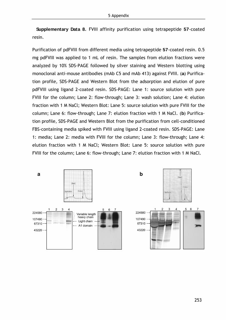
5 Appendix
253
Supplementary Data 8. FVIII affinity purification using tetrapeptide 57-coated
resin.
Purification of pdFVIII from different media using tetrapeptide 57-coated resin. 0.5
mg pdFVIII was applied to 1 mL of resin. The samples from elution fractions were
analyzed by 10% SDS-PAGE followed by silver staining and Western blotting using
monoclonal anti-mouse antibodies (mAb C5 and mAb 413) against FVIII. (a) Purifica-
tion profile, SDS-PAGE and Western Blot from the adsorption and elution of pure
pdFVIII using ligand 2-coated resin. SDS-PAGE: Lane 1: source solution with pure
FVIII for the column; Lane 2: flow-through; Lane 3: wash solution; Lane 4: elution
fraction with 1 M NaCl; Western Blot: Lane 5: source solution with pure FVIII for the
column; Lane 6: flow-through; Lane 7: elution fraction with 1 M NaCl. (b) Purifica-
tion profile, SDS-PAGE and Western Blot from the purification from cell-conditioned
FBS-containing media spiked with FVIII using ligand 2-coated resin. SDS-PAGE: Lane
1: media; Lane 2: media with FVIII for the column; Lane 3: flow-through; Lane 4:
elution fraction with 1 M NaCl; Western Blot: Lane 5: source solution with pure
FVIII for the column; Lane 6: flow-through; Lane 7: elution fraction with 1 M NaCl.
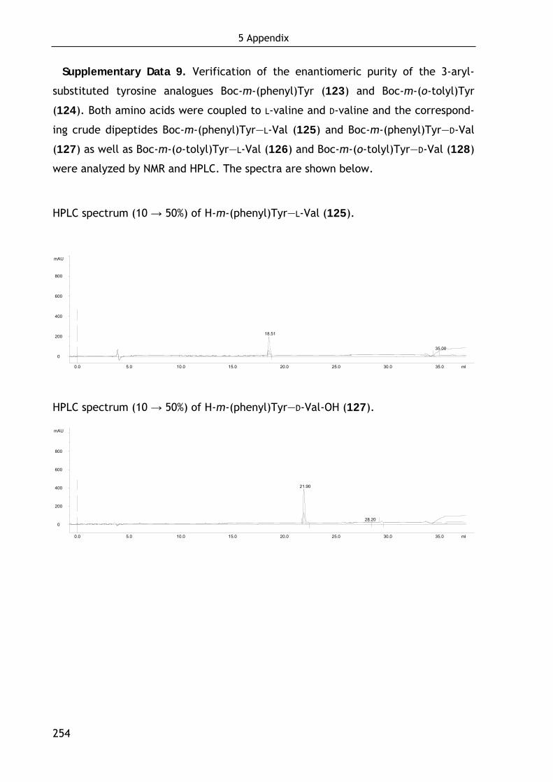
5 Appendix
254
Supplementary Data 9. Verification of the enantiomeric purity of the 3-aryl-
substituted tyrosine analogues Boc-m-(phenyl)Tyr (123) and Boc-m-(o-tolyl)Tyr
(124). Both amino acids were coupled to L-valine and D-valine and the correspond-
ing crude dipeptides Boc-m-(phenyl)Tyr—L-Val (125) and Boc-m-(phenyl)Tyr—D-Val
(127) as well as Boc-m-(o-tolyl)Tyr—L-Val (126) and Boc-m-(o-tolyl)Tyr—D-Val (128)
were analyzed by NMR and HPLC. The spectra are shown below.
HPLC spectrum (10 → 50%) of H-m-(phenyl)Tyr—L-Val (125).
0
200
400
600
800
mAU
0.0 5.0 10.0 15.0 20.0 25.0 30.0 35.0 ml
18.51
35.00
HPLC spectrum (10 → 50%) of H-m-(phenyl)Tyr—D-Val-OH (127).
0
200
400
600
800
mAU
0.0 5.0 10.0 15.0 20.0 25.0 30.0 35.0 ml
21.90
28.20
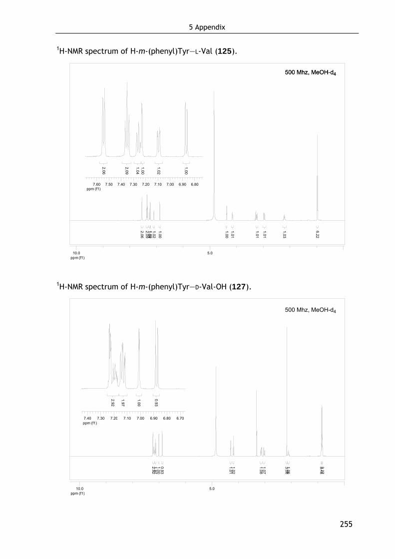
5 Appendix
255
1H-NMR spectrum of H-m-(phenyl)Tyr—L-Val (125).
ppm (f1)5.010.0
2.062.09
1.021.00
1.041.00
1.001.01
1.01
1.01
1.03
6.22
ppm (f1)6.806.907.007.107.207.307.407.507.60
2.06
2.09
1.02
1.00
1.041.00
1H-NMR spectrum of H-m-(phenyl)Tyr—D-Val-OH (127).
ppm (f1)5.010.0
3.273.18
1.063.06
1.071.08
1.021.01
0.931.001.972.92
ppm (f1)6.706.806.907.007.107.207.307.40
0.93
1.00
1.97
2.92
500 Mhz, MeOH-d4
500 Mhz, MeOH-d4500 Mhz, MeOH-d4
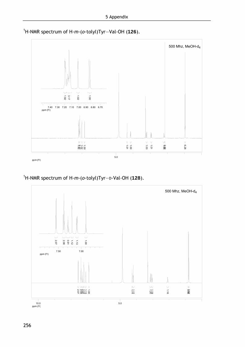
5 Appendix
256
1H-NMR spectrum of H-m-(o-tolyl)Tyr—Val-OH (126).
ppm (f1)5.0
1.001.023.171.92
1.011.00
1.03
1.01
1.053.00
6.28
ppm (f1)6.706.806.907.007.107.207.307.40
1.00
1.02
3.171.92
1H-NMR spectrum of H-m-(o-tolyl)Tyr—D-Val-OH (128).
ppm (f1)5.010.0
2.072.080.911.121.131.00
1.111.12
1.201.19
1.16
3.563.62
ppm (f1)7.007.50
2.07
2.08
0.911.12
1.13
1.00
500 Mhz, MeOH-d4
500 Mhz, MeOH-d4

5 Appendix
257
HPLC spectrum (10 → 50%) of H-m-(o-tolyl)Tyr—Val-OH (126).
0
200
400
600
800
mAU
0.0 5.0 10.0 15.0 20.0 25.0 30.0 35.0 ml
19.18
19.78
30.14
HPLC spectrum (10 → 50%) of H-m-(o-tolyl)Tyr—D-Val-OH (128).
0
200
400
600
800
mAU
0.0 5.0 10.0 15.0 20.0 25.0 30.0 35.0 ml
23.06
29.71
Supplementary Data 10. Verification of the enantiomeric purity of (S)-Boc-4-
hydroxy-naphthyl alanine (136) obtained by asymmetric hydrogenation. (S)-136
was coupled to L-valine to yield the (S,S)-dipeptide 137. Racemic 136 was coupled
in a similar manner to achieve the analogous diastereomeric mixture (S/R, S)-138.
Both dipeptides were analyzed by NMR and HPLC. The spectra are shown below.
HPLC spectrum (10 → 50%) of H-(S)-(4-hydroxy)Nal-Val-OH (136).
0
200
400
600
800 mAU
0.0 5.0 10.0 15.0 20.0 25.0 30.0 35.0 ml
12.19
14.71
15.45 19.82 23.69
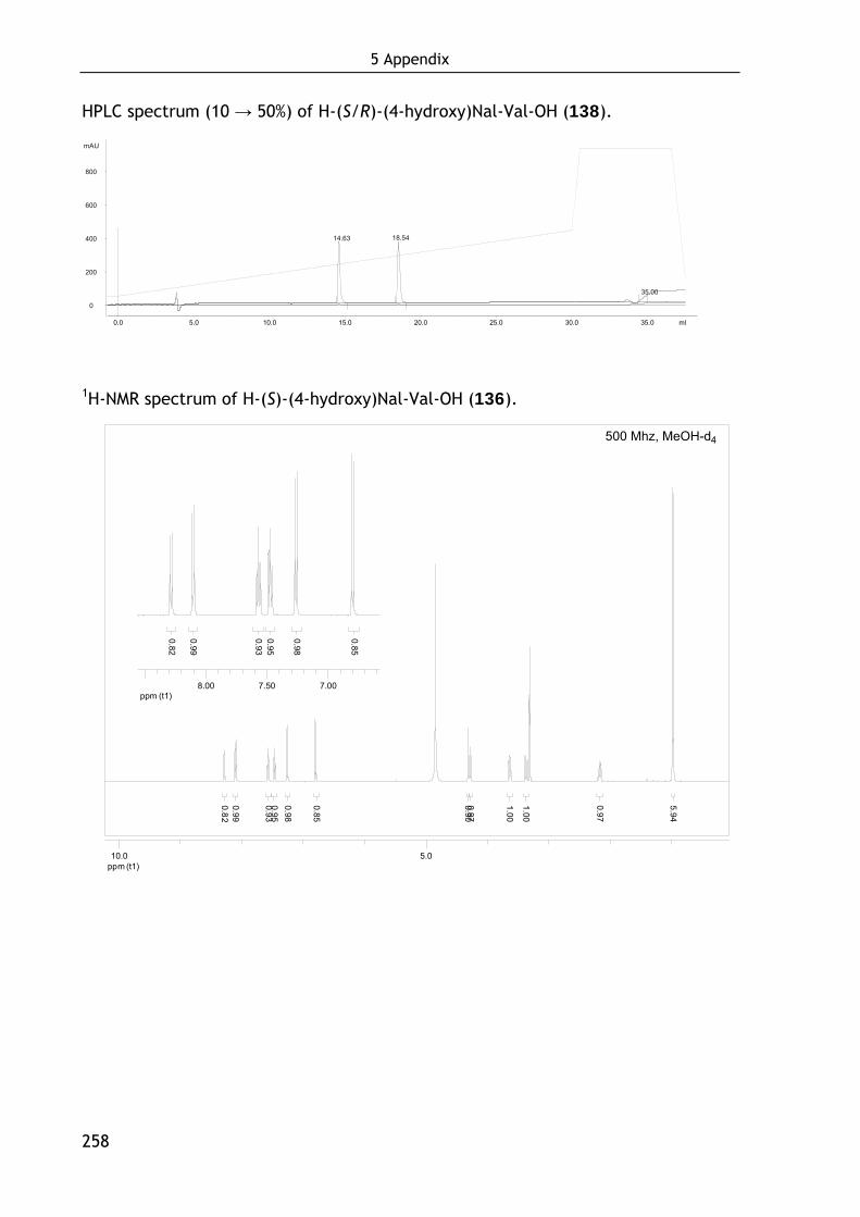
5 Appendix
258
HPLC spectrum (10 → 50%) of H-(S/R)-(4-hydroxy)Nal-Val-OH (138).
0
200
400
600
800 mAU
0.0 5.0 10.0 15.0 20.0 25.0 30.0 35.0 ml
14.63 18.54
35.00
1H-NMR spectrum of H-(S)-(4-hydroxy)Nal-Val-OH (136).
ppm (t1)5.010.0
0.820.99
0.930.950.98
0.85
1.00
1.00
0.900.97
0.97
5.94
ppm (t1)7.007.508.00
0.82
0.99
0.930.95
0.98
0.85
500 Mhz, MeOH-d4
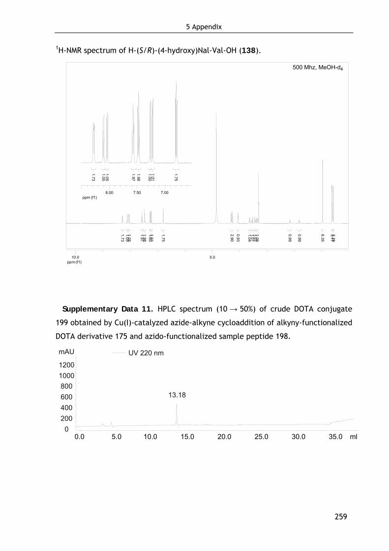
5 Appendix
259
1H-NMR spectrum of H-(S/R)-(4-hydroxy)Nal-Val-OH (138).
ppm (f1)5.010.0
1.731.051.05
1.971.981.001.01
1.75
0.93
1.041.071.071.06
0.99
0.99
6.35
3.173.28
2.90
ppm (f1)7.007.508.00
1.73
1.051.05
1.971.98
1.001.01
1.75
Supplementary Data 11. HPLC spectrum (10 → 50%) of crude DOTA conjugate
199 obtained by Cu(I)-catalyzed azide-alkyne cycloaddition of alkyny-functionalized
DOTA derivative 175 and azido-functionalized sample peptide 198.
UV 220 nm
0 200 400 600 800 1000 1200
mAU
0.0 5.0 10.0 15.0 20.0 25.0 30.0 35.0 ml
13.18
500 Mhz, MeOH-d4
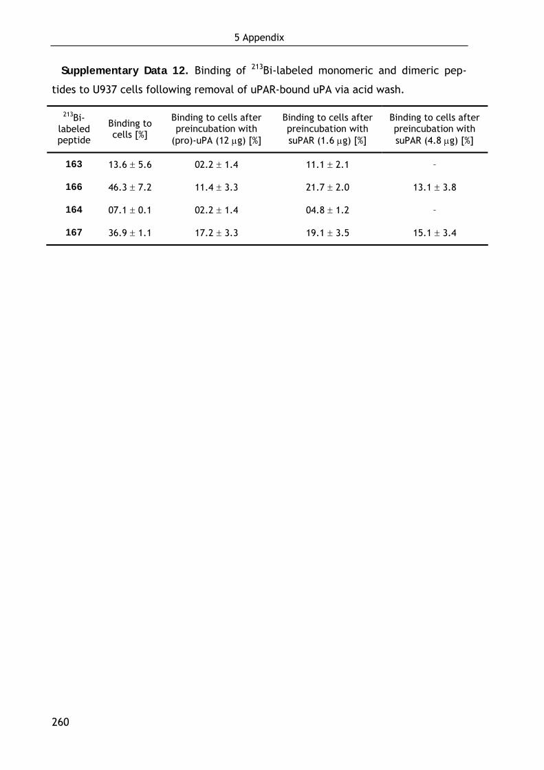
5 Appendix
260
Supplementary Data 12. Binding of 213Bi-labeled monomeric and dimeric pep-
tides to U937 cells following removal of uPAR-bound uPA via acid wash.
213Bi-labeled peptide
Binding to cells [%]
Binding to cells after preincubation with
(pro)-uPA (12 μg) [%]
Binding to cells after preincubation with suPAR (1.6 μg) [%]
Binding to cells after preincubation with suPAR (4.8 μg) [%]
163 13.6 ± 5.6 02.2 ± 1.4 11.1 ± 2.1 –
166 46.3 ± 7.2 11.4 ± 3.3 21.7 ± 2.0 13.1 ± 3.8
164 07.1 ± 0.1 02.2 ± 1.4 04.8 ± 1.2 –
167 36.9 ± 1.1 17.2 ± 3.3 19.1 ± 3.5 15.1 ± 3.4

6 Abbreviations and symbols
261
6 Abbreviations and Symbols
α1-AT α1-antitrypsin
α2-AP α2-antiplasmin
α2MR α2-macroglobulin receptor
δ chemical shift in NMR spectroscopy
λ wavelength
1D, 2D, 3D one-, two-, three dimensional
4-py 4-pyridylalanine
a activated
Ac acetyl
Ado 8-amino-3,6-dioxaoctanoic acid
AE105 Asp–Cha–Phe–(D-Ser)–(D-Arg)–Tyr–Leu–Trp–Ser
Aha 6-aminohexanoic acid
AIBN 2,2’-azobisisobutyronitrile
APC activated protein C
aq. aqueous
Ar, arom aromatic
ATF amino-terminal fragment
ATIII antithrombin III
BDD-rFVIII B domain deleted recombinant FVIII
BFCA bifunctional chelating agent

6 Abbreviations and symbols
262
biFVa bovine FVa
Bn benzyl
Boc tert-butyloxycarbonyl
br broad singulet
c concentration
c cyclic
calcd calculated
CCK cholecystokinin
Cha cyclohexyl alanine
CK casein kinase
CT computed tomography
d doublet/day
DBP dibenzoylperoxide
DBU 1,8-diazabicyclo[5.4.0]undec-7-ene
DCC dicyclohexylcarbodiimide
DCE dichloroethane
DCM dichloromethane
dd doublet of doublets
de diastereomeric excess
DIPEA diisopropylethyl amine
DMAP 4-dimethylamino pyridine
DME dimethoxyethane
DMEM Dulbecco's Modified Eagle Medium
DMF N,N-dimethylformamide
DMSO dimethyl sulfoxide
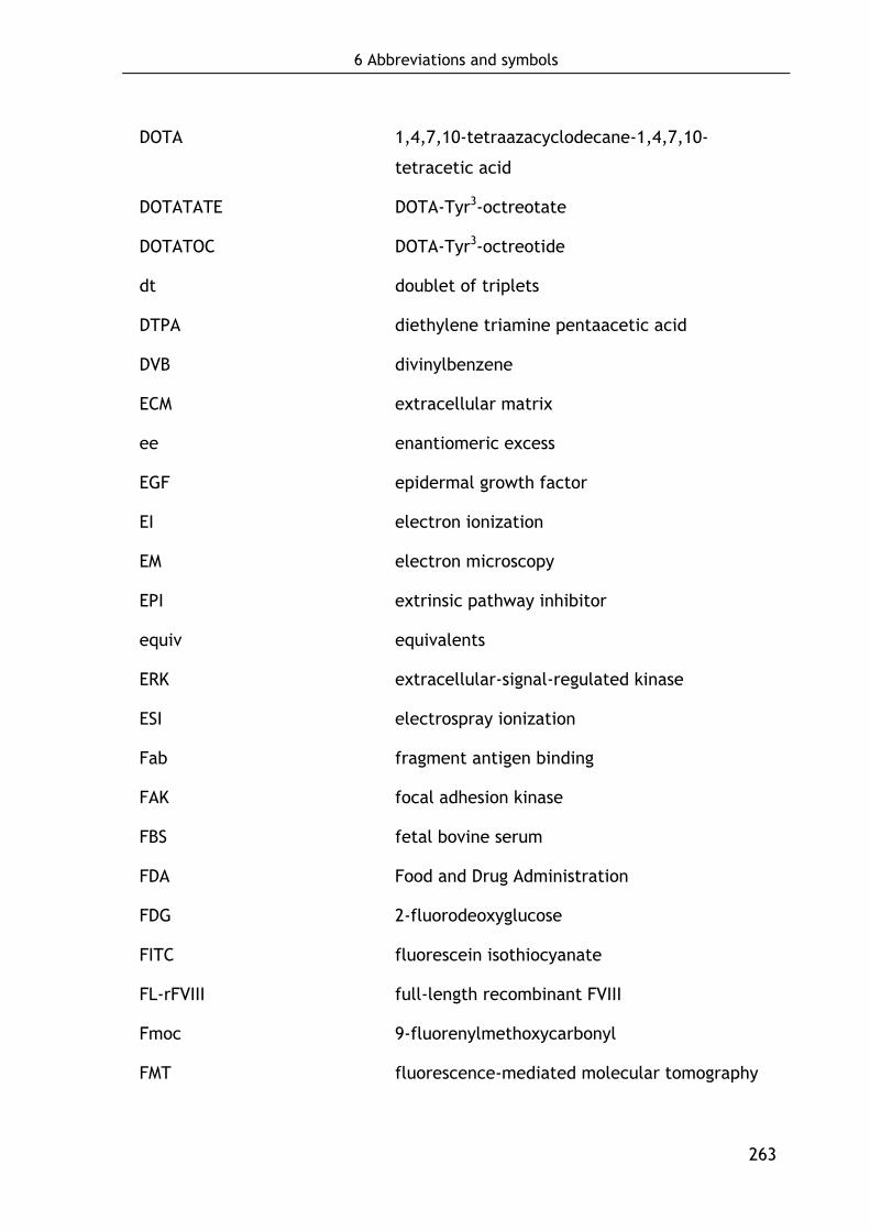
6 Abbreviations and symbols
263
DOTA 1,4,7,10-tetraazacyclodecane-1,4,7,10-
tetracetic acid
DOTATATE DOTA-Tyr3-octreotate
DOTATOC DOTA-Tyr3-octreotide
dt doublet of triplets
DTPA diethylene triamine pentaacetic acid
DVB divinylbenzene
ECM extracellular matrix
ee enantiomeric excess
EGF epidermal growth factor
EI electron ionization
EM electron microscopy
EPI extrinsic pathway inhibitor
equiv equivalents
ERK extracellular-signal-regulated kinase
ESI electrospray ionization
Fab fragment antigen binding
FAK focal adhesion kinase
FBS fetal bovine serum
FDA Food and Drug Administration
FDG 2-fluorodeoxyglucose
FITC fluorescein isothiocyanate
FL-rFVIII full-length recombinant FVIII
Fmoc 9-fluorenylmethoxycarbonyl
FMT fluorescence-mediated molecular tomography
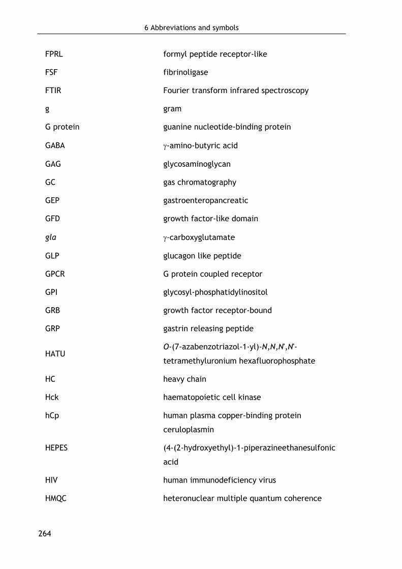
6 Abbreviations and symbols
264
FPRL formyl peptide receptor-like
FSF fibrinoligase
FTIR Fourier transform infrared spectroscopy
g gram
G protein guanine nucleotide-binding protein
GABA γ-amino-butyric acid
GAG glycosaminoglycan
GC gas chromatography
GEP gastroenteropancreatic
GFD growth factor-like domain
gla γ-carboxyglutamate
GLP glucagon like peptide
GPCR G protein coupled receptor
GPI glycosyl-phosphatidylinositol
GRB growth factor receptor-bound
GRP gastrin releasing peptide
HATU O-(7-azabenzotriazol-1-yl)-N,N,N',N'-
tetramethyluronium hexafluorophosphate
HC heavy chain
Hck haematopoietic cell kinase
hCp human plasma copper-binding protein
ceruloplasmin
HEPES (4-(2-hydroxyethyl)-1-piperazineethanesulfonic
acid
HIV human immunodeficiency virus
HMQC heteronuclear multiple quantum coherence
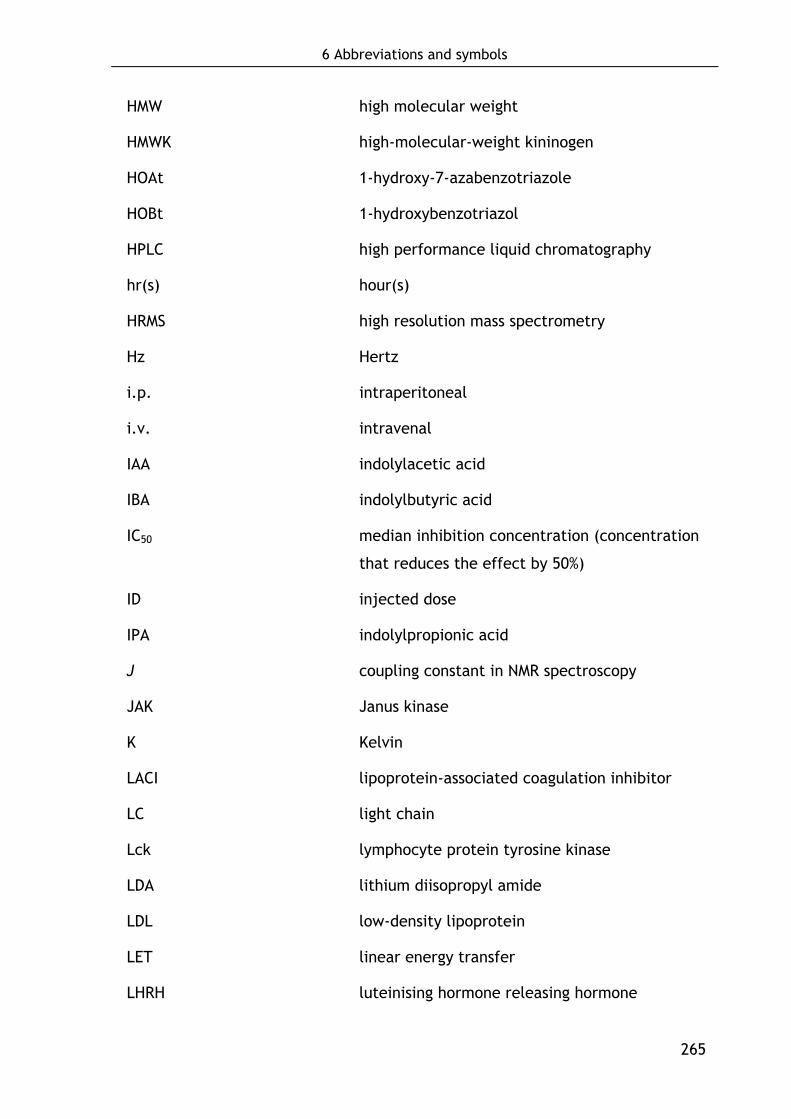
6 Abbreviations and symbols
265
HMW high molecular weight
HMWK high-molecular-weight kininogen
HOAt 1-hydroxy-7-azabenzotriazole
HOBt 1-hydroxybenzotriazol
HPLC high performance liquid chromatography
hr(s) hour(s)
HRMS high resolution mass spectrometry
Hz Hertz
i.p. intraperitoneal
i.v. intravenal
IAA indolylacetic acid
IBA indolylbutyric acid
IC50 median inhibition concentration (concentration
that reduces the effect by 50%)
ID injected dose
IPA indolylpropionic acid
J coupling constant in NMR spectroscopy
JAK Janus kinase
K Kelvin
LACI lipoprotein-associated coagulation inhibitor
LC light chain
Lck lymphocyte protein tyrosine kinase
LDA lithium diisopropyl amide
LDL low-density lipoprotein
LET linear energy transfer
LHRH luteinising hormone releasing hormone
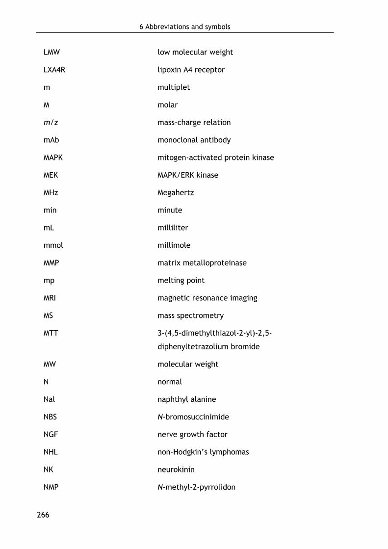
6 Abbreviations and symbols
266
LMW low molecular weight
LXA4R lipoxin A4 receptor
m multiplet
M molar
m/z mass-charge relation
mAb monoclonal antibody
MAPK mitogen-activated protein kinase
MEK MAPK/ERK kinase
MHz Megahertz
min minute
mL milliliter
mmol millimole
MMP matrix metalloproteinase
mp melting point
MRI magnetic resonance imaging
MS mass spectrometry
MTT 3-(4,5-dimethylthiazol-2-yl)-2,5-
diphenyltetrazolium bromide
MW molecular weight
N normal
Nal naphthyl alanine
NBS N-bromosuccinimide
NGF nerve growth factor
NHL non-Hodgkin’s lymphomas
NK neurokinin
NMP N-methyl-2-pyrrolidon
6 Abbreviations and symbols
267
NMR nuclear magnetic resonance
NOESY Nuclear Overhauser effect spectroscopy
NOTA 1,4,7-triazacyclononane-1,4,7-triyltriacetic
acid
OTf trifluoromethane sulfonate
p.i. post injection
PAI plasminogen activator inhibitor
Pbf 2,2,4,6,7-pentamethyldihydrobenzofuran-5-
sulfonyl
PBLG poly-γ-benzyl-L-glutamate
PBS phosphate-buffered saline
PC protein C
PCBP 4-n-pentyl-4’-cyanobiphenyl
pdFVIII plasma-derived human FVIII
PDMS polydimethylsiloxane
PELG poly-γ-ethyl-L-glutamate
PET positron emission tomography
PI3K phosphatidylinositol 3-kinase
PI-PLC/PI-PLD phosphatidylinositol-specific phospholipase C/D
PK prekallikrein
PKA protein kinase A
PKC protein kinase C
PL phospholipid
PMMA poly(methyl methacrylate)
ppm parts per million
PROK Proteinase K
6 Abbreviations and symbols
268
PS protein S, polystyrene
PVA poly(vinyl acetate)
q quartet
quant. quantitative
ran random
RDC residual dipolar coupling
rf reflux
Rf retention factor
rFVIII recombinant FVIII
RP reverse phase
rpm rotations per minute
rt room temperature
Rt retention time
s singulet
SAG strain induced alignment in a gel
SAR structure-activity relationships
SEM standard error of mean
SPECT single photon emission computed tomography
SPPS solid-phase peptide synthesis
sst somatostatin
STAT ignal transducer and activator of transcription
suPAR soluble urokinase-type plasminogen activator
receptor
t triplet
TAFI thrombin-activatable fibrinolysis inhibitor
TAT thrombin–antithrombin III complex

6 Abbreviations and symbols
269
TBAF tetrabutyl ammonium fluoride
TBS tert-butyldimethyl silyl
TBTU O-(benzotriazol-1-yl)-N,N,N',N'-
tetramethyluronium tetrafluoroborate
t-Bu tertiary butyl
TCP trityl chloride polystyrene (
TF tissue factor
TFA trifluoroacetic acid
TFPI tissue factor pathway inhibitor
TGF transforming growth factor
THF tetrahydrofurane
TIPS triisopropyl silane
TLC thin layer chromatography
TMOF trimethylorthoformate
TMS trimethylsilyl
TOF time of flight
tPA Tissue plasminogen activator
Trt trityl
uPA urokinase-type plasminogen activator
uPAR urokinase-type plasminogen activator receptor
UV/Vis ultraviolett/visible
v/v volume by volume
vWF von Willebrand factor
w/v weight by volume
WX360 (cyclo[21,29][D-Cys21Cys29]-uPA21-30)
6 Abbreviations and symbols
270
The nomenclature used in this work is based on the guidelines recommended by
Chemical Abstracts (Chemical Abstracts, ‘Index Guide’, 77, 210) and the IUPAC-IUB
commissions (IUPAC, Eur. J. Biochem. 1971, 21, 455-477; IUPAC, Pure Appl. Chem.
1996, 68, 1919; IUPAC Commission on Nomenclature of Organic Chemistry (CNOC)
and IUPAC-IUB Joint Commission on Biochemical Nomenclature (JCBN),
Biochemistry 1974, 10, 3983; IUPAC-IUB (JCBN), Eur. J. Biochem. 1984, 138, 9-37).
Expressions derived from the latin language are written in italic.
7 Bibliography
271
7 Bibliography
[1] D. Voet, J. G. Voet, Biochemie, 1st ed., VHC, Weinheim, New York, Basel,
Tokyo, 1994.
[2] E. W. Davie, O. D. Ratnoff, Science 1964, 145, 1310-1312.
[3] E. W. Davie, J Biol Chem 2003, 278(51), 50819-50832.
[4] S. Butenas, K. G. Mann, Biochemistry (Mosc) 2002, 67(1), 3-12.
[5] T. E. Adams, J. A. Huntington, Arterioscler. Thromb. Vasc. Biol. 2006,
26(8), 1738-1745.
[6] P. Bishop, J. Lawson, Nat. Rev. Drug. Discov. 2004, 3(8), 684-694.
[7] R. Zhuo, C. A. Siedlecki, E. A. Vogler, Biomaterials 2006, 27(24), 4325-4332.
[8] D. L. Tankersley, J. S. Finlayson, Biochemistry 1984, 23(2), 273-279.
[9] M. Silverberg, J. T. Dunn, L. Garen, A. P. Kaplan, J. Biol. Chem. 1980,
255(15), 7281-7286.
[10] P. H. Bolton-Maggs, K. J. Pasi, Lancet 2003, 361, 1801-1809.
[11] J. A. Huntington, T. P. Baglin, Trends Pharmacol. Sci. 2003, 24(11), 589-
595.
[12] P. J. Lenting, J. A. van Mourik, K. Mertens, Blood 1998, 92(11), 3983-3996.
[13] M. A. Panteleev, M. V. Ovanesov, D. A. Kireev, A. M. Shibeko, E. I.
Sinauridze, N. M. Ananyeva, A. A. Butylin, E. L. Saenko, F. I. Ataullakhanov,
Biophys. J. 2006, 90(5), 1489-1500.
[14] T. Bombeli, D. R. Spahn, Br. J. Anaesth. 2004, 93(2), 275-287.
[15] K. G. Mann, K. Brummel, S. Butenas, J. Thromb. Haemost. 2003, 1(7), 1504-
1514.
[16] R. Radcliffe, Y. Nemerson, J. Biol. Chem. 1975, 250(2), 388-395.
7 Bibliography
272
[17] M. Kjalke, A. Silveira, A. Hamsten, U. Hedner, M. Ezban, Arterioscler.
Thromb. Vasc. Biol. 2000, 20(7), 1835-1841.
[18] P. F. Neuenschwander, M. M. Fiore, J. H. Morrissey, J. Biol. Chem. 1993,
268(29), 21489-21492.
[19] L. V. Rao, T. Williams, S. I. Rapaport, Blood 1996, 87(9), 3738-3748.
[20] S. Butenas, K. G. Mann, Biochemistry 1996, 35(6), 1904-1910.
[21] M. Yamamoto, T. Nakagaki, W. Kisiel, J. Biol. Chem. 1992, 267(27), 19089-
19094.
[22] P. Wildgoose, W. Kisiel, Blood 1989, 73(7), 1888-1895.
[23] J. H. Lawson, M. Kalafatis, S. Stram, K. G. Mann, J. Biol. Chem. 1994,
269(37), 23357-23366.
[24] B. Osterud, S. I. Rapaport, Proc. Natl. Acad. Sci. USA 1977, 74(12), 5260-
5264.
[25] V. J. Bom, R. M. Bertina, Biochem. J. 1990, 265(2), 327-336.
[26] C. H. Gemmell, G. J. Broze, Jr., V. T. Turitto, Y. Nemerson, Blood 1990,
76(11), 2266-2271.
[27] K. W. Watt, T. Takagi, R. F. Doolittle, Proc. Natl. Acad. Sci. USA 1978,
75(4), 1731-1735.
[28] C. Fuss, J. C. Palmaz, E. A. Sprague, J. Vasc. Interv. Radiol. 2001, 12(6),
677-682.
[29] G. Cesarman-Maus, K. A. Hajjar, Br. J. Haematol. 2005, 129(3), 307-321.
[30] P. F. Bodary, K. J. Wickenheiser, D. T. Eitzman, Expert Rev. Mol. Med.
2002, 2002, 1-10.
[31] A. Rimon, Y. Shamash, B. Shapiro, J. Biol. Chem. 1966, 241(21), 5102-5107.
[32] M. Moroi, N. Aoki, J. Biol. Chem. 1976, 251(19), 5956-5965.
[33] N. Aoki, M. Moroi, M. Matsuda, K. Tachiya, J. Clin. Invest. 1977, 60(2), 361-
369.
[34] E. W. Brown, S. Ravindran, P. A. Patston, Blood Coagul. Fibrin. 2002, 13(8),
711-714.
[35] J. Graw, H. H. Brackmann, J. Oldenburg, R. Schneppenheim, M. Spannagl, R.
Schwaab, Nat. Rev. Genet. 2005, 6(6), 488-501.
7 Bibliography
273
[36] M. Jacquemin, M. De Maeyer, R. D'Oiron, R. Lavend'Homme, K. Peerlinck, J.
M. Saint-Remy, J. Thromb. Haemost. 2003, 1(3), 456-463.
[37] J. Oldenburg, N. M. Ananyeva, E. L. Saenko, Haemophilia 2004, 10(Suppl 4),
133-139.
[38] J. Klinge, N. M. Ananyeva, C. A. Hauser, E. L. Saenko, Semin. Thromb.
Hemost. 2002, 28(3), 309-322.
[39] E. L. Saenko, N. Ananyeva, D. Kouiavskaia, H. Schwinn, D. Josic, M. Shima,
C. A. E. Hauser, S. Pipe, Vox Sang. 2002, 83(2), 89-96.
[40] P. M. Mannucci, J. Thromb. Haemost. 2003, 1(7), 1349-1355.
[41] W. K. Hoots, Haemophilia 2001, 7(Suppl 1), 4-9.
[42] United Kingdom Haemophilia Centre Doctors' Organisation (UKHCDO),
Haemophilia 2003, 9(1), 1-23.
[43] A. Ishiwata, J. Mimuro, Y. Kashiwakura, M. Niimura, K. Takano, T. Ohmori,
S. Madoiwa, H. Mizukami, T. Okada, H. Naka, A. Yoshioka, K. Ozawa, Y.
Sakata, Thromb Res 2006, 118(5), 627-635.
[44] M. A. Kay, K. High, Proc. Natl. Acad. Sci. USA 1999, 96(18), 9973-9975.
[45] S. Connelly, J. Mount, A. Mauser, J. M. Gardner, M. Kaleko, A. McClelland,
C. D. Lothrop, Jr., Blood 1996, 88(10), 3846-3853.
[46] S. Connelly, J. L. Andrews, A. M. Gallo, D. B. Kayda, J. Qian, L. Hoyer, M. J.
Kadan, M. I. Gorziglia, B. C. Trapnell, A. McClelland, M. Kaleko, Blood 1998,
91(9), 3273-3281.
[47] C. Balague, J. Zhou, Y. Dai, R. Alemany, S. F. Josephs, G. Andreason, M.
Hariharan, E. Sethi, E. Prokopenko, H. Y. Jan, Y. C. Lou, D. Hubert-Leslie, L.
Ruiz, W. W. Zhang, Blood 2000, 95(3), 820-828.
[48] J. A. Bristol, A. Gallo-Penn, J. Andrews, N. Idamakanti, M. Kaleko, S.
Connelly, Hum. Gene Ther. 2001, 12(13), 1651-1661.
[49] F. Park, K. Ohashi, M. A. Kay, Blood 2000, 96(3), 1173-1176.
[50] Y. Yang, F. A. Nunes, K. Berencsi, E. E. Furth, E. Gonczol, J. M. Wilson,
Proc. Natl. Acad. Sci. USA 1994, 91(10), 4407-4411.
[51] V. R. Arruda, J. Thromb. Haemost. 2006, 4(6), 1215-1217.
7 Bibliography
274
[52] W. M. McCormack, Jr., M. P. Seiler, T. K. Bertin, K. Ubhayakar, D. J.
Palmer, P. Ng, T. C. Nichols, B. Lee, J. Thromb. Haemost. 2006, 4(6), 1218-
1225.
[53] K. P. Ponder, Curr. Opin. Hematol. 2006, 13(5), 301-307.
[54] A. Farrugia, Haemophilia 2004, 10(4), 327-333.
[55] P. M. Mannucci, J. Thromb. Haemost. 2003, 1(2), 216-217.
[56] W. K. Hoots, Transfus Med. Rev. 2001, 15(2), 11-19.
[57] W. K. Hoots, Transfus Med. Rev. 2001, 15(2 Suppl 1), 3-10.
[58] J. Gitschier, W. I. Wood, T. M. Goralka, K. L. Wion, E. Y. Chen, D. H. Eaton,
G. A. Vehar, D. J. Capon, R. M. Lawn, Nature 1984, 312(5992), 326-330.
[59] W. I. Wood, D. J. Capon, C. C. Simonsen, D. L. Eaton, J. Gitschier, B. Keyt,
P. H. Seeburg, D. H. Smith, P. Hollingshead, K. L. Wion, et al., Nature 1984,
312(5992), 330-337.
[60] J. J. Toole, J. L. Knopf, J. M. Wozney, L. A. Sultzman, J. L. Buecker, D. D.
Pittman, R. J. Kaufman, E. Brown, C. Shoemaker, E. C. Orr, et al., Nature
1984, 312(5992), 342-347.
[61] R. J. Kaufman, S. W. Pipe, Haemophilia 1998, 4(4), 370-379.
[62] E. L. Saenko, N. M. Ananyeva, M. Shima, C. A. Hauser, S. W. Pipe, J.
Thromb. Haemost. 2003, 1(5), 922-930.
[63] C. Lee, Thromb. Haemost. 1999, 82(2), 516-524.
[64] S. W. Pipe, J. M. Saint-Remy, C. E. Walsh, Haemophilia 2004, 10(Suppl 4),
55-63.
[65] J. P. Betts, BMJ 1998, 316(7141), 1385.
[66] A. Azzi, R. De Santis, M. Morfini, K. Zakrzewska, R. Musso, E. Santagostino,
G. Castaman, Blood 2001, 98(8), 2571-2573.
[67] R. K. Eriksson, C. Fenge, E. Lindner-Olson, C. Ljungqvist, J. Rosenquist, A. L.
Smeds, A. Ostlin, T. Charlebois, M. Leonard, B. D. Kelley, A. Ljungqvist,
Semin. Hematol. 2001, 38(2 (Suppl. 4)), 24-31.
[68] N. Ananyeva, A. Khrenov, F. Darr, R. Summers, A. Sarafanov, E. Saenko,
Expert Opin. Pharmacother. 2004, 5(5), 1061-1070.
[69] J. Barbara, P. Flanagan, BMJ 1998, 316(7133), 717-718.

7 Bibliography
275
[70] Medical and Scientific Advisory Council (MASAC) of the National Hemophilia
Foundation. MASAC recommencations concerning the treatment of
Hemophilia and other bleeeding disorders, MASAC Document #151 2003,
http://www.hemophilia.org/NHFWeb/Resource/StaticPages/menu0/menu5/
menu57/masac151.pdf.
[71] A. Bird, P. Isarangkura, D. Almagro, A. Gonzaga, A. Srivastava, Haemophilia
1998, 4(4), 481-485.
[72] G. A. Vehar, B. Keyt, D. Eaton, H. Rodriguez, D. P. O'Brien, F. Rotblat, H.
Oppermann, R. Keck, W. I. Wood, R. N. Harkins, et al., Nature 1984,
312(5992), 337-342.
[73] W. Wang, Y. J. Wang, D. N. Kelner, Int. J. Pharm. 2003, 259(1-2), 1-15.
[74] M. L. Koschinsky, W. D. Funk, B. A. van Oost, R. T. MacGillivray, Proc. Natl.
Acad. Sci. USA 1986, 83(14), 5086-5090.
[75] W. H. Kane, E. W. Davie, Proc. Natl. Acad. Sci. USA 1986, 83(18), 6800-
6804.
[76] J. J. Toole, D. D. Pittman, E. C. Orr, P. Murtha, L. C. Wasley, R. J. Kaufman,
Proc. Natl. Acad. Sci. USA 1986, 83(16), 5939-5942.
[77] A. V. Khrenov, N. M. Ananyeva, E. L. Saenko, Blood Coagul. Fibrin. 2006,
17(5), 379-388.
[78] P. J. Fay, M. T. Anderson, S. I. Chavin, V. J. Marder, Biochim. Biophys. Acta
1986, 871(3), 268-278.
[79] P. J. Fay, Blood Rev. 2004, 18(1), 1-15.
[80] S. S. Stoylova, P. J. Lenting, G. Kemball-Cook, A. Holzenburg, J. Biol. Chem.
1999, 274(51), 36573-36578.
[81] H. J. Weiss, I. I. Sussman, L. W. Hoyer, J. Clin. Invest. 1977, 60(2), 390-404.
[82] E. G. D. Tuddenham, L. W. Hoyer, Thromb. Haemost. 1977, 38(1), 9-9.
[83] E. L. Saenko, D. Scandella, J. Biol. Chem. 1997, 272(29), 18007-18014.
[84] M. Jacquemin, A. Benhida, K. Peerlinck, B. Desqueper, L. V. Elst, R.
Lavend'homme, R. d'Oiron, R. Schwaab, M. Bakkus, K. Thielemans, J. G.
Gilles, J. Vermylen, J. M. Saint-Remy, Blood 2000, 95(1), 156-163.
7 Bibliography
276
[85] R. J. Wise, A. J. Dorner, M. Krane, D. D. Pittman, R. J. Kaufman, J. Biol.
Chem. 1991, 266(32), 21948-21955.
[86] R. J. Kaufman, S. W. Pipe, Thromb. Haemost. 1999, 82(2), 201-208.
[87] J. Over, J. J. Sixma, M. H. Bruine, M. C. Trieschnigg, R. A. Vlooswijk, N. H.
Beeser-Visser, B. N. Bouma, J. Clin. Invest. 1978, 62(2), 223-234.
[88] K. Nogami, M. Shima, K. Hosokawa, M. Nagata, T. Koide, E. L. Saenko, I.
Tanaka, M. Shibata, A. Yoshioka, J. Biol. Chem. 2000, 275(33), 25774-
25780.
[89] L. M. Regan, P. J. Fay, J. Biol. Chem. 1995, 270(15), 8546-8552.
[90] R. T. Barrow, J. F. Healey, M. G. Jacquemin, J. M. Saint-Remy, P. Lollar,
Blood 2001, 97(1), 169-174.
[91] E. L. Saenko, D. Scandella, A. V. Yakhyaev, N. J. Greco, J. Biol. Chem.
1998, 273(43), 27918-27926.
[92] P. A. Foster, C. A. Fulcher, R. A. Houghten, T. S. Zimmerman, Blood 1990,
75(10), 1999-2004.
[93] K. P. Pratt, B. W. Shen, K. Takeshima, E. W. Davie, K. Fujikawa, B. L.
Stoddard, Nature 1999, 402(6760), 439-442.
[94] P. J. Fay, K. Koshibu, J. Biol. Chem. 1998, 273(30), 19049-19054.
[95] P. J. Fay, M. Mastri, M. E. Koszelak, H. Wakabayashi, J. Biol. Chem. 2001,
276(15), 12434-12439.
[96] P. J. Lenting, J. W. van de Loo, M. J. Donath, J. A. van Mourik, K. Mertens,
J. Biol. Chem. 1996, 271(4), 1935-1940.
[97] G. E. Gilbert, R. J. Kaufman, A. A. Arena, H. Miao, S. W. Pipe, J. Biol.
Chem. 2002, 277(8), 6374-6381.
[98] P. J. Fay, D. Scandella, J. Biol. Chem. 1999, 274(42), 29826-29830.
[99] P. J. Fay, T. Beattie, C. F. Huggins, L. M. Regan, J. Biol. Chem. 1994,
269(32), 20522-20527.
[100] K. Nogami, M. Shima, K. Hosokawa, T. Suzuki, T. Koide, E. L. Saenko, D.
Scandella, M. Shibata, S. Kamisue, I. Tanaka, A. Yoshioka, J. Biol. Chem.
1999, 274(43), 31000-31007.
7 Bibliography
277
[101] I. Zaitseva, V. Zaitsev, G. Card, K. Moshkov, B. Bax, A. Ralph, P. Lindley, J.
Biol. Inorg. Chem. 1996, 1(1), 15-23.
[102] S. Pemberton, P. Lindley, V. Zaitsev, G. Card, E. G. Tuddenham, G.
Kemball-Cook, Blood 1997, 89(7), 2413-2421.
[103] A. J. Gale, J. L. Pellequer, J. Thromb. Haemost. 2003, 1(9), 1966-1971.
[104] W. H. Hakeos, H. Miao, N. Sirachainan, G. Kemball-Cook, E. L. Saenko, R. J.
Kaufman, S. W. Pipe, Thromb. Haemost. 2002, 88(5), 781-787.
[105] T. E. Adams, M. F. Hockin, K. G. Mann, S. J. Everse, Proc. Natl. Acad. Sci.
USA 2004, 101(24), 8918-8923.
[106] S. Stoilova-McPhie, B. O. Villoutreix, K. Mertens, G. Kemball-Cook, A.
Holzenburg, Blood 2002, 99(4), 1215-1223.
[107] V. S. Purohit, K. Ramani, R. S. Kashi, M. J. Durrani, T. J. Kreiger, S. V.
Balasubramanian, Biochim Biophys Acta 2003, 1617(1-2), 31-38.
[108] L. Autin, M. A. Miteva, W. H. Lee, K. Mertens, K. P. Radtke, B. O.
Villoutreix, J. Thromb. Haemost. 2005, 3(9), 2044-2056.
[109] K. Takeshima, C. Smith, J. Tait, K. Fujikawa, Thromb. Haemost. 2003,
89(5), 788-794.
[110] H. Brandstetter, M. Bauer, R. Huber, P. Lollar, W. Bode, Proc. Natl. Acad.
Sci. USA 1995, 92(21), 9796-9800.
[111] P. J. Fay, P. V. Jenkins, Blood Rev. 2005, 19(1), 15-27.
[112] K. Mertens, P. H. Celie, J. A. Kolkman, P. J. Lenting, Thromb. Haemost.
1999, 82(2), 209-217.
[113] Cuatreca.P, M. Wilchek, C. B. Anfinsen, Proc. Natl. Acad. Sci. USA 1968,
61(2), 636-643.
[114] O. Mejan, V. Fert, M. Delezay, M. Delaage, R. Cheballah, A. Bourgois,
Thromb. Haemost. 1988, 59(3), 364-371.
[115] D. N. Fass, G. J. Knutson, J. A. Katzmann, Blood 1982, 59(3), 594-600.
[116] J. E. Addiego, E. Gomperts, S. L. Liu, P. Bailey, S. G. Courter, M. L. Lee, G.
G. Neslund, H. S. Kingdon, M. J. Griffith, Thromb. Haemost. 1992, 67(1),
19-27.
[117] B. G. Boedeker, Transfus Med. Rev. 1992, 6(4), 256-260.
7 Bibliography
278
[118] E. Gomperts, R. Lundblad, R. Adamson, Transfus Med. Rev. 1992, 6(4), 247-
251.
[119] J. J. Jakubik, S. M. Vicik, M. M. Tannatt, B. D. Kelley, Haemophilia 2004,
10(1), 69-74.
[120] R. Jiang, T. Monroe, R. McRogers, P. J. Larson, Haemophilia 2002, 8(Suppl.
2), 1-5.
[121] G. W. Jack, D. J. Beer, Methods Mol. Biol. 1996, 59, 187-196.
[122] R. K. Scopes, Protein purification: Principles and Practice, 3rd ed., Springer,
New York, 1994.
[123] A. C. Roque, C. R. Lowe, M. A. Taipa, Biotechnol. Prog. 2004, 20(3), 639-
654.
[124] N. E. Labrou, J. Chromatogr. B Analyt. Technol. Biomed. Life Sci. 2003,
790(1-2), 67-78.
[125] C. R. Lowe, A. R. Lowe, G. Gupta, J. Biochem. Biophys. Methods 2001,
49(1-3), 561-574.
[126] Y. D. Clonis, J. Chromatogr. A 2006, 1101(1-2), 1-24.
[127] E. L. Saenko, J.-M. Saint-Remy.
[128] C. R. Lowe, Curr. Opin. Chem. Biol. 2001, 5(3), 248-256.
[129] B. D. Kelley, M. Tannatt, R. Magnusson, S. Hagelberg, J. Booth, Biotechnol.
Bioeng. 2004, 87(3), 400-412.
[130] B. D. Kelley, J. Booth, M. Tannatt, Q. L. Wub, R. Ladner, J. Yuc, D. Potter,
A. Ley, J. Chromatogr. A 2004, 1038(1-2), 121-130.
[131] K. Amatschek, R. Necina, R. Hahn, E. Schallaun, H. Schwinn, D. Josic, A.
Jungbauer, J. High Resolut. Chromatogr. 2000, 23(1), 47-58.
[132] K. Pflegerl, R. Hahn, E. Berger, A. Jungbauer, J. Pept. Sci. 2002, 59(4), 174-
182.
[133] R. B. Merrifield, J. Am. Chem. Soc. 1963, 85, 2149-2154.
[134] B. Merrifield, Angew. Chem. Int. Edit. 1985, 24(10), 799-810.
[135] L. A. Carpino, G. Y. Han, J. Org. Chem. 1972, 37(22), 3404-3409.
[136] L. A. Carpino, B. J. Cohen, K. E. Stephens, S. Y. Sadataalaee, J. H. Tien, D.
C. Langridge, J. Org. Chem. 1986, 51(19), 3732-3734.

7 Bibliography
279
[137] G. B. Fields, R. L. Noble, Int. J. Pept. Protein Res. 1990, 35(3), 161-214.
[138] T. Curtius, J. Prakt. Chem. 1881, 24, 239-240.
[139] E. Fischer, Chem. Ber. 1902, 35, 1095 - 1106.
[140] F.Hofmeister, Ergeb. Physiol. Biol. Chem. Exp. Pharmacol 1902, 1, 759.
[141] E. Fischer, Chem. Ber. 1906, 39, 530-537.
[142] J. A. McLaren, Aust. J. Chem. 1958, 11, 360-365.
[143] M. Bergmann, L. Zervas, Chem. Ber. 1932, 65, 1192-1201.
[144] G. Barany, R. B. Merrifield, J. Am. Chem. Soc. 1977, 99(22), 7363-7365.
[145] R. Schwyzer, H. Kappeler, Helvetica Chimica Acta 1963, 46(5), 1550-1572.
[146] M. Bodanszky, V. Du Vigneaud, Nature 1959, 183(4671), 1324-1325.
[147] C. Chan, D. White, Fmoc solid phase peptide synthesis: a practical
approach, Oxford University Press, Oxford, 2000.
[148] K. Barlos, D. Gatos, J. Kallitsis, G. Papaphotiu, P. Sotiriu, W. Q. Yao, W.
Schafer, Tetrahedron Lett. 1989, 30(30), 3943-3946.
[149] P. Sieber, Tetrahedron Lett. 1987, 28(19), 2107-2110.
[150] L. A. Carpino, D. Sadat-Aalaee, H. G. Chao, R. H. DeSelm, J. Am. Chem. Soc.
1990, 112(26), 9651-9652.
[151] H. Chantrenne, Nature 1947, 160(4070), 603-604.
[152] J. Kovacs, Kisfalud.L, M. Q. Ceprini, J. Am. Chem. Soc. 1967, 89(1), 183-&.
[153] J. C. Sheehan, G. P. Hess, J. Am. Chem. Soc. 1955, 77(4), 1067-1068.
[154] M. Bodanszky, Principles of peptide synthesis, Springer Verlag, Berlin, 1984.
[155] F. Albericio, J. M. Bofill, A. El-Faham, S. A. Kates, J. Org. Chem. 1998,
63(26), 9678-9683.
[156] R. Knorr, A. Trzeciak, W. Bannwarth, D. Gillessen, Tetrahedron Lett. 1989,
30(15), 1927-1930.
[157] V. Dourtoglou, J. C. Ziegler, B. Gross, Tetrahedron Lett. 1978(15), 1269-
1272.
[158] B. Castro, J. R. Dormoy, G. Evin, C. Selve, Tetrahedron Lett. 1975(14),
1219-1222.
[159] J. Coste, D. Lenguyen, B. Castro, Tetrahedron Lett. 1990, 31(2), 205-208.
7 Bibliography
280
[160] J. M. Humphrey, A. R. Chamberlin, Chem. Rev. 1997, 97(6), 2243-2266.
[161] H. P. Koch, M. P. Kavanaugh, C. S. Esslinger, N. Zerangue, J. M. Humphrey,
S. G. Amara, A. R. Chamberlin, R. J. Bridges, Mol. Pharmacol. 1999, 56(6),
1095-1104.
[162] V. Dourtoglou, B. Gross, V. Lambropoulou, C. Zioudrou, Synthesis-Stuttgart
1984(7), 572-574.
[163] W. König, R. Geiger, Chem. Ber.-Recl. 1970, 103(7), 2024-2033.
[164] W. König, R. Geiger, Chem. Ber.-Recl. 1970, 103(7), 2034-2040.
[165] W. König, R. Geiger, Chem. Ber.-Recl. 1970, 103(3), 788-798.
[166] L. A. Carpino, J. Am. Chem. Soc. 1993, 115(10), 4397-4398.
[167] L. A. Carpino, A. Elfaham, J. Org. Chem. 1994, 59(4), 695-698.
[168] Y. X. Han, F. Albericio, G. Barany, J. Org. Chem. 1997, 62(13), 4307-4312.
[169] S. Knör, A. Khrenov, B. Laufer, E. L. Saenko, C. A. E. Hauser, H. Kessler., J.
Med. Chem. 2007, submitted.
[170] H. Gerlach, V. Prelog, Owtschin.Ja, Helv. Chim. Acta 1964, 47(8), 2294-&.
[171] V. Prelog, H. Gerlach, Helv. Chim. Acta 1964, 47(8), 2288-&.
[172] M. M. Shemyakin, Y. A. Ovchinnikov, V. T. Ivanov, Angew. Chem. Int. Edit.
1969, 8(7), 492-499.
[173] M. Chorev, M. Goodman, Accounts Chem. Res. 1993, 26(5), 266-273.
[174] M. Goodman, M. Chorev, Accounts Chem. Res. 1979, 12(1), 1-7.
[175] J. Wermuth, S. L. Goodman, A. Jonczyk, H. Kessler, J. Am. Chem. Soc.
1997, 119(6), 1328-1335.
[176] A. Paquet, Can. J. Chemistry 1981, 60, 976-980.
[177] V. Krchnak, A. S. Weichsel, D. Cabel, Z. Flegelova, M. Lebl, Mol. Divers.
1996, 1(3), 149-164.
[178] H. Nagase, in Handbook of Proteolytic Enzymes, Vol. 2 (Eds.: A. J. Barrett,
N. D. Rawlings, J. F. Woessner), Elsevier, London, 2004, pp. 512-523.
[179] N. Sewald, H.-D. Jakubke, Peptides: Chemistry and Biology, WILEY-VCH
Verlag GmbH, Weinheim, 2002.
[180] L. Zervas, I. Photaki, J. Am. Chem. Soc. 1962, 84(20), 3887-3897.
7 Bibliography
281
[181] F. Siedler, E. Weyher, L. Moroder, J. Pept. Sci. 1996, 2(4), 271-275.
[182] M. Goodman, L. Levine, J. Am. Chem. Soc. 1964, 86(14), 2918-2922.
[183] B. Henkel, L. S. Zhang, E. Bayer, Liebigs Ann.-Recl. 1997(10), 2161-2168.
[184] W. Steglich, G. Hofle, Angew. Chem. Int. Edit. 1969, 8(12), 981.
[185] H. M. L. Davies, R. J. Townsend, J. Org. Chem. 2001, 66(20), 6595-6603.
[186] P. Micuch, D. Seebach, Helv. Chim. Acta 2002, 85(6), 1567-1577.
[187] S. T. Hilton, T. C. T. Ho, G. Pljevaljcic, K. Jones, Org. Lett. 2000, 2(17),
2639-2641.
[188] T. T. Tidwell, Synthesis-Stuttgart 1990(10), 857-870.
[189] A. J. Mancuso, S. L. Huang, D. Swern, J. Org. Chem. 1978, 43(12), 2480-
2482.
[190] J. N. Denis, A. Correa, A. E. Greene, J. Org. Chem. 1991, 56(24), 6939-
6942.
[191] M. A. Blaskovich, G. A. Lajoie, J. Am. Chem. Soc. 1993, 115(12), 5021-5030.
[192] R. Grigg, W. S. MacLachlan, D. T. MacPherson, V. Sridharan, S. Suganthan,
M. Thornton-Pett, J. Zhang, Tetrahedron 2000, 56(35), 6585-6594.
[193] D. Gryko, J. Chalko, J. Jurczak, Chirality 2003, 15(6), 514-541.
[194] D. B. Dess, J. C. Martin, J. Org. Chem. 1983, 48(22), 4155-4156.
[195] D. B. Dess, J. C. Martin, J. Am. Chem. Soc. 1991, 113(19), 7277-7287.
[196] E. Masiukiewicz, B. Rzeszotarska, Org. Prep. Proced. Int. 1999, 31(5), 571-
572.
[197] D. A. Pearson, M. Blanchette, M. L. Baker, C. A. Guindon, Tetrahedron Lett.
1989, 30(21), 2739-2742.
[198] S. Knör, A. Khrenov, B. Laufer, A. Benhida, R. Schwaab, J. Oldenburg, N.
Beaufort, V. Magdolen, J.-M. Saint-Remy, E. L. Saenko, C. A. E. Hauser, H.
Kessler, J. Thromb. Haemost. 2007, submitted.
[199] S. Monroc, E. Badosa, L. Feliu, M. Planas, E. Montesinos, E. Bardaji, Peptides
2006, 27(11), 2567-2574.
[200] P. Y. Huang, G. A. Baumbach, C. A. Dadd, J. A. Buettner, B. L. Masecar, M.
Hentsch, D. J. Hammond, R. G. Carbonell, Bioorgan. Med. Chem. 1996,
4(5), 699-708.
7 Bibliography
282
[201] M. A. Jankowski, H. Patel, J. C. Rouse, L. A. Marzilli, S. B. Weston, P. J.
Sharpe, Haemophilia 2007, 13(1), 30-37.
[202] W. F. Schilow, E. Schoemer-Burkhardt, R. Seitz, Thromb. Haemost. 2004,
92(2), 427-428.
[203] S. Raut, S. Villard, S. Grailly, J. G. G. Gilles, C. Granier, J. M. R. Saint-
Remy, T. W. Barrowcliffe, Thromb. Haemost. 2003, 90(3), 385-397.
[204] P. C. Spiegel, M. Jacquemin, J. M. R. Saint-Remy, B. L. Stoddard, K. P.
Pratt, Blood 2001, 98(1), 13-19.
[205] K. Fischer, V. Lutz, O. Wilhelm, M. Schmitt, H. Graeff, P. Heiss, T.
Nishiguchi, N. Harbeck, H. Kessler, T. Luther, V. Magdolen, U. Reuning, FEBS
Lett. 1998, 438(1-2), 101-105.
[206] U. Reuning, M. Schmitt, B. Luber, V. Beck, V. Magdolen, Methods Mol. Med.
2006, 120, 427-440.
[207] R. Haubner, D. Finsinger, H. Kessler, Angew. Chem. Int. Edit. 1997, 36(13-
14), 1375-1389.
[208] H. Kessler, in Trends in Drug Research, Vol. 13 (Ed.: V. Claassen), Elsevier,
1990, pp. 73-84.
[209] H. Kessler, Angew. Chem. Int. Edit. 1982, 21, 512-523.
[210] D. Heckmann, Dissertation 2007, Technische Universität München.
[211] A. Y. Jeng, P. Savage, M. E. Beil, C. W. Bruseo, D. Hoyer, C. A. Fink, A. J.
Trapani, Clin. Sci. 2002, 103, 98S-101S.
[212] M. Goodman, H. Shao, Pure Appl. Chem. 1996, 68(6), 1303-1308.
[213] M. A. Dechantsreiter, E. Planker, B. Mathä, E. Lohof, G. Hölzemann, A.
Jonczyk, S. L. Goodman, H. Kessler, J. Med. Chem. 1999, 42(16), 3033-
3040.
[214] S. Knör, B. Laufer, H. Kessler, J. Org. Chem. 2006, 71(15), 5625-5630.
[215] W. Wang, J. Y. Zhang, C. Y. Xiong, V. J. Hruby, Tetrahedron Lett. 2002,
43(12), 2137-2140.
[216] M. J. Burk, J. R. Lee, J. P. Martinez, J. Am. Chem. Soc. 1994, 116(23),
10847-10848.
7 Bibliography
283
[217] M. J. Burk, M. F. Gross, T. G. P. Harper, C. S. Kalberg, J. R. Lee, J. P.
Martinez, Pure Appl. Chem. 1996, 68(1), 37-44.
[218] W. Wang, C. Y. Xiong, J. Y. Zhang, V. J. Hruby, Tetrahedron 2002, 58(15),
3101-3110.
[219] W. Wang, M. Y. Cai, C. Y. Xiong, J. Y. Zhang, D. Trivedi, V. J. Hruby,
Tetrahedron 2002, 58(36), 7365-7374.
[220] S. Kotha, K. Lahin, Biopolymers 2003, 69(4), 517-528.
[221] S. M. Birnbaum, L. Levintow, R. B. Kingsley, J. P. Greenstein, J. Biol. Chem.
1952, 194(1), 455-470.
[222] M. A. Vela, F. R. Fronczek, G. W. Horn, M. L. Mclaughlin, J. Org. Chem.
1990, 55(9), 2913-2918.
[223] U. Schmidt, A. Lieberknecht, J. Wild, Synthesis-Stuttgart 1984(1), 53-60.
[224] U. Schmidt, H. Griesser, V. Leitenberger, A. Lieberknecht, R. Mangold, R.
Meyer, B. Riedl, Synthesis-Stuttgart 1992(5), 487-490.
[225] M. J. Burk, J. Am. Chem. Soc. 1991, 113(22), 8518-8519.
[226] M. J. Burk, J. E. Feaster, W. A. Nugent, R. L. Harlow, J. Am. Chem. Soc.
1993, 115(22), 10125-10138.
[227] M. J. Burk, M. F. Gross, J. P. Martinez, J. Am. Chem. Soc. 1995, 117(36),
9375-9376.
[228] M. J. Burk, J. G. Allen, W. F. Kiesman, J. Am. Chem. Soc. 1998, 120(4), 657-
663.
[229] H. Kessler, M. Molter, Angew. Chem. Int. Edit. 1973, 12(12), 1011-1012.
[230] D. Hanahan, R. A. Weinberg, Cell 2000, 100(1), 57-70.
[231] E. Ruoslahti, Sci Am 1996, 275(3), 72-77.
[232] W. J. Guo, F. G. Giancotti, Nat. Rev. Mol. Cell Biol. 2004, 5(10), 816-826.
[233] F. Zhang, C. C. Tom, M. C. Kugler, T. T. Ching, J. A. Kreidberg, Y. Wei, H. A.
Chapman, J. Cell Biol. 2003, 163(1), 177-188.
[234] K. Danø, P. A. Andreasen, J. Grondahl-Hansen, P. Kristensen, L. S. Nielsen,
L. Skriver, Adv. Cancer Res. 1985, 44, 139-266.
[235] O. Wilhelm, U. Reuning, F. Jänicke, M. Schmitt, H. Graeff, Onkologie 1994,
17(4), 358-366.
7 Bibliography
284
[236] S. Cook, http://www.steve.gb.com/science/extracellular_matrix.html.
[237] A. M. Mercurio, Trends Cell Biol. 1995, 5(11), 419-423.
[238] D. R. Critchley, Curr. Opin. Cell Biol. 2000, 12(1), 133-139.
[239] E. Ruoslahti, Adv. Cancer Res. 1999, 76, 1-20.
[240] M. Lee, R. Fridman, S. Mobashery, Chem. Soc. Rev. 2004, 33(7), 401-409.
[241] M. Schmitt, H. Graeff, F. Jänicke, T. Genz, B. Lampe, Prospects in Diagnosis
and Treatment of Breast Cancer: Proceedings of the Joint International
Symposium, Munich, Germany, 10-11 November 1993, Elsevier Science,
Amsterdam, 1994.
[242] D. R. Edwards, G. Murphy, Nature 1998, 394(6693), 527-528.
[243] M. Johnsen, L. R. Lund, J. Romer, K. Almholt, K. Danø, Curr. Opin. Cell Biol.
1998, 10(5), 667-671.
[244] M. Egeblad, Z. Werb, Nat. Rev. Cancer 2002, 2(3), 161-174.
[245] N. Johansson, M. Ahonen, V. M. Kahari, Cell. Mol. Life Sci. 2000, 57(1), 5-
15.
[246] J. E. Koblinski, M. Ahram, B. F. Sloane, Clinica Chimica Acta 2000, 291(2),
113-135.
[247] S. Nozaki, Y. Endo, H. Nakahara, K. Yoshizawa, T. Ohara, E. Yamamoto,
Anti-Cancer Drugs 2006, 17(10), 1109-1117.
[248] M. Schmitt, O. G. Wilhelm, U. Reuning, A. Krüger, N. Harbeck, E. Lengyel,
H. Graeff, B. Gänsbacher, H. Kessler, M. Bürgle, J. Stürzebecher, S. Sperl, V.
Magdolen, Fibrinolysis & Proteolysis 2000, 14(2-3), 114-132.
[249] P. A. Andreasen, R. Egelund, H. H. Petersen, Cell. Mol. Life Sci. 2000,
57(1), 25-40.
[250] M. Schmitt, F. Jänicke, H. Graeff, Fibrinolysis 1992, 6, 3-26.
[251] F. J. Castellino, V. A. Ploplis, Thromb. Haemost. 2005, 93(4), 647-654.
[252] J. S. Rao, Nat. Rev. Cancer 2003, 3(7), 489-501.
[253] K. Uhland, Cell. Mol. Life Sci. 2006, 63(24), 2968-2978.
[254] W. G. Stetler-Stevenson, L. A. Liotta, D. E. Kleiner, Jr., FASEB J 1993,
7(15), 1434-1441.

7 Bibliography
285
[255] P. Basset, J. P. Bellocq, C. Wolf, I. Stoll, P. Hutin, J. M. Limacher, O. L.
Podhajcer, M. P. Chenard, M. C. Rio, P. Chambon, Nature 1990, 348(6303),
699-704.
[256] M. M. Mohamed, B. F. Sloane, Nat. Rev. Cancer 2006, 6(10), 764-775.
[257] B. F. Sloane, Semin. Cancer Biol. 1990, 1(2), 137-152.
[258] H. Rochefort, Breast Cancer Res. Treat. 1990, 16(1), 3-13.
[259] K. Danø, N. Behrendt, G. Høyer-Hansen, M. Johnsen, L. R. Lund, M. Ploug, J.
Romer, Thromb. Haemost. 2005, 93(4), 676-681.
[260] S. W. Hall, J. E. Humphries, S. L. Gonias, J. Biol. Chem. 1991, 266(19),
12329-12336.
[261] A. Meissauer, M. D. Kramer, V. Schirrmacher, G. Brunner, Exp. Cell Res.
1992, 199(2), 179-190.
[262] G. E. Stillfried, D. N. Saunders, M. Ranson, Breast Cancer Res. 2007, 9(1),
R14.
[263] V. Ellis, N. Behrendt, K. Danø, J. Biol. Chem. 1991, 266(19), 12752-12758.
[264] A. Gils, P. J. Declerck, Thromb. Haemost. 1998, 80(4), 531-541.
[265] R. W. Carrell, P. E. Stein, Biol. Chem. Hoppe Seyler 1996, 377(1), 1-17.
[266] D. Olson, J. Pollanen, G. Høyer-Hansen, E. Rønne, K. Sakaguchi, T. C. Wun,
E. Appella, K. Danø, F. Blasi, J. Biol. Chem. 1992, 267(13), 9129-9133.
[267] F. Al-Ejeh, D. Croucher, M. Ranson, Exp. Cell. Res. 2004, 297(1), 259-271.
[268] M. V. Cubellis, T. C. Wun, F. Blasi, EMBO J. 1990, 9(4), 1079-1085.
[269] D. Croucher, D. N. Saunders, M. Ranson, J. Biol. Chem. 2006, 281(15),
10206-10213.
[270] R. P. Czekay, T. A. Kuemmel, R. A. Orlando, M. G. Farquhar, Mol. Biol. Cell
2001, 12(5), 1467-1479.
[271] A. Nykjaer, M. Conese, E. I. Christensen, D. Olson, O. Cremona, J. Gliemann,
F. Blasi, EMBO J. 1997, 16(10), 2610-2620.
[272] M. Conese, A. Nykjaer, C. M. Petersen, O. Cremona, R. Pardi, P. A.
Andreasen, J. Gliemann, E. I. Christensen, F. Blasi, J. Cell. Biol. 1995,
131(6 Pt 1), 1609-1622.
[273] S. Mayor, H. Riezman, Nat. Rev. Mol. Cell Biol. 2004, 5(2), 110-120.
7 Bibliography
286
[274] M. Conese, F. Blasi, Biol. Chem. Hoppe Seyler 1995, 376(3), 143-155.
[275] L. H. Engelholm, B. S. Nielsen, K. Danø, N. Behrendt, Trends Cardiovas.
Med. 2001, 11(1), 7-13.
[276] H. Jin, Y. M. P. Song, G. Boel, J. Kochar, V. Pancholi, J. Mol. Biol. 2005,
350(1), 27-41.
[277] J. Sturge, D. Wienke, L. East, G. E. Jones, C. M. Isacke, J. Cell. Biol. 2003,
162(5), 789-794.
[278] T. C. Wun, L. Ossowski, E. Reich, J. Biol. Chem. 1982, 257(12), 7262-7268.
[279] Y. Nagamine, R. L. Medcalf, P. Munoz-Canoves, Thromb. Haemost. 2005,
93(4), 661-675.
[280] A. P. Hansen, A. M. Petros, R. P. Meadows, D. G. Nettesheim, A. P. Mazar, E.
T. Olejniczak, R. X. Xu, T. M. Pederson, J. Henkin, S. W. Fesik, Biochemistry
1994, 33(16), 4847-4864.
[281] W. A. Günzler, G. J. Steffens, F. Otting, G. Buse, L. Flohe, Hoppe Seylers Z.
Physiol. Chem. 1982, 363(2), 133-141.
[282] G. J. Steffens, W. A. Günzler, F. Otting, E. Frankus, L. Flohe, Hoppe Seylers
Z. Physiol. Chem. 1982, 363(9), 1043-1058.
[283] P. H. Quax, J. M. Grimbergen, M. Lansink, A. H. Bakker, M. C. Blatter, D.
Belin, V. W. van Hinsbergh, J. H. Verheijen, Arterioscler. Thromb. Vasc.
Biol. 1998, 18(5), 693-701.
[284] R. W. Stephens, A. M. Bokman, H. T. Myohanen, T. Reisberg, H. Tapiovaara,
N. Pedersen, J. Grondahl-Hansen, M. Llinas, A. Vaheri, Biochemistry 1992,
31(33), 7572-7579.
[285] W. Strassburger, A. Wollmer, J. E. Pitts, I. D. Glover, I. J. Tickle, T. L.
Blundell, G. J. Steffens, W. A. Günzler, F. Otting, L. Flohe, FEBS Lett. 1983,
157(2), 219-223.
[286] S. Sperl, U. Jacob, N. Arroyo de Prada, J. Stürzebecher, O. G. Wilhelm, W.
Bode, V. Magdolen, R. Huber, L. Moroder, Proc. Natl. Acad. Sci. USA 2000,
97(10), 5113-5118.
[287] G. Spraggon, C. Phillips, U. K. Nowak, C. P. Ponting, D. Saunders, C. M.
Dobson, D. I. Stuart, E. Y. Jones, Structure 1995, 3(7), 681-691.
7 Bibliography
287
[288] N. Schmiedeberg, Dissertation 2002, Technische Universität München.
[289] H. Kobayashi, M. Schmitt, L. Goretzki, N. Chucholowski, J. Calvete, M.
Kramer, W. A. Günzler, F. Jänicke, H. Graeff, J. Biol. Chem. 1991, 266(8),
5147-5152.
[290] E. Koivunen, M. L. Huhtala, U. H. Stenman, J. Biol. Chem. 1989, 264(24),
14095-14099.
[291] B. B. Wolf, J. Vasudevan, J. Henkin, S. L. Gonias, J. Biol. Chem. 1993,
268(22), 16327-16331.
[292] L. Goretzki, M. Schmitt, K. Mann, J. Calvete, N. Chucholowski, M. Kramer,
W. A. Günzler, F. Jänicke, H. Graeff, FEBS Lett. 1992, 297(1-2), 112-118.
[293] M. Schmitt, L. Goretzki, F. Jänicke, J. Calvete, M. Eulitz, H. Kobayashi, N.
Chucholowski, H. Graeff, Biomed. Biochim. Acta 1991, 50(4-6), 731-741.
[294] J. D. Vassalli, D. Baccino, D. Belin, J. Cell. Biol. 1985, 100(1), 86-92.
[295] N. Behrendt, E. Rønne, K. Danø, Biol. Chem. Hoppe Seyler 1995, 376(5),
269-279.
[296] N. Behrendt, M. Ploug, L. Patthy, G. Houen, F. Blasi, K. Danø, J. Biol. Chem.
1991, 266(12), 7842-7847.
[297] J. D. Vassalli, Fibrinolysis 1994, 8, 172-181.
[298] M. Ploug, N. Behrendt, D. Løber, K. Danø, Semin. Thromb. Hemost. 1991,
17(3), 183-193.
[299] F. Blasi, N. Behrendt, M. V. Cubellis, V. Ellis, L. R. Lund, M. T. Masucci, L. B.
Møller, D. P. Olson, N. Pedersen, M. Ploug, et al., Cell. Differ. Dev. 1990,
32(3), 247-253.
[300] M. V. Cubellis, M. L. Nolli, G. Cassani, F. Blasi, J. Biol. Chem. 1986,
261(34), 15819-15822.
[301] E. Appella, E. A. Robinson, S. J. Ullrich, M. P. Stoppelli, A. Corti, G. Cassani,
F. Blasi, J. Biol. Chem. 1987, 262(10), 4437-4440.
[302] M. P. Stoppelli, A. Corti, A. Soffientini, G. Cassani, F. Blasi, R. K. Assoian,
Proc. Natl. Acad. Sci. USA 1985, 82(15), 4939-4943.
7 Bibliography
288
[303] S. Temerinac, S. Klippel, E. Strunck, S. Roder, M. Lubbert, W. Lange, M.
Azemar, G. Meinhardt, H. E. Schaefer, H. L. Pahl, Blood 2000, 95(8), 2569-
2576.
[304] L. B. Møller, M. Ploug, F. Blasi, Eur. J. Biochem. 1992, 208(2), 493-500.
[305] L. B. Møller, J. Pollanen, E. Rønne, N. Pedersen, F. Blasi, J. Biol. Chem.
1993, 268(15), 11152-11159.
[306] N. Behrendt, E. Rønne, M. Ploug, T. Petri, D. Løber, L. S. Nielsen, W. D.
Schleuning, F. Blasi, E. Appella, K. Danø, J. Biol. Chem. 1990, 265(11),
6453-6460.
[307] A. L. Roldan, M. V. Cubellis, M. T. Masucci, N. Behrendt, L. R. Lund, K.
Danø, E. Appella, F. Blasi, EMBO J. 1990, 9(2), 467-474.
[308] M. Ploug, H. Rahbek-Nielsen, P. F. Nielsen, P. Roepstorff, K. Danø, J. Biol.
Chem. 1998, 273(22), 13933-13943.
[309] Q. Huai, A. P. Mazar, A. Kuo, G. C. Parry, D. E. Shaw, J. Callahan, Y. D. Li,
C. Yuan, C. B. Bian, L. Q. Chen, B. Furie, B. C. Furie, D. B. Cines, M. D.
Huang, Science 2006, 311(5761), 656-659.
[310] P. Llinas, M. H. Le Du, H. Gårdsvoll, K. Danø, M. Ploug, B. Gilquin, E. A.
Stura, A. Menez, EMBO J. 2005, 24(9), 1655-1663.
[311] Y. Wang, J. Dang, L. K. Johnson, J. J. Selhamer, W. F. Doe, Eur. J. Biochem.
1995, 227(1-2), 116-122.
[312] I. P. Witz, J. Cell. Biochem. 2000, 77(S34), 61-66.
[313] M. Ploug, V. Ellis, FEBS Lett. 1994, 349(2), 163-168.
[314] N. Ohkura, S. Inoue, K. Ikeda, K. Hayashi, Biochem. Biophys. Res. Commun.
1994, 204(3), 1212-1218.
[315] J. Petranka, J. Zhao, J. Norris, N. B. Tweedy, R. E. Ware, P. J. Sims, W. F.
Rosse, Blood Cells Mol. Dis. 1996, 22(3), 281-296.
[316] C. M. Fletcher, R. A. Harrison, P. J. Lachmann, D. Neuhaus, Structure 1994,
2(3), 185-199.
[317] R. Eshel, A. Zanin, O. Sagi-Assif, T. Meshel, N. I. Smorodinsky, O. Dwir, R.
Alon, R. Brakenhoff, G. van Dongen, I. P. Witz, J. Biol. Chem. 2000,
275(17), 12833-12840.

7 Bibliography
289
[318] T. P. Gumley, I. F. McKenzie, M. S. Sandrin, Immunol. Cell. Biol. 1995,
73(4), 277-296.
[319] M. Bürgle, Dissertation 1998, Technische Universität München.
[320] O. G. Wilhelm, S. Wilhelm, G. M. Escott, V. Lutz, V. Magdolen, M. Schmitt,
D. B. Rifkin, E. L. Wilson, H. Graeff, G. Brunner, J. Cell. Physiol. 1999,
180(2), 225-235.
[321] G. Brunner, C. N. Metz, H. Nguyen, J. Gabrilove, S. R. Patel, M. A. Davitz, D.
B. Rifkin, E. L. Wilson, Blood 1994, 83(8), 2115-2125.
[322] H. Gårdsvoll, K. Danø, M. Ploug, J. Biol. Chem. 1999, 274(53), 37995-38003.
[323] M. Ploug, H. Rahbek-Nielsen, V. Ellis, P. Roepstorff, K. Danø, Biochemistry
1995, 34(39), 12524-12534.
[324] M. Ploug, S. Østergaard, L. B. Hansen, A. Holm, K. Danø, Biochemistry 1998,
37(11), 3612-3622.
[325] M. Ploug, S. Østergaard, A. Holm, C. Holst-Hansen, R. W. Stephens, K. Danø,
PCT Int. Appl. 2000, WO 00/01802, 1-75.
[326] K. Bdeir, A. Kuo, A. Mazar, B. S. Sachais, W. Xiao, S. Gawlak, S. Harris, A. A.
Higazi, D. B. Cines, J. Biol. Chem. 2000, 275(37), 28532-28538.
[327] K. K. Wary, A. Mariotti, C. Zurzolo, F. G. Giancotti, Cell 1998, 94(5), 625-
634.
[328] H. A. Chapman, Y. Wei, D. I. Simon, D. A. Waltz, Thromb. Haemost. 1999,
82(2), 291-297.
[329] S. Monier, R. G. Parton, F. Vogel, J. Behlke, A. Henske, T. V. Kurzchalia,
Mol. Biol. Cell 1995, 6(7), 911-927.
[330] M. Resnati, I. Pallavicini, J. M. Wang, J. Oppenheim, C. N. Serhan, M.
Romano, F. Blasi, Proc. Natl. Acad. Sci. USA 2002, 99(3), 1359-1364.
[331] L. Ossowski, J. A. Aguirre-Ghiso, Curr. Opin. Cell Biol. 2000, 12(5), 613-620.
[332] F. Blasi, P. Carmeliet, Nat. Rev. Mol. Cell Biol. 2002, 3(12), 932-943.
[333] N. Sidenius, F. Blasi, Cancer Metastasis Rev. 2003, 22(2-3), 205-222.
[334] H. A. Chapman, Curr. Opin. Cell. Biol. 1997, 9(5), 714-724.
[335] W. Xue, I. Mizukami, R. F. Todd, 3rd, H. R. Petty, Cancer Res 1997, 57(9),
1682-1689.

7 Bibliography
290
[336] W. Xue, A. L. Kindzelskii, R. F. Todd, 3rd, H. R. Petty, J Immunol 1994,
152(9), 4630-4640.
[337] M. Yebra, G. C. Parry, S. Stromblad, N. Mackman, S. Rosenberg, B. M.
Mueller, D. A. Cheresh, J Biol Chem 1996, 271(46), 29393-29399.
[338] D. I. Simon, Y. Wei, L. Zhang, N. K. Rao, H. Xu, Z. Chen, Q. Liu, S.
Rosenberg, H. A. Chapman, J Biol Chem 2000, 275(14), 10228-10234.
[339] H. R. Petty, R. F. Todd, 3rd, Immunol Today 1996, 17(5), 209-212.
[340] J. Bohuslav, V. Horejsi, C. Hansmann, J. Stockl, U. H. Weidle, O. Majdic, I.
Bartke, W. Knapp, H. Stockinger, J Exp Med 1995, 181(4), 1381-1390.
[341] A. L. Kindzelskii, M. M. Eszes, R. F. Todd, 3rd, H. R. Petty, Biophys J 1997,
73(4), 1777-1784.
[342] P. Franco, I. Vocca, M. V. Carriero, D. Alfano, L. Cito, I. Longanesi-Cattani,
P. Grieco, L. Ossowski, M. P. Stoppelli, J Cell Sci 2006, 119(Pt 16), 3424-
3434.
[343] P. Chaurasia, J. A. Aguirre-Ghiso, O. D. Liang, H. Gårdsvoll, M. Ploug, L.
Ossowski, J Biol Chem 2006, 281(21), 14852-14863.
[344] J. A. Aguirre Ghiso, K. Kovalski, L. Ossowski, J. Cell. Biol. 1999, 147(1), 89-
104.
[345] T. Tarui, A. P. Mazar, D. B. Cines, Y. Takada, J. Biol. Chem. 2001, 276(6),
3983-3990.
[346] J. A. Aguirre-Ghiso, D. Liu, A. Mignatti, K. Kovalski, L. Ossowski, Mol. Biol.
Cell 2001, 12(4), 863-879.
[347] D. H. Nguyen, A. D. Catling, D. J. Webb, M. Sankovic, L. A. Walker, A. V.
Somlyo, M. J. Weber, S. L. Gonias, J. Cell. Biol. 1999, 146(1), 149-164.
[348] Y. Koshelnick, M. Ehart, P. Hufnagl, P. C. Heinrich, B. R. Binder, J. Biol.
Chem. 1997, 272(45), 28563-28567.
[349] I. Dumler, A. Weis, O. A. Mayboroda, C. Maasch, U. Jerke, H. Haller, D. C.
Gulba, J. Biol. Chem. 1998, 273(1), 315-321.
[350] I. Dumler, A. Kopmann, A. Weis, O. A. Mayboroda, K. Wagner, D. C. Gulba,
H. Haller, Arterioscler. Thromb. Vasc. Biol. 1999, 19(2), 290-297.
7 Bibliography
291
[351] N. Sidenius, A. Andolfo, R. Fesce, F. Blasi, J. Biol. Chem. 2002, 277(31),
27982-27990.
[352] O. D. Liang, K. Bdeir, R. L. Matz, T. Chavakis, K. T. Preissner, J. Biochem.
(Tokyo) 2003, 134(5), 661-666.
[353] G. Deng, S. A. Curriden, G. Hu, R. P. Czekay, D. J. Loskutoff, J. Cell.
Physiol. 2001, 189(1), 23-33.
[354] S. Stefansson, D. A. Lawrence, Nature 1996, 383(6599), 441-443.
[355] Y. Okumura, Y. Kamikubo, S. A. Curriden, J. Wang, T. Kiwada, S. Futaki, K.
Kitagawa, D. J. Loskutoff, J. Biol. Chem. 2002, 277(11), 9395-9404.
[356] N. Sidenius, F. Blasi, FEBS Lett. 2000, 470(1), 40-46.
[357] O. D. Liang, T. Chavakis, S. M. Kanse, K. T. Preissner, J. Biol. Chem. 2001,
276(31), 28946-28953.
[358] G. Høyer-Hansen, N. Behrendt, M. Ploug, K. Danø, K. T. Preissner, FEBS
Lett. 1997, 420(1), 79-85.
[359] K. T. Preissner, D. Seiffert, Thromb. Res. 1998, 89(1), 1-21.
[360] B. R. Binder, J. Mihaly, G. W. Prager, Thromb. Haemost. 2007, 97(3), 336-
342.
[361] D. H. Nguyen, D. J. Webb, A. D. Catling, Q. Song, A. Dhakephalkar, M. J.
Weber, K. S. Ravichandran, S. L. Gonias, J. Biol. Chem. 2000, 275(25),
19382-19388.
[362] B. Degryse, M. Resnati, S. A. Rabbani, A. Villa, F. Fazioli, F. Blasi, Blood
1999, 94(2), 649-662.
[363] F. Fazioli, M. Resnati, N. Sidenius, Y. Higashimoto, E. Appella, F. Blasi,
EMBO J. 1997, 16(24), 7279-7286.
[364] V. S. Misheneva, Ukr. Biokhim. Zh. 1963, 35, 566-572.
[365] J. F. Davidson, G. P. McNicol, G. L. Frank, T. J. Anderson, A. S. Douglas, Br.
Med. J. 1969, 1(5636), 88-91.
[366] B. Astedt, E. Lundgren, G. Roos, G. Abu Sinna, Thromb. Res. 1978, 13(6),
1031-1037.
[367] B. Astedt, L. Holmberg, Nature 1976, 261(5561), 595-597.

7 Bibliography
292
[368] K. Mengele, N. Harbeck, U. Reuning, V. Magdolen, M. Schmitt,
Hamostaseologie 2005, 25(3), 301-310.
[369] C. Dellas, D. J. Loskutoff, Thromb. Haemost. 2005, 93(4), 631-640.
[370] R. D. Balsara, F. J. Castellino, V. A. Ploplis, J. Biol. Chem. 2006, 281(32),
22527-22536.
[371] U. Reuning, V. Magdolen, O. Wilhelm, K. Fischer, V. Lutz, H. Graeff, M.
Schmitt, Int. J. Oncol. 1998, 13(5), 893-906.
[372] Y. B. Chen, R. J. Kelm, R. C. Budd, B. E. Sobel, D. J. Schneider, Journal of
Cellular Biochemistry 2004, 92(1), 178-188.
[373] G. Deng, S. A. Curriden, S. J. Wang, S. Rosenberg, D. J. Loskutoff, Journal
of Cell Biology 1996, 134(6), 1563-1571.
[374] N. Harbeck, R. E. Kates, K. Gauger, A. Willems, M. Kiechle, V. Magdolen, M.
Schmitt, Thromb. Haemost. 2004, 91(3), 450-456.
[375] F. Jänicke, M. Schmitt, H. Graeff, Semin Thromb Hemost 1991, 17(3), 303-
312.
[376] F. Jänicke, M. Schmitt, L. Pache, K. Ulm, N. Harbeck, H. Höfler, H. Graeff,
Breast Cancer Res Treat 1993, 24(3), 195-208.
[377] H. C. Kwaan, J. Wang, K. Svoboda, P. J. Declerck, Br J Cancer 2000, 82(10),
1702-1708.
[378] P. Rossignol, B. Ho-Tin-Noe, R. Vranckx, M. C. Bouton, O. Meilhac, H. R.
Lijnen, M. C. Guillin, J. B. Michel, E. Angles-Cano, J Biol Chem 2004,
279(11), 10346-10356.
[379] M. Schmitt, N. Harbeck, C. Thomssen, O. Wilhelm, V. Magdolen, U. Reuning,
K. Ulm, H. Höfler, F. Jänicke, H. Graeff, Thromb Haemost 1997, 78(1), 285-
296.
[380] N. Harbeck, C. Thomssen, Zentralbl. Gynakol. 2003, 125(9), 362-367.
[381] M. J. Duffy, P. O'Grady, D. Devaney, L. O'Siorain, J. J. Fennelly, H. J. Lijnen,
Cancer 1988, 62(3), 531-533.
[382] F. Jänicke, M. Schmitt, K. Ulm, W. Gossner, H. Graeff, Lancet 1989,
2(8670), 1049.
[383] C. E. de Bock, Y. Wang, Med. Res. Rev. 2004, 24(1), 13-39.
7 Bibliography
293
[384] M. J. Duffy, Curr. Pharm. Des. 2004, 10(1), 39-49.
[385] J. A. Foekens, F. Buessecker, H. A. Peters, U. Krainick, W. L. van Putten, M.
P. Look, J. G. Klijn, M. D. Kramer, Cancer Res. 1995, 55(7), 1423-1427.
[386] J. A. Foekens, H. A. Peters, M. P. Look, H. Portengen, M. Schmitt, M. D.
Kramer, N. Brunner, F. Jänicke, M. E. Meijer-van Gelder, S. C. Henzen-
Logmans, W. L. van Putten, J. G. Klijn, Cancer Res. 2000, 60(3), 636-643.
[387] V. Pillay, C. R. Dass, P. F. Choong, Trends Biotechnol. 2007, 25(1), 33-39.
[388] J. Romer, B. S. Nielsen, M. Ploug, Curr. Pharm. Des. 2004, 10(19), 2359-
2376.
[389] R. Mazzieri, F. Furlan, S. D'Alessio, M. Chiaravalli, F. Blasi, Thromb.
Haemost. 2005, 93(4), A11-A11.
[390] R. Mazzieri, F. Furlan, S. D'Alessio, E. Zonari, F. Talotta, P. Verde, F. Blasi,
Oncogene 2007, 26(5), 725-732.
[391] R. Mazzieri, F. Blasi, Thromb. Haemost. 2005, 93(4), 641-646.
[392] R. Mazzieri, S. D'Alessio, R. K. Kenmore, L. Ossowski, F. Blasi, Thromb.
Haemost. 2005, 93(4), A18-A18.
[393] S. Sperl, M. M. Mueller, O. G. Wilhelm, M. Schmitt, V. Magdolen, L. Moroder,
Drug News Perspect. 2001, 14(7), 401-411.
[394] D. E. Milenic, E. D. Brady, M. W. Brechbiel, Nat. Rev. Drug Discov. 2004,
3(6), 488-499.
[395] D. M. Goldenberg, R. M. Sharkey, Q. J. Nucl. Med. Mol. Imaging 2006, 50(4),
248-264.
[396] J. C. Reubi, H. R. Mäcke, E. P. Krenning, J. Nucl. Med. 2005, 46 Suppl 1,
67S-75S.
[397] M. Ginj, H. Zhang, B. Waser, R. Cescato, D. Wild, X. Wang, J. Erchegyi, J.
Rivier, H. R. Mäcke, J. C. Reubi, Proc. Natl. Acad. Sci. USA 2006, 103(44),
16436-16441.
[398] D. Artemov, J. Cell. Biochem. 2003, 90(3), 518-524.
[399] M. V. Knopp, H. von Tengg-Kobligk, P. L. Choyke, Mol. Cancer Ther. 2003,
2(4), 419-426.

7 Bibliography
294
[400] V. Ntziachristos, C. H. Tung, C. Bremer, R. Weissleder, Nat. Med. 2002, 8,
757-760.
[401] M. Rudin, R. Weissleder, Nat. Rev. Drug Discov. 2003, 2(2), 123-131.
[402] W. A. Weber, N. Avril, M. Schwaiger, Strahlenther. Onkol. 1999, 175(8),
356-373.
[403] Z. Keidar, O. Israel, Y. Krausz, Semin. Nucl. Med. 2003, 33(3), 205-218.
[404] M. Glaser, S. K. Luthra, F. Brady, Int. J. Oncol. 2003, 22(2), 253-267.
[405] L. Varagnolo, M. P. Stokkel, U. Mazzi, E. K. Pauwels, Nucl. Med. Biol. 2000,
27(2), 103-112.
[406] H. Anderson, P. Price, Eur. J. Cancer 2000, 36(16), 2028-2035.
[407] P. Rigo, P. Paulus, B. J. Kaschten, R. Hustinx, T. Bury, G. Jerusalem, T.
Benoit, J. Foidart-Willems, Eur J Nucl Med 1996, 23(12), 1641-1674.
[408] H. Lundqvist, V. Tolmachev, Biopolymers 2002, 66(6), 381-392.
[409] E. P. Krenning, D. J. Kwekkeboom, W. H. Bakker, W. A. Breeman, P. P.
Kooij, H. Y. Oei, M. van Hagen, P. T. Postema, M. de Jong, J. C. Reubi, et
al., Eur J Nucl Med 1993, 20(8), 716-731.
[410] M. Gotthardt, M. Fischer, I. Naeher, J. B. Holz, H. Jungclas, H. W. Fritsch,
M. Behe, B. Goke, K. Joseph, T. M. Behr, Eur J Nucl Med Mol Imaging 2002,
29(5), 597-606.
[411] T. M. Behr, M. P. Behe, Semin Nucl Med 2002, 32(2), 97-109.
[412] E. Garcia-Garayoa, P. Blauenstein, M. Bruehlmeier, A. Blanc, K. Iterbeke, P.
Conrath, D. Tourwe, P. A. Schubiger, J Nucl Med 2002, 43(3), 374-383.
[413] D. E. Milenic, E. D. Brady, M. W. Brechbiel, Nat. Rev. Drug. Discov. 2004,
3(6), 488-499.
[414] O. H. Aina, T. C. Sroka, M. L. Chen, K. S. Lam, Biopolymers 2002, 66(3),
184-199.
[415] J. A. Katzenellenbogen, J. Nucl. Med. 1995, 36(6), S8-S13.
[416] J. Fichna, A. Janecka, Bioconjugate Chem. 2003, 14(1), 3-17.
[417] R. E. Weiner, M. L. Thakur, Appl. Radiat. Isotopes 2002, 57(5), 749-763.
[418] A. Heppeler, S. Froidevaux, A. N. Eberle, H. R. Maecke, Curr. Med. Chem.
2000, 7(9), 971-994.
7 Bibliography
295
[419] S. Kneifel, D. Cordier, S. Good, M. C. S. Ionescu, A. Ghaffari, S. Hofer, M.
Kretzschmar, M. Tolnay, C. Apostolidis, B. Waser, M. Arnold, J. Mueller-
Brand, H. R. Maecke, J. C. Reubi, A. Merlo, Clin. Cancer Res. 2006, 12(12),
3843-3850.
[420] J. P. Norenberg, B. J. Krenning, I. R. Konings, D. F. Kusewitt, T. K. Nayak, T.
L. Anderson, M. de Jong, K. Garmestani, M. W. Brechbiel, L. K. Kvols, Clin.
Cancer Res. 2006, 12(3 Pt 1), 897-903.
[421] M. Behe, G. Kluge, W. Becker, M. Gotthardt, T. M. Behr, J. Nucl. Med. 2005,
46(6), 1012-1015.
[422] S. Panigone, A. D. Nunn, Q. J. Nucl. Med. Mol. Imaging 2006, 50(4), 310-
321.
[423] D. Wild, M. Behe, A. Wicki, D. Storch, B. Waser, M. Gotthardt, B. Keil, G.
Christofori, J. C. Reubi, H. R. Mäcke, J. Nucl. Med. 2006, 47(12), 2025-2033.
[424] M. de Jong, W. A. Breeman, R. Valkema, B. F. Bernard, E. P. Krenning, J.
Nucl. Med. 2005, 46 Suppl 1, 13S-17S.
[425] M. de Jong, W. A. Breeman, B. F. Bernard, W. H. Bakker, M. Schaar, A. van
Gameren, J. E. Bugaj, J. Erion, M. Schmidt, A. Srinivasan, E. P. Krenning, Int
J Cancer 2001, 92(5), 628-633.
[426] O. Ugur, P. J. Kothari, R. D. Finn, P. Zanzonico, S. Ruan, I. Guenther, H. R.
Maecke, S. M. Larson, Nucl Med Biol 2002, 29(2), 147-157.
[427] J. Carlsson, E. Forssell Aronsson, S. O. Hietala, T. Stigbrand, J. Tennvall,
Radiother Oncol 2003, 66(2), 107-117.
[428] D. J. Kwekkeboom, J. Mueller-Brand, G. Paganelli, L. B. Anthony, S.
Pauwels, L. K. Kvols, M. O'Dorisio T, R. Valkema, L. Bodei, M. Chinol, H. R.
Maecke, E. P. Krenning, J. Nucl. Med. 2005, 46 Suppl 1, 62S-66S.
[429] O. Couturier, S. Supiot, M. Degraef-Mougin, A. Faivre-Chauvet, T. Carlier, J.
F. Chatal, F. Davodeau, M. Cherel, Eur. J. Nucl. Med. Mol. Imaging 2005,
32(5), 601-614.
[430] C. Seidl, H. Schrock, S. Seidenschwang, R. Beck, E. Schmid, M. Abend, K. F.
Becker, C. Apostolidis, T. Nikula, E. Kremmer, M. Schwaiger, R.
Senekowitsch-Schmidtke, Eur. J. Nucl. Med. Mol. Imaging 2005, 32(3), 274-
285.

7 Bibliography
296
[431] M. R. Zalutsky, O. R. Pozzi, Q. J. Nucl. Med. Mol. Imaging 2004, 48(4), 289-
296.
[432] J. Elgqvist, H. Andersson, T. Back, I. Claesson, R. Hultborn, H. Jensen, B. R.
Johansson, S. Lindegren, M. Olsson, S. Palm, E. Warnhammar, L. Jacobsson,
J. Nucl. Med. 2006, 47(8), 1342-1350.
[433] R. Huber, C. Seidl, E. Schmid, S. Seidenschwang, K. F. Becker, C.
Schuhmacher, C. Apostolidis, T. Nikula, E. Kremmer, M. Schwaiger, R.
Senekowitsch-Schmidtke, Clin. Cancer Res. 2003, 9(10), 3922s-3928s.
[434] R. Senekowitsch-Schmidtke, C. Schuhmacher, K. F. Becker, T. K. Nikula, C.
Seidl, I. Becker, M. Miederer, C. Apostolidis, C. Adam, R. Huber, E.
Kremmer, K. Fischer, M. Schwaiger, Cancer Res. 2001, 61(7), 2804-2808.
[435] T. Nayak, J. Norenberg, T. Anderson, R. Atcher, Cancer Biother. Radio.
2005, 20(1), 52-57.
[436] S. J. Mather, Mol. Biosyst. 2007, 3(1), 30-35.
[437] B. M. Tashtoush, A. A. Traboulsi, L. Dittert, A. A. Hussain, Anal. Biochem.
2001, 288(1), 16-21.
[438] P. J. Fraker, J. C. Speck, Biochemical and Biophysical Research
Communications 1978, 80(4), 849-857.
[439] A. A. Wilson, R. F. Dannals, H. T. Ravert, J. J. Frost, H. N. Wagner Jr., J.
Med. Chem. 1989, 32(5), 1057-1062.
[440] G. Vaidyanathan, D. J. Affleck, J. Li, P. Welsh, M. R. Zalutsky, Bioconjugate
Chem. 2001, 12(3), 428-438.
[441] G. Espuna, G. Arsequell, G. Valencia, J. Barluenga, J. M. Alvarez-Gutierrez,
A. Ballesteros, J. M. Gonzalez, Angewandte Chemie-International Edition
2004, 43(3), 325-329.
[442] G. Espuna, D. Andreu, J. Barluenga, X. Perez, A. Planas, G. Arsequell, G.
Valencia, Biochemistry 2006, 45(19), 5957-5963.
[443] S. M. Ametamey, V. Treyer, J. Streffer, M. T. Wyss, M. Schmidt, M. Blagoev,
S. Hintermann, Y. Auberson, F. Gasparini, U. C. Fischer, A. Buck, J. Nucl.
Med. 2007, 48(2), 247-252.
7 Bibliography
297
[444] S. M. Ametamey, L. J. Kessler, M. Honer, M. T. Wyss, A. Buck, S.
Hintermann, Y. P. Auberson, F. Gasparini, P. A. Schubiger, J. Nucl. Med.
2006, 47(4), 698-705.
[445] I. Madar, B. Bencherif, J. Lever, R. F. Heitmiller, S. C. Yang, M. Brock, J.
Brahmer, H. Ravert, R. Dannals, J. J. Frost, J. Nucl. Med. 2007, 48(2), 207-
213.
[446] W. B. Mathews, J. A. Monn, H. T. Ravert, D. P. Holt, D. D. Schoepp, R. F.
Dannals, J. Labelled Compd. Rad. 2006, 49(9), 829-834.
[447] J. Liu, V. Kepe, A. Zabjek, A. Petric, H. C. Padgett, N. Satyamurthy, J. R.
Barrio, Mol. Imaging Biol. 2007, 9(1), 6-16.
[448] M. Ogawa, K. Hatano, S. Oishi, Y. Kawasumi, N. Fujii, M. Kawaguchi, R. Doi,
M. Imamura, M. Yamamoto, K. Ajito, T. Mukai, H. Saji, K. Ito, Nucl. Med.
Biol. 2003, 30(1), 1-9.
[449] R. Haubner, H.-J. Wester, W. A. Weber, C. Mang, S. I. Ziegler, S. L.
Goodman, R. Senekowitsch-Schmidtke, H. Kessler, M. Schwaiger, Cancer
Res. 2001, 61(5), 1781-1785.
[450] D. S. Wilbur, Bioconjugate Chem. 1992, 3(6), 433-470.
[451] T. Poethko, M. Schottelius, G. Thumshirn, U. Hersel, M. Herz, G. Henriksen,
H. Kessler, M. Schwaiger, H. J. Wester, J Nucl Med 2004, 45(5), 892-902.
[452] M. W. Brechbiel, O. A. Gansow, Bioconjug Chem 1991, 2(3), 187-194.
[453] Y. H. Jang, M. Blanco, S. Dasgupta, D. A. Keire, J. E. Shively, W. A.
Goddard, Journal of the American Chemical Society 1999, 121(26), 6142-
6151.
[454] M. Li, C. F. Meares, Bioconjug Chem 1993, 4(4), 275-283.
[455] B. Sadeghi, C. Arvieux, O. Glehen, A. C. Beaujard, M. Rivoire, J. Baulieux, E.
Fontaumard, A. Brachet, J. L. Caillot, J. L. Faure, J. Porcheron, J. L. Peix,
Y. Francois, J. Vignal, F. N. Gilly, Cancer 2000, 88(2), 358-363.
[456] D. M. Parkin, F. Bray, J. Ferlay, P. Pisani, CA-Cancer J. Clin. 2005, 55(2),
74-108.
7 Bibliography
298
[457] G. Bolis, A. Villa, P. Guarnerio, C. Ferraris, N. Gavoni, G. Giardina, M.
Melpignano, G. Scarfone, F. Zanaboni, F. Parazzini, Cancer 1996, 77(1), 128-
131.
[458] J. S. Rao, C. Gondi, C. Chetty, S. Chittivelu, P. A. Joseph, S. S. Lakka, Mol.
Cancer Ther. 2005, 4(9), 1399-1408.
[459] B. Setyono-Han, J. Stürzebecher, W. A. Schmalix, B. Muehlenweg, A. M.
Sieuwerts, M. Timmermans, V. Magdolen, M. Schmitt, J. G. Klijn, J. A.
Foekens, Thromb. Haemost. 2005, 93(4), 779-786.
[460] U. Reuning, S. Sperl, C. Kopitz, H. Kessler, A. Krüger, M. Schmitt, V.
Magdolen, Curr. Pharm. Des. 2003, 9(19), 1529-1543.
[461] T. K. Stutchbury, F. Al-Ejeh, G. E. Stillfried, D. R. Croucher, J. Andrews, D.
Irving, M. Links, M. Ranson, Mol. Cancer Ther. 2007, 6(1), 203-212.
[462] C. F. Qu, E. Y. Song, Y. Li, S. M. Rizvi, C. Raja, R. Smith, A. Morgenstern, C.
Apostolidis, B. J. Allen, Clin. Exp. Metastasis 2005, 22(7), 575-586.
[463] Y. J. Song, C. F. Qu, S. M. Rizvi, Y. Li, G. Robertson, C. Raja, A.
Morgenstern, C. Apostolidis, A. C. Perkins, B. J. Allen, Cancer Lett. 2006,
234(2), 176-183.
[464] S. M. A. Rizvi, Y. Li, E. Y. J. Song, C. F. Qu, C. Raja, A. Morgenstern, C.
Apostollidis, B. J. Allen, Cancer Biol. Ther. 2006, 5(4), 386-393.
[465] N. Schmiedeberg, M. Schmitt, C. Rol, V. Truffault, M. Sukopp, M. Bürgle, O.
G. Wilhelm, W. Schmalix, V. Magdolen, H. Kessler, J. Med. Chem. 2002,
45(23), 4984-4994.
[466] M. Ploug, S. Østergaard, H. Gårdsvoll, K. Kovalski, C. Holst-Hansen, A. Holm,
L. Ossowski, K. Danø, Biochemistry-US 2001, 40(40), 12157-12168.
[467] V. Magdolen, M. Bürgle, N. A. de Prada, N. Schmiedeberg, C. Riemer, F.
Schroeck, J. Kellermann, K. Degitz, O. G. Wilhelm, M. Schmitt, H. Kessler,
Biol. Chem. 2001, 382(8), 1197-1205.
[468] M. Bürgle, M. Koppitz, C. Riemer, H. Kessler, B. König, U. H. Weidle, J.
Kellermann, F. Lottspeich, H. Graeff, M. Schmitt, L. Goretzki, U. Reuning,
O. Wilhelm, V. Magdolen, Biol. Chem. 1997, 378(3-4), 231-237.
[469] E. Appella, E. A. Robinson, S. J. Ullrich, M. P. Stoppelli, A. Corti, G. Cassani,
F. Blasi, J. Biol. Chem. 1987, 262(10), 4437-4440.
7 Bibliography
299
[470] V. Magdolen, P. Rettenberger, M. Koppitz, L. Goretzki, H. Kessler, U. H.
Weidle, B. König, H. Graeff, M. Schmitt, O. Wilhelm, Eur. J. Biochem. 1996,
237(3), 743-751.
[471] R. J. Goodson, M. V. Doyle, S. E. Kaufman, S. Rosenberg, Proc. Natl. Acad.
Sci. USA 1994, 91(15), 7129-7133.
[472] R. J. Tressler, P. A. Pitot, J. R. Stratton, L. D. Forrest, S. Q. Zhuo, R. J.
Drummond, S. Fong, M. V. Doyle, L. V. Doyle, H. Y. Min, S. Rosenberg, APMIS
1999, 107(1), 168-173.
[473] M. Ploug, Biochemistry 1998, 37(47), 16494-16505.
[474] M. Ploug, S. Østergaard, L. B. L. Hansen, A. Holm, K. Danø, Biochemistry
1998, 37(11), 3612-3622.
[475] K. S. Lam, M. Lebl, V. Krchnak, Chem. Rev. 1997, 97(2), 411-448.
[476] B. Jacobsen, H. Gårdsvoll, G. Juhl Funch, S. Østergaard, V. Barkholt, M.
Ploug, Protein Expr. Purif. 2007, 52(2), 286-296.
[477] L. H. Engelholm, N. Behrendt, Biol. Chem. 2001, 382(3), 435-442.
[478] M. Ploug, H. Gårdsvoll, T. J. D. Jørgensen, L. L. Hansen, K. Danø, Biochem.
Soc. T. 2002, 30, 177-183.
[479] H. Gårdsvoll, B. Gilquin, M. H. Le Du, A. Menez, T. J. D. Jørgensen, M.
Ploug, J. Biol. Chem. 2006, 281(28), 19260-19272.
[480] C. Barinka, G. Parry, J. Callahan, D. E. Shaw, A. Kuo, K. Bdeir, D. B. Cines,
A. Mazar, J. Lubkowski, J. Mol. Biol. 2006, 363(2), 482-495.
[481] S. Knör, S. Sato, T. Huber, A. Morgenstern, F. Bruchertseifer, M. Schmitt, H.
Kessler, R. Senekowitsch-Schmidtke, V. Magdolen, C. Seidl, Eur. J. Nucl.
Med. Mol. Imaging 2007, submitted.
[482] M. Sukopp, Dissertation 2003, Technische Universität München.
[483] G. Thumshirn, U. Hersel, S. L. Goodman, H. Kessler, Chem.-Eur. J. 2003,
9(12), 2717-2725.
[484] H. J. Wester, H. Kessler, J. Nucl. Med. 2005, 46(12), 1940-1945.
[485] M. Mammen, S. K. Choi, G. M. Whitesides, Angew. Chem. Int. Edit. 1998,
37(20), 2755-2794.
7 Bibliography
300
[486] R. M. Owen, C. B. Carlson, J. W. Xu, P. Mowery, E. Fasella, L. L. Kiessling,
Chembiochem 2007, 8(1), 68-82.
[487] A. Heppeler, S. Froidevaux, H. R. Mäcke, E. Jermann, M. Behe, P. Powell, M.
Hennig, Chem. Eur. J. 1999, 5(7), 1974-1981.
[488] C. Will, O. Wilhelm, S. Hohl, V. Möbus, U. Weidle, R. Kreienberg, F. Jänicke,
M. Schmitt, H. Graeff, Int. J. Oncol. 1994, 5(4), 753-761.
[489] E. Vegt, J. F. Wetzels, F. G. Russel, R. Masereeuw, O. C. Boerman, J. E. van
Eerd, F. H. Corstens, W. J. Oyen, J. Nucl. Med. 2006, 47(3), 432-436.
[490] J. E. van Eerd, E. Vegt, J. F. Wetzels, F. G. Russel, R. Masereeuw, F. H.
Corstens, W. J. Oyen, O. C. Boerman, J. Nucl. Med. 2006, 47(3), 528-533.
[491] J. C. Reubi, Endocr. Rev. 2003, 24(4), 389-427.
[492] B. Lambert, M. Cybulla, S. M. Weiner, C. Van De Wiele, H. Ham, R. A.
Dierckx, A. Otte, Radiat. Res. 2004, 161(5), 607-611.
[493] E. J. Rolleman, R. Valkema, M. de Jong, P. P. Kooij, E. P. Krenning, Eur. J.
Nucl. Med. Mol. Imaging 2003, 30(1), 9-15.
[494] L. Bodei, M. Cremonesi, S. Zoboli, C. Grana, M. Bartolomei, P. Rocca, M.
Caracciolo, H. R. Mäcke, M. Chinol, G. Paganelli, Eur. J. Nucl. Med. Mol.
Imaging 2003, 30(2), 207-216.
[495] J. S. Jaggi, S. V. Seshan, M. R. McDevitt, G. Sgouros, E. Hyjek, D. A.
Scheinberg, Int. J. Radiat. Oncol. Biol. Phys. 2006, 64(5), 1503-1512.
[496] T. Kakizaki, Y. Yokoyama, M. Natsuhori, N. Yamada, M. Hashimoto, K. Sato,
N. Ito, G. B. Daniel, J. Vet. Pharmacol. Ther. 2005, 28(6), 559-564.
[497] M. Schottelius, M. Schwaiger, H.-J. Wester, Tetrahedron Lett. 2003, 44(11),
2393-2396.
[498] K. P. Eisenwiener, P. Powell, H. R. Mäcke, Bioorg. Med. Chem. Lett. 2000,
10(18), 2133-2135.
[499] P. L. Anelli, L. Lattuada, M. Gabellini, P. Recanati, Bioconjugate Chem.
2001, 12(6), 1081-1084.
[500] L. Camera, S. Kinuya, K. Garmestani, C. Wu, M. W. Brechbiel, L. H. Pai, T.
J. McMurry, O. A. Gansow, I. Pastan, C. H. Paik, J. A. Carasquillo, J. Nucl.
Med. 1994, 35(5), 882-889.
7 Bibliography
301
[501] T. J. McMurry, M. Brechbiel, K. Kumar, O. A. Gansow, Bioconjugate Chem.
1992, 3(2), 108-117.
[502] L. M. De Leon-Rodriguez, Z. Kovacs, G. R. Dieckmann, A. D. Sherry, Chem.
Eur. J. 2004, 10(5), 1149-1155.
[503] M. Oliver, M. R. Jorgensen, A. D. Miller, Synlett 2004(3), 453-456.
[504] J. J. Peterson, R. H. Pak, C. F. Meares, Bioconjugate Chem. 1999, 10(2),
316-320.
[505] G. Thumshirn, U. Hersel, H. Kessler, Chem. Eur. J. 2003, 9, 2717-2725.
[506] T. Poethko, M. Schottelius, G. Thumshirn, U. Hersel, M. Herz, G. Henriksen,
H. Kessler, M. Schwaiger, H. J. Wester, J. Nucl. Med. 2004, 45(5), 892-902.
[507] P. C. Lin, S. H. Ueng, M. C. Tseng, K. J.L., K. T. Huang, S. C. Yu, A. K. Adak,
Y. J. Chen, C. C. Lin, Angew. Chem. Int. Edit. 2006, 45(26), 4286-4290.
[508] J. Hovinen, Chem. Biodivers. 2006, 3(3), 296-303.
[509] H. C. Kolb, M. G. Finn, K. B. Sharpless, Angew. Chem. Int. Edit. 2001,
40(11), 2004-2021.
[510] K. Rose, J. Am. Chem. Soc. 1994, 116(1), 30-33.
[511] J. Shao, J. P. Tam, J. Am. Chem. Soc. 1995, 117(14), 3893-3899.
[512] C. W. Tornoe, C. Christensen, M. Meldal, J. Org. Chem. 2002, 67(9), 3057-
3064.
[513] D. D. Diaz, K. Rajagopal, E. Strable, J. Schneider, M. G. Finn, J. Am. Chem.
Soc. 2006, 128(18), 6056-6057.
[514] P. Marceau, C. Bure, A. F. Delmas, Bioorg. Med. Chem. Lett. 2005, 15(24),
5442-5445.
[515] T. Poethko, M. Schottelius, G. Thumshirn, M. Herz, R. Haubner, G.
Henriksen, H. Kessler, M. Schwaiger, H.-J. Wester, Radiochim. Acta 2004,
92(4-6), 317-327.
[516] G. Tuchscherer, D. Grell, M. Mathieu, M. Mutter, J. Pept. Res. 1999, 54(3),
185-194.
[517] D. Boturyn, J. L. Coll, E. Garanger, M. C. Favrot, P. Dumy, J. Am. Chem.
Soc. 2004, 126(18), 5730-5739.

7 Bibliography
302
[518] M. Kurth, A. Pelegrin, K. Rose, R. E. Offord, S. Pochon, J. P. Mach, F.
Buchegger, J. Med. Chem. 1993, 36(9), 1255-1261.
[519] F. Wahl, M. Mutter, Tetrahedron Lett. 1996, 37(38), 6861-6864.
[520] L. E. Canne, A. R. Ferré-D'Amaré, S. K. Burley, S. B. H. Kent, J. Am. Chem.
Soc. 1995, 117(11), 2998-3007.
[521] L. S. Zhang, T. R. Torgerson, X. Y. Liu, S. Timmons, A. D. Colosia, J.
Hawiger, J. P. Tam, Proc. Natl. Acad. Sci. USA 1998, 95(16), 9184-9189.
[522] R. Huisgen, Angew. Chem. Int. Edit. 1963, 75(13), 604-637.
[523] R. Huisgen, Angew. Chem. Int. Edit. 1963, 75(16-7), 742-754.
[524] R. Huisgen, Pure Appl. Chem. 1989, 61(4), 613-628.
[525] R. Huisgen, R. Knorr, L. Mobius, G. Szeimies, Chem. Ber.-Recl. 1965, 98(12),
4014-4021.
[526] A. Krasinski, Z. Radic, R. Manetsch, J. Raushel, P. Taylor, K. B. Sharpless, H.
C. Kolb, J. Am. Chem. Soc. 2005, 127(18), 6686-6692.
[527] P. Wu, A. K. Feldman, A. K. Nugent, C. J. Hawker, A. Scheel, B. Voit, J.
Pyun, J. M. Frechet, K. B. Sharpless, V. V. Fokin, Angew. Chem. Int. Edit.
2004, 43(30), 3928-3932.
[528] B. Khanetskyy, D. Dallinger, C. O. Kappe, J. Comb. Chem. 2004, 6(6), 884-
892.
[529] Q. Wang, T. R. Chan, R. Hilgraf, V. V. Fokin, K. B. Sharpless, M. G. Finn, J.
Am. Chem. Soc. 2003, 125(11), 3192-3193.
[530] S. Sen Gupta, K. S. Raja, E. Kaltgrad, E. Strable, M. G. Finn, Chem.
Commun. 2005(34), 4315-4317.
[531] S. Punna, E. Kaltgrad, M. G. Finn, Bioconjugate Chem. 2005, 16(6), 1536-
1541.
[532] S. Knör, A. Modlinger, T. Poethko, M. Schottelius, H. J. Wester, H. Kessler,
Chem. Eur. J. 2007, in press.
[533] A. Modlinger, Dissertation Technische Universität München, Verlag Dr. Hut,
München, 2005.
7 Bibliography
303
[534] S. Froidevaux, A. N. Eberle, M. Christe, L. Sumanovski, A. Heppeler, J. S.
Schmitt, K. Eisenwiener, C. Beglinger, H. R. Mäcke, Int. J. Cancer 2002,
98(6), 930-937.
[535] S. Froidevaux, A. N. Eberle, Biopolymers 2002, 66(3), 161-183.
[536] M. de Jong, W. A. P. Breeman, W. H. Bakker, P. P. M. Kooij, B. F. Bernard,
L. J. Hofland, T. J. Visser, A. Srinivasan, M. A. Schmidt, J. L. Erion, J. E.
Bugaj, H. R. Mäcke, E. P. Krenning, Cancer Res. 1998, 58(3), 437-441.
[537] C. H. Van Eijck, Brit. J. Surg. 2005, 92(11), 1333-1334.
[538] G. Weckbecker, I. Lewis, R. Albert, H. A. Schmid, D. Hoyer, C. Bruns, Nat.
Rev. Drug. Discov. 2003, 2(12), 999-1017.
[539] H. J. Wester, M. Schottelius, T. Poethko, K. Bruus-Jensen, M. Schwaiger,
Cancer Biother. Radio. 2004, 19(2), 231-244.
[540] L. J. Gooßen, Chem. Commun. 2001, 669-670.
[541] P. Magnus, T. Gallagher, J. Chem. Soc. Chem. Comm. 1984(6), 389-390.
[542] R. F. Borch, L. D. Weber, Grudzins.Cv, D. A. Peterson, J. Org. Chem. 1972,
37(8), 1141-&.
[543] M. B. Soellner, K. A. Dickson, B. L. Nilsson, R. T. Raines, J. Am. Chem. Soc.
2003, 125(39), 11790-11791.
[544] J. T. Lundquist, J. C. Pelletier, Org. Lett. 2001, 3(5), 781-783.
[545] P. B. Alper, S. C. Hung, C. H. Wong, Tetrahedron Lett. 1996, 37(34), 6029-
6032.
[546] D. T. S. Rijkers, H. H. R. van Vugt, H. J. F. Jacobs, R. M. J. Liskamp,
Tetrahedron Lett. 2002, 43(20), 3657-3660.
[547] K. Sonogashira, Y. Tohda, N. Hagihara, Tetrahedron Lett. 1975(50), 4467-
4470.
[548] S. R. Nagarajan, B. Devadas, M. E. Zupec, S. K. Freeman, D. L. Brown, H. F.
Lu, P. P. Mehta, N. S. Kishore, C. A. McWherter, D. P. Getman, J. I. Gordon,
J. A. Sikorski, J. Med. Chem. 1997, 40(10), 1422-1438.
[549] D. A. Evans, T. C. Britton, J. A. Ellman, R. L. Dorow, J. Am. Chem. Soc.
1990, 112(10), 4011-4030.
7 Bibliography
304
[550] S. Chandrasekaran, A. F. Kluge, J. A. Edwards, J. Org. Chem. 1977, 42(24),
3972-3974.
[551] V. D. Bock, H. Hiemstra, J. H. van Maarseveen, Eur. J. Org. Chem. 2006(1),
51-68.
[552] D. E. Prasuhn, Jr., R. M. Yeh, A. Obenaus, M. Manchester, M. G. Finn, Chem.
Commun. 2007, 12, 1269-1271.
[553] B. Luy, H. Kessler, in Handbook of Modern NMR-Methods (Ed.: D. Craik),
2007, p. in press.
[554] F. Kramer, M. V. Deshmukh, H. Kessler, S. J. Glaser, Concept. Magn. Reson.
A 2004, 21A(1), 10-21.
[555] A. Bax, Protein Sci. 2003, 12(1), 1-16.
[556] C. R. Sanders, B. J. Hare, K. P. Howard, J. H. Prestegard, Prog. Nucl. Mag.
Res. Sp. 1994, 26, 421-444.
[557] B. Simon, M. Sattler, Angew. Chem. Int. Edit. 2002, 41(3), 437-440.
[558] N. Tjandra, A. Bax, Science 1997, 278(5340), 1111-1114.
[559] J. R. Tolman, J. M. Flanagan, M. A. Kennedy, J. H. Prestegard, P. Natl.
Acad. Sci. USA 1995, 92(20), 9279-9283.
[560] J. H. Prestegard, Nat. Struct. Mol. Biol. 1998, 5, 517-522.
[561] R. Tycko, F. J. Blanco, Y. Ishii, J. Am. Chem. Soc. 2000, 122(38), 9340-9341.
[562] H. J. Sass, G. Musco, S. J. Stahl, P. T. Wingfield, S. Grzesiek, J. Biomol. NMR
2000, 18(4), 303-309.
[563] Y. Ishii, M. A. Markus, R. Tycko, J. Biomol. NMR 2001, 21(2), 141-151.
[564] J. J. Chou, S. Gaemers, B. Howder, J. M. Louis, A. Bax, J. Biomol. NMR
2001, 21(4), 377-382.
[565] M. R. Hansen, L. Mueller, A. Pardi, Nat. Struct. Mol. Biol. 1998, 5(12), 1065-
1074.
[566] M. Rückert, G. Otting, J. Am. Chem. Soc. 2000, 122(32), 7793-7797.
[567] K. Fleming, D. Gray, S. Prasannan, S. Matthews, J. Am. Chem. Soc. 2000,
122(21), 5224-5225.
[568] M. Martin-Pastor, C. A. Bush, J. Biomol. NMR 2001, 19(2), 125-139.
[569] D. I. Freedberg, J. Am. Chem. Soc. 2002, 124(10), 2358-2362.
7 Bibliography
305
[570] H. Neubauer, J. Meiler, W. Peti, C. Griesinger, Helvetica Chimica Acta
2001, 84(1), 243-258.
[571] H. F. Azurmendi, C. A. Bush, Carbohydrate Res. 2002, 337(10), 905-915.
[572] C. M. Thiele, J. Org. Chem. 2004, 69(22), 7403-7413.
[573] C. M. Thiele, S. Berger, Org. Lett. 2003, 5(5), 705-708.
[574] L. Verdier, P. Sakhaii, M. Zweckstetter, C. Griesinger, J. Magn. Reson. 2003,
163(2), 353-359.
[575] A. Mangoni, V. Esposito, A. Randazzo, Chem. Commun. 2003(1), 154-155.
[576] C. Aroulanda, V. Boucard, F. Guibe, J. Courtieu, D. Merlet, Chem. Eur. J.
2003, 9(18), 4536-4539.
[577] J. L. Yan, F. Delaglio, A. Kaerner, A. D. Kline, H. P. Mo, M. J. Shapiro, T. A.
Smitka, G. A. Stephenson, E. R. Zartler, J. Am. Chem. Soc. 2004, 126(15),
5008-5017.
[578] J. L. Yan, T. Corpora, P. Pradhan, J. H. Bushweller, J. Biomol. NMR 2002,
22(1), 9-20.
[579] B. Luy, K. Kobzar, H. Kessler, Angew. Chem. Int. Edit. 2004, 43(9), 1092-
1094.
[580] J. C. Freudenberger, P. Spiteller, R. Bauer, H. Kessler, B. Luy, J. Am. Chem.
Soc. 2004, 126(45), 14690-14691.
[581] P. Lesot, D. Merlet, A. Loewenstein, J. Courtieu, Tetrahedron-Asymmetr.
1998, 9(11), 1871-1881.
[582] C. Canlet, D. Merlet, P. Lesot, A. Meddour, A. Loewenstein, J. Courtieu,
Tetrahedron-Asymmetr. 2000, 11(9), 1911-1918.
[583] M. Panar, W. D. Phillips, J. Am. Chem. Soc. 1968, 90(14), 3880-&.
[584] A. Meddour, I. Canet, A. Loewenstein, J. M. Pechine, J. Courtieu, J. Am.
Chem. Soc. 1994, 116(21), 9652-9656.
[585] B. Bendiak, T. T. Fang, D. N. M. Jones, Can. J. Chem. 2002, 80(8), 1032-
1050.
[586] B. Deloche, E. T. Samulski, Macromolecules 1981, 14(3), 575-581.
[587] P. Sentosa, F. Fujara, H. Sillescu, Makromol. Chem. 1992, 193(9), 2421-
2431.

7 Bibliography
306
[588] L. C. ter Beek, F. M. Linseisen, Macromolecules 1998, 31(15), 4986-4989.
[589] R. J. Cormier, M. L. Kilfoil, P. T. Callaghan, Phys. Rev. E 2001, 6405(5), -.
[590] E. T. Samulski, Polymer 1985, 26(2), 177-189.
[591] P. Haberz, J. Farjon, C. Griesinger, Angew. Chem. Int. Edit. 2005, 44(3),
427-429.
[592] S. Meier, D. Haussinger, S. Grzesiek, J. Biomol. NMR 2002, 24(4), 351-356.
[593] B. Luy, K. Kobzar, S. Knör, J. Furrer, D. Heckmann, H. Kessler, J. Am. Chem.
Soc. 2005, 127(17), 6459-6465.
[594] J. C. Freudenberger, S. Knör, K. Kobzar, D. Heckmann, T. Paululat, H.
Kessler, B. Luy, Angew. Chem. Int. Edit. 2005, 44(3), 423-426.
[595] B. Luy, A. Frank, H. Kessler, in Drug Properties: Measurement and
computation (Ed.: R. Mannhold), Wiley-VCH, Weinheim, 2007, p. in press.
[596] A. D. Jenkins, P. Kratochvil, R. F. T. Stepto, U. W. Suter, Pure Appl. Chem.
1996, 68(12), 2287-2311.
[597] G. Odian, Principles of polymerization, 4th ed., Wiley, Hoboken, NJ, 2004.
[598] J. M. G. Cowie, Chemie und Physik der synthetischen Polymeren., 2nd ed.,
Vieweg, Braunschweig/Wiesbaden, 1997.
[599] T. Cierpicki, J. H. Bushweller, J. Am. Chem. Soc. 2004, 126(49), 16259-
16266.
[600] P. W. Kuchel, B. E. Chapman, N. Muller, W. A. Bubb, D. J. Philp, A. M.
Torres, J. Magn. Reson. 2006, 180(2), 256-265.
[601] T. Alfrey, G. Goldfinger, J. Chem. Phys. 1944, 12(6), 205-209.
[602] F. R. Mayo, F. M. Lewis, J. Am. Chem. Soc. 1944, 66, 1594-1601.
[603] F. T. Wall, J. Am. Chem. Soc. 1944, 66, 2050-2057.
[604] J. Brandrup, Polymer handbook, Wiley, Hoboken, N. J., 1999.
[605] H. Jacobelli, M. Bartholin, A. Guyot, J. App. Polym. Sci. 1979, 23(3), 927-
939.
[606] E. VivaldoLima, P. E. Wood, A. E. Hamielec, A. Penlidis, Ind. Eng. Chem.
Res. 1997, 36(4), 939-965.
[607] O. Fuchs, in Polymer Handbook (Eds.: J. Brandrup, E. H. Immergut), Wiley,
New York, 1989, pp. 379-407.
7 Bibliography
307
[608] B. Luy, K. Kobzar, S. Knör, J. Furrer, D. Heckmann, H. Kessler, J. Am. Chem.
Soc. 2005, 127(17), 6459-6465.
[609] T. J. Wenzel, J. D. Wilcox, Chirality 2003, 15(3), 256-270.
[610] D. Parker, Chem. Rev. 1991, 91, 1441-1457.
[611] E. Sackmann, S. Meiboom, L. C. Snyder, A. E. Meixner, R. E. Dietz, J. Am.
Chem. Soc. 1968, 90(13), 3567-3569.
[612] M. Sarfati, P. Lesot, D. Merlet, J. Courtieu, Chem. Commun. 2000(21),
2069-2081.
[613] M. Sarfati, J. Courtieu, P. Lesot, Chem. Commun. 2000(13), 1113-1114.
[614] C. Aroulanda, D. Merlet, J. Courtieu, P. Lesot, J. Am. Chem. Soc. 2001,
123(48), 12059-12066.
[615] D. Merlet, J. W. Emsley, P. Lesot, J. Courtieu, J. Chem. Phys. 1999,
111(15), 6890-6896.
[616] A. S. Tracey, P. Diehl, FEBS Lett. 1975, 59(1), 131-132.
[617] K. Baczko, C. Larpent, P. Lesot, Tetrahedron-Asymmetry 2004, 15(6), 971-
982.
[618] A. S. Tracey, Mol. Phys. 1977, 33(2), 339-350.
[619] E. Lafontaine, J. P. Bayle, J. Courtieu, J. Am. Chem. Soc. 1989, 111(21),
8294-8296.
[620] C. Aroulanda, M. Sarfati, J. Courtieu, P. Lesot, Enantiomer 2001, 6(5), 281-
287.
[621] B. E. Weiss-Lopez, M. Azocar, R. Montecinos, B. K. Cassels, R. Araya-
Maturana, Langmuir 2001, 17(22), 6910-6914.
[622] I. Canet, J. Courtieu, A. Loewenstein, A. Meddour, J. M. Pechine, J. Am.
Chem. Soc. 1995, 117(24), 6520-6526.
[623] E. Lafontaine, J. M. Pechine, J. Courtieu, C. L. Mayne, Liq. Cryst. 1990,
7(2), 293-298.
[624] J. M. Pechine, A. Meddour, J. Courtieu, Chem. Commun. 2002(16), 1734-
1735.
[625] K. Kobzar, H. Kessler, B. Luy, Angew. Chem. Int. Edit. 2005, 44(20), 3145-
3147.
7 Bibliography
308
[626] M. M. Green, J. W. Park, T. Sato, A. Teramoto, S. Lifson, R. L. B. Selinger, J.
V. Selinger, Angew. Chem. Int. Edit. 1999, 38(21), 3139-3154.
[627] M. M. Green, M. P. Reidy, R. J. Johnson, G. Darling, D. J. Oleary, G. Willson,
J. Am. Chem. Soc. 1989, 111(16), 6452-6454.
[628] N. A. Lakomek, T. Carlomagno, S. Becker, C. Griesinger, J. Meiler, J.
Biomol. NMR 2006, 34(2), 101-115.
[629] M. D. Mukrasch, P. Markwick, J. Biernat, M. Bergen, P. Bernado, C.
Griesinger, E. Mandelkow, M. Zweckstetter, M. Blackledge, J. Am. Chem.
Soc. 2007, 129(16), 5235-5243.
[630] J. Gante, H. Kessler, C. Gibson, in Houben-Weyl: Methods of Organic
Chemistry, Vol. E 22c (Eds.: A. F. M. Goodmann, L. Moroder, C. Toniolo),
Thieme Verlag, Stuttgart, New York, 2003, p. 311.
[631] T. Fukuyama, X. Q. Chen, G. Peng, J. Am. Chem. Soc. 1994, 116(7), 3127-
3128.
[632] H. M. Davies, R. J. Townsend, J. Org. Chem. 2001, 66(20), 6595-6603.
[633] V. J. Jasys, P. R. Kelbaugh, D. M. Nason, D. Phillips, K. J. Rosnack, N. A.
Saccomano, J. G. Stroh, R. A. Volkmann, J. Am. Chem. Soc. 1990, 112(18),
6696-6704.
[634] W. H. Walker, S. E. Rokita, J. Org. Chem. 2002, 68(4), 1563-1566.
[635] B. Carboni, A. Benalil, M. Vaultier, J. Org. Chem. 1993, 58(14), 3736-3741.
Curriculum vitae
309
Curriculum vitae
Personal Sebastian Knör, Dipl.-Chem. Univ.
Address Fürstenrieder Straße 10, 80687 München (Germany) Nationality German Sex male E-mail [email protected] Date and place of birth 22.03.1977, Burghausen (Germany) Marital status married
Professional Experiences
Since Oct. 2002 Research associate, Technische Universität, München Department Chemistry, Institute of Organic Chemistry, Prof. Dr. H. Kessler
University & Education
Since Oct. 2002 PhD thesis, Technische Universität, München Department Chemistry, Institute of Organic Chemistry, Prof. Dr. H. Kessler „Design and synthesis of FVIII- and uPAR-selective Ligands and cross-linked polymers as media for NMR-spectroscopy“ Date of doctoral degree ceremony: 3rd quarter 2007
10.1996 – 03.2002 Studies in Chemistry, Ludwig-Maximilians-Universität, München (10.1996 – 09.1998 TU München)
Main courses: Organic Chemistry, Macromolecular Chemistry Diploma thesis, Prof. Dr. W. Steglich, Institute of Organic Chemistry: “Studies towards the total synthesis of the mushroom metabolite (+)-Calopin”, Grade 1.0 (very good) 25th March, 2002; Diploma in Chemistry, Grade 1.0 (very good)
Curriculum vitae
310
1987 – 1996 High school: Tassilo-Gymnasium Simbach am Inn General certificate (Abitur), Grade 1.5 (very good) Main courses: Physics and Chemistry
Awards
2001 Book Award of the Department of Chemistry (LMU München) for excellence in oral diploma exams
1996 Book Award of the Tassilo-Gymnasium Simbach am Inn for excellence in A-level (Abitur) exams
Oral Presentations
S. Knör. Development and solution phase synthesis of a novel peptide-mimetic factor VIII affinity ligand. 3rd Summer School Medicinal Chemistry, Regensburg, 25. – 27. Sept., 2006.
Continuing education
Sept. 2006 Workshop Presentation, College Team, Job Center München
Sept. 2006 Third Summer School Medicinal Chemistry, Research Training Group Regensburg
Mai 2006 – July 2006 English for technical purposes II, University Language Center, Technische Universität München
Since April 2006 French, Language School München Aktiv, München
Feb. 2006 50th Annual Conference of the Society of Thrombosis and Haemostasis Research e. V., Basel (Schweiz)
June 2005 Workshop Medicinal Chemistry, Lilly Deutschland GmbH, München
July 2004 Users Seminar, Linear ion trap and Fourier transform ion cyclotron resonance mass spectroscopy, Thermo Electron Corp., München
Oct. 2003 Project Management, Dr. Nestel (German Chemical Society e. V.), München
Sept. 2003 Workshop Bioprocessing, Merck KGaA, München
Oct. 2002 – April 2003 Modern Greek, Language School Lingua-Nova, München
Recommended