Chapter 8 – Nervous System
Two message centers: Functions of these systems:
1. *
2. *
Overview of the Nervous System
Parts: General Functions:
Functions
Sensory input: Sensation via nerves
Integration: interpretation of data by brain and spinal cord
Motor Output: Motor response
Divisions of the Nervous System
The central Nervous System (CNS)
Peripheral Nervous System (PNS)
Consists of Cranial and spinal nerves
Two subdivisions
Afferent (sensory) Efferent (motor)
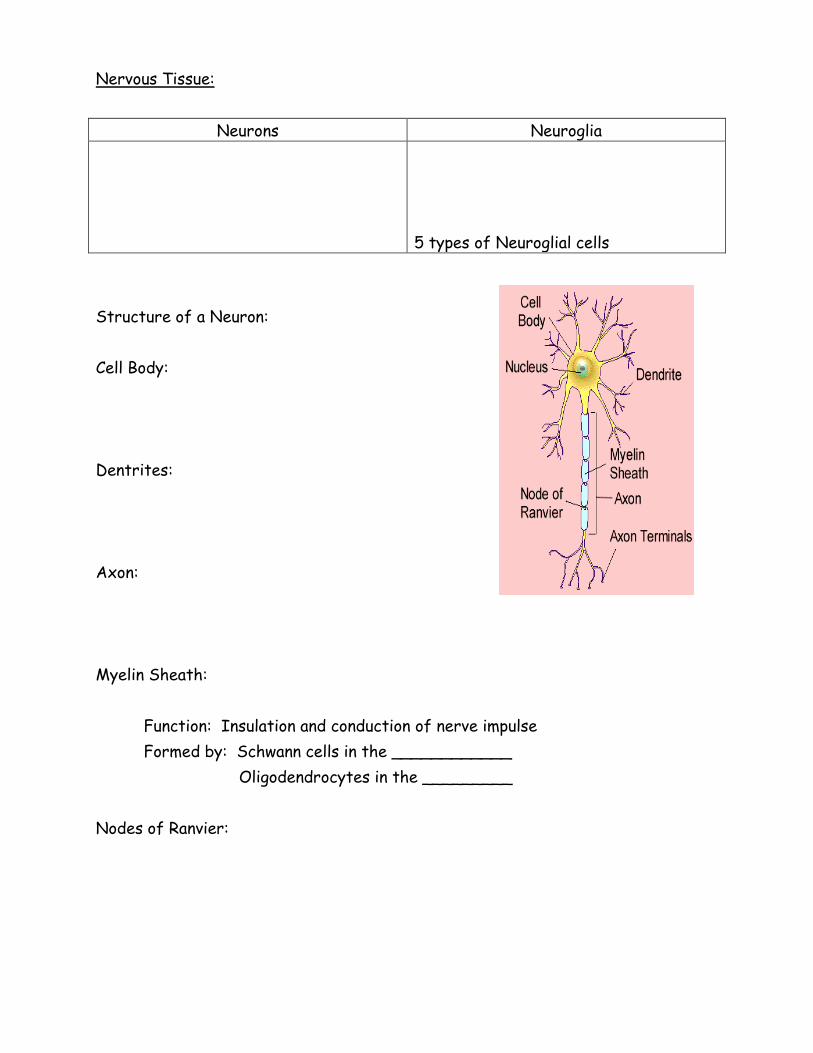
Nervous Tissue:
Neurons Neuroglia
5 types of Neuroglial cells
Structure of a Neuron:
Cell Body:
Dentrites:
Axon:
Myelin Sheath:
Function: Insulation and conduction of nerve impulse
Formed by: Schwann cells in the ____________
Oligodendrocytes in the _________
Nodes of Ranvier:

Structural Classification of Neurons:
Unipolar:___________ axon that enters and exits the cell body
Bipolar: one dendrite and one axon
Found :
Multipolar: many dendrites and _____ axon
Motor Neurons
Efferent
Sensory Neurons
Afferent
Interneurons
Association Neurons
Where
Found?
Function?
Structural
Classification Uni-
Bi- polar
Multi-
Pathway
Stimulated by

Sensory Neuron: Interneuron:
Neuroglial Cells and Transmission of Action Potential
6 Types of Neuroglial or Glial Cells:
Glia = greek word for glue
Special types of ________________________ that help ___________
and provide _________________ for nerve cells.
___________________ neurons and
______________________________.
Supply ____________ and _______________ to neurons.
______________________ one neuron from another.
Destroy ___________________ and remove ______________neurons.
Motor Neuron:

Name of the Cell Found in CNS or PNS Function
Astrocytes
Oligodendrocytes
Microglia Cells
Ependymal Cells
Schwann Cells
Satellite Cells
Nerve Impulse
An __________________________ that travels along a nerve fiber.
Travels about _________________________________________.
Rapid ____________________ and ________________ of a small
portion of the plasma membrane
Nerve Signal Conduction
_____________________ potential followed by an _______________
potential
Resting Potential
Electrical charge across the ______________________
Inside of cell is _______ compared to the outside
Sodium ions _________
Potassium ions ________
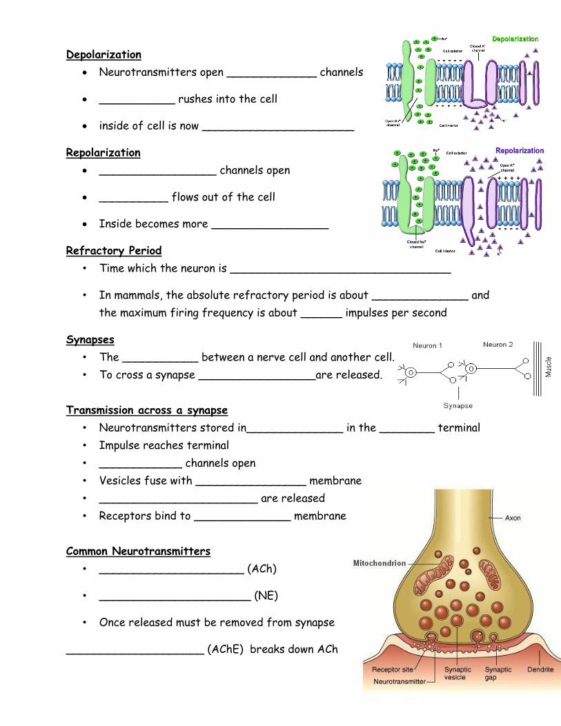
Depolarization
Neurotransmitters open _____________ channels
___________ rushes into the cell
inside of cell is now ______________________
Repolarization
_________________ channels open
__________ flows out of the cell
Inside becomes more _________________
Refractory Period
• Time which the neuron is ________________________________
• In mammals, the absolute refractory period is about ______________ and
the maximum firing frequency is about ______ impulses per second
Synapses
• The ___________ between a nerve cell and another cell.
• To cross a synapse _________________are released.
Transmission across a synapse
• Neurotransmitters stored in______________ in the ________ terminal
• Impulse reaches terminal
• ____________ channels open
• Vesicles fuse with ________________ membrane
• _______________________ are released
• Receptors bind to ______________ membrane
Common Neurotransmitters
• _____________________ (ACh)
• ______________________ (NE)
• Once released must be removed from synapse
____________________ (AChE) breaks down ACh

Brain Structure and Function
CNS: The Brain and Spinal Cord
Gray matter-
White matter-
Meninges: The Protective Coverings
Dura Mater (Outermost) Arachnoid Mater Pia Mater (Innermost)
Dura mater-
• Tough, fibrous, _____________________ tissue
• Made of ___________________________________ layers
• Separation of layers to form
_______________________________________________
– Collection of _____________________ and extra _________________
Arachnoid Mater-
• ________________________________ connective tissue
• CSF in the _______________________________ space
– CSF-clear fluid, _______________________________________
Pia Mater-
• Very thin layer
• Follows ________________________________________________________
CSF
• Made by the ____________________________________ lining the ventricles
• Fills all ________________________ and the __________________________
• Hydrocephalus-_________________________________________ in an infant
The Spinal Cord
• Is an extension of the brain
• Exits the cranial cavity via _________________________________________
• Runs through the _______________________________________________
• Ends between L ____ & L ____
• Cauda equina (horse’s tail)
The Vertebral Column
• Made of individual vertebrae separated by ________________________
– _____________________
– Disk herniation

Anatomy of the Spinal Cord
• Inner _______________________ with a central canal
• Outer ________________________________________
Spinal Nerves
• Posterior (dorsal) root of a spinal nerve:
– _____________________ (__________________________) fibers
• Anterior (ventral) root of a spinal nerve:
– _______________________ (_________________________) fibers
• Spinal nerve – joining of posterior & anterior roots
The Human Brain:
Gyri-
Sulci-
Major Sections of the Human Brain
The cerebrum
The diencephalon:
– Hypothalamus
– Thalamus
– Pineal gland
The cerebellum
The brainstem

Peripheral Nervous System (PNS)
All the rest of the nerves of your body
Two branches
o _______________ Nervous System
o _______________ Nervous System
Nerve Anatomy
____________________ = covers nerve itself
____________________ = covers bundles of axons
____________________ = covers individual axons
Somatic NS
_______ pairs of Cranial Nerves
_______ pairs of Spinal Nerves
Types of Nerves
Mixed = ____________________________
Sensory = ___________________________
Motor = ____________________________
Cranial Nerves Spinal Nerves

Nerve Plexus: ______________________________________
Name of Plexus Nerves found in
plexus
Services what
part of the body
Cranial
Nerve #
Name of Cranial
Nerve
Function Type of Nerve
I Sense of Smell
II Sense of sight
III Movement of eyelid and eyeball
IV Muscles of the eyes
V Muscles for chewing (motor) & pain
and touch for face and mouth
(sensory)
VI Muscles for eye movement
VII Sense of taste (sensory) & facial
expressions (motor)
VIII Sense of Hearing
IX Sense of taste (sensory), blood
pressure, tongue movement (motor)
X Innervates smooth muscle of the
gut(motor) & feelings of
distension/bloating (sensory)
XI Movement of neck muscles
XII Movement of the tongue
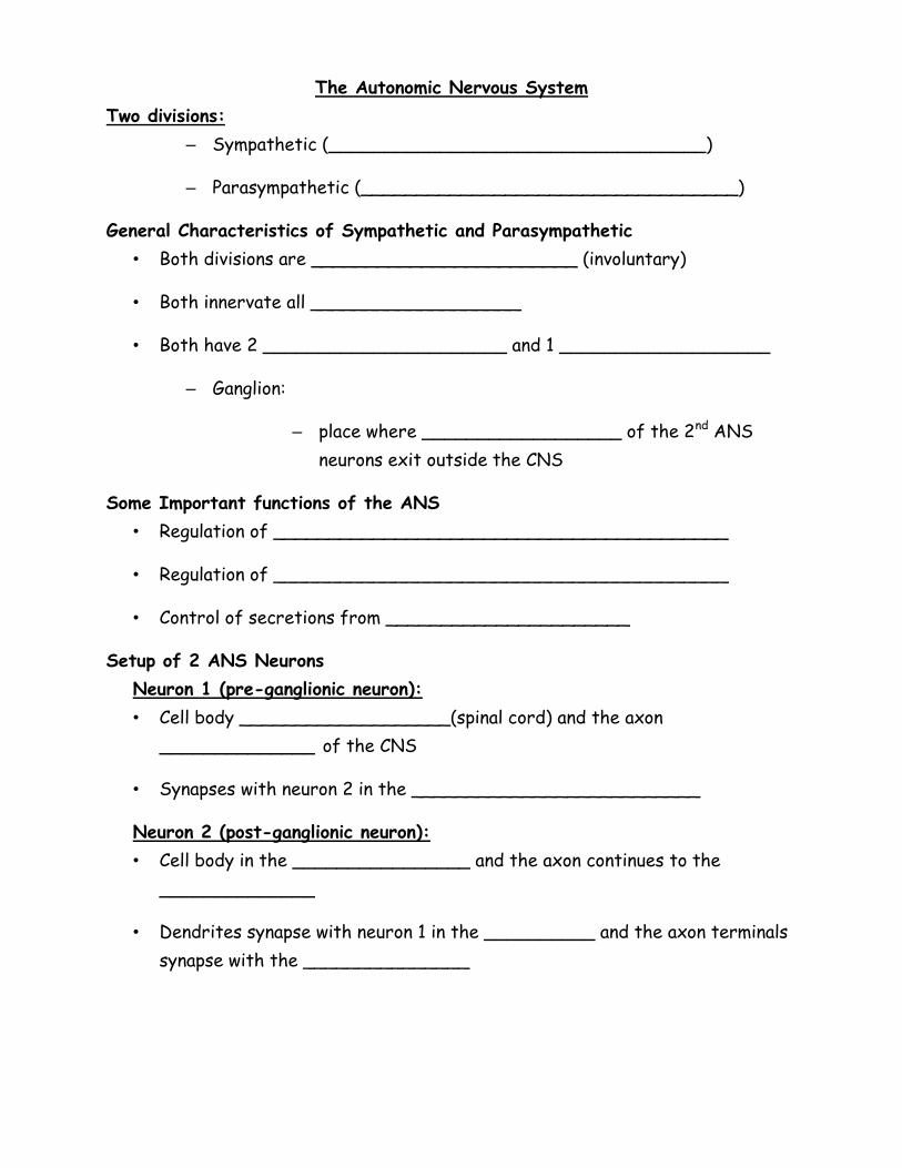
The Autonomic Nervous System
Two divisions:
– Sympathetic (__________________________________)
– Parasympathetic (__________________________________)
General Characteristics of Sympathetic and Parasympathetic
• Both divisions are ________________________ (involuntary)
• Both innervate all ___________________
• Both have 2 ______________________ and 1 ___________________
– Ganglion:
– place where __________________ of the 2nd ANS
neurons exit outside the CNS
Some Important functions of the ANS
• Regulation of _________________________________________
• Regulation of _________________________________________
• Control of secretions from ______________________
Setup of 2 ANS Neurons
Neuron 1 (pre-ganglionic neuron):
• Cell body ___________________(spinal cord) and the axon
______________ of the CNS
• Synapses with neuron 2 in the __________________________
Neuron 2 (post-ganglionic neuron):
• Cell body in the ________________ and the axon continues to the
______________
• Dendrites synapse with neuron 1 in the __________ and the axon terminals
synapse with the _______________
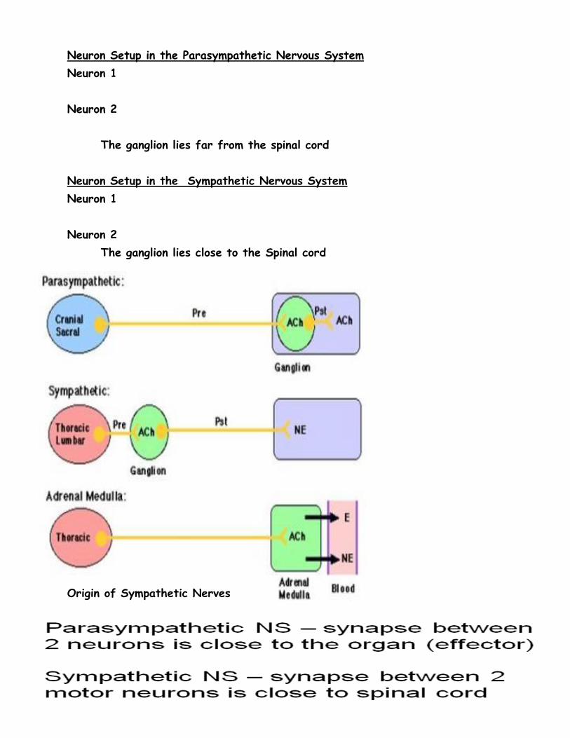
Neuron Setup in the Parasympathetic Nervous System
Neuron 1
Neuron 2
The ganglion lies far from the spinal cord
Neuron Setup in the Sympathetic Nervous System
Neuron 1
Neuron 2
The ganglion lies close to the Spinal cord
Origin of Sympathetic Nerves

Origin of Sympathetic Nerves __Origin of Parasympathetic Nerves
Functions of the Sympathetic Division
prepares the body for
emergencies (Fight or flight)
• increases heart rate
• raises blood pressure
(vasoconstriction)
• dilates the pupils
• dilates the trachea and bronchi
• Converts liver glycogen into
glucose
• shunts blood away from the skin
and organs (vasoconstriction)
• pushes blood toward the skeletal
muscles, brain, and heart
• inhibits peristalsis in the
gastrointestinal (GI) tract
• inhibits contraction of the
bladder and rectum
Functions of the Parasympathetic
Division
• The “housekeeper” division
• “Rest and Digest”
• Manages functions associated
with a relaxed state
• Contraction of the pupils
• Promotes digestion of food
• Slows down the heart rate
and decreased heart
contraction
• Increased blood flow to the
visceral organs (GI tract),
normal peristalsis
Sympathetic Neurotransmitters
Parasympathetic Neurotransmitters
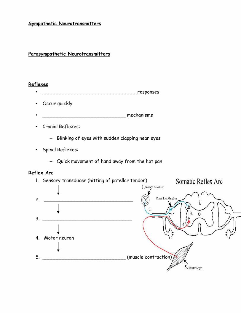
Reflexes
• ________________________________responses
• Occur quickly
• ____________________________ mechanisms
• Cranial Reflexes:
– Blinking of eyes with sudden clapping near eyes
• Spinal Reflexes:
– Quick movement of hand away from the hot pan
Reflex Arc
1. Sensory transducer (hitting of patellar tendon)
2. ______________________________
3. ______________________________
4. Motor neuron
5. ____________________________ (muscle contraction)
Recommended