Embed Size (px)
Citation preview
2
The Role of Astrocytes in Epileptogenesis
Jesús Pastor1,2 and Rafael G. Sola1,3 1Epilepsy Unit, Hospital Universitario La Princesa 2Faculty of Medicine, Universidad San Pablo-CEU
3Faculty of Medicine, UAM, Madrid Spain
1. Introduction
Epilepsy is a major neurological disorder that affects 0.5-2.0% of the global population (Hauser, 1998; Picot et al.; 2008). The term “epilepsy” likely encompasses a number of various etiological and pathophysiological processes. The International League Against Epilepsy (ILAE) has classified the epilepsies as being either focal or generalized. In all likelihood, this classification implies different causes and anatomical/functional substrates. Epileptogenesis (the processes by which epilepsy develops within an otherwise normal brain) probably differs for the various types of epilepsy (Lüders, 2008). In this sense, epileptogenesis of (for example) temporal lobe epilepsy (TLE) with hippocampal sclerosis (HS), would be different from that observed in patients with cortical dysplasia; this would hold true for other cryptogenic or idiopathic focal epilepsies as well. Therefore, to date, it has not been possible to establish a unique underlying cause or mechanism that is involved in the transition from normal to epileptic cortical tissue, although there are several hypotheses to explain the different types of seizures and epilepsies. From a therapeutic point of view, the epilepsies in humans can be divided into pharmaco
sensitive and pharmaco resistant. In the former, the patient is successfully treated by one or
more anti-epileptic drugs (AED). Nearly two-thirds of patients respond to anticonvulsant
therapy. However, in a significant percentage of patients, the seizures cannot be controlled
by drugs, despite verifying both maximal dosage and an adequate treatment duration. The
percentages of medical intractability among epileptic patients are: focal epilepsy, 24%;
idiopathic epilepsy, 9.3%; and catastrophic epilepsy, 66.7% (Lardizabal, 2008). In some cases, a
viable therapeutic option is surgery. Generally, the candidates for this treatment are patients
who suffer from focal or partial epilepsy. In these patients, it is possible—at least in theory—to
identify a well-defined area of the cortex that is responsible for the seizures; this is known as
the epileptic zone (EZ). From an operational point of view, the EZ is defined as the region in
which excision or disconnection relieves the patient from seizures. This option frequently
involves removing the brain section where the EZ is located, and this possibility offers a
unique opportunity to gain access to the real processes (the neural networks, membrane
properties and synaptic dynamics) underlying partial epilepsy in human patients.
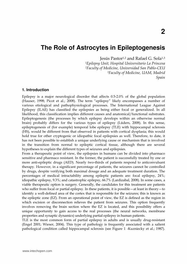
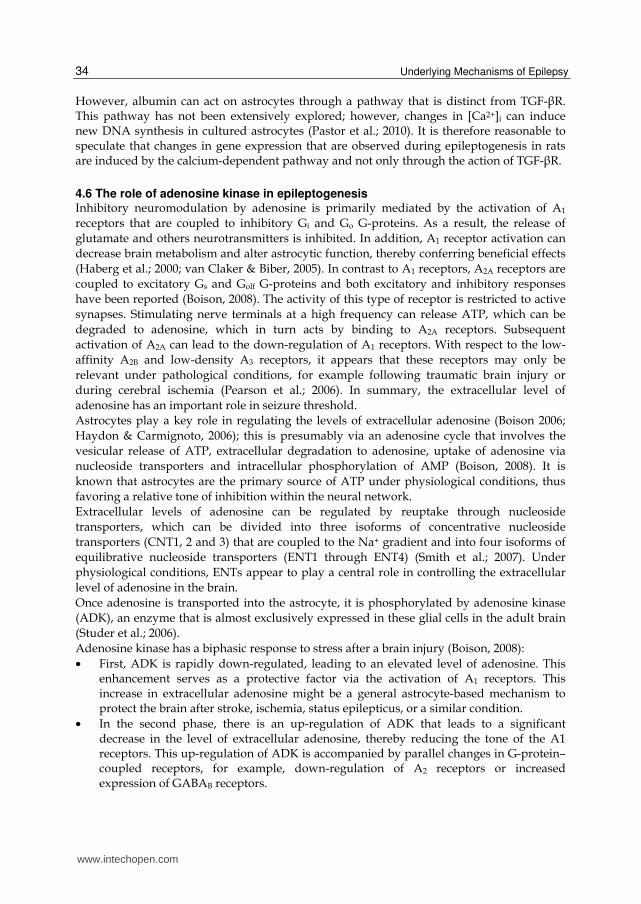
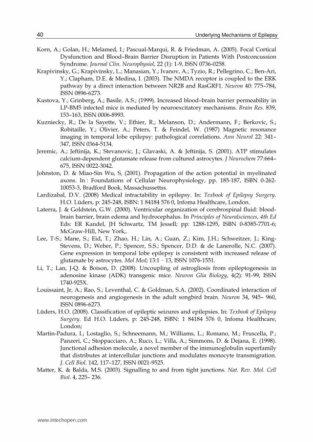
TLE is the most common form of partial epilepsy in adults and is usually drug-resistant (Engel 2001; Wieser, 2004). This type of pathology is frequently associated with a salient pathological condition called hippocampal sclerosis (see Figure 1 -Kuzniecky et al.; 1987).
www.intechopen.com
Underlying Mechanisms of Epilepsy 20
For several decades, HS has been considered the source of the electrical events that cause spontaneous seizures (Falconer, 1974; Spencer 1998). However, HS is found in approximately 40-65% of patients who undergo surgery for TLE (de Lanerolle et al.; 2003), and whether hippocampal sclerosis is the cause or the consequence of repeated seizures is still a matter of debate (Jefferys, 1999; Boison, 2008). The progression of epileptogenesis to chronic epilepsy often leads to pharmacoresistance and affects up to 30% of all patients with epilepsy, particularly those with TLE (Engel, 2001). A wide range of brain injuries and brain lesions are associated with an increased risk of epilepsy. In fact, such symptomatic etiologies account for 30-49% of all unprovoked seizures and epilepsy (Herman, 2002). The most common risk factors are cerebrovascular disease (21%), tumor (11%) and traumatic brain injury (7%) (Forsgren, 1990). A feature that is common to all of these conditions is the compromised permeability of the blood-brain barrier (BBB). Although idiopathic epilepsy accounts for up to 65% of cases, these patients likely share some pathophysiological mechanisms with injury-induced epilepsies. Much is known regarding neuronal electrophysiology in epileptic tissues (Avoli et al.; 2005; Pastor et al.; 2006); however, there is little information regarding the transition from a normal brain to an epileptic brain. Recently, it was shown in both rats and humans that astrocytes could be involved in some types of partial seizure (de Lanerolle et al.; 2010). Astrocytes are situated in key pivotal places that may play a role in epilepsy in several ways. Indeed, astrocytes control the trafficking of substances between capillary vessels and neurons. Another important function is in the homeostasis of extracellular ions, principally potassium (K+), which is directly involved in neuronal excitability. Finally, astrocytes are mediators of the neuron–to-neuron synapse through the tripartite synapse. In addition to astrocytes, another important factor is a change in the permeability of the BBB, which is formed by endothelial cells, although glial cells contribute yet to its function.
Fig. 1. Microphotograph of a Nissl-stained hippocampus obtained from a patient who underwent surgery for TLE. A loss of cells is evident in the CA1, CA3 and hilus regions.
www.intechopen.com

The Role of Astrocytes in Epileptogenesis 21
Albumin, the most abundant protein in blood, has also been implicated in epileptogenesis (Seiffert et al.; 2004; Herrera-Peco et al.; 2008) together with changes in BBB permeability or the inflammatory cascade, although the role of these mechanisms in epilepsy are still poorly understood (de Lanerolle et al.; 2010). Although not conclusive, different results point to astrocytes as being important mediators of the mechanisms involved in epileptogenesis and the origin and spreading of seizures. In this chapter, we review some of the recent theories and results concerning these topics, with a special emphasis on results obtained from human patients. We shall analyze the role of BBB permeability in the leakage of albumin into the brain’s extracellular space and the possible role of this protein in the activation of astrocytes. Although other reviews have focused on the role of the TGF-┚ receptor (TGF-┚R) in the action of albumin, we shall review a putative role for a different albumin receptor that is currently not well characterized but clearly has different properties than TGF-┚R.
2. A brief introduction to astrocyte physiology
Glial cells outnumber neurons by approximately 10-50:1. Glial cells can be divided into two major classes called microglia and macroglia.
• Microglia are phagocytes that mobilize in response to brain injury (for example, infection or disease). From a physiological and embryological point of view, they are unrelated to the others cells of the nervous system. Microglia are activated and recruited during infection, seizure and injury or disease, including multiple sclerosis, stroke, acquired immunodeficiency syndrome (AIDS)-related dementia and Parkinson’s disease. In this regard, microglia serve as major antigen-presenting cells.
• Macroglia comprise three different types of cells:
• Oligodendrocytes and Schwann cells are located in the Central Nervous System (CNS) and Peripheral Nervous System (PNS), respectively. Both cell types provide insulation for neuronal axons. Each oligodendrocyte envelops an average of 15 axonal internodes. However, each Schwann cell envelops only one internode on a single axon. The insulation provided by these cells is necessary for saltatory nerve conduction (Donaldson & Mia-Sin Wu, 2001).
• Astrocytes are the most numerous glial cells and tend to have long processes, some of which terminate as end-feet on the surface of neurons and, among other things, supply nutrients. Some astrocytes project their end-feet to capillaries, where they help endothelial cells form the BBB. However, their exact definition and the types of astrocytes that exist remain incompletely understood (Haydon & Carmignoto, 2006; de Lanerolle et al.; 2010).
Traditionally, the main role of astrocytes has been considered to be to provide support to neurons (Barres, 1991). However, over the previous decades, astrocytes have been found to play several roles other than supportive. Seminal works showed that astrocytes express receptors for neurotransmitters (Porter & McCarthy, 1997) and can respond to a local application of glutamate with a calcium elevation that travels as a wave through a syncytium (Corner-Bell et al.; 1990). Together, these data suggest that astrocytes possess the capacity to signal using spike-like calcium transients. In fact, these calcium spikes can travel over long distances and serve as a tool for communication. Astrocytes are normally identified by their expression of glial fibrillary acidic protein (GFAP). However, glial cells can be further categorized according to their functional properties as follows (Matthias et al.; 2003, Wallraff et al.; 2004; Jabs et al.; 2008):
www.intechopen.com
Underlying Mechanisms of Epilepsy 22
• Cells with large outward K+ current and attenuated inward K+ current (IKir). The resting
potential (Vr) of these cells is therefore relatively depolarized (∼-31 mV). Most of these cells contain TTX-sensitive Na+ currents but are unable to generate action potentials. These cells express alpha-amino-3-hydroxy-5-methyl-4-isoxazolepropionic acid (AMPA) type receptors (GluR), but not glutamate transporter currents. Finally, these cells completely lack gap junction coupling but receive spontaneous synaptic input from glutamatergic and ┛-aminobutyric acid neurons (GABAergic).
• Cells that resemble protoplasmic astrocytes. These cells have a more negative Vr (∼-70 mV), and IKir is present. These cells express glutamate transporter currents (GluT) but not GluR currents. Finally, these cells are extensively coupled via gap junctions.
• A third cell type closely resembles the GluR cell type described above. These are considered a third class of macroglia and have various names, including neuron glia-2 (NG2) cells, oligodendrocyte precursor cells, polydendrocytes, synantocytes and complex cells (Pauker & Bergles, 2006). These cells express chondroitin sulfate proteoglycan, do not express GFAP and lack GluT currents and gap junctions. As with GluR astrocytes, they express AMPA receptors as well as GABA receptors; moreover, they receive synaptic terminals. Current injection into these cells elicits action potentials that are reversibly blocked by TTX. To complicate matters further, two classes of NG2 cells have been recently identified (Káradóttir et al.; 2008): one class expresses voltage-gated Na+ currents together with voltage-gated K+ currents, whereas the other does not express Na+ currents. Presumably, both of these cell classes additionally express voltage-gated Ca2+ currents.
2.1 Plasma membrane properties of astrocytes Some astrocytes exhibit a high resting K+ conductance and possess gap junction coupling;
thus, the first major function that was assigned to these cells was the clearance of
extracellular K+ following neuronal activity. However, glial cells express a set of receptors
that is similar to neurons, but at different relative densities. This suggests that astrocytes
perform other functions than buffering K+. Among their intrinsic membrane proteins are
metabotropic glutamate receptors (mGluR) (Zur Nieden and Deitmer, 2006), purinergic
receptors (Jeremic et al.; 2001) and GABA type-B receptors. Other ionotropic receptors, such
as AMPA, are also present. An elevated amount of GluR1 in the AMPA receptors of reactive
astrocytes has been reported, which suggests an increased responsiveness of these astrocytes
to glutamate (Seifert et al.; 2004). This change has been observed in astrocytes that were
obtained from sclerotic tissue. Moreover, in animal models, mGluR3, mGluR5 and mGluR8
up-regulation has been reported in the hippocampus (Steinhäuser & Seifert, 2002). These
receptors are involved in the astrocyte’s response to glial and neurotransmitter. During
normal brain function activity, astrocytes play a major role in the clearance of glutamate that
is released from the nerve terminal into the extracellular space.
Astrocytes achieve this through the activity of two glutamate transporter molecules, namely
the excitatory amino acid transporter (EAAT) subtypes EAAT1 and EAAT2 (de Lanerolle et
al.; 2010). The GABA transporter GAT3 is usually only weakly expressed (if at all) in
astrocytes. Aquaporin 4 (AQP4) is a water transporter molecule that is found on astrocytes.
The distribution of these transporter molecules is asymmetric, being more densely
expressed on the perivascular astrocytic end-feet than on the membrane that faces the
neuropil (de Lanerolle et al.; 2010).
www.intechopen.com
The Role of Astrocytes in Epileptogenesis 23
Several studies demonstrated the presence of voltage-dependent ionic channels, including
Na+, K+ and Ca2+ channels (Barres et al.; 1990; Sontheimer et al.; 1991; Sontheimer &
Waxman, 1993). Voltage-gated Na+ channels are present in astrocytes in various brain
regions. It has been postulated that this might serve to regulate [Na+]i and thereby control
the activity of the Na+/glutamate transporter or Na+/K+ ATPase exchanger (Sontheimer et
al.; 1994). Calcium channels have also been identified in both cultured astrocytes and acute
astrocyte preparations (Verkhratsky & Steinhäuser, 2000). Astrocytes play a major role in K+
homeostasis in the central nervous system. During neuronal activity, [K+]o is temporally
increased, which depolarizes nearby membranes. Astrocytes help move K+ away from
regions of high concentration to restore the normal extracellular concentration (Steinhäuser
& Seifert, 2002). Inwardly rectifying potassium (Kir) channels in astrocytes play a major role
in removing K+ from the extracellular space.
2.2 Calcium signaling in astrocytes Glial cells respond with changing [Ca2+]i to various neurotransmitters, the most important of
which are glutamate, ATP, adenosine and GABA (Haydon & Carmignoto, 2006). These
transmitters activate metabotropic receptors that are coupled to second messenger systems.
Astrocytes not only exhibit changes in [Ca2+]i after extracellular neurotransmitter release, but spontaneous calcium oscillations can also occur in the absence of neuronal activity (Nett et al.; 2002; Parri & Crunelli 2003; Zur Nieden & Deitmer, 2006). It has been established that both evoked and spontaneous astrocyte calcium increases depend on calcium release from internal stores (Fiacco & McCarthy, 2006). A neurotransmitter-induced increase in astrocytic calcium activates astrocytic metabotropic Gq-coupled receptors that drive the release of calcium from the endoplasmic reticulum (ER) upon activation of phospholipase C (PLC) and conversion of phosphotidylinositol bisphosphate to inositol triphosphate (IP3) (Nadal et al.; 1995; Porter & McCarthy, 1996; Araque et al.; 1998a, Newman 2005; Pastor et al.; 2010). Spontaneous [Ca2+]i changes are not completely understood, but some theories that are
supported by various experimental data have been postulated. These include a mechanism
involving voltage-gated calcium channels and calcium induced-calcium release (Parri &
Crunelli, 2003), activation of mGluR by the quantal release of small amounts of glio or
neurotransmitters (possibly spillover from synaptic region) (Zur Neider & Deitmer, 2006)
and a “switch” in the intrinsic activity of metabotropic receptors (Pasti et al.; 1997).
Although the ryanodine receptor has been implicated in calcium changes in epilepsy
(Tashiro et al.; 2002), its role has not yet been clarified because several studies failed to show
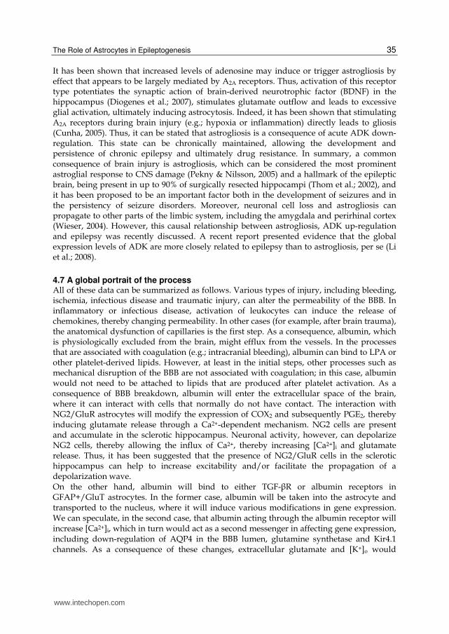
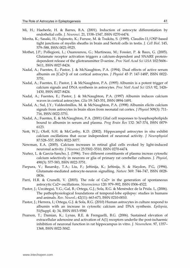
any significant contribution in astrocytes that were obtained from epileptic patients (see
Figure 2 and reference Pastor et al.; 2010). In astrocytes, a local calcium increase following IP3 receptor activation often propagates as a ‘‘calcium wave’’ some distance from its point of origin (Nadal et al.; 1997). The propagation of calcium waves is believed to occur via calcium diffusing locally (after its initial release) to neighboring IP3 receptors, where it acts as a co-agonist with IP3 to release stored calcium from adjacent compartments (Fiacco & McCarthy, 2006). Upon stimulation, astrocytes in culture display a calcium wave that propagates intercellularly through thousands of cells (Cornell-Bell et al.; 1990). The mechanisms that are involved in the propagation of intercellular calcium waves in cultured astroglia include: (1) gap junctions between astrocytes that allow the intercellular diffusion of second messengers such as IP3, and (2) the calcium-induced release of ATP that diffuses extracellularly to
www.intechopen.com

Underlying Mechanisms of Epilepsy 24
neighboring astrocytes to activate purinergic receptors that are coupled to internal calcium stores (Cotrina et al.; 2000; Suadicani et al.; 2004). There is no evidence of the existence of intercellular astrocyte calcium waves in intact tissue, suggesting that long-range glial signaling may be an artifact of cultured astroglia; however, it has been proposed that this process might occur in pathological conditions such as epilepsy (Peters et al.; 2003). In general, these observations suggest that widespread intercellular calcium waves are not likely to occur between astrocytes under physiological conditions in situ. However, a recent in vivo study showed that “Ca2+ signals (…) can propagate through the cell and couple to adjacent astrocytes in the form of a Ca2+ wave” (Ding et al.; 2007). These changes in [Ca2+]i can act as a second messenger in various cellular process, including the release of gliotransmitters or the modification of gene expression patterns.
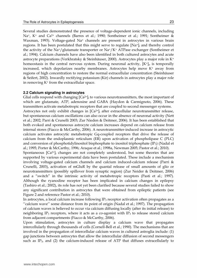
Fig. 2. Ryanodine fails to block the response to bovine plasma albumin (Alb; 20 mg/ml) in astrocytes obtained from epileptic patients (unpublished data).
2.3 Interactions between astrocytes and neurons Astrocytes were initially considered to be supportive cells. However, during the last years of the past century, it became clear that glial cells can also contribute to information processing by neurons. Various studies have led to the proposal of the “tripartite synapse”, in which the astrocyte monitors synaptic activity and provides feedback by modulating the strength of the synaptic connection (Araque et al.; 1999; 2001). This concept is based on studies showing that glial cells respond to neuronal activity with an [Ca2+]i increase, which in turn triggers the release of gliotransmitters that can cause feedback regulation of neuronal activity. Calcium increases in astrocytes are coupled to the regulated release of neuroactive
molecules and classic neurotransmitters that include ATP, inflammatory cytokines,
vasoactive compounds, D-serine and glutamate (Parpura et al.; 1994 ; Coco et al.; 2003 ;
Zonta et al.; 2003; Mothet et al.; 2005).
Other modes of glutamate release by astrocytes are calcium-independent and include
release through large pores, including gap junction hemi-channels and purinergic P2X7
receptors (Ye et al.; 2003; Fellin et al.; 2006), a reversal of glutamate transport (Anderson &
Swanson, 2000) and by volume-sensitive anion channels (Takano et al.; 2005). However,
calcium-independent modes of astrocytic glutamate release have only been observed under
www.intechopen.com
The Role of Astrocytes in Epileptogenesis 25
pathological conditions, such as severe energy depletion, highly increased external K+ and
in divalent cation-free extracellular solutions. Therefore, a considerable body of data
suggests that the calcium-dependent release of glutamate from astrocytes occurs via a
regulated exocytotic vesicular mechanism.
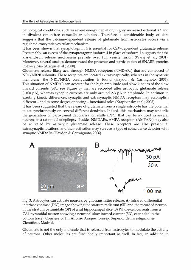
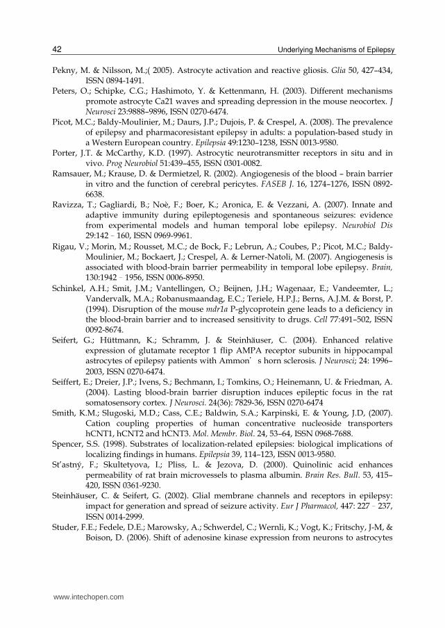
It has been shown that synaptotagmin 4 is essential for Ca2+-dependent glutamate release. Presumably, an excess of the synaptotagmin isoform 4 in place of isoform 1 suggests that the kiss-and-run release mechanism prevails over full vesicle fusion (Wang et al.; 2001). Moreover, several studies demonstrated the presence and participation of SNARE proteins in exocytosis (Araque et al.; 2000). Glutamate release likely acts through NMDA receptors (NMDARs) that are composed of NR1/NR2B subunits. These receptors are located extrasynaptically, whereas in the synaptic membrane, the NR1/NR2A configuration is found (Haydon & Carmignoto, 2006). This situation of NMDAR can account for the high amplitude and slow kinetics of the slow inward currents (SIC; see Figure 3) that are recorded after astrocytic glutamate release
(∼100 pA), whereas synaptic currents are only around 2-3 pA in amplitude. In addition to exerting kinetic differences, synaptic and extrasynaptic NMDA receptors may accomplish different—and to some degree opposing—functional roles (Krapivinsky et al.; 2003). It has been suggested that the release of glutamate from a single astrocyte has the potential to act synchronously on several different dendrites. Indeed, this mechanism may underlie the generation of paroxysmal depolarization shifts (PDS) that can be induced in several neurons in a rat model of epilepsy. Besides NMDARs, AMPA receptors (AMPARs) may also be activated by astrocytic glutamate release. These receptors are also present at extrasynaptic locations, and their activation may serve as a type of coincidence detector with synaptic NMDARs (Haydon & Carmignoto, 2006).
Fig. 3. Astrocytes can activate neurons by gliotransmitter release. A) Infrared differential interface contrast (DIC) image showing the stratum radiatum (SR) and the recorded neuron in the stratum pyramidale (SP) of a rat hippocampal slice. B) Whole-cell currents from a CA1 pyramidal neuron showing a neuronal slow inward current (SIC, expanded in the bottom trace). Courtesy of Dr. Alfonso Araque, Consejo Superior de Investigaciones Científicas, Madrid.
Glutamate is not the only molecule that is released from astrocytes to modulate the activity of neurons. Other molecules are functionally important as well. In fact, in addition to
www.intechopen.com

Underlying Mechanisms of Epilepsy 26
requiring glutamate for NMDAR activation, the co-agonist D-serine may also be important. Astrocytes are the only cells that express the enzyme serine racemase, making glial cells the only source for this molecule in the brain (Wolosker et al.; 1999). It has been suggested that vesicles might contain both D-serine and glutamate, thereby allowing their simultaneous release upon increased [Ca2+]i (Haydon & Carmignoto, 2006). Another important gliotransmitter that is released by astrocytes is ATP, and the mechanisms that are involved are the subject of debate. There is evidence that the release of both ATP and glutamate involve phospholipase C, but it has been suggested that ATP release depends on the diacylglycerol pathway (Wang et al.; 2000), although this hypothesis is still under debate. Released at micromolar concentrations ATP has powerful effects on adjacent neurons acting through purinergic receptors (P2). ATP can activate P2 presynaptic receptors. In fact, the activation of P2Y1 receptors in CA1 interneurons—most likely after the release of ATP from both neurons and astrocytes—may induce action potentials in these neurons and lead to increased GABAergic synaptic inhibition of pyramidal neurons (Bowser & Khakh, 2004). However, an excitatory effect of ATP has been described in which glutamate-mediated miniature end-plate synaptic currents are increased (Gordon et al.; 2005). Once ATP has been released into the extracellular space, a variety of ectonucleotidases degrade ATP into adenosine. Adenosine is a ubiquitous modulator of synaptic transmission and neuronal activity and acts through specific receptors. Indeed, there are high-affinity inhibitory adenosine A1 and excitatory A2A receptors that are activated by nanomolar concentrations of adenosine as well as low-affinity or low-abundance A2B and A3 receptors. The most relevant function of adenosine is to induce presynaptic inhibition of transmitter release and therefore has potent anticonvulsant and neuroprotective functions (Boison, 2008). It is important to note that adenosine—either released or produced by ATP degradation—can diffuse from the synaptic cleft and act laterally to regulate the strength of neighboring synapses in a mechanism named heterosynaptic suppression, which is mediated by the accumulation of adenosine acting through A1 receptors (Zhang et al.; 2003). We will see below in detail the effect of adenosine on seizure threshold (see 4.5 The role of adenosine kinase in epileptogenesis); however, suffice it to say that adenosine acting through A1 receptors has a potent anticonvulsant effect. In addition to the release of gliotransmitters, it is important to consider that one cortical astrocyte makes contacts with approximately 300-600 neuronal dendrites. These “local modules” of astrocytic integration can affect the function of several groups of neurons or synapsis, thus working in a more coherent mode (Halassa et al.; 2007). Astrocytes play an important role in the metabolism of the brain through their exclusive role in the degradation of both glucose and glutamate. The importance of astrocytes in these processes lies in their expression of key enzymes that are not normally found in neurons. Of course, these processes are extremely important for appropriate brain function but are beyond the scope of this review. The reader is referred to Hertz et al. (1999) and Herrera-Peco et al. (2008).
2.4 Astrocytes are activated by albumin Albumin (C123H193N35O37) is the most abundant protein in blood, representing more than 50% of all proteins. Its molecular weight is 2754.06 g/mol, and its concentration in plasma is around 35-55 mg/ml (Nadal et al.; 2001). Among its functions is the binding of several polar lipids, particularly lysophosphatidic acid (LPA), other fatty acids and sphingomyelins that are derived from platelet activation (Fuentes, E.t al.; 1999). Albumin is also the main
www.intechopen.com
The Role of Astrocytes in Epileptogenesis 27
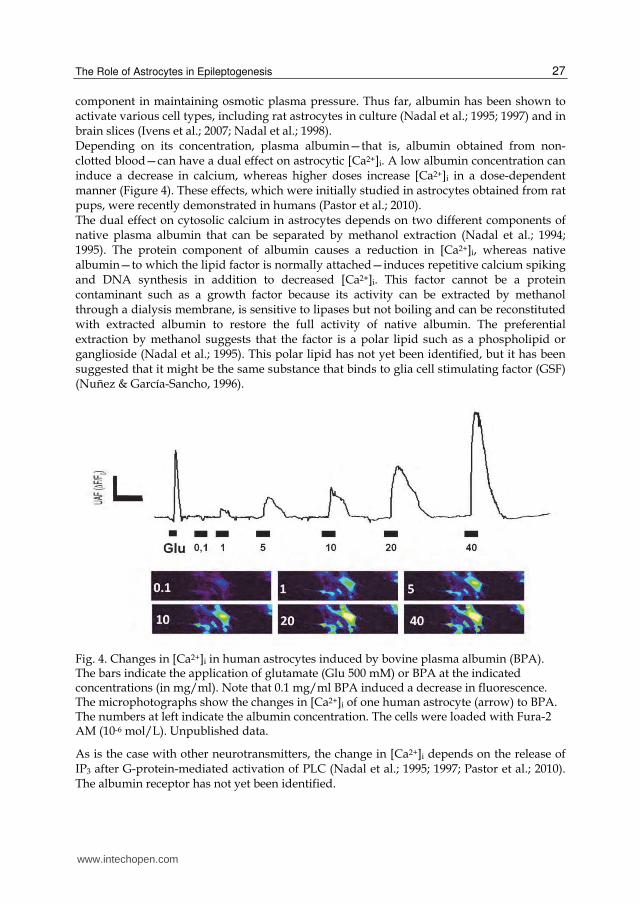
component in maintaining osmotic plasma pressure. Thus far, albumin has been shown to activate various cell types, including rat astrocytes in culture (Nadal et al.; 1995; 1997) and in brain slices (Ivens et al.; 2007; Nadal et al.; 1998). Depending on its concentration, plasma albumin—that is, albumin obtained from non-clotted blood—can have a dual effect on astrocytic [Ca2+]i. A low albumin concentration can induce a decrease in calcium, whereas higher doses increase [Ca2+]i in a dose-dependent manner (Figure 4). These effects, which were initially studied in astrocytes obtained from rat pups, were recently demonstrated in humans (Pastor et al.; 2010). The dual effect on cytosolic calcium in astrocytes depends on two different components of native plasma albumin that can be separated by methanol extraction (Nadal et al.; 1994; 1995). The protein component of albumin causes a reduction in [Ca2+]i, whereas native albumin—to which the lipid factor is normally attached—induces repetitive calcium spiking and DNA synthesis in addition to decreased [Ca2+]i. This factor cannot be a protein contaminant such as a growth factor because its activity can be extracted by methanol through a dialysis membrane, is sensitive to lipases but not boiling and can be reconstituted with extracted albumin to restore the full activity of native albumin. The preferential extraction by methanol suggests that the factor is a polar lipid such as a phospholipid or ganglioside (Nadal et al.; 1995). This polar lipid has not yet been identified, but it has been suggested that it might be the same substance that binds to glia cell stimulating factor (GSF) (Nuñez & García-Sancho, 1996).
Fig. 4. Changes in [Ca2+]i in human astrocytes induced by bovine plasma albumin (BPA). The bars indicate the application of glutamate (Glu 500 mM) or BPA at the indicated concentrations (in mg/ml). Note that 0.1 mg/ml BPA induced a decrease in fluorescence. The microphotographs show the changes in [Ca2+]i of one human astrocyte (arrow) to BPA. The numbers at left indicate the albumin concentration. The cells were loaded with Fura-2 AM (10-6 mol/L). Unpublished data.
As is the case with other neurotransmitters, the change in [Ca2+]i depends on the release of IP3 after G-protein-mediated activation of PLC (Nadal et al.; 1995; 1997; Pastor et al.; 2010). The albumin receptor has not yet been identified.
www.intechopen.com
Underlying Mechanisms of Epilepsy 28
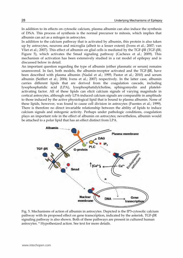
In addition to its effects on cytosolic calcium, plasma albumin can also induce the synthesis of DNA. This process of synthesis is the normal precursor to mitosis, which implies that albumin can act as a mitogen in astrocytes. In addition to the calcium pathway that is activated by albumin, this protein is also taken up by astrocytes, neurons and microglia (albeit to a lesser extent) (Ivens et al.; 2007; van Vliet et al.; 2007). This effect of albumin on glial cells is mediated by the TGF-┚R (TGF-┚R; Figure 5), which activates the Smad signaling pathway (Cacheux et al.; 2009). This mechanism of activation has been extensively studied in a rat model of epilepsy and is discussed below in detail. An important question regarding the type of albumin (either plasmatic or serum) remains unanswered. In fact, both models, the albumin-receptor activated and the TGF-βR, have been described with plasma albumin (Nadal et al.; 1995; Pastor et al.; 2010) and serum albumin (Seiffert et al.; 2004; Ivens et al.; 2007) respectively. In the latter case, albumin carries different lipids that are derived from the coagulation cascade, including lysophosphatidic acid (LPA), lysophosphatidylcholine, sphingomyelin and platelet-activating factor. All of these lipids can elicit calcium signals of varying magnitude in cortical astrocytes, although only LPA-induced calcium signals are comparable in amplitude to those induced by the active physiological lipid that is bound to plasma albumin. None of these lipids, however, was found to cause cell division in astrocytes (Fuentes et al.; 1999). There is therefore no direct invariable relationship between the ability of lipids to induce calcium signals and mitogenic activity. Perhaps under pathologic conditions, coagulation plays an important role in the effect of albumin on astrocytes; nevertheless, albumin would be attached to a polar lipid that has an effect distinct from LPA.
Fig. 5. Mechanisms of action of albumin in astrocytes. Depicted is the IP3-cytosolic calcium pathway with its proposed effect on gene transcription, indicated by the asterisk. TGF-┚R signaling pathway is also shown. Both of these pathways are present in cultured human astrocytes. * Hypothesized action. See text for more details.
www.intechopen.com

The Role of Astrocytes in Epileptogenesis 29
3. The structure and function of the blood-brain barrier
The BBB is a diffusion barrier that impedes the influx of most compounds between the blood and the brain, thereby isolating the brain from the external environment. Three cellular elements of the brain’s microvasculature comprise the BBB and are responsible for its properties. These components are: • Endothelial cells. Tight junctions (TJs) are special transmembrane proteins that are
located between cerebral endothelial cells, where they form a diffusion barrier that selectively excludes most blood-borne substances from entering the brain. There is some evidence of cross-talk with astrocytes. Indeed, endothelial cells seem to be the primary source of leukemia inhibitory factor (LIF), which helps induce astrocyte differentiation (Mi et al.; 2001). Other molecules that are presumably released by endothelial cells to affect astrocytes include bradykinin and glutamate. Moreover, endothelial cells are involved in neurogenesis, primarily via vascular endothelial growth factor (VEGF) (Loussaint et al.; 2002).
• Astrocyte end-feet. These structures tightly ensheath the vessel wall and appear to be critical for the induction and maintenance of the TJ barrier, although astrocytes are not believed to have a role in the mammalian BBB. However, astrocytes are important because they help induce the generation of BBB structures in endothelial cells. Astrocytes
can act on endothelial cells by releasing cytokines such as TGF-β and glial cell-derived neurotrophic factor (GDNF) (Utsumi et al.; 2000, Ramsauer et al.; 2002). Intracellular calcium waves mediate bidirectional astrocyte-endothelial interactions. In co-culture models, two signaling mechanisms are presumably involved: 1) astrocytes and endothelial cells can exchange calcium signals via a pathway that is dependent on intracellular IP3 and gap junctions; and 2) diffusion of a purinergic messenger. However, in vivo, astrocytes and endothelial cells are not in contact, and therefore, these findings need to be confirmed. Nevertheless, the dilation of arterioles triggered by neuronal activity depends on glutamate-mediated [Ca2+]i oscillations in astrocytes (Zonta et al.; 2003).
• Pericytes are undifferentiated cells that wrap around the endothelial cells of microvessels, including capillaries, venules and arterioles. Pericytes are believed to provide structural support and vasodynamic capacity to the microvasculature (Ballabh et al.; 2004). Pericytes can also help to stabilize capillaries that are recently formed by endothelial cells, thereby regulating angiogenesis (Balabanov & Dore-Duffy, 1998).
The BBB differs from endothelial cells in the rest of the body by the absence of fenestrations, extensive tight junctions (TJs) and bare pinocytic vesicular transport. The building blocks of the BBB are comprised of several molecules and subcellular structures that confer its unique properties, the most important of which are: • Complex junctions in the BBB are formed by TJs and adherens junctions. Tight junctions
are sites of apparent fusion between leaflets of the plasma membranes of adjacent cells, whereas adherens junctions are composed of cadherin-catenin complexes and their associated proteins (Matter & Balda, 2003). Cytoplasmic proteins link membrane proteins to actin, which is the principal protein of the cytoskeleton.
• Tight-junction–associated membrane proteins, including occludins, claudins and junctional adhesion molecules (Furuse et al.; 1993; Martin-Pardura et al.; 1998; Morita et al.; 1999). These proteins form the extracellular component of TJs.
• Cytoplasmic accessory proteins, including zonula occludens (ZO) proteins, cingulin and several others (Itoh et al.; 1999). Actin binds to ZO proteins, thereby providing structural support to the endothelial cells (Haskins et al.; 1998).
www.intechopen.com
Underlying Mechanisms of Epilepsy 30
Dysfunction of the BBB exacerbates a number of neurologic diseases, including stroke and neuroinflammatory disorders. Under pathological conditions, different chemical mediators that increase BBB permeability are released. Among these mediators are glutamate,
aspartate, taurine, ATP, endothelin-I, tumor necrosis factor (TNF)-α and interleukin-1β
(IL-1β), all of which are produced by astrocytes (Kutsova et al.; 1999; Abbot 2000; 2002). Moreover, endothelial cells can release bradykinin, 5-HT, histamine, substance P, platelet activating factor and free radicals (Abbot, 2002; St’astný et al.; 2000). The function of the BBB is to control the traffic of substances between the blood and the brain. This is achieved in three ways:
• Diffusion of lipid-soluble substances. The BBB permits the rapid exchange of lipid-soluble gases such as O2 and CO2, an exchange that is limited only by the surface area of the blood vessel and cerebral blood flow. Barrier vessels are impermeable to molecules that are poorly soluble in lipids. The permeability coefficient of the BBB for many substances is directly proportional to the lipid solubility of the substance, which is measured as its oil-water partition coefficient (Laterra & Goldstein, 2000).
• Facilitated and energy-dependent receptor-mediated transport of specific water-soluble substances. Most substances that must cross the BBB are not lipid soluble and therefore need a specific carrier-mediated transport system. One of the most important substances is glucose, which is transported by the glucose transporter isotype-1, or Glut1 (Guerin et al.; 1999). Amino acids are also transported by three distinct carrier systems. Another transport system is related to the multidrug resistance to chemotherapy (MDR) transporter (Schinkel et al.; 1994).
• Ion channels. Various studies support the existence of a nonselective luminal ion channel that is inhibited by both amiloride and atrial natriuretic peptide. The existence of luminal Na+/H+ and Cl-/HCO3- exchangers has also been suggested. The external membrane of brain endothelial cells contains a relatively high density of Na+/K+-ATPase pump. In conjunction with K+ channels in astrocytes, this abluminal endothelial pump may play an important role in removing extracellular K+ that is released during intense neuronal activity. In addition, the nonselective luminal ion channel, a distinct abluminal K+ channel and the abluminal Na+/K+-ATPase may work together to tightly regulate the entry of Na+ and the release or recycling of K+ (Haydon & Carmignoto, 2006). Aquaporin-4 is the major water channel expressed in brain perivascular astrocytic processes and is the principal system for regulating the traffic of water between the brain and blood. Thus, its role in cerebral edema is very important.
4. The role of astrocytes in epileptogenesis
Until recently, little was known regarding the process by which a normal brain becomes epileptic following an external or internal insult, for example following traumatic brain injury or seizures that are induced by fever or infectious disease. These symptomatic epilepsies account for a significant number of patients; however, for patients with idiopathic or cryptogenic epilepsy, the antecedents remain unknown. In general, except for well-defined etiologies (e.g.; cortical dysplasia), the brain’s transition from normal to epileptic—namely, epileptogenesis—remains obscure. However, several lines of evidence involving astrocytes have begun to emerge during the
last decade, providing a somewhat coherent framework. These lines of evidence can be
grouped according to the different structures that are involved.
www.intechopen.com

The Role of Astrocytes in Epileptogenesis 31
4.1 Modification of astrocyte membrane proteins Diverse changes have been described for the expression of intrinsic plasma membrane proteins in astrocytes at the epileptic seizure foci. In sclerotic hippocampi, reactive astrocytes express mGluR2/3, mGluR4 and mGluR8. Similarly, mGluR3, mGluR5 and mGluR8 are up-regulated in the hippocampus in experimental animal models of TLE. Activation of these receptors may lead to an increase in [Ca2+]i and Ca2+ wave propagation, thereby leading to the release of glutamate from astrocytes (Volterra & Meldolesi, 2005). Using microdialysis, increased levels of in situ extracellular glutamate have been found in sclerotic seizure foci (During & Spencer, 1993; Cavus et al.; 2002). Moreover, the down-regulation of EAAT1 and EAAT2 has also been reported (Proper et al.; 2002). However, other groups were unable to confirm this observation (Tessler et al.; 1998). In sclerotic astrocytes, the GABA transporter GAT3 is up-regulated. In the same vein, microdialysis in epileptic patients has revealed decreased levels of extracellular GABA in the epileptic foci during the ictal state (During & Spencer 1993). The increased expression of the transporter would help to reduce extracellular GABA levels, thereby decreasing inhibitory tone during the ictal state. Expression of the protein AQP4 is reduced on the perivascular membrane of the astrocyte in sclerotic hippocampi, whereas its expression is unchanged on the membrane that faces the neuropil. This change may cause a decrease in water extrusion from the neuropil to the vessel (Eid et al.; 2005). In addition to changes in the expression of transport proteins in epileptic foci, ion channels in astrocytes are also modified. Cultured astrocytes obtained from patient hippocampus and entorhinal cortex1 displayed much larger tetrodotoxin (TTX)-sensitive Na+ currents than astrocytes from non-sclerotic hippocampi (O’connor et al.; 1998). However, as with other results, there are discrepancies regarding the increased Na+ current (Hinterkeuser et al.; 2000). Changes in the differential expression of calcium channels have been reported in the sclerotic hippocampus, but the functional significance of this change remains unclear (de Lanerolle et al.; 2010). Impaired K+ buffering has been detected in sclerotic patient hippocampi, suggesting that there are defective channels in the sclerotic region, particularly in CA1 (Gabriel et al.; 1998). The buffering of K+, which is likely performed by the GFAP+ subset of Glu-like astrocytes (and carried out by the Kir channels), depends on a parallel flux of water through the plasma membrane to avoid hyperosmolarity. Water molecules and K+ are taken up by the astrocytic membrane of the neuropil and pushed into the blood and cerebrospinal fluid (CSF) through the end-food pole. A loss of AQP4 would impede the movement of water and contribute to increased [K+]o. Concomitantly, the down-regulation of Kir4.1 channels during a critical time window has been described in a rat model of epilepsy (Ivens et al.; 2007), as we discuss in detail below. Clearly, this process can contribute to further increase the concentration of extracellular K+.
4.2 Changes in genes expression and metabolism In the sclerotic hippocampus, changes in various enzymes that are involved in the metabolism of astrocytes and the interaction between astrocytes and neurons have been 1The entorhinal cortex is part of the parahippocampal gyrus and is the main source of input to the hippocampus through the perforant pathway and the collaterals of the temporoammonic pathway.
www.intechopen.com
Underlying Mechanisms of Epilepsy 32
described (Herrera-Peco et al.; 2008). In this regard, a loss of glutamine synthetase in CA1 and CA3, an increase in the activity of glutamate dehydrogenase and/or a decrease in the activity of lactate dehydrogenase have all been reported. Besides, extracellular levels of lactate are elevated in the sclerotic hippocampus (de Lanerolle et al.; 2010). On the other hand, several astrocyte-related genes are up-regulated in sclerotic regions. These changes in the pattern of gene expression are consistent with increased gliosis or immune and inflammatory responses (Lee et al.; 2007). This is discussed in detail below. It is likely that much of these changes are a consequence of epilepsy rather than a cause. Therefore, more data are needed before we can formulate an exact picture of the genetic changes that are related to epileptogenesis.
4.3 Changes in BBB permeability It has long been recognized that there is a proliferation of the microvasculature in the sclerotic hippocampus, an observation that has been recently confirmed (Rigau et al.; 2007). However, on the contrary, there are recent reports showing a significant loss of microvessels in the CA1 region of sclerotic hippocampi (Kastanauskaite et al.; 2009). In any case, most groups agree on the presence of blood vessel changes in TLE-associated hippocampal sclerosis. Several molecules that are located in the perivascular end-feet exhibit changes in sclerotic regions. AQP4 and dystrophin are reduced, whereas CD44 and plectin 1 are increased. Other molecules such as the chemokines CCL2 and CCL3 are up-regulated, as are the chemokine receptors CCR1 and CCR2 (de Lanerolle, 2010). It is believed that these glial-derived chemokines can guide circulating leukocytes through endothelial junctions and into the brain’s extracellular space. These chemokines can also help mediate the extravasation of albumin (de Lanerolle et al.; 2010). Therefore, these changes can alter the permeability of the BBB, thereby allowing molecules that normally are excluded to access the brain. Indeed, it has been shown that the BBB is leakier during seizures (de Lanerolle et al.; 2010). Furthermore, immunohistochemical localization of albumin in resected hippocampi from TLE patients showed strong albumin immunoreactivity in the parenchyma throughout the hippocampus next to the blood vessels. Neurons and astrocytes located around the vessels were also albumin-positive, thus demonstrating an increase in BBB permeability. Such extravasations of albumin were not observed in control hippocampi (van Vliet et al.; 2007). In an animal model, the extravasation of serum albumin to the brain’s extracellular space was associated with a prominent activation of astrocytes, but no inflammatory response or marked cell loss. This activation was associated with the development of epileptiform discharges within 4–7 days (Seiffert et al.; 2004). Changes other than permeability have been reported in the BBB. The erythropoietin receptor is strongly expressed in the capillaries of sclerotic hippocampi, which suggests an increased uptake of erythropoietin into the hippocampus (Eid et al.; 2004).
4.4 Inflammatory and immune responses in the brain Astrocytes contribute to the inflammatory response in the CNS. Glial cells can produce a range of immunologically relevant molecules, including class II major histocompatibility complex antigens, cytokines and chemokines. Studies have shown that glial cells—including both astrocytes and microglia—express high levels of pro-inflammatory cytokines in regions that are recruited in the generation and propagation of seizures. In astrocytes and microglia
of patients with TLE and hippocampal sclerosis, the IL-1β system is activated, particularly in end-feet perivascular regions and in endothelial cells in sclerotic brain regions (Ravizza et
www.intechopen.com
The Role of Astrocytes in Epileptogenesis 33
al.; 2007). These changes can affect the permeability of the BBB, perhaps by disrupting tight-
junction organization (Del Machio et al.; 1996). It has been showed that IL-1β has a pro-ictogenic role that is mediated by the receptor IL-1R1 (Vezzani et al.; 2002). Exposure to IL-
1β or TNF-α exacerbates the excitotoxic neuronal damage that is induced by NMDA or AMPA, thus suggesting that cell survival depends on the tissue concentration and duration of action of cytokines. This deleterious effect is mediated by the induction of toxic mediators to affect neuronal excitability.
The damage in the brain that is induced by IL-1β can be mediated by an increase in the function of NMDA receptors or by an increase in extracellular glutamate. In this sense, IL-1β increases NMDA-mediated Ca2+ influx into neurons, thereby promoting excitotoxicity (Vezzani et al.; 2008). Other well-established mechanisms include the inhibition of
glutamate uptake by glial cells and an increase in glutamate release via TNF-α production (Bezzi et al.; 2001). Additionally, IL-1β activates nitric oxide (NO) synthase, increasing the glutamate release. Presumably, all of these mechanisms lead to increased glutamatergic transmission and toxicity. However, GABAergic transmission is also affected by inflammation. Indeed, IL-1β has been shown to inhibit GABAA receptors, thereby contributing to hyperexcitability (Wang et al.; 2000).
Other cytokines that are produced by glial cells—such as IL-6, TNF-α and prostaglandins—have been shown to have either pro-ictogenic or inhibitory effects on seizures in a concentration-dependent manner, and this depends on the type of receptors involved and the origin of the cells (Vezzani et al.; 2008). It has been reported that TNF-α activates the rapid recruitment AMPA receptors that lack the GluR2 subunit to the neuronal membrane; this molecular conformation is permissive for Ca2+ influx, thereby increasing neurotoxicity. Moreover, in the sclerotic hippocampus, there is an up-regulation of the p65 subunit of the nuclear factor kappa-light-chain-enhancer of activated B cells (NF-κB) (Crespel et al.; 2002), a protein complex that control DNA transcription and is involved in the cellular responses to stress. In the sclerotic hippocampus, immunoreactivity of complement proteins was increased in regions of neuronal loss (including CA1, CA3 and the hylus of the dentate gyrus). All of these data imply a direct participation of the inflammatory pathway in epilepsy.
4.5 The participation of albumin in the activation of astrocytes Albumin is usually not in contact with astrocytes or neurons. Indeed, its concentration in the
extracellular space of the CNS (∼35 ng/ml) is three orders of magnitude lower than in blood (Nadal et al.; 2001). The effect of albumin on glial cells depends on both the protein itself and the lipids to which it is attached (Nadal et al.; 1995; 1997). Recently, it was elegantly demonstrated in rats that albumin can act directly on astrocytes, thereby facilitating epileptogenesis (Seiffert et al.; 2004, Ivens et al.; 2007; Van Vliet et al.; 2007). It has been shown that direct brain exposure to serum albumin is associated with albumin uptake into astrocytes that is mediated by TGF-┚R. This uptake is followed by the down-regulation of Kir4.1 channels in astrocytes, which results in reduced buffering of extracellular potassium. This in turn leads to an increased activity-dependent accumulation of extracellular potassium, which results in facilitated NMDAR–mediated neuronal hyperexcitability and ultimately epileptiform activity (Ivens et al.; 2007). Presumably, these effects are mediated by GFAP+/GluT astrocytes. On the other hand, albumin can act on NG2/GluR astrocytes; in these cells, TGF-┚R can induce the transcription factor NF-κB, which can modify the expression of cyclooxygenase 2 (COX-2).
www.intechopen.com
Underlying Mechanisms of Epilepsy 34
However, albumin can act on astrocytes through a pathway that is distinct from TGF-┚R. This pathway has not been extensively explored; however, changes in [Ca2+]i can induce new DNA synthesis in cultured astrocytes (Pastor et al.; 2010). It is therefore reasonable to speculate that changes in gene expression that are observed during epileptogenesis in rats are induced by the calcium-dependent pathway and not only through the action of TGF-┚R.
4.6 The role of adenosine kinase in epileptogenesis Inhibitory neuromodulation by adenosine is primarily mediated by the activation of A1
receptors that are coupled to inhibitory Gi and Go G-proteins. As a result, the release of
glutamate and others neurotransmitters is inhibited. In addition, A1 receptor activation can
decrease brain metabolism and alter astrocytic function, thereby conferring beneficial effects
(Haberg et al.; 2000; van Claker & Biber, 2005). In contrast to A1 receptors, A2A receptors are
coupled to excitatory Gs and Golf G-proteins and both excitatory and inhibitory responses
have been reported (Boison, 2008). The activity of this type of receptor is restricted to active
synapses. Stimulating nerve terminals at a high frequency can release ATP, which can be
degraded to adenosine, which in turn acts by binding to A2A receptors. Subsequent
activation of A2A can lead to the down-regulation of A1 receptors. With respect to the low-
affinity A2B and low-density A3 receptors, it appears that these receptors may only be
relevant under pathological conditions, for example following traumatic brain injury or
during cerebral ischemia (Pearson et al.; 2006). In summary, the extracellular level of
adenosine has an important role in seizure threshold.
Astrocytes play a key role in regulating the levels of extracellular adenosine (Boison 2006;
Haydon & Carmignoto, 2006); this is presumably via an adenosine cycle that involves the
vesicular release of ATP, extracellular degradation to adenosine, uptake of adenosine via
nucleoside transporters and intracellular phosphorylation of AMP (Boison, 2008). It is
known that astrocytes are the primary source of ATP under physiological conditions, thus
favoring a relative tone of inhibition within the neural network.
Extracellular levels of adenosine can be regulated by reuptake through nucleoside
transporters, which can be divided into three isoforms of concentrative nucleoside
transporters (CNT1, 2 and 3) that are coupled to the Na+ gradient and into four isoforms of
equilibrative nucleoside transporters (ENT1 through ENT4) (Smith et al.; 2007). Under
physiological conditions, ENTs appear to play a central role in controlling the extracellular
level of adenosine in the brain.
Once adenosine is transported into the astrocyte, it is phosphorylated by adenosine kinase
(ADK), an enzyme that is almost exclusively expressed in these glial cells in the adult brain
(Studer et al.; 2006).
Adenosine kinase has a biphasic response to stress after a brain injury (Boison, 2008):
• First, ADK is rapidly down-regulated, leading to an elevated level of adenosine. This enhancement serves as a protective factor via the activation of A1 receptors. This increase in extracellular adenosine might be a general astrocyte-based mechanism to protect the brain after stroke, ischemia, status epilepticus, or a similar condition.
• In the second phase, there is an up-regulation of ADK that leads to a significant decrease in the level of extracellular adenosine, thereby reducing the tone of the A1 receptors. This up-regulation of ADK is accompanied by parallel changes in G-protein–coupled receptors, for example, down-regulation of A2 receptors or increased expression of GABAB receptors.
www.intechopen.com
The Role of Astrocytes in Epileptogenesis 35
It has been shown that increased levels of adenosine may induce or trigger astrogliosis by effect that appears to be largely mediated by A2A receptors. Thus, activation of this receptor type potentiates the synaptic action of brain-derived neurotrophic factor (BDNF) in the hippocampus (Diogenes et al.; 2007), stimulates glutamate outflow and leads to excessive glial activation, ultimately inducing astrocytosis. Indeed, it has been shown that stimulating A2A receptors during brain injury (e.g.; hypoxia or inflammation) directly leads to gliosis (Cunha, 2005). Thus, it can be stated that astrogliosis is a consequence of acute ADK down-regulation. This state can be chronically maintained, allowing the development and persistence of chronic epilepsy and ultimately drug resistance. In summary, a common consequence of brain injury is astrogliosis, which can be considered the most prominent astroglial response to CNS damage (Pekny & Nilsson, 2005) and a hallmark of the epileptic brain, being present in up to 90% of surgically resected hippocampi (Thom et al.; 2002), and it has been proposed to be an important factor both in the development of seizures and in the persistency of seizure disorders. Moreover, neuronal cell loss and astrogliosis can propagate to other parts of the limbic system, including the amygdala and perirhinal cortex (Wieser, 2004). However, this causal relationship between astrogliosis, ADK up-regulation and epilepsy was recently discussed. A recent report presented evidence that the global expression levels of ADK are more closely related to epilepsy than to astrogliosis, per se (Li et al.; 2008).
4.7 A global portrait of the process All of these data can be summarized as follows. Various types of injury, including bleeding, ischemia, infectious disease and traumatic injury, can alter the permeability of the BBB. In inflammatory or infectious disease, activation of leukocytes can induce the release of chemokines, thereby changing permeability. In other cases (for example, after brain trauma), the anatomical dysfunction of capillaries is the first step. As a consequence, albumin, which is physiologically excluded from the brain, might efflux from the vessels. In the processes that are associated with coagulation (e.g.; intracranial bleeding), albumin can bind to LPA or other platelet-derived lipids. However, at least in the initial steps, other processes such as mechanical disruption of the BBB are not associated with coagulation; in this case, albumin would not need to be attached to lipids that are produced after platelet activation. As a consequence of BBB breakdown, albumin will enter the extracellular space of the brain, where it can interact with cells that normally do not have contact. The interaction with NG2/GluR astrocytes will modify the expression of COX2 and subsequently PGE2, thereby inducing glutamate release through a Ca2+-dependent mechanism. NG2 cells are present and accumulate in the sclerotic hippocampus. Neuronal activity, however, can depolarize NG2 cells, thereby allowing the influx of Ca2+, thereby increasing [Ca2+]i and glutamate release. Thus, it has been suggested that the presence of NG2/GluR cells in the sclerotic hippocampus can help to increase excitability and/or facilitate the propagation of a depolarization wave. On the other hand, albumin will bind to either TGF-┚R or albumin receptors in GFAP+/GluT astrocytes. In the former case, albumin will be taken into the astrocyte and transported to the nucleus, where it will induce various modifications in gene expression. We can speculate, in the second case, that albumin acting through the albumin receptor will increase [Ca2+]i, which in turn would act as a second messenger in affecting gene expression, including down-regulation of AQP4 in the BBB lumen, glutamine synthetase and Kir4.1 channels. As a consequence of these changes, extracellular glutamate and [K+]o would
www.intechopen.com

Underlying Mechanisms of Epilepsy 36
increase These modifications in the external milieu will lead to changes in the excitability of neurons, presumably by modifying NMDA function. In addition to changing extracellular albumin concentration, the primary injury can lead to a general protective reaction that consists of ADK down-regulation. If the injure is not too severe, general homeostatic mechanisms might control and reverse this process. However, if the damage is severe enough, the down-regulation will be followed by an up-regulation of ADK, thereby decreasing the anticonvulsant effect of adenosine and triggering the appearance of astrogliosis, followed by recurrent seizures and resistance to drugs.
5. Discussion and conclusions
We have revisited some diverse aspects related to epilepsy and astrocytes, including changes in gene expression patterns, integral membrane proteins and vasculature permeability in the brain. Presently, one cannot be certain which of these changes are causal or consequential to the epileptic process is. However, there cannot be doubt with regard to the participation of these changes in the disease. The ultimate cause of epilepsy is most likely multifactorial and is both intrinsic (e.g.; genetics) and extrinsic (e.g.; stroke, inflammation, infection); however, for the first time we can now present an overview of many of the processes that lead from a normal brain to an epileptic brain. There can be no doubt that considering this process is extremely important. Properly understanding the process of epileptogenesis might lead to the development of new approaches directed at preventing the appearance of a first seizure or recurrent seizures after a window period, addressing the increased permeability in the BBB or the efflux of albumin into the brain and perhaps blocking of the astrocytes with agents directed against either the TGF-┚ or calcium-dependent pathway. In this same vein— but perhaps a bit more delayed in the therapy cascade—new pharmacological approaches for increasing the anticonvulsant effects of adenosine can be studied. However, therapeutics is not the only potential benefit of the emergence of this theory. Diagnosis can be improved by measuring the levels of molecules in the brain by PET or SPECT scanning. For example, the levels of adenosine or TGF-┚ receptors in patients may be measured in the future, which could be valuable in identifying the epileptic zone in patients who are being evaluated for epilepsy surgery. A large body of data has been gleaned from animal models. However, animal models and the human pathology are not exactly equivalent; therefore, most of these hypothesis must be tested in humans, where the actual illness occurs. It is important to keep this point in mind if we wish to provide relief to epileptic patients. In this sense, it has been shown in humans that cortical slowing is a typical finding after a mild traumatic brain injury, and this may be associated with persistently increased BBB permeability and a regional cerebral blood flow (rCBF) deficit (Korn et al.; 2005). The general portrait that has been outlined in this chapter likely raises as many questions as it answers. For example:
• Is this model correct in human patients? Obviously, this is the most important question. Several lines of evidence suggest that this model— or a modified model—could be responsible for partial epilepsies in humans. However, this hypothesis needs to be demonstrated definitively.
www.intechopen.com
The Role of Astrocytes in Epileptogenesis 37
• Is there any difference between plasma and serum albumin? It is likely that not all CNS injuries will be associated with coagulation. Therefore, it is important to discern which mechanism of astrocyte activation (TGF-┚R or calcium-dependent) is acting in a given pathological condition.
• Which of these pathways (TGF-┚R or calcium-dependent), if any, act in humans? Do both pathways act in all epileptogenic processes, or do they act differentially depending on the etiology?
• What is the role of astrogliosis in epilepsy? Is it merely a finding or a causal process? To summarize, we can affirm that astrocytes are undoubtedly very important players in the process of epileptogenesis, although their true role still needs to be completely uncovered.
6. Acknowledgment
This work was supported by a grant from the Plan Nacional de Investigación Científica, Desarrollo e Innovación Tecnológica (I+D+I), Instituto de Salud Carlos III, PS09/02116 and Convocatoria de ayudas internas para la investigación, Universidad San Pablo-CEU USP-PPC09/09.
7. References
Abbott, N.J. (2000). Inflammatory mediators and modulation of blood–brain barrier permeability. Cell. Mol. Neurobiol. 20, 131–147, ISSN 0272-4340.
Abbott, N.J. (2002). Astrocyte–endothelial interactions and blood–brain barrier permeability. J. Anat. 200, 629– 638, 0021-8782.
Anderson, C.M. & Swanson, R.A. (2000). Astrocyte glutamate transport: Review of properties, regulation, and physiological functions. Glia 32: 1–14, ISSN 0894-1491.
Araque, A.; Parpura, V.; Sanzgiri, R.P. & Haydon, P.G. (1998). Glutamate-dependent astrocyte modulation of synaptic transmission between cultured hippocampal neurons. Eur J Neurosci 10:2129–2142, ISSN 0953-816X.
Araque, A.; Li, N.; Doyle, R.T. & Haydon, P.G. (2000). SNARE protein dependent glutamate release from astrocytes. J Neurosci 20: 666–673, ISSN 0270-6474.
Araque, A.; Carmignoto, G. & Haydon, P.G. (2001). Dynamic signaling between astrocytes and neurons. Annu Rev Physiol 63: 795–813, ISSN 0066-4278.
Avoli M, Louvel J, Pumain R & Köhling F (2005). Cellular and molecular mechanisms of epilepsy in the human brain. Prog Neurobiol; 77: 166-200, ISSN 0301-0082.
Balabanov, R. & Dore-Duffy, P. (1998). Role of the CNS microvascular pericyte in the blood–brain barrier. J. Neurosci. Res. 53, 637– 644, ISSN 0270-6474.
Ballabh, P.; Braum, A. & Nedergaard, M. (2004). The blood-brain barrier: an overview. Structure, regulation and clinical implications. Neurobiol Dis 16: 1-13, ISSN 0969-9961.
Barres, B.A. (1991). Glial ion channels. Curr. Opin. Neurobiol, 1: 345-359, ISSN 0959-4388. Boison, D. (2006). Adenosine kinase, epilepsy and stroke: mechanisms and therapies. Trends
Pharmacol. Sci. 27, 652–658, ISSN 0165-6147. Boison, D. (2008). The adenosine kinase hypothesis of epileptogenesis. Prog Neurobiol 84:
249-262, ISSN 0301-0082. Bowser, D.N. & Khakh, B.S. (2004). ATP excites interneurons and astrocytes to increase
synaptic inhibition in neuronal networks. J Neurosci 24: 8606–8620, ISSN 0270-6474.
www.intechopen.com
Underlying Mechanisms of Epilepsy 38
Cacheaux, L.P.; Ivens, S.; David, Y.; Lakhter, A.J.; Bar-Klein, G.; Shapira, M.; Heinemann, U.; Friedman, A. & Kaufer, D. (2009) Transcriptome Profiling Reveals TGF-┚ Signaling Involvement in Epileptogenesis. J. Neurosci.; 29(28): 8927– 8935, ISSN 0270-6474.
Coco, S.; Calegari, F.; Pravettoni, E.; Pozzi, D.; Taverna, E.; Rosa, P.; Matteoli, M. &, Verderio, C. (2003). Storage and release of ATP from astrocytes in culture. J Biol Chem 278:1354–1362, ISSN 0021-9258.
Cornell-Bell, A.H.; Finkbeiner, S.M.; Cooper, M.S. & Smith, S.J. (1990). Glutamate induces calcium waves in cultured astrocytes: Long-range glial signaling. Science 247:470–473, ISSN 0036-8075.
Cotrina, M.L.; Lin, J.H.; Lopez-Garcia, J.C.; Naus, C.C. & Nedergaard, M. (2000). ATP-mediated glia signaling. J Neurosci 20:2835–2844, ISSN 0270-6474.
Crespel, A.; Coubes, P.; Rousset, M.C,, Brana. C,, Rougier, A.; Rondouin, G.; Bockaert, J.; Baldy-Moulinier, M. & Lerner-Natoli, M. (2002). Inflammatory reactions in human medial temporal lobe epilepsy with hippocampal sclerosis. Brain Res; 952:159 –
169, ISSN 0006-8993. Cunha, R.A. (2005). Neuroprotection by adenosine in the brain: from A1 receptor activation
to A2A receptor blockade. Purinergic Signal. 1, 111–134, ISSN 1573-9538. de Lanerolle, N.C.; Lee, T-S. & Spencer, D.D. (2010) Astrocytes and epilepsy.
Neurotherapeutics 7 (4):424-438, ISSN 1933-7213. de Lanerolle, N.C.; Kim, J.H.; Williamson, A.; Spencer, S.S.; Zaveri, H.P.; Eid, T. & Spencer,
D.D. (2003). A retrospective analysis of hippocampal pathology in human temporal lobe epilepsy: evidence for distinctive patient subcategories. Epilepsia; 44:677– 687,
ISSN 0013-9580. Del Maschio, A.; Zanetti, A.; Corada, M.; Rival, Y.; Ruco, L.; Lampugnani, M.G.; Dejana, E.
(1996) Polymorphonuclear leukocyte adhesion triggers the disorganization of endothelial cell-to-cell adherens junctions. J Cell Biol 135:497–510, ISSN 0021-9525.
Ding, S.; Fellin, T.; Zhu, Y.; Lee, S-Y.; Auberson, Y.P.; Meany, D.F.; Coulter, D.A.; Carmignoto, G. & Haydon, P.G. (2007). Enhanced astrocytic Ca2+ signals contribute to neuronal excitotoxicity after status epilepticus. J Neurosci, 27(40): 10674-10684, ISSN 0270-6474.
Diogenes, M.J.; Assaife-Lopes, N.; Pinto-Duarte, A.; Ribeiro, J.A. & Sebastiao, A.M. (2007). Influence of age on BDNF modulation of hippocampal synaptic transmission: interplay with adenosine A(2A) receptors. Hippocampus 17, 577–585, ISSN 1050-9631.
Eid, T.; Brines, M.; Cerami, A.; Spencer, D.D.; Kim, J.H.; Schweitzer, J.S.; Ottersen, O.P. & de Lanerolle, N.C. (2004). Increased expression of erythropoietin receptor on blood vessels in the human epileptogenic hippocampus with sclerosis. J Neuropathol Exp Neurol; 63:73– 83, ISSN 0022-3069.
Engel, J Jr. (2001). Mesial temporal lobe epilepsy: what have we learned? Neuroscientist 7, 340–352.
Falconer, M.A. (1974). Mesial temporal (Ammon's horn) sclerosis as a common cause of epilepsy. Aetiology, treatment, and prevention. Lancet 2, 767–770, ISSN 0140-6736.
Fellin, T.; Pozzan, T. & Carmignoto, G. (2006). Purinergic receptors mediate two distinct glutamate release pathways in hippocampal astrocytes. J Biol Chem 281:4274–4284, ISSN 0021-9258.
www.intechopen.com

The Role of Astrocytes in Epileptogenesis 39
Fiacco, T. & McCarthy, K.D. (2005). Astrocytes calcium elevations : properties, propagation, and effects on brain signaling. Glia, 54 : 676-690, ISSN 0894-1491.
Forsgren, L. (1990) Prospective incidence study and clinical characterization of seizures in newly referred adults. Epilepsia; 31:292–301, ISSN 0013-9580.
Fuentes, E.; Nadal, A. & McNaughton, P. (1999) Lysophospholipids Trigger Calcium Signals but Not DNASynthesis in Cortical Astrocytes. Glia 28:272–276, ISSN 0894-1491.
Furuse, M.; Sasaki, H. & Tsukita, S. (1999). Manner of interaction of heterogeneous claudin species within and between tight junction strands. J. Cell Biol. 147, 891– 903, ISSN 0021-9525.
Gordon, G.R.; Baimoukhametova, D.V.; Hewitt, S.A.; Rajapaksha, W.R.; Fisher, T.E.; & Bains, J.S. (2005). Norepinephrine triggers release of glial ATP to increase postsynaptic efficacy. Nat Neurosci 8: 1078–1086, ISSN 1097-6256.
Guerin, C.; Laterra, J.; Hruban, R.; Brem, H.; Drewes, L.R. & Goldstein, G.W. (1990). The glucose transporter and blood-brain barrier of human brain tumors. Ann Neurol 28:758–765, ISSN 0364-5134.
Haberg, A.; Qu, H.; Haraldseth, O.; Unsgard, G. & Sonnewald, U. (2000). In vivo effects of adenosine A1 receptor agonist and antagonist on neuronal and astrocytic intermediary metabolism studied with ex vivo 13C NMR spectroscopy. J. Neurochem. 74, 327–333, ISSN 0022-3042.
Halassa, M.M.; Fellin, T.; Takano, H.; Dong, J.H. & Haydon, P.G. (2007). Synaptic islands defined by the territory of a single astrocyte. J. Neurosci. 27, 6473–6477, ISSN 0270-6474.
Haskins, J.; Gu, L.; Wittchen, E.S.; Hibbard, J. & Stevenson, B.R. (1998). ZO-3, a novel member of the MAGUK protein family found at the tight junction, interacts with ZO-1 and occludin. J. Cell Biol. 141, 199–208, ISSN 0021-9525.
Hauser, W.A. (1998) Incidence and prevalence. In Epilepsy. A comprehensive textbook Vol. 1. Engel J Jr, Pedley T, pp. 47-57. Lippincott-Raven, ISBN: 0-397-51639-8, Philadelphia.
Haydon, P.G. & Carmignoto, G. (2006) Astrocyte control of synaptic transmission and neurovascular coupling. Physiol Rev.; 86(3):1009-31, ISSN 0031-9333.
Herman, S.T. (2002). Epilepsy after brain insult. Targeting epileptogenesis. Neurology 59 (supple 5); S21-S26, ISSN 0028-3878.
Herrera-Peco, I.; Sola, R.G.; Osejo, V.; Wix-Ramos, R. & Pastor, J. (2008). Role of astrocytes activated by albumin in epileptogenesis. Rev Neurol 47(11): 582-587, ISSN 0210-0010.
Hertz, L.; Dringen, R.; Schousboe, A. & Robinson, S.R. (1999). Astrocytes: glutamate producers for neurons. J Neurosci Res; 57:417– 428, ISSN 0360-4012.
Itoh, M.; Furuse, M.; Morita, K.; Kubota, K.; Saitou, M. & Tsukita. S, (1999). Direct binding of three tight junction-associated MAGUKs, ZO-1, ZO- 2, and ZO-3, with the COOH termini of claudins. J. Cell Biol. 147, 1351–1363, ISSN 0021-9525.
Ivens, S.; Kaufer, D.; Flores, L.P.; Bechmann, I.; Zumsteg, D.; Tomkins, O.; Seiffert, E.; Heinemann, U. & Friedman, A. (2007) TGF-beta receptor-mediated albumin uptake into astrocytes is involved in neocortical epileptogenesis. Brain 130(Pt2):553-547, ISSN 0006-8950.
Jefferys, J.G. (1999). Hippocampal sclerosis and temporal lobe epilepsy: cause or consequence? Brain 122 (6), 1007–1008, ISSN 0006-8950.
www.intechopen.com
Underlying Mechanisms of Epilepsy 40
Korn, A.; Golan, H.; Melamed, I.; Pascual-Marqui, R. & Friedman, A. (2005). Focal Cortical Dysfunction and Blood–Brain Barrier Disruption in Patients With Postconcussion Syndrome. Journal Clin. Neurophysiol, 22 (1): 1-9, ISSN 0736-0258.
Krapivinsky, G.; Krapivinsky, L.; Manasian, Y.; Ivanov, A.; Tyzio, R.; Pellegrino, C.; Ben-Ari, Y.; Clapham, D.E. & Medina, I. (2003). The NMDA receptor is coupled to the ERK pathway by a direct interaction between NR2B and RasGRF1. Neuron 40: 775–784, ISSN 0896-6273.
Kustova, Y.; Grinberg, A.; Basile, A.S.; (1999). Increased blood–brain barrier permeability in LP-BM5 infected mice is mediated by neuroexcitatory mechanisms. Brain Res. 839, 153–163, ISSN 0006-8993.
Kuzniecky, R.; De la Sayette, V.; Ethier, R.; Melanson, D.; Andermann, F.; Berkovic, S.; Robitaille, Y.; Olivier, A.; Peters, T. & Feindel, W. (1987) Magnetic resonance imaging in temporal lobe epilepsy: pathological correlations. Ann Neurol 22: 341–347, ISSN 0364-5134.
Jeremic, A.; Jeftinija, K.; Stevanovic, J.; Glavaski, A. & Jeftinija, S. (2001). ATP stimulates calcium-dependent glutamate release from cultured astrocytes. J Neurochem 77:664–675, ISSN 0022-3042.
Johnston, D. & Miao-Sin Wu, S, (2001). Propagation of the action potential in myelinated axons. In : Foundations of Cellular Neurophysiology, pp. 185-187, ISBN 0-262-10053-3, Bradford Book, Massachussettss.
Lardizabal, D.V. (2008) Medical intractability in epilepsy. In: Texbook of Epilepsy Surgery. H.O. Lüders, p: 245-248, ISBN: 1 84184 576 0, Infoma Healthcare, London.
Laterra, J. & Goldstein, G.W. (2000). Ventricular organization of cerebrospinal fluid: blood-brain barrier, brain edema and hydrocephalus. In Principles of Neuralsciences, 4th Ed Eds: ER Kandel, JH Schwartz, TM Jessell; pp: 1288-1295, ISBN 0-8385-7701-6; McGraw-Hill, New York,.
Lee, T-S.; Mane, S.; Eid, T.; Zhao, H.; Lin, A.; Guan, Z.; Kim, J.H.; Schweitzer, J.; King-Stevens, D.; Weber, P.; Spencer, S.S.; Spencer, D.D. & de Lanerolle, N.C. (2007). Gene expression in temporal lobe epilepsy is consistent with increased release of glutamate by astrocytes. Mol Med; 13:1–13, ISSN 1076-1551.
Li, T.; Lan, J-Q. & Boison, D. (2008). Uncoupling of astrogliosis from epileptogenesis in adenosine kinase (ADK) transgenic mice. Neuron Glia Biology, 4(2): 91-99, ISSN 1740-925X.
Louissaint, Jr, A.; Rao, S.; Leventhal, C. & Goldman, S.A. (2002). Coordinated interaction of neurogenesis and angiogenesis in the adult songbird brain. Neuron 34, 945– 960, ISSN 0896-6273.
Lüders, H.O. (2008). Classification of epileptic seizures and epilepsies. In: Texbook of Epilepsy Surgery. Ed H.O. Lüders, p: 245-248, ISBN: 1 84184 576 0, Infoma Healthcare, London;
Martin-Padura, I.; Lostaglio, S.; Schneemann, M.; Williams, L.; Romano, M.; Fruscella, P.; Panzeri, C.; Stoppacciaro, A.; Ruco, L.; Villa, A.; Simmons, D. & Dejana, E. (1998). Junctional adhesion molecule, a novel member of the immunoglobulin superfamily that distributes at intercellular junctions and modulates monocyte transmigration. J. Cell Biol. 142, 117–127, ISSN 0021-9525.
Matter, K. & Balda, M.S. (2003). Signalling to and from tight junctions. Nat. Rev. Mol. Cell Biol. 4, 225– 236.
www.intechopen.com
The Role of Astrocytes in Epileptogenesis 41
Mi, H.; Haeberle, H. & Barres, B.A. (2001). Induction of astrocyte differentiation by endothelial cells. J. Neurosci. 21, 1538–1547, ISSN 0270-6474.
Morita, K.; Sasaki, H.; Fujimoto, K.; Furuse, M. & Tsukita, S. (1999). Claudin-11/OSP-based tight junctions of myelin sheaths in brain and Sertoli cells in testis. J. Cell Biol. 145, 579–588, ISSN 0021-9525.
Mothet, J.P.; Pollegioni, L.; Ouanounou, G.; Martineau, M.; Fossier, P. & Baux, G. (2005). Glutamate receptor activation triggers a calcium-dependent and SNARE protein-dependent release of the gliotransmitter D-serine. Proc Natl Acad Sci USA 102:5606–5611, ISSN 0027-8424.
Nadal, A.; Fuentes, E.; Pastor, J. & McNaughton, P.A. (1994). Dual effects of active serum albumin on [Ca2+]i of rat cortical astrocytes. J Physiol 45 P: 147-148P, ISSN 0022-3751.
Nadal, A.; Fuentes, E.; Pastor, J. & McNaughton, P.A. (1995) Albumin is a potent trigger of calcium signals and DNA synthesis in astrocytes. Proc Natl Acad Sci USA 92, 1426-1430, ISSN 0027-8424.
Nadal, A.; Fuentes, E.; Pastor, J. & McNaughton, P.A. (1997) Albumin induces calcium waves in cortical astrocytes. Glia 19: 343-351, ISSN 0894-1491.
Nadal, A.; Sul, J.Y.; Valdeolmillos, M. & McNaughton, P.A. (1998) Albumin elicits calcium signals from astrocytes in brain slices from neonatal rat cortex. J Physiol 509(3): 711-716, ISSN 0022-3751.
Nadal, A.; Fuentes, E. & McNaughton, P.A. (2001) Glial cell responses to lysophospholipids bound to albumin in serum and plasma. Prog Brain Res 132: 367-374, ISSN 0079-6123.
Nett, W.J.; Oloff, S.H. & McCarthy, K.D. (2002). Hippocampal astrocytes in situ exhibit calcium oscillations that occur independent of neuronal activity. J Neurophysiol 87:528–537, ISSN 0022-3077.
Newman, E.A. (2005). Calcium increases in retinal glial cells evoked by light-induced neuronal activity. J Neurosci 25:5502–5510, ISSN 0270-6474.
Nuñez, L. & García-Sancho, J. (1996). Two different constituents of plasma increas cytosolic calcium selectively in neurons or glia of primary rat cerebellar cultures. J. Physiol, 490(3): 577-583, ISSN 0022-3751.
Parpura, V.; Basarsky, T.A.; Liu, F.; Jeftinija, K.; Jeftinija, S. & Haydon, P.G, (1994). Glutamate-mediated astrocyte-neuron signalling. Nature 369: 744–747, ISSN 0028-0836.
Parri, H.R. & Crunelli, V. (2003). The role of Ca2+ in the generation of spontaneous astrocytic Ca2+ oscillations. Neuroscience 120: 979–992, ISSN 0306-4522.
Pastor, J.; Uzcátegui, Y.G.; Gal, B.; Ortega, G.J.; Sola, R.G. & Menéndez de la Prida. L, (2006). The pathophysiological foundations of temporal-lobe epilepsy: studies in humans and animals. Rev. Neurol.; 42(11): 663-673, ISSN 0210-0010.
Pastor, J.; Herrera, I.; Ortega, G.J. & Sola, R.G. (2010) Human astrocytes in culture respond to albumin with an increase in cytosolic calcium and DNA synthesis. Epilepsia, 51(Suppl. 4): 34, ISSN 0013-9580
Pearson, T.; Damian, K.; Lynas, R.E. & Frenguelli, B.G. (2006). Sustained elevation of extracellular adenosine and activation of A(1) receptors underlie the post-ischaemic inhibition of neuronal function in rat hippocampus in vitro. J. Neurochem. 97, 1357–1368, ISSN 0022-3042.
www.intechopen.com
Underlying Mechanisms of Epilepsy 42
Pekny, M. & Nilsson, M.;( 2005). Astrocyte activation and reactive gliosis. Glia 50, 427–434, ISSN 0894-1491.
Peters, O.; Schipke, C.G.; Hashimoto, Y. & Kettenmann, H. (2003). Different mechanisms promote astrocyte Ca21 waves and spreading depression in the mouse neocortex. J Neurosci 23:9888–9896, ISSN 0270-6474.
Picot, M.C.; Baldy-Moulinier, M.; Daurs, J.P.; Dujois, P. & Crespel, A. (2008). The prevalence of epilepsy and pharmacoresistant epilepsy in adults: a population-based study in a Western European country. Epilepsia 49:1230–1238, ISSN 0013-9580.
Porter, J.T. & McCarthy, K.D. (1997). Astrocytic neurotransmitter receptors in situ and in vivo. Prog Neurobiol 51:439–455, ISSN 0301-0082.
Ramsauer, M.; Krause, D. & Dermietzel, R. (2002). Angiogenesis of the blood – brain barrier in vitro and the function of cerebral pericytes. FASEB J. 16, 1274–1276, ISSN 0892-6638.
Ravizza, T.; Gagliardi, B.; Noè, F.; Boer, K.; Aronica, E. & Vezzani, A. (2007). Innate and adaptive immunity during epileptogenesis and spontaneous seizures: evidence from experimental models and human temporal lobe epilepsy. Neurobiol Dis 29:142–160, ISSN 0969-9961.
Rigau, V.; Morin, M.; Rousset, M.C.; de Bock, F.; Lebrun, A.; Coubes, P.; Picot, M.C.; Baldy-Moulinier, M.; Bockaert, J.; Crespel, A. & Lerner-Natoli, M. (2007). Angiogenesis is associated with blood-brain barrier permeability in temporal lobe epilepsy. Brain, 130:1942–1956, ISSN 0006-8950.
Schinkel, A.H.; Smit, J.M.; Vantellingen, O.; Beijnen, J.H.; Wagenaar, E.; Vandeemter, L.; Vandervalk, M.A.; Robanusmaandag, E.C.; Teriele, H.P.J.; Berns, A.J.M. & Borst, P. (1994). Disruption of the mouse mdr1a P-glycoprotein gene leads to a deficiency in the blood-brain barrier and to increased sensitivity to drugs. Cell 77:491–502, ISSN 0092-8674.
Seifert, G.; Hüttmann, K.; Schramm, J. & Steinhäuser, C. (2004). Enhanced relative expression of glutamate receptor 1 flip AMPA receptor subunits in hippocampal astrocytes of epilepsy patients with Ammon’s horn sclerosis. J Neurosci; 24: 1996–
2003, ISSN 0270-6474. Seiffert, E.; Dreier, J.P.; Ivens, S.; Bechmann, I.; Tomkins, O.; Heinemann, U. & Friedman, A.
(2004). Lasting blood-brain barrier disruption induces epileptic focus in the rat somatosensory cortex. J Neurosci. 24(36): 7829-36, ISSN 0270-6474
Smith, K.M.; Slugoski, M.D.; Cass, C.E.; Baldwin, S.A.; Karpinski, E. & Young, J.D, (2007). Cation coupling properties of human concentrative nucleoside transporters hCNT1, hCNT2 and hCNT3. Mol. Membr. Biol. 24, 53–64, ISSN 0968-7688.
Spencer, S.S. (1998). Substrates of localization-related epilepsies: biological implications of localizing findings in humans. Epilepsia 39, 114–123, ISSN 0013-9580.
St’astný, F.; Skultetyova, I.; Pliss, L. & Jezova, D. (2000). Quinolinic acid enhances permeability of rat brain microvessels to plasma albumin. Brain Res. Bull. 53, 415– 420, ISSN 0361-9230.
Steinhäuser, C. & Seifert, G. (2002). Glial membrane channels and receptors in epilepsy: impact for generation and spread of seizure activity. Eur J Pharmacol, 447: 227–237,
ISSN 0014-2999. Studer, F.E.; Fedele, D.E.; Marowsky, A.; Schwerdel, C.; Wernli, K.; Vogt, K.; Fritschy, J-M, &
Boison, D. (2006). Shift of adenosine kinase expression from neurons to astrocytes
www.intechopen.com
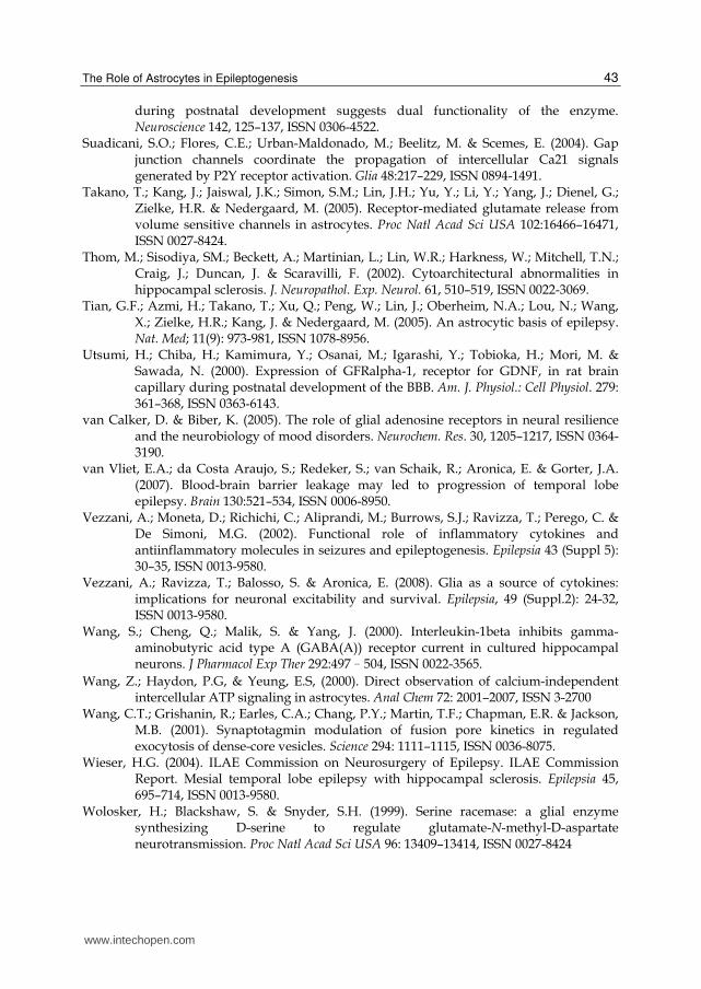
The Role of Astrocytes in Epileptogenesis 43
during postnatal development suggests dual functionality of the enzyme. Neuroscience 142, 125–137, ISSN 0306-4522.
Suadicani, S.O.; Flores, C.E.; Urban-Maldonado, M.; Beelitz, M. & Scemes, E. (2004). Gap junction channels coordinate the propagation of intercellular Ca21 signals generated by P2Y receptor activation. Glia 48:217–229, ISSN 0894-1491.
Takano, T.; Kang, J.; Jaiswal, J.K.; Simon, S.M.; Lin, J.H.; Yu, Y.; Li, Y.; Yang, J.; Dienel, G.; Zielke, H.R. & Nedergaard, M. (2005). Receptor-mediated glutamate release from volume sensitive channels in astrocytes. Proc Natl Acad Sci USA 102:16466–16471, ISSN 0027-8424.
Thom, M.; Sisodiya, SM.; Beckett, A.; Martinian, L.; Lin, W.R.; Harkness, W.; Mitchell, T.N.; Craig, J.; Duncan, J. & Scaravilli, F. (2002). Cytoarchitectural abnormalities in hippocampal sclerosis. J. Neuropathol. Exp. Neurol. 61, 510–519, ISSN 0022-3069.
Tian, G.F.; Azmi, H.; Takano, T.; Xu, Q.; Peng, W.; Lin, J.; Oberheim, N.A.; Lou, N.; Wang, X.; Zielke, H.R.; Kang, J. & Nedergaard, M. (2005). An astrocytic basis of epilepsy. Nat. Med; 11(9): 973-981, ISSN 1078-8956.
Utsumi, H.; Chiba, H.; Kamimura, Y.; Osanai, M.; Igarashi, Y.; Tobioka, H.; Mori, M. & Sawada, N. (2000). Expression of GFRalpha-1, receptor for GDNF, in rat brain capillary during postnatal development of the BBB. Am. J. Physiol.: Cell Physiol. 279: 361–368, ISSN 0363-6143.
van Calker, D. & Biber, K. (2005). The role of glial adenosine receptors in neural resilience and the neurobiology of mood disorders. Neurochem. Res. 30, 1205–1217, ISSN 0364-3190.
van Vliet, E.A.; da Costa Araujo, S.; Redeker, S.; van Schaik, R.; Aronica, E. & Gorter, J.A. (2007). Blood-brain barrier leakage may led to progression of temporal lobe epilepsy. Brain 130:521–534, ISSN 0006-8950.
Vezzani, A.; Moneta, D.; Richichi, C.; Aliprandi, M.; Burrows, S.J.; Ravizza, T.; Perego, C. & De Simoni, M.G. (2002). Functional role of inflammatory cytokines and antiinflammatory molecules in seizures and epileptogenesis. Epilepsia 43 (Suppl 5): 30–35, ISSN 0013-9580.
Vezzani, A.; Ravizza, T.; Balosso, S. & Aronica, E. (2008). Glia as a source of cytokines: implications for neuronal excitability and survival. Epilepsia, 49 (Suppl.2): 24-32, ISSN 0013-9580.
Wang, S.; Cheng, Q.; Malik, S. & Yang, J. (2000). Interleukin-1beta inhibits gamma-aminobutyric acid type A (GABA(A)) receptor current in cultured hippocampal neurons. J Pharmacol Exp Ther 292:497–504, ISSN 0022-3565.
Wang, Z.; Haydon, P.G, & Yeung, E.S, (2000). Direct observation of calcium-independent intercellular ATP signaling in astrocytes. Anal Chem 72: 2001–2007, ISSN 3-2700
Wang, C.T.; Grishanin, R.; Earles, C.A.; Chang, P.Y.; Martin, T.F.; Chapman, E.R. & Jackson, M.B. (2001). Synaptotagmin modulation of fusion pore kinetics in regulated exocytosis of dense-core vesicles. Science 294: 1111–1115, ISSN 0036-8075.
Wieser, H.G. (2004). ILAE Commission on Neurosurgery of Epilepsy. ILAE Commission Report. Mesial temporal lobe epilepsy with hippocampal sclerosis. Epilepsia 45, 695–714, ISSN 0013-9580.
Wolosker, H.; Blackshaw, S. & Snyder, S.H. (1999). Serine racemase: a glial enzyme synthesizing D-serine to regulate glutamate-N-methyl-D-aspartate neurotransmission. Proc Natl Acad Sci USA 96: 13409–13414, ISSN 0027-8424
www.intechopen.com
Underlying Mechanisms of Epilepsy 44
Ye, Z.C.; Wyeth, M.S.; Baltan-Tekkok, S. & Ransom, B.R. (2003). Functional hemichannels in astrocytes: A novel mechanism of glutamate release. J Neurosci 23:3588–3596, ISSN 0270-6474.
Zhang, J.M.; Wang, H.K.; Ye, C.Q.; Ge, W.; Chen, Y.; Jiang, Z.L.; Wum, C.P.; Poo, M.M.; & Duan, S. (2003). ATP released by astrocytes mediates glutamatergic activity-dependent heterosynaptic suppression. Neuron 40: 971–982, ISSN 0896-6273.
Zonta, M.; Angulo, M.C.; Gobbo, S.; Rosengarten, B.; Hossmann, K.A.; Pozzan, T. & Carmignoto, G. (2003). Neuron-to-astrocyte signaling is central to the dynamic control of brain microcirculation. Nat. Neurosci. 6, 43– 50, ISSN 1097-6256.
Zur Nieden, R. & Deitmer, J.W. (2006). The role of metabotropic glutamate receptors for the generation of calcium oscillations in rat hippocampal astrocytes in situ. Cereb Cortex 16:676–687, ISSN 1047-3211.
www.intechopen.com
Underlying Mechanisms of EpilepsyEdited by Prof. Fatima Shad Kaneez
ISBN 978-953-307-765-9Hard cover, 354 pagesPublisher InTechPublished online 26, September, 2011Published in print edition September, 2011
InTech EuropeUniversity Campus STeP Ri Slavka Krautzeka 83/A 51000 Rijeka, Croatia Phone: +385 (51) 770 447 Fax: +385 (51) 686 166www.intechopen.com
InTech ChinaUnit 405, Office Block, Hotel Equatorial Shanghai No.65, Yan An Road (West), Shanghai, 200040, China
Phone: +86-21-62489820 Fax: +86-21-62489821
This book is a very provocative and interesting addition to the literature on Epilepsy. It offers a lot of appealingand stimulating work to offer food of thought to the readers from different disciplines. Around 5% of the totalworld population have seizures but only 0.9% is diagnosed with epilepsy, so it is very important to understandthe differences between seizures and epilepsy, and also to identify the factors responsible for its etiology so asto have more effective therapeutic regime. In this book we have twenty chapters ranging from causes andunderlying mechanisms to the treatment and side effects of epilepsy. This book contains a variety of chapterswhich will stimulate the readers to think about the complex interplay of epigenetics and epilepsy.
How to referenceIn order to correctly reference this scholarly work, feel free to copy and paste the following:
Jesu ́s Pastor and Rafael G. Sola (2011). The Role of Astrocytes in Epileptogenesis, Underlying Mechanisms ofEpilepsy, Prof. Fatima Shad Kaneez (Ed.), ISBN: 978-953-307-765-9, InTech, Available from:http://www.intechopen.com/books/underlying-mechanisms-of-epilepsy/the-role-of-astrocytes-in-epileptogenesis

© 2011 The Author(s). Licensee IntechOpen. This chapter is distributedunder the terms of the Creative Commons Attribution-NonCommercial-ShareAlike-3.0 License, which permits use, distribution and reproduction fornon-commercial purposes, provided the original is properly cited andderivative works building on this content are distributed under the samelicense.