Embed Size (px)
Citation preview
Cellular/Molecular
Taurine Release by Astrocytes Modulates OsmosensitiveGlycine Receptor Tone and Excitability in the AdultSupraoptic Nucleus
Katrina Y. Choe,1 James E. Olson,2 and Charles W. Bourque1
1Center for Research in Neuroscience, Research Institute of the McGill University Health Centre, Montreal General Hospital, Montreal, Quebec, Canada H3G1A4, and 2Department of Emergency Medicine/Department of Neuroscience, Cell Biology, and Physiology, Wright State University, Dayton, Ohio 45435
Cells can release the free amino acid taurine through volume-regulated anion channels (VRACs), and it has been hypothesized thattaurine released from glial cells is capable of inhibiting action potential (AP) firing by activating neuronal glycine receptors (GlyRs)(Hussy et al., 1997). Although an inhibitory GlyR tone is widely observed in the brain, it remains unknown whether this specificallyreflects gliotransmission because most neurons also express VRACs and other endogenous molecules can activate GlyRs. We found thatVRACs are absent in neurons of the rat supraoptic nucleus (SON), suggesting that glial cells are the exclusive source of taurine in thisnucleus. Application of strychnine to rat hypothalamic explants caused a depolarization of SON neurons associated with a decrease ofchloride conductance and could excite these cells in the absence of fast synaptic transmission. This inhibitory GlyR tone was eliminatedby pharmacological blockade of VRACs, by cellular taurine depletion, by metabolic inactivation of glia with fluorocitrate, and afterretraction of astrocytic processes that intercalate neuronal somata and dendrites. Finally, GlyR tone varied inversely with extracellularfluid tonicity to mediate the osmotic control of AP firing by SON neurons. These findings establish taurine as a physiological gliotrans-mitter and show that gliotransmission is a spatially constrained process that can be modulated by the morphological rearrangement ofastrocytes.
IntroductionChanges in extracellular osmolality cause proportional changesin the electrical activity of magnocellular neurosecretory neuronsin the supraoptic nucleus (SON) to regulate the release of vaso-pressin (VP; an antidiuretic hormone) and oxytocin (OT; a na-triuretic hormone in rodents) and thus promote body fluidhomeostasis (Bourque, 2008). Specifically, hyperosmotic condi-tions excite SON neurons to promote VP/OT secretion and re-duce blood osmolality, whereas hypo-osmotic conditions inhibitelectrical activity and basal hormone secretion to elevate osmo-lality. Previous studies have shown that the excitation of SONneurons during hypertonicity is mediated by the intrinsic osmo-sensitivity of these cells (Oliet and Bourque, 1993a) and by excit-atory synaptic inputs from other osmosensitive neurons (Leng etal., 1989). However, recent work has suggested that taurine re-lease by astrocytes might contribute significantly to the inhibition
of SON neurons via the activation of chloride flux through gly-cine receptors (GlyRs) under hypo-osmotic conditions (Hussy etal., 2000). Although this form of signaling could play an impor-tant role in the SON and other brain regions, the existence oftaurinergic gliotransmission remains controversial.
The hypothesis that taurinergic gliotransmission might con-tribute to the osmotic control of SON neurons was developedaround three main observations. First, it is well established thattaurine is a potent agonist at GlyRs (Lynch, 2004), includingthose expressed on SON neurons (Hussy et al., 1997). Second,pharmacological blockade of GlyRs with strychnine can exciteSON neurons during extracellular recordings in vivo, and thiseffect is augmented by lowering plasma osmolality (Hussy et al.,1997). Third, it has been shown that hypotonic solutions stimu-late taurine release from isolated SON in vitro and that this effectis reduced by the glia-specific toxin fluorocitrate (FC) (Deleuze etal., 1998). Although these observations are consistent with theexistence of taurinergic gliotransmission, important factors com-plicate this interpretation. For example, a recent study reportedthat the activation of chloride channels (GABAA receptors) mayhave a depolarizing effect on VP neurons in the rat SON (Haam etal., 2012). If this is the case, the excitatory effects of locally appliedstrychnine observed in this nucleus would have to be caused by ablockade of GlyRs on local interneurons, rather than those on VPneurons themselves. Another factor confounding the taurinergicgliotransmission hypothesis is that two other agonists of GlyRs,namely glycine and �-alanine (Mori et al., 2002; Lynch, 2004), arepresent in the extracellular fluid (Shibanoki et al., 1993), and it is
Received March 20, 2012; revised July 5, 2012; accepted July 13, 2012.Author contributions: K.Y.C. and C.W.B. designed research; K.Y.C. and J.E.O. performed research; K.Y.C. and J.E.O.
analyzed data; K.Y.C. and C.W.B. wrote the paper.This work was supported by Canadian Institutes of Health Research Operating Grants MOP-9939 and MOP-82818
to C.W.B., who was also the recipient of a James McGill Research Chair. The Research Institute of the McGill UniversityHealth Center was supported by the Fonds de la Recherche en Sante du Quebec. We thank Denise Cook and Dr. KeithMurai for providing cultured astrocytes and Jim Leasure for help with the HPLC analysis.
Correspondence should be addressed to Charles W. Bourque, Center for Research in Neuroscience, ResearchInstitute of the McGill University Health Centre, Montreal General Hospital, 1650 Cedar Avenue, Montreal, QC,Canada H3G 1A4. E-mail: [email protected].
DOI:10.1523/JNEUROSCI.1380-12.2012Copyright © 2012 the authors 0270-6474/12/3212518-10$15.00/0
12518 • The Journal of Neuroscience, September 5, 2012 • 32(36):12518 –12527
not known whether GlyRs are functionally activated by taurine orthese other molecules in situ. Finally, it is well known that bothneurons and glial cells can potentially release taurine (Olson andLi, 2000), making it difficult to establish whether any observableGlyR tone is mediated specifically by an agonist released fromglial cells.
In this study, we used a combination of approaches to addressthese issues. Our results provide compelling evidence for the ex-istence of taurinergic GlyR-dependent gliotransmission in the ratSON and reveal that this form of communication plays an im-portant physiological role that relies on a spatially intimate rela-tionship between neurons and surrounding astrocytes.
Materials and MethodsAll procedures involving animals were performed according to protocol1190 approved by the Facility Animal Care Committee of McGillUniversity.
Electrophysiology in superfused hypothalamic explants. Acute hypotha-lamic explants were prepared from adult male Long–Evans rats (80 –160g) as described previously (Ghamari-Langroudi and Bourque, 2001) andsuperfused (�1 ml/min) with warm (31–33°C) oxygenated (95% O2/5%CO2) artificial CSF (ACSF; pH 7.35) comprising (in mM) 104 NaCl, 26NaHCO3, 1.23 NaH2PO4, 3 KCl, 1 MgCl2, 2 CaCl2, 10 D-glucose, andmannitol added to the desired osmolality. Intracellular recordings weremade via pipettes filled with 2 M potassium acetate (100 –130 M�).Steady-state voltage– current ( V–I) relationships were measured in theabsence and presence of 1 �M strychnine, and membrane conductancewas determined as the inverse of the slope of the V–I plot between �70and �60 mV. Reversal potential (EREV) was determined from the inter-section point of V–I plots recorded in different conditions. We believethat the EREV for chloride (ECl) is unaffected by our particular recordingconditions (2 M K acetate, high-resistance sharp electrode, very low hold-ing current). In an attempt to quantify this, we measured time-dependent changes in IPSP reversal potential in a subset of neurons. Nosignificant change was observed between the first and fifth minute(�1.5 � 1.3 mV; n � 3; p � 0.05) or between the 5th and 25th minute(�0.3 � 0.3 mV; n � 3; p � 0.05). Extracellular recordings of AP firingwere made from preparations superfused with ACSF containing 4 mM
KCl and 1 mM CaCl2 using micropipettes with lower resistance (15–20M�) again filled with 2 M potassium acetate, and the voltage signal wasbandpass filtered between 600 and 1500 Hz. Basal activity was deter-mined as the average rate of firing observed during 3 min before appli-cation of strychnine. Changes in firing rate were calculated as thedifference between basal activity and the average rate of discharge duringthe last 30 s of the strychnine application. Neurons with unstable baselineactivity were excluded from the analysis.
Whole-cell patch-clamp recordings in slices. Coronal (transverse) hypo-thalamic slices (300 �m thick) were prepared from adult male Long–Evans rats (80 –160 g) as described previously (Trudel and Bourque,2010) and perfused at 1.5 ml/min (31–33°C) with oxygenated (95%O2/5% CO2) ACSF comprising (in mM) 114 NaCl, 26 NaHCO3, 1.23NaH2PO4, 3 KCl, 1 MgCl2, 2 CaCl2, 10 D-glucose, and 0.01 bicuculline.Whole-cell voltage-clamp recordings from SON neurons were per-formed using pipettes filled with a solution comprising (in mM) 110K-gluconate, 10 HEPES, 10 KCl, and MgCl2 (access resistance, 5.5–17.5M�). Current–voltage analysis was performed on cells held at �60 mVusing a 1 s voltage ramp command applied from �130 to �45 mV.Membrane conductance was determined as the slope of the I–V curve.
Volume-regulated anion channel recording in isolated neurons and glia.Mouse astrocyte cultures were prepared by triturating hippocampi re-moved from anesthetized postnatal day 0 –2 mice in glia growth mediumcomprising MEM supplemented with 1% penicillin, 0.6% glucose, and10% horse serum (Invitrogen). Cultured astrocytes were replated ontoPetri dishes 30 min before recording. Acutely isolated neurons were ob-tained from blocks of SON (�1 mm 3) obtained from anesthetized Long–Evans rats (80 –160 g) and incubated in an oxygenated (100% O2) PIPESsolution containing 20 mM NaCl, 5 mM KCl, 1 mM MgCl2, 20 mM PIPES,1 mM CaCl2, 10 mM D-glucose, pH 7.3, 0.5 mg/ml Protease-X, and 0.5
mg/ml Protease-XIV at room temperature for 30 min. Blocks rinsed inthe enzyme-free PIPES solution were triturated, and the suspension wasplated onto Petri dishes. For whole-cell patch clamp, electrodes (3–5M�) were filled with a solution comprising (in mM) 110 CsCl, 1 MgSO4,10 HEPES, and 1 EGTA; the pH was adjusted to 7.35 with NaOH andosmolality to 280 mOsm/kg with mannitol. Cells were superfused with asolution (pH 7.4) consisting of (in mM) 110 CsCl, 2 CaCl2, 1 MgSO4, and10 HEPES; mannitol was added to 300 mOsm/kg. Hypo-osmotic stimuliwere applied by switching to HEPES lacking mannitol (250 mOsm/kg).
Chemicals and drugs. Strychnine HCl (Sigma-Aldrich) was dissolved indistilled water as a 1 mM stock and kept at 4°C. 4-[(2-butyl-6,7-dichloro-2-cyclopentyl-2,3-dihydro-1-oxo-1 H-inden-5-yl)oxy] (DCPIB; TocrisBioscience) was dissolved in DMSO at 5 mM concentration and kept at�20°C. These stocks were added directly to the ACSF as required for eachexperiment. Taurine (Sigma-Aldrich) was dissolved in ACSF at the concen-tration required. In taurine depletion experiments, guanodinoethane sulfo-nate (GES; Toronto Research Chemicals) was dissolved in ACSF (1 mM). Formetabolic inactivation of glia, ACSF containing 65 �M DL-fluorocitrate wasprepared by precipitation of the barium salt of fluorocitric acid (Sigma-Aldrich). Synaptic blockade was achieved by adding 10 �M (�)-bicucullinemethochloride (Sigma-Aldrich) and 2 mM kynurenic acid (Sigma-Aldrich)to the ACSF.
Immunohistochemistry. Coronal sections (50 �m thick) obtained using avibratome were stained using antibodies against GlyRs (mAb4a mousemonoclonal antibody; Synaptic Systems) diluted 1:200 in PBS. Sections werethen incubated in a fluorescent secondary antibody (Invitrogen; diluted1:500), mounted in SlowFade Gold Antifade reagent (Invitrogen), and ob-served under a confocal laser-scanning microscope (Leica).
Electrophoresis and Western blotting. Equal amounts of SON tissuewere triturated and homogenized in a HEPES-based buffer containingprotease inhibitor (Roche) and 1.0% Triton X-100 (Sigma-Aldrich) andincubated at 4°C for 30 – 60 min. After centrifugation at 13,000 rpm for15 min, protein concentration in the supernatant was measured using theDc protein assay (Bio-Rad) and Ultrospec 2100 pro UV/visible spectro-photometer. Dithiothreitol (DTT) (Sigma-Aldrich) and a loading buffercontaining bromophenyl blue (Sigma-Aldrich) were added to the super-natant and incubated at 80°C for 5 min. Samples were loaded onto aNuPAGE 4 –12% Bis-Tris gel (Invitrogen), and proteins were separatedby electrophoresis at 150 V for 1 h. Proteins were transferred to a PDVFmembrane in transfer buffer (192 mM glycine, 25 mM Tris-HCl, 20%methanol, pH 8.3) at 70 V for 1 h. Membranes were blocked with Trisbuffer containing 1% casein at 4°C overnight and incubated with themAb4a antibody at 1:500 for 1 h and with HRP goat anti-mouse second-ary antibody (1:5000; Cedarlane Laboratories Limited) for 30 min. De-tection was performed using an enhanced chemiluminescence kit(PerkinElmer Life and Analytical Sciences). Membranes were stripped byincubation with 100 mM glycine at pH 2.8 for 30 min and probed withmouse anti-GADPH antibody (1:5000; Abcam) for normalization. Theintensity of the bands was quantified using ImageJ analysis software (ver-sion 1.46; NIH).
Statistics. All values in this study are reported as mean plus or minusthe SEM. Statistical differences between mean values were tested usingStudent’s one-tailed, two-tailed, or paired t test, as appropriate, with SigmaS-tat 2.03 software (SPSS). Comparisons of linear regression parameters wereperformed using Prism 5 software (GraphPad Software). Differences be-tween values were considered to be significant when p � 0.05.
ResultsAlthough our study was designed to examine the regulation of VPneurons, we did not formally identify all of the SON neuronsrecorded as VP or oxytocin secreting. However, 70% of SONneurons in Long–Evans rats are known to express VP, and all ofour recordings were obtained from cells located in the mostventral part of the nucleus, which is composed almost exclu-sively of VP neurons (Rhodes et al., 1981). Many of the cells werecorded from were identified as putative VP-secreting neu-rons because they displayed spontaneous phasic firing (Pou-lain and Wakerley, 1982).
Choe et al. • Taurinergic Gliotransmission in Supraoptic Nucleus J. Neurosci., September 5, 2012 • 32(36):12518 –12527 • 12519
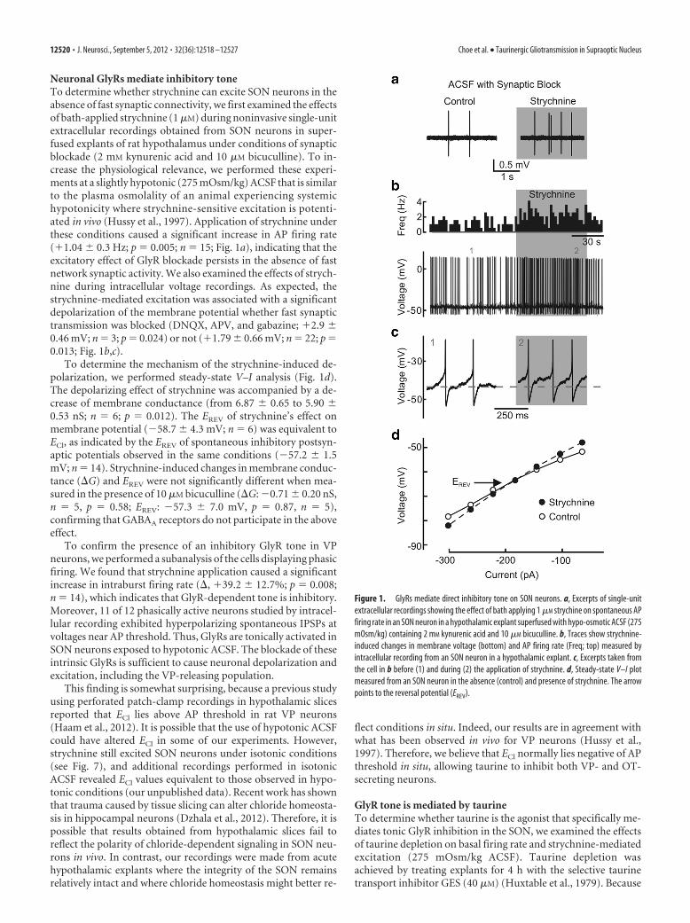
Neuronal GlyRs mediate inhibitory toneTo determine whether strychnine can excite SON neurons in theabsence of fast synaptic connectivity, we first examined the effectsof bath-applied strychnine (1 �M) during noninvasive single-unitextracellular recordings obtained from SON neurons in super-fused explants of rat hypothalamus under conditions of synapticblockade (2 mM kynurenic acid and 10 �M bicuculline). To in-crease the physiological relevance, we performed these experi-ments at a slightly hypotonic (275 mOsm/kg) ACSF that is similarto the plasma osmolality of an animal experiencing systemichypotonicity where strychnine-sensitive excitation is potenti-ated in vivo (Hussy et al., 1997). Application of strychnine underthese conditions caused a significant increase in AP firing rate(�1.04 � 0.3 Hz; p � 0.005; n � 15; Fig. 1a), indicating that theexcitatory effect of GlyR blockade persists in the absence of fastnetwork synaptic activity. We also examined the effects of strych-nine during intracellular voltage recordings. As expected, thestrychnine-mediated excitation was associated with a significantdepolarization of the membrane potential whether fast synaptictransmission was blocked (DNQX, APV, and gabazine; �2.9 �0.46 mV; n � 3; p � 0.024) or not (�1.79 � 0.66 mV; n � 22; p �0.013; Fig. 1b,c).
To determine the mechanism of the strychnine-induced de-polarization, we performed steady-state V–I analysis (Fig. 1d).The depolarizing effect of strychnine was accompanied by a de-crease of membrane conductance (from 6.87 � 0.65 to 5.90 �0.53 nS; n � 6; p � 0.012). The EREV of strychnine’s effect onmembrane potential (�58.7 � 4.3 mV; n � 6) was equivalent toECl, as indicated by the EREV of spontaneous inhibitory postsyn-aptic potentials observed in the same conditions (�57.2 � 1.5mV; n � 14). Strychnine-induced changes in membrane conduc-tance (G) and EREV were not significantly different when mea-sured in the presence of 10 �M bicuculline (G: �0.71 � 0.20 nS,n � 5, p � 0.58; EREV: �57.3 � 7.0 mV, p � 0.87, n � 5),confirming that GABAA receptors do not participate in the aboveeffect.
To confirm the presence of an inhibitory GlyR tone in VPneurons, we performed a subanalysis of the cells displaying phasicfiring. We found that strychnine application caused a significantincrease in intraburst firing rate (, �39.2 � 12.7%; p � 0.008;n � 14), which indicates that GlyR-dependent tone is inhibitory.Moreover, 11 of 12 phasically active neurons studied by intracel-lular recording exhibited hyperpolarizing spontaneous IPSPs atvoltages near AP threshold. Thus, GlyRs are tonically activated inSON neurons exposed to hypotonic ACSF. The blockade of theseintrinsic GlyRs is sufficient to cause neuronal depolarization andexcitation, including the VP-releasing population.
This finding is somewhat surprising, because a previous studyusing perforated patch-clamp recordings in hypothalamic slicesreported that ECl lies above AP threshold in rat VP neurons(Haam et al., 2012). It is possible that the use of hypotonic ACSFcould have altered ECl in some of our experiments. However,strychnine still excited SON neurons under isotonic conditions(see Fig. 7), and additional recordings performed in isotonicACSF revealed ECl values equivalent to those observed in hypo-tonic conditions (our unpublished data). Recent work has shownthat trauma caused by tissue slicing can alter chloride homeosta-sis in hippocampal neurons (Dzhala et al., 2012). Therefore, it ispossible that results obtained from hypothalamic slices fail toreflect the polarity of chloride-dependent signaling in SON neu-rons in vivo. In contrast, our recordings were made from acutehypothalamic explants where the integrity of the SON remainsrelatively intact and where chloride homeostasis might better re-
flect conditions in situ. Indeed, our results are in agreement withwhat has been observed in vivo for VP neurons (Hussy et al.,1997). Therefore, we believe that ECl normally lies negative of APthreshold in situ, allowing taurine to inhibit both VP- and OT-secreting neurons.
GlyR tone is mediated by taurineTo determine whether taurine is the agonist that specifically me-diates tonic GlyR inhibition in the SON, we examined the effectsof taurine depletion on basal firing rate and strychnine-mediatedexcitation (275 mOsm/kg ACSF). Taurine depletion wasachieved by treating explants for 4 h with the selective taurinetransport inhibitor GES (40 �M) (Huxtable et al., 1979). Because
Figure 1. GlyRs mediate direct inhibitory tone on SON neurons. a, Excerpts of single-unitextracellular recordings showing the effect of bath applying 1 �M strychine on spontaneous APfiring rate in an SON neuron in a hypothalamic explant superfused with hypo-osmotic ACSF (275mOsm/kg) containing 2 mM kynurenic acid and 10 �M bicuculline. b, Traces show strychnine-induced changes in membrane voltage (bottom) and AP firing rate (Freq; top) measured byintracellular recording from an SON neuron in a hypothalamic explant. c, Excerpts taken fromthe cell in b before (1) and during (2) the application of strychnine. d, Steady-state V–I plotmeasured from an SON neuron in the absence (control) and presence of strychnine. The arrowpoints to the reversal potential (EREV).
12520 • J. Neurosci., September 5, 2012 • 32(36):12518 –12527 Choe et al. • Taurinergic Gliotransmission in Supraoptic Nucleus
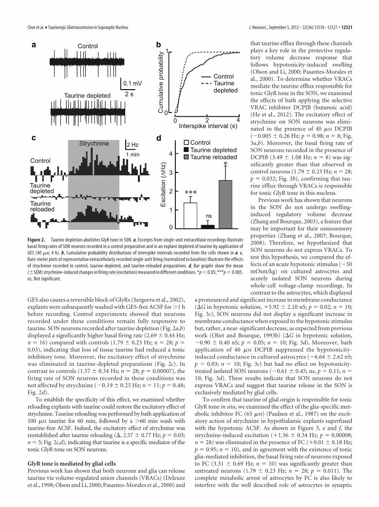
GES also causes a reversible block of GlyRs (Sergeeva et al., 2002),explants were subsequently washed with GES-free ACSF for �1 hbefore recording. Control experiments showed that neuronsrecorded under these conditions remain fully responsive totaurine. SON neurons recorded after taurine depletion (Fig. 2a,b)displayed a significantly higher basal firing rate (2.69 � 0.44 Hz;n � 16) compared with controls (1.79 � 0.23 Hz; n � 28; p �0.03), indicating that loss of tissue taurine had reduced a tonicinhibitory tone. Moreover, the excitatory effect of strychninewas eliminated in taurine-depleted preparations (Fig. 2c). Incontrast to controls (1.57 � 0.34 Hz; n � 28; p � 0.00007), thefiring rate of SON neurons recorded in these conditions wasnot affected by strychnine (�0.19 � 0.25 Hz; n � 11; p � 0.48;Fig. 2d).
To establish the specificity of this effect, we examined whetherreloading explants with taurine could restore the excitatory effect ofstrychnine. Taurine reloading was performed by bath application of100 �M taurine for 60 min, followed by a �60 min wash withtaurine-free ACSF. Indeed, the excitatory effect of strychnine wasreestablished after taurine reloading (, 2.57 � 0.77 Hz; p � 0.03;n � 5; Fig. 2c,d), indicating that taurine is a specific mediator of thetonic GlyR tone on SON neurons.
GlyR tone is mediated by glial cellsPrevious work has shown that both neurons and glia can releasetaurine via volume-regulated anion channels (VRACs) (Deleuzeet al., 1998; Olson and Li, 2000; Pasantes-Morales et al., 2000) and
that taurine efflux through these channelsplays a key role in the protective regula-tory volume decrease response thatfollows hypotonicity-induced swelling(Olson and Li, 2000; Pasantes-Morales etal., 2000). To determine whether VRACsmediate the taurine efflux responsible fortonic GlyR tone in the SON, we examinedthe effects of bath applying the selectiveVRAC inhibitor DCPIB (butanoic acid)(He et al., 2012). The excitatory effect ofstrychnine on SON neurons was elimi-nated in the presence of 40 �M DCPIB(�0.005 � 0.26 Hz; p � 0.98; n � 8; Fig.3a,b). Moreover, the basal firing rate ofSON neurons recorded in the presence ofDCPIB (3.49 � 1.08 Hz; n � 8) was sig-nificantly greater than that observed incontrol neurons (1.79 � 0.23 Hz; n � 28;p � 0.032; Fig. 3b), confirming that tau-rine efflux through VRACs is responsiblefor tonic GlyR tone in this nucleus.
Previous work has shown that neuronsin the SON do not undergo swelling-induced regulatory volume decrease(Zhang and Bourque, 2003), a feature thatmay be important for their osmosensoryproperties (Zhang et al., 2007; Bourque,2008). Therefore, we hypothesized thatSON neurons do not express VRACs. Totest this hypothesis, we compared the ef-fects of an acute hypotonic stimulus (�50mOsm/kg) on cultured astrocytes andacutely isolated SON neurons duringwhole-cell voltage-clamp recordings. Incontrast to the astrocytes, which displayed
a pronounced and significant increase in membrane conductance(G in hypotonic solution, �5.92 � 2.10 nS; p � 0.02; n � 10;Fig. 3c), SON neurons did not display a significant increase inmembrane conductance when exposed to the hypotonic stimulusbut, rather, a near-significant decrease, as expected from previouswork (Oliet and Bourque, 1993b) (G in hypotonic solution,�0.90 � 0.40 nS; p � 0.05; n � 10; Fig. 3d). Moreover, bathapplication of 40 �M DCPIB suppressed the hypotonicity-induced conductance in cultured astrocytes (�6.64 � 2.62 nS;p � 0.03; n � 10; Fig. 3c) but had no effect on hypotonicity-treated isolated SON neurons (�0.61 � 0.45; ns, p � 0.11; n �10; Fig. 3d). These results indicate that SON neurons do notexpress VRACs and suggest that taurine release in the SON isexclusively mediated by glial cells.
To confirm that taurine of glial origin is responsible for tonicGlyR tone in situ, we examined the effect of the glia-specific met-abolic inhibitor FC (65 �M) (Paulsen et al., 1987) on the excit-atory action of strychnine in hypothalamic explants superfusedwith the hypotonic ACSF. As shown in Figure 3, e and f, thestrychnine-induced excitation (�1.56 � 0.34 Hz; p � 0.00008;n � 28) was eliminated in the presence of FC (�0.01 � 0.18 Hz;p � 0.95; n � 10), and in agreement with the existence of tonicglia-mediated inhibition, the basal firing rate of neurons exposedto FC (3.31 � 0.69 Hz; n � 10) was significantly greater thanuntreated neurons (1.79 � 0.23 Hz; n � 28; p � 0.011). Thecomplete metabolic arrest of astrocytes by FC is also likely tointerfere with the well described role of astrocytes in synaptic
Figure 2. Taurine depletion abolishes GlyR tone in SON. a, Excerpts from single-unit extracellular recordings illustratebasal firing rates of SON neurons recorded in a control preparation and in an explant depleted of taurine by application ofGES (40 �M; 4 h). b, Cumulative probability distributions of interspike intervals recorded from the cells shown in a. c,Rate-meter plots of representative extracellularly recorded single-unit firing (normalized to baseline) illustrate the effectsof strychnine recorded in control, taurine-depleted, and taurine-reloaded preparations. d, Bar graphs show the mean(�SEM) strychnine-induced changes in firing rate (excitation) measured in different conditions. *p � 0.05; ***p � 0.005.ns, Not significant.
Choe et al. • Taurinergic Gliotransmission in Supraoptic Nucleus J. Neurosci., September 5, 2012 • 32(36):12518 –12527 • 12521
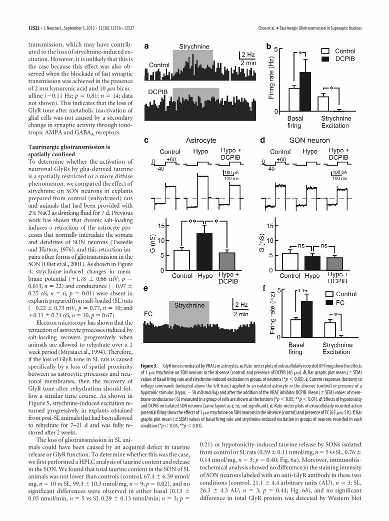
transmission, which may have contrib-uted to the loss of strychnine-induced ex-citation. However, it is unlikely that this isthe case because this effect was also ob-served when the blockade of fast synaptictransmission was achieved in the presenceof 2 mM kynurenic acid and 10 �M bicuc-ulline (�0.11 Hz; p � 0.81; n � 14; datanot shown). This indicates that the loss ofGlyR tone after metabolic inactivation ofglial cells was not caused by a secondarychange in synaptic activity through iono-tropic AMPA and GABAA receptors.
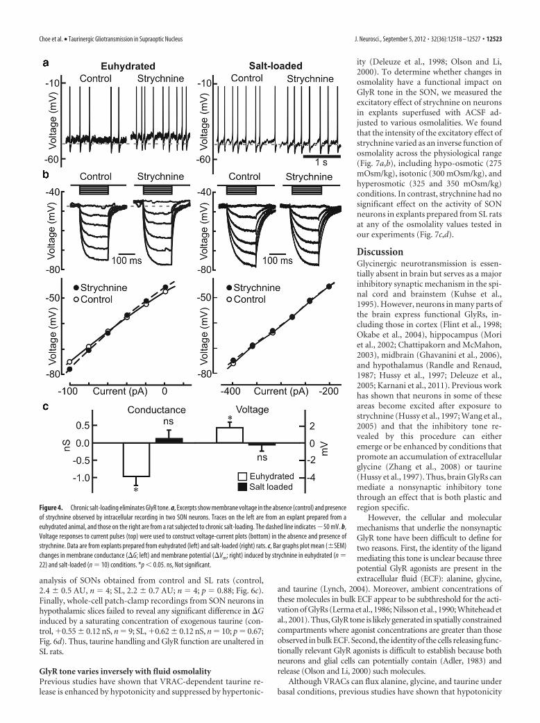
Taurinergic gliotransmission isspatially confinedTo determine whether the activation ofneuronal GlyRs by glia-derived taurineis a spatially restricted or a more diffusephenomenon, we compared the effect ofstrychnine on SON neurons in explantsprepared from control (euhydrated) ratsand animals that had been provided with2% NaCl as drinking fluid for 7 d. Previouswork has shown that chronic salt-loadinginduces a retraction of the astrocyte pro-cesses that normally intercalate the somataand dendrites of SON neurons (Tweedleand Hatton, 1976), and this retraction im-pairs other forms of gliotransmission in theSON (Oliet et al., 2001). As shown in Figure4, strychnine-induced changes in mem-brane potential (�1.78 � 0.66 mV; p �0.013; n � 22) and conductance (�0.97 �0.25 nS; n � 6; p � 0.01) were absent inexplants prepared from salt-loaded (SL) rats(�0.22 � 0.73 mV, p � 0.77, n � 10; and�0.11 � 0.24 nS, n � 10, p � 0.67).
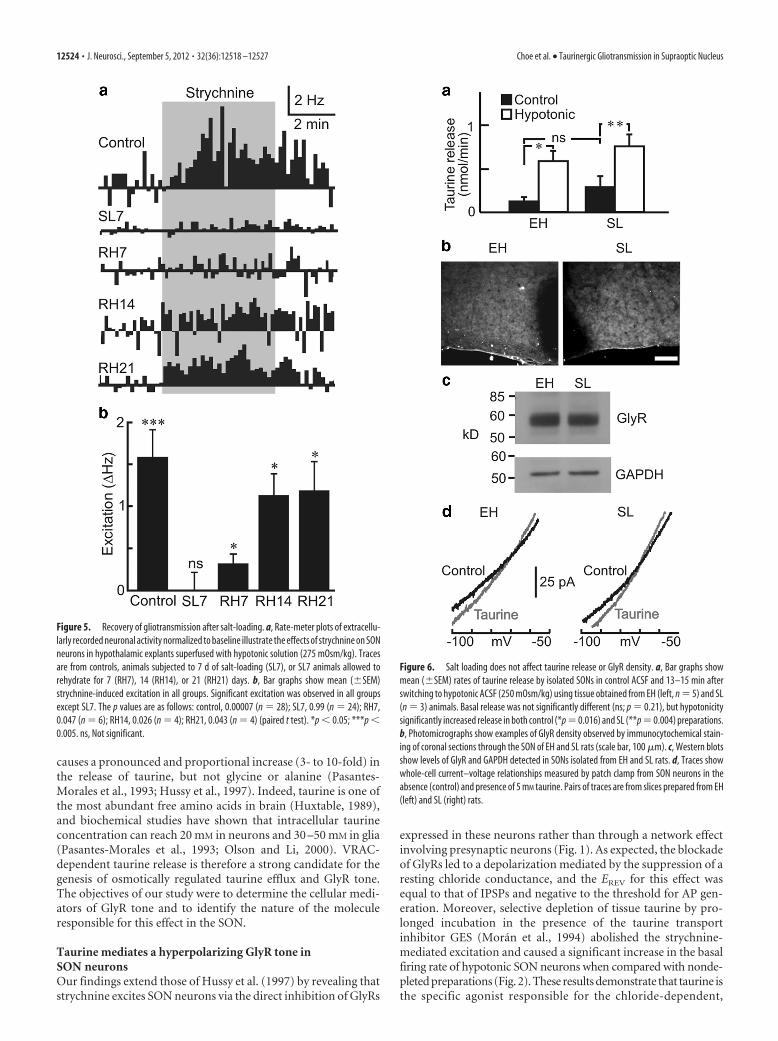
Electron microscopy has shown that theretraction of astrocyte processes induced bysalt-loading recovers progressively whenanimals are allowed to rehydrate over a 2week period (Miyata et al., 1994). Therefore,if the loss of GlyR tone in SL rats is causedspecifically by a loss of spatial proximitybetween as astrocytic processes and neu-ronal membranes, then the recovery ofGlyR tone after rehydration should fol-low a similar time course. As shown inFigure 5, strychnine-induced excitation re-turned progressively in explants obtainedfrom post-SL animals that had been allowedto rehydrate for 7–21 d and was fully re-stored after 2 weeks.
The loss of gliotransmission in SL ani-mals could have been caused by an acquired defect in taurinerelease or GlyR function. To determine whether this was the case,we first performed a HPLC analysis of taurine content and releasein the SON. We found that total taurine content in the SON of SLanimals was not lower than controls (control, 67.4 � 6.39 nmol/mg, n � 10 vs SL, 99.3 � 10.3 nmol/mg, n � 9; p � 0.02), and nosignificant differences were observed in either basal (0.13 �0.05 nmol/min, n � 5 vs SL 0.29 � 0.13 nmol/min; n � 3; p �
0.21) or hypotonicity-induced taurine release by SONs isolatedfrom control or SL rats (0.59 � 0.11 nmol/mg, n � 5 vs SL, 0.76 �0.14 nmol/mg, n � 3; p � 0.40; Fig. 6a). Moreover, immunohis-tochemical analysis showed no difference in the staining intensityof SON neurons labeled with an anti-GlyR antibody in these twoconditions [control, 21.1 � 4.4 arbitrary units (AU), n � 3; SL,26.3 � 4.3 AU, n � 3; p � 0.44; Fig. 6b), and no significantdifference in total GlyR protein was detected by Western blot
Figure 3. GlyR tone is mediated by VRACs in astrocytes. a, Rate-meter plots of extracellularly recorded AP firing show the effectsof 1 �M strychnine on SON neurons in the absence (control) and presence of DCPIB (40 �M). b, Bar graphs plot mean (�SEM)values of basal firing rate and strychnine-induced excitation in groups of neurons (*p � 0.05). c, Current responses (bottom) tovoltage commands (indicated above the left trace) applied to an isolated astrocyte in the absence (control) or presence of ahypotonic stimulus (Hypo; �50 mOsmol/kg) and after the addition of the VRAC inhibitor DCPIB. Mean (�SEM) values of mem-brane conductance ( G) measured in a group of cells are shown at the bottom (*p � 0.05; **p � 0.01). d, Effects of hypotonicityand DCPIB on isolated SON neurons (same layout as c; ns, not significant). e, Rate-meter plots of extracellularly recorded actionpotential firing show the effects of 1 �M strychnine on SON neurons in the absence (control) and presence of FC (65 �M; 5 h). f, Bargraphs plot mean (�SEM) values of basal firing rate and strychnine-induced excitation in groups of neurons recorded in eachcondition (*p � 0.05; **p � 0.01).
12522 • J. Neurosci., September 5, 2012 • 32(36):12518 –12527 Choe et al. • Taurinergic Gliotransmission in Supraoptic Nucleus
analysis of SONs obtained from control and SL rats (control,2.4 � 0.5 AU, n � 4; SL, 2.2 � 0.7 AU; n � 4; p � 0.88; Fig. 6c).Finally, whole-cell patch-clamp recordings from SON neurons inhypothalamic slices failed to reveal any significant difference in Ginduced by a saturating concentration of exogenous taurine (con-trol, �0.55 � 0.12 nS, n � 9; SL, �0.62 � 0.12 nS, n � 10; p � 0.67;Fig. 6d). Thus, taurine handling and GlyR function are unaltered inSL rats.
GlyR tone varies inversely with fluid osmolalityPrevious studies have shown that VRAC-dependent taurine re-lease is enhanced by hypotonicity and suppressed by hypertonic-
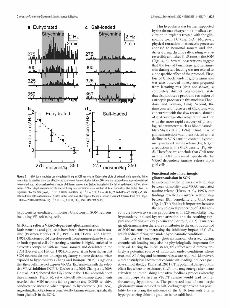
ity (Deleuze et al., 1998; Olson and Li,2000). To determine whether changes inosmolality have a functional impact onGlyR tone in the SON, we measured theexcitatory effect of strychnine on neuronsin explants superfused with ACSF ad-justed to various osmolalities. We foundthat the intensity of the excitatory effect ofstrychnine varied as an inverse function ofosmolality across the physiological range(Fig. 7a,b), including hypo-osmotic (275mOsm/kg), isotonic (300 mOsm/kg), andhyperosmotic (325 and 350 mOsm/kg)conditions. In contrast, strychnine had nosignificant effect on the activity of SONneurons in explants prepared from SL ratsat any of the osmolality values tested inour experiments (Fig. 7c,d).
DiscussionGlycinergic neurotransmission is essen-tially absent in brain but serves as a majorinhibitory synaptic mechanism in the spi-nal cord and brainstem (Kuhse et al.,1995). However, neurons in many parts ofthe brain express functional GlyRs, in-cluding those in cortex (Flint et al., 1998;Okabe et al., 2004), hippocampus (Moriet al., 2002; Chattipakorn and McMahon,2003), midbrain (Ghavanini et al., 2006),and hypothalamus (Randle and Renaud,1987; Hussy et al., 1997; Deleuze et al.,2005; Karnani et al., 2011). Previous workhas shown that neurons in some of theseareas become excited after exposure tostrychnine (Hussy et al., 1997; Wang et al.,2005) and that the inhibitory tone re-vealed by this procedure can eitheremerge or be enhanced by conditions thatpromote an accumulation of extracellularglycine (Zhang et al., 2008) or taurine(Hussy et al., 1997). Thus, brain GlyRs canmediate a nonsynaptic inhibitory tonethrough an effect that is both plastic andregion specific.
However, the cellular and molecularmechanisms that underlie the nonsynapticGlyR tone have been difficult to define fortwo reasons. First, the identity of the ligandmediating this tone is unclear because threepotential GlyR agonists are present in theextracellular fluid (ECF): alanine, glycine,
and taurine (Lynch, 2004). Moreover, ambient concentrations ofthese molecules in bulk ECF appear to be subthreshold for the acti-vation of GlyRs (Lerma et al., 1986; Nilsson et al., 1990; Whitehead etal., 2001). Thus, GlyR tone is likely generated in spatially constrainedcompartments where agonist concentrations are greater than thoseobserved in bulk ECF. Second, the identity of the cells releasing func-tionally relevant GlyR agonists is difficult to establish because bothneurons and glial cells can potentially contain (Adler, 1983) andrelease (Olson and Li, 2000) such molecules.
Although VRACs can flux alanine, glycine, and taurine underbasal conditions, previous studies have shown that hypotonicity
Figure 4. Chronic salt-loading eliminates GlyR tone. a, Excerpts show membrane voltage in the absence (control) and presenceof strychnine observed by intracellular recording in two SON neurons. Traces on the left are from an explant prepared from aeuhydrated animal, and those on the right are from a rat subjected to chronic salt-loading. The dashed line indicates �50 mV. b,Voltage responses to current pulses (top) were used to construct voltage-current plots (bottom) in the absence and presence ofstrychnine. Data are from explants prepared from euhydrated (left) and salt-loaded (right) rats. c, Bar graphs plot mean (�SEM)changes in membrane conductance (G; left) and membrane potential (Vm; right) induced by strychnine in euhydrated (n �22) and salt-loaded (n � 10) conditions. *p � 0.05. ns, Not significant.
Choe et al. • Taurinergic Gliotransmission in Supraoptic Nucleus J. Neurosci., September 5, 2012 • 32(36):12518 –12527 • 12523
causes a pronounced and proportional increase (3- to 10-fold) inthe release of taurine, but not glycine or alanine (Pasantes-Morales et al., 1993; Hussy et al., 1997). Indeed, taurine is one ofthe most abundant free amino acids in brain (Huxtable, 1989),and biochemical studies have shown that intracellular taurineconcentration can reach 20 mM in neurons and 30 –50 mM in glia(Pasantes-Morales et al., 1993; Olson and Li, 2000). VRAC-dependent taurine release is therefore a strong candidate for thegenesis of osmotically regulated taurine efflux and GlyR tone.The objectives of our study were to determine the cellular medi-ators of GlyR tone and to identify the nature of the moleculeresponsible for this effect in the SON.
Taurine mediates a hyperpolarizing GlyR tone inSON neuronsOur findings extend those of Hussy et al. (1997) by revealing thatstrychnine excites SON neurons via the direct inhibition of GlyRs
expressed in these neurons rather than through a network effectinvolving presynaptic neurons (Fig. 1). As expected, the blockadeof GlyRs led to a depolarization mediated by the suppression of aresting chloride conductance, and the EREV for this effect wasequal to that of IPSPs and negative to the threshold for AP gen-eration. Moreover, selective depletion of tissue taurine by pro-longed incubation in the presence of the taurine transportinhibitor GES (Moran et al., 1994) abolished the strychnine-mediated excitation and caused a significant increase in the basalfiring rate of hypotonic SON neurons when compared with nonde-pleted preparations (Fig. 2). These results demonstrate that taurine isthe specific agonist responsible for the chloride-dependent,
Figure 5. Recovery of gliotransmission after salt-loading. a, Rate-meter plots of extracellu-larly recorded neuronal activity normalized to baseline illustrate the effects of strychnine on SONneurons in hypothalamic explants superfused with hypotonic solution (275 mOsm/kg). Tracesare from controls, animals subjected to 7 d of salt-loading (SL7), or SL7 animals allowed torehydrate for 7 (RH7), 14 (RH14), or 21 (RH21) days. b, Bar graphs show mean (�SEM)strychnine-induced excitation in all groups. Significant excitation was observed in all groupsexcept SL7. The p values are as follows: control, 0.00007 (n � 28); SL7, 0.99 (n � 24); RH7,0.047 (n � 6); RH14, 0.026 (n � 4); RH21, 0.043 (n � 4) (paired t test). *p � 0.05; ***p �0.005. ns, Not significant.
Figure 6. Salt loading does not affect taurine release or GlyR density. a, Bar graphs showmean (�SEM) rates of taurine release by isolated SONs in control ACSF and 13–15 min afterswitching to hypotonic ACSF (250 mOsm/kg) using tissue obtained from EH (left, n � 5) and SL(n � 3) animals. Basal release was not significantly different (ns; p � 0.21), but hypotonicitysignificantly increased release in both control (*p � 0.016) and SL (**p � 0.004) preparations.b, Photomicrographs show examples of GlyR density observed by immunocytochemical stain-ing of coronal sections through the SON of EH and SL rats (scale bar, 100 �m). c, Western blotsshow levels of GlyR and GAPDH detected in SONs isolated from EH and SL rats. d, Traces showwhole-cell current–voltage relationships measured by patch clamp from SON neurons in theabsence (control) and presence of 5 mM taurine. Pairs of traces are from slices prepared from EH(left) and SL (right) rats.
12524 • J. Neurosci., September 5, 2012 • 32(36):12518 –12527 Choe et al. • Taurinergic Gliotransmission in Supraoptic Nucleus
hypotonicity-mediated inhibitory GlyR tone in SON neurons,including VP-releasing cells.
GlyR tone reflects VRAC-dependent gliotransmissionBoth neurons and glial cells have been shown to contain tau-rine (Pasantes-Morales et al., 1993, 2000; Decavel and Hatton,1995). GlyR tone could therefore result from taurine release by eitheror both types of cells. Interestingly, taurine is highly enriched inastrocytes compared with neuronal somata and dendrites in theSON (Decavel and Hatton, 1995). Moreover, it has been shown thatSON neurons do not undergo regulatory volume decrease whenexposed to hypotonicity (Zhang and Bourque, 2003), suggestingthat these cells may not express VRACs. Experiments with the selec-tive VRAC inhibitor DCPIB (Decher et al., 2001; Zhang et al., 2008;He et al., 2012) showed that GlyR tone in the SON is dependent onthese channels (Fig. 3a,b), yet whole-cell patch-clamp experimentsrevealed that SON neurons fail to generate any DCPIB-sensitiveconductance increase when exposed to hypotonicity (Fig. 3c,d),suggesting that GlyR tone is generated by taurine released specificallyfrom glial cells in the SON.
This hypothesis was further supportedby the absence of strychnine-mediated ex-citation in explants treated with the glia-specific toxin FC (Fig. 3e,f). Moreover,physical retraction of astrocytic processesapposed to neuronal somata and den-drites during chronic salt-loading in vivoreversibly abolished GlyR tone in the SON(Figs. 4, 5). Several observations suggestthat the loss of taurinergic gliotransmis-sion during salt-loading was not related toa nonspecific effect of the protocol. First,loss of GlyR-dependent gliotransmissionwas also observed in explants preparedfrom lactating rats (data not shown), acompletely distinct physiological statethat also induces a profound retraction ofastrocytic processes in this nucleus (Theo-dosis and Poulain, 1984). Second, thetime course of recovery of GlyR tone wasconcurrent with the slow reestablishmentof glial coverage after rehydration and notwith the more rapid recovery of physio-logical parameters such as blood osmola-lity (Miyata et al., 1994). Third, loss ofgliotransmission was not associated with adecline in SON taurine content, hypoto-nicity-induced taurine release (Fig. 6a), ora reduction in the GlyR density (Fig. 6b–d). Therefore, we conclude that GlyR tonein the SON is caused specifically byVRAC-dependent taurine release fromglial cells.
Functional role of taurinergicgliotransmission in SONIn agreement with the inverse relationshipbetween osmolality and VRAC-mediatedtaurine release (Hussy et al., 1997), ourfindings revealed an inverse relationshipbetween ECF osmolality and GlyR tone(Fig. 7). This finding is important becausethe physiological properties of SON neu-
rons are known to vary in proportion with ECF osmolality, i.e.,hypotonicity-induced hyperpolarization and the resulting sup-pression of firing activity (Voisin and Bourque, 2002). Tauriner-gic gliotransmission therefore contributes to the osmotic controlof SON neurons by increasing the inhibitory impact of GlyRs,which reduces firing rate under hypo-osmotic conditions.
The loss of taurinergic gliotransmission observed duringchronic salt-loading may also be physiologically important forsurvival. During the initial stages, this effect would remove en-tirely a potential source of inhibition under conditions wheremaximal AP firing and hormone release are required. Moreover,a recent study has shown that chronic salt-loading induces a pos-itive shift of the ECl (Kim et al., 2011). The potential danger of thiseffect lies where an excitatory GlyR tone may emerge after acuterehydration, establishing a positive feedback process wherebyan inappropriate rise in VP/OT release would lead to life-threatening hyponatremia. The protracted loss of taurinergicgliotransmission induced by salt-loading may prevent this possi-bility by restoring the influence of the GlyR tone only after ahyperpolarizing chloride gradient is reestablished.
Figure 7. GlyR tone mediates osmoregulated firing in SON neurons. a, Rate-meter plots of extracellularly recorded firingnormalized to baseline show the effects of strychnine on the electrical activity of SON neurons recorded from explants obtainedfrom euhydrated rats superfused with media of different osmolalities (values indicated at the left of each trace). b, Plots showmean (�SEM) strychnine-induced changes in firing rate (excitation) as a function of ACSF osmolality. The dashed line is aregression fit of the data (slope, �0.021 � 0.007 Hz/mOsm � kg �1; p � 0.0012; n � 28, 17, 22, and 4 for each point). c, d, Dataobtained from salt-loaded animals treated in the same way. The slope of the regression in d was not different from zero (slope,�0.0024 � 0.05 Hz/mOsm � kg �1; p � 0.57; n � 24, 14, 7, and 13 for each point).
Choe et al. • Taurinergic Gliotransmission in Supraoptic Nucleus J. Neurosci., September 5, 2012 • 32(36):12518 –12527 • 12525
Possible role of taurinergic gliotransmission in otherbrain regionsSince extrasynaptic GlyRs are expressed in many types of neu-rons, our results raise the possibility that taurinergic gliotrans-mission may also play a role in other parts of the brain. Indeed,taurine has been suggested to play a protective role during brainpathologies associated with swelling, such as ischemia (Benesovaet al., 2009) and hyponatremia (Chvatal et al., 2007). In addition,neuronal hyperactivity can significantly increase extracellular[K�] (Gutnick et al., 1979), and elevated levels of extracellular[K�] can promote swelling-induced taurine release by surround-ing glial cells (Pasantes-Morales and Schousboe, 1989). Tauriner-gic gliotransmission could therefore mediate feedback inhibitionof neurons under hyperexcitable states associated with seizuresand epilepsy. Indeed, taurine’s protective role against epilepticactivity is well documented (Pasantes-Morales et al., 1987).
Finally, the loss of GlyR tone after salt-loading indicates that aclose spatial proximity between VRAC-containing astrocyticprocesses and neuronal membranes expressing GlyRs is requiredfor this process. Indeed, taurine is efficiently transported fromthe extracellular compartment to the intracellular compartmentvia specific transporters (Huxtable, 1989). Therefore, we specu-late that glial VRACs and neuronal GlyRs are normally locatedwithin local cell– cell “microdomains” where levels of extracellu-lar taurine are sufficient for receptor activation. If this is the case,then it is possible that the retraction of astrocytic processes dis-rupts this relationship and that the spatial mismatch betweentaurine release and GlyRs prevents their activation because of theeffect of distributed taurine uptake on bulk extracellular taurinelevels. Interestingly, morphological astrocyte plasticity has beenshown to occur in other parts of the brain [e.g., suprachiasmaticnucleus (Becquet et al., 2008), brain stem (Hirrlinger et al., 2004),and cortex (Jones et al., 1996)], and recent imaging studies haveshown that astrocytic processes are highly motile and capable ofextending and retracting on a time scale of minutes (Haber et al.,2006). Therefore, it is possible that taurinergic gliotransmission isa process that can be regulated dynamically in response tochanges in astrocyte morphology.
ReferencesAdler R (1983) Taurine uptake by chick embryo retinal neurons and glial
cells in purified culture. J Neurosci Res 10:369 –379.Becquet D, Girardet C, Guillaumond F, Francois-Bellan AM, Bosler O
(2008) Ultrastructural plasticity in the rat suprachiasmatic nucleus. Pos-sible involvement in clock entrainment. Glia 56:294 –305.
Benesova J, Hock M, Butenko O, Prajerova I, Anderova M, Chvatal A (2009)Quantification of astrocyte volume changes during ischemia in situ re-veals two populations of astrocytes in the cortex of GFAP/EGFP mice.J Neurosci Res 87:96 –111.
Bourque CW (2008) Central mechanisms of osmosensation and systemicosmoregulation. Nat Rev Neurosci 9:519 –531.
Chattipakorn SC, McMahon LL (2003) Strychnine-sensitive glycine recep-tors depress hyperexcitability in rat dentate gyrus. J Neurophysiol89:1339 –1342.
Chvatal A, Anderova M, Hock M, Prajerova I, Neprasova H, Chvatal V,Kirchhoff F, Sykova E (2007) Three-dimensional confocal morphome-try reveals structural changes in astrocyte morphology in situ. J NeurosciRes 85:260 –271.
Decavel C, Hatton GI (1995) Taurine immunoreactivity in the rat su-praoptic nucleus: prominent localization in glial cells. J Comp Neurol354:13–26.
Decher N, Lang HJ, Nilius B, BruggemannA, Busch AE, Steinmeyer K (2001)DCPIB is a novel selective blocker of I(Cl,swell) and prevents swelling-induced shortening of guinea-pig atrial action potential duration. Br JPharmacol 134:1467–1479.
Deleuze C, Duvoid A, Hussy N (1998) Properties and glial origin of
osmotic-dependent release of taurine from the rat supraoptic nucleus.J Physiol 507:463– 471.
Deleuze C, Alonso G, Lefevre IA, Duvoid-Guillou A, Hussy N (2005) Extra-synaptic localization of glycine receptors in the rat supraoptic nucleus:further evidence for their involvement in glia-to-neuron communication.Neuroscience 133:175–183.
Dzhala V, Valeeva G, Glykys J, Khazipov R, Staley K (2012) Traumatic al-terations in GABA signaling disrupt hippocampal network activity in thedeveloping brain. J Neurosci 32:4017– 4031.
Flint AC, Liu X, Kriegstein AR (1998) Nonsynaptic glycine receptor activa-tion during early neocortical development. Neuron 20:43–53.
Ghamari-Langroudi M, Bourque CW (2001) Ionic basis of the caesium-induced depolarisation in rat supraoptic nucleus neurones. J Physiol536:797– 808.
Ghavanini AA, Mathers DA, Kim HS, Puil E (2006) Distinctive glyciner-gic currents with fast and slow kinetics in thalamus. J Neurophysiol95:3438 –3448.
Gutnick MJ, Heinemann U, Lux HD (1979) Stimulus induced and seizurerelated changes in extracellular potassium concentration in cat thalamus(VPL). Electroencephalogr Clin Neurophysiol 47:329 –344.
Haam J, Popescu IR, Morton LA, Halmos KC, Teruyama R, Ueta Y, Tasker JG(2012) GABA is excitatory in adult vasopressinergic neuroendocrinecells. J Neurosci 32:572–582.
Haber M, Zhou L, Murai KK (2006) Cooperative astrocyte and dendriticspine dynamics at hippocampal excitatory synapses. J Neurosci26:8881– 8891.
He D, Luo X, Wei W, Xie M, Wang W, Yu Z (2012) DCPIB, a specificinhibitor of volume-regulated anion channels (VRACs), inhibits astro-cyte proliferation and cell cycle progression via G1/S arrest. J Mol Neuro-sci 46:249 –257.
Hirrlinger J, Hulsmann S, Kirchhoff F (2004) Astroglial processes showspontaneous motility at active synaptic terminals in situ. Eur J Neurosci20:2235–2239.
Hussy N, Deleuze C, Pantaloni A, Desarmenien MG, Moos F (1997) Agonistaction of taurine on glycine receptors in rat supraoptic magnocellularneurones: possible role in osmoregulation. J Physiol 502:609 – 621.
Hussy N, Deleuze C, Desarmenien MG, Moos FC (2000) Osmotic regula-tion of neuronal activity: a new role for taurine and glial cells in a hypo-thalamic neuroendocrine structure. Prog Neurobiol 62:113–134.
Huxtable RJ (1989) Taurine in the central nervous system and the mamma-lian actions of taurine. Prog Neurobiol 32:471–533.
Huxtable RJ, Laird HE 2nd, Lippincott SE (1979) The transport of taurine inthe heart and the rapid depletion of tissue taurine content by guanidino-ethyl sulfonate. J Pharmacol Exp Ther 211:465– 471.
Jones TA, Hawrylak N, Greenough WT (1996) Rapid laminar-dependentchanges in GFAP immunoreactive astrocytes in the visual cortex of ratsreared in a complex environment. Psychoneuroendocrinology21:189 –201.
Karnani MM, Venner A, Jensen LT, Fugger L, Burdakov D (2011) Directand indirect control of orexin/hypocretin neurons by glycine receptors.J Physiol 589:639 – 651.
Kim JS, Kim WB, Kim YB, Lee Y, Kim YS, Shen FY, Lee SW, Park D, Choi HJ,Hur J, Park JJ, Han HC, Colwell CS, Cho YW, Kim YI (2011) Chronichyperosmotic stress converts GABAergic inhibition into excitation in va-sopressin and oxytocin neurons in the rat. J Neurosci 31:13312–13322.
Kuhse J, Betz H, Kirsch J (1995) The inhibitory glycine receptor: architec-ture, synaptic localization and molecular pathology of a postsynaptic ion-channel complex. Curr Opin Neurobiol 5:318 –323.
Leng G, Blackburn RE, Dyball RE, Russell JA (1989) Role of anteriorperi-third ventricular structures in the regulation of supraoptic neu-ronal activity and neurohypophysial hormone secretion in the rat.J Neuroendocrinol 1:35– 46.
Lerma J, Herranz AS, Herreras O, Abraira V, Martíndel Rio R (1986) In vivodetermination of extracellular concentration of amino acids in the rathippocampus. A method based on brain dialysis and computerized anal-ysis. Brain Res 384:145–155.
Lynch JW (2004) Molecular structure and function of the glycine receptorchloride channel. Physiol Rev 84:1051–1095.
Miyata S, Nakashima T, Kiyohara T (1994) Structural dynamics of neuralplasticity in the supraoptic nucleus of the rat hypothalamus during dehy-dration and rehydration. Brain Res Bull 34:169 –175.
Moran J, Maar TE, Pasantes-Morales H (1994) Impaired cell volume
12526 • J. Neurosci., September 5, 2012 • 32(36):12518 –12527 Choe et al. • Taurinergic Gliotransmission in Supraoptic Nucleus
regulation in taurine deficient cultured astrocytes. Neurochem Res19:415– 420.
Mori M, Gahwiler BH, Gerber U (2002) Beta-alanine and taurine as endog-enous agonists at glycine receptors in rat hippocampus in vitro. J Physiol539:191–200.
Nilsson P, Hillered L, Ponten U, Ungerstedt U (1990) Changes in corticalextracellular levels of energy-related metabolites and amino acids follow-ing concussive brain injury in rats. J Cereb Blood Flow Metab 10:631– 637.
Okabe A, Kilb W, Shimizu-Okabe C, Hanganu IL, Fukuda A, Luhmann HJ(2004) Homogenous glycine receptor expression in cortical plate neu-rons and Cajal-Retzius cells of neonatal rat cerebral cortex. Neuroscience123:715–724.
Oliet SH, Bourque CW (1993a) Steady-state osmotic modulation of cat-ionic conductance in neurons of rat supraoptic nucleus. Am J Physiol265:R1475–R1479.
Oliet SH, Bourque CW (1993b) Mechanosensitive channels transduce os-mosensitivity in supraoptic neurons. Nature 364:341–343.
Oliet SH, Piet R, Poulain DA (2001) Control of glutamate clearance andsynaptic efficacy by glial coverage of neurons. Science 292:923–926.
Olson JE, Li GZ (2000) Osmotic sensitivity of taurine release from hip-pocampal neuronal and glial cells. Adv Exp Med Biol 483:213–218.
Pasantes-Morales H, Schousboe A (1989) Release of taurine from astrocytesduring potassium-evoked swelling. Glia 2:45–50.
Pasantes-Morales H, Arzate ME, Quesada O, Huxtable RJ (1987) Highersusceptibility of taurine-deficient rats to seizures induced by4-aminopyridine. Neuropharmacology 26:1721–1725.
Pasantes-Morales H, Alavez S, Sanchez Olea R, Moran J (1993) Contribu-tion of organic and inorganic osmolytes to volume regulation in rat braincells in culture. Neurochem Res 18:445– 452.
Pasantes-Morales H, Franco R, Torres-Marquez ME, Hernandez-FonsecaK,Ortega A (2000) Amino acid osmolytes in regulatory volume decreaseand isovolumetric regulation in brain cells: contribution and mecha-nisms. Cell Physiol Biochem 10:361–370.
Paulsen RE, Contestabile A, Villani L, Fonnum F (1987) An in vivo modelfor studying function of brain tissue temporarily devoid of glial cell me-tabolism: the use of fluorocitrate. J Neurochem 48:1377–1385.
Poulain DA, Wakerley JB (1982) Electrophysiology of hypothalamic mag-nocellular neurones secreting oxytocin and vasopressin. Neuroscience7:773– 808.
Randle JC, Renaud LP (1987) Actions of gamma-aminobutyric acid on
rat supraoptic nucleus neurosecretory neurones in vitro. J Physiol387:629 – 647.
Rhodes CH, Morrell JI, Pfaff DW (1981) Immunohistochemical analysis ofmagnocellular elements in rat hypothalamus: distribution and numbersof cells containing neurophysin, oxytocin, and vasopressin. J Comp Neu-rol 198:45– 64.
Sergeeva OA, Chepkova AN, Haas HL (2002) Guanidinoethyl sulphonate isa glycine receptor antagonist in striatum. Br J Pharmacol 137:855– 860.
Shibanoki S, Kogure M, Sugahara M, Ishikawa K (1993) Effect of systemicadministration of N-methyl-D-aspartic acid on extracellular taurine levelmeasured by microdialysis in the hippocampal CA1 field and striatum ofrats. J Neurochem 61:1698 –1704.
Theodosis DT, Poulain DA (1984) Evidence for structural plasticity in thesupraoptic nucleus of the rat hypothalamus in relation to gestation andlactation. Neuroscience 11:183–193.
Trudel E, Bourque CW (2010) Central clock excites vasopressin neuronsby waking osmosensory afferents during late sleep. Nat Neurosci13:467– 474.
Tweedle CD, Hatton GI (1976) Ultrastructural comparisons of neurons ofsupraoptic and circularis nuclei in normal and dehydrated rats. Brain ResBull 1:103–121.
Voisin DL, Bourque CW (2002) Integration of sodium and osmosensorysignals in vasopressin neurons. Trends Neurosci 25:199 –205.
Wang F, Xiao C, Ye JH (2005) Taurine activates excitatory non-synapticglycine receptors on dopamine neurones in ventral tegmental area ofyoung rats. J Physiol 565:503–516.
Whitehead KJ, Manning JP, Smith CG, Bowery NG (2001) Determinationof the extracellular concentration of glycine in the rat spinal cord dorsalhorn by quantitative microdialysis. Brain Res 910:192–194.
Zhang LH, Gong N, Fei D, Xu L, Xu TL (2008) Glycine uptake regulateshippocampal network activity via glycine receptor-mediated tonic inhi-bition. Neuropsychopharmacology 33:701–711.
Zhang Y, Zhang H, Feustel PJ, Kimelberg HK (2008) DCPIB, a specific in-hibitor of volume regulated anion channels (VRACs), reduces infarct sizein MCAo and the release of glutamate in the ischemic cortical penumbra.Exp Neurol 210:514 –520.
Zhang Z, Bourque CW (2003) Osmometry in osmosensory neurons. NatNeurosci 6:1021–1022.
Zhang Z, Kindrat AN, Sharif-Naeini R, Bourque CW (2007) Actin filamentsmediate mechanical gating during osmosensory transduction in rat su-praoptic nucleus neurons. J Neurosci 27:4008 – 4013.
Choe et al. • Taurinergic Gliotransmission in Supraoptic Nucleus J. Neurosci., September 5, 2012 • 32(36):12518 –12527 • 12527