Case ReportTubular Carcinoma of the Breast: Advantages andLimitations of Breast Tomosynthesis
Filipa Vilaverde,1 Ana Rocha,2 and Alcinda Reis1
1Servico de Imagiologia, Centro Hospitalar de Entre o Douro e Vouga, Santa Maria da Feira, Portugal2Servicio de Radiologıa, Hospital Povisa, Vigo, Spain
Correspondence should be addressed to Filipa Vilaverde; [email protected]
Received 1 October 2016; Accepted 4 December 2016
Academic Editor: Roberto Grassi
Copyright © 2016 Filipa Vilaverde et al.This is an open access article distributed under the Creative CommonsAttribution License,which permits unrestricted use, distribution, and reproduction in any medium, provided the original work is properly cited.
Tubular carcinoma of the breast is a rare variant of invasive ductal carcinoma. We report a case of 42-year-old asymptomaticfemale with a histopathological proven multifocal tubular carcinoma, studied by mammography, Tomosynthesis, Ultrasound, andMagnetic Resonance. Herein, we discuss the advantages and limitations of Tomosynthesis, an emerging imaging technique, in thisparticular case.
1. Introduction
Mammography, the accepted technique for breast cancerscreening, presents a global sensitivity of about 85%, reducingbreast cancer deaths by 15 to 30% [1, 2]. Mammographicscreening false negatives vary from 6 to 46% between seriesand are much more common in dense breasts. This is mainlyrelated to the fact that a three-dimensional structure—thebreast—is being studied with a two-dimensional image withsubsequent tissue overlap [1, 3].
Digital Breast Tomosynthesis (DBT) is a new tool thatcan be expected to partially obviate this problem by reducingor eliminating tissue overlap. DBT technology enables theacquisition of a three-dimensional volume of thin-sectiondata, and images are reconstructed in conventional orien-tations by using reconstruction algorithms similar to thoseused in computed tomography (CT). DBT is being progres-sively implemented in breast imaging clinics, as early clinicaldata have shown that it improves the accuracy of screeningand diagnostic breast imaging, addressing some of the long-standing limitations of conventional mammography [4–6].
However, as with any new technique, several issues mustbe considered when implementing DBT into daily practice,and some limitations of the technique must be recognized.
This case illustrates some potential advantages and limi-tations of DBT.
2. Clinical Case
A 42-year-old asymptomatic female presented to our institu-tion for routine screening breast study.The patient had familyhistory of breast cancer (her mother at 45 years). Physicalexamination was normal.
Mammography showed an architectural distortion inthe upper quadrants of the left breast, only clearly seen inthe mediolateral oblique view (Figure 1). DBT—routinelyperformed at the patient’s hospital—localized the lesion tothe upper outer quadrant and further characterized it asa small irregular mass with long spicules (Figure 2). Atargeted Ultrasound (US) showed a 10mm solid nodule, withposterior acoustic shadowing (Figure 3). A tru-cut biopsyguided by US was performed, revealing a tubular carcinoma.
Breast Magnetic Resonance Imaging (MRI) was done toevaluate the local extension and showed, apart from thisnodule, an additional one 8mm in the upper inner quadrant,with the same imaging findings at MRI and US, and at adistance of 35mm from the former (Figure 4). It was biopsiedat second-look US, and the histologic diagnosis was alsotubular carcinoma.
3. Discussion
Tubular carcinoma of the breast is a well-differentiated typeof invasive ductal carcinoma that forms neoplastic tubules
Hindawi Publishing CorporationCase Reports in RadiologyVolume 2016, Article ID 3906195, 4 pageshttp://dx.doi.org/10.1155/2016/3906195
2 Case Reports in Radiology
(a) (b)
Figure 1: Left breast mammography, mediolateral oblique (a) and craniocaudal views (b). There is an architectural distortion in the upperquadrants of the left breast (white circle), not clearly seen in the craniocaudal view: Rocha A, Servicio de Radiologia, Hospital Povisa, Vigo,Spain.
(a)
(b)
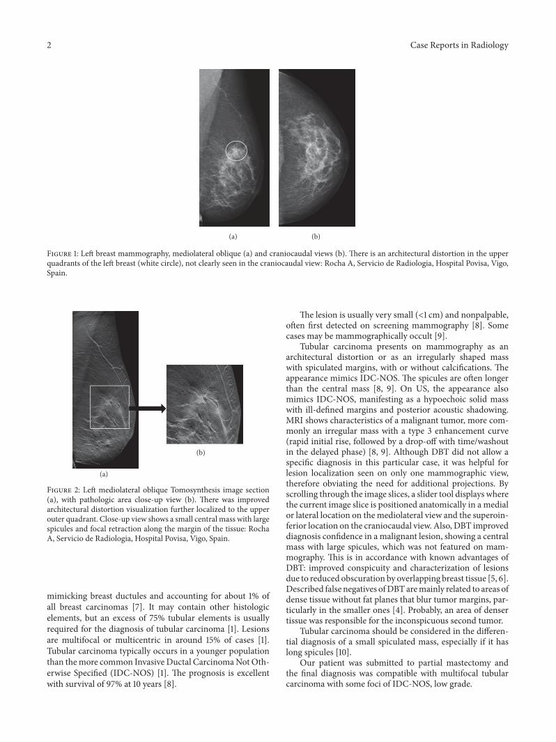
Figure 2: Left mediolateral oblique Tomosynthesis image section(a), with pathologic area close-up view (b). There was improvedarchitectural distortion visualization further localized to the upperouter quadrant. Close-up view shows a small central mass with largespicules and focal retraction along the margin of the tissue: RochaA, Servicio de Radiologia, Hospital Povisa, Vigo, Spain.
mimicking breast ductules and accounting for about 1% ofall breast carcinomas [7]. It may contain other histologicelements, but an excess of 75% tubular elements is usuallyrequired for the diagnosis of tubular carcinoma [1]. Lesionsare multifocal or multicentric in around 15% of cases [1].Tubular carcinoma typically occurs in a younger populationthan themore common Invasive Ductal CarcinomaNot Oth-erwise Specified (IDC-NOS) [1]. The prognosis is excellentwith survival of 97% at 10 years [8].
The lesion is usually very small (<1 cm) and nonpalpable,often first detected on screening mammography [8]. Somecases may be mammographically occult [9].
Tubular carcinoma presents on mammography as anarchitectural distortion or as an irregularly shaped masswith spiculated margins, with or without calcifications. Theappearance mimics IDC-NOS. The spicules are often longerthan the central mass [8, 9]. On US, the appearance alsomimics IDC-NOS, manifesting as a hypoechoic solid masswith ill-defined margins and posterior acoustic shadowing.MRI shows characteristics of a malignant tumor, more com-monly an irregular mass with a type 3 enhancement curve(rapid initial rise, followed by a drop-off with time/washoutin the delayed phase) [8, 9]. Although DBT did not allow aspecific diagnosis in this particular case, it was helpful forlesion localization seen on only one mammographic view,therefore obviating the need for additional projections. Byscrolling through the image slices, a slider tool displays wherethe current image slice is positioned anatomically in a medialor lateral location on themediolateral view and the superoin-ferior location on the craniocaudal view. Also, DBT improveddiagnosis confidence in a malignant lesion, showing a centralmass with large spicules, which was not featured on mam-mography. This is in accordance with known advantages ofDBT: improved conspicuity and characterization of lesionsdue to reduced obscuration by overlapping breast tissue [5, 6].Described false negatives ofDBTaremainly related to areas ofdense tissue without fat planes that blur tumor margins, par-ticularly in the smaller ones [4]. Probably, an area of densertissue was responsible for the inconspicuous second tumor.
Tubular carcinoma should be considered in the differen-tial diagnosis of a small spiculated mass, especially if it haslong spicules [10].
Our patient was submitted to partial mastectomy andthe final diagnosis was compatible with multifocal tubularcarcinoma with some foci of IDC-NOS, low grade.

Case Reports in Radiology 3
(a) (b)
Figure 3: Ultrasound, transverse (a) and longitudinal (b) sections. A 10mm solid nodule was shown, with irregular and ill-defined margins,taller rather than wider shaped, with a hyperechoic rim and posterior acoustic shadowing: Rocha A, Servicio de Radiologia, Hospital Povisa,Vigo, Spain.
(a) (b)
Figure 4: Breast Magnetic Resonance T1-weighted images, after gadolinium administration and axial maximum intensity projection (MIP)reformations. In addition to the previously biopsied lesion in the upper outer quadrant, there was another nodule 35mm apart, in the upperinner quadrant. It measured 8mm and was also submitted to US-guided biopsy: Rocha A, Servicio de Radiologia, Hospital Povisa, Vigo,Spain.
4. Conclusion
DBT improves radiologist’s diagnostic confidence allowingdifferentiation between fibroglandular tissue overlap and atrue lesion, without the need of additional mammographicacquisitions. As tubular carcinoma frequently presents withan architectural distortion or as a small mass, it represents apotential tumor benefiting from DBT.
Competing Interests
The authors declare that there is no conflict of interestsregarding the publication of this paper.
References
[1] Cardenosa, Clinical Breast Imaging, A Patient Focused TeachingFile, Lippincott Williams & Wilkins, Philadelphia, Pa, USA,2006.
[2] S. M. Moss, H. Cuckle, A. Evans, L. Johns, M. Waller, and L.Bobrow, “Effect of mammographic screening from age 40 years
on breast cancer mortality at 10 years’ follow-up: a randomisedcontrolled trial,” The Lancet, vol. 368, no. 9552, pp. 2053–2060,2006.
[3] A. B. Miller, C. Wall, C. J. Baines, P. Sun, T. To, and S. A.Narod, “Twenty five year follow-up for breast cancer incidenceandmortality of the CanadianNational Breast Screening Study:randomised screening trial,” British Medical Journal, vol. 348,article g366, 2014.
[4] M. P. Jeong, E. A. Franken Jr., M. Garg, L. L. Fajardo, and L.T. Niklason, “Breast tomosynthesis: present considerations andfuture applications,”RadioGraphics, vol. 27, pp. S231–S240, 2007.
[5] H. R. Peppard, B. E. Nicholson, C.M. Rochman, J. K.Merchant,R. C. Mayo, and J. A. Harvey, “Digital breast tomosynthesisin the diagnostic setting: indications and clinical applications,”Radiographics, vol. 35, no. 4, pp. 975–990, 2015.
[6] R. Gartner Roth, A. D. A. Maidment, S. P. Weinstein, S. OrelRoth, and E. F. Conant, “Digital breast tomosynthesis: lessonslearned from early clinical implementation,” Radiographics, vol.34, no. 4, pp. E89–E102, 2014.
[7] D. Cyrlak, P. M. Carpenter, and N. B. Rawal, “Breast imagingcase of the day,” Radiographics, vol. 19, no. 1, pp. 245–247, 1999.
4 Case Reports in Radiology
[8] J. A. Harvey, “Unusual breast cancers: useful clues to expandingthe differential diagnosis,” Radiology, vol. 242, no. 3, pp. 683–694, 2007.
[9] D. G. Sheppard, G. J. Whitman, P. T. Huynh, A. A. Sahin, B. D.Fornage, and C. B. Stelling, “Tubular carcinoma of the breast:mammographic and sonographic features,”American Journal ofRoentgenology, vol. 174, no. 1, pp. 253–257, 2000.
[10] G. Gatta, G. Di Grezia, A. Ancona et al., “Underestimation ofatypical lobular hyperplasia and lobular carcinoma in situ atstereotaxic 11-gauge vacuum-assisted breast biopsy,” EuropeanJournal of Inflammation, vol. 11, no. 3, pp. 825–835, 2013.

Submit your manuscripts athttp://www.hindawi.com
Stem CellsInternational
Hindawi Publishing Corporationhttp://www.hindawi.com Volume 2014
Hindawi Publishing Corporationhttp://www.hindawi.com Volume 2014
MEDIATORSINFLAMMATION
of
Hindawi Publishing Corporationhttp://www.hindawi.com Volume 2014
Behavioural Neurology
EndocrinologyInternational Journal of
Hindawi Publishing Corporationhttp://www.hindawi.com Volume 2014
Hindawi Publishing Corporationhttp://www.hindawi.com Volume 2014
Disease Markers
Hindawi Publishing Corporationhttp://www.hindawi.com Volume 2014
BioMed Research International
OncologyJournal of
Hindawi Publishing Corporationhttp://www.hindawi.com Volume 2014
Hindawi Publishing Corporationhttp://www.hindawi.com Volume 2014
Oxidative Medicine and Cellular Longevity
Hindawi Publishing Corporationhttp://www.hindawi.com Volume 2014
PPAR Research
The Scientific World JournalHindawi Publishing Corporation http://www.hindawi.com Volume 2014
Immunology ResearchHindawi Publishing Corporationhttp://www.hindawi.com Volume 2014
Journal of
ObesityJournal of
Hindawi Publishing Corporationhttp://www.hindawi.com Volume 2014
Hindawi Publishing Corporationhttp://www.hindawi.com Volume 2014
Computational and Mathematical Methods in Medicine
OphthalmologyJournal of
Hindawi Publishing Corporationhttp://www.hindawi.com Volume 2014
Diabetes ResearchJournal of
Hindawi Publishing Corporationhttp://www.hindawi.com Volume 2014
Hindawi Publishing Corporationhttp://www.hindawi.com Volume 2014
Research and TreatmentAIDS
Hindawi Publishing Corporationhttp://www.hindawi.com Volume 2014
Gastroenterology Research and Practice
Hindawi Publishing Corporationhttp://www.hindawi.com Volume 2014
Parkinson’s Disease
Evidence-Based Complementary and Alternative Medicine
Volume 2014Hindawi Publishing Corporationhttp://www.hindawi.com
Recommended