Embed Size (px)
Citation preview
i
CARCINOMA OF RENAL
TUBULAR ORIGIN
(excision specimens)
STRUCTURED REPORTING
PROTOCOL
(2nd Edition 2018)
Incorporating the
International Collaboration on Cancer Reporting (ICCR)
Dataset for the reporting of Invasive Carcinoma of Renal Tubular Origin
www.ICCR-Cancer.org

ii
Core Document versions:
1. ICCR Dataset for the Reporting of Invasive Carcinoma of Renal Tubular Origin
1st edition v1.1
2. AJCC Cancer Staging Manual 8th edition
3. World Health Organization (WHO). Classification of tumours. Pathology and
genetics of the urinary system and male genital organs. 4th edition.

iii
ISBN: 978‐1‐76000‐921‐2
Publications number (SHPN): (CI) 180558
Online copyright
© RCPA 2017
This work (Protocol) is copyright. You may download, display, print and
reproduce the Protocol for your personal, non-commercial use or use within your
organisation subject to the following terms and conditions:
1. The Protocol may not be copied, reproduced, communicated or displayed, in
whole or in part, for profit or commercial gain.
2. Any copy, reproduction or communication must include this RCPA copyright
notice in full.
3. With the exception of Chapter 6 - the checklist, no changes may be made
to the wording of the Protocol including any Standards, Guidelines,
commentary, tables or diagrams. Excerpts from the Protocol may be used
in support of the checklist. References and acknowledgments must be
maintained in any reproduction or copy in full or part of the Protocol.
4. In regard to Chapter 6 of the Protocol - the checklist:
o The wording of the Standards may not be altered in any way and must be
included as part of the checklist.
o Guidelines are optional and those which are deemed not applicable may be
removed.
o Numbering of Standards and Guidelines must be retained in the checklist,
but can be reduced in size, moved to the end of the checklist item or
greyed out or other means to minimise the visual impact.
o Additional items for local use may be added but must not be numbered as
a Standard or Guideline, in order to avoid confusion with the RCPA
checklist items.
o Formatting changes in regard to font, spacing, tabulation and sequencing
may be made.
o Commentary from the Protocol may be added or hyperlinked to the
relevant checklist item.
Apart from any use as permitted under the Copyright Act 1968 or as set out
above, all other rights are reserved. Requests and inquiries concerning
reproduction and rights should be addressed to RCPA, 207 Albion St, Surry Hills,
NSW 2010, Australia.
First published: July 2018, 2nd Edition (version 2.0)

iv
Disclaimer
The Royal College of Pathologists of Australasia ("College") has developed these
protocols as an educational tool to assist pathologists in reporting of relevant
information for specific cancers. Each protocol includes “standards” and
“guidelines” which are indicators of ‘minimum requirements’ and
‘recommendations’, which reflect the opinion of the relevant expert authoring
groups. The use of these standards and guidelines is subject to the clinician’s
judgement in each individual case.
The College makes all reasonable efforts to ensure the quality and accuracy of the
protocols and to update the protocols regularly. However subject to any
warranties, terms or conditions which may be implied by law and which cannot be
excluded, the protocols are provided on an "as is" basis. The College does not
warrant or represent that the protocols are complete, accurate, error-free, or up
to date. The protocols do not constitute medical or professional advice. Users
should obtain appropriate medical or professional advice, or where appropriately
qualified, exercise their own professional judgement relevant to their own
particular circumstances. Users are responsible for evaluating the suitability,
accuracy, currency, completeness and fitness for purpose of the protocols.
Except as set out in this paragraph, the College excludes: (i) all warranties, terms
and conditions relating in any way to; and (ii) all liability (including for
negligence) in respect of any loss or damage (including direct, special, indirect or
consequential loss or damage, loss of revenue, loss of expectation, unavailability
of systems, loss of data, personal injury or property damage) arising in any way
from or in connection with; the protocols or any use thereof. Where any statute
implies any term, condition or warranty in connection with the provision or use of
the protocols, and that statute prohibits the exclusion of that term, condition or
warranty, then such term, condition or warranty is not excluded. To the extent
permitted by law, the College's liability under or for breach of any such term,
condition or warranty is limited to the resupply or replacement of services or
goods.

v
Contents
Scope ................................................................................................................ vi
Abbreviations ................................................................................................... vii
Definitions ....................................................................................................... viii
Introduction ....................................................................................................... 1
Authority and development ................................................................................ 5
1 Pre-analytical ........................................................................................... 9
2 Specimen handling and macroscopic findings ........................................ 11
3 Microscopic findings ............................................................................... 15
4 Ancillary studies findings ....................................................................... 22
5 Synthesis and overview ......................................................................... 23
6 Structured checklist ............................................................................... 26
7 Formatting of pathology reports ............................................................ 46
Appendix 1 Pathology request information .......................................... 47
Appendix 2 Guidelines for formatting of a pathology report ................ 52
Appendix 3 Example of a pathology report .......................................... 53
Appendix 4 WHO classification of renal neoplasia. ............................... 56
References ....................................................................................................... 57

vi
Scope
This protocol contains standards and guidelines for the preparation of structured
reports for excision specimens of the kidneys. Urothelial carcinoma arising from
the upper renal tract, Wilms tumours and other nephroblastic and mesenchymal
tumours are not included.
The protocol is designed for the reporting of specimens of single laterality ie left
or right. If bilateral tumours are submitted then separate datasets should be
completed. If the tumour is multifocal due to intra-renal spread then a single form
should be used. However, if two or more synchronous malignancies are present
(usually identified by the presence of differing morphologies) then a separate
form should be used for each tumour.
Structured reporting aims to improve the completeness and usability of pathology
reports for clinicians, and improve decision support for cancer treatment. The
protocol provides the framework for the reporting of renal cancer, whether as a
minimum data set or fully comprehensive report.

vii
Abbreviations
AJCC American Joint Committee on Cancer
ALK anaplastic lymphoma kinase
ccRCC clear cell renal cell carcinoma
CG Commentary for a guideline
CS Commentary for a standard
FISH Fluorescent in-situ hybridization
HLRCC hereditary leiomyomatosis and renal cell carcinoma associated
renal cell carcinoma
ICCR International Collaboration on Cancer Reporting
ISUP International Society of Urological Pathology
LIS laboratory information system
LVI lymphovascular invasion
PBS Pharmaceutical Benefits Scheme
RCC Renal cell carcinoma
RCPA Royal College of Pathologists of Australasia
TNM tumour-node-metastasis
UICC International Union Against Cancer
WHO World Health Organization

viii
Definitions
The table below provides definitions for general or technical terms used in this
protocol. Readers should take particular note of the definitions for ‘standard’,
‘guideline’ and ‘commentary’, because these form the basis of the protocol.
Ancillary study An ancillary study is any pathology investigation that may form
part of a cancer pathology report but is not part of routine
histological assessment.
Clinical
information
Patient information required to inform pathological assessment,
usually provided with the specimen request form, also referred to
as “pre-test information”.
Commentary Commentary is text, diagrams or photographs that clarify the
standards (see below) and guidelines (see below), provide
examples and help with interpretation, where necessary (not every
standard or guideline has commentary).
Commentary is used to:
• define the way an item should be reported, to foster
reproducibility
• explain why an item is included (e.g. how does the item assist
with clinical management or prognosis of the specific cancer).
• cite published evidence in support of the standard or guideline
• state any exceptions to a standard or guideline.
In this document, commentary is prefixed with ‘CS’ (for
commentary on a standard) or ‘CG’ (for commentary on a
guideline), numbered to be consistent with the relevant standard
or guideline, and with sequential alphabetic lettering within each
set of commentaries (eg CS1.01a, CG2.05b).
General
commentary
General commentary is text that is not associated with a specific
standard or guideline. It is used:
• to provide a brief introduction to a chapter, if necessary
• for items that are not standards or guidelines but are included
in the protocol as items of potential importance, for which there
is currently insufficient evidence to recommend their inclusion.
(Note: in future reviews of protocols, such items may be
reclassified as either standards or guidelines, in line with
diagnostic and prognostic advances, following evidentiary
review).

ix
Guideline Guidelines are recommendations; they are not mandatory, as
indicated by the use of the word ‘should’. Guidelines cover items
that are unanimously agreed should be included in the dataset but
are not supported by NHMRC level III-2 evidence.1 These elements
may be clinically important and recommended as good practice but
are not yet validated or regularly used in patient management.
Guidelines include key information other than that which is
essential for clinical management, staging or prognosis of the
cancer such as macroscopic observations and interpretation, which
are fundamental to the histological diagnosis and conclusion eg
macroscopic tumour details, block identification key, may be
included as either required or recommended elements by
consensus of the expert committee. Such findings are essential
from a clinical governance perspective, because they provide a
clear, evidentiary decision-making trail.
Guidelines are not used for research items.
In this document, guidelines are prefixed with ‘G’ and numbered
consecutively within each chapter (eg G1.10).
Macroscopic
findings
Measurements, or assessment of a biopsy specimen, made by the
unaided eye.
Microscopic
findings
In this document, the term ‘microscopic findings’ refers to histo-
morphological assessment.
Predictive factor A predictive factor is a measurement that is associated with
response or lack of response to a particular therapy.
Prognostic
factor
A prognostic factor is a measurement that is associated with
clinical outcome in the absence of therapy or with the application
of a standard therapy. It can be thought of as a measure of the
natural history of the disease.
Standard Standards are mandatory, as indicated by the use of the term
‘must’. Standards are essential for the clinical management,
staging or prognosis of the cancer. These elements will either have
evidentiary support at Level III-2 or above (based on prognostic
factors in the NHMRC levels of evidence1 document). In rare
circumstances, where level III-2 evidence is not available an
element may be made a Standard where there is unanimous
agreement in the expert committee. An appropriate staging
system eg Pathological TNM staging would normally be included as
a required element. These elements must be recorded and at the
discretion of the pathologist included in the pathology report
according to the needs of the recipient of the report.
The summation of all standards represents the minimum dataset
for the cancer.
In this document, standards are prefixed with ‘S’ and numbered
consecutively within each chapter (eg S1.02).

x
Structured
report
A report format which utilises standard headings, definitions and
nomenclature with required information.
Synoptic report A structured report in condensed form (as a synopsis or precis).
Synthesis Synthesis is the process in which two or more pre-existing
elements are combined, resulting in the formation of something
new.
The Oxford dictionary defines synthesis as “the combination of
components or elements to form a connected whole”.
In the context of structured pathology reporting, synthesis
represents the integration and interpretation of information from
two or more modalities to derive new information.

1
Introduction
Renal Parenchymal Malignancy (Renal Cell Carcinoma)
Renal cell carcinoma was not described in antiquity, with the first confirmed case
of RCC being reported in France in 1810.2 The first classification of renal
neoplasia was produced in 1824 and since then a variety of classifications have
been proposed.3 Despite these early attempts to classify RCC, it is only in the last
two decades that there has been any real appreciation as to the wide variety of
morphotypes of RCC that exist.
In the first edition of the WHO classification, published in 1981, epithelial
malignancies of the renal parenchyma were classified as Renal Cell Carcinoma
and Other.4 The publication of the Mainz Classification in 1986 and the work of
the Heidelberg (1996) and Rochester (1997) Consensus Groups provided the
basis for classifying RCC into a variety of sub-types, each with differing clinical,
histological and genetic features.5,6 These conclusions were reinforced by the third
WHO classification working group who met in 2002, with the final classification
being released in 2004.7 In this classification ten distinctive sub-types of renal
parenchymal neoplasia were recognized, with a further category – that of Renal
Cell Carcinoma – Unclassified being reserved for those tumours whose features
differ from those of the recognized in the 2004 classification. It is from the group
of tumours classified as Renal Cell Carcinoma – Unclassified that several novel
variants of renal epithelial malignancy have been identified and since the
publication of the 2004 WHO Classification, a further six tumour sub-types have
been recognized.8 The classification of RCC was expanded in the ISUP Vancouver9
classification published in 2014, and in the 2016 4th edition of the WHO
classification.10
The failure to appreciate from the outset that RCC is a group of tumours rather
than a single tumour entity, has had a major impact upon outcome prediction for
these forms of malignancy. In particular, the failure to identify tumour sub-type
in data sets has served to introduced an uncontrolled variable into statistical
analyses and this has served to undermine the credibility of numerous prognostic
studies.11
More recently major studies have validated the sub-classification of RCC on the
basis of tumour-related outcome data.12-14 These studies have also attempted to
identify prognostic parameters for each sub-type of RCC and specifically, there
has been considerable emphasis on the evaluation of the predictive importance of
tumour stage and grade.15 This is of particular importance as RCCs as a group
have a considerable morbidity and mortality accounting for 2% of cancer deaths
worldwide. In the United States, the annual incidence of renal cell carcinoma has
increased by 46.9% over the past 17 years rising from 27,200 cases in 1990 to
an estimated 63,990 cases in 2017.16,17 In Australia, in 2017 the estimated age
adjusted incidence of RCC is 12 cases per 100,00018 while in New Zealand the
incidence was 8.1 cases per 100,000 in 2013.19
Importance of histopathological reporting
Information derived from the careful assessment and dissection of the gross
specimen, the judicious selection of tissues for histological examination and the

2
provision of a pathology report that contains information of both clinical and
prognostic utility is central to contemporary medical practice.
The information contained within pathology reports on specimens removed for the
management of RCC provide guidance for further treatment options and permit
assessment of outcome.
It is recognized that some morphotypes of RCC have a less aggressive clinical
course than others and as a consequence consideration may be given to
undertaking further surgical interventions if a patient subsequently develops
metastatic disease. Further, for those patients who have disease that is found to
be incurable following surgery, a variety of chemotherapeutic options are
available, and current protocols relate to specific tumour sub-types. For both of
these scenarios it is clear that subsequent management is informed by the
pathology report that details the morphology of the primary tumour.
It is well recognized that the most important single prognostic parameter for RCC
is tumour stage. Information regarding the completeness of surgical excision and
involvement of anatomic boundaries by tumour is essential for staging purposes.
Evaluation of other features contained with a standard report for RCC, such as
tumour grade relating to specific morphotypes of RCC, the presence of
sarcomatoid or rhabdoid differentiation, and the presence and degree of tumour
necrosis provide information that is essential for determining prognosis in
individual cases.
Benefits of structured reporting
The pathology report lays the foundation for a patient’s cancer journey and
conveys information which:
• Provides the definitive diagnosis
• Includes critical information for Tumour-Node-Metastasis (TNM) staging
• Evaluates the adequacy of the surgical excision
• Provides morphological and biological prognostic markers which determine
personalised cancer therapy
However, the rapid growth in ancillary testing such as immunohistochemistry,
flow cytometry, cytogenetics, and molecular studies, have made the task of
keeping abreast of advances on specific cancer investigations extremely difficult
for pathologists. The use of structured reporting checklists by pathologists
ensures that all key elements are included in the report specifically those which
have clinical management, staging or prognostic implications. Consequently
minimum or comprehensive datasets for the reporting of cancer have been
developed20,21 around the world. Both the United Kingdom,22 and United States23
have produced standardised cancer reporting protocols or “datasets” for national
use for many years.
The use of cancer reporting checklists improves completeness and quality of
cancer reporting and thereby ensures an improved outcome for cancer patients.
This has long term cost implications for public health by ensuring the most
effective and timely treatment based on accurate and complete information.
The use of a structured reporting format also facilitates easy extraction of the
necessary information by secondary users of the information ie cancer registries.

3
International Collaboration on Cancer Reporting
The International Collaboration on Cancer Reporting (ICCR), founded in 2011 by
the Australasian (RCPA), US (CAP) and UK (RCPath) Colleges of Pathology and
the Canadian Association of Pathology (CAP-ACP) in association with the Canadian
Partnership Against Cancer (CPAC), was established to explore the possibilities of
a collaborative approach to the development of common, internationally
standardised and evidence-based cancer reporting protocols for surgical
pathology specimens.
The ICCR, recognising that standardised cancer datasets have been shown to
provide significant benefits for patients and efficiencies for organisations through
the ease and completeness of data capture24-27 undertook to use the best
international approaches and the knowledge and experience of expert
pathologists, and produce cancer datasets which would ensure that cancer reports
across the world will be of the same high quality – ensuring completeness,
consistency, clarity, conciseness and above all, clinical utility.
Representatives from the four countries participating in the initial collaboration
undertook a pilot project in 2011 to develop four cancer datasets - Lung,
Melanoma, Prostate (Radical Prostatectomy), and Endometrium. Following on
from the success of this pilot project, the ICCR was joined by the European
Society of Pathology (ESP) in 2013 and in 2014 incorporated a not-for-profit
organisation focussed on the development of internationally agreed evidence-
based datasets developed by world leading experts. The ICCR Datasets are made
freely available from its website www.ICCR-Cancer.org
Design of this protocol
This structured reporting protocol has been developed incorporating the ICCR
dataset on renal cancer (excision specimens) as the foundation.
This protocol includes all of the ICCR cancer dataset elements as well as
additional information, elements and commentary as agreed by the RCPA expert
committee. It provides a comprehensive framework for the assessment and
documentation of pathological features of renal cancer in excision specimens.
ICCR dataset elements for renal cancer in excision specimens are included
verbatim. ICCR Required elements are mandatory and therefore represented as
standards in this document. ICCR Recommended elements, that is, those which
are not mandatory but are recommended, may be included as guidelines or
upgraded to a standard based on the consensus opinion of the local expert
committee.
The ICCR elements are identified in each chapter with the ICCR logo placed
before the Standard or Guideline number or bullet and the ICCR element
description and commentary is boarded by a grey box as shown below:
G3.02 The intraglandular extent should be recorded as a percentage.

4
Additional commentary by the RCPA expert committee may be added to an ICCR
element but is not included in the grey bordered area nor indicated with an ICCR
logo eg
G2.03 If present, the laterality of the lymph nodes submitted may be
recorded as left, right or bilateral.
CS2.03a If present, record site and number. All lymph node
tissue should be submitted for histological
examination.
Further information on the ICCR is available at www.iccr-cancer.org
Checklist
Consistency and speed of reporting is improved by the use of discrete data
elements recorded from the checklist. Items suited to tick boxes are distinguished
from more complex elements requiring free text or narrative. A structured or
discrete approach to responses is favoured, however the pathologist is
encouraged to include free text or narrative where necessary to document any
other relevant issues, to give reasons for coming to a particular opinion and to
explain any points of uncertainty.
Report format
The structure provided by the following chapters, headings and subheadings
describes the elements of information and their groupings, but does not
necessarily represent the format of either a pathology report (Chapter 7) or
checklist (Chapter 6). These, and the structured pathology request form
(Appendix 1) are templates that represent information from this protocol,
organised and formatted differently to suit different purposes.
Key documentation
• Guidelines for Authors of Structured Cancer Pathology Reporting Protocols,
Royal College of Pathologists of Australasia, 200928
• World Health Organization (WHO). Classification of tumours. Pathology and
genetics of the urinary system and male genital organs. Humphrey PA, Moch
H, Reuter VE, Ulbright TM editors. 4th edition. Lyon, France: IARC
Press;2016.10
• AJCC Cancer Staging Manual, 8th edition, American Joint Committee on
Cancer, 201629
Updates since last edition
Inclusion of ICCR agreed REQUIRED and RECOMMENDED elements.
5
Authority and development
This section provides information about the process undertaken to develop this
protocol.
This 2nd edition of the protocol is an amalgam of two separate processes:
1. This protocol is based on the ICCR Dataset for the Reporting of Invasive
Carcinoma of Renal Tubular Origin 1st edition. All ICCR elements from this
dataset, both required (mandatory) and recommended (optional), are
included in this protocol, verbatim. (It should be noted that RCPA feedback
from all Anatomical Pathology fellows and specifically the local expert
committee was sought during the development process of the ICCR
dataset.) Details of the ICCR development process and the international
expert authoring committee responsible for the ICCR dataset are available
on the ICCR website: iccr-cancer.org.
2. Additional elements, values and commentary have been included as
deemed necessary by the local expert committee. In addition, the
standard inclusions of RCPA protocols eg example reports, request
information etc, have also been added.
Authorship - 2nd edition
Professor Brett Delahunt (Lead author), Pathologist
Dr David Clouston, Pathologist
Adjunct Professor Warick Delprado, Pathologist
Dr Anne O’Donnell, Medical Oncologist
Clinical Professor James Kench, Pathologist
Professor Hemamali Samaratunga, Pathologist
Dr Simon Wood, Urologist
Authorship – 1st edition (2011)
Professor Brett Delahunt (Lead author), Pathologist
Dr Adrian Charles, Paediatric Pathologist
Dr David Clouston, Pathologist
Adjunct Professor Warick Delprado, Pathologist
Dr Anne O’Donnell, Medical Oncologist
Dr Thomas Eade, Radiation Oncologist
Professor James Kench, Pathologist
Dr Howard Lau, Urologist
Associate Professor Hemamali Samaratunga, Pathologist

6
Editorial manager
Meagan Judge, Royal College of Pathologists of Australasia.
Acknowledgements
The Kidney cancer expert committee wish to thank all the pathologists and
clinicians who contributed to the discussion around this document.

7
Stakeholders
ACT Health
ACT Cancer Registry
Australian Cancer Network
Australian Commission on Safety and Quality in Health Care
Australian Digital Health Agency
Australian Institute of Health and Welfare
Cancer Australia
Cancer Council ACT
Cancer Council Queensland
Cancer Council Victoria
Cancer Council Western Australia
Cancer Institute NSW
Cancer Services Advisory Committee (CanSAC)
Cancer Voices NSW
Clinical Oncology Society of Australia (COSA)
Department of Health, Australia
Department of Health, New Zealand
Faculty of Radiation Oncology Genito-Urinary Group (FROGG)
Health Informatics Society of Australia (HISA)
Independent Review Group of Pathologists
Medical Software Industry Association (MSIA)
National Pathology Accreditation Advisory Council (NPAAC)
New Zealand Cancer Registry
Northern Territory Cancer Registry
Pathology Australia
Public Pathology Australia
Queensland Cooperative Oncology Group (QCOG)
RCPA Anatomical Pathology Advisory Committee (APAC)
Representatives from laboratories specialising in anatomical pathology across
Australia
Royal Australasian College of Physicians (RACP)
South Australia Cancer Registry
Standards Australia
Tasmanian Cancer Registry
The Australian and New Zealand Urogenital and Prostate Cancer Trials Group
(ANZUP)
The Medical Oncology Group of Australia
The Prostate Cancer Foundation of Australia (PCFA)

8
The Prostate Cancer Foundation of New Zealand (PCFNZ)
The Royal Australasian College of Surgeons (RACS)
The Royal Australian and New Zealand College of Radiologists (RANZCR)
The Royal Australian College of General Practitioners (RACGP)
The Royal College of Pathologists of Australasia (RCPA)
The Urological Society of Australia And New Zealand (USANZ)
Western Australia Clinical Oncology Group (WACOG)
Development process
This protocol has been developed following the ten-step process set out in
Guidelines for Authors of Structured Cancer Pathology Reporting Protocols.28
Where no reference is provided, the authority is the consensus of the local expert
group for local inclusions and the ICCR Dataset Authoring Committee for ICCR
components denoted with the ICCR logo.

9
1 Pre-analytical
This chapter relates to information that should be recorded on receipt of the
specimen in the laboratory.
The pathologist is reliant on the quality of information received from the clinicians
or requestor. Some of this information may be received in generic pathology
request forms, however, the additional information required by the pathologist
specifically for the reporting of renal cancer is outlined in Appendix 1. Appendix 1
also includes a standardised request information sheet that may be useful in
obtaining all relevant information from the requestor.
Surgical handling procedures affect the quality of the specimen and
recommendations for appropriate surgical handling are included in Appendix 1.
S1.01 All demographic information provided on the request form and
with the specimen must be recorded.
CS1.01a The Royal College of Pathologists of Australasia (RCPA) The
Pathology Request-Test-Report Cycle — Guidelines for
Requesters and Pathology Providers must be adhered to.30 This
document specifies the minimum information to be provided by
the requesting clinician for any pathology test.
CS1.01b Whether or not the patient identifies as Aboriginal and/ or
Torres Strait Islander. This is in support of a government
initiative to monitor the health of indigenous Australians
particularly in relation to cancer.
CS1.01c The patient’s health identifiers may include the patient’s
Medical Record Number as well as a national health number
such as a patient’s Individual Healthcare Identifier (IHI)
(Australia) or the National Healthcare Identifier (New Zealand).
S1.02 All clinical information as documented on the request form must
be recorded verbatim.
CS1.02a The request information may be recorded as a single text
(narrative) field or it may be recorded in a structured format.
CS1.02b The copy doctors requested on the request form must be
recorded.
S1.03 The pathology accession number of the specimen must be
recorded.
S1.04 The principal clinician involved in the patient’s care and
responsible for investigating the patient must be recorded.
CS1.04a The principal clinician can provide key information regarding
the clinical presentation of the patient. Follow up may be
10
required with the principle clinician for a number of reasons:
• The clinical assessment and staging may be incomplete at
the time of biopsy.
• The pathology request is often authored by the clinician
performing the surgical excision/biopsy rather than the
clinician who is investigating and managing the patient.
• The identity of this clinician is often not indicated on the
pathology request form
In practice therefore, it is important in such cases that the
reporting pathologist should be able to communicate with the
managing clinician for clarification.
CS1.04b The Australian Healthcare identifiers i.e. Healthcare Provider
Identifier - Individual (HPI-I) and Healthcare Provider
Identifier - Organisation (HPI-O) should be included, where
possible, to identify the principal clinician involved in the
patient's care.
G1.01 Any clinical information received in other communications from the
requestor or other clinician should be recorded together with the source of
that information.

11
2 Specimen handling and macroscopic findings
This chapter relates to the procedures required after the information has been
handed over from the requesting clinician, and the specimen has been received in
the laboratory.
Tissue banking
➢ Pathologists may be asked to provide tissue samples from fresh specimens
for tissue banking or research purposes. The decision to provide tissue
should only be made if the pathologist is sure that the diagnostic process
will not be compromised. As a safeguard, research use of the tissue
samples may be put on hold until the diagnostic process is complete.
Specimen handling
➢ Detailed fixation and specimen handling instructions are available from the
RCPA online Cut-up Manual:
www.rcpa.edu.au/Library/Practising-Pathology/Macroscopic-Cut-Up
Macroscopic findings
S2.01 The labelling of the specimen(s) must be clearly recorded.
G2.01 The nature of the specimen at the time of reception should be given.
CG2.01a Choose from fresh or fixed (identify fixative), intact or
morcellated.
S2.02 Laterality of the specimen must be recorded.
CS2.02a Specimen laterality information is needed for identification
and specimen orientation and patient safety purposes.
S2.03 The operative procedure must be recorded.
CS2.03a The type of surgical procedure is important in determining
the assessment of surgical margins. Specifically in the case
of partial nephrectomy specimens it is important that the
intra-renal surgical margin be carefully evaluated so as to
ensure that no residual tumour is present in the remaining
kidney.
A radical nephrectomy specimen is defined as a resection

12
of Gerota’s fascia and its entire contents including the
kidney, perinephric fat and lymphatics and a length of
ureter, and may or may not be accompanied by the
adrenal gland. This is the principle treatment for
neoplasia.
A simple nephrectomy is the removal of a kidney only with
a portion of ureter. This is primarily utilised for removal of
non-tumorous kidney.
A partial nephrectomy specimen may vary from a simple
enucleation of the tumour to part of a kidney containing
variable portions of calyceal or renal pelvic collecting
system.
S2.04 Any accompanying or attached structures must be recorded.
G2.02 Whether or not tissue has been removed from the specimen prior to
submission should be recorded.
CG2.02a Pathologic evaluation requires a detailed examination of
the complete surgical specimen. If tissue has been
removed prior to examination this could compromise
diagnosis, staging and prognostic assessment.
G2.03 The kidney should be measured in 3 dimensions.
G2.04 The lengths of the renal vein, renal artery and ureter should be
measured.
G2.05 The specimen should be weighed.
G2.06 Evidence of adherence of renal capsule to visceral surface of perirenal
fat should be recorded.
CG2.06a If there is any degree of adherence of the renal capsule to
the visceral surface of the perirenal fat as this is evidence
of co-existing renal pathology.
G2.07 Any abnormalities of the cortical surface should be recorded.
G2.08 The site(s) of tumour in the kidney should be described.
CG2.08a The position of the tumour in relation to the boundaries of
the kidney and the surgical resection margin for radical
nephrectomy and partial nephrectomy specimens is
important for staging purposes. The position of the tumour
in relation to the renal cortex or medulla may also have
diagnostic importance. This is especially important for
small tumours where a site of origin within the medulla
would support a diagnosis of collecting duct carcinoma or
medullary carcinoma.1

13
Locations of medulla and renal cortex should be mentioned
under ‘other (specify)’.
S2.05 Tumour focality ie the number of tumours must be recorded.
CS2.05a Renal cell carcinomas are usually solitary, however, if
multifocal tumours are present, this is important to record.
Carcinomas in the setting of acquired cystic kidney disease
are often multiple. Multiple tumours may also be a clue
that one may be dealing with hereditary renal cell
carcinoma. Von Hippel Lindau, Birt-Hogg-Dubé and
hereditary papillary carcinoma syndromes are
characteristically associated with multiple tumours.
In a case of multiple carcinomas, it is important to record
the diagnostic and prognostic parameters associated with
the most significant tumours (largest, highest pT-category,
highest grade). The histological subtype of the tumours
may be similar or different and occasionally diverse
morpho-types may be found. When numerous carcinomas
are present some authors have suggested that the details
of the 5 largest tumours should be recorded.31
S2.06 The maximum dimension of the tumour must be recorded.
CS2.06a The maximum dimension of the tumour is required for
staging purposes as it constitutes the defining feature of
the pT1 and pT2 categories of the TNM staging
classification.29 Further it has been shown that for clear cell
renal cell carcinoma tumour size correlates with outcome
as a continuous variable.32
Measurement of tumour size should be undertaken
following detailed dissection of the gross specimen and the
greatest dimension should be recorded. Tumour extending
into extracapsular tissue and/or the renal sinus, in
continuity with the primary tumour intra-renal should be
included in the measurement. Tumour within the real vein
should not be included in this measurement. If multiple
tumours are present the greatest dimension of the five
largest tumours should be recorded.31
S2.07 The macroscopic extent of invasion must be recorded.
CS2.07a The identification of tumour directly infiltrating the renal
sinus or large vessels has prognostic significance and this
information is required for staging purposes.33,34
CS2.07b Tumour involvement of the adrenal gland (either direct
from the kidney of by metastatic spread) is associated with
a poor prognosis and needs to be documented for staging
purposes.

14
G2.09 The appearance of the cut surface of the tumour should be described.
CG2.09a Whether the tumour is solid or cystic should be recorded.
CG2.09b Macroscopic evidence of tumour necrosis should be given.
The presence of necrosis within RCC has prognostic
significance for clear cell renal cell carcinoma and
chromophobe renal cell carcinoma but not for papillary
renal cell carcinoma.35
CG2.09c The colour of the tumour should be given. Variegated
tumours with yellow areas are often rich in fat and this
appearance is most frequently seen in a clear cell renal cell
carcinoma. Chromophobe renal cell carcinomas and
sarcomatoid carcinomas are often pale.7
CG2.09d The consistency of the tumour should be provided.
Sarcomatoid carcinomas are often firm while papillary renal
cell carcinomas usually have a friable consistency.36
G2.10 Any abnormal features of the kidney should be recorded.
S2.08 A block identification key listing the nature and origin of all
tissue blocks must be recorded.
G2.11 A descriptive or narrative field should be provided to record any
macroscopic information that is not recorded in the above standards
and guidelines, and that would normally form part of the macroscopic
description.
CG2.11a The traditional macroscopic narrative recorded at the time
of specimen dissection is often reported separately from
the cancer dataset. Although this remains an option, it is
recommended that macroscopic information be recorded
within the overall structure of this protocol.
CG2.11b Much of the information recorded in a traditional
macroscopic narrative is covered in the standards and
guidelines above and in many cases, no further description
is required.
CG2.11c A traditional macroscopic description may be required
when the Laboratory Information System (LIS) does not
allow a structured approach.
CG2.11d Where the LIS offers an electronic interface for structured
data entry the need for narrative can be significantly
reduced to describe only information not otherwise
captured.

15
3 Microscopic findings
Microscopic findings relates to purely histological (morphological) assessment.
Information derived from multiple investigational modalities, or from two or more
chapters, is described in Chapter 5.
S3.01 The histological tumour type must be recorded (refer to
Appendix 4).
CS3.01a Many of the various sub-types of renal epithelial neoplasia
exhibit differing clinical behaviour and prognosis.8,10,29,36-40
This has been confirmed in large single and multicentre
studies for the main tumour sub-types. Several series have
also clearly demonstrated that many of the newly
described entities of renal malignancy have a prognosis
that differs from that of clear cell renal cell carcinoma.40 In
addition to this protocols for the various types of adjuvant
anti-angiogenic therapy relate to specific tumour sub-
types.41
The 2013 International Society of Urological Pathology
(ISUP) Vancouver Classification of adult renal tumours
identified an emerging/provisional category of renal cell
carcinoma (RCC).9 While appearing distinctive, these rare
tumours had not been fully characterized by morphology,
immunohistochemistry and molecular studies. This
category was also included in the fourth edition of the
World Health Organization (WHO) classification of renal
neoplasia. In the WHO classification oncocytoid RCC post-
neuroblastoma, thyroid-like follicular RCC, anaplastic
lymphoma kinase (ALK) rearrangement-associated RCC,
RCC with (angio) leiomyomatous stroma, eosinophilic solid
and cystic clear cell renal cell carcinoma, and biphasic
squamoid and alveolar renal cell carcinoma are included in
this category. These entities should be classified under
‘other’ with the name specified.
Papillary RCC has traditionally been subdivided into Type 1
and Type 2.42 Recent studies have shown these tumours to
be clinically and biologically distinct. Type 1 tumours are
associated with alterations in the MET pathway while type
2 tumours are associated with activation of the NRF2-ARE
pathway. On the basis of molecular features type 2
tumours may be sub-divided into at least 3 subtypes.43
Type 1 and type 2 tumours show differing
immunohistochemical staining with type 1 tumours more
frequently expressing cytokeratin 7 in comparison to type
2.9,10,42,43
Oncocytic papillary renal cell carcinoma is a category
included in the fourth edition of the WHO renal tumour
classification.10 While not fully characterized, this tumour is
16
best included in the broader papillary category.
Papillary RCC is associated with a more favourable
outcome than clear cell renal cell carcinoma (ccRCC),
collecting duct carcinoma and hereditary leiomyomatosis
and renal cell carcinoma – associated renal cell carcinoma
(HLRCC)10,40 Papillary subtyping is also of prognostic
significance with type 1 tumours having a better prognosis
then those with type 2 morphology.40,42,43
S3.02 The histological WHO/ISUP tumour grade must be recorded.
CS3.02a Grade should be assigned based on the single high power
field showing the greatest degree of nucleolar size and/or
nuclear pleomorphism.
This grading system is the World Health Organization/
International Society of Urological Pathology (WHO/ISUP)
grading system for renal cell carcinoma which is
recommended in the 2016 WHO.10,40 This system has been
validated as a prognostic parameter for clear cell and
papillary renal cell carcinoma.40,45,46 It has not been
validated for other types of renal cell carcinoma but may
be used for descriptive purposes.47 The current
recommendation is that chromophobe renal cell carcinoma
is not graded.10,48
S3.03 Evidence of sarcomatoid morphology must be recorded.
CS3.03a The presence of sarcomatoid morphology is seen in
approximately 5% of renal cell carcinomas and is
associated with a poor prognosis.40,49-52 Numerous studies
have confirmed that sarcomatoid morphology may occur
within any of the main subtypes of renal cell carcinoma and
represents high grade disease.9,10 The five year survival for
patients with sarcomatoid morphology is of the order of 15
to 22%.9,10,49-52 The outcome associated with sarcomatoid
morphology is stage dependent.53 The presence of
sarcomatoid morphology is incorporated into the
WHO/ISUP grading system (Grade 4).40
G3.01 The extent of any sarcomatoid component should be reported.
CG3.01a While there is no recommended or agreed method to
calculate the sarcomatoid component at this stage.40 It
has been suggested that the proportion of tumour showing
sarcomatoid differentiation has prognostic significance. In
particular, significantly different survivals were
demonstrated for tumours divided with a cutpoint of 50%
sarcomatoid component.52
S3.04 Evidence of rhabdoid morphology must be recorded.

17
CS3.04a Similar to the sarcomatoid morphology, rhabdoid
morphology is a feature of high grade disease.40,54
Tumours showing this phenotype resemble rhabdoid cells
having bulky eosinophilic cytoplasm and an eccentric
nucleus, often with a prominent nucleolus.9,10 Rhabdoid
change is associated with a poor prognosis. It has been
shown that 71% of patients with rhabdoid morphology
developed metastases with a mean follow-up of 4.5
months. Within 2 years it was also noted that 43% of
patients in this series had died, with a median survival rate
of 8-31months.40,54-56 In approximately 25% of tumours
with rhabdoid morphology, there is co-existing sarcomatoid
carcinoma.10 The presence of rhabdoid morphology is
incorporated into the WHO/ISUP grading system (Grade
4).40
G3.02 The extent of any rhabdoid component should be reported.
CG3.02a There is currently no firm evidence to demonstrate that the
volume of cells showing rhabdoid morphology is of
prognostic significance.40
S3.05 Evidence of tumour necrosis must be recorded.
CS3.05a The presence of tumour necrosis has been shown to be a
prognostic indicator for clear cell renal cell carcinoma and
chromophobe renal cell carcinoma independent of tumour
stage.35,40 Papillary renal cell carcinoma typically contains
foci of necrosis, however the prognostic significance of this
is, at best debated. At present, it is recommended that the
presence of both macroscopic and microscopic
(coagulative) necrosis be recorded.40 For patients who
have undergone pre-surgical renal embolization, the
degree of tumour-related necrosis cannot be assessed.
G3.03 The extent of necrosis in cases of clear cell renal cell carcinoma should
be reported.
CG3.03a The presence of tumour necrosis has been shown to be a
prognostic indicator for clear cell renal cell carcinoma and
has limited or no prognostic implications for papillary renal
cell carcinoma. It has been shown that tumour necrosis
>10% is associated with a less favourable outcome, while
for TNM stage 1 and 2 tumours a cutpoint of 20% of the
area of the tumour showing necrosis has been suggested
to have prognostic significance.57 At present the prognostic
significance of the amount of necrosis within a tumour is
uncertain. Despite this it has been recommended that this
be recorded as a percentage.40
S3.06 The extent of tumour invasion must be recorded as “tumour
limited to the kidney” or recorded for each of the following:
18
• Tumour spread beyond the renal capsule
• Tumour in renal sinus
• Tumour extends beyond Gerota’s fascia
• Tumour in major veins (renal vein or its segmental
branches, inferior vena cava)
• Tumour in pelvicalyceal system
• Tumour in adrenal gland
• Tumour in other organs/structures
CS3.06a The identification of tumour directly infiltrating the renal
sinus or large vessels has prognostic significance and this
information is required for staging purposes.6,29 Careful
gross examination of the specimen to assess large vessel
invasion for example of the renal vein or beyond (if
applicable) should be undertaken.
The renal sinus is an important pathway of spread of renal
cell carcinoma and is often an under-recognized
phenomenon.58 The renal sinus fat should be carefully
assessed and generously sampled in order to detect renal
sinus fat involvement. There is evolving literature
suggesting that renal sinus fat involvement predicts a more
aggressive outcome than peripheral perinephric fat
invasion.59,60
When renal carcinoma involves the adrenal gland, it is
important to document whether the involvement is
contiguous spread of tumour or a separate
(noncontiguous) nodule of carcinoma, the latter
representing metastatic disease (pM1).29
Refer to Figure 1.
Extra-renal extension of tumour is a feature of pT3 and
pT4 staging categories of the TNM staging classification.
Extension of tumour beyond Gerota’s fascia is a feature of
the pT4 staging category of the TNM staging system.29
The renal sinus is the compartment that lies between the
renal parenchymal and the renal pelvis and calyces. This
compartment contains varying amounts of fat and is rich in
lymphatics. As a consequence infiltration of the renal sinus
is the principal route for the extension of tumour beyond
the kidney.56 Renal sinus invasion is present when there is
tumour in contact with renal sinus fat, loose connective
tissue clearly beyond the renal parenchyma of the renal
sinus and in endothelial-lined spaces (with or without
mural smooth muscle) within the renal sinus.56 This is
most commonly seen in clear cell renal cell carcinoma and
appears to be associated with tumour size. In particular it
19
has been noted that in clear cell renal cell carcinomas ≥
7cm in diameter, renal sinus invasion was seen in > 90%
of cases.59,60 Involvement of the renal sinus by tumour is a
feature of pT3a tumour staging category of the TNM
classification. It is likely that renal sinus invasion is
preceded by involvement of renal sinus veins. It has also
been shown that involvement of lymphatics within the
renal sinus is of prognostic significance.61
If renal sinus invasion is seen on gross inspection of the
specimen, then only one confirmatory section need be
taken. If there is no evidence of renal sinus invasion
grossly, then sampling should consist of at least three
blocks of tissue.31
Adrenal gland: It is now recognized that direct spread of
tumour to the ipsilateral adrenal gland has an outcome
similar to pT4 tumour.62,63 In earlier TNM classifications this
was included in the pT3a category, however, in view of
these recent findings this was included as a feature of the
pT4 category. In contrast a discrete, separate nodule in the
adrenal gland is considered M1 disease.29
Other organs: The presence of metastatic disease is a
feature of the pM1 staging category of the TNM staging
classification.29
G3.04 The presence or absence of tumour within the renal vein wall should
be recorded.
G3.05 The presence or absence of lymphovascular invasion should be
recorded.
CG3.05a Lymphovascular invasion includes intratumoral,
peritumoral and perirenal space invasion.31 In the renal
sinus, it may be difficult to distinguish microscopic
lymphovascular invasion from involvement of thin walled
veins lacking smooth muscle. From a practical perspective,
the presence of either pattern should be considered as
renal sinus involvement (pT3a).
Microvascular invasion has been shown to correlate with
the development of metastases and with survival,
independent of tumour size, primary tumour category, and
grade.64
In both clear cell and papillary RCC, tumour spread is
predominantly haematogenous via the sinus veins, renal
vein and vena cava to the lung. Infiltration of the perirenal
fat can result in retroperitoneal spread. Lymphatic spread
to the nodes of the renal hilum may also occur and is more
common in papillary RCC than with ccRCC.29
S3.07 Lymph node status must be recorded.

20
CS3.07a In earlier editions of the UICC/AJCC of the TNM
classification, the number of lymph nodes infiltrated by
tumour was used to differentiate the different pN
categories. This has been simplified to now consist of
presence or absence of lymph node involvement by
tumour.10 It has, however been shown that survival does
decrease with an increase in the number of lymph nodes
involved (>4).65
CS3.07b The number of lymph nodes examined and the number of
positive lymph nodes must be stated.
G3.06 In the presence of positive lymph nodes, the presence or absence of
extranodal extension should be recorded.
CG3.06a It has been shown that extra-nodal extension of metastatic
tumour has prognostic significance independent of size of
lymph node metastases and tumour grade.66
S3.08 The status of surgical margins should be reported and if
involved the specific margins must be reported.
CS3.08a Assessment of surgical margins is important in determining
if residual tumour is present. In a partial nephrectomy
specimen, the renal parenchymal margin should be inked
and histologically assessed. Most partial nephrectomy
specimens also contain a portion of perinephric fat
overlying the tumour site. The perirenal fat margin should
also be assessed. In situations where no perirenal fat is
submitted, the renal capsular margin should be inked and
examined histologically. In radical nephrectomy specimens
the ureteric, major vascular (renal vein, renal artery) and
soft tissue (Gerota’s fascia, renal sinus) margins should be
examined and documented in the report.
CS3.08b Where contiguous tumour extension into the renal vein or
inferior vena cava occurs involvement of the vascular
margin should only be reported where there is tumour
infiltration of the transected vessel wall. Tumour in the
lumen of the vessel does not represent an involved margin.
G3.07 For partial nephrectomy specimens, the distance from the tumour to
the closest surgical margin should be recorded.
S3.09 The nature of any co-existing renal pathology in non-neoplastic
kidney must be reported.
CS3.09a It is important to recognize that medical kidney diseases
may be present in nonneoplastic renal tissue in
nephrectomy and nephroureterectomy specimens.67,68
Arterionephrosclerosis (or hypertensive nephropathy) and
diabetic nephropathy are most frequently seen, and in two

21
separate series medical renal disease was seen in 17% to
60% of cases. The findings of greater than 20% global
glomerulosclerosis or advanced diffuse diabetic
glomerulosclerosis are predictive of significant decline in
renal function 6 months after radical nephrectomy.68
Evaluation for medical renal disease should be performed in
each case; PAS and/or Jones methenamine silver stains
should applied if necessary. Consultation with a
nephropathologist should be pursued as needed.
G3.08 Any additional relevant comments should be recorded.

22
4 Ancillary studies findings
Ancillary studies may be used to determine lineage, clonality or disease
classification or subclassification; as prognostic biomarkers; or to indicate the
likelihood of patient response to specific biologic therapies.
Some studies, such as Her-2 testing, are required under the Pharmaceutical
Benefits Scheme, to enable certain specific therapies to be prescribed.
G4.01 Whether or not ancillary tests are performed should be recorded and
the results incorporated into the pathology report.
CG4.01a Ancillary studies are being increasingly utilized for
subtyping of renal cell neoplasms. It is now recognized that
Immunohistochemical assessment of tumours can be
diagnostically helpful.69
CG4.01b Fluorescent in-situ hybridization (FISH) can be used to
confirm a diagnosis of translocation carcinoma (MiT family
tumour) and has been shown to be of utility in
distinguishing oncocytoma from chromophobe renal cell
carcinoma.10 Cytogenetics may be undertaken in some
instances; however, this is not usually performed as part
of the routine assessment of a renal tumour. It is now
recognized that immunohistochemical assessment of
tumours can be diagnostically helpful.
CG4.01c There are currently no ancillary tests that are accepted as
having prognostic significance for renal cell neoplasms. 69,70,71,72

23
5 Synthesis and overview
Information that is synthesised from multiple modalities and therefore cannot
reside solely in any one of the preceding chapters is described here.
For example. tumour stage is synthesised from multiple classes of information –
clinical, macroscopic and microscopic.
By definition, synthetic elements are inferential rather than observational, often
representing high-level information that is likely to form part of the report
‘Summary’ or ‘Diagnosis’ section in the final formatted report.
Overarching case comment is synthesis in narrative format. Although it may not
necessarily be required in any given report, the provision of the facility for
overarching commentary in a cancer report is essential.
S5.01 The tumour stage must be recorded according to the AJCC TNM
Classification 2016 (8th Edition).
CS5.01a Refer to Figures 1-4 below.
Figure 1.
A: Diagram showing the renal sinus fat (S) and its rich venous system
that envelops the collecting system. The renal capsule terminates
(arrow) just inside the vestibule of the hilus.
B: A renal malignancy is constrained by the renal capsule (arrow), yet no
fibrous capsule impedes its growth into the vascular tissue of the renal
sinus (curved arrows).
From Bonsib et al.58 The American Journal of Surgical Pathology. © 2000 Wolters
Kluwer Health. Reproduced with permission.

24
Figure 2: T3a Invasion into perirenal and/or renal sinus fat but not
beyond Gerota’s fascia.
Figure 3: T4 Invasion beyond Gerota’s fascia.
Figure 4: T4 Direct extension of tumour into ipsilateral adrenal gland.
Used with permission of the American College of Surgeons, Chicago, Illinois. The
original and primary source for this information is the AJCC Cancer Staging Atlas,
Second Edition (2012) published by Springer Science+Business Media.
S5.02 The year of publication and/or edition of the cancer staging
system used in S5.01 must be included in the report.
25
G5.01 The “Diagnostic summary” section of the final formatted report should
include:
a. Specimen laterality (S2.03)
b. Operative procedure (S2.04)
c. Tumour type (S3.01)
d. Tumour grade (S3.02)
e. Tumour stage (S5.01)
f. Involvement of surgical margin (completeness of excision) (S3.08)
S5.03 The reporting system must provide a field for free text or
narrative in which the reporting pathologist can give
overarching case comment, if required.
CS5.03a This field may be used, for example, to:
– document any noteworthy adverse gross and/or
histological features
– explain the decision-making pathway, or any
elements of clinicopathological ambiguity, or factors
affecting diagnostic certainty, thereby allowing
communication of diagnostic subtlety or nuance that
is beyond synoptic capture
– document further consultation or results still
pending.
CS5.03b Use of this field is at the discretion of the reporting
pathologist.
G5.02 The edition/version number of the RCPA protocol on which the report
is based should be included on the final report.
CG5.02a For example, the pathology report may include the
following wording at the end of the report: “the data fields
within this formatted report are aligned with the criteria as
set out in the RCPA document “ XXXXXXXXXX” XXXX
Edition dated XXXXXXX”.

26
6 Structured checklist
The following checklist includes the standards and guidelines for this protocol
which must be considered when reporting, in the simplest possible form. The
summation of all “Standards” is equivalent to the “Minimum Data Set” for renal
parenchymal malignancy. For emphasis, standards (mandatory elements) are
formatted in bold font.
S6.01 The structured checklist provided may be modified as required
but with the following restrictions:
a. All standards and their respective naming conventions,
definitions and value lists must be adhered to.
b. Guidelines are not mandatory but are recommendations and
where used, must follow the naming conventions, definitions
and value lists given in the protocol.
G6.01 The order of information and design of the checklist may be varied
according to the laboratory information system (LIS) capabilities and as
described in Functional Requirements for Structured Pathology
Reporting of Cancer Protocols.73
CG6.01a Where the LIS allows dissociation between data entry and
report format, the structured checklist is usually best
formatted to follow pathologist workflow. In this situation,
the elements of synthesis or conclusions are necessarily at
the end. The report format is then optimised independently
by the LIS.
CG6.01b Where the LIS does not allow dissociation between data
entry and report format, (for example where only a single
text field is provided for the report), pathologists may elect
to create a checklist in the format of the final report. In this
situation, communication with the clinician takes precedence
and the checklist design is according to principles given in
Chapter 7.
G6.02 Where the checklist is used as a report template (see G6.01), the
principles in Chapter 7 and Appendix 2 apply.
CG6.02a All extraneous information, tick boxes and unused values
should be deleted.
G6.03 Additional comment may be added to an individual response where
necessary to describe any uncertainty or nuance in the selection of a
prescribed response in the checklist. Additional comment is not required
where the prescribed response is adequate.

27
S6.01 The structured checklist provided may be modified as required but with
the following restrictions:
c. All standards and their respective naming conventions, definitions
and value lists must be adhered to.
d. Guidelines are not mandatory but are recommendations and where
used, must follow the naming conventions, definitions and value lists
given in the protocol.
G6.01 The order of information and design of the checklist may be varied
according to the laboratory information system (LIS) capabilities and as
described in Functional Requirements for Structured Pathology
Reporting of Cancer Protocols.73
CG6.01a Where the LIS allows dissociation between data entry and
report format, the structured checklist is usually best
formatted to follow pathologist workflow. In this situation,
the elements of synthesis or conclusions are necessarily at
the end. The report format is then optimised independently
by the LIS.
CG6.01b Where the LIS does not allow dissociation between data
entry and report format, (for example where only a single
text field is provided for the report), pathologists may elect
to create a checklist in the format of the final report. In this
situation, communication with the clinician takes precedence
and the checklist design is according to principles given in
Chapter 7.
G6.02 Where the checklist is used as a report template (see G6.01), the
principles in Chapter 7 and Appendix 2 apply.
CG6.02a All extraneous information, tick boxes and unused values
should be deleted.
G6.03 Additional comment may be added to an individual response where
necessary to describe any uncertainty or nuance in the selection of a
prescribed response in the checklist. Additional comment is not required
where the prescribed response is adequate.

28
Item descriptions in italics are conditional on previous responses.
Values in all caps are headings with sub values.
S/G Item description Response type Conditional
Pre-analytical
S1.01 Demographic information
provided
S1.02 Information provided on
request form
Not provided
OR
Text
OR
Structured entry as below:
Relevant past medical history Text
Predisposing factors (including
genetic status)
Text
Pre-operative treatment Multi selection value list (select all that
apply):
• Tumour embolization
• Cryoablation
• Radio frequency ablation

29
• External-beam radiation therapy (EBRT)
• Other, specify
Relevant family history Text
Extent of disease Text
Previous biopsy/surgical
specimens
Text
Clinical or differential diagnosis Text
Laterality Single selection value list:
• Left
• Right
• Other eg horseshoe kidney, specify
Nature of operation Single selection value list:
• Radical nephrectomy
• Simple nephrectomy
• Partial nephrectomy
• Other, specify
Operative findings Text
Surgical intent Single selection value list:
• Curative
• Palliative

30
Tissue removed for research
or other purposes
Single selection value list:
• No
• Yes, specify details of tissue removed
New primary lesion or
recurrence
Single selection value list:
• New primary
• Recurrence – regional, describe
• Recurrence – distant, describe
S1.03 Pathology accession number Alpha-numeric
S1.04 Principal clinician Text
G1.01 Comments Text
Macroscopic findings
S2.01 Specimen labelled as Text
G2.01 Nature of specimen Single selection value list:
• Fixed, specify fixative
• Fresh
AND
Single selection value list:
• Intact
• Morcellated

31
S2.02 Specimen laterality Not specified
OR
Single selection value list:
• Left
• Right
• Other eg horseshoe kidney, specify
S2.03 Operative procedure Not specified
OR
Single selection value list:
• Radical nephrectomy
• Simple nephrectomy
• Partial nephrectomy
• Other, specify
S2.04 Accompanying/ attached
structures
None submitted
OR
Multi selection value list (select all that
apply):
• Adrenal gland
• Lymph nodes, provide details
• Other organs, provide details

32
G2.02 Tissue removed from specimen
prior to submission
Single selection value list:
• Not stated
• No
• Yes, provide details
G2.03 Kidney dimensions Numeric: __x__x__mm
G2.04 Length of renal vein Numeric: __mm
Length of renal artery Numeric: __mm
Length of ureter Numeric: __mm
G2.05 Specimen weight Numeric: __g
G2.06 Adherence of renal capsule to
visceral surface of perirenal fat
Single selection value list:
• Not identified
• Present
G2.07 Cortical surface abnormalities
Text
G2.08 Tumour site(s) Single selection value list:
• Not provided

33
• Cannot be assessed
OR
Multi selection value list (select all that
apply):
• Upper pole
• Mid zone
• Lower pole
• Cortex
• Medulla
• Other, specify
S2.05 Tumour focality Single selection value list:
• Cannot be assessed
• Unifocal
• Multifocal, specify number of tumours (if
possible)
S2.06 Maximum tumour dimension Numeric: __mm
Notes
If multiple tumours the maximum dimension of
the largest five should be recorded.
S2.07 Macroscopic extent of
invasion
Single selection value list:
• Tumour confined to kidney
OR
Multi selection value list (select all that
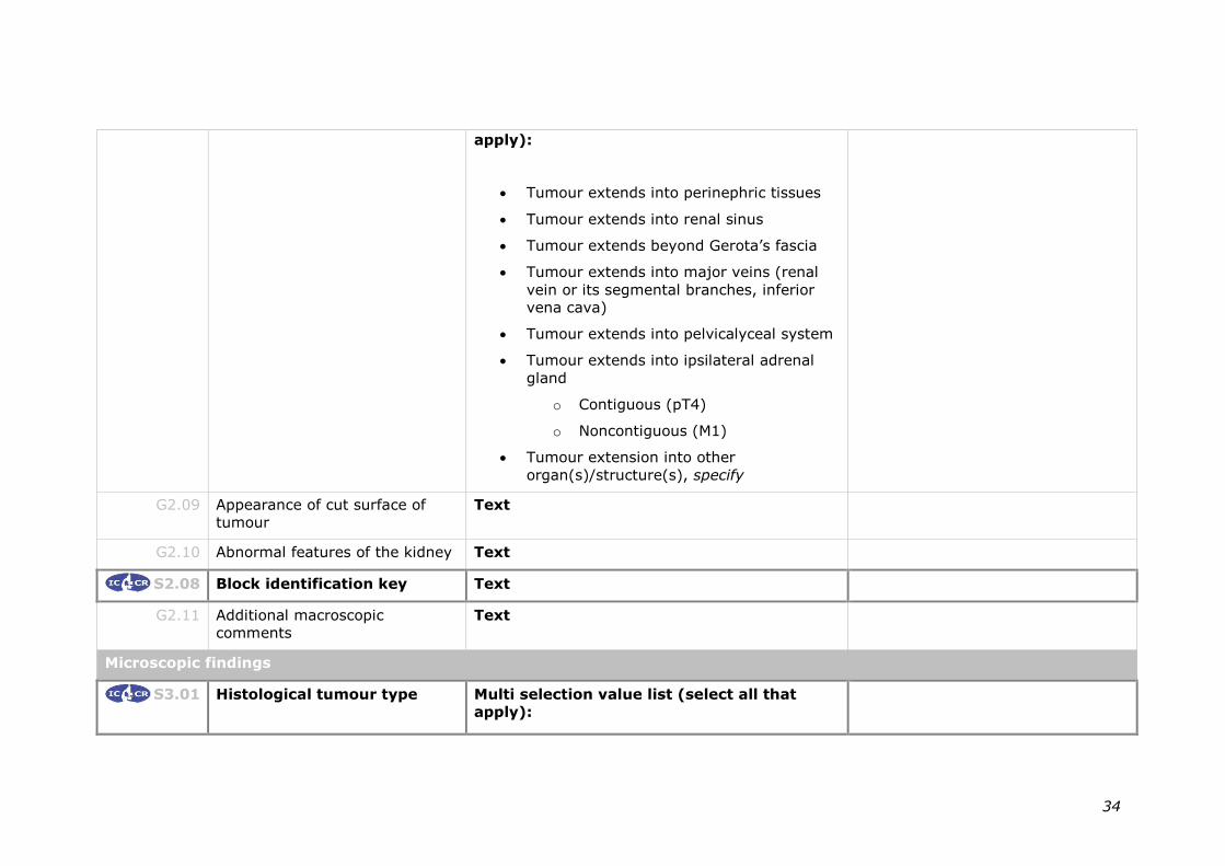
34
apply):
• Tumour extends into perinephric tissues
• Tumour extends into renal sinus
• Tumour extends beyond Gerota’s fascia
• Tumour extends into major veins (renal
vein or its segmental branches, inferior
vena cava)
• Tumour extends into pelvicalyceal system
• Tumour extends into ipsilateral adrenal
gland
o Contiguous (pT4)
o Noncontiguous (M1)
• Tumour extension into other
organ(s)/structure(s), specify
G2.09 Appearance of cut surface of
tumour
Text
G2.10 Abnormal features of the kidney Text
S2.08 Block identification key Text
G2.11 Additional macroscopic
comments
Text
Microscopic findings
S3.01 Histological tumour type
Multi selection value list (select all that
apply):

35
• Clear cell renal cell carcinoma
• Multilocular clear cell renal cell neoplasm
of low malignant potential
• Papillary renal cell carcinoma
o Type 1
o Type 2
o Oncocytic
o NOS
• Chromophobe renal cell carcinoma
o Hybrid oncocytic chromophobe
tumour
• Collecting duct carcinoma
• Renal medullary carcinoma
• MiT family translocation renal cell
carcinoma
o Xp11 translocation renal cell
carcinoma
o t(6;11) renal cell carcinoma
o Other, specify
• Mucinous tubular and spindle cell
carcinoma
• Tubulocystic renal cell carcinoma
• Acquired cystic disease associated renal
cell carcinoma
• Clear cell papillary/tubulopapillary renal

36
cell carcinoma
• Hereditary leiomyomatosis and renal cell
carcinoma-associated renal cell carcinoma
• Succinate dehydrogenase (SDH) deficient
renal carcinoma
• Renal cell carcinoma, unclassified
• Other, specify
Notes:
Occasionally more than one histologic type of
carcinoma occurs within the same kidney
specimen. Each tumour type should be
separately recorded.
S3.02 Histological tumour grade –
WHO/ISUP
Single selection value list:
• Not applicable
• Grade X - Cannot be assessed
• Grade 1 - Nucleoli absent or
inconspicuous and basophilic at 400x
magnification
• Grade 2 - Nucleoli conspicuous and
eosinophilic at 400x magnification, visible
but not prominent at 100x magnification
• Grade 3 - Nucleoli conspicuous and
eosinophilic at 100x magnification
• Grade 4 - Extreme nuclear pleomorphism
and/or multi nuclear giant cells and/or
rhabdoid and/or sarcomatoid
differentiation

37
S3.03 Sarcomatoid morphology Single selection value list:
• Not identified
• Present
If present, consider reporting
G3.01
G3.01 Extent of sarcomatoid
component
Numeric: __%
S3.04 Rhabdoid morphology Single selection value list:
• Not identified
• Present
If present, consider reporting
G3.02
G3.02 Extent of rhabdoid
component
Numeric: __%
S3.05 Necrosis Single selection value list:
• Cannot be assessed
• Not identified
• Present
o Microscopic coagulative necrosis
o Macroscopic tumour necrosis
If present, consider reporting
G3.03
G3.03 Extent of necrosis (Applicable to
clear cell renal cell carcinoma
only)
Numeric: __%
S3.06 Extent of invasion Single selection value list:
• Tumour limited to the kidney
OR

38
Complete each of the following elements
and consider reporting G3.04-5.
Tumour spread beyond the
renal capsule
Single selection value list:
• Cannot be assessed
• Not identified
• Present
Tumour in renal sinus Single selection value list:
• Cannot be assessed
• Not identified
• Present in fat
• Present in vascular spaces
• Present in fat and vascular spaces
Tumour extends beyond
Gerota’s fascia
Single selection value list:
• Cannot be assessed
• Not identified
• Present
Tumour in major veins (renal
vein or its segmental
branches, inferior vena cava)
Single selection value list:
• Cannot be assessed
• Not identified
• Present
Tumour in pelvicalyceal Single selection value list:

39
system • Cannot be assessed
• Not identified
• Present
Tumour in adrenal gland Single selection value list:
• Not provided
• Cannot be assessed
• Not identified
• Present - direct extension
• Present - metastasis
Tumour in other
organs/structures
Single selection value list:
• Not provided
• Cannot be assessed
• Not identified
• Present, specify sites
G3.04 Tumour in renal vein wall Single selection value list:
• Cannot be assessed
• Not identified
• Present
G3.05 Lymphovascular invasion Single selection value list:
• Not identified
• Present

40
S3.07 LYMPH NODE STATUS This is conditional on receipt of
LNs in S2.05.
Number of lymph nodes
examined
Numeric: ___
OR
Number cannot be determined
Number of positive lymph
nodes
Numeric: ___ Required only if the number of
LN’s can be assessed.
If >0 consider recording G3.06
G3.06 Extranodal extension Single selection value list:
• Cannot be assessed
• Not identified
• Present
S3.08 Margin status
Single selection value list:
• Cannot be assessed
• Not involved
• Involved
If involved specify the sites
If not involved, consider
reporting G3.07
Site(s) Multi selection value list (select all that
apply):
• Renal parenchymal margin (partial
nephrectomy only)
• Renal capsular margin (partial
nephrectomy only)
• Perinephric fat margin (partial

41
nephrectomy only)
• Gerota’s fascial margin
• Renal vein margin
• Ureteral margin
• Other, specify
G3.07 Distance from tumour to closest
surgical margin (Applicable to
partial nephrectomy specimens
only)
Numeric:___mm
S3.09 Co-existing pathology in non-
neoplastic kidney
Single selection value list:
• None identified
• Insufficient tissue for evaluation (<5 mm
tissue adjacent to the tumour)
OR
Multi selection value list (select all that
apply):
• Glomerular disease, specify type
• Tubulointerstitial disease, specify type
• Vascular disease, specify type
• Cyst(s), specify type
• Tubular (papillary) adenoma(s)
• Other, specify

42
G3.08 Additional microscopic comment Text
Ancillary findings
G4.01 Ancillary studies
Single selection value list:
• Not performed
• Performed, specify tests and results
Synthesis and overview
S5.01
PATHOLOGICAL STAGING
(AJCC 8TH EDITION)
TNM descriptors Choose if applicable:
• m - multiple primary tumours at a single
site
• r - recurrent tumours after a disease free
period
• y - classification is performed during or
following multimodality treatment
Primary tumour (T) Single selection value list :
TX Primary tumour cannot be assessed
T0 No evidence of primary tumour
T1 Tumour ≤ 7 cm in greatest dimension,
limited to the kidney
T1a Tumour ≤ 4 cm in greatest dimension,
limited to the kidney
T1b Tumour > 4 cm but ≤ 7 cm in greatest

43
dimension, limited to the kidney
T2 Tumour > 7 cm in greatest dimension,
limited to the kidney
T2a Tumour > 7 cm but ≤ 10 cm in greatest
dimension, limited to the kidney
T2b Tumour >10 cm, limited to the kidney
T3 Tumour extends into major veins or
perinephric tissues, but not into the
ipsilateral adrenal gland and not beyond
Gerota’s fascia
T3a Tumour extends into the renal vein or its
segmental branches, or invades
pelvicalyceal system, or invades perirenal
and/or renal sinus fat but not beyond
Gerota’s fascia
T3b Tumour extends into the vena cava below
the diaphragm
T3c Tumour extends into the vena cava above
the diaphragm or invades the wall of the
vena cava
T4 Tumour invades beyond Gerota’s fascia
(including contiguous extension into the
ipsilateral adrenal gland)

44
Regional lymph nodes (N) Single selection value list :
NX Regional lymph nodes cannot be assessed.
N0 No regional lymph node metastasis
N1 Metastasis in regional lymph node(s)
Distant metastasis (M) Single selection value list :
Not applicable
OR
M1 Distant metastasis
S5.02 Year and/or edition of
staging system
Numeric: year
AND/OR
Text: Edition eg 1st, 2nd etc
G5.01 Diagnostic summary
Include:
a. Specimen laterality (S2.03)
b. Operative procedure (S2.04)
c. Tumour type (S3.01)
d. Tumour grade (S3.02)
Text
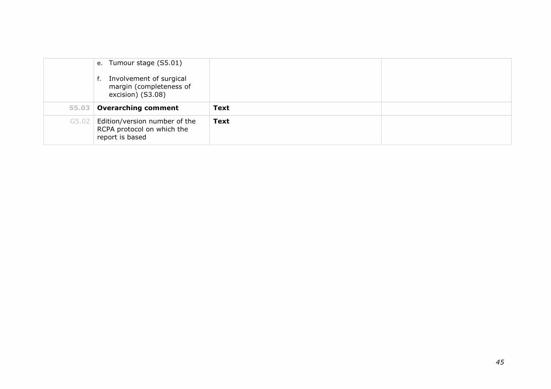
45
e. Tumour stage (S5.01)
f. Involvement of surgical
margin (completeness of
excision) (S3.08)
S5.03 Overarching comment Text
G5.02 Edition/version number of the
RCPA protocol on which the
report is based
Text

46
7 Formatting of pathology reports
Good formatting of the pathology report is essential for optimising communication
with the clinician, and will be an important contributor to the success of cancer
reporting protocols. The report should be formatted to provide information clearly
and unambiguously to the treating doctors, and should be organised with their
use of the report in mind. In this sense, the report differs from the structured
checklist, which is organised with the pathologists’ workflow as a priority.
Uniformity in the format as well as in the data items of cancer reports between
laboratories makes it easier for treating doctors to understand the reports; it is
therefore seen as an important element of the systematic reporting of cancer. For
guidance on formatting pathology reports, please refer to Appendix 2.

47
Appendix 1 Pathology request information and surgical handling procedures
This appendix describes the information that should be collected before the
pathology test. Some of this information can be provided on generic pathology
request forms; any additional information required specifically for the reporting of
renal cancer may be provided by the clinician on a separate request information
sheet. An example request information sheet is included below. Elements which
are in bold text are those which pathologists consider to be required information.
Those in non-bold text are recommended.
Also included in this appendix are the procedures that are recommended before
handover of specimens to the laboratory.
Patient information
➢ Adequate demographic and request information should be
provided with the specimen.
• Items relevant to cancer reporting protocols include:
• patient name
• date of birth
• sex
• identification and contact details of requesting doctor
• date of request
• Whether or not the patient identifies as Aboriginal and/ or Torres
Strait Islander. This is in support of a government initiative to
monitor the health of indigenous Australians particularly in
relation to cancer.
➢ The patient’s health identifiers should be provided.
• The patient’s health identifiers may include the patient’s Medical
Record Number as well as a national health number such as a
patient’s Individual Healthcare Identifier (IHI) (Australia) or the
National Healthcare Identifier (New Zealand).
➢ The Australian Healthcare identifiers i.e. Healthcare Provider Identifier -
Individual (HPI-I) and Healthcare Provider Identifier - Organisation (HPI-
O) should be use, where possible, to identify the requesting doctor.

48
Clinical Information
➢ Any relevant past medical history should be provided.
➢ Any relevant predisposing factors, including genetic status where
appropriate, should be provided.
➢ If the patient has received any pre-operative treatment this should be
recorded.
• Pre-operative treatments may significantly alter the gross and
microscopic appearance of the tumour.
➢ Any relevant family history should be provided.
• Familial syndromes that predispose individuals to renal
neoplasia are well recognized and several of these are
associated with specific sub-types of RCC.
➢ Details of the extent of disease as determined from both clinical
assessment and imaging studies should be provided.
• Relevant information as to the extent of disease obtained from
examination of the patient and from imaging studies is
necessary for the accurate staging of the tumour.
➢ The details of any previous biopsy or surgical specimens removed
from the patient should be provided.
• The details of any tissue removed from the patient will provide
information as to the extent of the tumour. Diagnostic
information regarding the nature of the tumour as determined
by biopsy, may inform handling of the specimen, especially if
genetic studies are being considered. Comparison of the finding
from previous biopsies/surgical specimens may alert the
pathologist to a second, possibly occult, malignancy.
➢ The laterality of the lesion must be recorded.
• Laterality information is needed for identification purposes.
➢ The clinical diagnosis or differential diagnosis should be recorded.
• Providing the provisional clinical diagnosis or differential
diagnosis improves clinicopathological correlation and improves
diagnostic accuracy.
➢ The nature of the operation must be recorded.
• Whether the surgical procedure is a radical, total or partial
nephrectomy must be stated as this will influence the

49
assessment of surgical margins. Specifically in the case of
partial nephrectomy specimens it is important that the intra-
renal surgical margin be carefully evaluated so as to ensure
that no residual tumour is present in the remaining kidney.
• If the kidney has been removed laparoscopically and/or the
specimen has been morcellated this must be stated.74
➢ The operative findings should be recorded.
• The operative findings may provide additional data that is
necessary for accurate staging of the patient.
➢ The surgical intent should be stated.
• The surgeon should provide an indication as to whether the
intent of the operation is palliative or curative. This will inform
the assessment of surgical excision margins.
➢ If tissue has been removed from the specimen for research or
other purposes, this must be stated and details as to nature of
the tissue removed provided.
• Pathologic evaluation requires a detailed examination of the
complete surgical specimen. If tissue has been removed prior
to examination this could compromise diagnosis, staging and
prognostic assessment.
➢ Record if this is a new primary cancer or a recurrence of a
previous cancer, if known.
• The term recurrence defines the return, reappearance or
metastasis of cancer (of the same histology) after a disease
free period.
Recurrence should be classified as distant metastases or
regional (local) recurrence.
Regional (local) recurrence refers to the recurrence of cancer
cells at the same site as the original (primary) tumour or the
regional lymph nodes.
Distant metastasis refers to the spread of cancer of the same
histologic type as the original (primary) tumour to distant
organs or distant lymph nodes.
• This information will provide an opportunity for previous
reports to be reviewed during the reporting process, which may
provide valuable information to the pathologist. This
information also has implications for recording cancer incidence
and evidence based research.

50
Surgical handling
➢ The specimen should be capable of orientation if the status of specific
surgical margins is critical in determining the need for, or extent of,
further surgery.
• Where there are no anatomical landmarks, specimen
orientation may be indicated with marking sutures or other
techniques. If a specimen is oriented, the orientation should be
indicated on the specimen request form (this may be facilitated
by the use of a diagram).

51
Example request information sheet
The above Request Information Sheet is published to the RCPA website.

52
Appendix 2 Guidelines for formatting of a pathology report
Layout
Headings and spaces should be used to indicate subsections of the report, and
heading hierarchies should be used where the LIS allows it. Heading hierarchies
may be defined by a combination of case, font size, style and, if necessary,
indentation.
• Grouping like data elements under headings and using ‘white space’ assists in
rapid transfer of information.75
Descriptive titles and headings should be consistent across the protocol, checklist
and report.
When reporting on different tumour types, similar layout of headings and blocks
of data should be used, and this layout should be maintained over time.
• Consistent positioning speeds data transfer and, over time, may reduce the
need for field descriptions or headings, thus reducing unnecessary information
or ‘clutter’.
Within any given subsection, information density should be optimised to assist in
data assimilation and recall.
• Configuring reports in such a way that they ‘chunk’ data elements into a
single unit will help to improve recall for the clinician.75
• ‘Clutter’ should be reduced to a minimum.75 Thus, information that is not part
of the protocol (e.g. billing information, Snomed codes, etc) should not appear
on the reports or should be minimized.
• Injudicious use of formatting elements (e.g. too much bold, underlining or use
of footnotes) constitutes clutter and may distract the reader from the key
information.
Where a structured report checklist is used as a template for the actual report,
any values provided in the checklist but not applying to the case in question must
be deleted from the formatted report.
Reports should be formatted with an understanding of the potential for the
information to mutate or be degraded as the report is transferred from the LIS to
other health information systems.
As a report is transferred between systems:
• text characteristics such as font type, size, bold, italics and colour are often
lost
• tables are likely to be corrupted as vertical alignment of text is lost when fixed
font widths of the LIS are rendered as proportional fonts on screen or in print
• spaces, tabs and blank lines may be stripped from the report, disrupting the
formatting
• supplementary reports may merge into the initial report.

53
Appendix 3 Example of a pathology report

54

55

56
Appendix 4 World Health Organization (WHO) classification of renal neoplasia
• Clear cell renal cell carcinoma
• Multilocular clear cell renal cell neoplasm of low malignant potential
• Papillary renal cell carcinoma
o Type 1
o Type 2
o Oncocytic
o NOS
• Chromophobe renal cell carcinoma
o Hybrid oncocytic chromophobe tumour
• Collecting duct carcinoma
• Renal medullary carcinoma
• MiT family translocation renal cell carcinoma
o Xp11 translocation renal cell carcinoma
o t(6;11) renal cell carcinoma
• Mucinous tubular and spindle cell carcinoma
• Tubulocystic renal cell carcinoma
• Acquired cystic disease associated renal cell carcinoma
• Clear cell papillary/tubulopapillary renal cell carcinoma
• Hereditary leiomyomatosis and renal cell carcinoma-associated renal cell
carcinoma
• Succinate dehydrogenase (SDH) deficient renal carcinoma
• Renal cell carcinoma, unclassified
© WHO/International Agency for Research on Cancer (IARC). Reproduced with permission

57
References
1 Merlin T, Weston A and Tooher R (2009). Extending an evidence
hierarchy to include topics other than treatment: revising the Australian
'levels of evidence'. BMC Med Res Methodol 9:34.
2 Rayer PFO (1841). Cancer des reins. In: Traité des Maladies Des Reins et
des Altératons de la Sécrétion Urinaire., J.-B. Bailliere, Paris.
3 Delahunt B and Eble JN (2005). History of the development of the
classification of renal cell neoplasia. Clin Lab Med 25(2):231-246, v.
4 Mostofi FK, Sesterhenn IA, Sobin LH, Chaves E, Galindo L, Gouygon C,
Krayevsky NA, McGovern VJ, Rogbeen MS and Thackray AC (1981).
Histological Typing of Kidney Tumours. International Histological
Classification of Tumours No. 25, World Health Organization, Geneva.
5 Kovacs G, Akhtar M, Beckwith BJ, Bugert P, Cooper CS, Delahunt B, Eble
JN, Fleming S, Ljungberg B, Medeiros LJ, Moch H, Reuter VE, Ritz E, Roos
G, Schmidt D, Srigley JR, Stőrkel S, van den Berg E and Zbar B (1997).
The Heidelberg Classification of Renal Cell Tumours. Journal of Pathology
183:131-133.
6 Stőrkel S, Eble JN, Adlakha K, Amin M, Blute ML, Bostwick DG, Darson M,
Delahunt B and Iczkowski K (1997). Classification of renal cell carcinoma.
Cancer 80:987-989.
7 WHO (World Health Organization) (2004). World Health Organization
Classification of Tumours. Pathology and Genetics of Tumours of the
Urinary System and Male Genital Organ. Eble JN, Sauter G, Epstein JI
and Sesterhenn IA. IARC Press, Lyon, France.
8 Srigley JR and Delahunt B (2009). Uncommon and recently described
renal carcinomas. Modern Pathology 22:S2-S23.
9 Srigley JR, Delahunt B, Eble JN, Egevad L, Epstein JI, Grignon D, Hes O,
Moch H, Montironi R, Tickoo SK, Zhou M and Argani P (2013). The
International Society of Urological Pathology (ISUP) Vancouver
Classification of Renal Neoplasia. Am J Surg Pathol 37(10):1469-1489.
10 WHO (World Health Organization) (2016). Classification of tumours.
Pathology and genetics of the urinary system and male genital organs,
4th edition. Humphrey PA, Moch H, Reuter VE and Ulbright TM. IARC
Press, Lyon, France.

58
11 Delahunt B, Bethwaite PB and Nacey JN (2007). Outcome prediction for
renal cell carcinoma: evaluation of prognostic factors for tumours divided
according to histological subtype. Pathology 39(5):459-465.
12 Amin MB, Amin MB, Tamboli P, Javidan J, Stricker H, de-Peralta
Venturina M, Deshpande A and Menon M (2002). Prognostic impact of
histologic subtyping of adult renal epithelial neoplasms: an experience of
405 cases. Am J Surg Pathol 26(3):281-291.
13 Gudbjartsson T, Hardarson S, Petursdottir V, Thoroddsen A, Magnusson J
and Einarsson GV (2005). Histological subtyping and nuclear grading of
renal cell carcinoma and their implications for survival: a retrospective
nation-wide study of 629 patients. Eur Urol 48(4):593-600.
14 Patard JJ, Leray E, Rioux-Leclercq N, Cindolo L, Ficarra V, Zisman A, De
La Taille A, Tostain J, Artibani W, Abbou CC, Lobel B, Guille F, Chopin DK,
Mulders PF, Wood CG, Swanson DA, Figlin RA, Belldegrun AS and Pantuck
AJ (2005). Prognostic value of histologic subtypes in renal cell carcinoma:
a multicenter experience. J Clin Oncol 23(12):2763-2771.
15 Delahunt B (2009). Advances and controversies in grading and staging of
renal cell carcinoma. Mod Pathol 22 Suppl 2:S24-36.
16 American Cancer Society (2018). Cancer Statistics Centre: Kidney and
renal pelvis. Available from:
https://cancerstatisticscenter.cancer.org/?_ga=2.73208531.1930618672
.1513216747-50642755.1513216747#!/cancer-
site/Kidney%20and%20renal%20pelvis. (Accessed 28th Feb 2018).
17 Collins S, McKiernan J and Landman J (2006). Update on the
epidemiology and biology of renal cortical neoplasms. J Endourol
20(12):975-985.
18 Cancer Australia (2018). Kidney cancer statistics. Available from:
https://kidney-cancer.canceraustralia.gov.au/statistics. (Accessed 28th
Feb 2018).
19 New Zealand Ministry of Health (2013). Cancer: New registrations and
deaths. Available from https://www.health.govt.nz/publication/cancer-
new-registrations-and-deaths-2013. (Accessed 14th Dec 2017).
20 Australian Cancer Network Colorectal Cancer Guidelines Revision
Committee (2005). Guidelines for the Prevention, Early Detection and
Management of Colorectal Cancer. The Cancer Council Australia and
Australian Cancer Network, Sydney.

59
21 Maughan NJ, Morris E, Forman D and Quirke P (2007). The validity of the
Royal College of Pathologists' colorectal cancer minimum dataset within a
population. British Journal of Cancer 97(10):1393–1398.
22 Royal College of Pathologist (2017). Cancer datasets and tissue
pathways. Available from:
https://www.rcpath.org/profession/publications/cancer-datasets.html.
(Accessed 19th Dec 2017).
23 College of American Pathologists (2015). Cancer protocol templates.
Available from: http://www.cap.org/web/home/resources/cancer-
reporting-tools/cancer-protocol-templates?_adf.ctrl-
state=10jd5draq2_17&_afrLoop=78742816534289#!%40%40%3F_afrLo
op%3D78742816534289%26_adf.ctrl-state%3D4596lsm96_4.
http://www.cap.org/apps/cap.portal?_nfpb=true&cntvwrPtlt_actionOverri
de=%2Fportlets%2FcontentViewer%2Fshow&_windowLabel=cntvwrPtlt&
cntvwrPtlt%7BactionForm.contentReference%7D=committees%2Fcancer
%2Fcancer_protocols%2Fprotocols_index.html&_state=maximized&_pag
eLabel=cntvwr (Accessed 19th Feb 2016).
24 Cross SS, Feeley KM and Angel CA (1998). The effect of four
interventions on the informational content of histopathology reports of
resected colorectal carcinomas. J Clin Oncol 51(6):481–482.
25 Mathers M, Shrimankar J, Scott D, Charlton F, Griffith C and Angus B
(2001). The use of a standard proforma in breast cancer reporting. J Clin
Pathol 54(10):809–811.
26 Srigley JR, McGowan T, MacLean A, Raby M, Ross J, Kramer S and Sawka
C (2009). Standardized synoptic cancer pathology reporting: A
population-based approach. J Surg Oncol 99(8):517–524.
27 Gill AJ, Johns AL, Eckstein R, Samra JS, Kaufman A, Chang DK, Merrett
ND, Cosman PH, Smith RC, Biankin AV and Kench JG (2009). Synoptic
reporting improves histopathological assessment of pancreatic resection
specimens. Pathology 41(2):161–167.
28 RCPA (Royal College of Pathologists of Australasia (2009 ). Guidelines for
Authors of Structured Cancer Pathology Reporting Protocols. RCPA, Surry
Hills NSW.
29 Amin MB, Edge SB and Greene FL et al (eds) (2017). AJCC Cancer
Staging Manual. 8th ed., Springer, New York.
30 RCPA (Royal College of Pathologists of Australasia) (2004). Chain of
Information Custody for the Pathology Request-Test-Report Cycle —
Guidelines for Requesters and Pathology Providers. RCPA, Surry Hills,
NSW.

60
31 Trpkov K, Grignon DJ, Bonsib SM, Amin MB, Billis A, Lopez-Beltran A,
Samaratunga H, Tamboli P, Delahunt B, Egevad L, Montironi R and
Srigley JR (2013). Handling and staging of renal cell carcinoma: the
International Society of Urological Pathology Consensus (ISUP)
conference recommendations. Am J Surg Pathol 37(10):1505-1517.
32 Delahunt B, Kittelson JM, McCredie MR, Reeve AE, Stewart JH and Bilous
AM (2002). Prognostic importance of tumor size for localized conventional
(clear cell) renal cell carcinoma: assessment of TNM T1 and T2 tumor
categories and comparison with other prognostic parameters. Cancer
94(3):658-664.
33 Bonsib SM (2004). The renal sinus is the principal invasive pathway: a
prospective study of 100 renal cell carcinomas. Am J Surg Pathol
28(12):1594-1600.
34 Bonsib SM (2007). Renal veins and venous extension in clear cell renal
cell carcinoma. Mod Pathol 20(1):44-53.
35 Cheville JC, Lohse CM, Zincke H, Weaver AL and Blute ML (2003).
Comparison of outcome and prognostic features among histologic
suptypes of renal cell carcinoma. Am J Surg Pathol 27:612-624.
36 Murphy WM, Grignon DJ and Perlman EJ (eds) (2004). Tumours of the
Kidney, Bladder, and Related Urinary Structures. AFIP Atlas of Tumour
Pathology Series 4. American Registry of Pathology, Washington DC.
37 Kim H, Cho NH and Kim D et al (2004). Renal cell carcinoma in South
Korea: A multicenter study. Hum Pathol 35:1556-1563.
38 Ljungberg B, Alamdri FI and Stenling R et al (1999). Prognostic
significance of the Heidelberg Classification of renal cell carcinoma. Eur
Urol 36:565-569.
39 Moch H, Grasser T and Amin MB (2000). Prognostic utility of the recently
recommended histologic classification and revised TNM staging system of
renal cell carcinoma. A Swiss experience with 588 tumours. Cancer
89:604-614.
40 Delahunt B, Cheville JC, Martignoni G, Humphrey PA, Magi-Galluzzi C,
McKenney J, Egevad L, Algaba F, Moch H, Grignon DJ, Montironi R,
Srigley JR and the members of the ISUP Renal Tumor Panel (2013). The
International Society of Urological Pathology (ISUP) Grading System for
Renal Cell Carcinoma and Other Prognostic Parameters. Am J Surg
Pathol. 37:1490-1504.
61
41 O'Brien MF, Russo P and Motzer RJ (2008). Sunitinib therapy in renal cell
carcinoma. BJU Int 101(11):1339-1342.
42 Delahunt B, Eble JN, McCredie MR, Bethwaite PB, Stewart JH and Bilous
AM (2001). Morphologic typing of papillary renal cell carcinoma:
comparison of growth kinetics and patient survival in 66 cases. Hum
Pathol 32(6):590-595.
43 Cancer Genome Atlas Research Network (2016). Comprehensive
Molecular Characterization of Papillary Renal-Cell Carcinoma. N Engl J
Med 374(2):135-145.
44 Delahunt B and Eble JN (2002). Renal cell neoplasia. Pathology 34:13-20.
45 Sika-Paotonu D, Bethwaite PB and McCredie MRE et al (2006). Nucleolar
grade but not Fuhrman grade is applicable to papillary renal cell
carcinoma. Am J Surg Pathol 30:1091-1096.
46 Delahunt B, Sika-Paotonu D and Bethwaite PB et al (2011). Grading of
clear cell renal cell carcinoma should be based on nucleolar prominence.
Am J Surg Pathol 135:1134-1139.
47 Delahunt B, Egevad L, Samaratunga H, Martignoni G, Nacey JN and
Srigley JR (2016). Gleason and Fuhrman no longer make the grade.
Histopathology 68(4):475-481.
48 Delahunt B, Sika-Paotonu D and Bethwaite PB et al (2007). Fuhrman
grading is not appropriate for chromophobe renal cell carcinoma. Am J
Surg Pathol 31:957-960.
49 Cheville JC, Lohse CM and Zincke H et al (2004). Sarcomatoid renal cell
carcinoma. An examination of underlying histologic subtype and an
analysis of associations with patient outcome. Am J Surg Pathol 28:435-
441.
50 Cangiano T, Liao J and Naitoh J et al (1999). Sarcomatoid renal cell
carcinoma: biologic behavior, prognosis and response to combined
surgical resection and immunotherapy. J Clin Oncol 17:523-528.
51 Delahunt B (1999). Sarcomatoid renal cell carcinoma. The final common
dedifferentiation pathway of renal epithelial malignancies. Pathology
31:185-190.
52 de Peralta-Venturina M, Moch H and Amin M et al (2001). Sarcomatoid
differentiation in renal cell carcinoma. A study of 101 cases. Am J Surg
Pathol 25:275-278.

62
53 Mian BM, Bhadkamkar N and Slaton JW et al (2002). Prognostic factors
and survival of patients with sarcomatoid renal cell carcinoma. J Urol
167:64-70.
54 Kuroiwa K, Kinoshita Y and Shiratsuchi H et al (2002). Renal cell
carcinoma with rhabdoid features: an aggressive neoplasm.
Histopathology 41:538-548.
55 Gokden N, Nappi O and Swanson PE et al (2000). Renal cell carcinoma
with rhabdoid features. Am J Surg Pathol 24:1329-1338.
56 Leroy X, Zini L and Buob D et al (2007). Renal cell carcinoma with
rhabdoid features. Arch Pathol Lab Med 131:102-106.
57 Klatte T, Said JW and de Martino M et al (2009). Presence of tumour
necrosis is not a significant predictor of survival in clear cell renal cell
carcinoma: higher prognostic accuracy of extent based rather than
presence/absence classification. J Urol 181:1558-1564.
58 Bonsib SM, Gibson D, Mhoon M and Greene GF (2000). Renal sinus
involvement in renal cell carcinoma. Am J Surg Pathol. 24(3):451-458.
59 Bonsib SM (2005). T2 clear cell renal cell carcinoma is a rare entity: a
study of 120 clear cell renal cell carcinomas. J Urol. 174(4 Pt 1):1199-
1202.
60 Thompson RH, Leibovich BC and Cheville JC et al (2005). Is renal sinus
fat invasion the same as perinephric fat invasion for pT3a renal cell
carcinoma? J Urol. 174(4 Pt 1):1218-1221.
61 Bonsib SM (2006). Renal lymphatics, and lymphatic involvement in sinus
vein invasive (pT3b) clear cell renal cell carcinoma: a study of 40 cases.
Mod Pathol 19:746-753.
62 Thompson RH, Cheville JC and Lohse CM et al (2005). Reclassification of
patients with pT3 and pT4 renal cell carcinoma improves prognostic
accuracy. Cancer 104:53-60.
63 Ficcara V, Novara G and Iafrate M et al (2007). Proposal for
reclassificationof the TNM staging systemin patients with locally advanced
(pT3-4) renal cell carcinoma according to the cancer-related outcome.
Eur Urol 51:722-729.

63
64 Lang H, Lindner V and Letourneux H et al (2004). Prognostic value of
microscopic venous invasion in renal cell carcinoma: long term follow-up.
Eur Urol 46:331-335.
65 Terrone C, Cracco C and Porpiglia et al (2006). Reassessing the current
TNM lymph node staging for renal cell carcinoma. Eur Urol 49:324-331.
66 Dimashkieh HH, Lohse CM, Blute ML, Kwon ED, Leibovich BC and Cheville
JC (2006). Extranodal extension in regional lymph nodes is associated
with outcome in patients with renal cell carcinoma. J Urol 176(5):1978-
1982; discussion 1982-1973.
67 Henriksen KJ, Meehan SM and Chang A (2007). Non-neoplastic renal
diseases are often unrecognized in adult tumor nephrectomy specimens:
a review of 246 cases. Am J Surg Pathol. 31(11):1703-1708.
68 Bijol V, Mendez GP, Hurwitz S, Rennke HG and Nose V (2006). Evaluation
of the non-neoplastic pathology in tumor nephrectomy specimens:
predicting the risk of progressive failure. Am J Surg Pathol 30(5):575-
584.
69 Reuter VE, Argani P, Zhou M and Delahunt B (2014). Best practices
recommendations in the application of immunohistochemistry in the
kidney tumors: report from the International Society of Urologic
Pathology consensus conference. Am J Surg Pathol 38(8):e35-49.
70 Tan PH, Cheng L, Rioux-Leclercq N, Merino MJ, Netto G, Reuter VE, Shen
SS, Grignon DJ, Montironi R, Egevad L, Srigley JR, Delahunt B and Moch
H (2013). Renal tumors: diagnostic and prognostic biomarkers. Am J
Surg Pathol 37(10):1518-1531.
71 Sobin L, Gospodarowicz M, Wittekind C and International Union against
Cancer (eds) (2009). TNM Classification of Malignant Tumours, Wiley-
Blackwell, Chichester, UK and Hoboken, New Jersey.
72 Edge SE, Byrd DR, Compton CC, Fritz AG, Greene FL and Trotti A (eds)
(2010). AJCC Cancer Staging Manual 7th ed., New York, NY.: Springer.
73 Royal College of Pathologists of Australasia (2011). Functional
Requirements for Laboratory Information Systems to support Structured
Pathology Reporting of Cancer Protocols
https://www.rcpa.edu.au/Library/Practising-Pathology/Structured-
Pathology-Reporting-of-Cancer/Implementation.
74 Rabban JT, Meng MV, Yeh B, Koppie T, Ferrell L and Stoller ML (2001).
Kidney morcellation in laparoscopic nephrectomy for tumor:

64
recommendations for specimen sampling and pathologic tumor staging.
Am J Surg Pathol 25(9):1158-1166.
75 Valenstein PN (2008). Formatting pathology reports: applying four design
principles to improve communication and patient safety. Arch Path Lab
Med. 132(1):84–94.