Embed Size (px)
Citation preview
RESEARCH ARTICLE
Zebrafish P54 RNA helicases are cytoplasmic granule residentsthat are required for development and stress resilienceCecilia Zampedri1, Maryana Tinoco-Cuellar1, Samantha Carrillo-Rosas2,*, Abigail Diaz-Tellez1,Jose Luis Ramos-Balderas2, Francisco Pelegri3 and Ernesto Maldonado1,‡
ABSTRACTStress granules are cytoplasmic foci that directly respond to theprotein synthesis status of the cell. Various environmental insults,such as oxidative stress or extreme heat, block protein synthesis;consequently, mRNA will stall in translation, and stress granules willimmediately form and become enriched with mRNAs. P54 DEAD boxRNA helicases are components of RNA granules such as P-bodiesand stress granules. We studied the expression, in cytoplasmic foci,of both zebrafish P54 RNA helicases (P54a and P54b) duringdevelopment and found that they are expressed in cytoplasmicgranules under both normal conditions and stress conditions. Inzebrafish embryos exposed to heat shock, some proportion of P54aandP54b helicasesmove to larger granules that exhibit the propertiesof genuine stress granules. Knockdown of P54a and/or P54b inzebrafish embryos produces developmental abnormalities restrictedto the posterior trunk; further, these embryos do not form stressgranules, and their survival upon exposure to heat-shock conditionsis compromised. Our observations fit the model that cells lackingstress granules have no resilience or ability to recover once the stresshas ended, indicating that stress granules play an essential role in theway organisms adapt to a changing environment.
KEY WORDS: P54 RNA helicase, P-bodies, Stress granules,Zebrafish
INTRODUCTIONIn eukaryotic cells,mRNA regulation is carried out inmultiple parts ofthe cell and during multiple stages of the cell cycle. Importantreservoirs for mRNAs are the RNA granules, which include P-bodies(Oh et al., 2013; Parker and Sheth, 2007), stress granules (Andersonand Kedersha, 2008; Kedersha et al., 2002; Sahoo et al., 2012), germgranules (in germ cells) (Updike and Strome, 2010), and other types ofRNA-containing cytoplasmic foci. This classification of RNAgranules is based on their composition, size and cell of origin. Themechanism leading to the formation ofRNAgranules remains amatter
of debate (Ramaswami et al., 2013; Weber and Brangwynne, 2012).A remarkable aspect of RNA-containing granules is the absence ofencapsulating membranes which leaves RNA and associated RNA-binding proteins free to shuttle in and out of granules in a dynamicequilibrium, rendering such aggregates unstable by nature (Buchanand Parker, 2009). For this reason, the isolation of RNA-containinggranules from cells and their further characterization in vitro remainissues that have proven difficult to address. The different classes ofRNA granules share common features. They possess mRNAs in arepressed state that may re-initate translation in response to specificsignals (Bhattacharyya et al., 2006; Brengues et al., 2005; Nagamoriet al., 2011). Further, they exhibit dynamic interactions with oneanother, such as docking, fusion, or apparent maturation from onegranule type to the next (Hoyle et al., 2007; Kedersha et al., 2005).Meanwhile, RNA granules share certain components, such as RNA-binding proteins and certain mRNAs (Buchan and Parker, 2009), andfrequently, some components shuffle from one type of granule toanother granule type as cellular conditions change (Buchan et al.,2008; Kedersha et al., 2005; Mollet et al., 2008).
One of the most-studied shared components of different types ofgranules is the DEAD-box P54/RCK RNA helicase. This protein is amember of a helicase DDX6 subfamily, conserved in invertebratesand vertebrates,with homologues in human (RCK/P54),mouse (P54),Xenopus (Xp54), Drosophila (Me31B), Caenorhabditis elegans(Cgh-1),Planaria (DjCBC-1), and Saccharomyces cerevisiae (Dhh1)(Navarro and Blackwell, 2005; Navarro et al., 2001; Rajyaguru andParker, 2009; Weston and Sommerville, 2006; Yoshida-Kashikawaet al., 2007). In mammalian cells, depletion of P54/RCK protein leadsto the disappearance of P-bodies and prevents their de novo assemblyin response to triggers such as arsenite, which means that P54/RCK iscentral to P-body assembly (Serman et al., 2007).
It also has been reported that P54/RCK interacts with P-bodies/decapping proteins (Bish et al., 2015) and with the RISC complex,which mediates translational silencing by miRNAs (Chu and Rana,2006). Ddx6 also interacts with two stress granule proteins (GRAN1and GRAN2), even under normal conditions, when visible mRNPstructures are absent, suggesting that Ddx6 may be a key factor inmodulating the contents of P-bodies and stress granules (Bish et al.,2015). Xp54 in Xenopus is known as a component of the CPEBrepressor complex in oocytes (Ladomery et al., 1997; Minshallet al., 2001), and in C. elegans, the P54 homolog Cgh-1 (Navarroand Blackwell, 2005) promotes both mRNA stability in P-granulesin oocytes and mRNA decay in somatic cell granules (Boag et al.,2008; Noble et al., 2008). In zebrafish (Danio rerio), stress granuleswere identified, while studying FUS (fused in sarcoma) abnormalcytoplasmic localization, by the use of antibodies against classicstress granules markers such as TIAL-1 (T-cell internal antigenlike-1) (Bosco et al., 2010) and eIF3e (Acosta et al., 2014).
Stress granules are formed in response to various stressors, andtheir components include translation initiation factors (eIF3, eIF4A,Received 3 November 2015; Accepted 26 July 2016
1EvoDevo Laboratory, Unidad de Sistemas Arrecifales, Instituto de Ciencias delMar y Limnologıa, Universidad Nacional Autonoma de Mexico, Puerto Morelos,Quintana Roo, Mexico, 77580. 2Departamento de Biologıa Celular y del Desarrollo,Instituto de Fisiologıa Celular, Universidad Nacional Autonoma deMexico, D.F. Mexico, Mexico, 04510. 3Laboratory of Genetics, University ofWisconsin-Madison, Wisconsin 53706, USA.*Present address: Institut Genetique Biologie Moleculaire Cellulaire, Illkirch 67400,France.
‡Author for correspondence ([email protected])
E.M., 0000-0002-9627-967X
This is an Open Access article distributed under the terms of the Creative Commons AttributionLicense (http://creativecommons.org/licenses/by/3.0), which permits unrestricted use,distribution and reproduction in any medium provided that the original work is properly attributed.
1473
© 2016. Published by The Company of Biologists Ltd | Biology Open (2016) 5, 1473-1484 doi:10.1242/bio.015826
BiologyOpen
by guest on September 17, 2018http://bio.biologists.org/Downloaded from

eIF4E and eIF4G), stalled mRNAs (Anderson and Kedersha, 2008;Anderson et al., 2015), 40S ribosomal subunits, and RNA bindingproteins (RBPs). Some examples for these RBPs are TIA1 (T-cellinternal antigen 1), TIAL-1 (T-cell internal antigen like-1)(Kedersha et al., 2000), and G3BP1 (Ras-GTPase-activatingprotein SH3-domain binding protein 1) (Tourriere et al., 2003).These granules are in a dynamic equilibrium with polysomalmRNA; it was observed in photobleaching experiments that theyrespond directly to the translational status of the cell (Mollet et al.,2008). Global inhibition of protein synthesis induces coalescence ofstress granule components; nevertheless, the molecular details of theaggregation process are not clearly understood.In this work, we characterize P54/RCK homologs P54a and P54b
in zebrafish, particularly their residence in cytoplasmic foci thatare similar to P-bodies and to stress granules. Using a combinationof heat-shock treatments and classical translation inhibitors(cycloheximide and puromycin) we found that P54 granulesexhibit the properties of classical stress granules. This work alsoshows that P54 granules are essential for whole-organismdevelopment and resilience or the ability to recover after heat shock.
RESULTSZebrafish have two P54 DEAD box RNA helicasesHuman Rck/p54 and Caenorhabditis elegans cgh-1 belong toa family of DEAD box RNA helicases, closely related to eIF4Athat allows translation initiation by mRNA unwinding (Linderand Fuller-Pace, 2013). In the zebrafish (Danio rerio) genome, wefound two open reading frames in the Ensembl databases;
ENSDARP00000081816.5 (Chromosome 18:43.87 Mb) andENSDARP00000129311.1 (Chromosome 16:25.76 Mb). Accordingto protein sequence multiple alignment and phylogenetic analysis(Fig. 1) we found both genes to be orthologs of CGH-1 inC. elegans, Me31B inDrosophila melanogaster, DDX6 in Xenopuslaevis and the human Rck/p54 family of DEAD box RNA helicases.We named them P54a and P54b, respectively. All conserveddomains from this DEAD box protein family are also conserved inzebrafish P54a and P54b, including the ATP-binding domain I andRNA-binding motifs IV and V. The conserved NLS (nuclearlocalization signal) and NES (nuclear export signal) sequences,only found in P54 RNA helicases from vertebrates, were also foundin zebrafish P54 proteins (Fig. 1A). In a phylogenetic tree ofselected DEAD box RNA helicases, the eIF4A branch is clearly anoutgroup from the P54/RCK/Cgh-1 branch (Fig. 1B). All knowngenomes from teleost fishes contain both P54a and P54b RNAhelicases (data not shown); in zebrafish, the presence of duplicatedgenes is a common feature due to an ancient genome duplicationduring the evolution of ray-finned fish (Glasauer and Neuhauss,2014). P54a appears to be more closely related to P54 frommammals than P54b (93.8% and 85% identity with the humanortholog, respectively).
P54a and P54b are both expressed in cytoplasmic granulesduring zebrafish developmentP54 DEAD box RNA helicases have been studied in severalorganisms and are usually found in cytoplasmic granules with RNAprocessing functions (Presnyak and Coller, 2013). We used an
Fig. 1. Domain structure and evolutionaryconservation of the P54 RNA helicases P54aand P54b from zebrafish. (A) Conserveddomains in P54RNA helicases (NLS, Q, I, NES,Ia, Ib, II, III, IV, V and VI) are indicated in coloredboxes. Zebrafish P54a and P54b proteins arecompared with human RCK (Uniprot P26196)and CGH_1 from C. elegans (WormbaseC07H6.5) proteins. The central white boxesrepresent the conserved helicase region, and itspercentage identity with RCK is shown. Grayboxes indicate C- and N-terminal variableregions. (B) Maximum likelihood tree of P54-related proteins. The scale indicatessubstitutions per site. The outgroup isrepresented by eIF4A RNA helicases fromvarious organisms. CGH-1, Me31B, DDX6 andRCK are different names for the P54 RNAhelicase. Abbreviations: H. sapiens (Homosapiens), D. rerio (Danio rerio), C. elegans(Caenorhabditis elegans), D. melanogaster(Drosophila melanogaster), O. latipes (Oryziaslatipes or medaka fish), X. laevis (Xenopuslaevis), M. musculus (Mus musculus).
1474
RESEARCH ARTICLE Biology Open (2016) 5, 1473-1484 doi:10.1242/bio.015826
BiologyOpen
by guest on September 17, 2018http://bio.biologists.org/Downloaded from

antibody against P54 proteins (see theMaterial andMethods sectionand Fig. S1) to locate the expression of P54 RNA helicases duringdifferent developmental stages in zebrafish. P54 was observed bothin cytoplasmic granules and diffused in the cytoplasm, beginningvery early in development, at the 4-cell stage [1 hour post-fertilization (hpf); Fig. 2A] and later at the sphere (4 hpf), 10-somites (10 hpf) stage and in 24 hpf embryos (Fig. 2B–D).Although both zebrafish P54 helicases have nuclear localizationsequences, we did not observe any nuclear labeling. Because P54has been found in germ granules in C. elegans (P-granules) andDrosophila melanogaster (Polar granules) (Nakamura et al., 2001;Navarro et al., 2001), we compared the expression of P54 with two
known markers for zebrafish germ granules; phosphorylated non-muscle myosin (NMII-p) and Vasa. Unexpectedly the expressionpattern of P54 at the 16-cell stage (1.5 hpf ) did not resemble thelabeling reported for NMII-p at the cell division furrows (Nair et al.,2013), neither resemble the typical 24 hpf germ granule anti-Vasastaining (Braat et al., 2000) (Fig. 2E–H). These results indicate thatP54 RNA helicases may not associate with germ granules inzebrafish. However, we observed that P54 immunostaining duringzebrafish development (Fig. 2A–H) resembles the labeling ofcytoplasmic granules known as P-bodies, where P54 RNA helicaseshomologs are known to reside (Minshall et al., 2009; Navarro andBlackwell, 2005; Presnyak and Coller, 2013).
Fig. 2. P54 proteins are expressed in cytoplasmic granules during zebrafish development. (A–E,G) Whole-mount immunostaining was performed with theanti-P54 antibody in WT zebrafish embryos at (A) the 4-cell stage, (B) the sphere stage, (C) the 10-somite stage, and (D,E,G) 24 hpf. In these embryos, P54helicases were located in cytoplasmic granules. Immunostaining (green) and DAPI (blue) were visualized by epifluorescence microscopy. (E–H) P54-positivegranules were different from germline granules, observed by immunostaining with anti-NMII-p (16-cell stage) or anti-Vasa (24 hpf), two known markers ofgerm granules. Arrowheads indicate P54-positive granules, and arrows indicate granules labeled by anti-NMII-p in germplasm or anti-Vasa in germ granules.(I–K) Live embryos at 24 hpf expressing the P54a-mCherry and P54b-EGFP fusion proteins that were also located in cytoplasmic granules. Arrowsindicate cytoplasmic granules where P54a and P54b co-localize. (L–W) Expression of P54a-EGFP and P54b-EGFP fusion reporters and immunostaining withanti-Dcp2 (a marker for P-bodies) in 24 hpf embryos maintained at normal temperature (28.5°C) (L–N,R–T) or exposed for 2 h to heat-shock conditions (37°C)(O–Q,U–W). P54a and P54b fusion proteins co-localize with or are in close contact with Dcp2-labeled granules. Under heat-shock conditions, P54a-EGFP andP54b-EGFP were observed in larger granules that can be seen in close contact with smaller Dcp2-positive granules. Small granules, such as P-bodies, arelabeled with arrowheads, and larger granules are indicated with arrows. For some images, a general view of the embryos is shown in the inset, wherewhite boxesindicate the regions where the analysis was performed.
1475
RESEARCH ARTICLE Biology Open (2016) 5, 1473-1484 doi:10.1242/bio.015826
BiologyOpen
by guest on September 17, 2018http://bio.biologists.org/Downloaded from

Our anti-P54 serum could not distinguish whether these putativeP-bodies contain both P54a and P54b RNA helicases. In order tosolve this problem, we made C-terminal mCherry and EGFP fusionproteins with both P54a and P54b, respectively, and expressedthese fusion proteins in zebrafish embryos. Both P54a-mCherryand P54b-EGFP fusion proteins were found to co-localize incytoplasmic granules (Fig. 2I–K); some labeling was also observeddiffused in the cytoplasm. This is a similar localization pattern to theone observed using the anti-P54 serum (Fig. 2D; Fig. S2C). In orderto determine whether these cytoplasmic granules correspond toP-bodies, embryos expressing either P54a-EGFP or P54b-EGFPwere immunostained using a bona fide marker for P-bodies, theanti-Dcp2 antibody, that binds to the mRNA deccaping protein-2.We observed similar punctuated pattern for P54a/P54b and Dcp2 butunexpectedly very few of them show co-localization (Fig. 2L–N,R–T). These data suggest that some P54a and P54b foci couldcorrespond to P-bodies even though they are located in differentgranules.While P-bodies are constitutive cytoplasmic granules required
for RNA storage and decay, some other cytoplasmic granules onlyappear under stress conditions (Anderson et al., 2015). These areknown as stress granules, and it has been shown that P54 RNAhelicases are also components of stress granules in other organisms(Wilczynska et al., 2005). Zebrafish embryos expressing P54a-EGFP or P54b-EGFP were exposed to heat-shock conditions andimmunostained with anti-Dcp2. As expected, P-bodies (Dcp2
positive) did not change in size with the heat-shock treatment, but,interestingly, P54a and P54b fusion proteins aggregated in largerfoci during this stress condition (Fig. 2O–Q,U–W). These resultssuggest that zebrafish have large, heat-shock-dependent P54a andP54b cytoplasmic granules that resemble stress granules. It isimportant to mention that most of our observations were carried outat the trunk anterior regions of the 24 hpf embryo, and were frommuscle cells or epithelial cells from the skin (Fig. S2).
P54 RNA helicases from zebrafish are components of stressgranulesTo learn more about P54 granules dynamics during heat shock, weexposed 24 hpf embryos grown at 28.5°C, to 37°C for 30 min, 1 hor 2 h, followed by fixation and immunostaining using the anti-P54specific serum. Under normal conditions, P54 was observed insmall, discrete foci in the cytoplasm of most cells in the embryos(Fig. 3A). It was only after heat-shock that P54 RNA helicases wereobserved in larger granules, and the ratio of small to larger granuleswas increased as heat shock conditions were extended (Fig. 3B–D).In normal conditions, the average diameter measured for P54a-EGFP and P54b-EGFP cytoplasmic granules was 0.3 and 0.29 μm,respectively. By contrast, in heat shock conditions, P54a-EGFP andP54b-EGFP cytoplasmic granules have an average diameter of 1.6and 1.0 μm respectively (Fig. S3). It is interesting that P54a-EGFPgranules are larger than P54b-EGFP cytoplasmic granules, underheat shock conditions.
Fig. 3. Heat-shock induced the association of P54a and P54b RNA helicases with large cytoplasmic granules. (A,E–G,K–M,Q) Wild-type 24 hpf zebrafishembryos were maintained under normal conditions at 28.5°C or (B–D,H–J,N–P,R,S) under heat-shock conditions at 37°C. (A–D) Anti-P54 immunostaining inembryos subjected to heat-shock for 30 min, 1 h and 2 h. Nuclei are counterstained with DAPI. The top right insets are general views, and the amplifiedregions are indicated in white boxes. (E–J) Expression of the P54a-EGFP in combination with TIAL-1 immunostaining (as a stress granule marker). In the inset,some P54a-expressing granules co-localize with or are in close contact with TIAL-1-positive granules. (K–P) P54b-EGFP expression in small (at 28.5°C) or large(at 37°C) granules, combined with immunostaining with anti-TIAL-1. The insets show both P54b-expressing granules and TIAL-1-positive granules. Arrowheadspoint to small granules, such as P-bodies, and arrows indicate larger granules, such as TIAL-1-positive (red) stress granules. (Q–S) Knockdown of P54a andP54b expression by splice-blocking morpholinos prevents the formation of stress granules labeled by anti-TIAL-1. For each panel, the percentage of embryosshowing the same pattern is indicated, and the total number of fish embryos tested is shown in parentheses. White arrowheads point to small granules (putativeP-bodies) and white arrows to large granules (putative stress granules).
1476
RESEARCH ARTICLE Biology Open (2016) 5, 1473-1484 doi:10.1242/bio.015826
BiologyOpen
by guest on September 17, 2018http://bio.biologists.org/Downloaded from
TIAL-1 is classicmarker for stress granules inmammals (Kedershaet al., 1999;Moutaoufik et al., 2014). Therefore, to compare P54 heat-shock-induced cytoplasmic granules with TIAL-1-labeled stressgranules we combined anti-TIAL-1 immunostaining and theexpression of P54 fusion proteins. Specifically, we used 24 hpfembryos expressing either P54a-EGFP or P54b-EGFP reporters thatalso were immunostained with the anti-TIAL-1 antibody. We foundthat recombinant P54a-EGFP and P54b-EGFP reporters are localizedin larger cytoplasmic granules induced by heat-shock (Fig. 3E,H,K,L;Fig. S3). As expected, the same dynamics was also observed in stressgranules labeled with the anti-TIAL-1 antibody (Fig. 3F,I,L,O).However, we did not observe co-localization between P54a/P54bfusion proteins and anti-TIAL-1-labeled stress granules. Interestinglywe did observe P54a-EGFP and P54b-EGFP granules adjacent tostress granules (Fig. 3G,J,M,P). It is not unusual to find cytoplasmicRNA granules of different types (such as stress granules andP-bodies) in close contact with each other; this has been seen as anindication of active flow of RNAs and proteins between them(Anderson and Kedersha, 2008).Next, we reasoned that if P54 RNA helicases are essential for
stress granule formation, knocking down their expression may affectthe induction of heat shock stress granules. We knocked down theexpression of both P54a and P54b in zebrafish by microinjection ofsplice-blocking morpholinos (see the Materials and Methodssection and Fig. S1). This blocked the formation of TIAL-1-labeled stress granules under heat shock conditions, but theexpression of those granules was rescued by the co-injection of‘in vitro’ synthesized mature p54mRNA ( p54-mRNA; Fig. 3Q–S).The absence of stress granules has also been observed upon thetreatment of HeLa cells with the protein synthesis inhibitorcycloheximide (Wilczynska et al., 2005), which blocks translation
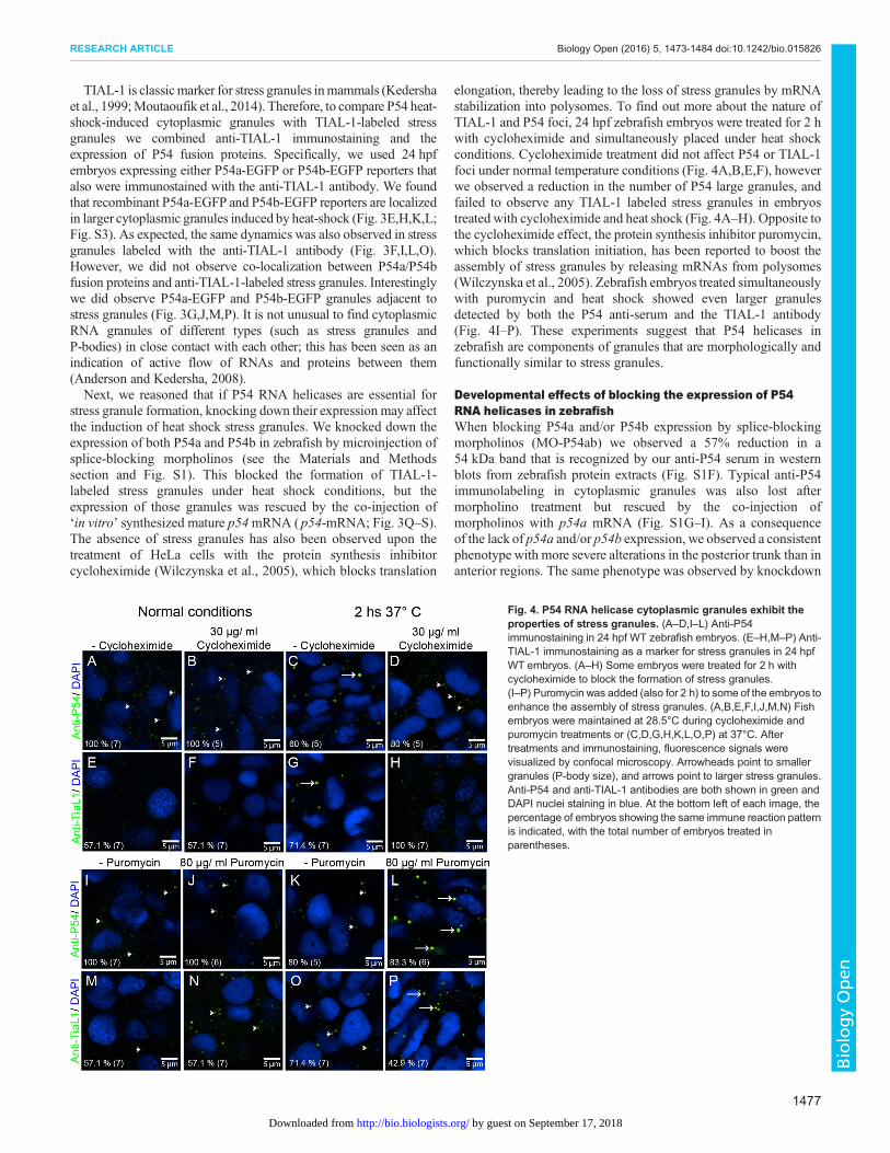
elongation, thereby leading to the loss of stress granules by mRNAstabilization into polysomes. To find out more about the nature ofTIAL-1 and P54 foci, 24 hpf zebrafish embryos were treated for 2 hwith cycloheximide and simultaneously placed under heat shockconditions. Cycloheximide treatment did not affect P54 or TIAL-1foci under normal temperature conditions (Fig. 4A,B,E,F), howeverwe observed a reduction in the number of P54 large granules, andfailed to observe any TIAL-1 labeled stress granules in embryostreated with cycloheximide and heat shock (Fig. 4A–H). Opposite tothe cycloheximide effect, the protein synthesis inhibitor puromycin,which blocks translation initiation, has been reported to boost theassembly of stress granules by releasing mRNAs from polysomes(Wilczynska et al., 2005). Zebrafish embryos treated simultaneouslywith puromycin and heat shock showed even larger granulesdetected by both the P54 anti-serum and the TIAL-1 antibody(Fig. 4I–P). These experiments suggest that P54 helicases inzebrafish are components of granules that are morphologically andfunctionally similar to stress granules.
Developmental effects of blocking the expression of P54RNA helicases in zebrafishWhen blocking P54a and/or P54b expression by splice-blockingmorpholinos (MO-P54ab) we observed a 57% reduction in a54 kDa band that is recognized by our anti-P54 serum in westernblots from zebrafish protein extracts (Fig. S1F). Typical anti-P54immunolabeling in cytoplasmic granules was also lost aftermorpholino treatment but rescued by the co-injection ofmorpholinos with p54a mRNA (Fig. S1G–I). As a consequenceof the lack of p54a and/or p54b expression, we observed a consistentphenotype with more severe alterations in the posterior trunk than inanterior regions. The same phenotype was observed by knockdown
Fig. 4. P54 RNA helicase cytoplasmic granules exhibit theproperties of stress granules. (A–D,I–L) Anti-P54immunostaining in 24 hpf WT zebrafish embryos. (E–H,M–P) Anti-TIAL-1 immunostaining as a marker for stress granules in 24 hpfWT embryos. (A–H) Some embryos were treated for 2 h withcycloheximide to block the formation of stress granules.(I–P) Puromycin was added (also for 2 h) to some of the embryos toenhance the assembly of stress granules. (A,B,E,F,I,J,M,N) Fishembryos were maintained at 28.5°C during cycloheximide andpuromycin treatments or (C,D,G,H,K,L,O,P) at 37°C. Aftertreatments and immunostaining, fluorescence signals werevisualized by confocal microscopy. Arrowheads point to smallergranules (P-body size), and arrows point to larger stress granules.Anti-P54 and anti-TIAL-1 antibodies are both shown in green andDAPI nuclei staining in blue. At the bottom left of each image, thepercentage of embryos showing the same immune reaction patternis indicated, with the total number of embryos treated inparentheses.
1477
RESEARCH ARTICLE Biology Open (2016) 5, 1473-1484 doi:10.1242/bio.015826
BiologyOpen
by guest on September 17, 2018http://bio.biologists.org/Downloaded from
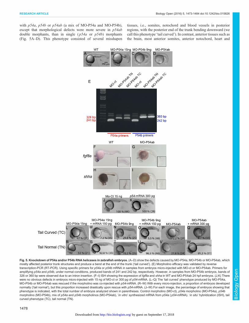
with p54a, p54b or p54ab (a mix of MO-P54a and MO-P54b),except that morphological defects were more severe in p54abdouble morphants, than in single ( p54a or p54b) morphants(Fig. 5A–D). This phenotype consisted of several misshapen
tissues, i.e., somites, notochord and blood vessels in posteriorregions, with the posterior end of the trunk bending downward (wecall this phenotype ‘tail curved’). In contrast, anterior tissues such asthe brain, most anterior somites, anterior notochord, heart and
Fig. 5. Knockdown of P54a and/or P54b RNA helicases in zebrafish embryos. (A–D) show the defects caused by MO-P54a, MO-P54b or MO-P54ab, whichmostly affected posterior trunk structures and produce a bend at the end of the trunk (‘tail curved’). (E) Morpholino efficacy was validated by reversetranscription-PCR (RT-PCR). Using specific primers for p54a or p54b mRNA in samples from embryos micro-injected with MO-ct or MO-P54ab. Primers foramplifying p54a and p54b, under normal conditions, produced bands of 241 and 242 bp, respectively. However, in samples from MO-P54b embryos, bands of326 or 360 bp were observed due to an intron insertion. (F–I) ISH showing the expression of fgf8a and shha in WT and MO-P54ab 24 hpf embryos. (J,K) Therewere no obvious defects in embryos micro-injected with 15 ng of MO-ct or 300 pg of p54-mRNA. (L–Q) The ‘tail curved’ phenotype produced by MO-P54a,MO-P54b or MO-P54ab was rescued if the morpholino was co-injected with p54-mRNA. (R–W) With every micro-injection, a proportion of embryos developednormally (‘tail normal’), but this proportion increased drastically upon rescue with p54-mRNA. (J–W) For each image, the percentage of embryos showing thatphenotype is indicated, with the total number of embryos analyzed shown in parentheses. Control morpholino (MO-ct), p54a morpholino (MO-P54a), p54bmorpholino (MO-P54b), mix of p54a and p54b morpholinos (MO-P54ab), ‘in vitro’ synthesized mRNA from p54a (p54-mRNA). ‘in situ’ hybridization (ISH), tailcurved phenotype (TC), tail normal (TN).
1478
RESEARCH ARTICLE Biology Open (2016) 5, 1473-1484 doi:10.1242/bio.015826
BiologyOpen
by guest on September 17, 2018http://bio.biologists.org/Downloaded from

anterior blood vessels appear normal or only mildly affected(Fig. 5A–D).Pleiotropic zebrafish mutants are known to show defects first in
anterior regions, since the rate of cell division is higher in the brainthat in the rest of the body during early stages of development.However, this is not the case for p54a and p54b morphants,indicating that it is a specific phenotype. Splice-blockingmorpholinos were efficient at disrupting p54a and p54b RNAmaturation because we observed intron insertion events for bothgenes when microinjecting either MO-P54a or MO-P54a (data notshown) or when microinjecting a mix of the two morpholinos(Fig. 5E).Two mRNAs known to be expressed in both the head and the
posterior trunk ( fgf8a and shha) were tested by in situ hybridization(ISH) in p54ab double morphants. We observed that, while anteriordomains continued to express these particular genes, the posteriorregions lost expression when both P54a and P54b were knockeddown (Fig. 5F–I). Other markers, such as krox20, bmp4, nog1, flhand ntl, which are specific for anterior and/or posterior regions, werealso tested by ISH, but no significant differences were foundbetween wild type (WT) and morphants (Fig. S4). In order todetermine whether this phenotype is in fact induced by the loss ofP54a and/or P54b expression, we conducted a rescue experimentby co-injecting MO-P54a with p54a-mRNA, MO-P54b withp54a-mRNA or MO-P54ab with p54a-mRNA. The ‘tail curved’phenotype that is usually present at rates of 47% (MO-P54a), 29.8%(MO-P54b) and 72% (MO-P54ab) was observed at only 7.2% (MO-P54a), 3.5% (MO-P54b) and 8.7% (MO-P54ab) in the presence ofp54a-mRNA (Fig. 5J–W). As a control, embryos injected with thesame amount of p54a-mRNA alone did not have phenotypic defects(Fig. 5K). These experiments suggest that knocking down P54a andP54b induces defects in the development of posterior trunkstructures.
P54 RNA helicases are required for resilience after stresstreatmentIt is known that translation is blocked by cellular stress such as heatshock and that while polysomes are disassembled, mRNAs stalledin translation become accumulated in stress granules. Stressgranules are essential for conserving anabolic energy andpreserving essential mRNAs later required for repairing stress-induced molecular damage (Bergkessel and Reese, 2004; Yamasakiand Anderson, 2008). We next tested whether the knockdown ofP54 RNA helicases affects survival after heat-shock stress. Controland morphant embryos at 24 hpf were heat-shocked for 2 h at 37°Cand then allowed to recover under normal conditions (at 28.5°C).For the purposes of this analysis, morphant embryos were classifiedinto two groups, ‘tail curved’ (with phenotypic defects) and ‘tailnormal’ (with no evident phenotypic defects). Data were collectedhourly during the first 4 h of recovery for each group of embryos andthereafter twice per day for the 5 days after the stress treatment(Fig. 6).When growing at normal temperature double-morphants MO-
P5ab (co-injected with a mix of MO-P54a and MO-P54b) havehigher rates of survival, approximately 95% after five days ofdevelopment. In fact, they survive at a similar rate to zebrafishembryos micro-injected with a control morpholino (Fig. 6A,B).Heat shock alone reduced the survival rate of embryos micro-injected with control morpholino to approximately 85–88%.However, survival rates for double morphants (MO-P54ab) dropto 45% after heat shock. Phenotypic ‘tail curved’ P54ab morphantsshowed slightly lower survival rates (Fig. 6B) than ‘tail normal’
P54ab morphants (Fig. 6A). Furthermore, co-injection of MO-P54ab with p54a mRNA rescues embryos from lethality after heatshock to almost normal rates of survival (86%) (Fig. 6C). This resultsuggests that P54 RNA helicases are required for survival after heatshock treatment. Because we previously observed that knockingdown P54 RNA helicases impairs the formation of stress granules(Fig. 3Q–R), we propose that P54ab morphants are less likely torecover after heat shock due to the loss of stress granules.
DISCUSSIONWe found that zebrafish possess two P54 Dead box RNA helicasescoded by the genes p54a and p54b on chromosomes 18 and 16,respectively. Phylogenetic analysis show that these are co-orthologsof the tetrapod p54 RNA helicase gene and likely appear in theteleost whole-genome duplication (Glasauer and Neuhauss, 2014).Both P54a and P54b are expressed in cytoplasmic granules inzebrafish embryos, consistent with previous results that P54 RNAhelicases are components of P-bodies and stress granules (Bishet al., 2015; Minshall et al., 2009; Navarro et al., 2001; Paz-Gomezet al., 2014; Serman et al., 2007). Zebrafish P54 cytoplasmicgranules resemble P-bodies under normal conditions and stressgranules under heat-shock conditions. P54 RNA helicase and Dcp2(mRNA decapping enzyme) are frequently used as markers forP-bodies (Ingelfinger et al., 2002; Minshall et al., 2009). Inzebrafish, P-bodies labeled with anti-Dcp2 were similar in size toP54-containing granules. While P54a-mCherry and P54b-EGFPfusion proteins were often found in the same granules, we did notobserve co-localization of P54 fluorescent reporters withDcp2-positive granules, this was unexpected and may beexplained by the existence of different classes of P-bodies, evenin the same cell, as was proposed before (Parker and Sheth, 2007).The cytoplasmic granules containing P54 helicases in zebrafish donot resemble germ granules but are highly similar to stress granules.
Stress granules are reversible aggregates of RNA-bindingproteins and translation initiation factors and contain untranslatedmRNAs. These granules assemble in response to stress conditionsand are typically larger than P-bodies (Anderson et al., 2015). Wefound that by treating zebrafish embryos with a heat shock theformation of large cytoplasmic granules was induced. We proposethese are stress granules because (i) they were heat-induced, (ii) theywere larger than P-bodies, and (iii) they were labeled with the anti-TIAL-1 antibody (a known marker for stress granules) (Kedershaet al., 1999; Moutaoufik et al., 2014). In normal conditions fusionproteins (like P54a-EGFP, P54a-EGFP or P54a-mCherry) weredetected in cytoplasmic granules with an average diameter of0.3 μm, while in heat shock conditions the same fusion proteinswere found in cytoplasmic granules with an average size of 1 to1.6 μm in diameter. Our measurements are equivalent to previousreports for P-bodies and stress granules, respectively (Eulalio et al.,2007; Thomas et al., 2009). However, we failed to observe co-localization between P54 fusion reporters and anti-TIAL-1-positivecytoplasmic granules. It is interesting that very often we detectedP54 heat shock granules adjacent to TIAL-1 granules. A similarobservation was recently made in zebrafish cells in culture, whereanti-eIF3e antibody-labeled stress granules were found in closecontact with FUS-GFP-containing granules and coincidently bothtypes of granules were induced under stress conditions (Acostaet al., 2014). In our case, both P54 large granules and TIAL-1granules only appeared after embryo heat exposure.
We also observed that P54 heat-induced large granules werelocated in the same cells and in close contact with smaller P54granules. It is known that stress granules and P-bodies are both
1479
RESEARCH ARTICLE Biology Open (2016) 5, 1473-1484 doi:10.1242/bio.015826
BiologyOpen
by guest on September 17, 2018http://bio.biologists.org/Downloaded from

present in stress conditions and that they are frequently close to eachother (Kedersha et al., 2005; Souquere et al., 2009; Wilczynskaet al., 2005). At the same time it was proposed that specific proteinsor mRNAs are exchanged between P-bodies and stress granules(Buchan et al., 2008; Stoecklin and Kedersha, 2013); however,formation of both types of granules was induced in stress conditions(Acosta et al., 2014). Our observations were carried out mainly inepithelial cells from the skin and muscle cells, since these wereeasier to image in the 24 hpf treated embryos (Fig. S2). Even though
stress granules have been reported to be widely distributed in manycell types and do not seem to be cell specific, some RNA granules,like SX-bodies (Thomas and Boccaccio, 2016) were only detectedin neurons, therefore further work will be required to determine ifthere are different types of stress granules and if some of these couldbe cell-specific. We observed that knocking down the expression ofP54a or P54b with morpholinos prevented the formation of stressgranules in heat-shock conditions, as detected by the anti-TIAL-1antibody. This effect could be rescued by co-injection of p54a
Fig. 6. Resilience after heat shock. Comparison of survival rates between embryos maintained at 28°C (normal conditions) or exposed to heat-shockconditions at 37°C for 2 h (heat shock). Embryos were micro-injected with MO-Ct or MO-P54ab. The MO-P54ab micro-injected embryos were separated into twogroups. (A) Morphants showing the tail normal phenotype and (B) morphants showing the tail curved phenotype. (C) Some embryos were co-injected withMO-P54ab plus P54a-mRNA in order to rescue the lack of P54. In each experiment the number of embryos used was between 30 and 40. For each category, thetotal number of embryos was counted every hour during the first 4 h of recovery and thereafter twice per day for five days. Error bars were made with standarddeviation values.
1480
RESEARCH ARTICLE Biology Open (2016) 5, 1473-1484 doi:10.1242/bio.015826
BiologyOpen
by guest on September 17, 2018http://bio.biologists.org/Downloaded from
mRNA, suggesting that P54 helicases were required for stressgranule assembly in zebrafish. This observation is in agreementwith reports where P54 is required for stress granule assembly(Mollet et al., 2008; Thomas et al., 2009). Intriguingly, someauthors working with mammalian cells have found the opposite;that P54 RNA helicases are not essential for stress granule formation(Serman et al., 2007) even though they are required for P-bodyassembly (Andrei et al., 2005; Chu and Rana, 2006). One possibilityis that there are different mechanisms for stress granules assemblybetween mammals and zebrafish.Stress-induced repression of translation is widely accepted to be
connected to the formation of stress granules (Lascarez-Lagunaset al., 2014; Mollet et al., 2008), and we found this to be true as wellfor P54a and P54b heat-induced granules in zebrafish embryos.In our experiments, P54 heat-induced granules and TIAL-1-labeled stress granules showed the same responses to theblockade of translation by cycloheximide or by puromycin. Whilecycloheximide prevents stress granule assembly and forces thedisassembly of pre-formed stress granules due to mRNAstabilization into polysomes, puromycin enriches the pool ofmRNAs in stress granules (Wilczynska et al., 2005). Becausethese antibiotics change the amount of mRNA available for stressgranules, P54 granules formed during heat shock are the directconsequence of translation arrest. Therefore, if the size of the P54-containing granules is determined by the supply of mRNAs stalledin translation, then they act just as genuine stress granules.In addition to the apparent loss of stress granules upon P54a or
P54b knockdown, we observed defects in the development of theposterior trunk region in treated zebrafish embryos, specificallyaffecting posterior somites, whereas anterior tissues were notseverely affected. The consistently worse phenotype in posteriorregions was surprising because P54-stained granules (labeled by theanti-P54 serum) were broadly expressed in 24 hpf zebrafishembryos. The fact that we observed the same phenotype uponknocking down the expression of either P54a or P54b suggests thatthese two RNA helicases perform similar functions. This is notunexpected since all eight functional domains (Q, I, Ia, Ib, III, IV, Vand VI) are highly conserved between the two genes and because itis not uncommon that duplicated genes in zebrafish have the samefunction. P54a and P54b fluorescent reporters were also observed toco-localize in the same cytoplasmic granules when co-expressed,suggesting then same function and same subcellular localization.Unfortunately, this could not be confirmed with our anti-P54antibody since it could not differentiate between P54a and P54b.RT-PCR (reverse transcription PCR) analysis from whole embryosshowed that both are expressed from early on and in the samedevelopmental stages (data not shown). The fact that doublemorphants (co-injected with a mix of MO-P54a and MO-P54b)showed worse defects than single MO-P54a or MO-P54bmorphants suggests functional redundancy as has been reportedfor other duplicate genes in zebrafish (Campos et al., 2012; Douganet al., 2003). This is also supported by the fact that MO-P54bmorphants andMO-P54ab double morphants are efficiently rescuedby the micro-injection of p54a-mRNA.Surprisingly, even in severely affected double-morphant
embryos, most of the genes we assayed by in situ hybridizationshowed no disruption in expression patterns. However, in doublemorphants, the expression of both shha and fgf8a was completelylost in the posterior trunk but still observed in anterior regions. Oneof the main characteristics of P54 RNA helicase morphants is thatposterior somites were clearly misshapen. The genes shh and fgf8participate in somite development (Boulet and Capecchi, 2012;
Resende et al., 2010), raising the possibility that P54 RNA helicasesor the cytoplasmic granules they form are somehow part of the shh-and/or fgf8-related mechanisms of somite development. Notochordunderdevelopment impairs shh and fgf8 expression, but thenotochord marker ntl was indeed expressed in single and doublemorphants from P54 RNA helicases, showing that notochordformation is not impaired. We looked for a relationship between thefunction of stress granules and somite development in zebrafish andfound that P97a and P97b, homologous proteins to the translationinitiation factor eIF4G, are components of stress granules and bothare required in zebrafish for mesoderm formation (Nousch et al.,2007). At the same time we found that the mammalian Disheveled(Dvl) protein, an effector of the Wnt signaling pathway, negativelyregulates the assembly of stress granules (Sahoo et al., 2012) and itis known that the Wnt pathway is involved in zebrafish somiteformation (Bajard et al., 2014).
Repression of translation is a general response to different stressesin many organisms, which involves many different molecular eventsand cellular mechanisms. During stress mRNAs stall in translationinitiation and are transferred from polysomes to stress granules. Atthe same time, the selective translation of mRNAs encoding repairenzymes is initiated (Yamasaki and Anderson, 2008). It is knownthat the lack of some stress granule components affect cell survivalafter an induced stress; for example, the serine/threonine kinaseRSK2 (Eisinger-Mathason et al., 2008) or the cytoplasmicdeacetylase HDAC6 (Kwon et al., 2007). It is widely acceptedthat stress granules are required for a rapid recovery after stress(Anderson and Kedersha, 2006; Kedersha and Anderson, 2007) butfor this to happen, it has been shown that the presence of P54 RNAhelicase is important. For example, yeast do no reinitiate the cellcycle after an induced stress in the absence of their P54 homolog(known as DHH1) (Bergkessel and Reese, 2004).
We explored the resilience of zebrafish embryos with decreasedlevels of P54 RNA helicases and stress granules. In double MO-P54ab-morphant embryos we found that the ability to recover fromheat-shock stress is strongly impaired and only rescued by theaddition of exogenous p54a mRNA. These data support the ideathat P54 RNA helicases are essential for the formation of stressgranules, and therefore for the resilience of organisms after heatshock. It has been observed that once the stress has ended mRNAsstored in stress granules move back to polysomes (Mollet et al.,2008); therefore, stress granules may prevent mRNA degradationduring stress conditions (Kedersha et al., 2005). It is also possiblethat zebrafish P54 RNA helicases have a role in protecting mRNAsfor degradation during the heat shock, and for that to happen alarge amount of P54 is necessary in the cells. It has been estimatedthat mammalian cells in culture may contain as many as twomillion P54 molecules per cell (Ernoult-Lange et al., 2012). Sincethe number of mRNAs per cell has been calculated to be from20,000 to 300,000 (Ernoult-Lange et al., 2012; von der Haar,2008) there seems to be at least a sevenfold molar excess of P54RNA helicases with respect to mRNAs.
P54 RNA helicases also interact with microtubules (Rajgor et al.,2014) so it is also possible that these RNA helicase participate inmRNA transport during stress. It has also been proposed that stressgranules prevent apoptosis by sequestration of apoptosis-inducingfactors like RACK1 (Buchan and Parker, 2009). Stress granules alsoinhibit apoptosis by reducing the production of reactive oxygenspecies (Takahashi et al., 2013). In our experiments zebrafishembryos with reduced levels of P54 RNA helicases and stressgranules could be affected by mRNA mislocalization anddegradation, as well as increased apoptosis. In conclusion, stress
1481
RESEARCH ARTICLE Biology Open (2016) 5, 1473-1484 doi:10.1242/bio.015826
BiologyOpen
by guest on September 17, 2018http://bio.biologists.org/Downloaded from
granules and P54 RNA helicases may be important as part ofmechanisms for recovery after stress conditions.
MATERIALS AND METHODSZebrafish strains and growth conditionsAll procedures performed with animals were approved by the Office ofLaboratory AnimalWelfare (OLAW) of the United States National Institutesof Health (NIH), approval #A5281-01. Wild-type zebrafish (Danio rerio)embryos were obtained from natural crosses of our Tab-WIK strain, a crossfrom the strains TAB-14 andWIK obtained from the Zebrafish InternationalResource Center (ZIRC). Adult zebrafish were maintained in a recirculationsystem (Aquatic Habitats) with a constant pH, a 28°C temperature and alight:dark cycle of 10:14 h (Trevarrow, 2004). Some experiments werecarried out with the wild-type strain AB that was maintained in the zebrafishfacility at the Genetics-Biotechnology Center at the University ofWisconsin-Madison. Freshly fertilized embryos were incubated at 28.5°Cin EmbryoMedium (5 mMNaCl, 0.17 mMKCl, 0.33 mMCaCl2, 0.33 mMMgSO4) (Westerfield, 1993).
Bioinformatics analysisWe used the human RCK amino acid sequence (NM_001257191) as bait inBLAST searches against the zebrafish genome sequence using Ensembldatabases.We obtained two annotated sequences, ENSDARP00000081816.5(Chromosome 18:43.87 Mb) and ENSDARP00000129311.1 (Chromosome16:25.76 Mb). These proteins were identified as P54 RNA DEAD boxhelicases after multiple alignment and phylogenetic tree construction. P54RNA helicase sequences from zebrafish and other organisms (human RCK,mouse DDX6, C elegans CGH_1, Drosophila Me31B, Xenopus DDX6,medaka DDX6_A and medaka DDX6_B) were aligned using ClustalW2.Pro-Test 3.2 was used to calculate the substitution model, and RAxML wasused to generate the phylogenetic tree (Stamatakis et al., 2008) and todetermine the identity between homologous sequences. The phylogenetic treeoutgroup consisted of the homologous proteins zebrafish eIF4A_1A,zebrafish eIF4A_1B, human eIF4A and C. elegans eIF4A.
Anti-P54 serum testingTo investigate the expression of P54 RNA helicases in zebrafish duringdevelopment, we developed a rabbit antiserum against the synthetic peptideYDDRFNLKGIEEQL derived from a C-terminal sequence from zebrafishP54a. This antibody may also recognize P54b because the correspondingsequence (SEDRFNLKGIEDQL) is highly conserved as shown in aprotein alignment (Fig. S1B), where 13 out of 14 residues were eitheridentical or conserved substitutions. In western blot analysis from wholeembryo extracts, both P54 proteins were indistinguishable, with calculatedmolecular weights of 53.9 and 54.3 for P54a and P54b, respectively. Theanti-P54 serum identified a broad band of approximately 54 kDa, (Fig. S1A)that was not labeled by the pre-immune serum. The relative density for eachP54 and tubulin band, in the western blot experiment, was calculated usedImageJ (Schneider et al., 2012). Relative density for P54 bands wasnormalized against its corresponding relative density tubulin loading controlband. When tested by whole-mount immunostaining in 24 hpf embryos, theP54 serum labeled cytoplasmic granules in most cells, and again, thepre-immune serum did not show any specific labeling (Fig. S1C,D).
Morpholino knock downSplice-blocking morpholinos were designed to block the donor splice site atthe exon 1-intron 1 boundary in both p54a and p54b (Bill et al., 2009) (see(Table S1). After titration by RT-PCR (reverse transcription-PCR), micro-injection of 15 ng ofMO-P54a or 9 ng ofMO-P54b was found to induce thecomplete loss of normal p54a and p54b mRNAs, producing insertions ofintron 1 in both cases. The primers for testing the expression of p54a andp54b mRNAs (cDNAs) using RT-PCR were based on exon 1 and exon 3sequences (see Table S1). Micro-injection of a mix of 15 ng of MO-P54aand 9 ng of MO-P54b produced the same results by RT-PCR and aphenotype (discussed in the Results section) similar to those produced byseparate MO-P54a and MO-P54b micro-injections. Western blot analysis ofdouble morphants (MO-P54a+MO-P54b) showed a 57% decrease in the
expression of the 54 kDa band as identified by the anti-P54 serum andcalculated as relative densities using ImageJ (Fig. S1E,F). In whole-mountimmunostaining, we observed the loss of the typical anti-P54 labeling indouble MO-P54a+MO-P54b morphants. Such labeling of cytoplasmicgranules was rescued by co-injection of the mix MO-P54a+MO-P54b+p54-mRNA. For western blotting, protein extracts of 12 WT or morphantembryos at 24 hpf were run in each lane of a polyacrylamide gel. Aftertransfer, rabbit polyclonal serum against P54 was used at a dilution of1:2000. The loading control, rabbit-anti-α-tubulin (Abcam, ab15246) wasused at a dilution of 1:500. The secondary antibody, goat polyclonal anti-rabbit IgG-HRP (Abcam, ab97051) was used at a dilution of 1:10,000.Micro-injections were carried out using air pulses at 25 psi with a MINJ-1micro-injector (Tritech Research) using pulled glass needles (P-1000 SutterInstruments) with 1 mm outer diameter and 0.58 mm inner diameter,positioned with a three-axis Narishige micromanipulator. RT-PCR wascarried out using TrIzol-based total RNA extraction of 24 hpf WT ormorphant zebrafish embryos, followed by first-strand cDNA synthesis usingSuperScript® III Reverse Transcriptase (Thermo Fischer) with oligo dTprimers according to the manufacturer’s recommendations. PCRwas carriedout using primers from; actin (as a control), p54a-mRNA and p54b-mRNA(see Table S1).
Zebrafish whole-mount immunostainingZebrafish embryos at different stages of development were fixed overnightin cold 4% paraformaldehyde-PBS (PFA). Embryos were then dehydratedwith methanol for storage at −20°C and rehydrated, washed in PBST [PBS+0.1% Tween 20], permeabilized with cold acetone (20 s), blocked withgoat serum for at least 1 h at room temperature. Thereafter, they wereincubated with rabbit anti-P54 serum (1:2000), rabbit pre-immune serum,rabbit anti-Dcp2 (Novus Biologicals, NBP2-16109; 1:2000), rabbit anti-TIAL-1 (Novus Biologicals ,NBP1-79932; 1:2000), rabbit anti-phosphorylated non-muscle myosin (NMII-p) (Cell Signaling, 3671) orrabbit anti-DDX4 (anti-Vasa) (Abcam, ab13840), these were diluted inPBST and incubated overnight at 4°C. Labeling was detected usingsecondary antibody (1:1000) goat anti-rabbit IgG conjugated with AlexaFluor 488 (Jackson ImmunoResearch, 111-545-003), and nuclei werecounterstained with DAPI (4′,6-diamidino-2-phenylindole).
Fusion reporter proteins expression and RNA rescue assayFull open reading frames (ORFs) for both p54a and p54b were obtained byRT-PCR (see primers sequences in Table S1), digested with BamHI andClaI enzymes and cloned in pCS2 or pCS2+8CmCherry for expression. ForEGPF expression both genes were cloned in the Gateway cloning system(Thermo Fischer) entry vector pDONR 221 and further recombined intothe expression vector pCDNA 6.2/EmGFP-DEST. In all cases fusionfluorescent reporter proteins were placed at the C-terminal end of P54proteins. For expression 75 pg of each plasmid were micro-injected asmentioned above. For rescue experiments capped p54-mRNA was in vitrosynthesized using the mMessage mMachine SP6 kit (Ambion) and 300 pgwere micro-injected in fish embryos.
Whole mount in situ hybridizationPlasmids were linearized before the in vitro transcription using T7, T3 orSP6 RNA polymerases. Digoxigenin-labeled antisense probes were used todetect mRNA expression of fgf8a, shha, krox20, bmp4, nog1, flh and ntl. Ananti-digoxigenin antibody (Roche Life Sciences, 11093274910) conjugatedwith alkaline phosphatase was used to detect the hybridization pattern in24 hpf zebrafish embryos pre-fixed with cold 4% PFA and permeabilized bya short treatment with proteinase K. All plasmids containing fragments(approximately 400 to 1000 bp) of the tested genes were kindly provided byProfessor Isaac Skrome at the University of Miami, except for nog1 and flh,which were cloned by us (Table S1).
Visualizing zebrafish embryosTreated embryos were mounted in 2% agarose in embryo medium, andimages were obtained with a confocal microscope (FV10i Olympus). Someimages were obtained with an epifluorescence microscope (Axioimager
1482
RESEARCH ARTICLE Biology Open (2016) 5, 1473-1484 doi:10.1242/bio.015826
BiologyOpen
by guest on September 17, 2018http://bio.biologists.org/Downloaded from
Zeiss) equipped with an AxioCam MRc camera and the ZEN image capturesoftware (Zeiss). All images were processed with Illustrator CS6 software(Adobe). Embryos treated for ISH were photographed with a Sony Cybershotcamera DSC-H20 attached to a Stereoscopic microscope SMZ-645 (Nikon)by means of a MM99 adaptor (Martin Microscopes Company). In embryosfrom immunostaining or expression experiments two images were obtained,one in a low amplification (10×) and one in high amplification (60×). The lowamplification imagewas used as a reference of the body regionwere the imagewas obtained (see insets in Fig. 2D,G–H,K and Fig. 3A–D,Q–S). All imagesfrom 24 hpf embryos were obtained from anterior regions of the trunk. Thecells more often photographed were epithelial cells from the skin and in somecases muscle cells. We were able to identify the cell type using DIC(differential interference contrast) illumination (Fig. S2), aided by the cellshape (GFP expression) or nuclei form (DAPI stained), see for exampleepithelial cells in (Fig. S2E,G,I) versus muscle cells in (Fig. S2H,Q).Calculations for the average diameter of P54a-EGFP and P54b-EGFPcytoplasmic granules were made using ImageJ (Schneider et al., 2012). Firstwe calculated the area (μm2) for all the granules in five cells in each condition(including normal temperature and heat shock) and the averaged area wasconverted to diameter, assuming each granule to be a circle (Fig. S3).
Heat-shock assays and survival assaysSynchronized zebrafish embryos were incubated in Petri dishes at 28.5°Cuntil 24 hpf and were subsequently transferred to an incubator for heat shock(30–40 embryos per plate) and incubated at 37°C for 30 min to 2 h. Forimmunostaining experiments, heat-shocked embryos were fixed in cold 4%PFA immediately after heat shock; for survival assays fish embryos weremoved to 28.5°C after the heat shock, and the number of surviving embryoswas registered during the following 4 days. For survival experiments, datawere obtained from multiple experiments, and the total survival percentagewas plotted. Three experiments were conducted for morphant animals, andthree experiments were conducted for rescued animals. Analysis was carriedout using Prism 6 software (GraphPad).
Cycloheximide and puromycin treatmentsInhibition of translationwas achieved under normal or heat-shock conditionsusing 30 μg/ml cycloheximide (Roche Diagnostics) or 80 μg/ml puromycin(Sigma) in Embryo Medium for 2 h. After treatment, embryos wereimmediately fixed in cold 4% PFA.
AcknowledgementsThe authors thank to Rosa E. Navarro for her valuable comments and for her supportalong this project. We also thank Laura Silvia Salinas-Velazquez and Jorge Castillofor the help provided.
Competing interestsThe authors declare no competing or financial interests.
Author contributionsE.M., C.Z. and F.P. conceived the experiments and secured funding. E.M. and C.Z.wrote the manuscript. C.Z. performed the experiments. M.T.-C. performed ISHexperiments. S.C.R., A.D.-T. and J.L.R.-B. provided reagents, expertise and feedback.
FundingThis work was supported by PAPPIIT-UNAM [grant number IN208512], ConsejoNacional de Ciencia y Tecnologıa [grant number 166046 to E.M.] and by theNational Institutes of Health [grant number R01 GM 065303 to F.P.]. C.Z. isa doctoral student from ‘Programa de Doctorado en Ciencias Biomedicas,Universidad Nacional Autonoma deMexico (UNAM). Consejo Nacional de Ciencia yTecnologıa provided Fellowships [226284 to C.Z. and 5086998/290019 to M.T.-C.].
Supplementary informationSupplementary information available online athttp://bio.biologists.org/lookup/doi/10.1242/bio.015826.supplemental
ReferencesAcosta, J. R., Goldsbury, C., Winnick, C., Badrock, A. P., Fraser, S. T., Laird,A. S., Hall, T. E., Don, E. K., Fifita, J. A., Blair, I. P. et al. (2014). Mutant humanFUS is ubiquitously mislocalized and generates persistent stress granules inprimary cultured transgenic zebrafish cells. PLoS ONE 9, e90572.
Anderson, P. and Kedersha, N. (2006). RNA granules. J. Cell Biol. 172, 803-808.
Anderson, P. and Kedersha, N. (2008). Stress granules: the Tao of RNA triage.Trends Biochem. Sci. 33, 141-150.
Anderson, P., Kedersha, N. and Ivanov, P. (2015). Stress granules, P-bodies andcancer. Biochim. Biophys. Acta 1849, 861-870.
Andrei, M. A., Ingelfinger, D., Heintzmann, R., Achsel, T., Rivera-Pomar, R. andLuhrmann, R. (2005). A role for eIF4E and eIF4E-transporter in targeting mRNPsto mammalian processing bodies. RNA 11, 717-727.
Bajard, L., Morelli, L. G., Ares, S., Pecreaux, J., Julicher, F. and Oates, A. C.(2014). Wnt-regulated dynamics of positional information in zebrafishsomitogenesis. Development 141, 1381-1391.
Bergkessel, M. and Reese, J. C. (2004). An essential role for the Saccharomycescerevisiae DEAD-box helicase DHH1 in G1/S DNA-damage checkpoint recovery.Genetics 167, 21-33.
Bhattacharyya, S. N., Habermacher, R., Martine, U., Closs, E. I. and Filipowicz,W. (2006). Stress-induced reversal of microRNA repression and mRNA P-bodylocalization in human cells. Cold Spring Harb. Symp. Quant. Biol. 71, 513-521.
Bill, B. R., Petzold, A. M., Clark, K. J., Schimmenti, L. A. and Ekker, S. C. (2009).A primer for morpholino use in zebrafish. Zebrafish 6, 69-77.
Bish, R., Cuevas-Polo, N., Cheng, Z., Hambardzumyan, D., Munschauer, M.,Landthaler, M. and Vogel, C. (2015). Comprehensive protein interactomeanalysis of a key RNA helicase: detection of novel stress granule proteins.Biomolecules 5, 1441-1466.
Boag, P. R., Atalay, A., Robida, S., Reinke, V. and Blackwell, T. K. (2008).Protection of specific maternal messenger RNAs by the P body protein CGH-1(Dhh1/RCK) during Caenorhabditis elegans oogenesis. J. Cell Biol. 182, 543-557.
Bosco, D. A., Lemay, N., Ko, H. K., Zhou, H., Burke, C., Kwiatkowski, T. J., Jr,Sapp, P., McKenna-Yasek, D., Brown, R. H., Jr and Hayward, L. J. (2010).Mutant FUS proteins that cause amyotrophic lateral sclerosis incorporate intostress granules. Hum. Mol. Genet. 19, 4160-4175.
Boulet, A. M. and Capecchi, M. R. (2012). Signaling by FGF4 and FGF8 is requiredfor axial elongation of the mouse embryo. Dev. Biol. 371, 235-245.
Braat, A. K., van deWater, S., Goos, H., Bogerd, J. and Zivkovic, D. (2000). Vasaprotein expression and localization in the zebrafish. Mech. Dev. 95, 271-274.
Brengues, M., Teixeira, D. and Parker, R. (2005). Movement of eukaryotic mRNAsbetween polysomes and cytoplasmic processing bodies. Science 310, 486-489.
Buchan, J. R. and Parker, R. (2009). Eukaryotic stress granules: the ins and outs oftranslation. Mol. Cell 36, 932-941.
Buchan, J. R., Muhlrad, D. and Parker, R. (2008). P bodies promote stress granuleassembly in Saccharomyces cerevisiae. J. Cell Biol. 183, 441-455.
Campos, C., Valente, L. M. P. and Fernandes, J. M. O. (2012). Molecular evolutionof zebrafish dnmt3 genes and thermal plasticity of their expression duringembryonic development. Gene 500, 93-100.
Chu, C.-Y. and Rana, T. M. (2006). Translation repression in human cells bymicroRNA-induced gene silencing requires RCK/p54. PLoS Biol. 4, e210.
Dougan, S. T., Warga, R. M., Kane, D. A., Schier, A. F. and Talbot, W. S. (2003).The role of the zebrafish nodal-related genes squint and cyclops in patterning ofmesendoderm. Development 130, 1837-1851.
Eisinger-Mathason, T. S. K., Andrade, J., Groehler, A. L., Clark, D. E., Muratore-Schroeder, T. L., Pasic, L., Smith, J. A., Shabanowitz, J., Hunt, D. F., Macara,I. G. et al. (2008). Codependent functions of RSK2 and the apoptosis-promotingfactor TIA-1 in stress granule assembly and cell survival. Mol. Cell 31, 722-736.
Ernoult-Lange, M., Baconnais, S., Harper, M., Minshall, N., Souquere, S.,Boudier, T., Benard,M., Andrey, P., Pierron, G., Kress, M. et al. (2012). Multiplebinding of repressedmRNAs by the P-body protein Rck/p54.RNA 18, 1702-1715.
Eulalio, A., Behm-Ansmant, I., Schweizer, D. and Izaurralde, E. (2007). P-bodyformation is a consequence, not the cause, of RNA-mediated gene silencing.Mol.Cell. Biol. 27, 3970-3981.
Glasauer, S. M. K. and Neuhauss, S. C. F. (2014). Whole-genome duplication inteleost fishes and its evolutionary consequences. Mol. Genet. Genomics 289,1045-1060.
Hoyle, N. P., Castelli, L. M., Campbell, S. G., Holmes, L. E. A. and Ashe, M. P.(2007). Stress-dependent relocalization of translationally primed mRNPs tocytoplasmic granules that are kinetically and spatially distinct from P-bodies.J. Cell Biol. 179, 65-74.
Ingelfinger, D., Arndt-Jovin, D. J., Luhrmann, R. and Achsel, T. (2002). Thehuman LSm1-7 proteins colocalize with the mRNA-degrading enzymes Dcp1/2and Xrnl in distinct cytoplasmic foci. RNA 8, 1489-1501.
Kedersha, N. and Anderson, P. (2007). Mammalian stress granules andprocessing bodies. Methods Enzymol. 431, 61-81.
Kedersha, N. L., Gupta, M., Li, W., Miller, I. and Anderson, P. (1999). RNA-binding proteins TIA-1 and TIAR link the phosphorylation of eIF-2 alpha to theassembly of mammalian stress granules. J. Cell Biol. 147, 1431-1442.
Kedersha, N., Cho, M. R., Li, W., Yacono, P. W., Chen, S., Gilks, N., Golan, D. E.and Anderson, P. (2000). Dynamic shuttling of TIA-1 accompanies therecruitment of mRNA to mammalian stress granules. J. Cell Biol. 151, 1257-1268.
Kedersha, N., Chen, S., Gilks, N., Li, W., Miller, I. J., Stahl, J. and Anderson, P.(2002). Evidence that ternary complex (eIF2-GTP-tRNA(i)(Met))-deficientpreinitiation complexes are core constituents of mammalian stress granules.Mol. Biol. Cell 13, 195-210.
1483
RESEARCH ARTICLE Biology Open (2016) 5, 1473-1484 doi:10.1242/bio.015826
BiologyOpen
by guest on September 17, 2018http://bio.biologists.org/Downloaded from

Kedersha, N., Stoecklin, G., Ayodele, M., Yacono, P., Lykke-Andersen, J.,Fritzler, M. J., Scheuner, D., Kaufman, R. J., Golan, D. E. and Anderson, P.(2005). Stress granules and processing bodies are dynamically linked sites ofmRNP remodeling. J. Cell Biol. 169, 871-884.
Kwon, S., Zhang, Y. and Matthias, P. (2007). The deacetylase HDAC6 is a novelcritical component of stress granules involved in the stress response.Genes Dev.21, 3381-3394.
Ladomery, M., Wade, E. and Sommerville, J. (1997). Xp54, the Xenopushomologue of human RNA helicase p54, is an integral component of storedmRNP particles in oocytes. Nucleic Acids Res. 25, 965-973.
Lascarez-Lagunas, L. I., Silva-Garcia, C. G., Dinkova, T. D. and Navarro, R. E.(2014). LIN-35/Rb causes starvation-induced germ cell apoptosis via CED-9/Bcl2downregulation in Caenorhabditis elegans. Mol. Cell. Biol. 34, 2499-2516.
Linder, P. and Fuller-Pace, F. V. (2013). Looking back on the birth of DEAD-boxRNA helicases. Biochim. Biophys. Acta 1829, 750-755.
Minshall, N., Thom, G. and Standart, N. (2001). A conserved role of a DEAD boxhelicase in mRNA masking. RNA 7, 1728-1742.
Minshall, N., Kress, M., Weil, D. and Standart, N. (2009). Role of p54 RNAhelicase activity and its C-terminal domain in translational repression, P-bodylocalization and assembly. Mol. Biol. Cell 20, 2464-2472.
Mollet, S., Cougot, N., Wilczynska, A., Dautry, F., Kress, M., Bertrand, E. andWeil, D. (2008). Translationally repressedmRNA transiently cycles through stressgranules during stress. Mol. Biol. Cell 19, 4469-4479.
Moutaoufik, M. T., El Fatimy, R., Nassour, H., Gareau, C., Lang, J., Tanguay,R. M., Mazroui, R. and Khandjian, E. W. (2014). UVC-induced stress granules inmammalian cells. PLoS ONE 9, e112742.
Nagamori, I., Cruickshank, V. A. and Sassone-Corsi, P. (2011). Regulation of anRNA granule during spermatogenesis: acetylation of MVH in the chromatoid bodyof germ cells. J. Cell Sci. 124, 4346-4355.
Nair, S., Marlow, F., Abrams, E., Kapp, L., Mullins, M. C. and Pelegri, F. (2013).The chromosomal passenger protein birc5b organizes microfilaments and germplasm in the zebrafish embryo. PLoS Genet. 9, e1003448.
Nakamura, A., Amikura, R., Hanyu, K. and Kobayashi, S. (2001). Me31Bsilences translation of oocyte-localizing RNAs through the formation ofcytoplasmic RNP complex during Drosophila oogenesis. Development 128,3233-3242.
Navarro, R. E. andBlackwell, T. K. (2005). Requirement for P granules andmeiosisfor accumulation of the germline RNA helicase CGH-1. Genesis 42, 172-180.
Navarro, R. E., Shim, E. Y., Kohara, Y., Singson, A. and Blackwell, T. K. (2001).cgh-1, a conserved predicted RNA helicase required for gametogenesis andprotection from physiological germline apoptosis in C. elegans.Development 128,3221-3232.
Noble, S. L., Allen, B. L., Goh, L. K., Nordick, K. andEvans, T. C. (2008). MaternalmRNAs are regulated by diverse P body-related mRNP granules during earlyCaenorhabditis elegans development. J. Cell Biol. 182, 559-572.
Nousch, M., Reed, V., Bryson-Richardson, R. J., Currie, P. D. and Preiss, T.(2007). The eIF4G-homolog p97 can activate translation independent of caspasecleavage. RNA 13, 374-384.
Oh, J.-Y., Kwon, A., Jo, A., Kim, H., Goo, Y.-S., Lee, J.-A. and Kim, H. K. (2013).Activity-dependent synaptic localization of processing bodies and their role indendritic structural plasticity. J. Cell Sci. 126, 2114-2123.
Parker, R. and Sheth, U. (2007). P bodies and the control of mRNA translation anddegradation. Mol. Cell 25, 635-646.
Paz-Gomez, D., Villanueva-Chimal, E. and Navarro, R. E. (2014). The DEAD BoxRNA helicase VBH-1 is a new player in the stress response in C. elegans. PLoSONE 9, e97924.
Presnyak, V. and Coller, J. (2013). The DHH1/RCKp54 family of helicases: anancient family of proteins that promote translational silencing. Biochim. Biophys.Acta 1829, 817-823.
Rajgor, D., Mellad, J. A., Soong, D., Rattner, J. B., Fritzler, M. J. and Shanahan,C. M. (2014). Mammalian microtubule P-body dynamics are mediated by nesprin-1. J. Cell Biol. 205, 457-475.
Rajyaguru, P. and Parker, R. (2009). CGH-1 and the control of maternal mRNAs.Trends Cell Biol. 19, 24-28.
Ramaswami, M., Taylor, J. P. and Parker, R. (2013). Altered ribostasis: RNA-protein granules in degenerative disorders. Cell 154, 727-736.
Resende, T. P., Ferreira, M., Teillet, M.-A., Tavares, A. T., Andrade, R. P. andPalmeirim, I. (2010). Sonic hedgehog in temporal control of somite formation.Proc. Natl. Acad. Sci. USA 107, 12907-12912.
Sahoo, P. K., Murawala, P., Sawale, P. T., Sahoo, M. R., Tripathi, M. M., Gaikwad,S. R., Seshadri, V. and Joseph, J. (2012). Wnt signalling antagonizes stressgranule assembly through a Dishevelled-dependent mechanism. Biol. Open 1,109-119.
Schneider, C. A., Rasband,W. S. and Eliceiri, K. W. (2012). NIH Image to ImageJ:25 years of image analysis. Nat. Methods 9, 671-675.
Serman, A., Le Roy, F., Aigueperse, C., Kress, M., Dautry, F. andWeil, D. (2007).GW body disassembly triggered by siRNAs independently of their silencingactivity. Nucleic Acids Res. 35, 4715-4727.
Souquere, S., Mollet, S., Kress, M., Dautry, F., Pierron, G. and Weil, D. (2009).Unravelling the ultrastructure of stress granules and associated P-bodies inhuman cells. J. Cell Sci. 122, 3619-3626.
Stamatakis, A., Hoover, P. and Rougemont, J. (2008). A rapid bootstrap algorithmfor the RAxMLWeb servers. Syst. Biol. 57, 758-771.
Stoecklin, G. and Kedersha, N. (2013). Relationship of GW/P-bodies with stressgranules. Adv. Exp. Med. Biol. 768, 197-211.
Takahashi, M., Higuchi, M., Matsuki, H., Yoshita, M., Ohsawa, T., Oie, M. andFujii, M. (2013). Stress granules inhibit apoptosis by reducing reactive oxygenspecies production. Mol. Cell. Biol. 33, 815-829.
Thomas, M. G. and Boccaccio, G. L. (2016). Novel mRNA-silencing bodies at thesynapse: a never-ending story. Commun. Integr. Biol. 9, e1139251.
Thomas, M. G., Martinez Tosar, L. J., Desbats, M. A., Leishman, C. C. andBoccaccio, G. L. (2009). Mammalian Staufen 1 is recruited to stress granules andimpairs their assembly. J. Cell Sci. 122, 563-573.
Tourriere, H., Chebli, K., Zekri, L., Courselaud, B., Blanchard, J. M., Bertrand, E.and Tazi, J. (2003). The RasGAP-associated endoribonuclease G3BPassembles stress granules. J. Cell Biol. 160, 823-831.
Trevarrow, B. (2004). Zebrafish facilities for small and large laboratories. MethodsCell Biol. 77, 565-591.
Updike, D. and Strome, S. (2010). P granule assembly and function inCaenorhabditis elegans germ cells. J. Androl. 31, 53-60.
von der Haar, T. (2008). A quantitative estimation of the global translational activityin logarithmically growing yeast cells. BMC Syst. Biol. 2, 87.
Weber, S. C. and Brangwynne, C. P. (2012). Getting RNA and protein in phase.Cell 149, 1188-1191.
Westerfield, M. (1993). The Zebrafish Book: A Guide for the Laboratory use ofZebrafish (Brachydanio rerio). Eugene, OR: M. Westerfield.
Weston, A. and Sommerville, J. (2006). Xp54 and related (DDX6-like) RNAhelicases: roles in messenger RNP assembly, translation regulation and RNAdegradation. Nucleic Acids Res. 34, 3082-3094.
Wilczynska, A., Aigueperse, C., Kress, M., Dautry, F. and Weil, D. (2005). Thetranslational regulator CPEB1 provides a link between dcp1 bodies and stressgranules. J. Cell Sci. 118, 981-992.
Yamasaki, S. and Anderson, P. (2008). Reprogramming mRNA translation duringstress. Curr. Opin. Cell Biol. 20, 222-226.
Yoshida-Kashikawa, M., Shibata, N., Takechi, K. and Agata, K. (2007). DjCBC-1,a conserved DEAD box RNA helicase of the RCK/p54/Me31B family, is acomponent of RNA-protein complexes in planarian stem cells and neurons. Dev.Dyn. 236, 3436-3450.
1484
RESEARCH ARTICLE Biology Open (2016) 5, 1473-1484 doi:10.1242/bio.015826
BiologyOpen
by guest on September 17, 2018http://bio.biologists.org/Downloaded from