Embed Size (px)
Citation preview
Molecular adjuvant interleukin-33enhances the antifertility effect of
Lagurus lagurus zona pellucida 3 DNAvaccine administered by the mucosal route
Y.X. Tu, X.P. Li, Z. Kadir and F.C. Zhang
Xinjiang Key Laboratory of Biological Resources and Genetic Engineering, College of Life Science and Technology,
Xinjiang University, Urumqi, China
Abstract
It has been shown that cytokines can act as molecular adjuvant to enhance the immune response induced by DNA vaccines,
but it is unknown whether interleukin 33 (IL-33) can enhance the immunocontraceptive effect induced by DNA vaccines. In the
present study, we explored the effects of murine IL-33 on infertility induced by Lagurus lagurus zona pellucida 3 (Lzp3)
contraceptive DNA vaccine administered by the mucosal route. Plasmid pcD-Lzp3 and plasmid pcD-mIL-33 were encapsulated
with chitosan to generate the nanoparticle chi-(pcD-Lzp3+pcD-mIL-33) as the DNA vaccine. Sixty female ICR mice, divided
into 5 groups (n=12/group), were intranasally immunized on days 0, 14, 28, and 42. After intranasal immunization, the anti-
LZP3-specific IgG in serum and IgA in vaginal secretions and feces were determined by ELISA. The results showed that chi-
(pcD-Lzp3+pcD-mIL-33) co-immunization induced the highest levels of serum IgG, secreted mucosal IgA, and T cell
proliferation. Importantly, mice co-immunized with chi-(pcD-Lzp3+pcD-mIL-33) had the lowest birth rate and mean litter size,
which correlated with high levels of antibodies. Ovaries from infertile female mice co-immunized with chi-(pcD-Lzp3+pcD-mIL-33) showed abnormal development of ovarian follicles, indicated by atretic follicles and loss of oocytes. Our results
demonstrated that intranasal delivery of the molecular adjuvant mIL-33 with chi-pcD-Lzp3 significantly increased infertility by
enhancing both systemic and mucosal immune responses. Therefore, chi-(pcD-Lzp3+pcD-mIL-33) co-immunization could be
a strategy for controlling the population of wild animal pests.
Key words: DNA immunocontraceptive vaccine; Interleukin 33; Intranasal co-immunization; Antifertility effect; Anti-LZP3-
specific IgG; Lagurus lagurus
Introduction
Immunocontraception prevents pregnancy or oocyte
fertilization by immunological mechanisms (1-3). The
potential of immunocontraceptive vaccines to control the
population of pest animals and wildlife such as foxes,
mice, rabbits, African elephants, and white-tailed deer has
been investigated (4-6). Mammalian zona pellucida
glycoprotein (ZP) is made up of three sulfated glycopro-
teins (ZP1, ZP2, ZP3 or ZPA, ZPB, ZPC) that surround
the oocyte (7). ZP3 glycoprotein, a primary sperm
receptor, plays a critical role in specific sperm-oocyte
combining and triggers the acrosome reaction (8,9). It has
been regarded as a promising target antigen for develop-
ing an immunocontraceptive vaccine because ZP3 anti-
bodies (Abs) can block sperm-oocyte binding (9-11).
It has been shown that inoculation of murine cytome-
galovirus-expressing mouse ZP3 (mZP3) (2) or purified
mZP3 protein expressed by vaccinia virus (12) induces an
anti-ZP3 immune response and results in a strong
immunocontraceptive effect in immunized mice. Except
for the direct effects of ZP3 Abs blocking the sperm binding
site, some studies showed that ovarian pathological
features characterized by depletion of primordial follicles
and loss of follicles might cause the infertility induced by
ZP3 protein vaccination (7,13). Accumulated evidence
indicates that both T cell and Ab-mediated reactions could
cause ovarian pathology (14-16). For humans, a safe
immunocontraceptive vaccine should not induce ovarian
pathology. A number of research groups focused their
Correspondence: F.C. Zhang, Xinjiang Key Laboratory of Biological Resources and Genetic Engineering, College of Life Science and
Technology, Xinjiang University, 14 Shengli Road, Urumqi 830046, Xinjiang, China. Fax: ++86-991-858-3517.
E-mail: [email protected]
Received April 15, 2013. Accepted July 23, 2013. First published online December 2, 2013.
Brazilian Journal of Medical and Biological Research (2013) 46: 1064-1073, http://dx.doi.org/10.1590/1414-431X20133126
ISSN 1414-431X
Braz J Med Biol Res 46(12) 2013 www.bjournal.com.br
investigations on developing a safe and effective immuno-
contraceptive vaccine (10,17,18). However, for wildlife
pests such as Lagurus lagurus (a wild pest in the Xinjiang
desert grassland, in Northwest China), immunocontracep-
tive efficiency should be the first issue of concern.
Therefore, we tried to develop an effective DNA vaccine
expressing LZP3 to decrease the fertility of L. lagurus. DNAvaccines can induce both cellular and humoral responses,
but the level of immune response needs to be further
improved, especially humoral responses.
The signaling of interleukin 33 (IL-33), a novel cytokine
of the IL-1 family, and its receptor ST2 promotes
generation of proinflammatory cytokines, chemokines,
and Th2-associated cytokines in many cells of the
immune system such as Th2 lymphocytes, basophils,
eosinophils, mast cells, and natural killer cells (19,20). IL-
33 has strong immunomodulatory functions and predomi-
nantly induces Th2 immune responses (21,22) by
activating dendritic cells (23). The Th2 immune response
is important for induction of Ab production because Th2
cells effectively activate B cells to secrete Abs (24). Th2
immune responses to ZP3 antigens also play a role in
autoimmune infertility caused by immunization with ZP3
proteins (12). Therefore, we chose IL-33 as a molecular
adjuvant for our DNA vaccine.
Conventional vaccine delivery technologies are based
on injection into the body of a mixture of protective
components with an immunostimulatory agent (25).
Direct injection of zona pellucida antigens into the body is
not feasible for controlling the overpopulation of widely
distributed pest animals (26). Mucosal vaccination has the
advantage of needle-free administration, induces both
systemic and mucosal immune responses, and can be
used for mass vaccination (25). Our previous studies
showed that nasal immunization induced both IgA and IgG
Ab responses to mZP3 in mouse models (27,28). In order
to protect DNA from degradation, chitosan, a biodegrad-
able cationic polysaccharide, was used to encapsulate the
plasmid DNA as in our previous studies (14,27).
In this study, LZP3 DNA vaccine (pcD-Lzp3) and a
molecular adjuvant Mus musculus IL-33 (pcD-mIL-33)were encapsulated with chitosan and delivered intrana-
sally into ICR mice, an outbred Swiss-derived model. The
results showed that mice intranasally co-immunized with
chitosan (chi)-(pcD-Lzp3+pcD-mIL-33) produced signifi-
cantly higher levels of systemic and mucosal immune
responses than mice immunized with chi-pcD-Lzp3 alone
and decreased the birth rate, suggesting that IL-33 is a
good candidate for developing an immunocontraceptive
DNA vaccine for L. lagurus.
Material and Methods
AnimalsFemale ICR mice (6-8 weeks old) were purchased
from the Center for Disease Control and Prevention
(Xinjiang, China). Animal experimental procedures were
in compliance with regulations issued by the Science
and Technology Department [approval number SCXK
(Xin) 2003-0002] of Xinjiang Uygur Autonomous Region,
China.
LZP3 antigen and plasmidsThe core coding region of the L. lagurus ZP3 gene
(Lzp3, GenBank No. AF515621) was cloned into pGEX-
4T-1 to generate pGEX-4T-1-Lzp3 plasmids, which were
expressed in Escherichia coli BL21(DE3) as a GST-LZP3
fusion protein. GST-LZP3 was purified and quantified
using a Bradford micro-assay kit (Tiangen, China), and
was used as a specific antigen in ELISA or T-cell
proliferation assays. Lzp3 was inserted into the eukaryotic
expression plasmid pcDNA3.0 to produce a pcD-Lzp3
construct, which was used as the DNA vaccine. mIL-33encoding the open reading frame of M. musculus IL-33was inserted into the pcDNA3.0 plasmid to yield a pcD-
mIL-33 plasmid that was used as the molecular adjuvant.
pcD-mIL-33 was prepared on a large scale and trans-
fected into mouse hepatocytes by a hydrodynamic-based
transfection method to detect the expression of mIL-33
(29). Briefly, 37.5 mg (12.5 mg/mL) plasmid was injected
via the tail vein using a 21-gauge needle syringe. The
plasmid solution injection was completed within 8-10 s
and did not exceed 8-10% body weight according to the
age and weight of mice. Total RNA was extracted from
hepatocytes 8 h after injection, and the expression of the
target gene was assayed by RT-PCR.
Preparation of chitosan-DNA complex nanoparticlesChitosan (170 kD, 85% deacetylated) was purchased
from Xindie Chitin Co., Ltd., China. Chitosan-DNA
nanoparticles were prepared as described previously
(14,27). In brief, chitosan was dissolved completely in
1% acetic acid, then 0.14% chitosan solution (w/v) in 0.1
M sodium acetate buffer, pH 5.7, and 100 mg/mL plasmid
(pcD-Lzp3, pcD-mIL-33, or pcDNA3) solution in 0.2 M
sodium sulfate were incubated at 556C for 30 min
separately, then were mixed together quickly and shaken
vigorously for 60 s. The DNA plasmids were encapsulated
with chitosan in a 1:7 ratio. The resulting nanoparticles
were named chi-pcDNA3, chi-pcD-Lzp3, and chi-pcD-
mIL-33, respectively. Equal amounts of pcD-Lzp3 and
pcD-mIL-33 were encapsulated together with chitosan to
generate the nanoparticle chi-(pcD-Lzp3++pcD-mIL-33).The mixture was kept at 46C for 30 min, then centrifuged
at 10,000 g at 46C for 20 min. Pellets were resuspended
in 0.9% sterile saline, and the final concentration of
plasmid DNA in the chitosan-DNA complex was
2.5 mg/mL.
Examination of chitosan-DNA nanoparticlesFor scanning electron microscopy observation, 20 mL
chitosan-DNA nanoparticle solution was pipetted onto a
IL-33 enhances the antifertility effect of Lzp3 DNA vaccine 1065
www.bjournal.com.br Braz J Med Biol Res 46(12) 2013
glass slide and sprayed with silver powder after being
dried for 30 min. The surface of the chitosan-DNA
nanoparticles was observed using scanning electron
microscopy.
The protective effect of chitosan against nuclease
degradation of DNA plasmids was checked by gel
electrophoresis. The chitosan-coated plasmids and the
corresponding naked plasmids (3.4 mL) were separately
digested with 0.3 mL 5 U/mL DNase I (Takara, China) at
376C for 30 min in a 20-mL digestion system. Two
microliters of 66 loading buffer was added to deactivate
DNase I after each reaction. The protective effect of
chitosan on DNA was detected by 0.7% agarose gel
electrophoresis.
Intranasal immunization with chitosan-DNAnanoparticles
Sixty ICR female mice were randomly divided into 5
groups (n=12/group). The five groups were randomly
administered chi-(pcD-Lzp3+pcD-mIL-33), chi-pcD-Lzp3,chi-pcD-mIL-33, chi-pcDNA3, and chitosan. Inoculation
was conducted intranasally on days 0, 14, 28, and 42 by
dropping 40 mL of the chitosan-DNA nanoparticle solution
(containing 100 mg plasmid DNA) into a nasal cavity with a
Gilson pipette. In the co-immunization group, each mouse
was inoculated with a total of 80 mL chi-(pcD-Lzp3+pcD-
mIL-33) nanoparticle solution (containing 100 mg pcD-
Lzp3 and 100 mg pcD-mIL-33). In the blank control group,
each mouse received 40 mL chitosan solution.
Sample collectionSerum was collected from mice at 2, 4, 6, and 8 weeks
after the first immunization and stored at ––206C until
ELISA detection for IgG, and at the same time vaginal
washings and fecal extracts were collected to measure
secretory IgA (sIgA). The vaginal washings were obtained
by washing the mouse’s vaginal cavity with 150 mL sterile
PBS containing 1% bovine serum albumin (BSA-PBS)
nine times with a Gilson pipette. The vaginal washing was
centrifuged at 10,000 g at 46C for 20 min. The super-
natant was collected, 10 mM protease inhibitor phenyl-
methylsulfonyl fluoride (PMSF) was added, and then it
was stored at ––206C. The fecal sample was dissolved in
1 mL PBS at a final concentration of 0.1 g/mL and kept at
46C for 2 h, and then vigorously shaken for 5 min. The
fecal sample was centrifuged at 10,000 g for 20 min at
46C, and the supernatant was collected and detected by
ELISA.
Assessment of specific IgG and sIgA responsesThe levels of anti-LZP3 Abs in the serum samples,
vaginal washings, and fecal extracts were determined
using ELISA. A 96-well microtiter plate (Jet Biofil1, China)
was coated with 5 mg/mL recombinant LZP3 protein (in
10 mL 0.05 M bicarbonate buffer, pH 9.6) at 46C over-
night, and then was blocked with 3% BSA-PBST (PBS
with 0.05% Tween 20) for 2 h at 376C. The serum and
vaginal washings were diluted at 1:100 and 1:4 in 1%
BSA-PBST, respectively. One hundred microliters of
serum dilution, vaginal washing dilution, or supernatant
of fecal extract was added to each well and incubated at
376C for 1 h. The plate was washed four times with
350 mL PBST with a microplate washer. After washing,
100 mL secondary, goat anti-mouse IgG conjugated with
horseradish peroxidase (Southern Biotechnology
Associates, USA) at a dilution of 1:8000 in 1% BSA-
PBST was added to each well and incubated at 376C for
1 h. Other secondary Ab IgA subtypes (Southern
Biotechnology Associates) were diluted at 1:10,000. To
display the color, 100 mg 3,39,5,59-tetramethylbenzidine
(TMB) (Shanghai Lanji Science and Technology
Development Co., Ltd., China) was dissolved in 10 mL
dimethyl sulfoxide (DMSO), then 100 mL of the TMB
solution and 42 mL 0.75% H2O2 were added to 10 mL
0.025 M phosphate-citrate buffer, and 100 mL of the
resulting solution was added to each well. Finally, 50 mL 2
M H2SO4 was added to each well to stop the reaction. The
absorbance at 450/655 nm was read with a microplate
spectrophotometer (Benchmark PlusTM, Bio-Rad, USA).
Antibody titers were determined by the ratio of the
absorbance values (postimmune/preimmune §2.0) with
successive dilution of the serum of each sample.
T cell proliferationOn day 7 after the final immunization, spleens were
collected from immunized mice. A suspension of single
splenocytes was made and 16105 cells/well were
inoculated onto a 96-well plate in RPMI-1640 medium
with 10% fetal bovine serum and stimulated in vitro by
each of the following antigens: 20 mg/mL ConA as a
positive control, 10 mg/mL LZP3 protein as a specific
antigen, 2.0 mg/mL BSA as an irrelevant antigen, or
medium. The plate was incubated with 5% CO2 for 48 h at
376C.
To determine lymphocyte proliferation, 20 mL MTT
solution [3-(4,5-dimethylthiazol-2-yl)-2,5-diphenyltetrazo-
lium bromide, 5 mg/mL in PBS, pH 7.2-7.4] was added
to each well and further incubated for 4 h at 376C. One
hundred microliters of DMSO was added to each well, and
the absorbance was measured at 570 nm using a
microplate spectrophotometer (Benchmark PlusTM, Bio-
Rad). Data are reported as stimulation index, calculated
as the mean absorbance of triplicate wells stimulated with
an antigen, and divided by the mean absorbance of
triplicate wells stimulated with the medium (Aantigen/
Acontrol, used as control) (27,28).
Detection of contraceptive efficacyImmunized ICR female mice were mated on day 7
after the last immunization with healthy fertile male ICR
mice. Mating was performed by housing two immunized
female mice together with a male mouse in one cage. The
1066 Y.X. Tu et al.
Braz J Med Biol Res 46(12) 2013 www.bjournal.com.br
male and female mice were co-habited for 16 days to
assess the efficacy of the contraceptive.
Histological analysis of the ovariesMouse ovaries were fixed in 4% paraformaldehyde
buffer, dehydrated and embedded in paraffin, and subse-
quently sectioned at a thickness of 5 mm. The slides were
stained with hematoxylin and eosin (HE), and then
observed and photographed using a Leica inverted
fluorescence microscope (Leica, DMI Series, Germany).
Statistical analysisData are reported as means±SE, and were analyzed
using the one-sided Student t-test and one-way analysis
of variance with the GraphPad Prism 4 software. A value
of P,0.05 was considered to be statistically significant.
Results
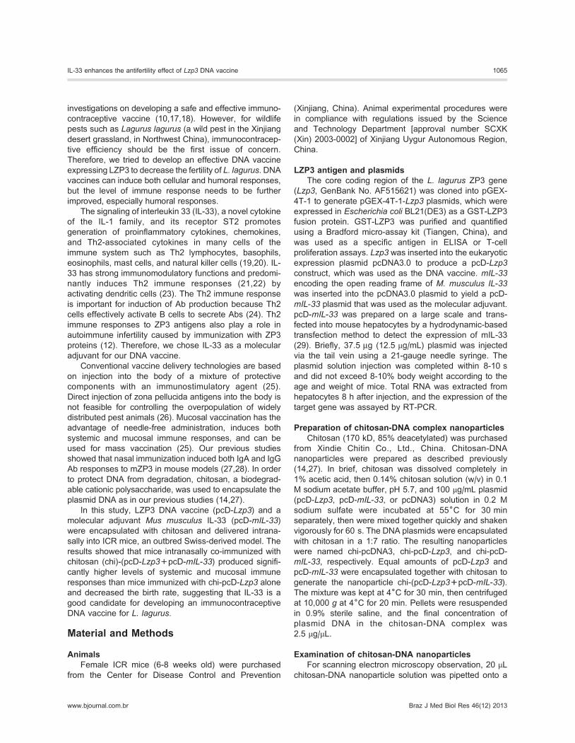
Characterization of chitosan-DNA nanoparticlesThe morphology of the chitosan-DNA nanoparticles
was observed with scanning electronmicroscopy (Figure 1).
The size of chitosan-DNA nanoparticles was approximately
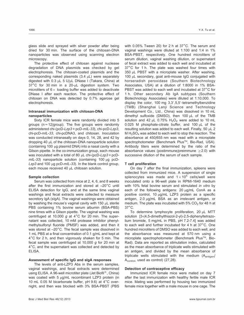
100-1000 nm. The protective effect of nanoparticles was
detected using DNase I digestion. The nanoparticles
completely protected DNA plasmids from digestion by
DNase I, whereas the naked DNA was completely
degraded by DNase I after 30 min (Figure 2). These
results show that chitosan could protect DNA vaccine from
degradation.
Transcription expression of mIL-33 plasmid in mousehepatocytes
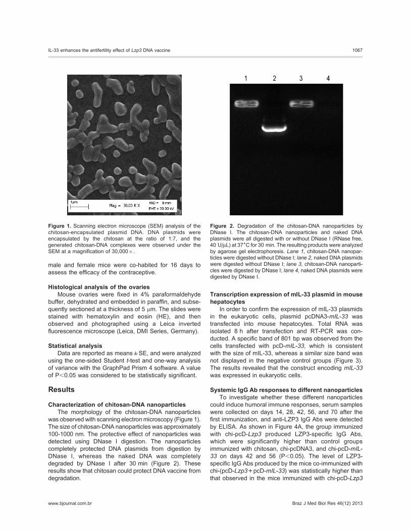
In order to confirm the expression of mIL-33 plasmids
in the eukaryotic cells, plasmid pcDNA3-mIL-33 was
transfected into mouse hepatocytes. Total RNA was
isolated 8 h after transfection and RT-PCR was con-
ducted. A specific band of 801 bp was observed from the
cells transfected with pcD-mIL-33, which is consistent
with the size of mIL-33, whereas a similar size band was
not displayed in the negative control groups (Figure 3).
The results revealed that the construct encoding mIL-33was expressed in eukaryotic cells.
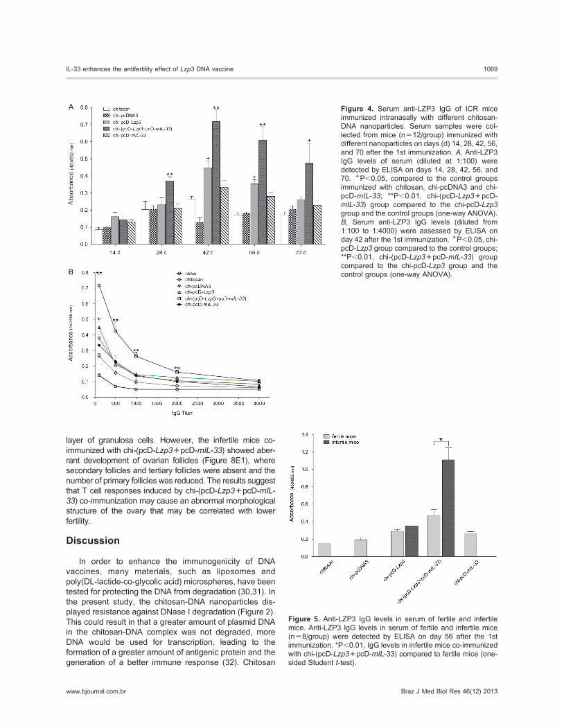
Systemic IgG Ab responses to different nanoparticlesTo investigate whether these different nanoparticles
could induce humoral immune responses, serum samples
were collected on days 14, 28, 42, 56, and 70 after the
first immunization, and anti-LZP3 IgG Abs were detected
by ELISA. As shown in Figure 4A, the group immunized
with chi-pcD-Lzp3 produced LZP3-specific IgG Abs,
which were significantly higher than control groups
immunized with chitosan, chi-pcDNA3, and chi-pcD-mIL-
33 on days 42 and 56 (P,0.05). The level of LZP3-
specific IgG Abs produced by the mice co-immunized with
chi-(pcD-Lzp3+pcD-mIL-33) was statistically higher than
that observed in the mice immunized with chi-pcD-Lzp3
Figure 1. Scanning electron microscope (SEM) analysis of the
chitosan-encapsulated plasmid DNA. DNA plasmids were
encapsulated by the chitosan at the ratio of 1:7, and the
generated chitosan-DNA complexes were observed under the
SEM at a magnification of 30,0006.
Figure 2. Degradation of the chitosan-DNA nanoparticles by
DNase I. The chitosan-DNA nanoparticles and naked DNA
plasmids were all digested with or without DNase I (RNase free,
40 U/mL) at 376C for 30 min. The resulting products were analyzed
by agarose gel electrophoresis. Lane 1, chitosan-DNA nanopar-
ticles were digested without DNase I; lane 2, naked DNA plasmids
were digested without DNase I; lane 3, chitosan-DNA nanoparti-
cles were digested by DNase I; lane 4, naked DNA plasmids were
digested by DNase I.
IL-33 enhances the antifertility effect of Lzp3 DNA vaccine 1067
www.bjournal.com.br Braz J Med Biol Res 46(12) 2013
and in the control groups on days 28, 42, 56 (P,0.01) and
70 (P,0.05 vs control groups; Figure 4A). LZP3-specific
IgG Abs were detected as early as 28 days in chi-(pcD-
Lzp3+pcD-mIL-33) co-immunized mice, suggesting that
chi-(pcD-Lzp3+pcD-mIL-33) co-immunization not only
enhanced the level of IgG Ab but also quickly induced
Ab production. The level of Abs reached the maximum on
day 42. The highest IgG titer of the group co-immunized
with chi-(pcD-Lzp3+pcD-mIL-33) was 4000 on day 42
(P,0.01 vs chi-pcD-Lzp3 and control groups; P,0.05 vs
control groups; Figure 4B). Importantly, we analyzed the
level of IgG Ab in infertile and fertile mice after mating.
The levels of IgG Abs in infertile mice were significantly
higher than those in fertile mice in the co-immunized
group (P,0.01; Figure 5), suggesting that infertility was
correlated with the higher level of Abs. The results
showed that mIL-33 as a molecular adjuvant for pcD-
Lzp3 DNA vaccine could enhance systemic Ab responses
against ZP3 through mucosal immunization.
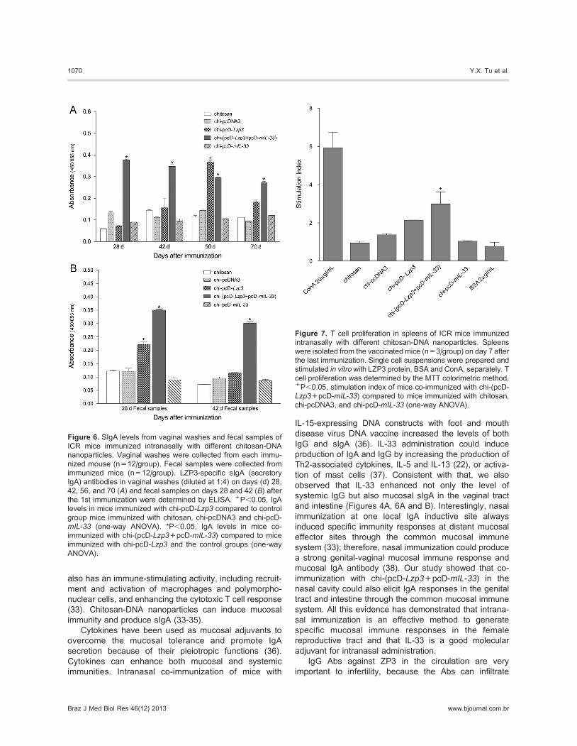
Mucosal IgA responsesAfter testing the systemic IgG Ab responses, we
detected a mucosal response, antigen-specific sIgA, in
the vaginal washes and fecal extracts by ELISA. Chi-
(pcD-Lzp3+pcD-mIL-33) co-immunization quickly
induced high levels of LZP3-specific sIgA in the vaginal
washes and reached peak responses on day 28 after the
first immunization. The sIgA levels in response to co-
immunization were statistically higher than those in mice
immunized with chi-pcD-Lzp3 and the control groups on
days 28, 42, and 70. On day 56, both chi-(pcD-
Lzp3+pcD-mIL-33) and chi-pcD-Lzp3 induced higher
sIgA levels than control groups, but the sIgA level of
chi-pcD-Lzp3 quickly decreased on day 70 (Figure 6A).
Both chi-(pcD-Lzp3+pcD-mIL-33) and chi-pcD-Lzp3induced LZP3-specific sIgA in fecal extracts on day 28,
and the level of sIgA induced by chi-(pcD-Lzp3+pcD-
mIL-33) was significantly higher than that induced by chi-
pcD-Lzp3 and the control groups (P,0.05 vs chi-pcD-
Lzp3 and control groups; Figure 6B). LZP3-specific sIgA
in mice immunized with chi-pcD-Lzp3 decreased to basal
levels on day 42, but a high level of LZP3-specific sIgA in
mice co-immunized with chi-(pcD-Lzp3+pcD-mIL-33)
remained on day 42. These results suggest that mIL-33
not only increases the level of sIgA induced by LZP3 DNA
vaccine but also enhances the longevity of sIgA.
T cell proliferation in vitroThe above results show that mIL-33 enhanced
systemic and mucosal Ab responses induced by LZP3
DNA vaccine. We next examined whether mIL-33
enhanced cellular responses. T cell proliferation in vitrowas assessed by the MTT method. The level of T cell
proliferation induced by chi-(pcD-Lzp3+pcD-mIL-33) wassignificantly higher than that of other groups (P,0.05;
Figure 7). The level of T cell proliferation induced by chi-
pcD-Lzp3 was somewhat higher than that of the control
groups but was not significantly different. The results
indicated that IL-33 as a mucosal adjuvant also enhanced
antigen-specific cellular immune responses.
Analysis of antifertilityTo detect the antifertility effect of the mice immunized
with different nanoparticles, the rate of fertility was
recorded. The results showed that mice of the co-
immunized group had the lowest birth rate and mean litter
size compared with the other groups. Three of 8 female
mice in the co-immunized group were infertile, the other 5
females had 54 pups in total, and the average litter size for
the entire group was 6.75 pups, that is, significantly lower
than that of other groups (Table 1). Only one mouse in the
group immunized with chi-pcD-Lzp3 was infertile, and the
average litter size was 9.25. All females in other control
groups were fertile. These results showed that the fertility
of mice co-immunized with chi-(pcD-Lzp3+pcD-mIL-33)by intranasal delivery was reduced.
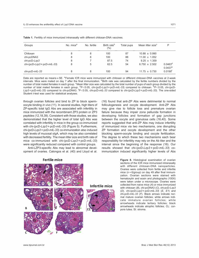
Ovarian pathologyOvaries were collected from immunized mice 6 weeks
after mating. Ovarian slides were prepared and stained
with HE. The results indicated that the fertile mice
immunized with chi-(pcD-Lzp3+pcD-mIL-33) or chi-pcD-
Lzp3 or other control groups had normal follicles in
numbers and in morphology with primordial, primary,
secondary, tertiary, and mature follicles (Figure 8A-F). The
mature follicles contained oocytes surrounded by a single
Figure 3. Expression of pcDNA3-mIL-33 in mouse hepatocytes.
Total RNA was extracted from mouse hepatocytes 8 h after
transfection with pcDNA3-mIL-33 using the hydrodynamics-
based transfection method. The templates used for RT-PCR
were as follows: lane 1, distilled water as a negative control; lane2, cDNA from hepatocytes of mouse transfected with pcDNA3 as
a negative control; lane 3, RNA from hepatocytes of mouse
transfected with pcDNA3-mIL-33 as a negative control; lane 4,cDNA from hepatocytes of mouse transfected with pcDNA3-mIL-33; lane 5, pcDNA3-mIL-33 plasmid served as a positive
template. M1: 1500 bp DNA marker; M2: DL15,000++2000 bp
DNA marker.
1068 Y.X. Tu et al.
Braz J Med Biol Res 46(12) 2013 www.bjournal.com.br
layer of granulosa cells. However, the infertile mice co-
immunized with chi-(pcD-Lzp3+pcD-mIL-33) showed aber-
rant development of ovarian follicles (Figure 8E1), where
secondary follicles and tertiary follicles were absent and the
number of primary follicles was reduced. The results suggest
that T cell responses induced by chi-(pcD-Lzp3+pcD-mIL-33) co-immunization may cause an abnormal morphological
structure of the ovary that may be correlated with lower
fertility.
Discussion
In order to enhance the immunogenicity of DNA
vaccines, many materials, such as liposomes and
poly(DL-lactide-co-glycolic acid) microspheres, have been
tested for protecting the DNA from degradation (30,31). In
the present study, the chitosan-DNA nanoparticles dis-
played resistance against DNase I degradation (Figure 2).
This could result in that a greater amount of plasmid DNA
in the chitosan-DNA complex was not degraded, more
DNA would be used for transcription, leading to the
formation of a greater amount of antigenic protein and the
generation of a better immune response (32). Chitosan
Figure 4. Serum anti-LZP3 IgG of ICR mice
immunized intranasally with different chitosan-
DNA nanoparticles. Serum samples were col-
lected from mice (n=12/group) immunized with
different nanoparticles on days (d) 14, 28, 42, 56,
and 70 after the 1st immunization. A, Anti-LZP3IgG levels of serum (diluted at 1:100) were
detected by ELISA on days 14, 28, 42, 56, and
70. +P,0.05, compared to the control groups
immunized with chitosan, chi-pcDNA3 and chi-
pcD-mIL-33; **P,0.01, chi-(pcD-Lzp3+pcD-
mIL-33) group compared to the chi-pcD-Lzp3group and the control groups (one-way ANOVA).
B, Serum anti-LZP3 IgG levels (diluted from
1:100 to 1:4000) were assessed by ELISA on
day 42 after the 1st immunization. +P,0.05, chi-
pcD-Lzp3 group compared to the control groups;
**P,0.01, chi-(pcD-Lzp3+pcD-mIL-33) group
compared to the chi-pcD-Lzp3 group and the
control groups (one-way ANOVA).
Figure 5. Anti-LZP3 IgG levels in serum of fertile and infertile
mice. Anti-LZP3 IgG levels in serum of fertile and infertile mice
(n=8/group) were detected by ELISA on day 56 after the 1st
immunization. *P,0.01, IgG levels in infertile mice co-immunized
with chi-(pcD-Lzp3+pcD-mIL-33) compared to fertile mice (one-
sided Student t-test).
IL-33 enhances the antifertility effect of Lzp3 DNA vaccine 1069
www.bjournal.com.br Braz J Med Biol Res 46(12) 2013
also has an immune-stimulating activity, including recruit-
ment and activation of macrophages and polymorpho-
nuclear cells, and enhancing the cytotoxic T cell response
(33). Chitosan-DNA nanoparticles can induce mucosal
immunity and produce sIgA (33-35).
Cytokines have been used as mucosal adjuvants to
overcome the mucosal tolerance and promote IgA
secretion because of their pleiotropic functions (36).
Cytokines can enhance both mucosal and systemic
immunities. Intranasal co-immunization of mice with
IL-15-expressing DNA constructs with foot and mouth
disease virus DNA vaccine increased the levels of both
IgG and sIgA (36). IL-33 administration could induce
production of IgA and IgG by increasing the production of
Th2-associated cytokines, IL-5 and IL-13 (22), or activa-
tion of mast cells (37). Consistent with that, we also
observed that IL-33 enhanced not only the level of
systemic IgG but also mucosal sIgA in the vaginal tract
and intestine (Figures 4A, 6A and B). Interestingly, nasal
immunization at one local IgA inductive site always
induced specific immunity responses at distant mucosal
effector sites through the common mucosal immune
system (33); therefore, nasal immunization could produce
a strong genital-vaginal mucosal immune response and
mucosal IgA antibody (38). Our study showed that co-
immunization with chi-(pcD-Lzp3+pcD-mIL-33) in the
nasal cavity could also elicit IgA responses in the genital
tract and intestine through the common mucosal immune
system. All this evidence has demonstrated that intrana-
sal immunization is an effective method to generate
specific mucosal immune responses in the female
reproductive tract and that IL-33 is a good molecular
adjuvant for intranasal administration.
IgG Abs against ZP3 in the circulation are very
important to infertility, because the Abs can infiltrate
Figure 6. SIgA levels from vaginal washes and fecal samples of
ICR mice immunized intranasally with different chitosan-DNA
nanoparticles. Vaginal washes were collected from each immu-
nized mouse (n=12/group). Fecal samples were collected from
immunized mice (n=12/group). LZP3-specific sIgA (secretory
IgA) antibodies in vaginal washes (diluted at 1:4) on days (d) 28,
42, 56, and 70 (A) and fecal samples on days 28 and 42 (B) afterthe 1st immunization were determined by ELISA. +P,0.05, IgA
levels in mice immunized with chi-pcD-Lzp3 compared to control
group mice immunized with chitosan, chi-pcDNA3 and chi-pcD-
mIL-33 (one-way ANOVA). *P,0.05, IgA levels in mice co-
immunized with chi-(pcD-Lzp3+pcD-mIL-33) compared to mice
immunized with chi-pcD-Lzp3 and the control groups (one-way
ANOVA).
Figure 7. T cell proliferation in spleens of ICR mice immunized
intranasally with different chitosan-DNA nanoparticles. Spleens
were isolated from the vaccinatedmice (n=3/group) on day 7 after
the last immunization. Single cell suspensions were prepared and
stimulated in vitrowith LZP3 protein, BSA and ConA, separately. T
cell proliferation was determined by the MTT colorimetric method.+P,0.05, stimulation index of mice co-immunized with chi-(pcD-
Lzp3+pcD-mIL-33) compared to mice immunized with chitosan,
chi-pcDNA3, and chi-pcD-mIL-33 (one-way ANOVA).
1070 Y.X. Tu et al.
Braz J Med Biol Res 46(12) 2013 www.bjournal.com.br
through ovarian follicles and bind to ZP to block sperm-
oocyte binding in vivo (11). In several studies, high titers of
ZP-specific total IgG Abs are associated with infertility in
mice immunized with the recombinant ZP3 protein or ZP3
peptides (12,18,39). Consistent with those studies, we also
demonstrated that the higher level of total IgG Abs was
correlated with infertility in mice in the group co-immunized
with chi-(pcD-Lzp3+pcD-mIL-33) (Figure 5). Furthermore,
chi-(pcD-Lzp3+pcD-mIL-33) co-immunization also induced
high levels of mucosal sIgA, which may be also correlated
with decreased fertility. Themean litter size and birth rate of
mice co-immunized with chi-(pcD-Lzp3+pcD-mIL-33)
were significantly reduced compared with control groups.
Anti-LZP3-specific Abs may lead to abnormal devel-
opment of ovaries. Calongos et al. (40) and Lloyd et al.
(16) found that anti-ZP Abs were detrimental to normal
folliculogenesis and oocyte development. Anti-ZP Abs
may give rise to follicle loss and premature ovarian
failure because they impair zona pellucida formation in
developing follicles and formation of gap junctions
between the oocyte and granulosa cells (16,40). Some
reports suggested that anti-ZP Abs may induce infertility
of immunized mice via two mechanisms, one disrupting
ZP formation and oocyte development and the other
blocking sperm-oocyte binding and oocyte fertilization.
The degree to which these two mechanisms each bear
responsibility for infertility may rely on the Ab titer and the
interval since the beginning of the response (16). Our
results showed that chi-(pcD-Lzp3+pcD-mIL-33) co-
immunization induced significantly higher levels of Abs
Table 1. Fertility of mice immunized intranasally with different chitosan-DNA vaccines.
Groups No. micea No. fertile Birth rateb
(%)Total pups Mean litter sizec P
Chitosan 8 8 100 87 10.88 ± 0.990
chi-pcDNA3 8 8 100 88 11.00 ± 1.052
chi-pcD-Lzp3 8 7 87.5 74 9.25 ± 1.359
chi-(pcD-Lzp3+pcD-mIL-33) 8 5 62.5 54 6.750 ± 2.502 0.0463d
0.0437e
chi-pcD-mIL-33 8 8 100 94 11.75 ± 0.730 0.0190f
Data are reported as means±SE. aFemale ICR mice were immunized with chitosan or different chitosan-DNA vaccines at 2-week
intervals. Mice were mated on day 7 after the final immunization. bBirth rate was calculated by the fertile numbers divided by the
number of total mated females in each group. cMean litter size was calculated by the total number of pups of each group divided by the
number of total mated females in each group. dP,0.05, chi-(pcD-Lzp3+pcD-mIL-33) compared to chitosan. eP,0.05, chi-(pcD-
Lzp3+pcD-mIL-33) compared to chi-pcDNA3. fP,0.05, chi-pcD-mIL-33 compared to chi-(pcD-Lzp3+pcD-mIL-33). The one-sided
Student t-test was used for statistical analyses.
Figure 8. Histological examination of ovarian
sections of the ICR mice immunized intranasally
with different chitosan-DNA nanoparticles.
Ovaries were collected from fertile and infertile
mice (n=6/group) on day 49 after final immuni-
zation. Ovarian sections were stained with
hematoxylin and eosin and photographs (100X)
were taken under a microscope. Ovaries were
collected from naive mice (A) or mice immunized
with chitosan (B), chi-pcDNA3 (C), chi-pcD-Lzp3(D), chi-(pcD-Lzp3+pcD-mIL-33) (E, E1) and
chi-pcD-mIL-33 (F). Black arrows indicate nor-
mal mature ovarian follicles; white arrows indi-
cate immature ovarian fol l ic les; white
arrowheads indicate tertiary follicles; black
arrowheads indicate atrophic follicles; CL: cor-
pora lutea; St: stroma.
IL-33 enhances the antifertility effect of Lzp3 DNA vaccine 1071
www.bjournal.com.br Braz J Med Biol Res 46(12) 2013
in infertile mice than in fertile mice, suggesting that the
infertility may be due to the Abs blocking sperm-oocyte
binding. We also observed abnormal development of
follicles in infertile mice co-immunized with chi-(pcD-
Lzp3+pcD-mIL-33), which may be correlated with the
high level of T cell proliferation, and the abnormal
development of follicles may also contribute to infertility.
Taken together, our results show that IL-33 is a good
molecular adjuvant to enhance systemic and mucosal
Ab responses and cellular responses induced by LZP3
DNA vaccine. Importantly, chi-(pcD-Lzp3+pcD-mIL-33)co-immunization significantly increased infertility. We
provide a potential strategy to control the population of
wild animal pests.
Acknowledgments
We would like to thank Dr. Ji Ma, Dr. Jinyao Li and Ms.
Jiayinaguli Zhumabai for the critical reading and for
revising the manuscript. We also thank Mr. Yonghai
Liang for his technical support. Research supported by
the Research Starting Foundation for Doctors of Xinjiang
University (#BS120112) to Dr. Y.X. Tu, and the Natural
Science Foundation of China (#30760136) to F.C. Zhang.
References
1. Cooper DW, Larsen E. Immunocontraception of mammalian
wildlife: ecological and immunogenetic issues. Reproduc-
tion 2006; 132: 821-828, doi: 10.1530/REP-06-0037.
2. Lloyd ML, Shellam GR, Papadimitriou JM, Lawson MA.
Immunocontraception is induced in BALB/c mice inoculated
with murine cytomegalovirus expressing mouse zona
pellucida 3. Biol Reprod 2003; 68: 2024-2032, doi:
10.1095/biolreprod.102.012880.
3. O’Rand MG, Widgren EE, Sivashanmugam P, Richardson
RT, Hall SH, French FS, et al. Reversible immunocontra-
ception in male monkeys immunized with eppin. Science
2004; 306: 1189-1190, doi: 10.1126/science.1099743.
4. Hardy CM, Hinds LA, Kerr PJ, Lloyd ML, Redwood AJ,
Shellam GR, et al. Biological control of vertebrate pests
using virally vectored immunocontraception. J Reprod
Immunol 2006; 71: 102-111, doi: 10.1016/j.jri.2006.04.006.
5. Kirkpatrick JF, Lyda RO, Frank KM. Contraceptive vaccines
for wildlife: a review. Am J Reprod Immunol 2011; 66: 40-50,
doi: 10.1111/j.1600-0897.2011.01003.x.
6. Lo SC, Zeenathul NA, Sheikh Omar AR, Mohd Azmi ML.
Current ZP3-based immunocontraceptive vaccine for free
ranging wild pest. Pertanika J Trop Agric Sci 2011; 34: 1-16.
7. Naz RK, Gupta SK, Gupta JC, Vyas HK, Talwar AG. Recent
advances in contraceptive vaccine development: a mini-
review. Hum Reprod 2005; 20: 3271-3283, doi: 10.1093/
humrep/dei256.
8. Srivastava N, Santhanam R, Sheela P, Mukund S, Thakral
SS, Malik BS, et al. Evaluation of the immunocontraceptive
potential of Escherichia coli-expressed recombinant dog
ZP2 and ZP3 in a homologous animal model. Reproduction
2002; 123: 847-857, doi: 10.1530/rep.0.1230847.
9. Bagavant H, Fusi FM, Baisch J, Kurth B, David CS, Tung
KS. Immunogenicity and contraceptive potential of a human
zona pellucida 3 peptide vaccine. Biol Reprod 1997; 56:
764-770, doi: 10.1095/biolreprod56.3.764.
10. Xiang RL, Zhou F, Yang Y, Peng JP. Construction of the
plasmid pCMV4-rZPC’ DNA vaccine and analysis of its
contraceptive potential. Biol Reprod 2003; 68: 1518-1524,
doi: 10.1095/biolreprod.102.007849.
11. Zhang X, Lou YH, Koopman M, Doggett T, Tung KS, Curtiss
R III. Antibody responses and infertility in mice following oral
immunization with attenuated Salmonella typhimurium
expressing recombinant murine ZP3. Biol Reprod 1997;
56: 33-41, doi: 10.1095/biolreprod56.1.33.
12. Clydesdale G, Pekin J, Beaton S, Jackson RJ, Vignarajan
S, Hardy CM. Contraception in mice immunized with
recombinant zona pellucida subunit 3 proteins correlates
with Th2 responses and the levels of interleukin 4
expressed by CD4++ cells. Reproduction 2004; 128: 737-
745, doi: 10.1530/rep.1.00310.
13. Hardy CM, Beaton S, Hinds LA. Immunocontraception in
mice using repeated, multi-antigen peptides: immunization
with purified recombinant antigens. Mol Reprod Dev 2008;
75: 126-135, doi: 10.1002/mrd.20745.
14. Ma X, Li J, Zhang F. Intranasal co-delivery with the mouse
zona pellucida 3 and GM-CSF expressing constructs
enhances humoral immune responses and contraception
in mice. Scand J Immunol 2012; 76: 521-527, doi: 10.1111/
j.1365-3083.2012.02765.x.
15. Li J, Jin H, Zhang F, Du X, Zhao G, Yu Y, et al. Treatment of
autoimmune ovarian disease by co-administration with
mouse zona pellucida protein 3 and DNA vaccine through
induction of adaptive regulatory T cells. J Gene Med 2008;
10: 810-820, doi: 10.1002/jgm.1200.
16. Lloyd ML, Papadimitriou JM, O’Leary S, Robertson SA,
Shellam GR. Immunoglobulin to zona pellucida 3 mediates
ovarian damage and infertility after contraceptive vaccina-
tion in mice. J Autoimmun 2010; 35: 77-85, doi: 10.1016/
j.jaut.2010.03.002.
17. Choudhury S, Kakkar V, Suman P, Chakrabarti K, Vrati S,
Gupta SK. Immunogenicity of zona pellucida glycoprotein-3
and spermatozoa YLP(12) peptides presented on Johnson
grass mosaic virus-like particles. Vaccine 2009; 27: 2948-
2953, doi: 10.1016/j.vaccine.2009.03.002.
18. Li J, Jin H, Zhang A, Li Y, Wang B, Zhang F. Enhanced
contraceptive response by co-immunization of DNA and
protein vaccines encoding the mouse zona pellucida 3 with
minimal oophoritis in mouse ovary. J Gene Med 2007; 9:
1095-1103, doi: 10.1002/jgm.1069.
19. Cayrol C, Girard JP. The IL-1-like cytokine IL-33 is
inactivated after maturation by caspase-1. Proc Natl Acad
Sci U S A 2009; 106: 9021-9026, doi: 10.1073/
pnas.0812690106.
20. Yasuoka S, Kawanokuchi J, Parajuli B, Jin S, Doi Y, Noda
M, et al. Production and functions of IL-33 in the central
nervous system. Brain Res 2011; 1385: 8-17, doi: 10.1016/
j.brainres.2011.02.045.
21. Miller AM, Xu D, Asquith DL, Denby L, Li Y, Sattar N, et al.
1072 Y.X. Tu et al.
Braz J Med Biol Res 46(12) 2013 www.bjournal.com.br
IL-33 reduces the development of atherosclerosis. J Exp
Med 2008; 205: 339-346, doi: 10.1084/jem.20071868.
22. Schmitz J, Owyang A, Oldham E, Song Y, Murphy E,
McClanahan TK, et al. IL-33, an interleukin-1-like cytokine
that signals via the IL-1 receptor-related protein ST2 and
induces T helper type 2-associated cytokines. Immunity
2005; 23: 479-490, doi: 10.1016/j.immuni.2005.09.015.
23. Rank MA, Kobayashi T, Kozaki H, Bartemes KR, Squillace
DL, Kita H. IL-33-activated dendritic cells induce an atypical
TH2-type response. J Allergy Clin Immunol 2009; 123:
1047-1054, doi: 10.1016/j.jaci.2009.02.026.
24. Bergmann C, Van Hemmen JL, Segel LA. Th1 or Th2: how
an appropriate T helper response can be made. Bull Math
Biol 2001; 63: 405-430, doi: 10.1006/bulm.2000.0215.
25. Van Roosmalen ML, Kanninga R, El Khattabi M, Neef J,
Audouy S, Bosma T, et al. Mucosal vaccine delivery of
antigens tightly bound to an adjuvant particle made from
food-grade bacteria. Methods 2006; 38: 144-149, doi:
10.1016/j.ymeth.2005.09.015.
26. Mackenzie SM, McLaughlin EA, Perkins HD, French N,
Sutherland T, Jackson RJ, et al. Immunocontraceptive
effects on female rabbits infected with recombinant myxoma
virus expressing rabbit ZP2 or ZP3. Biol Reprod 2006; 74:
511-521, doi: 10.1095/biolreprod.105.046268.
27. Zhang A, Li J, Zhao G, Geng S, Zhuang S, Wang B, et al.
Intranasal co-administration with the mouse zona pellucida
3 expressing construct and its coding protein induces
contraception in mice. Vaccine 2011; 29: 6785-6792, doi:
10.1016/j.vaccine.2010.12.061.
28. Kadir Z, Ma X, Li J, Zhang F. Granulocyte-macrophage
colony-stimulating factor enhances the humoral immune
responses of mouse zona pellucida 3 vaccine strategy
based on DNA and protein coadministration in BALB/c mice.
Reprod Sci 2013; 20: 400-407, doi: 10.1177/19337191124
59236.
29. Wang G, Tschoi M, Spolski R, Lou Y, Ozaki K, Feng C, et al.
In vivo antitumor activity of interleukin 21 mediated by
natural killer cells. Cancer Res 2003; 63: 9016-9022.
30. Gursel M, Tunca S, Ozkan M, Ozcengiz G, Alaeddinoglu G.
Immunoadjuvant action of plasmid DNA in liposomes.Vaccine
1999; 17: 1376-1383, doi: 10.1016/S0264-410X(98)00383-1.
31. He XW, Wang F, Jiang L, Li J, Liu SK, Xiao ZY, et al.
Induction of mucosal and systemic immune response by
single-dose oral immunization with biodegradable micro-
particles containing DNA encoding HBsAg. J Gen Virol
2005; 86: 601-610, doi: 10.1099/vir.0.80575-0.
32. Bivas-Benita M, van Meijgaarden KE, Franken KL,
Junginger HE, Borchard G, Ottenhoff TH, et al. Pulmonary
delivery of chitosan-DNA nanoparticles enhances the
immunogenicity of a DNA vaccine encoding HLA-A*0201-
restricted T-cell epitopes of Mycobacterium tuberculosis.
Vaccine 2004; 22: 1609-1615, doi: 10.1016/j.vaccine.
2003.09.044.
33. Xu W, Shen Y, Jiang Z, Wang Y, Chu Y, Xiong S. Intranasal
delivery of chitosan-DNA vaccine generates mucosal SIgA
and anti-CVB3 protection. Vaccine 2004; 22: 3603-3612,
doi: 10.1016/j.vaccine.2004.03.033.
34. Xu J, Dai W, Wang Z, Chen B, Li Z, Fan X. Intranasal
vaccination with chitosan-DNA nanoparticles expressing pneu-
mococcal surface antigen a protects mice against nasophar-
yngeal colonization by Streptococcus pneumoniae. Clin
Vaccine Immunol 2011; 18: 75-81, doi: 10.1128/CVI.00263-10.
35. Khatri K, Goyal AK, Gupta PN, Mishra N, Vyas SP. Plasmid
DNA loaded chitosan nanoparticles for nasal mucosal
immunization against hepatitis B. Int J Pharm 2008; 354:
235-241, doi: 10.1016/j.ijpharm.2007.11.027.
36. WangX,ZhangX,KangY, JinH,DuX,ZhaoG,et al. Interleukin-
15 enhance DNA vaccine elicited mucosal and systemic
immunity against foot and mouth disease virus. Vaccine 2008;
26: 5135-5144, doi: 10.1016/j.vaccine.2008.03.088.
37. Xu D, Jiang HR, Kewin P, Li Y, Mu R, Fraser AR, et al. IL-33
exacerbates antigen-induced arthritis by activating mast
cells. Proc Natl Acad Sci U S A 2008; 105: 10913-10918,
doi: 10.1073/pnas.0801898105.
38. Holmgren J, Czerkinsky C. Mucosal immunity and vaccines.
Nat Med 2005; 11: S45-S53, doi: 10.1038/nm1213.
39. Hardy CM, ten Have JF, Mobbs KJ, Hinds LA. Assessment
of the immunocontraceptive effect of a zona pellucida 3
peptide antigen in wild mice. Reprod Fertil Dev 2002; 14:
151-155, doi: 10.1071/RD01112.
40. Calongos G, Hasegawa A, Komori S, Koyama K. Harmful
effects of anti-zona pellucida antibodies in folliculogenesis,
oogenesis, and fertilization. J Reprod Immunol 2009; 79:
148-155, doi: 10.1016/j.jri.2008.06.003.
IL-33 enhances the antifertility effect of Lzp3 DNA vaccine 1073
www.bjournal.com.br Braz J Med Biol Res 46(12) 2013