Embed Size (px)
DESCRIPTION
Issue 18 of Young Scientists Journal - Inspiring and nurturing the scientists of the future
Citation preview
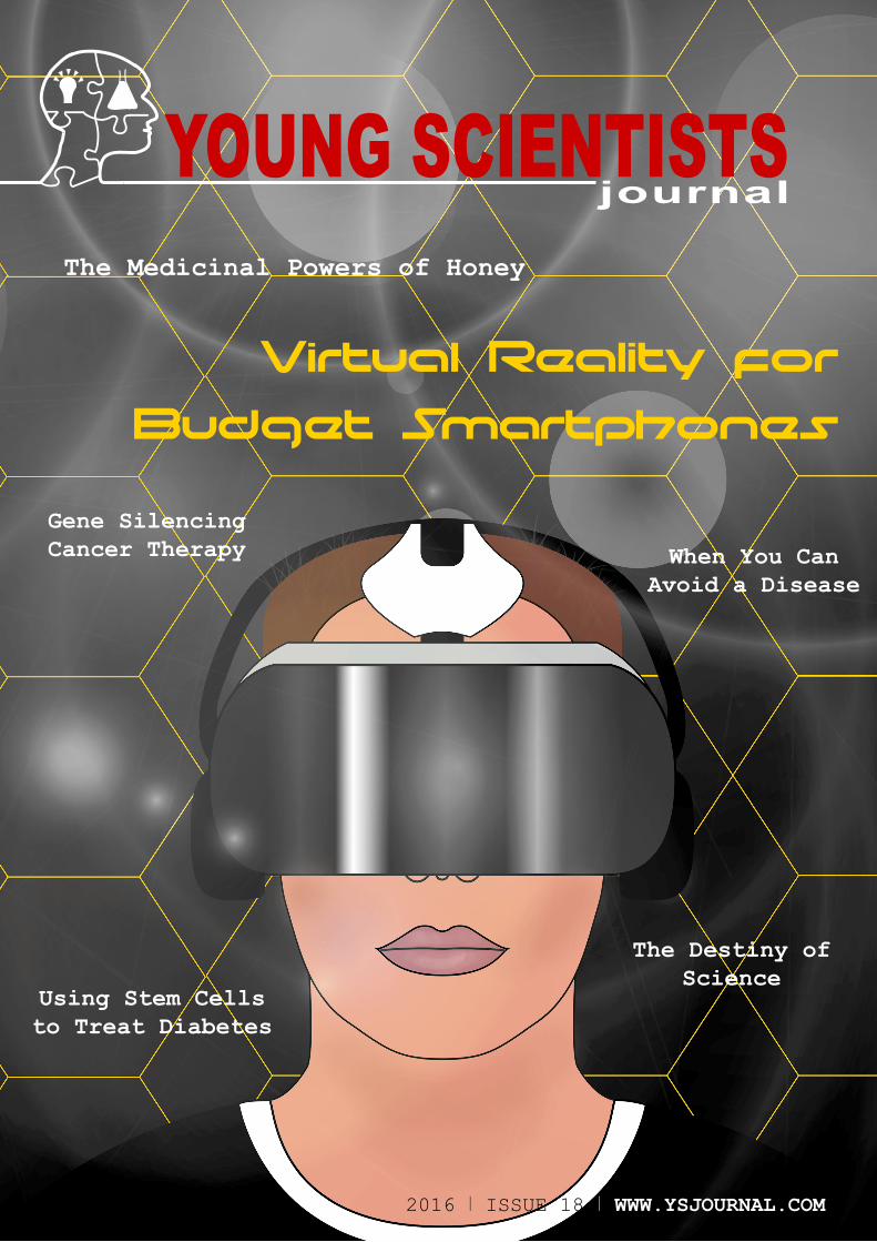
2016 I ISSUE 18 I WWW.YSJOURNAL.COM 12016 I ISSUE 18 I WWW.YSJOURNAL.COM
Virtual Reality for
Budget Smartphones
Using Stem Cells to Treat Diabetes
The Destiny of Science
When You Can Avoid a Disease
Gene Silencing Cancer Therapy
YOUNG SCIENTISTSThe Medicinal Powers of Honey

2 WWW.YSJOURNAL.COM I ISSUE 18 I 2016
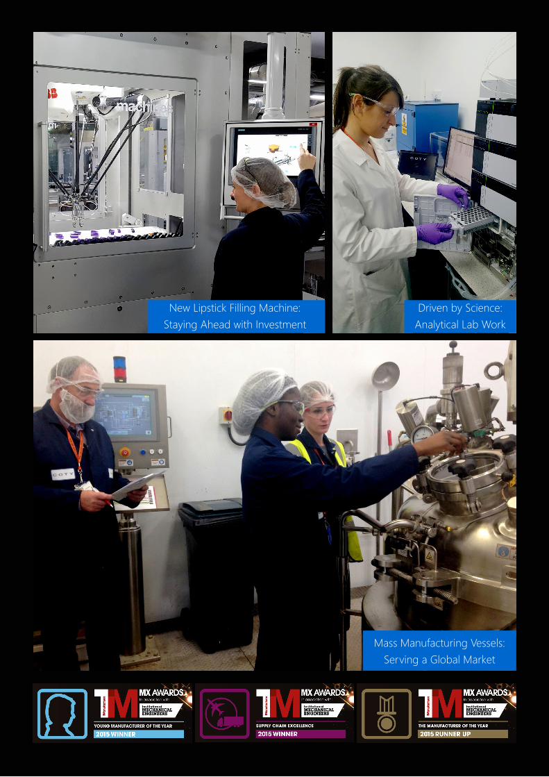
Coty is a market leading global fragrance and beauty brand which has a history spanning over 100 years. With an entrepreneurial spirit
and vision, we are an established company with new and innovative ideas. We have a portfolio of some of the world’s most prestigious and recognised beauty and fragrance brands which continues to grow from strength to strength.
Coty Manufacturing in Ashford, Kent, is proud to be a part of this. Our Ashford site is Coty’s largest in-house supplier of cosmetics globally. The brands we manufacture include Rimmel, Astor, Miss Sporty, NYC, Manhattan, Les Cosmétiques, and CK One. We produce over 150 million units annually in Ashford, with Rimmel being our largest brand overall. Our manufacturing processes are technical, highly automated, and utilise the latest in cosmetic manufacturing technology.
Our business is driven by the credo Faster. Further. Freer. We capture trends quickly. We catch opportunities as soon as they appear in the market, and our fast decision-making process allows us to leapfrog over competitors and keep us at the front of the pack.
This ethos and the highly skilled men and women at Coty Ashford enable us to support and lead many new and exciting product launches each year. This involves scaling up from laboratory batches to full industrialization of our final products. In total our production volume in Ashford has increased over 30% in four years, and we are seizing further growth opportunities in both our traditional markets and emerging markets.
This growth has led to exciting employment opportunities including work experience projects for students, internships, apprenticeships, and full time employment in engineering and other fields. Being part of a global business means there are plenty of learning opportunities and limitless career paths for talents.
Having a culture that is unique enables our employees and our company to be successful. If you want to be a part of this exciting journey get in touch.
CONNECT WITH US
/cotyinc
@cotyinc
/company/coty
www.coty.com/company/careers
ADVERTISEMENT
2016 I ISSUE 18 I WWW.YSJOURNAL.COM 3
New Lipstick Filling Machine:Staying Ahead with Investment
Driven by Science: Analytical Lab Work
Mass Manufacturing Vessels: Serving a Global Market
It has been a busy and exciting few months for the team. In July, we were delighted to release our special issue in partnership
with the Royal Society, a link we hope will continue long into the future as we encourage schools in receipt of their Partnership Grants to publish with Young Scientists Journal.
In October, we held our second Science Communication Conference at The King’s School in Canterbury. It was a great occasion, attended by 250 people from 20 schools across the UK and Ireland, read more on page 8. Our next conference will be held on 18th October 2016 at St Anne’s College, Oxford - be sure to keep up to date on our website.
Films of all those who presented posters on their science research at the conference can be viewed on our YouTube channel and some feature as articles in this issue.
You will also find articles from authors around the world, on a number of different topics; for instance, there is an article by Peter He from Tiffin Boys’ School, who carried out a project looking into the use of wireless virtual reality for basic smartphones – an intriguing possibility for the future, reflected in our front cover artwork.
Then, you can meet our conference poster presentation winner, Elliot Young, who talks about his investigation on the antibacterial properties of Manuka honey, carried out when he was at Wisbech Grammar School.
In the field of biology, we also have an article written by Ammara Jones from St. Dominic’s Sixth Form College on lung cancer and why it is such a deadly disease. Girinath Nandakumar from Bolton School (one of our hub schools) discusses how gene silencing might serve as a possible treatment for cancer in the future.
The problem of global warming continues to grow in size, so much so, Adam Shine, also from Bolton School, looks at whether or not the problem of rising carbon dioxide levels can be resolved. Also, Benjamin Shi, who studies at Watford Grammar School, takes a look at the transmission electron microscope.
Later in this issue is an article by our Outreach Team Leader, Sanjay Kubsad, currently in his first year at Washington State University, who looks into the future of science and what we can possibly do to re-ignite public scientific interest.
On top of our next conference, we also have some exciting plans coming next
year. Together with the Royal Horticultural Society we are looking at launching a themed issue with a writing competition. We’re also looking at setting up a partnership with the citizen science project, Zooniverse, and strengthening our partnership with CREST. The British Council would like to promote us in schools across the world, helping us to set up hub schools meaning that even more people across the world can get involved with the journal. There’s all this to come and more, so keep your eyes peeled for developments next year!
We hope you like Issue 18 and reading it encourages you to get involved. Check out www.ysjournal.com for all the latest updates including the articles as we publish them. If you find yourself inspired by this issue and would like to write and publish your article with us or get involved in other aspects of running the journal, head over to contact us on our website.
Claire Nicholson, Chief Editor
A word from our mentor…I am delighted to share the good news of a generous grant awarded to the Young Scientists Journal by the Royal Commission for the 1851 Exhibition. The Commission’s aim is “to increase the means of industrial education and extend the influence of science and art upon productive industry”. The ‘special award’ we have been granted will be used to further the engagement of the journal with state schools in the UK. So, if your school is interested in becoming a ‘hub school’, or you are organising an event we might be interested in, do get in touch: [email protected].
I’d also like to welcome three new members to our International Advisory Board:
• Professor Sir Martyn Poliakoff, Vice-President and Foreign Secretary of the Royal Society and professor of chemistry at the University of Nottingham
• Dr. Claire McNulty, Director of Science at the British Council and developmental biologist.
• Rod Edwards, CEO of Young Engineers
Our thanks to the whole team of Advisors for their support and expertise.
Christina Astin, Co-founder & Mentor
ISSUE 18 Editorial
Claire Nicholson
www.ysjournal.com/YSJournal @YSJournal@ysjournal
2016 I ISSUE 18 I WWW.YSJOURNAL.COM 5
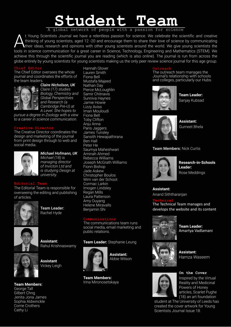
Student TeamA global network of people with a passion for science
At Young Scientists Journal we have a relentless passion for science. We celebrate the scientific and creative thinking of young scientists, aged 12 -20 and encourage them to share their love of science by communicating their ideas, research and opinions with other young scientists around the world. We give young scientists the
tools in science communication for a great career in Science, Technology, Engineering and Mathematics (STEM). We achieve this through the scientific journal you are reading (which is also online). The journal is run from across the globe entirely by young scientists for young scientists making us the only peer review science journal for this age group.
Chief EditorThe Chief Editor oversees the whole journal and coordinates the efforts of the team leaders.
Claire Nicholson, UKClaire (17) studies Biology, Chemistry and Global Perspectives and Research (a Cambridge Pre-U) at A Level. She hopes to
pursue a degree in Zoology with a view to a career in science communication.
Creative DirectorThe Creative Director coordinates the design and marketing of the journal from print design through to web and social media.
Michael Hofmann, UKMichael (18) is managing director of Invicton Ltd and is studying Design at university.
Editorial TeamThe Editorial Team is responsible for overseeing the editing and publishing of articles.
Team Leader: Rachel Hyde
Assistant: Rahul Krishnaswamy
Assistant Vickey Leigh
Team Members:George TallGilbert ChngJenita Jona JamesSophia AldwinckleCorrie CrothersCathy Li
Hannah GloverLauren SmithFiona BellMustafa MajeedNathan DayPierce McLoughlinSamir ChitnavisSunniva HaynesJamie HowieLizzy AvissIman MouloudiFiona BellToby CliftonAnju AnnaParis JaggersJames TunsleySansith HewapathiranaBen HallPeter HeSaumya MaheshwariAminah AhmedRebecca WilliamsJoseph McGrath WilliamsFionn BishopJade AskewChristopher BoulosWim van der Schoot Cormac Larkin Imogen LindsleyRegan Mills Laura PattersonAmy OuyangHelene MiravallsBenjamin Shi
CommunicationsThe communications team runs social media, email marketing and public relations.
Team Leader: Stephanie Leung
Assistant:Abbie Wilson
Team Members:Irina Mironosetskaya
OutreachThe outreach team manages the Journal’s relationship with schools and colleges, particularly our Hubs.
Team Leader: Sanjay Kubsad
Assistant: Gurneet Bhela
Team Members: Nick Curtis
Research-in-Schools Leader: Rose Meddings
Assistant:Anand SiththaranjanTechnicalThe Technical Team manages and develops the website and its content
Team Leader: Amartya Vadlamani
Assistant: Hamza Waseem
On the CoverInspired by the Virtual Reality and Medicinal Powers of Honey articles, Scarlet Pughe (18) an art foundation
student at The University of Leeds has created the cover artwork for Young Scientists Journal Issue 18.
8Conference 2015 Report
10News
12The Medicinal Powers of Honey
17
Through the Silicon Looking Glass
20The Destiny of Science
22Using Stem Cells to Treat Diabetes
26V-Band Photometry in V404 Cygni
29Gene Silencing as a Therapy for Cancer
33Transmission Electron Microscope
36Spider Silk in Medicine
47When You Can Avoid a Disease
50Virtual Reality for Budget Smartphones
58Treating Lung Cancer
62Aesthetically Pleasing Graphs
6 WWW.YSJOURNAL.COM I ISSUE 18 I 2016
ContentsIssue 18
36
29
58 26
12
22
2016 I ISSUE 18 I WWW.YSJOURNAL.COM 7
8 WWW.YSJOURNAL.COM I ISSUE 18 I 2016
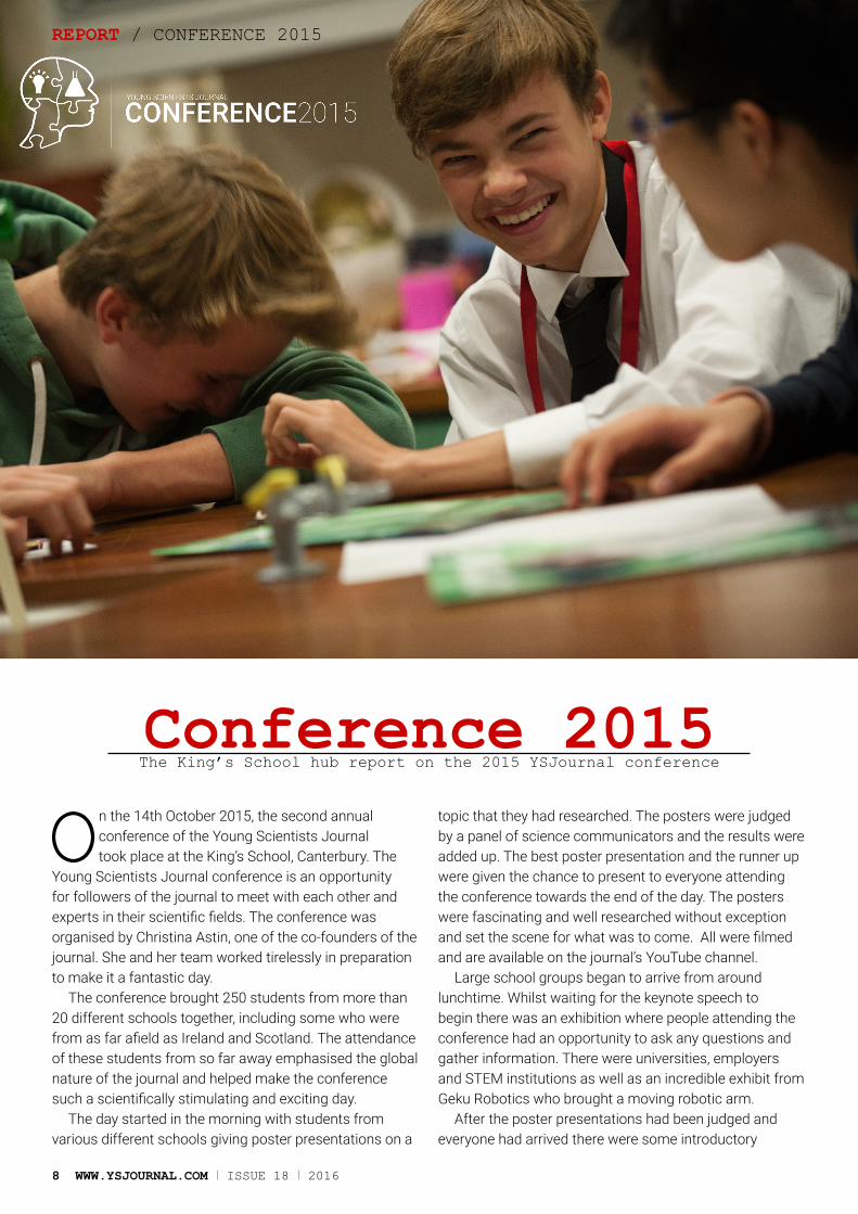
On the 14th October 2015, the second annual conference of the Young Scientists Journal took place at the King’s School, Canterbury. The
Young Scientists Journal conference is an opportunity for followers of the journal to meet with each other and experts in their scientific fields. The conference was organised by Christina Astin, one of the co-founders of the journal. She and her team worked tirelessly in preparation to make it a fantastic day.
The conference brought 250 students from more than 20 different schools together, including some who were from as far afield as Ireland and Scotland. The attendance of these students from so far away emphasised the global nature of the journal and helped make the conference such a scientifically stimulating and exciting day.
The day started in the morning with students from various different schools giving poster presentations on a
topic that they had researched. The posters were judged by a panel of science communicators and the results were added up. The best poster presentation and the runner up were given the chance to present to everyone attending the conference towards the end of the day. The posters were fascinating and well researched without exception and set the scene for what was to come. All were filmed and are available on the journal’s YouTube channel.
Large school groups began to arrive from around lunchtime. Whilst waiting for the keynote speech to begin there was an exhibition where people attending the conference had an opportunity to ask any questions and gather information. There were universities, employers and STEM institutions as well as an incredible exhibit from Geku Robotics who brought a moving robotic arm.
After the poster presentations had been judged and everyone had arrived there were some introductory
Conference 2015The King’s School hub report on the 2015 YSJournal conference
REPORT / CONFERENCE 2015
2016 I ISSUE 18 I WWW.YSJOURNAL.COM 9
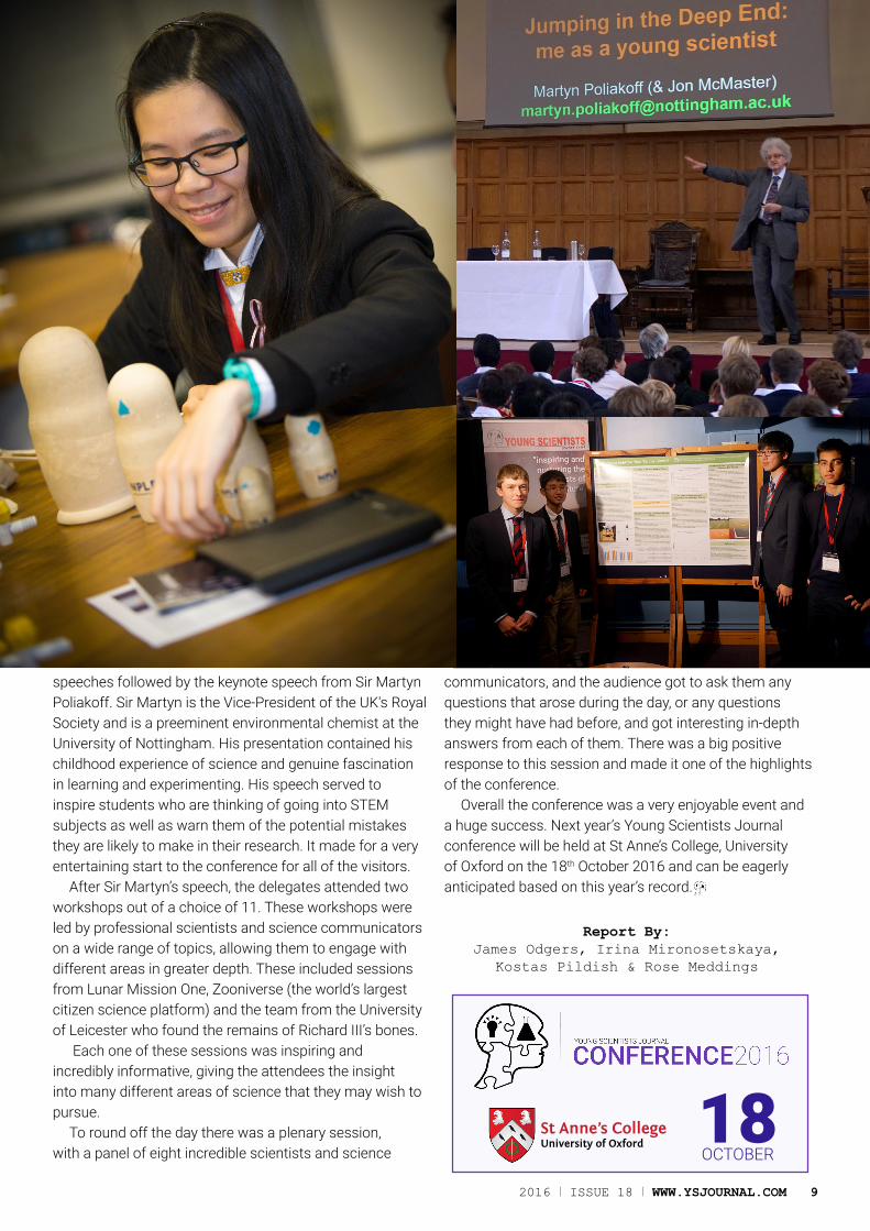
speeches followed by the keynote speech from Sir Martyn Poliakoff. Sir Martyn is the Vice-President of the UK's Royal Society and is a preeminent environmental chemist at the University of Nottingham. His presentation contained his childhood experience of science and genuine fascination in learning and experimenting. His speech served to inspire students who are thinking of going into STEM subjects as well as warn them of the potential mistakes they are likely to make in their research. It made for a very entertaining start to the conference for all of the visitors.
After Sir Martyn’s speech, the delegates attended two workshops out of a choice of 11. These workshops were led by professional scientists and science communicators on a wide range of topics, allowing them to engage with different areas in greater depth. These included sessions from Lunar Mission One, Zooniverse (the world’s largest citizen science platform) and the team from the University of Leicester who found the remains of Richard III’s bones.
Each one of these sessions was inspiring and incredibly informative, giving the attendees the insight into many different areas of science that they may wish to pursue.
To round off the day there was a plenary session, with a panel of eight incredible scientists and science
communicators, and the audience got to ask them any questions that arose during the day, or any questions they might have had before, and got interesting in-depth answers from each of them. There was a big positive response to this session and made it one of the highlights of the conference.
Overall the conference was a very enjoyable event and a huge success. Next year’s Young Scientists Journal conference will be held at St Anne’s College, University of Oxford on the 18th October 2016 and can be eagerly anticipated based on this year’s record.
18OCTOBER
St Anne’s CollegeUniversity of Oxford
Report By:James Odgers, Irina Mironosetskaya,
Kostas Pildish & Rose Meddings

News
In October 2015 the World Health Organisation (WHO) announced that eating 50g of processed meat a day - less than two slices of bacon - could increase the chances of cancer by 18%. For some time now, the general public have been told there is evidence showing a link between eating processed meat and the risk of bowel cancer. However, this announcement from WHO confirms this.
Gravitational lensing, a phenomenon created by the strong gravity of nearby clusters, has allowed the Hubble Telescope to discover 227 new galaxies. These galaxies are fainter and further than most known galaxies, and are estimated to be 13 billion light years away. This means that we now have data regarding the appearance of the universe at the youthful age of 700 million years, during the re-ionization of the universe - a subject that is little understood.
57% of the world’s population is not connected to the Internet. To combat this, Google is now trying to provide Internet connections via helium-filled balloons while Facebook is experimenting with drones. Although both projects are still in the early stages, Google seems to have had more success as it has made successful flights, something Facebook hopes to do by the end of the year.
2015 is the first year on record that average global temperatures have been over 1 degree Celsius higher than pre-industrial levels. While this year there has been a strong El Niño, a Pacific weather effect that increases global temperatures, the UK Met Office still asserts that “it’s clear that it is human influence driving our modern climate into uncharted territory.” With is in mind nearly 200 world leaders reached a landmark agreement at COP21 in Paris to try and limit global temperature increases to well below 2oC.
Quantum entanglement of photons has been investigated at higher energies than ever before. In the experiment, the photons are held within mirrors and when measured appear to have the same properties. These subtle effects are very difficult to measure at increasingly higher energies as the photons can become excited and change state, destroying symmetries induced by the experiment. This data is proof for quantum entanglement on large scales.
[SPACE.COM]
[BBC/GOOGLE]
[PHYSICS-ASTRONOMY.COM]
[BBC]
10 WWW.YSJOURNAL.COM I ISSUE 18 I 2016
NewsScience NewsAll the latest from around the world
[PHYS.ORG]

News NewsNews
Scientists at the universities of Lancaster and Manchester have built brand new systems that could be the future of security protocols. The researchers discovered a way to identify any object by building tiny metallic structures and incorporating deliberate design flaws. These atomic-scale imperfections are impossible to copy as they involve the manipulation of single atoms. The devices require no password and can be built into any material.
NASA’s Mars Reconnaissance Orbiter has strong evidence for the existence of flowing water on Mars, using spectral analysis on the ‘dark streaks’ on the Red Planet, where hydrated minerals have been detected. However, this doesn’t confirm life on Mars due to its lack of an atmosphere. NASA says that solar winds have slowly been removing the atmosphere for billions of years, at a rate of 100g of gas a second and even higher during solar storms.
One-year-old Layla Richards, diagnosed with leukaemia seems to be cured after the first successful medical use of gene editing. T-cells, a type of immune cell, were harvested and genetically modified to attack cancer cells. These were given to Layla, and within weeks there was no sign of cancerous cells in her bone marrow. Although researchers say it is too soon to judge the effectiveness of the procedure, it is undoubtedly a huge breakthrough.
Stronger hair, smoother skin and controlled levels of blood sugar are just some of the benefits of using coconut oil. Surprisingly, coconut oil can also help to ease the symptoms of Parkinson’s disease. A sufferer of this disease began to consume 8 spoons of coconut oil each day and within months he began to experience improvements in mobility. Remarkably, he has even regained his sense of smell.
A study by University College London suggests that patients who suffer from frontotemporal dementia were reported by family and friends in a survey to have had a drastic change in humour. This was said to be the development of inappropriate humour, such as laughing at tragic events. Looking at such symptoms will allow earlier and more accurate diagnoses for dementia and so make possible treatment more effective.
[BBC/JOHNSTONHEALTH.ORG]
[NEW SCIENTIST / GOSH.NHS.UK]
[SKY]
[BBC/GETTY IMAGES]
[SCIENTIFIC REPORTS 5]
2016 I ISSUE 18 I WWW.YSJOURNAL.COM 11
Science NewsBy Becky Payne - Bolton, UK

The Medicinal Powers of Honey
EXPERIMENT / THE MEDICINAL POWERS OF HONEY
AbstractHoney: sweet, delicious and great on toast! After proving a hit in our kitchen cupboards, honey is now making its way into our medicine cabinets too; next time you have a sore throat, you may be reaching for the honey jar. This study looks at three different types of honey and their medicinal qualities against bacteria. Standard processed honey, unprocessed honey and medicinal grade Manuka honey all neutralized samples of both Gram-negative and Gram-positive bacteria, proving all of these honeys to have antibacterial properties. The Manuka honey, however, had a greater antibacterial effect against both bacteria, suggesting that Manuka honey is the best for medicinal use.
Elliot (16) experimentally compares the antibacterial qualities of three different types of honey.
Introduction
Research in 2008 by Professor Thomas Henle from the University of Dresden suggested that Manuka honey is better than other honeys in terms of its
antibacterial qualities, and that this is down to the high methylglyoxal (MGO) levels in the Manuka honey.[1]
This project aims to compare three different types of honey: processed, unprocessed and Manuka. By doing this comparison, the difference in antibacterial qualities between the three types of honey can be assessed, testing the differences between honeys and whether the difference is statistically significant.
Killing BacteriaCertain substances found in all types of honey aid in their antibacterial properties. Due to honey’s high sugar concentration, bacterial cells can become dehydrated and die due to osmosis between the honey and the bacterial cell cytoplasm.
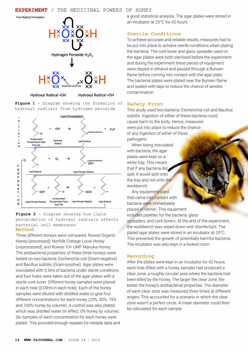
Hydrogen peroxide is found in all types of honey, forming from a reaction between glucose and oxygen and catalysed by glucose oxidase. A hydrogen peroxide molecule has the ability to form two hydroxyl radicals. These hydroxyl radicals are known as relative oxygen species (ROS) or free radicals, as they each contain an unpaired electron, which makes them highly reactive.[2]
Bacterial cells contain a cell membrane, consisting of a phospholipid bilayer (this contains phosphorous heads, glycerol backbones and fatty acid tails). A hydroxyl radical (formed from the hydrogen peroxide in the honey) will ‘attack’ a carbon-hydrogen bond in the fatty acid tail. This will produce a lipid radical (and water), which can then react with oxygen to produce a lipid peroxyl radical. This radical can now react with other fatty acid tails in the phospholipid bilayer, producing a lipid peroxyl radical and another lipid radical. This forms a cycle called lipid
peroxidation (see diagram), which alters the structure of the bacterial cell membrane, causing the bacterial cell not to function properly and thus killing the bacteria.[3]
Some bacteria contain the enzyme catalase, which breaks down hydrogen peroxide (found in honey) into water and oxygen, thus preventing the lipid peroxidation of the bacterial plasma membrane. However, due to honey’s acidity (averaging at a pH of 3.9) the catalase enzyme is denatured, preventing the breakdown of hydrogen peroxide and allowing lipid peroxidation to continue to kill the bacteria.[4]
Manuka honey is deemed to have a stronger antibacterial effect due to the presence of methylglyoxal in the honey.[1] Manuka honey is produced with the nectar from the Leptospermum Scoparium, found in New Zealand. This nectar produces honey that contains significant levels of methylglyoxal. This methylglyoxal is deemed to be the factor which gives manuka honey its stronger antibacterial properties. Manuka honey is graded in UMF (Unique Manuka Factor) which commercially ranges from 5+ UMF to around 30+ UMF.[5]
next time you have a sore throat, you may be reaching for the honey jar
“12 WWW.YSJOURNAL.COM I ISSUE 18 I 2016

THE MEDICINAL POWERS OF HONEY / EXPERIMENT
2016 I ISSUE 18 I WWW.YSJOURNAL.COM 13
14 WWW.YSJOURNAL.COM I ISSUE 18 I 2016
EXPERIMENT / THE MEDICINAL POWERS OF HONEY
MethodThree different honeys were compared: Rowse Organic Honey (processed); Norfolk Cottage Local Honey (unprocessed); and Rowse 10+ UMF Manuka Honey. The antibacterial properties of these three honeys were tested on two bacteria: Escherichia coli (Gram-negative) and Bacillus subtilis (Gram-positive). Agar plates were inoculated with 0.5ml of bacteria under sterile conditions and four holes were taken out of the agar plates with a sterile cork borer. Different honey samples were placed in each hole (0.09ml in each hole). Each of the honey samples were diluted with distilled water to give four different concentrations for each honey (25%, 50%, 75% and 100% honey by volume). A control was also plated, which was distilled water (in effect, 0% honey by volume). Six samples of each concentration for each honey were plated. This provided enough repeats for reliable data and
a good statistical analysis. The agar plates were stored in an incubator at 25°C for 42 hours.
Sterile ConditionsTo achieve accurate and reliable results, measures had to be put into place to achieve sterile conditions when plating the bacteria. The cork borer and glass spreader used on the agar plates were both sterilised before the experiment and during the experiment these pieces of equipment were dipped in ethanol and passed through a Bunsen flame before coming into contact with the agar plate. The bacterial plates were plated near the Bunsen flame and sealed with tape to reduce the chance of aerobic contamination.
Safety FirstThis study used two bacteria: Escherichia coli and Bacillus subtilis. Ingestion of either of these bacteria could cause harm to the body. Hence, measures were put into place to reduce the chance of any ingestion of either of these pathogens:
When being inoculated with bacteria, the agar plates were kept on a white tray. This meant that if any bacteria did spill, it would spill onto the tray and not onto the workbench.
Any equipment used that came into contact with bacteria were immediately placed in vilcron. This equipment included pipettes for the bacteria, glass spreaders, and cork borers. At the end of the experiment, the workbench was wiped down with disinfectant. The plated agar plates were stored in an incubator at 25°C. This prevented the growth of potentially harmful bacteria. The incubator was also kept in a locked room.
RecordingAfter the plates were kept in an incubator for 42 hours, each hole (filled with a honey sample) had produced a clear zone; a roughly circular area where the bacteria had been killed by the honey. The larger the clear zone, the better the honey’s antibacterial properties. The diameter of each clear zone was measured three times at different angles. This accounted for a scenario in which the clear zone wasn’t a perfect circle. A mean diameter could then be calculated for each sample.
Figure 1 - Diagram showing the formation of hydroxyl radicals from hydrogen peroxide
Figure 2 - Diagram showing how lipid peroxidation of hydroxyl radicals affects bacterial cell membranes
2016 I ISSUE 18 I WWW.YSJOURNAL.COM 15
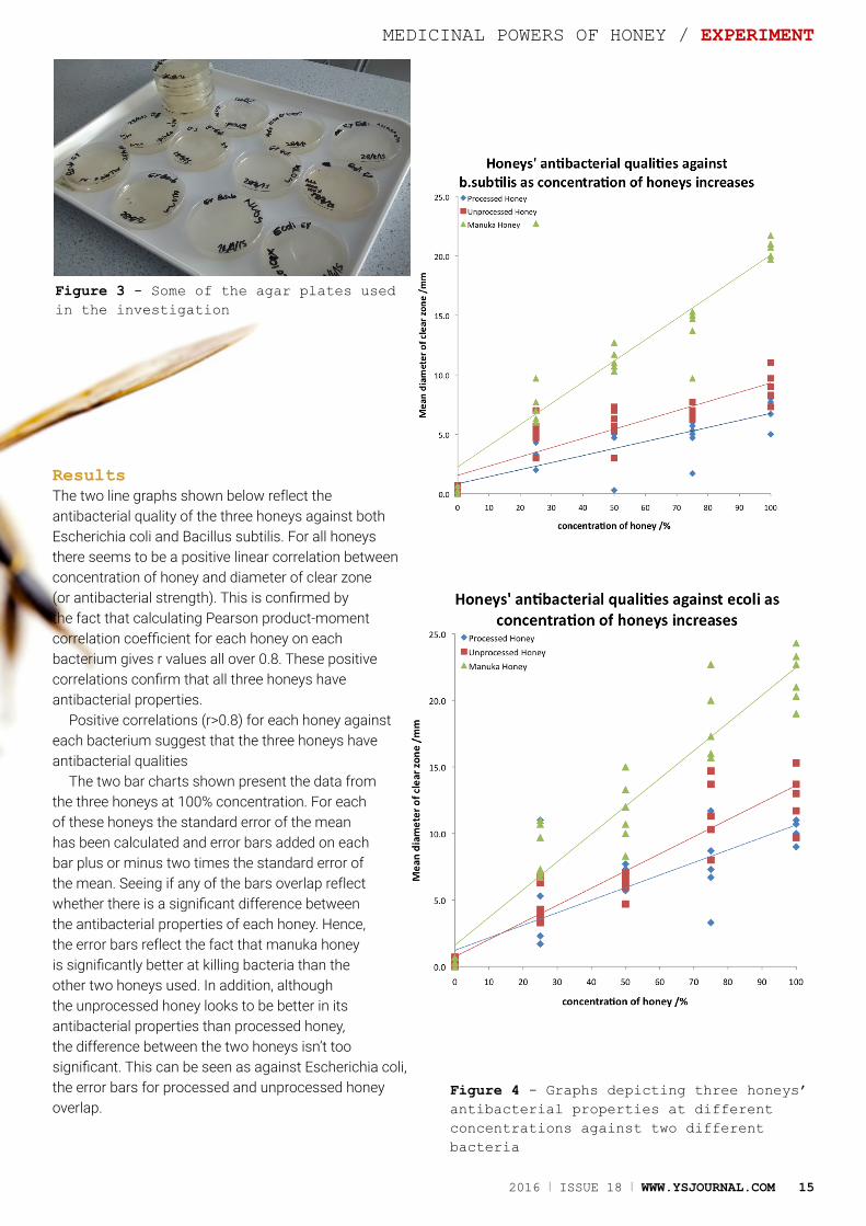
ResultsThe two line graphs shown below reflect the antibacterial quality of the three honeys against both Escherichia coli and Bacillus subtilis. For all honeys there seems to be a positive linear correlation between concentration of honey and diameter of clear zone (or antibacterial strength). This is confirmed by the fact that calculating Pearson product-moment correlation coefficient for each honey on each bacterium gives r values all over 0.8. These positive correlations confirm that all three honeys have antibacterial properties.
Positive correlations (r>0.8) for each honey against each bacterium suggest that the three honeys have antibacterial qualities
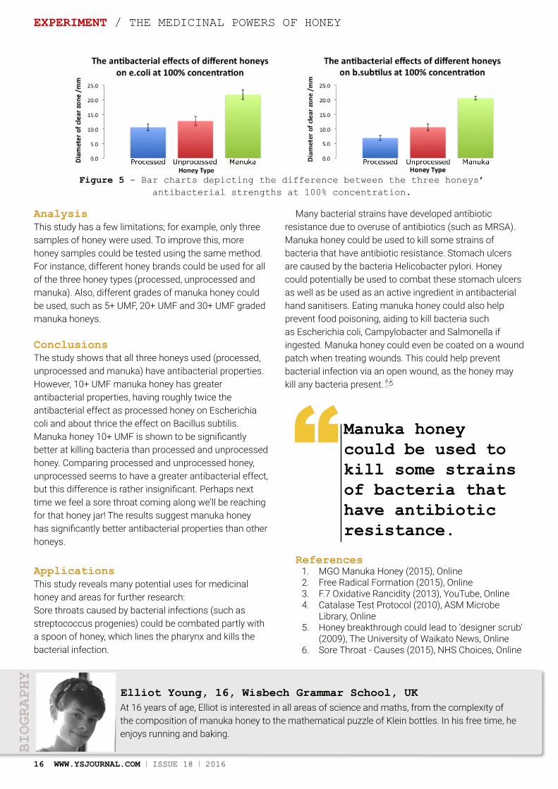
The two bar charts shown present the data from the three honeys at 100% concentration. For each of these honeys the standard error of the mean has been calculated and error bars added on each bar plus or minus two times the standard error of the mean. Seeing if any of the bars overlap reflect whether there is a significant difference between the antibacterial properties of each honey. Hence, the error bars reflect the fact that manuka honey is significantly better at killing bacteria than the other two honeys used. In addition, although the unprocessed honey looks to be better in its antibacterial properties than processed honey, the difference between the two honeys isn’t too significant. This can be seen as against Escherichia coli, the error bars for processed and unprocessed honey overlap.
Figure 3 - Some of the agar plates used in the investigation
Figure 4 - Graphs depicting three honeys’ antibacterial properties at different concentrations against two different bacteria
THE MEDICINAL POWERS OF HONEY / EXPERIMENT
16 WWW.YSJOURNAL.COM I ISSUE 18 I 2016
AnalysisThis study has a few limitations; for example, only three samples of honey were used. To improve this, more honey samples could be tested using the same method. For instance, different honey brands could be used for all of the three honey types (processed, unprocessed and manuka). Also, different grades of manuka honey could be used, such as 5+ UMF, 20+ UMF and 30+ UMF graded manuka honeys.
ConclusionsThe study shows that all three honeys used (processed, unprocessed and manuka) have antibacterial properties. However, 10+ UMF manuka honey has greater antibacterial properties, having roughly twice the antibacterial effect as processed honey on Escherichia coli and about thrice the effect on Bacillus subtilis. Manuka honey 10+ UMF is shown to be significantly better at killing bacteria than processed and unprocessed honey. Comparing processed and unprocessed honey, unprocessed seems to have a greater antibacterial effect, but this difference is rather insignificant. Perhaps next time we feel a sore throat coming along we’ll be reaching for that honey jar! The results suggest manuka honey has significantly better antibacterial properties than other honeys.
ApplicationsThis study reveals many potential uses for medicinal honey and areas for further research:Sore throats caused by bacterial infections (such as streptococcus progenies) could be combated partly with a spoon of honey, which lines the pharynx and kills the bacterial infection.
Many bacterial strains have developed antibiotic resistance due to overuse of antibiotics (such as MRSA). Manuka honey could be used to kill some strains of bacteria that have antibiotic resistance. Stomach ulcers are caused by the bacteria Helicobacter pylori. Honey could potentially be used to combat these stomach ulcers as well as be used as an active ingredient in antibacterial hand sanitisers. Eating manuka honey could also help prevent food poisoning, aiding to kill bacteria such as Escherichia coli, Campylobacter and Salmonella if ingested. Manuka honey could even be coated on a wound patch when treating wounds. This could help prevent bacterial infection via an open wound, as the honey may kill any bacteria present.
References1. MGO Manuka Honey (2015), Online2. Free Radical Formation (2015), Online3. F.7 Oxidative Rancidity (2013), YouTube, Online4. Catalase Test Protocol (2010), ASM Microbe
Library, Online5. Honey breakthrough could lead to ‘designer scrub’
(2009), The University of Waikato News, Online6. Sore Throat - Causes (2015), NHS Choices, Online
At 16 years of age, Elliot is interested in all areas of science and maths, from the complexity of the composition of manuka honey to the mathematical puzzle of Klein bottles. In his free time, he enjoys running and baking.
Elliot Young, 16, Wisbech Grammar School, UK
BIOGRAPHY
EXPERIMENT / THE MEDICINAL POWERS OF HONEYY
Manuka honey could be used to kill some strains of bacteria that have antibiotic resistance.
“
Figure 5 - Bar charts depicting the difference between the three honeys’ antibacterial strengths at 100% concentration.
THROUGH THE SILICON LOOKING GLASS / REVIEW ARTICLE
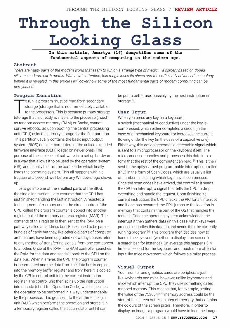
Through the Silicon Looking GlassIn this article, Amartya (16) demystifies some of the fundamental aspects of computing in the modern age.
AbstractThere are many parts of the modern world that seem to run on a strange type of magic – a sorcery based on doped silicates and rare earth metals. With a little attention, this magic loses its sheen and the sufficiently advanced technology behind it is revealed. In this article I will cover how some of the most fundamental parts of modern computing can be demystified.
Program Execution
To run, a program must be read from secondary storage (storage that is not immediately available to the processor). This is because primary storage
(storage that is directly available to the processor), such as random access memory (RAM) or Cache, cannot survive reboots. So upon booting, the central processing unit (CPU) asks the primary storage for the first partition. This partition usually contains the basic input output system (BIOS) on older computers or the unified extended firmware interface (UEFI) loader on newer ones. The purpose of these pieces of software is to set up hardware in a way that allows it to be used by the operating system (OS), and usually to start the boot loader which finally loads the operating system. This all happens within a fraction of a second, well before any Windows logo shows up.
Let’s go into one of the smallest parts of the BIOS, the single Instruction. Let’s assume that the CPU has just finished handling the last instruction. A register, a fast segment of memory under the direct control of the CPU, called the program counter is copied into another register called the memory address register (MAR). The contents of this register is then sent to the RAM on a pathway called an address bus. Buses used to be parallel bundles of cable but they, like other old parts of computer architecture, have been upgraded - nowadays buses refer to any method of transferring signals from one component to another. Once at the RAM, the RAM controller searches the RAM for the data and sends it back to the CPU on the data bus. When it arrives the CPU, the program counter is incremented and the data from the data bus is copied into the memory buffer register and from here it is copied by the CPU’s control unit into the current instruction register. The control unit then splits up the instruction into opcode (short for ‘Operation Code’) which specifies the operation to be performed in a way understandable by the processor. This gets sent to the arithmetic logic unit (ALU) which performs the operation and stores it in a temporary register called the accumulator until it can
be put to better use, possibly by the next instruction in storage [3].
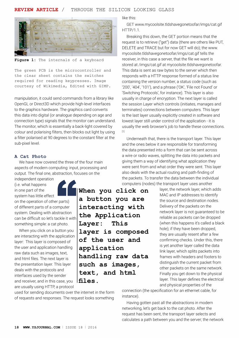
User InputWhen you press any key on a keyboard, a switch (mechanical or conductive) under the key is compressed, which either completes a circuit (in the case of a mechanical keyboard) or increases the current flowing under the key (in the case of a capacitive one). Either way, this action generates a detectable signal which is sent to a microprocessor on the keyboard itself. The microprocessor handles and processes this data into a form that the rest of the computer can read. [2] This is then sent to the aptly-named programmable interrupt controller (PIC) in the form of Scan Codes, which are usually a list of numbers indicating which keys have been pressed. Once the scan codes have arrived, the controller it sends the CPU an Interrupt, a signal that tells the CPU to drop everything and handle the request. Upon finishing its current instruction, the CPU checks the PIC for an interrupt and if one has occurred, the CPU jumps to the location in memory that contains the part of the OS that handles the request. Once the operating system acknowledges the interrupt it then gathers data (in this case, what keys were pressed), bundles this data up and sends it to the currently running program [4]. This program then decides how to handle the key-event (whether to display it on screen in a search bar, for instance). On average this happens 3-4 times a second for the keyboard, and much more often for input like mice movement which follows a similar process.
Visual OutputYour monitor and graphics cards are peripherals just like keyboards and mice; however, unlike keyboards and mice which interrupt the CPU, they use something called mapped memory. This means that, for example, setting the value of the 753664th [5] memory address could be the start of the screen buffer, an area of memory that contains the colours of the screen pixels. Therefore, in order to display an image, a program would have to load the image
2016 I ISSUE 18 I WWW.YSJOURNAL.COM 17
18 WWW.YSJOURNAL.COM I ISSUE 18 I 2016
REVIEW ARTICLE / THROUGH THE SILICON LOOKING GLASS
into RAM and then copy it into the buffer that the graphics card expects. If the program requires complex image
manipulation, it could send commands from a library like OpenGL or Direct3D which provide high-level interfaces to the graphics hardware. The graphics card converts this data into digital (or analogue depending on age and connection type) signals that the monitor can understand. The monitor, which is essentially a back-light covered by colour and polarising filters, then blocks out light by using a filter polarised at 90 degrees to the constant filter at the sub-pixel level.
A Cat PhotoWe have now covered the three of the four main
aspects of modern computing: input, processing and output. The final one, abstraction, focuses on the independent operation (i.e. what happens in one part of the system has little effect on the operation of other parts) of different parts of a computer system. Dealing with abstraction can be difficult so let’s tackle it with something simple: a cat photo.
When you click on a button you are interacting with the application layer: This layer is composed of the user and application handling raw data such as images, text, and html files. The next layer is the presentation layer. This layer deals with the protocols and interfaces used by the sender and receiver, and in this case, you are usually using HTTP, a protocol used for sending documents over the internet in the form of requests and responses. The request looks something
like this:GET www.mycoolsite.tldshavegonetoofar/imgs/cat.gif
HTTP/1.1.Breaking this down, the GET portion means that the
request is to retrieve (“get”) data (there are others like PUT, DELETE and TRACE but for now GET will do); the www.mycoolsite.tldshavegonetoofar/imgs/cat.gif tells the receiver, in this case a server, that the file we want is stored at /imgs/cat.gif at mycoolsite.tldshavegonetoofar. This data is sent as raw bytes to the server which then responds with a HTTP response formed of a status line containing the version number, a status code (such as ‘200’, ‘404’, ‘101’), and a phrase (‘OK’, ‘File not Found’ or ‘Switching Protocols’, for instance). This layer is also usually in charge of encryption. The next layer is called the session Layer which controls (initiates, manages and terminates) connections between computers. This layer is the last layer usually explicitly created in software and lowest layer still under control of the application - it is usually the web browser’s job to handle these connections. [1]
Underneath that, there is the transport layer. This layer and the ones below it are responsible for transforming the data presented into a form that can be sent across a wire or radio waves, splitting the data into packets and giving them a way of identifying what application they were sent from and what order they were sent. This layer also deals with the actual routing and path-finding of the packets. To transfer the data between the individual computers (nodes) the transport layer uses another
layer, the network layer, which adds MAC and IP addresses to identify the source and destination nodes. Delivery of the packets on the network layer is not guaranteed to be reliable as packets can be dropped (when this happens it’s called a black hole); if they have been dropped, they are usually resent after a few confirming checks. Under this, there is yet another layer called the data link layer, which splits packets into frames with headers and footers to distinguish the current packet from other packets on the same network. Finally you get down to the physical layer. This layer defines the electrical and physical properties of the
connection (the specification for an ethernet cable, for instance).
Having gotten past all the abstractions in modern networking, let’s get back to the cat photo. After the request has been sent, the transport layer selects and calculates a path between you and the server; the network
Figure 1: The internals of a keyboard
The green PCB is the microcontroller and the clear sheet contains the switches required for reading keypresses. Image courtesy of Wikimedia, Edited with GIMP.
When you click on a button you are interacting with the Application Layer: This layer is composed of the user and application handling raw data such as images, text, and html files.
“
2016 I ISSUE 18 I WWW.YSJOURNAL.COM 19
THROUGH THE SILICON LOOKING GLASS/ REVIEW ARTICLE
layer handles the transmission to the next router in the path; the data-link and physical layers actually transfer the data to the next router in the chain. At the next router, it performs the same set of calculations and sends it on to the next router, and so on until the destination is reached. At the destination, the server’s layers reverse the alterations made by the client’s layers, stripping headers and extracting the raw data, so that the server application can read the actual message body. It then sees that this is an image request and then creates a response with the image’s bytes embedded. This response is sent back in the same way and the client (your computer) decodes it, decompresses it, and finally puts it on your screen.
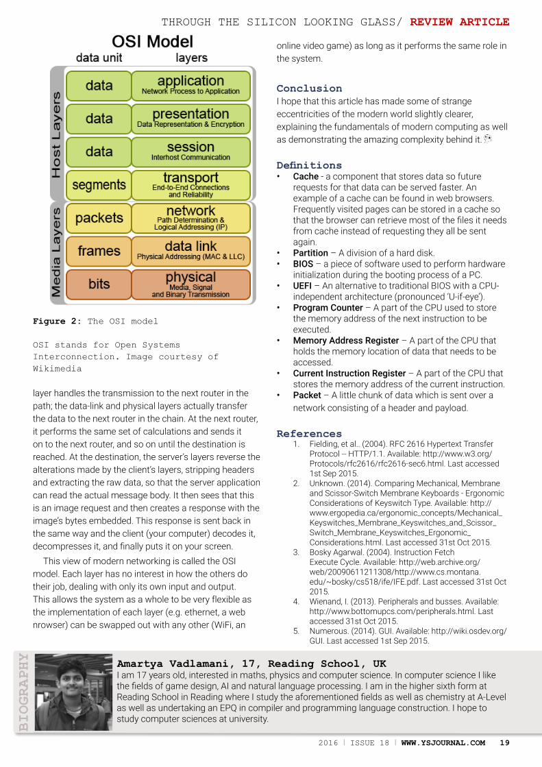
This view of modern networking is called the OSI model. Each layer has no interest in how the others do their job, dealing with only its own input and output. This allows the system as a whole to be very flexible as the implementation of each layer (e.g. ethernet, a web nrowser) can be swapped out with any other (WiFi, an
online video game) as long as it performs the same role in the system.
ConclusionI hope that this article has made some of strange eccentricities of the modern world slightly clearer, explaining the fundamentals of modern computing as well as demonstrating the amazing complexity behind it.
Definitions• Cache - a component that stores data so future
requests for that data can be served faster. An example of a cache can be found in web browsers. Frequently visited pages can be stored in a cache so that the browser can retrieve most of the files it needs from cache instead of requesting they all be sent again.
• Partition – A division of a hard disk.• BIOS – a piece of software used to perform hardware
initialization during the booting process of a PC.• UEFI – An alternative to traditional BIOS with a CPU-
independent architecture (pronounced ‘U-if-eye’).• Program Counter – A part of the CPU used to store
the memory address of the next instruction to be executed.
• Memory Address Register – A part of the CPU that holds the memory location of data that needs to be accessed.
• Current Instruction Register – A part of the CPU that stores the memory address of the current instruction.
• Packet – A little chunk of data which is sent over a network consisting of a header and payload.
References1. Fielding, et al.. (2004). RFC 2616 Hypertext Transfer
Protocol -- HTTP/1.1. Available: http://www.w3.org/Protocols/rfc2616/rfc2616-sec6.html. Last accessed 1st Sep 2015.
2. Unknown. (2014). Comparing Mechanical, Membrane and Scissor-Switch Membrane Keyboards - Ergonomic Considerations of Keyswitch Type. Available: http://www.ergopedia.ca/ergonomic_concepts/Mechanical_Keyswitches_Membrane_Keyswitches_and_Scissor_Switch_Membrane_Keyswitches_Ergonomic_Considerations.html. Last accessed 31st Oct 2015.
3. Bosky Agarwal. (2004). Instruction Fetch Execute Cycle. Available: http://web.archive.org/web/20090611211308/http://www.cs.montana.edu/~bosky/cs518/ife/IFE.pdf. Last accessed 31st Oct 2015.
4. Wienand, I. (2013). Peripherals and busses. Available: http://www.bottomupcs.com/peripherals.html. Last accessed 31st Oct 2015.
5. Numerous. (2014). GUI. Available: http://wiki.osdev.org/GUI. Last accessed 1st Sep 2015.
I am 17 years old, interested in maths, physics and computer science. In computer science I like the fields of game design, AI and natural language processing. I am in the higher sixth form at Reading School in Reading where I study the aforementioned fields as well as chemistry at A-Level as well as undertaking an EPQ in compiler and programming language construction. I hope to study computer sciences at university.
BIOGRAPHY
Figure 2: The OSI model
OSI stands for Open Systems Interconnection. Image courtesy of Wikimedia
Amartya Vadlamani, 17, Reading School, UK

20 WWW.YSJOURNAL.COM I ISSUE 18 I 2016
The Destiny of Science
Sanjay Kubsad discusses the future of Science and why public interest in science is dwindling.
AbstractThe decline in interest for science by the general public can be explained by the human tendency to perceive logarithmically rather than linearly. Science education can address this problem to alter the destiny of science.
Introduction
Science faces a new and perhaps its greatest threat yet. The public interest in scientific progress is destined to wane. The roots of this can already be
seen. Public apathy to science continues to grow.When NASA first sent the man to the moon, millions were glued to the television. They watched as Neil Armstrong took one giant leap for mankind. Yet, according to Michael Tribbe, author of No Requiem for the Space Age, surveys conducted by the New York Times one year after showed that the majority of Americans could not remember his name[1]. How is it that one of the greatest achievements in science loses interest so quickly? The reason for can be explained by the mind’s mathematical perception of the world.
Our Mathematical Perception Humans have an in-built sense for recognizing proportions. We perceive the world relative to itself. It can
be evidenced in how we view age. It is not the wonder of childhood that makes it seem like the longest part of life. This is because although we age linearly, we see everything else logarithmically.
This is why the first years of our lives seemed to linger for a longer duration. This is because every new year that we age is a smaller fraction of all the years we have lived before that. The logarithmic perception extends far beyond age. When driving from Seattle to Spokane in the US (a 280 mile journey), adding ten miles to the trip will not be easily noticed. However, driving ten more miles to find a restaurant within a city will seem more noticeable. The human brain is particularly impacted by relativism. It does hold advantages. The logarithmic programming of our brains allows us to estimate in a manner, as Journalist Ben Thomas puts it, “that reduces relative risk rather than absolute risk.”[2] This allows for quick decisions in an information heavy world.
REVIEW ARTICLE / THE DESTINY OF SCIENCE
2016 I ISSUE 18 I WWW.YSJOURNAL.COM 21
Threats to Innovations Although a logarithmic method of perceiving life as a whole holds merits in sorting out the constant influx of data, it makes it inevitable for the public to lose interest in science. This is because as they age discoveries also become more common.
We perceive each new discovery that is made as a smaller fraction of all the discoveries ever made before. This concept makes it impossible to heighten the public’s interest in scientific progress.
The name Albert Einstein is so fondly remembered among the public, but one of today’s most influential physicists, Roger Penrose is not in the public eye. This is because Einstein was one of the first to breakout as a rock star physicist in the 20th century. And then, Marie Curie, Jonas Salk, Niels Bohr, Francis Crick and James Watson swarmed the pedestal. Our innate logarithmic perception made any equally revolutionary future discoveries by scientists a smaller fraction of the existing set of discoveries. Although natural, this trend brings problems. Scientists depend on public approval to secure funding so an apathetic public results in less money to sustain innovation. Furthermore interest in scientific progress needs to be met with as equal excitement from the public as it is with the scientists. The imbalance leads to an innovation plateau. Today, technology is solely driven by consumers. Improvements in smartphones, internet and computing technologies, although significant, are in the end, simply improvements not innovations. Although our cars are faster and cleaner, the paradigm has not shifted as it had done in the mid-20th century.
Different ApproachThe answer to this problem is not media coverage. The BP oil spill of 2010 was extensively covered yet did not spearhead the oil industry or drive the Green Revolution. (See definitions) As another example, Media outlets frequently cover gene therapy but the public has not batted an eye.
The answer lies in revolutionizing the way the public views innovation. Our natural instinct to view scientific
progress logarithmically needs to be transformed by the education system to be viewed additively. This means that education systems need to teach young people to treat every discovery with a baseline amount of respect. Steps can be taken to educate, not of each innovation itself, but of the impact of each innovation to achieve this goal. Furthermore, scientific development must seek to explore new terrain rather than simply solving the problems of today. For example, the US at the time of space launches did not have the most efficient commercial airline planes, yet NASA embarked on creating the space shuttle. Developing the space shuttle not only uncovered more engineering phenomenona but also created technologies
which improved the commercial airlines. As an existentialist (see definitions) Søren Kierkegaard puts it, "life can only be understood backwards; but it must be lived forwards."[3]
Similarly, the problems today can only be solved when we continue to innovate. Granted, by following this path, science will take risky turns, but in the process it will uncover deeper
truths of the world around us and drive us closer to our final form and destiny.
References1. Why Americans lost interest in putting men on the
Moon - BBC News. Retrieved June 8, 20152. Thomas, B. What’s Halfway Between 1 and 9?
Kids and Scientists Say 3. Retrieved June 8, 20153. Additional Information: Soren Kierkegaard |
biography - Danish philosopher. Retrieved June 8, 2015
4. Image: NASA 1969
What is the Green Revolution? The Green Revolution is a large increase in crop production particularly in developing countries. To do this, there is a high use of artificial fertilisers, pesticides and other chemicals to achieve high productivity.
What is an Existentialist?An Existentialist is someone who has the philosophy of emphasizing individual existence, choice and freedom. They take on the view that humans define their own path and meaning in life, they also strive to make rational and well thought out decisions despite living in the society we do.
Sanjay Kubsad lives in Seattle, Washington and is a freshman at the University of Washington working towards a major’s degree in Biology. He has been with the journal since 2013, participating first as an editor and recently as an Outreach Team Leader. He loves everything science and a good cup of coffee!
Sanjay Kubsad, 17, University of Washington, Seattle, USA
BIOGRAPHY
We perceive each new discovery that is made as a smaller fraction of all the discoveries ever made before
“
THE DESTINY OF SCIENCE / REVIEW ARTICLE

22 WWW.YSJOURNAL.COM I ISSUE 18 I 2016
Using Stem Cells to Treat Diabetes
In this article Christopher (17) explores the application of stem cells in type 1 diabetes treatment.
AbstractChristopher describes the background knowledge needed to understand this potential application such as what the disease is, and the different types of stem cells. He also debates some of the ethical issues associated with the use of stem cells which is a major controversy surrounding this research.
Introduction
Type 1 Diabetes Mellitus is a disease estimated to effect between 11-22 million people worldwide [1], accounting for 5-10% of all cases of diabetes (the
other 90% is due to type 2 and gestational diabetes)[2]. Every year, around 80,000 children worldwide develop the disease [3]. Furthermore, type 1 disease is estimated to cost the NHS £1 billion per year [4].Various studies are being conducted into the use of different types of stem cells to treat type 1 diabetes
What is Type 1 Diabetes?Type 1 Diabetes Mellitus (T1DM) is a metabolic disorder leading to chronic hyperglycaemia (abnormally high blood glucose levels). The real root of the disease is the autoimmune destruction of the beta cells of the islets of the Langerhans in the pancreas. Beta cells are the cells which produce insulin, which is the hormone which lowers blood glucose levels [5], this means that people with T1DM are insulin-deficient, and thus can’t lower their blood glucose level. T1DM usually develops in children, although the exact cause isn’t known. The most common symptoms of diabetes are polyuria (increased urination), polydipsia
(increased thirst), and polyphagia (increased hunger). There is no cure [6] and if left untreated, T1DM can lead to serious chronic complications [7]. The main 3 chronic complications arise from microangiopathy (damage to small blood vessels) due to the high blood glucose concentrations. The endothelium of vessels become thicker in order to take in more glucose. More glycoproteins form on the surface of the endothelial cells and so blood vessels become thicker but weaker. They then leak and bleed, so certain areas of the body do not get enough blood. Diabetic retinopathy occurs
when there is a lack of blood going to the retina. It can cause macular edema (swelling of the macula) and, ultimately, blindness [8]. Diabetic nephropathy (damage to the capillaries in the kidney glomeruli) can lead to chronic renal failure, eventually requiring dialysis. Finally, diabetic neuropathy, the most common of the complications, is damage to nerves in the body, affecting movement, touch, and the autonomic nervous system (the nerves serving vital organs which control important functions e.g. heart rate, respiration rate and digestion). Furthermore, diabetes doubles the risk of cardiovascular disease [9].
What are Stem Cells?Stem cells are undifferentiated cells that have the ability to differentiate into specialised cells. Stem cells in mammals fall into 2 main categories: embryonic stem cells, which are found in embryos,
and adult stem cells, which are found in different tissues in the body. There are 2 unique stem cell properties: firstly ‘self-renewal’. Stem cells have the capacity to divide multiple times whilst maintaining their undifferentiated state. There are 2 mechanisms by which this occurs. The first is obligatory asymmetric replication, where the stem cell divides into one mother cell, which is identical to the original stem cell, and a second cell, which is differentiated, called a daughter cell. The second mechanism is stochastic differentiation, where the stem cell divides into daughter cells that are differentiated, and then another stem cell divides by mitosis to produce two more identical stem
REVIEW ARTICLE / USING STEM CELLS TO TREAT DIABETES
Harvard used stem cells to create insulin producing cells
2016 I ISSUE 18 I WWW.YSJOURNAL.COM 23
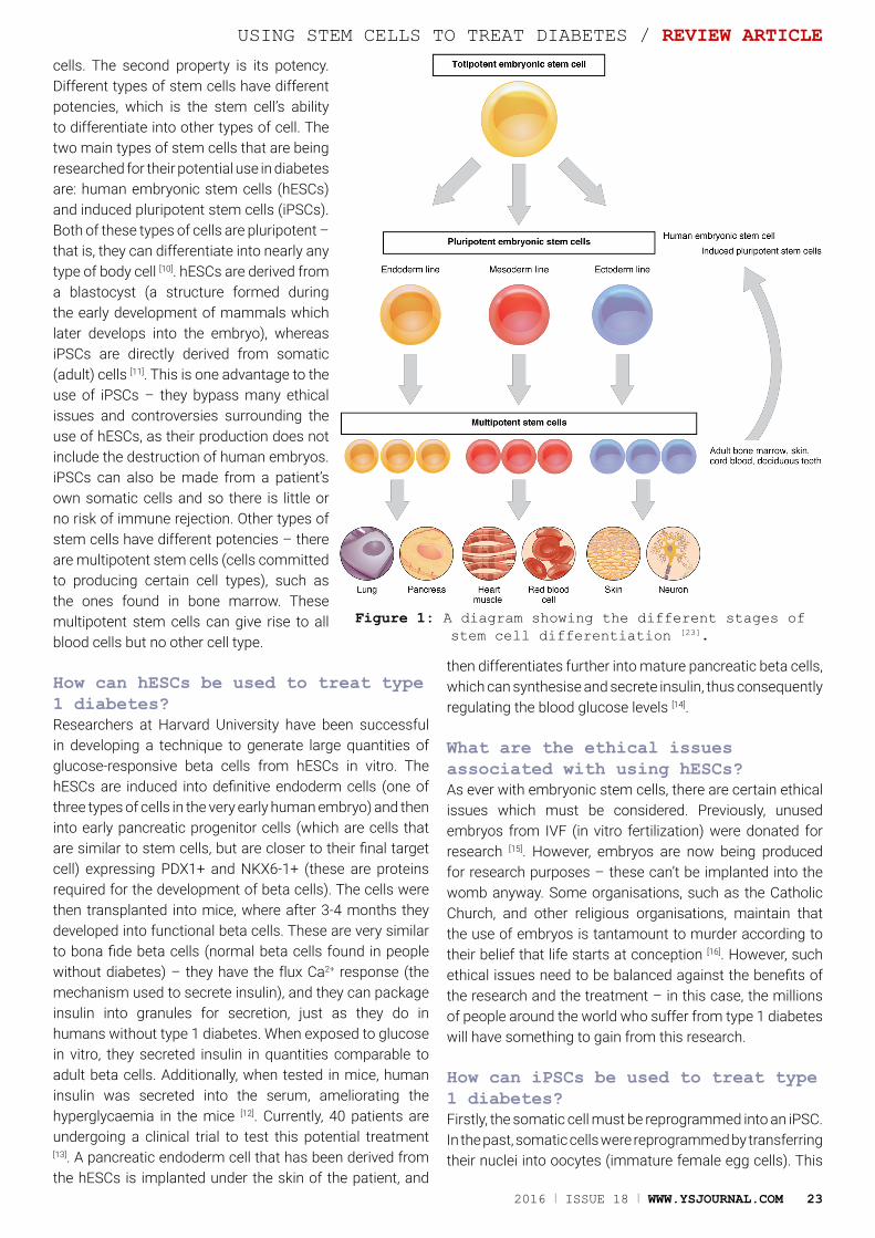
cells. The second property is its potency. Different types of stem cells have different potencies, which is the stem cell’s ability to differentiate into other types of cell. The two main types of stem cells that are being researched for their potential use in diabetes are: human embryonic stem cells (hESCs) and induced pluripotent stem cells (iPSCs). Both of these types of cells are pluripotent – that is, they can differentiate into nearly any type of body cell [10]. hESCs are derived from a blastocyst (a structure formed during the early development of mammals which later develops into the embryo), whereas iPSCs are directly derived from somatic (adult) cells [11]. This is one advantage to the use of iPSCs – they bypass many ethical issues and controversies surrounding the use of hESCs, as their production does not include the destruction of human embryos. iPSCs can also be made from a patient’s own somatic cells and so there is little or no risk of immune rejection. Other types of stem cells have different potencies – there are multipotent stem cells (cells committed to producing certain cell types), such as the ones found in bone marrow. These multipotent stem cells can give rise to all blood cells but no other cell type.
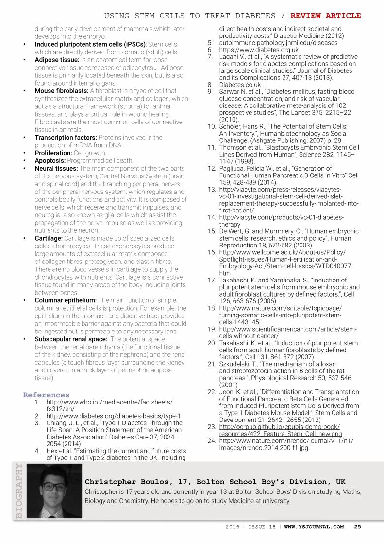
How can hESCs be used to treat type 1 diabetes?Researchers at Harvard University have been successful in developing a technique to generate large quantities of glucose-responsive beta cells from hESCs in vitro. The hESCs are induced into definitive endoderm cells (one of three types of cells in the very early human embryo) and then into early pancreatic progenitor cells (which are cells that are similar to stem cells, but are closer to their final target cell) expressing PDX1+ and NKX6-1+ (these are proteins required for the development of beta cells). The cells were then transplanted into mice, where after 3-4 months they developed into functional beta cells. These are very similar to bona fide beta cells (normal beta cells found in people without diabetes) – they have the flux Ca2+ response (the mechanism used to secrete insulin), and they can package insulin into granules for secretion, just as they do in humans without type 1 diabetes. When exposed to glucose in vitro, they secreted insulin in quantities comparable to adult beta cells. Additionally, when tested in mice, human insulin was secreted into the serum, ameliorating the hyperglycaemia in the mice [12]. Currently, 40 patients are undergoing a clinical trial to test this potential treatment [13]. A pancreatic endoderm cell that has been derived from the hESCs is implanted under the skin of the patient, and
then differentiates further into mature pancreatic beta cells, which can synthesise and secrete insulin, thus consequently regulating the blood glucose levels [14].
What are the ethical issues associated with using hESCs?As ever with embryonic stem cells, there are certain ethical issues which must be considered. Previously, unused embryos from IVF (in vitro fertilization) were donated for research [15]. However, embryos are now being produced for research purposes – these can’t be implanted into the womb anyway. Some organisations, such as the Catholic Church, and other religious organisations, maintain that the use of embryos is tantamount to murder according to their belief that life starts at conception [16]. However, such ethical issues need to be balanced against the benefits of the research and the treatment – in this case, the millions of people around the world who suffer from type 1 diabetes will have something to gain from this research.
How can iPSCs be used to treat type 1 diabetes?Firstly, the somatic cell must be reprogrammed into an iPSC. In the past, somatic cells were reprogrammed by transferring their nuclei into oocytes (immature female egg cells). This
USING STEM CELLS TO TREAT DIABETES / REVIEW ARTICLE
Figure 1: A diagram showing the different stages of stem cell differentiation [23].
24 WWW.YSJOURNAL.COM I ISSUE 18 I 2016
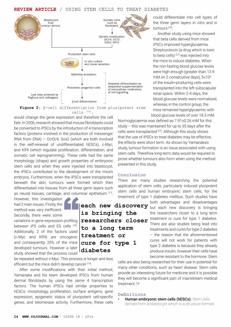
would change the gene expression and therefore the cell fate. In 2006, research showed that mouse fibroblasts could be converted to iPSCs by the introduction of 4 transcription factors (proteins involved in the production of messenger RNA from DNA) – Oct3/4, Sox2 (which are both involved in the self-renewal of undifferentiated hESCs), c-Myc, and Klf4 (which regulate proliferation, differentiation, and somatic cell reprogramming). These cells had the same morphology (shape) and growth properties of embryonic stem cells and when they were injected into blastocyst, the iPSCs contributed to the development of the mice’s embryos. Furthermore, when the iPSCs were transplanted beneath the skin, tumours were formed which then differentiated into tissues from all three germ layers such as neural tissues, cartilage, and columnar epithelium [17]. However, this investigation had 2 main issues. Firstly, the method was very inefficient. Secondly, there were some variations in gene expression profiling between iPS cells and ES cells [18]. Additionally, 2 of the factors used (c-Myc and Klf4) are oncogenic and consequently 20% of the mice developed tumours. However a later study showed that the process could be repeated without c-Myc. This process is longer and less efficient but the mice didn’t develop cancer [19].
After some modifications with their initial method, Yamanaka and his team developed iPSCs from human dermal fibroblasts by using the same 4 transcription factors. The human iPSCs had similar properties to hESCs: morphology, proliferation, surface antigens, gene expression, epigenetic status of pluripotent cell-specific genes, and telomerase activity. Furthermore, these cells
could differentiate into cell types of the three germ layers in vitro and in tumours [20].
Another study using mice showed that beta cells derived from mice iPSCs improved hyperglycaemia. Streptozotocin (a drug which is toxic to beta cells) [21] was injected into the mice to induce diabetes. When the non-fasting blood glucose levels were high enough (greater than 13.9 mM on 2 consecutive days), 5×10⁶ of the insulin-producing cells were transplanted into the left subscapular renal space. Within 2-4 days, the blood glucose levels were normalized, whereas in the control group, the mice remained hyperglycaemic with
blood glucose levels of over 18.3 mM. Normoglycaemia was defined as 7.91±0.26 mM for this study – this was maintained for up to 35 days after the cells were transplanted [22]. Although this study shows that the use of iPSCs to treat diabetes may be effective, the effects were short term. As shown by Yamanaka’s study, tumour formation is an issue associated with using stem cells. Therefore long-term data would be required to prove whether tumours also form when using the method presented in this study.
ConclusionThere are many studies researching the potential application of stem cells, particularly induced pluripotent stem cells and human embryonic stem cells, for the treatment of type 1 diabetes mellitus. Such studies have
both advantages and disadvantages but each new discovery is bringing the researchers closer to a long term treatment or cure for type 1 diabetes. There are also studies being lead into treatments and cures for type 2 diabetes – the reason that the aforementioned cures will not work for patients with type 2 diabetes is because they already produce insulin, however their cells have become resistant to the hormone. Stem
cells are also being researched for their use in potential for many other conditions, such as heart disease. Stem cells provide an interesting future for medicine and it is possible they will become a significant part of mainstream medical treatment.
Definitions• Human embryonic stem cells (hESCs): Stem cells
derived from a blastocyst which is a structure formed
REVIEW ARTICLE / USING STEM CELLS TO TREAT DIABETES
Figure 2: β-cell differentiation from pluripotent stem cells [24].
each new discovery is bringing the researchers closer to a long term treatment or cure for type 1 diabetes
“
2016 I ISSUE 18 I WWW.YSJOURNAL.COM 25
during the early development of mammals which later develops into the embryo.
• Induced pluripotent stem cells (iPSCs): Stem cells which are directly derived from somatic (adult) cells
• Adipose tissue: Is an anatomical term for loose connective tissue composed of adipocytes. Adipose tissue is primarily located beneath the skin, but is also found around internal organs.
• Mouse fibroblasts: A fibroblast is a type of cell that synthesizes the extracellular matrix and collagen, which act as a structural framework (stroma) for animal tissues, and plays a critical role in wound healing. Fibroblasts are the most common cells of connective tissue in animals.
• Transcription factors: Proteins involved in the production of mRNA from DNA.
• Proliferation: Cell growth.• Apoptosis: Programmed cell death.• Neural tissues: The main component of the two parts
of the nervous system; Central Nervous System (brain and spinal cord) and the branching peripheral nerves of the peripheral nervous system, which regulates and controls bodily functions and activity. It is composed of nerve cells, which receive and transmit impulses, and neuroglia, also known as glial cells which assist the propagation of the nerve impulse as well as providing nutrients to the neuron.
• Cartilage: Cartilage is made up of specialized cells called chondrocytes. These chondrocytes produce large amounts of extracellular matrix composed of collagen fibres, proteoglycan, and elastin fibres. There are no blood vessels in cartilage to supply the chondrocytes with nutrients. Cartilage is a connective tissue found in many areas of the body including joints between bones
• Columnar epithelium: The main function of simple columnar epithelial cells is protection. For example, the epithelium in the stomach and digestive tract provides an impermeable barrier against any bacteria that could be ingested but is permeable to any necessary ions
• Subscapular renal space: The potential space between the renal parenchyma (the functional tissue of the kidney, consisting of the nephrons) and the renal capsules (a tough fibrous layer surrounding the kidney and covered in a thick layer of perinephric adipose tissue).
References1. http://www.who.int/mediacentre/factsheets/
fs312/en/2. http://www.diabetes.org/diabetes-basics/type-13. Chiang, J. L., et al., “Type 1 Diabetes Through the
Life Span: A Position Statement of the American Diabetes Association” Diabetes Care 37, 2034–2054 (2014)
4. Hex et al. “Estimating the current and future costs of Type 1 and Type 2 diabetes in the UK, including
direct health costs and indirect societal and productivity costs.” Diabetic Medicine (2012)
5. autoimmune.pathology.jhmi.edu/diseases6. https://www.diabetes.org.uk7. Lagani V., et al., “A systematic review of predictive
risk models for diabetes complications based on large scale clinical studies.” Journal of Diabetes and its Complications 27, 407-13 (2013).
8. Diabetes.co.uk9. Sarwar N, et al., “Diabetes mellitus, fasting blood
glucose concentration, and risk of vascular disease: A collaborative meta-analysis of 102 prospective studies”, The Lancet 375, 2215–22 (2010).
10. Schöler, Hans R., “The Potential of Stem Cells: An Inventory.”, Humanbiotechnology as Social Challenge. (Ashgate Publishing, 2007) p. 28.
11. Thomson et al., “Blastocysts Embryonic Stem Cell Lines Derived from Human”, Science 282, 1145–1147 (1998).
12. Pagliuca, Felicia W., et al., “Generation of Functional Human Pancreatic β Cells In Vitro” Cell 159, 428-439 (2014).
13. http://viacyte.com/press-releases/viacytes-vc-01-investigational-stem-cell-derived-islet-replacement-therapy-successfully-implanted-into-first-patient/
14. http://viacyte.com/products/vc-01-diabetes-therapy
15. De Wert, G. and Mummery, C., “Human embryonic stem cells: research, ethics and policy”, Human Reproduction 18, 672-682 (2003)
16. http://www.wellcome.ac.uk/About-us/Policy/Spotlight-issues/Human-Fertilisation-and-Embryology-Act/Stem-cell-basics/WTD040077.htm
17. Takahashi, K. and Yamanaka, S., “Induction of pluripotent stem cells from mouse embryonic and adult fibroblast cultures by defined factors.”, Cell 126, 663-676 (2006)
18. http://www.nature.com/scitable/topicpage/turning-somatic-cells-into-pluripotent-stem-cells-14431451
19. http://www.scientificamerican.com/article/stem-cells-without-cancer/
20. Takahashi, K. et al., “Induction of pluripotent stem cells from adult human fibroblasts by defined factors.”, Cell 131, 861-872 (2007)
21. Szkudelski, T., “The mechanism of alloxan and streptozotocin action in B cells of the rat pancreas.”, Physiological Research 50, 537-546 (2001)
22. Jeon, K. et al., “Differentiation and Transplantation of Functional Pancreatic Beta Cells Generated from Induced Pluripotent Stem Cells Derived from a Type 1 Diabetes Mouse Model.”, Stem Cells and Development 21, 2642–2655 (2012)
23. http://oerpub.github.io/epubjs-demo-book/resources/422_Feature_Stem_Cell_new.png
24. http://www.nature.com/nrendo/journal/v11/n1/images/nrendo.2014.200-f1.jpg
Christopher is 17 years old and currently in year 13 at Bolton School Boys’ Division studying Maths, Biology and Chemistry. He hopes to go on to study Medicine at university.
Christopher Boulos, 17, Bolton School Boy’s Division, UK
BIOGRAPHY
USING STEM CELLS TO TREAT DIABETES / REVIEW ARTICLE
26 WWW.YSJOURNAL.COM I ISSUE 18 I 2016
ORIGINAL RESEARCH / V-BAND PHOTOMETRY IN V404 CYGNI
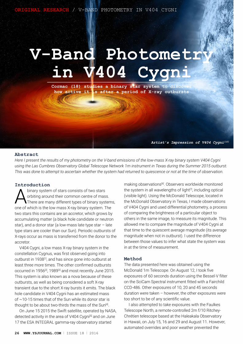
V-Band Photometry in V404 CygniCormac (18) studies a binary star system to discover how active it is after a period of X-ray outbursts
AbstractHere I present the results of my photometry on the V-band emissions of the low-mass X-ray binary system V404 Cygni using the Las Cumbres Observatory Global Telescope Network 1m instrument in Texas during the Summer 2015 outburst. This was done to attempt to ascertain whether the system had returned to quiescence or not at the time of observation.
Introduction
A binary system of stars consists of two stars orbiting around their common centre of mass. There are many different types of binary systems,
one of which is the low mass X-ray binary system. The two stars this contains are an accretor, which grows by accumulating matter (a black hole candidate or neutron star), and a donor star (a low-mass late type star – late type stars are cooler than our Sun). Periodic outbursts of X-rays occur as mass is transferred from the donor to the accretor.
V404 Cygni, a low mass X-ray binary system in the constellation Cygnus, was first observed going into outburst in 1938[1], and has since gone into outburst at least three more times. The other confirmed outbursts occurred in 1956[2], 1989[3] and most recently June 2015. This system is also known as a nova because of these outbursts, as well as being considered a soft X-ray transient due to the short X-ray bursts it emits. The black hole candidate in V404 Cygni has an estimated mass of ~10-15 times that of the Sun while its donor star is thought to be about two-thirds the mass of the Sun[4].
On June 15 2015 the Swift satellite, operated by NASA, detected activity in the area of V404 Cygni[5] and on June 17 the ESA INTEGRAL gamma-ray observatory started
making observations[6]. Observers worldwide monitored the system in all wavelengths of light[7], including optical (visible light). Using the McDonald Telescope, located in the McDonald Observatory in Texas, I made observations of V404 Cygni and used differential photometry, a process of comparing the brightness of a particular object to others in the same image, to measure its magnitude. This allowed me to compare the magnitude of V404 Cygni at that time to the quiescent average magnitude (its average magnitude when not in outburst). I used the difference between those values to infer what state the system was in at the time of measurement.
MethodThe data presented here was obtained using the McDonald 1m Telescope. On August 12, I took five exposures of 60 seconds duration using the Bessel-V filter on the SciCam Spectral instrument fitted with a Fairchild CCD-486. Other exposures of 10, 20 and 45 seconds duration were taken – however, the other exposures were too short to be of any scientific value.
I also attempted to take exposures with the Faulkes Telescope North, a remote-controlled 2m f/10 Ritchey-Chrétien telescope based at the Haleakala Observatory in Hawaii, on July 15, 16 and 29 and August 11. However, automated overrides and poor weather prevented the
Artist’s Impression of V404 Cygni[12]
2016 I ISSUE 18 I WWW.YSJOURNAL.COM 27
V-BAND PHOTOMETRY IN V404 CYGNI / ORIGINAL RESEARCH
intended exposures being taken. Both the McDonald Telescope and the Faulkes Telescope North are owned and operated by the Las Cumbres Observatory Global Telescope Network (LCOGTN). My observing time was allocated to me by the Faulkes Telescope Project, a UK-based charitable foundation supporting enquiry-based science education in second-level schools and an educational partner of LCOGTN.
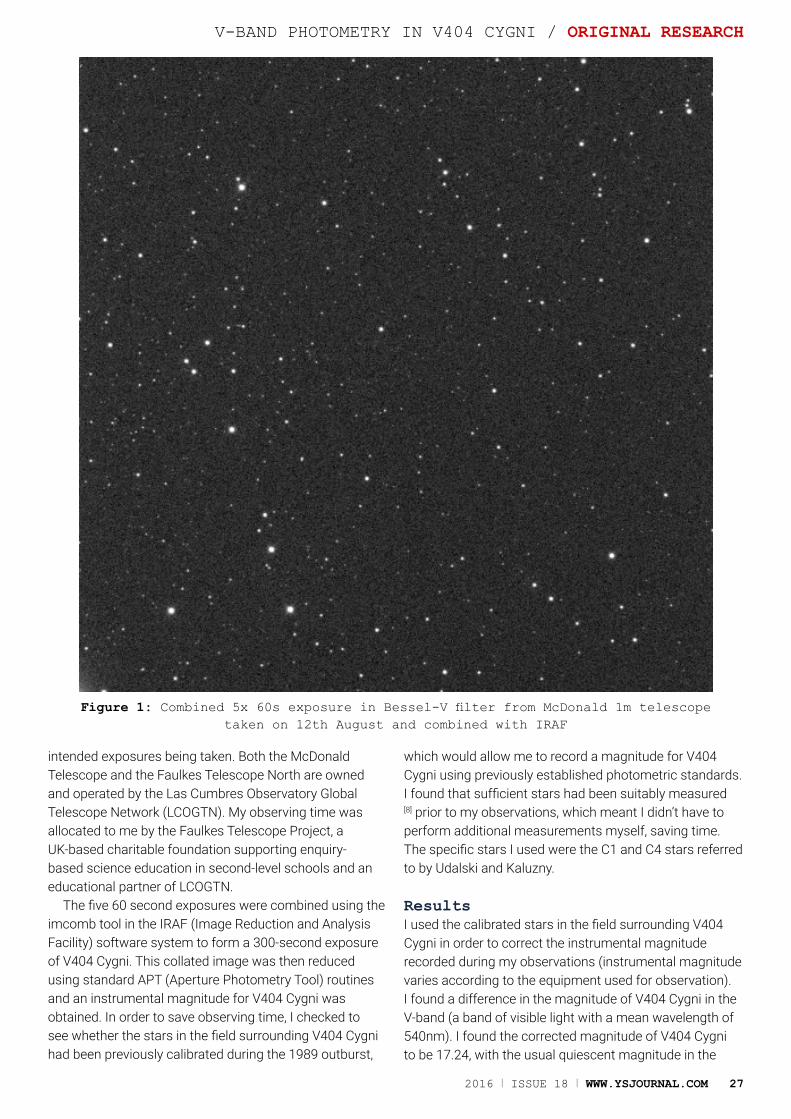
The five 60 second exposures were combined using the imcomb tool in the IRAF (Image Reduction and Analysis Facility) software system to form a 300-second exposure of V404 Cygni. This collated image was then reduced using standard APT (Aperture Photometry Tool) routines and an instrumental magnitude for V404 Cygni was obtained. In order to save observing time, I checked to see whether the stars in the field surrounding V404 Cygni had been previously calibrated during the 1989 outburst,
which would allow me to record a magnitude for V404 Cygni using previously established photometric standards. I found that sufficient stars had been suitably measured
[8] prior to my observations, which meant I didn’t have to perform additional measurements myself, saving time. The specific stars I used were the C1 and C4 stars referred to by Udalski and Kaluzny.
ResultsI used the calibrated stars in the field surrounding V404 Cygni in order to correct the instrumental magnitude recorded during my observations (instrumental magnitude varies according to the equipment used for observation). I found a difference in the magnitude of V404 Cygni in the V-band (a band of visible light with a mean wavelength of 540nm). I found the corrected magnitude of V404 Cygni to be 17.24, with the usual quiescent magnitude in the
Figure 1: Combined 5x 60s exposure in Bessel-V filter from McDonald 1m telescope taken on 12th August and combined with IRAF
28 WWW.YSJOURNAL.COM I ISSUE 18 I 2016
Cormac is a future astrophysicist who has completed placements at the University of St Andrews and University College Cork. He is also currently completing a CREST gold award with Armagh Observatory on the characterisation of massive OB stars in the Small Magellanic Cloud. He is in 5th year and studying for his Leaving Certificate, which he will sit in two years’ time. He is studying Maths, Applied Maths, Physics, Chemistry, Economics, German, English and Gaeilge.
Cormac Larkin, 18, Coláiste an Spioraid Naoimh, Ireland
BIOGRAPHY
ORIGINAL RESEARCH / V-BAND PHOTOMETRY IN V404 CYGNI
V-Band ranging between 18.3-18.4[9].
AnalysisFrom the data presented above, there is evidence to suggest that V404 Cygni had not yet reached total quiescence at the time of my observations. However, there is a noticeable decline in magnitude from the maximum values recorded during the peak of the outbursts, 12.1[10]. The most obvious thing to note here is that my observations were quite limited in scope due to restrictions on observation time and therefore they were not as comprehensive as I would have liked. My
other exposures were too short as I had no precedent with which to estimate appropriate exposure values. The unavailability of the Faulkes Telescope North due to unforeseen circumstances compounded the issue. My observations were also limited to only one band of the optical spectrum. The variation in magnitude observed is consistent with continuing activity in V404 Cygni on the scale of 300 second intervals. Although my data were limited, the difference of over one magnitude suggests that V404 Cygni had yet to return to total quiescence at the time of measurement.
Conclusions and Further WorkFurther observations would have been needed to confirm that the increased magnitude observed was indeed an indicator of persisting outburst activity in V404 Cygni. Exposures of different durations would reveal variations on other short time-scales. Exposures in other bands would also help to provide further proof over the optical range. From the limited observations I have conducted, there is evidence to suggest that V404 Cygni had not reached total quiescence at the time of my observations.
This finding was also corroborated by the findings of University College Dublin[11] at the Irish National Astronomy Meeting 2015 where I discussed my results with Prof. Hanlon.
This work was presented in poster format at the Irish National Astronomy Meeting 2015 and the Young Scientists Journal 2015 conference, where it was awarded 3rd place overall.
AcknowledgementI would like to thank my supervisor, Prof. Paul Callanan for all the help and guidance that made this project possible.
References1. A. A. Wachmann: Beobachtung von
Veränderlichen in der Umgebung von Kapteyn-Feldern der nördlichen Milchstraße. Teil I1 (Eichfeld 64). Astronomische Abhandlungen, Ergänzungshefte zu den Astronomischen Nachrichten, Bd. 11 Nr. 5. 48 S. DinA 4, mit 5 Abb. Berlin 1948, Akademie-Verlag.
2. Richter, G. A. 1989, Information Bulletin on Variable Stars, 3362, 1
3. Wagner, R.M. et al., 1990. The 1989 outburst of V404 cygni: A very unusual x-ray nova.
4. Shahbaz, T. et al., 1994. The mass of the black hole in V404 Cygni. Monthly Notices of the Royal Astronomical Society, 271(1), pp.L10–L14.
5. GCN Circular #179296. ATel #7662: INTEGRAL observations of intense
X-ray and optical flaring from V404 Cyg7. ATel #7735: V404 Cygni: coordination of multi-
wavelength observations and request for coverage during HST visits
8. Udalski, A. & Kaluzny, J., 1991. CCD photometry of the X-ray nova V404 Cygni after the 1989 outburst. Publications of the Astronomical Society of the Pacific, 103, p.198.
9. Shahbaz, T. et al., 2003. Multicolour observations of V404 Cyg with ULTRACAM. Monthly Notices of the Royal Astronomical Society, 346(4), pp.1116–1124.
10. ATel #7721: Optical (V-band) observations of V404 Cygni with the 0.3m telescope at Wheaton College Observatory
11. Murphy, D. et al., 2015. Watcher Monitoring of V404 Cygni in Outburst.
12. www.theblackstack.com
There is evidence to suggest that V404 Cygni had not yet reached total quiescence at the time of my observations
“

2016 I ISSUE 18 I WWW.YSJOURNAL.COM 29
Gene Silencing as a Therapy for Cancer
Girinath (17) explores the possible application of gene silencing in medicine, particularly cancer treatment.
GENE SILENCING AS A THERAPY FOR CANCER / REVIEW ARTICLE
AbstractIn this article I will explain what RNAi is, the genetic mechanisms behind it and why it deserves the considerable attention it has been receiving. Expanding on these basic mechanisms, I will go on to explore some of the latest RNAi trials involving Huntington’s disease and cancer. I will highlight the complications associated with the treatment of these complex diseases and importantly, why RNAi is seen as a viable therapeutic pathway. This article discusses the fact that cancer is not easy to treat because there is no medicine or treatment that can cure all cancers, due to the very distinct characteristics and reactions of each type. The method of gene silencing may be part of a solution for some cancer patients.
Introduction
Given that the expenditure for cancer care in the US alone is expected to reach $170 billion and that more than a third of men and women will
be diagnosed with cancer during their lives [1], there is an urgent need for effective treatments. Among the promising fields, RNA Interference (RNAi) and its role in gene silencing has drawn attention and seen steady progress over the years. Whilst the ability to turn genes on and off at will may seem like a biomedical superpower, the following research brings this ideal closer to reality and application as a treatment.
What is RNAi?RNAi is the process of gene expression silencing that
occurs after transcription (the copying of information from DNA to RNA) which functions as a defence mechanism against viruses and transposable elements. Whilst this is a natural process allowing both a host and a parasite to manipulate one another's gene expression [4], it may also hold the therapeutic potential to silence causative genes involved in tumourigenesis [6].
RNAi was first documented in 1998 by Andrew Fire and Craig Mello who were investigating how gene expression is regulated in C. elegans, a nematode. They were injecting mRNA that codes for muscle protein production which, if incompletely expressed, results in twitching movements. When they injected the coding (sense) strand and the complementary template (antisense) strand separately they observed no twitching. When injected together however, they observed the twitching characteristic of dysfunctional muscle protein genes. Fire and Mello hypothesised that the double-stranded RNA formed by the binding of the sense and antisense strand was somehow silencing the gene carrying that same code [2]. This discovery and hypothesis, proven and recorded
extensively, earned them the Nobel Prize in 2006 and has since become an area of extensive research.
Mechanisms of RNAi Gene SilencingThe process of RNAi is mediated by small interfering RNA, abbreviated to siRNA. It is worth noting that since
Dicer from G. intestinalis, the ribonuclease that
cleaves double stranded RNA into
siRNA [17]
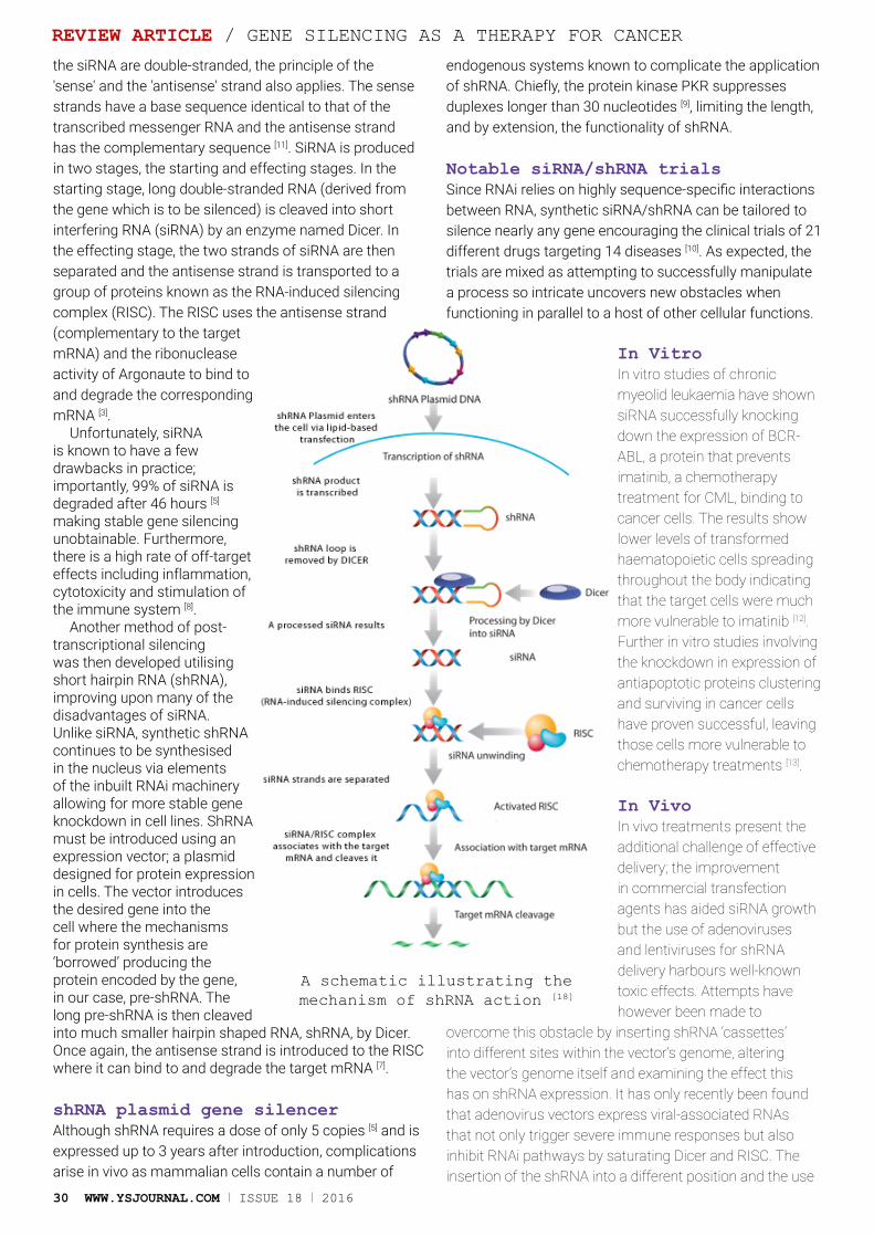
30 WWW.YSJOURNAL.COM I ISSUE 18 I 2016
the siRNA are double-stranded, the principle of the 'sense' and the 'antisense' strand also applies. The sense strands have a base sequence identical to that of the transcribed messenger RNA and the antisense strand has the complementary sequence [11]. SiRNA is produced in two stages, the starting and effecting stages. In the starting stage, long double-stranded RNA (derived from the gene which is to be silenced) is cleaved into short interfering RNA (siRNA) by an enzyme named Dicer. In the effecting stage, the two strands of siRNA are then separated and the antisense strand is transported to a group of proteins known as the RNA-induced silencing complex (RISC). The RISC uses the antisense strand (complementary to the target mRNA) and the ribonuclease activity of Argonaute to bind to and degrade the corresponding mRNA [3].
Unfortunately, siRNA is known to have a few drawbacks in practice; importantly, 99% of siRNA is degraded after 46 hours [5] making stable gene silencing unobtainable. Furthermore, there is a high rate of off-target effects including inflammation, cytotoxicity and stimulation of the immune system [8].
Another method of post-transcriptional silencing was then developed utilising short hairpin RNA (shRNA), improving upon many of the disadvantages of siRNA. Unlike siRNA, synthetic shRNA continues to be synthesised in the nucleus via elements of the inbuilt RNAi machinery allowing for more stable gene knockdown in cell lines. ShRNA must be introduced using an expression vector; a plasmid designed for protein expression in cells. The vector introduces the desired gene into the cell where the mechanisms for protein synthesis are ‘borrowed’ producing the protein encoded by the gene, in our case, pre-shRNA. The long pre-shRNA is then cleaved into much smaller hairpin shaped RNA, shRNA, by Dicer. Once again, the antisense strand is introduced to the RISC where it can bind to and degrade the target mRNA [7].
shRNA plasmid gene silencerAlthough shRNA requires a dose of only 5 copies [5] and is expressed up to 3 years after introduction, complications arise in vivo as mammalian cells contain a number of
endogenous systems known to complicate the application of shRNA. Chiefly, the protein kinase PKR suppresses duplexes longer than 30 nucleotides [9], limiting the length, and by extension, the functionality of shRNA.
Notable siRNA/shRNA trialsSince RNAi relies on highly sequence-specific interactions between RNA, synthetic siRNA/shRNA can be tailored to silence nearly any gene encouraging the clinical trials of 21 different drugs targeting 14 diseases [10]. As expected, the trials are mixed as attempting to successfully manipulate a process so intricate uncovers new obstacles when functioning in parallel to a host of other cellular functions.
In VitroIn vitro studies of chronic myeolid leukaemia have shown siRNA successfully knocking down the expression of BCR-ABL, a protein that prevents imatinib, a chemotherapy treatment for CML, binding to cancer cells. The results show lower levels of transformed haematopoietic cells spreading throughout the body indicating that the target cells were much more vulnerable to imatinib [12]. Further in vitro studies involving the knockdown in expression of antiapoptotic proteins clustering and surviving in cancer cells have proven successful, leaving those cells more vulnerable to chemotherapy treatments [13].
In VivoIn vivo treatments present the additional challenge of effective delivery; the improvement in commercial transfection agents has aided siRNA growth but the use of adenoviruses and lentiviruses for shRNA delivery harbours well-known toxic effects. Attempts have however been made to
overcome this obstacle by inserting shRNA ‘cassettes’ into different sites within the vector’s genome, altering the vector’s genome itself and examining the effect this has on shRNA expression. It has only recently been found that adenovirus vectors express viral-associated RNAs that not only trigger severe immune responses but also inhibit RNAi pathways by saturating Dicer and RISC. The insertion of the shRNA into a different position and the use
A schematic illustrating the mechanism of shRNA action [18]
REVIEW ARTICLE / GENE SILENCING AS A THERAPY FOR CANCER

2016 I ISSUE 18 I WWW.YSJOURNAL.COM 31
of VA-deleted adenovirus has been shown to reduce the drawbacks of this method and increase shRNA activity [14]. Whilst this approach has been applied to suppress hepatitis C replication [10], the opportunity to apply the method to cancer models still remains.
Another clinical trial that has drawn significant attention is being carried out by UCL and Isis pharmaceuticals and is focused, (in its early days), on safety as opposed to the complete knockdown of mutant Huntington expression. There are currently no treatments to prevent or even slow the progression of Huntington’s but RNAi gene silencing is the first therapeutic pathway to be trialled [15]. Whilst the results have not yet been published, we can see that the nature of RNAi can make it a versatile therapeutic tool against a number of genetic disorders.
Ex VivoEx vivo delivery of the therapeutic is also an option that has been applied by Duke University in a phase 1 study. The procedure, known as autologous cell therapy involves removing cells, modifying them with the appropriate treatment and re-implanting them into the patient. In a study targeting metastatic melanoma, the siRNA should enhance antigen presentation of the melanoma provoking a strong immune response, theoretically. Promisingly, according to Dr Pruitt from the University, none of the 10 patients have experienced toxicity from the process and have all shown antigen-specific immune responses [10].
Why is cancer so difficult to treat?Having explored the potential of RNAi, it is reasonable to ask why cancer remains so difficult to treat. The simple explanation is that cancer is not one disease but many hundreds of diseases. Whilst they may share common themes such as a resistance to apoptosis, forming their own blood supply (angiogenesis) and the ability to metastasise, each cancer achieves these in distinct ways. Even cancers of the same cell type can have quite different characteristics and within each cancer there are different populations of cells [16]. As a result, finding a single cure for cancer, even aided by discoveries such as RNAi, is unlikely. Instead, there must be numerous approaches to the problem, and among these, RNAi proves especially promising. It can be tailored to desired genes, allowing for specialised drugs for specialised diseases.
ConclusionsThe problem cancer presents lies in its specificity, making an umbrella treatment unlikely. Methods such as RNAi, however, provide an alternative approach that may not alone provide an effective treatment by itself but as we have learned, can work well in parallel with current chemotherapy.
The ability of RNAi to elicit specific transient or long-term gene knockdown has pushed the manipulation of this technique to make great strides since its discovery. As an already well-established tool to study gene function in labs today, the rate of progress has been staggering, considering the numerous technical obstacles such as delivery and integration of shRNA. The prospect of being able to inhibit
GENE SILENCING AS A THERAPY FOR CANCER / REVIEW ARTICLE
finding a single cure for cancer, even aided by discoveries such as RNAi, is unlikely“
Artist’s Impression of cancer cells dividing[newslocker]
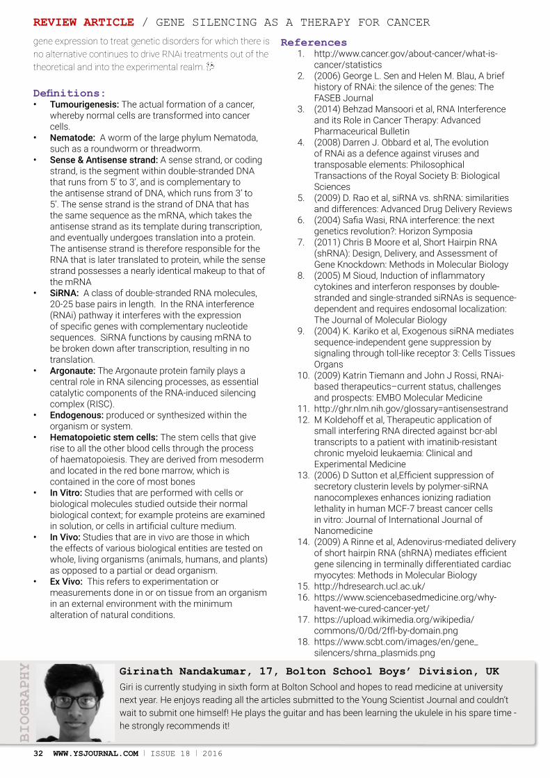
32 WWW.YSJOURNAL.COM I ISSUE 18 I 2016
gene expression to treat genetic disorders for which there is no alternative continues to drive RNAi treatments out of the theoretical and into the experimental realm.
Definitions:• Tumourigenesis: The actual formation of a cancer,
whereby normal cells are transformed into cancer cells.
• Nematode: A worm of the large phylum Nematoda, such as a roundworm or threadworm.
• Sense & Antisense strand: A sense strand, or coding strand, is the segment within double-stranded DNA that runs from 5’ to 3’, and is complementary to the antisense strand of DNA, which runs from 3’ to 5’. The sense strand is the strand of DNA that has the same sequence as the mRNA, which takes the antisense strand as its template during transcription, and eventually undergoes translation into a protein. The antisense strand is therefore responsible for the RNA that is later translated to protein, while the sense strand possesses a nearly identical makeup to that of the mRNA
• SiRNA: A class of double-stranded RNA molecules, 20-25 base pairs in length. In the RNA interference (RNAi) pathway it interferes with the expression of specific genes with complementary nucleotide sequences. SiRNA functions by causing mRNA to be broken down after transcription, resulting in no translation.
• Argonaute: The Argonaute protein family plays a central role in RNA silencing processes, as essential catalytic components of the RNA-induced silencing complex (RISC).
• Endogenous: produced or synthesized within the organism or system.
• Hematopoietic stem cells: The stem cells that give rise to all the other blood cells through the process of haematopoiesis. They are derived from mesoderm and located in the red bone marrow, which is contained in the core of most bones
• In Vitro: Studies that are performed with cells or biological molecules studied outside their normal biological context; for example proteins are examined in solution, or cells in artificial culture medium.
• In Vivo: Studies that are in vivo are those in which the effects of various biological entities are tested on whole, living organisms (animals, humans, and plants) as opposed to a partial or dead organism.
• Ex Vivo: This refers to experimentation or measurements done in or on tissue from an organism in an external environment with the minimum alteration of natural conditions.
References1. http://www.cancer.gov/about-cancer/what-is-
cancer/statistics2. (2006) George L. Sen and Helen M. Blau, A brief
history of RNAi: the silence of the genes: The FASEB Journal
3. (2014) Behzad Mansoori et al, RNA Interference and its Role in Cancer Therapy: Advanced Pharmaceurical Bulletin
4. (2008) Darren J. Obbard et al, The evolution of RNAi as a defence against viruses and transposable elements: Philosophical Transactions of the Royal Society B: Biological Sciences
5. (2009) D. Rao et al, siRNA vs. shRNA: similarities and differences: Advanced Drug Delivery Reviews
6. (2004) Safia Wasi, RNA interference: the next genetics revolution?: Horizon Symposia
7. (2011) Chris B Moore et al, Short Hairpin RNA (shRNA): Design, Delivery, and Assessment of Gene Knockdown: Methods in Molecular Biology
8. (2005) M Sioud, Induction of inflammatory cytokines and interferon responses by double-stranded and single-stranded siRNAs is sequence-dependent and requires endosomal localization: The Journal of Molecular Biology
9. (2004) K. Kariko et al, Exogenous siRNA mediates sequence-independent gene suppression by signaling through toll-like receptor 3: Cells Tissues Organs
10. (2009) Katrin Tiemann and John J Rossi, RNAi-based therapeutics–current status, challenges and prospects: EMBO Molecular Medicine
11. http://ghr.nlm.nih.gov/glossary=antisensestrand12. M Koldehoff et al, Therapeutic application of
small interfering RNA directed against bcr-abl transcripts to a patient with imatinib-resistant chronic myeloid leukaemia: Clinical and Experimental Medicine
13. (2006) D Sutton et al,Efficient suppression of secretory clusterin levels by polymer-siRNA nanocomplexes enhances ionizing radiation lethality in human MCF-7 breast cancer cells in vitro: Journal of International Journal of Nanomedicine
14. (2009) A Rinne et al, Adenovirus-mediated delivery of short hairpin RNA (shRNA) mediates efficient gene silencing in terminally differentiated cardiac myocytes: Methods in Molecular Biology
15. http://hdresearch.ucl.ac.uk/16. https://www.sciencebasedmedicine.org/why-
havent-we-cured-cancer-yet/17. https://upload.wikimedia.org/wikipedia/
commons/0/0d/2ffl-by-domain.png18. https://www.scbt.com/images/en/gene_
silencers/shrna_plasmids.png
Giri is currently studying in sixth form at Bolton School and hopes to read medicine at university next year. He enjoys reading all the articles submitted to the Young Scientist Journal and couldn’t wait to submit one himself! He plays the guitar and has been learning the ukulele in his spare time - he strongly recommends it!
BIOGRAPHY Girinath Nandakumar, 17, Bolton School Boys’ Division, UK
REVIEW ARTICLE / GENE SILENCING AS A THERAPY FOR CANCER
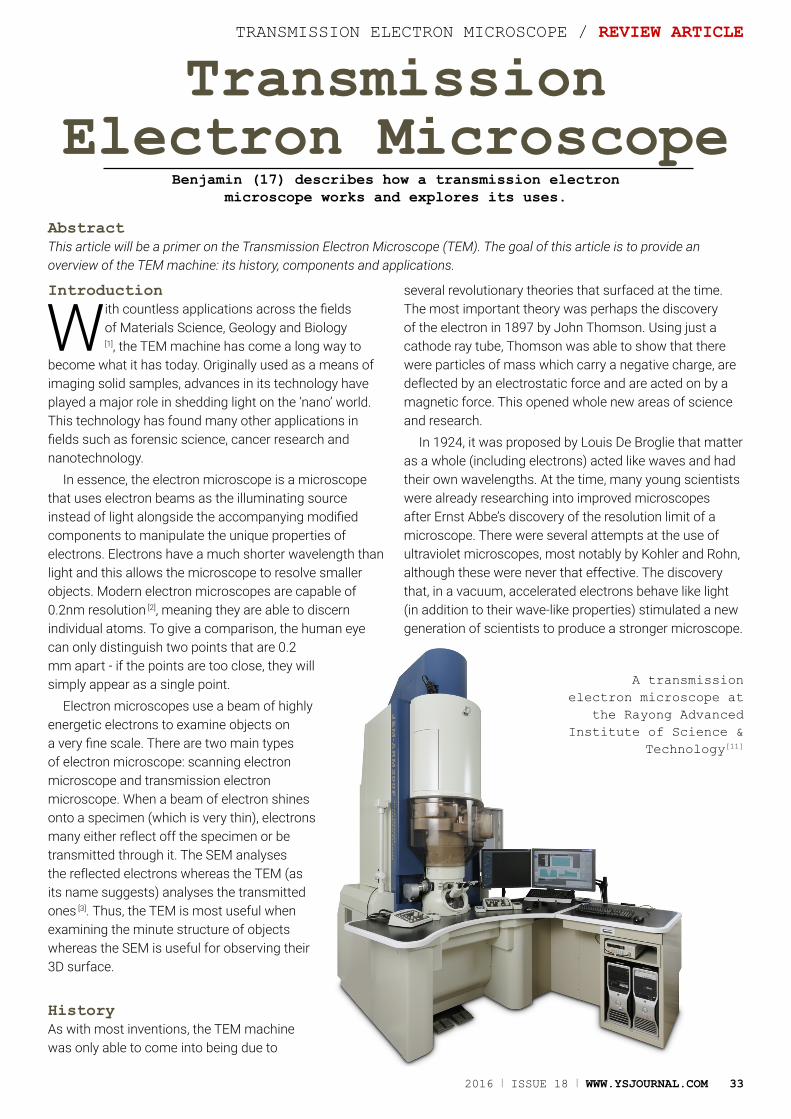
2016 I ISSUE 18 I WWW.YSJOURNAL.COM 33
Transmission Electron Microscope
Benjamin (17) describes how a transmission electron microscope works and explores its uses.
TRANSMISSION ELECTRON MICROSCOPE / REVIEW ARTICLE
AbstractThis article will be a primer on the Transmission Electron Microscope (TEM). The goal of this article is to provide an overview of the TEM machine: its history, components and applications.
Introduction
With countless applications across the fields of Materials Science, Geology and Biology
[1], the TEM machine has come a long way to become what it has today. Originally used as a means of imaging solid samples, advances in its technology have played a major role in shedding light on the ‘nano’ world. This technology has found many other applications in fields such as forensic science, cancer research and nanotechnology.
In essence, the electron microscope is a microscope that uses electron beams as the illuminating source instead of light alongside the accompanying modified components to manipulate the unique properties of electrons. Electrons have a much shorter wavelength than light and this allows the microscope to resolve smaller objects. Modern electron microscopes are capable of 0.2nm resolution [2], meaning they are able to discern individual atoms. To give a comparison, the human eye can only distinguish two points that are 0.2 mm apart - if the points are too close, they will simply appear as a single point.
Electron microscopes use a beam of highly energetic electrons to examine objects on a very fine scale. There are two main types of electron microscope: scanning electron microscope and transmission electron microscope. When a beam of electron shines onto a specimen (which is very thin), electrons many either reflect off the specimen or be transmitted through it. The SEM analyses the reflected electrons whereas the TEM (as its name suggests) analyses the transmitted ones [3]. Thus, the TEM is most useful when examining the minute structure of objects whereas the SEM is useful for observing their 3D surface.
HistoryAs with most inventions, the TEM machine was only able to come into being due to
several revolutionary theories that surfaced at the time. The most important theory was perhaps the discovery of the electron in 1897 by John Thomson. Using just a cathode ray tube, Thomson was able to show that there were particles of mass which carry a negative charge, are deflected by an electrostatic force and are acted on by a magnetic force. This opened whole new areas of science and research.
In 1924, it was proposed by Louis De Broglie that matter as a whole (including electrons) acted like waves and had their own wavelengths. At the time, many young scientists were already researching into improved microscopes after Ernst Abbe’s discovery of the resolution limit of a microscope. There were several attempts at the use of ultraviolet microscopes, most notably by Kohler and Rohn, although these were never that effective. The discovery that, in a vacuum, accelerated electrons behave like light (in addition to their wave-like properties) stimulated a new generation of scientists to produce a stronger microscope.
A transmission electron microscope at
the Rayong Advanced Institute of Science &
Technology[11]
34 WWW.YSJOURNAL.COM I ISSUE 18 I 2016
Equipped with all the necessary knowledge, several teams of scientists engaged in building the first electron microscope until 1931 when Ernst Ruska and Max Knoll finally produced a working model. Ruska subsequently received a Physics Nobel Prize for his work in 1986 [4].
ComponentsMany of the electron microscope components are analogous to those of a light microscope. These components have been modified for manipulating electrons however many of their purposes are the same. Rather than a light source, the electron microscope uses an electron gun and the glass lenses are replaced by electromagnetic lenses. Unlike glass lenses, the resolution and magnification of an electromagnetic lens is affected by changing the current through the lens whereas in a light microscope it is done by mechanically changing the lenses. Electron microscopes are normally built underground in order to reduce interference (in the form of vibrations) from environmental sources. As a precaution, they are also built in a room with thick walls as well as a vibration dampening table – you could drop a pencil on the table and it won’t bounce!
The transmission electron microscope can be compared to a slide projector [5]; it has an electron source which is made into a parallel beam by condenser lenses, which passes through an object and is then focussed onto a fluorescent screen.
Most TEM machines sport a columnar design with the electron source located at the top [6]. Typical components found inside the column are:
Electron gun - An electron gun consists of a filament (usually made of tungsten) surrounded by a Wehnelt cylinder that directs the beam in a tight, narrow beam. Most TEM machines work by heating the filament so that thermionic electrons are emitted by the tungsten. Alternatively, field emission guns may be used; these are used to pull electrons from atoms by producing a strong electrical field. An anode exists below the Wehnelt cylinder to accelerate the electrons generated. The specimen to be examined will be located below the accelerated beam.
Specimen chamber - Due to the nature of the TEMs, the sample used has to be immobilised and be made very thin. The specimen holder has one or two wells located at its end where the sample will be loaded onto. The specimen has to be kept very still so as to be able to produce clear images free of artefacts. As of such, the sample chamber is very sturdy.
Lenses - Similar to the optical microscope, the TEM has a lens system. However, this is a magnetic lens system rather than a glass one. Electron beams are not affected by glass lenses. However, magnetic fields can affect them as discovered by Han Busch in 1927. The TEM uses an electromagnet so that the strength of the magnetic field can be varied to control the direction and size of the electron beam.
Viewing chamber – Located at the base of the electron microscope, the viewing chamber is where the image of the magnified specimen is projected onto a screen. The surface onto which the image is projected onto is made of a fluorescent material which lights up depending on the density of electrons that arrives on the screen.
Vacuum chamber – Particles can interfere with the electron beams generated by the electron gun in two ways. Firstly, they can easily absorb some of the electrons and thus block the path of the electron beam. Alternatively, electrons may be scattered of knocked out by the particles and onto the specimen; this could distort the surface of the specimen.
How it worksThe electron beam generated by the electron gun is concentrated into a more powerful beam by the anode underneath it. It is directed towards the specimen, which sits on a copper grid in the specimen chamber. There, the beam passes through the specimen and based on the
varying thickness and density of the material, some of the beam may be scattered or reflected off of the specimen. This means that the electron beam at the other end of the specimen will have regions of varying electron densities. This is then magnified by the magnetic lenses and the image becomes visible when the
electron beam hits a fluorescent screen. The brightness of an area is determined by the electron density of that area of the beam. This image can then be viewed directly in the viewing chamber via a pair of binoculars or a camera.
This would normally produce a 2D image. Electron tomography can be used to produce 3D images. A beam of electrons is transmitted through the sample at incremental degrees of rotation around the centre of the target sample. This information can be collected and used to form an image by using computer software [7].
Applications and limitationsDespite being considered a rather old technique nowadays, the transmission electron microscope is still found at the forefront of many areas of science such
the transmission electron microscope is still found at the forefront of many areas of science such as materials science and biology
“
REVIEW ARTICLE / TRANSMISSION ELECTRON MICROSCOPE
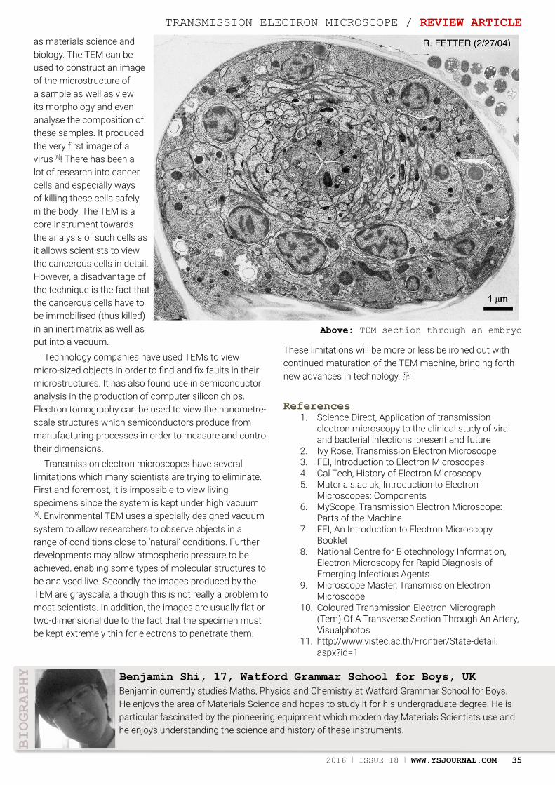
2016 I ISSUE 18 I WWW.YSJOURNAL.COM 35
as materials science and biology. The TEM can be used to construct an image of the microstructure of a sample as well as view its morphology and even analyse the composition of these samples. It produced the very first image of a virus [8]! There has been a lot of research into cancer cells and especially ways of killing these cells safely in the body. The TEM is a core instrument towards the analysis of such cells as it allows scientists to view the cancerous cells in detail. However, a disadvantage of the technique is the fact that the cancerous cells have to be immobilised (thus killed) in an inert matrix as well as put into a vacuum.
Technology companies have used TEMs to view micro-sized objects in order to find and fix faults in their microstructures. It has also found use in semiconductor analysis in the production of computer silicon chips. Electron tomography can be used to view the nanometre-scale structures which semiconductors produce from manufacturing processes in order to measure and control their dimensions.
Transmission electron microscopes have several limitations which many scientists are trying to eliminate. First and foremost, it is impossible to view living specimens since the system is kept under high vacuum [9]. Environmental TEM uses a specially designed vacuum system to allow researchers to observe objects in a range of conditions close to ‘natural’ conditions. Further developments may allow atmospheric pressure to be achieved, enabling some types of molecular structures to be analysed live. Secondly, the images produced by the TEM are grayscale, although this is not really a problem to most scientists. In addition, the images are usually flat or two-dimensional due to the fact that the specimen must be kept extremely thin for electrons to penetrate them.
These limitations will be more or less be ironed out with continued maturation of the TEM machine, bringing forth new advances in technology.
References 1. Science Direct, Application of transmission
electron microscopy to the clinical study of viral and bacterial infections: present and future
2. Ivy Rose, Transmission Electron Microscope3. FEI, Introduction to Electron Microscopes4. Cal Tech, History of Electron Microscopy5. Materials.ac.uk, Introduction to Electron
Microscopes: Components6. MyScope, Transmission Electron Microscope:
Parts of the Machine7. FEI, An Introduction to Electron Microscopy
Booklet8. National Centre for Biotechnology Information,
Electron Microscopy for Rapid Diagnosis of Emerging Infectious Agents
9. Microscope Master, Transmission Electron Microscope
10. Coloured Transmission Electron Micrograph (Tem) Of A Transverse Section Through An Artery, Visualphotos
11. http://www.vistec.ac.th/Frontier/State-detail.aspx?id=1
Benjamin currently studies Maths, Physics and Chemistry at Watford Grammar School for Boys. He enjoys the area of Materials Science and hopes to study it for his undergraduate degree. He is particular fascinated by the pioneering equipment which modern day Materials Scientists use and he enjoys understanding the science and history of these instruments.
Benjamin Shi, 17, Watford Grammar School for Boys, UK
BIOGRAPHY
Above: TEM section through an embryo
TRANSMISSION ELECTRON MICROSCOPE / REVIEW ARTICLE

Spider Silk in Medicine
Archana (16) studies the science behind spider silk and its applications in the medical industry.
AbstractMaterials have specific properties which make them useful for certain applications. Medicine is an area in which various material characteristics find a role in the human body. Spider silk fibre has revolutionary properties which make it an excellent new biomaterial. This review article will explore the chemistry, physics and biology of this material. Spider silk fibre has more recently also been reverse engineered into the physical state of an aqueous and ultra-thin film state. This opens up additional possibilities. It also allows implantation of silk material based devices within the human body in a manner that seamlessly conforms to biotic-abiotic interface. The versatile uses of spider silk in medicine include micro sutures (a type of joint found in the skull) with more strength, sturdy bio-scaffolds for regenerative medicine and tissue engineering for artificial skin and nerve grafts, tendon and ligament repair with the required strength and elasticity, weight-bearing artificial knee menisci in Orthopaedic surgery, liquid silk for biological wound dressings, silk micro particles for drug delivery and finally silk optics, bio-photonics, biosensors and bio electronics. The practical challenges and scope of research will revolve around the mass production and genetic engineering of spider silk, and reinventing a material that has existed for millennia.
REVIEW ARTICLE / SPIDER SILK IN MEDICINE
36 WWW.YSJOURNAL.COM I ISSUE 18 I 2016

2016 I ISSUE 18 I WWW.YSJOURNAL.COM 37
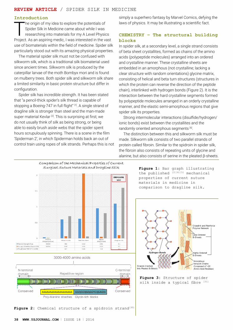
38 WWW.YSJOURNAL.COM I ISSUE 18 I 2016
Introduction
The origin of my idea to explore the potentials of Spider Silk in Medicine came about while I was researching into materials for my A Level Physics
Project. As an aspiring medic, I was interested in the vast use of biomaterials within the field of medicine. Spider silk particularly stood out with its amazing physical properties.
The material spider silk must not be confused with silkworm silk, which is a traditional silk biomaterial used since ancient times. Silkworm silk is produced by the caterpillar larvae of the moth Bombyx mori and is found on mulberry trees. Both spider silk and silkworm silk share a limited similarity in basic protein structure but differ in configuration.
Spider silk has incredible strength. It has been stated that “a pencil-thick spider’s silk thread is capable of stopping a Boeing-747 in full flight” [1]. A single strand of dragline silk is stronger than steel and the man-made super material Kevlar [2]. This is surprising at first; we do not usually think of silk as being strong, or being able to easily brush aside webs that the spider spent hours scrupulously spinning. There is a scene in the film ‘Spiderman 2’, in which Spiderman holds back an out of control train using ropes of silk strands. Perhaps this is not
simply a superhero fantasy by Marvel Comics, defying the laws of physics. It may be illustrating a scientific fact.
CHEMISTRY – The structural building blocksIn spider silk, at a secondary level, a single strand consists of beta sheet crystallites, formed as chains of the amino acids (polypeptide molecules) arranged into an ordered and crystalline manner. These crystalline sheets are embedded in an amorphous (not crystalline; lacking a clear structure with random orientations) glycine matrix, consisting of helical and beta turn structures (structures in which the protein can reverse the direction of the peptide chain), interlinked with hydrogen bonds (Figure 2). It is the interaction between the hard crystalline segments formed by polypeptide molecules arranged in an orderly crystalline manner, and the elastic semi-amorphous regions that give spider silk its properties.
Strong intermolecular interactions (disulfide/hydrogen/ionic bonds) exist between the crystallites and the randomly oriented amorphous segments [6].
The distinction between this and silkworm silk must be made. Silkworm silk consists of two parallel strands of protein called fibroin. Similar to the spidroin in spider silk, the fibroin also consists of repeating units of glycine and alanine, but also consists of serine in the pleated β-sheets.
REVIEW ARTICLE / SPIDER SILK IN MEDICINE
Figure 1: Bar graph illustrating the published [3][4][5] mechanical properties of current suture materials in medicine in comparison to dragline silk.
Figure 2: Chemical structure of a spidroin strand[30]
Figure 3: Structure of spider silk inside a typical fibre [31]
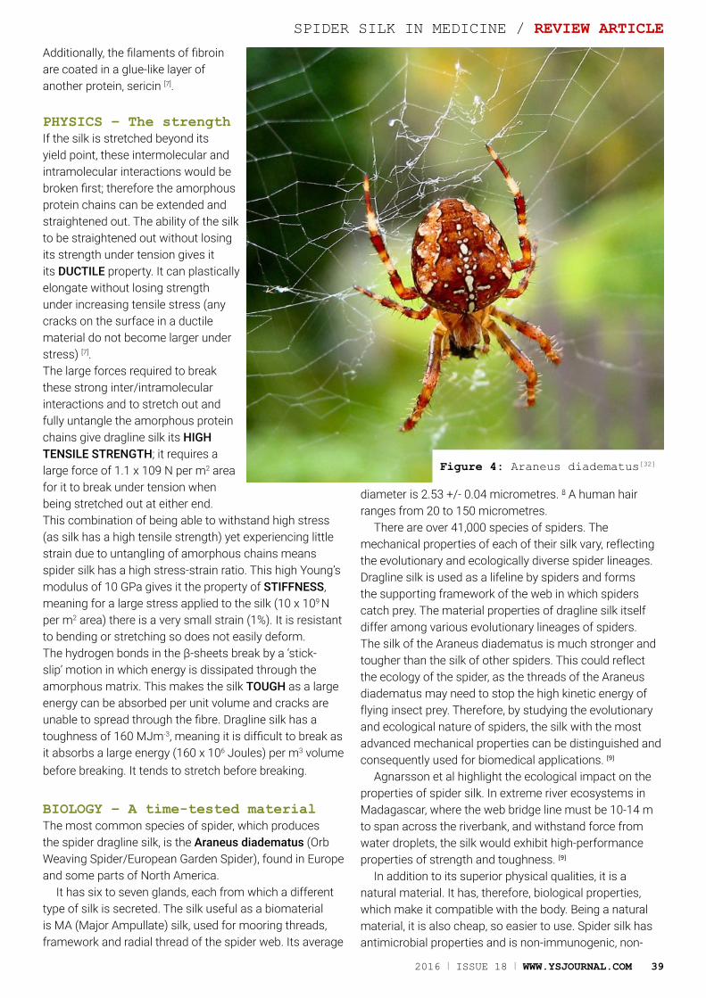
2016 I ISSUE 18 I WWW.YSJOURNAL.COM 39
Additionally, the filaments of fibroin are coated in a glue-like layer of another protein, sericin [7].
PHYSICS – The strengthIf the silk is stretched beyond its yield point, these intermolecular and intramolecular interactions would be broken first; therefore the amorphous protein chains can be extended and straightened out. The ability of the silk to be straightened out without losing its strength under tension gives it its DUCTILE property. It can plastically elongate without losing strength under increasing tensile stress (any cracks on the surface in a ductile material do not become larger under stress) [7]. The large forces required to break these strong inter/intramolecular interactions and to stretch out and fully untangle the amorphous protein chains give dragline silk its HIGH TENSILE STRENGTH; it requires a large force of 1.1 x 10⁹ N per m2 area for it to break under tension when being stretched out at either end.This combination of being able to withstand high stress (as silk has a high tensile strength) yet experiencing little strain due to untangling of amorphous chains means spider silk has a high stress-strain ratio. This high Young’s modulus of 10 GPa gives it the property of STIFFNESS, meaning for a large stress applied to the silk (10 x 109 N per m2 area) there is a very small strain (1%). It is resistant to bending or stretching so does not easily deform.The hydrogen bonds in the β-sheets break by a ‘stick-slip’ motion in which energy is dissipated through the amorphous matrix. This makes the silk TOUGH as a large energy can be absorbed per unit volume and cracks are unable to spread through the fibre. Dragline silk has a toughness of 160 MJm-3, meaning it is difficult to break as it absorbs a large energy (160 x 106 Joules) per m3 volume before breaking. It tends to stretch before breaking.
BIOLOGY – A time-tested materialThe most common species of spider, which produces the spider dragline silk, is the Araneus diadematus (Orb Weaving Spider/European Garden Spider), found in Europe and some parts of North America.
It has six to seven glands, each from which a different type of silk is secreted. The silk useful as a biomaterial is MA (Major Ampullate) silk, used for mooring threads, framework and radial thread of the spider web. Its average
diameter is 2.53 +/- 0.04 micrometres. 8 A human hair ranges from 20 to 150 micrometres.
There are over 41,000 species of spiders. The mechanical properties of each of their silk vary, reflecting the evolutionary and ecologically diverse spider lineages. Dragline silk is used as a lifeline by spiders and forms the supporting framework of the web in which spiders catch prey. The material properties of dragline silk itself differ among various evolutionary lineages of spiders. The silk of the Araneus diadematus is much stronger and tougher than the silk of other spiders. This could reflect the ecology of the spider, as the threads of the Araneus diadematus may need to stop the high kinetic energy of flying insect prey. Therefore, by studying the evolutionary and ecological nature of spiders, the silk with the most advanced mechanical properties can be distinguished and consequently used for biomedical applications. [9]
Agnarsson et al highlight the ecological impact on the properties of spider silk. In extreme river ecosystems in Madagascar, where the web bridge line must be 10-14 m to span across the riverbank, and withstand force from water droplets, the silk would exhibit high-performance properties of strength and toughness. [9]
In addition to its superior physical qualities, it is a natural material. It has, therefore, biological properties, which make it compatible with the body. Being a natural material, it is also cheap, so easier to use. Spider silk has antimicrobial properties and is non-immunogenic, non-
SPIDER SILK IN MEDICINE / REVIEW ARTICLE
Figure 4: Araneus diadematus[32]

40 WWW.YSJOURNAL.COM I ISSUE 18 I 2016
REVIEW ARTICLE / SPIDER SILK IN MEDICINE
Figure 6
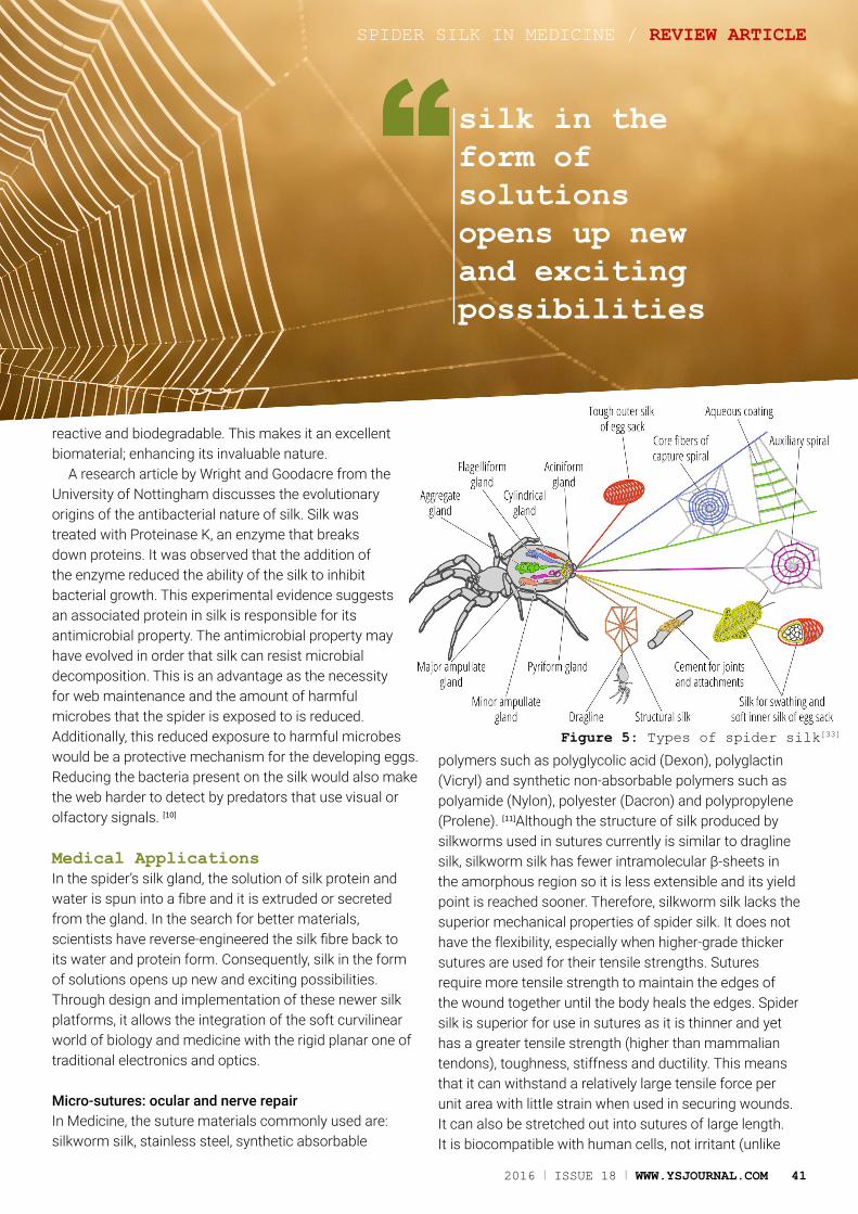
2016 I ISSUE 18 I WWW.YSJOURNAL.COM 41
reactive and biodegradable. This makes it an excellent biomaterial; enhancing its invaluable nature.
A research article by Wright and Goodacre from the University of Nottingham discusses the evolutionary origins of the antibacterial nature of silk. Silk was treated with Proteinase K, an enzyme that breaks down proteins. It was observed that the addition of the enzyme reduced the ability of the silk to inhibit bacterial growth. This experimental evidence suggests an associated protein in silk is responsible for its antimicrobial property. The antimicrobial property may have evolved in order that silk can resist microbial decomposition. This is an advantage as the necessity for web maintenance and the amount of harmful microbes that the spider is exposed to is reduced. Additionally, this reduced exposure to harmful microbes would be a protective mechanism for the developing eggs. Reducing the bacteria present on the silk would also make the web harder to detect by predators that use visual or olfactory signals. [10]
Medical ApplicationsIn the spider’s silk gland, the solution of silk protein and water is spun into a fibre and it is extruded or secreted from the gland. In the search for better materials, scientists have reverse-engineered the silk fibre back to its water and protein form. Consequently, silk in the form of solutions opens up new and exciting possibilities. Through design and implementation of these newer silk platforms, it allows the integration of the soft curvilinear world of biology and medicine with the rigid planar one of traditional electronics and optics.
Micro-sutures: ocular and nerve repairIn Medicine, the suture materials commonly used are: silkworm silk, stainless steel, synthetic absorbable
polymers such as polyglycolic acid (Dexon), polyglactin (Vicryl) and synthetic non-absorbable polymers such as polyamide (Nylon), polyester (Dacron) and polypropylene (Prolene). [11]Although the structure of silk produced by silkworms used in sutures currently is similar to dragline silk, silkworm silk has fewer intramolecular β-sheets in the amorphous region so it is less extensible and its yield point is reached sooner. Therefore, silkworm silk lacks the superior mechanical properties of spider silk. It does not have the flexibility, especially when higher-grade thicker sutures are used for their tensile strengths. Sutures require more tensile strength to maintain the edges of the wound together until the body heals the edges. Spider silk is superior for use in sutures as it is thinner and yet has a greater tensile strength (higher than mammalian tendons), toughness, stiffness and ductility. This means that it can withstand a relatively large tensile force per unit area with little strain when used in securing wounds. It can also be stretched out into sutures of large length. It is biocompatible with human cells, not irritant (unlike
SPIDER SILK IN MEDICINE / REVIEW ARTICLE
Figure 5: Types of spider silk[33]
silk in the form of solutions opens up new and exciting possibilities
“
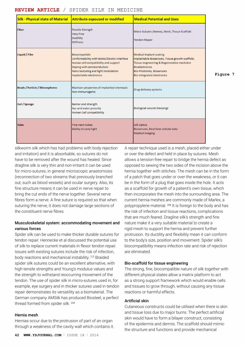
42 WWW.YSJOURNAL.COM I ISSUE 18 I 2016
silkworm silk which has had problems with body rejection and irritation) and it is absorbable, so sutures do not have to be removed after the wound has healed. Since dragline silk is very thin and non-irritant it can be used for micro-sutures, in general microscopic anastomosis (reconnection of two streams that previously branched out, such as blood vessels) and ocular surgery. Also, its fine structure means it can be used in nerve repair to bring the cut ends of the nerve together. Several nerve fibres form a nerve. A fine suture is required so that when suturing the nerve, it does not damage large sections of the constituent nerve fibres.
Musculoskeletal system: accommodating movement and various forcesSpider silk can be used to make thicker durable sutures for tendon repair. Hennecke et al discussed the potential use of silk to replace current materials in flexor tendon repair. Issues with existing sutures include the risk of infection, body reactions and mechanical instability. [12] Braided spider silk sutures could be an excellent alternative, with high tensile strengths and Young’s modulus values and the strength to withstand reoccurring movement of the tendon. The use of spider silk in micro-sutures used in, for example, eye surgery and in thicker sutures used in tendon repair demonstrates its versatility as a biomaterial. The German company AMSilk has produced Biosteel, a perfect thread formed from spider silk. [13]
Hernia meshHernias occur due to the protrusion of part of an organ through a weakness of the cavity wall which contains it.
A repair technique used is a mesh, placed either under or over the defect and held in place by sutures. Mesh allows a tension-free repair to bridge the hernia defect as opposed to sewing the two sides of the incision above the hernia together with stitches. The mesh can be in the form of a patch that goes under or over the weakness, or it can be in the form of a plug that goes inside the hole. It acts as a scaffold for growth of a patient’s own tissue, which then incorporates the mesh into the surrounding area. The current hernia meshes are commonly made of Marlex, a polypropylene material. [14] It is foreign to the body and has the risk of infection and tissue reactions, complications that are much feared. Dragline silk’s strength and fine nature make it a very suitable material to create a rigid mesh to support the hernia and prevent further protrusion. Its ductility and flexibility mean it can conform to the body’s size, position and movement. Spider silk’s biocompatibility means infection rate and risk of rejection are eliminated.
Bio-scaffold for tissue engineeringThe strong, fine, biocompatible nature of silk together with different physical states allow a matrix platform to act as a strong support framework which would enable cells and tissues to grow through, without causing any tissue reactions or harmful effects.
Artificial skinCutaneous constructs could be utilised when there is skin and tissue loss due to major burns. The perfect artificial skin would have to form a bilayer construct, consisting of the epidermis and dermis. The scaffold should mimic the structure and functions and provide mechanical
REVIEW ARTICLE / SPIDER SILK IN MEDICINE
Figure 7
2016 I ISSUE 18 I WWW.YSJOURNAL.COM 43
support and regulate cell activities. It should support cell attachment and migration and guide cell differentiation. In vitro investigation has shown that several types of human cells, including epidermal keratinocytes, dermal fibroblasts and endothelial cells, can be successfully cultured on silk scaffolds in various forms to form a single layer or bi-layered skin model if conditioned appropriately with nutrients, warmth and air. The silk film was prepared by dissolving the silk fibres in lithium bromide. It was biocompatible with minimal inflammatory reaction, haemocompatible with the underlying blood vessels and oxygen and water permeable, which is necessary as the skin acts as an interface. It supported the attachment and proliferation of cells.
Current research has looked into synthetic polymers like polyglycolic acid and polylactic acid as scaffold materials. These materials have poor mechanical properties. This has been evidenced by the deficiency of regenerated axons in nerve grafts. Collagen, a structural protein which is currently used, can lose its mechanical properties. Spider silk has better mechanical properties as well as being more biocompatible with no risk of rejection. [15] However, there are still challenges in creating artificial skin. It is difficult for a construct to restore all layers of skin, which include the functions of touch and temperature sensation, excretion, perspiration, thermoregulation, protection from ultraviolet light, synthetic function and its aesthetic function. Currently, silk is more feasibly used in the construction of the epidermal layer alone. This paper from the University of Brighton, England illustrates in great detail some of the currently marketed and clinically available tissue-engineered skin substitute products. [16]
Nerve graftThis same concept of silk being used as a support mesh on which cells grow back can be used in nerve grafts and silk filled conduit for larger nerve defects. Research is aiming to lead to treatment for damaged nerves with a large defect which require a scaffold support that allow nerve tissues to grow back and bridge the gap. [17]
The fine and strong nature of spider silk combined with its high biocompatibility and the ability to guide the migration of regenerating nerve cells and supporting (Schwann) cells make it a very useful material for regenerative medicine. The ultimate aim would be spinal cord repair using this approach.
Heart muscle regenerationBio-scaffolds can also be applied to regenerating heart cells in myocardial reconstruction. Currently, a magnesium
fluoride coated magnesium alloy LA63 is used to form the scaffold onto which heart cells are cultured and proliferate. This can surgically revitalize the heart whose muscles have died following a heart attack. LA63 lattice does have very high mechanical properties to withstand the continuous contraction of the myocardial muscle for a period of time. But these metallic constructs need precise engineering techniques. [18] The high tensile strength, high Young’s Modulus and high toughness of spider silk could perhaps make it a more advanced material for use in the myocardial scaffolds that are easily conformable to the moving contours and forces of the heart.
Tendon and ligament regenerationTissue engineering can also be applied to tendon and ligament repair. Both formed from collagen, which forms a fibrous connective tissue, tendons connect muscle to bone and ligaments connect two bones. Collagen can lose its fibrous and mechanical properties when manipulated for use in repair. Spider silk could serve as an improved alternative scaffold material for cells to proliferate and form the collagen fibrous tissue tendons and ligaments. Bone marrow-derived mesenchymal stem cells and anterior cruciate ligament fibroblasts from the knee can be cultured on a porous silk scaffold for tissue engineering. Research is also ongoing to develop a tendon and ligament scaffold by the synergistic incorporation of the silk and collagen. Furthermore, its advanced mechanical properties of high tensile strength and a high Young’s Modulus will enable it to withstand motion experienced when tendons and ligaments move. Its toughness also
means it can withstand movement as it will absorb a large amount of energy before fracturing. However, tissue engineered tendons and ligaments face challenges. The long-term stability and physical integrity of the silk scaffold constructs is not certain. [19]
Biological wound dressingThe people of the Carpathian Mountains across Central and Eastern Europe used spider silk for wounds. [10] The antimicrobial silk will mean it will not cause any reactions or immune response. Biological wound dressings such as hydrogels can be made as spider silk can be liquefied into a spidroin solution by the addition of acids or other additives. Hydrogels are three-dimensional polymer networks which are physically durable to swelling in aqueous solution but do not dissolve in these solutions. The biocompatible porous silk bioscaffold facilitates cell in-growth for wounds to heal.
Drug deliveryThe liquid silk can be made, mixed with a medical drug
SPIDER SILK IN MEDICINE / REVIEW ARTICLE
Dragline silk’s strength and fine nature make it a very suitable material to create a rigid mesh to support a hernia
“
and the solution can be converted into a solid particle state or very fine microsphere structures. This can be for the delivery of small molecule drugs to a particular site in the body. It will not cause immune reactions as it travels through the body to the site needed. [20]
Bio photonics/ silk opticsAdded to these benefits is the recent discovery that silk is a gifted manipulator of light. Light travels through silk almost as easily as it flows through glass fibres. The science of photonics includes the generation, emission, transmission, modulation, signal processing, switching, amplification, and detection/sensing of light. The term has an analogy to electronics. The term photonics developed as an outgrowth of the first practical semiconductor light emitters LED invented in the early 1960s and optical fibres developed in the 1970s. Nano-texturing allows the use of this silk platform as biosensors that utilises the behaviour of light along nano wavelength distances or barriers. The brilliant blue colour of the morpho butterfly is a natural example of the manipulation of light by the non-silk natural material of this butterfly. Diffraction refers to the phenomenon that occurs when a wave encounters an obstacle or a slit that is comparable in size to its wavelength. Maximum diffraction occurs when the aperture is equal to or smaller than the light wavelength (400 – 600 nm). In optics, a diffraction grating is an optical component with a periodic structure, which splits and diffracts light into several beams travelling in different directions. The directions of these beams depend on the spacing of the grating and the wavelength of the light so that the grating acts as the dispersive element. Silk films can be nano-textured or imprinted to manipulate light. These can then be used as biosensors. Light transmitted into the blood or cells will be modified by the incident substrate such as proteins, DNA, cells etc. The interaction of this light with the silk film can be used to bio-sense the biologic media. Silk photonics can be used to detect cellular real time data and may offer clues to cancer cell initiation, metabolic status, oxygenation status etc. Silk can alternatively be doped with
semiconductors or integrated with nanoelectronic circuitry to yield bioelectronics. The can lead to implantable bioelectronic sensors. [21] Silk in the form of tubes offers new possibilities compared to glass based fibre-optic technology. Silk is not as efficient as glass fibre in conducting light, but researchers are working on coating the silk fibre with appropriate chemicals to overcome this limitation. Glass based fibre optics requires precise manufacturing technology, while the spider manufactures silk for us! Natural silk is only five microns in diameter, less than a tenth of the width of a human hair (50 microns), while glass fibres vary from about 10-60microns internal diameters. [22] Silk optics opens up new avenues for medical imaging.
Liquid silk coatingThe biological evolution of silk into a non-immunogenic, non-toxic and non-reactive material offers the possibility of coating medical implants to help reduce the human body’s ability to recognise the foreign material in the implants. Silicone breast implants can elicit an adjacent tissue inflammatory response that can distort the implant, affect the consistency and feel of the implant or produce pain to the patient. AMSilk has produced silicone implants coated with silk protein BioShield-S1 that are currently in pre-clinical trials. [23]
Orthopaedic surgery – Artificial MeniscusMeniscal implants using dragline silk could withstand movement and compressive forces. A meniscus is a fibrocartilaginous structure that partly divides a joint cavity such as in the knee. This is a common injury in football players and can become a major health problem. The founder of Oxford Silk Group, Professor Fritz Vollrath, along with his team is currently developing implants for this. By dissolving in lithium bromide, the dragline silk becomes firmer. The resulting implant has high strength and toughness. A large force and energy are required before fracture so it can support the weight of the body. It will absorb a large energy before any damage can occur.
REVIEW ARTICLE / SPIDER SILK IN MEDICINE
44 WWW.YSJOURNAL.COM I ISSUE 18 I 2016
It integrates much better potentially with the human body than any plastic material. According to Vollrath, the meniscal implants, which are undergoing animal trials, could be used in humans in one to two years. [24]
Practical Aspects And Future Avenues For ResearchTranslating the ideas in academia to clinical practice unearths many practical challenges of using spider silk in the various medical fields mentioned above. The fact that dragline silk is a natural material is one practical advantage. Current materials involving petrochemical processes are not sustainable in the long run, but the use of silk would be.
Potential commercial mass production for many spider silks is still extremely finite. This arises from the difficulty of high-density spider farming due to their cannibalistic behaviour. Furthermore, only approximately 12 m of silk can be obtained from a spider web. This is an extremely small quantity compared to other materials such as from a silkworm cocoon, from which 600-900 m of silk can be yielded. This means alternative methods of manufacture are required.
Current research involves genetic engineering to produce silk similar to dragline silk on a mass scale. So far, it has not been possible to recreate an authentic copy of spider silk.
The difficulty of this can be understood to some extent by comparison to the current mass production of human insulin by genetic engineering. Gone are the days of pig or porcine insulins. The fundamental requisite is understanding the genetic code. The complexity of the genetic code in turn depends on the complexity of the proteins being coded for. Frederick Sanger characterized the primary structure of insulin in the late 1950s. Insulin is a smaller protein made of two polypeptide chains comprising of 51 amino acids in total and molecular
weight of 5808 Da. The relatively simple structure meant in 1978, the first genetic engineering company Genetech, could mass-produce genetically engineered human insulin.
In comparison, dragline silk presents a much more complex situation. The molecular weight is 200-350 KDa. The secondary structure is a complex co-polymer of alanine crystallites and glycine amorphous chains. The knowledge of spider silk genes coding for this structure is incomplete. Therefore, an exact mimic of a native dragline silk fibre has not been possible thus far. All transgene constructs for recombinant silk proteins have relied on partial cDNA. These truncated cDNAs encode only a fraction (typically 20% or less) of the repetitive region and the C-terminal domain. [25] Research must centre on alternative methods. Teulé et al outline research into the use of vectors to create transgenic silkworms encoding chimeric silkworm/spider silk protein genes. The engineered silk produced by the silkworm was a composite of chimeric silkworm and spider silk. It was tougher than the parental silkworm silk and equally tough as dragline silk. The experimental evidence demonstrates that silkworms can be genetically engineered to manufacture silk with properties close to native dragline silk, although exact replication has still not been achieved. [26] Bioengineering firm NexiaBiotech have genetically engineered goats to secrete spider silk proteins into its milk, which can then be separated off. [27] Some other companies that have embraced this new material challenge include ARAKNITEK collaborating with Utah State University, [28] AMSilk [13] and Spiber Technologies. [29]
ConclusionThe scientific world is always searching for novel biomaterials that could revolutionise our daily lives. The multifarious applications of spider silk in medicine have been clearly demonstrated in an array of medical areas. The potential
SPIDER SILK IN MEDICINE / REVIEW ARTICLE
The potential applications of spider silk stretch beyond the field of medicine
“2016 I ISSUE 18 I WWW.YSJOURNAL.COM 45
46 WWW.YSJOURNAL.COM I ISSUE 18 I 2016
applications of spider silk indeed stretch beyond the field of medicine. It is also being considered as a material for use in bulletproof clothing, ropes, nets, seat belts, parachutes and composite materials in some vehicles including aircraft. The day may arrive when a coffee cup made of spider silk can be thrown away without the fears from the current non-degradable polystyrene cups! It is remarkable how such a super material exists naturally, carefully mastered over thousands of years by evolutionary processes with superlative chemical, biological and physical properties. Perhaps mastering of the proteins and the permutative possibilities offered by the 20 amino acids should be the subject of tomorrow’s material science research. Most of us are unaware of the fact that spiders, a creature which for many of us triggers trepidation, contain the material that could revolutionise our way of life. Perhaps arachnophobes should reconsider their fear, for it could be a spider silk stitch or bendy spider silk bioelectronics that may save them in a hospital of the future!
DefinitionsAmorphous- A structure without a clear shape or form. Antimicrobial - An agent that kills microorganisms or inhibits their growth. Hernia- When a section of an organ moves out of where it is usually located.Orthopaedic Surgery - Surgery concerning the musculoskeletal system. Immunogenicity - The ability of a substance to cause an immune response.Young’s Modulus - The relationship between stress and strain.
References1. How thick should a spider silk thread be to stop a Boeing-747 in full
flight? [Internet]. [cited 2015 Jan 24]. Available from: http://ednieuw.home.xs4all.nl/Spiders/Info/SilkBoeing.html
2. From spiders, a material to rival Kevlar - Fortune [Internet]. [cited 2015 Jan 24]. Available from: http://fortune.com/2013/06/14/from-spiders-a-material-to-rival-kevlar/
3. Altman GH, Diaz F, Jakuba C, Calabro T, Horan RL, Chen J, et al. Silk-based biomaterials. Biomaterials. 2003 Feb;24(3):401–16.
4. Plastipedia: The Plastics Encyclopedia - Polypropylene (PP) [Internet]. [cited 2015 Jan 24]. Available from: http://www.bpf.co.uk/plastipedia/polymers/pp.aspx
5. Properties: Stainless Steel - Grade 316 (UNS S31600) [Internet]. [cited 2015 Jan 24]. Available from: http://www.azom.com/properties.aspx?ArticleID=863
6. Spider silk [Internet]. Wikipedia, the free encyclopedia. 2015 [cited 2015 Jan 24]. Available from: http://en.wikipedia.org/w/index.php?title=Spider_silk&oldid=643480027
7. Liu X, Zhang K-Q. Silk Fiber — Molecular Formation Mechanism, Structure- Property Relationship and Advanced Applications. In: Lesieur C, editor. Oligomerization of Chemical and Biological Compounds [Internet]. InTech; 2014 [cited 2015 Jan 24]. Available
from: http://www.intechopen.com/books/oligomerization-of-chemical-and-biological-compounds/silk-fiber-molecular-formation-mechanism-structure-property-relationship-and-advanced-applications
8. Singha K, Maity S, Singha M. Spinning and Applications of Spider Silk. Front Sci. 2012 Dec 1;2(5):92–100.
9. Agnarsson I, Kuntner M, Blackledge TA. Bioprospecting Finds the Toughest Biological Material: Extraordinary Silk from a Giant Riverine Orb Spider. Lalueza-Fox C, editor. PLoS ONE. 2010 Sep 16;5(9):e11234.
10. Wright S, Goodacre SL. Evidence for antimicrobial activity associated with common house spider silk. BMC Res Notes. 2012;5(1):326.
11. ETHICONTM Suture Needles | Ethicon [Internet]. [cited 2015 Jan 24]. Available from: http://www.ethicon.com/healthcare-professionals/products/wound-closure/suture-needles/ethicon-suture-needles
12. Hennecke K, Redeker J, Kuhbier JW, Strauss S, Allmeling C, Kasper C, et al. Bundles of Spider Silk, Braided into Sutures, Resist Basic Cyclic Tests: Potential Use for Flexor Tendon Repair. Roeder RK, editor. PLoS ONE. 2013 Apr 17;8(4):e61100.
13. AMSilk | high performance materials: Biosteel Spidersilk Fibers [Internet]. [cited 2015 Jan 25]. Available from: http://www.amsilk.com/en/products/biosteel-spidersilk-fibers.html
14. Mesh & Hernia Repair [Internet]. [cited 2015 Jan 24]. Available from: http://www.herniasolutions.com/mesh-hernia-repair
15. Sobajo C, Behzad F, Yuan X-F, Bayat A. Silk: a potential medium for tissue engineering. Eplasty. 2008;8:e47.
16. Shevchenko RV, James SL, James SE. A review of tissue-engineered skin bioconstructs available for skin reconstruction. J R Soc Interface. 2010 Feb 6;7(43):229–58.
17. Radtke C, Allmeling C, Waldmann K-H, Reimers K, Thies K, Schenk HC, et al. Spider Silk Constructs Enhance Axonal Regeneration and Remyelination in Long Nerve Defects in Sheep. Egles C, editor. PLoS ONE. 2011 Feb 25;6(2):e16990.
18. Schilling T, Brandes G, Tudorache I, Cebotari S, Hilfiker A, Meyer T, et al. In vivo degradation of magnesium alloy LA63 scaffolds for temporary stabilization of biological myocardial grafts in a swine model. Biomed Tech Eng [Internet]. 2013 Jan 1 [cited 2015 Jan 24];58(5). Available from: http://www.degruyter.com/view/j/bmte.2013.58.issue-5/bmt-2012-0047/bmt-2012-0047.xml
19. Zhang Q, Yan S, Li M. Silk Fibroin Based Porous Materials. Materials. 2009 Dec 9;2(4):2276–95.
20. AMSilk | high performance materials: Silkbeads [Internet]. [cited 2015 Jan 25]. Available from: http://www.amsilk.com/en/materials/silkbeads.html
21. Kim D-H, Viventi J, Amsden JJ, Xiao J, Vigeland L, Kim Y-S, et al. Dissolvable films of silk fibroin for ultrathin conformal bio-integrated electronics. Nat Mater. 2010 Jun;9(6):511–7.
22. The FOA Reference For Fiber Optics - Optical Fiber [Internet]. [cited 2015 Jan 25]. Available from: http://www.thefoa.org/tech/ref/basic/fiber.html
23. AMSilk | high performance materials: Implant Coating [Internet]. [cited 2015 Jan 25]. Available from: http://www.amsilk.com/en/products/implant-coating.html
24. Fritz Vollrath: “Who wouldn’t want to study spiders?’ | Science | The Guardian [Internet]. [cited 2015 Jan 25]. Available from: http://www.theguardian.com/science/2013/jan/12/fritz-vollrath-spiders-tim-adams
25. Ayoub NA, Garb JE, Tinghitella RM, Collin MA, Hayashi CY. Blueprint for a High-Performance Biomaterial: Full-Length Spider Dragline Silk Genes. DeSalle R, editor. PLoS ONE. 2007 Jun 13;2(6):e514.
26. Teule F, Miao Y-G, Sohn B-H, Kim Y-S, Hull JJ, Fraser MJ, et al. Silkworms transformed with chimeric silkworm/spider silk genes spin composite silk fibers with improved mechanical properties. Proc Natl Acad Sci. 2012 Jan 17;109(3):923–8.
27. spider-silk - Companies & Biotechnology Firms Investigating Spider Silk [Internet]. [cited 2015 Jan 25]. Available from: http://spider-silk.wikispaces.com/
28. ARAKNITEK [Internet]. [cited 2015 Jan 25]. Available from: http://www.araknitek.com/index.html
29. spiber Spiber applications [Internet]. [cited 2015 Jan 25]. Available from: http://www.spiber.se/index.php?langId=1&headId=3&pageId=3
30. Chemical structure of a spidroin strand, Copyright Spiber Technologies31. Araneus diadematus, CC-BY-2.0.32. Types of spider silk, Copyright Spiber Technologies.
Archana is currently in Year 13 at King Edward VI High School for Girls, Birmingham and studying Biology, Chemistry, Maths and Physics. She wants to study Medicine at university as she finds the way in which all the sciences are inextricably linked to form materials that revolutionise medical treatments very interesting.
BIOGRAPHY Archana Jain, 16, King Edward VI High School for Girls, UK
REVIEW ARTICLE / SPIDER SILK IN MEDICINE
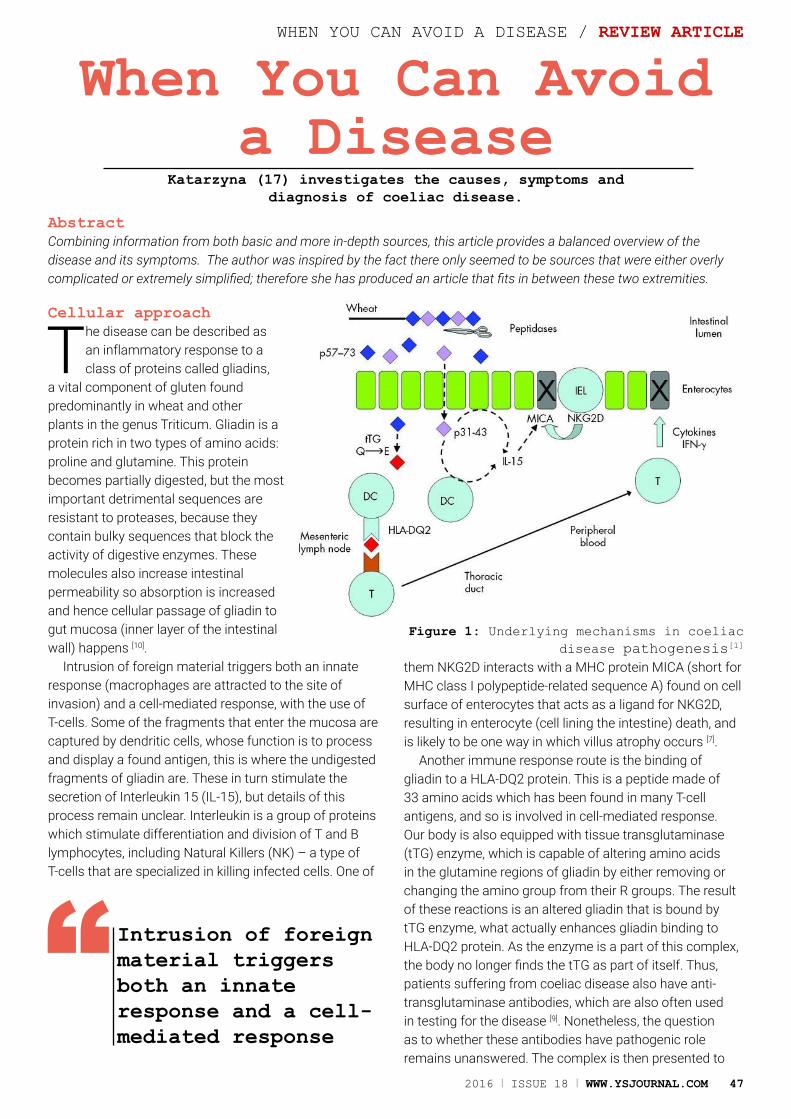
2016 I ISSUE 18 I WWW.YSJOURNAL.COM 47
When You Can Avoid a Disease
Katarzyna (17) investigates the causes, symptoms and diagnosis of coeliac disease.
AbstractCombining information from both basic and more in-depth sources, this article provides a balanced overview of the disease and its symptoms. The author was inspired by the fact there only seemed to be sources that were either overly complicated or extremely simplified; therefore she has produced an article that fits in between these two extremities.
Cellular approach
The disease can be described as an inflammatory response to a class of proteins called gliadins,
a vital component of gluten found predominantly in wheat and other plants in the genus Triticum. Gliadin is a protein rich in two types of amino acids: proline and glutamine. This protein becomes partially digested, but the most important detrimental sequences are resistant to proteases, because they contain bulky sequences that block the activity of digestive enzymes. These molecules also increase intestinal permeability so absorption is increased and hence cellular passage of gliadin to gut mucosa (inner layer of the intestinal wall) happens [10].
Intrusion of foreign material triggers both an innate response (macrophages are attracted to the site of invasion) and a cell-mediated response, with the use of T-cells. Some of the fragments that enter the mucosa are captured by dendritic cells, whose function is to process and display a found antigen, this is where the undigested fragments of gliadin are. These in turn stimulate the secretion of Interleukin 15 (IL-15), but details of this process remain unclear. Interleukin is a group of proteins which stimulate differentiation and division of T and B lymphocytes, including Natural Killers (NK) – a type of T-cells that are specialized in killing infected cells. One of
them NKG2D interacts with a MHC protein MICA (short for MHC class I polypeptide-related sequence A) found on cell surface of enterocytes that acts as a ligand for NKG2D, resulting in enterocyte (cell lining the intestine) death, and is likely to be one way in which villus atrophy occurs [7].
Another immune response route is the binding of gliadin to a HLA-DQ2 protein. This is a peptide made of 33 amino acids which has been found in many T-cell antigens, and so is involved in cell-mediated response. Our body is also equipped with tissue transglutaminase (tTG) enzyme, which is capable of altering amino acids in the glutamine regions of gliadin by either removing or changing the amino group from their R groups. The result of these reactions is an altered gliadin that is bound by tTG enzyme, what actually enhances gliadin binding to HLA-DQ2 protein. As the enzyme is a part of this complex, the body no longer finds the tTG as part of itself. Thus, patients suffering from coeliac disease also have anti-transglutaminase antibodies, which are also often used in testing for the disease [9]. Nonetheless, the question as to whether these antibodies have pathogenic role remains unanswered. The complex is then presented to
Figure 1: Underlying mechanisms in coeliac disease pathogenesis[1]
WHEN YOU CAN AVOID A DISEASE / REVIEW ARTICLE
Intrusion of foreign material triggers both an innate response and a cell-mediated response
“
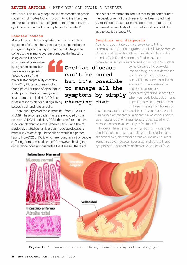
48 WWW.YSJOURNAL.COM I ISSUE 18 I 2016
the T-cells. This usually happens in the mesenteric lymph nodes (lymph nodes found in proximity to the intestine). This results in the release of gamma-Interferon (IFN-γ), a cytokine, which attracts macrophages to the site. [4]
Genetic causesMost of the problems originate from the incomplete digestion of gluten. Then, these untypical peptides are recognized by immune system and are destroyed. In doing so, the immune system damages the intestine lining as well. It seems to be caused completely by digestion errors, but there is also a genetic factor. A part of the major histocompatibility complex II (MHC II; it is a set of molecules found on cell surface of cells that is a vital part of the immune system in vertebrates) called HLA-DQ, is a protein responsible for distinguishing between self and foreign cells.
There are 8 types of these proteins - from HLA-DQ2 to DQ9. These polypeptide chains are encoded by the genes HLA-DQA1 and HLA-DQB1 that are found to have a loci on 6th chromosome. When a particular allele of previously stated genes, is present, coeliac disease is more likely to develop. These alleles result in a person having HLA-DQ2 or DQ8, which are found in 95% of people suffering from coeliac disease [5][8]. However, having the genes alone does not guarantee the disease - there are
also other environmental factors that might contribute to the development of the disease. It has been noted that a viral infection, that causes intestine inflammation and increased permeability of the small intestine, could also lead to coeliac disease [10].
Symptoms and diagnosisAs shown, both interactions give rise to killing enterocytes and thus degradation of villi. Malabsorption of many vital nutrients such as minerals and fat-soluble vitamins (A, D, E and K) from the food is due to a decreased absorption surface area in the intestine. Further
symptoms may include weight loss and fatigue due to decreased absorption of carbohydrates, iron deficiency anaemia, calcium and vitamin D malabsorption and hence secondary hyperparathyroidism - a condition when your body lacks calcium and phosphates, what triggers release of these minerals from bones so
that there are optimal levels of them in your blood, what in turn causes osteoporosis - a disorder in which your bones lose mass and bone mineral density is decreased what leads to increased vulnerability to fractures [6].
However, the most common symptoms include: pale skin, loose and greasy stool; pale, voluminous diarrhoea, abdominal pain, abdominal distension and mouth ulcers. Sometimes even lactose intolerance might arise. These symptoms are caused by incomplete digestion of food
Figure 2: A transverse section through bowel showing villus atrophy[2]
REVIEW ARTICLE / WHEN YOU CAN AVOID A DISEASE
Coeliac disease can’t be cured but it’s possible to manage all the symptoms by simply changing diet
“
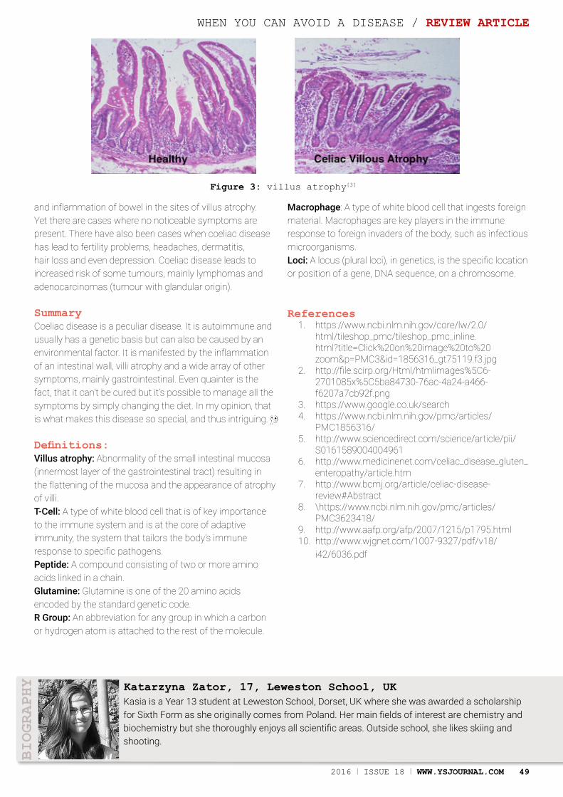
2016 I ISSUE 18 I WWW.YSJOURNAL.COM 49
and inflammation of bowel in the sites of villus atrophy. Yet there are cases where no noticeable symptoms are present. There have also been cases when coeliac disease has lead to fertility problems, headaches, dermatitis, hair loss and even depression. Coeliac disease leads to increased risk of some tumours, mainly lymphomas and adenocarcinomas (tumour with glandular origin).
SummaryCoeliac disease is a peculiar disease. It is autoimmune and usually has a genetic basis but can also be caused by an environmental factor. It is manifested by the inflammation of an intestinal wall, villi atrophy and a wide array of other symptoms, mainly gastrointestinal. Even quainter is the fact, that it can’t be cured but it’s possible to manage all the symptoms by simply changing the diet. In my opinion, that is what makes this disease so special, and thus intriguing.
Definitions:Villus atrophy: Abnormality of the small intestinal mucosa (innermost layer of the gastrointestinal tract) resulting in the flattening of the mucosa and the appearance of atrophy of villi.T-Cell: A type of white blood cell that is of key importance to the immune system and is at the core of adaptive immunity, the system that tailors the body’s immune response to specific pathogens.Peptide: A compound consisting of two or more amino acids linked in a chain.Glutamine: Glutamine is one of the 20 amino acids encoded by the standard genetic code.R Group: An abbreviation for any group in which a carbon or hydrogen atom is attached to the rest of the molecule.
Macrophage: A type of white blood cell that ingests foreign material. Macrophages are key players in the immune response to foreign invaders of the body, such as infectious microorganisms.Loci: A locus (plural loci), in genetics, is the specific location or position of a gene, DNA sequence, on a chromosome.
References1. https://www.ncbi.nlm.nih.gov/core/lw/2.0/
html/tileshop_pmc/tileshop_pmc_inline.html?title=Click%20on%20image%20to%20zoom&p=PMC3&id=1856316_gt75119.f3.jpg
2. http://file.scirp.org/Html/htmlimages%5C6-2701085x%5C5ba84730-76ac-4a24-a466-f6207a7cb92f.png
3. https://www.google.co.uk/search4. https://www.ncbi.nlm.nih.gov/pmc/articles/
PMC1856316/5. http://www.sciencedirect.com/science/article/pii/
S01615890040049616. http://www.medicinenet.com/celiac_disease_gluten_
enteropathy/article.htm7. http://www.bcmj.org/article/celiac-disease-
review#Abstract8. \https://www.ncbi.nlm.nih.gov/pmc/articles/
PMC3623418/9. http://www.aafp.org/afp/2007/1215/p1795.html10. http://www.wjgnet.com/1007-9327/pdf/v18/
i42/6036.pdf
Kasia is a Year 13 student at Leweston School, Dorset, UK where she was awarded a scholarship for Sixth Form as she originally comes from Poland. Her main fields of interest are chemistry and biochemistry but she thoroughly enjoys all scientific areas. Outside school, she likes skiing and shooting.
Katarzyna Zator, 17, Leweston School, UK
BIOGRAPHY
Figure 3: villus atrophy[3]
WHEN YOU CAN AVOID A DISEASE / REVIEW ARTICLE

Virtual Reality for Budget Smartphones
Peter He (15) explores wireless virtual reality for entry-level smartphones in this exciting project
PROJECT / VIRTUAL REALITY FOR BUDGET SMARTPHONES
AbstractIn the recent decade, Virtual Reality (VR) has become more common through a range of systems including computer-powered head-mounted displays (HMDs) and smartphone-powered headsets. It has presented both designers and developers alike with a new field filled with unexplored potential. In this article, a new and inexpensive approach to VR is explored, allowing entry-level smartphones to run powerful computer-based VR experiences wirelessly through Bluetooth with relatively high levels of performance. The project was recognised as a European and African Finalist in the Google Science Fair 2015
50 WWW.YSJOURNAL.COM I ISSUE 18 I 2016
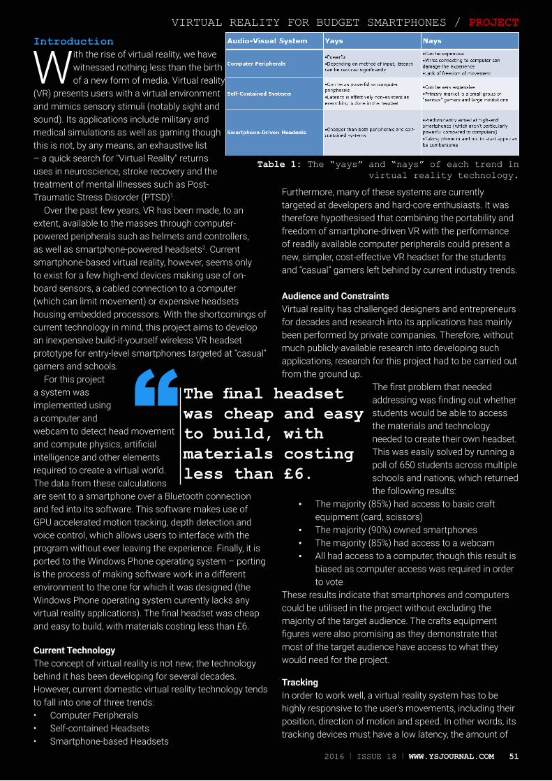
VIRTUAL REALITY FOR BUDGET SMARTPHONES / PROJECT
Introduction
With the rise of virtual reality, we have witnessed nothing less than the birth of a new form of media. Virtual reality
(VR) presents users with a virtual environment and mimics sensory stimuli (notably sight and sound). Its applications include military and medical simulations as well as gaming though this is not, by any means, an exhaustive list – a quick search for "Virtual Reality" returns uses in neuroscience, stroke recovery and the treatment of mental illnesses such as Post-Traumatic Stress Disorder (PTSD)1.
Over the past few years, VR has been made, to an extent, available to the masses through computer-powered peripherals such as helmets and controllers, as well as smartphone-powered headsets2. Current smartphone-based virtual reality, however, seems only to exist for a few high-end devices making use of on-board sensors, a cabled connection to a computer (which can limit movement) or expensive headsets housing embedded processors. With the shortcomings of current technology in mind, this project aims to develop an inexpensive build-it-yourself wireless VR headset prototype for entry-level smartphones targeted at “casual” gamers and schools.
For this project a system was implemented using a computer and webcam to detect head movement and compute physics, artificial intelligence and other elements required to create a virtual world. The data from these calculations are sent to a smartphone over a Bluetooth connection and fed into its software. This software makes use of GPU accelerated motion tracking, depth detection and voice control, which allows users to interface with the program without ever leaving the experience. Finally, it is ported to the Windows Phone operating system – porting is the process of making software work in a different environment to the one for which it was designed (the Windows Phone operating system currently lacks any virtual reality applications). The final headset was cheap and easy to build, with materials costing less than £6.
Current TechnologyThe concept of virtual reality is not new; the technology behind it has been developing for several decades. However, current domestic virtual reality technology tends to fall into one of three trends:• Computer Peripherals• Self-contained Headsets• Smartphone-based Headsets
Furthermore, many of these systems are currently targeted at developers and hard-core enthusiasts. It was therefore hypothesised that combining the portability and freedom of smartphone-driven VR with the performance of readily available computer peripherals could present a new, simpler, cost-effective VR headset for the students and “casual” gamers left behind by current industry trends.
Audience and ConstraintsVirtual reality has challenged designers and entrepreneurs for decades and research into its applications has mainly been performed by private companies. Therefore, without much publicly-available research into developing such applications, research for this project had to be carried out from the ground up.
The first problem that needed addressing was finding out whether students would be able to access the materials and technology needed to create their own headset. This was easily solved by running a poll of 650 students across multiple schools and nations, which returned the following results:
• The majority (85%) had access to basic craft equipment (card, scissors)
• The majority (90%) owned smartphones• The majority (85%) had access to a webcam• All had access to a computer, though this result is
biased as computer access was required in order to vote
These results indicate that smartphones and computers could be utilised in the project without excluding the majority of the target audience. The crafts equipment figures were also promising as they demonstrate that most of the target audience have access to what they would need for the project.
TrackingIn order to work well, a virtual reality system has to be highly responsive to the user’s movements, including their position, direction of motion and speed. In other words, its tracking devices must have a low latency, the amount of
Table 1: The “yays” and “nays” of each trend in virtual reality technology.
The final headset was cheap and easy to build, with materials costing less than £6.
“
2016 I ISSUE 18 I WWW.YSJOURNAL.COM 51
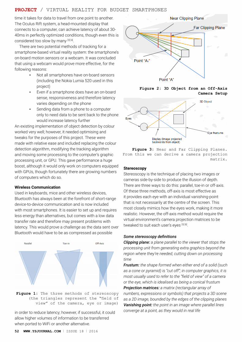
52 WWW.YSJOURNAL.COM I ISSUE 18 I 2016
time it takes for data to travel from one point to another. The Oculus Rift system, a head-mounted display that connects to a computer, can achieve latency of about 30-40ms in perfectly optimized conditions, though even this is considered too slow by many [3] [4].
There are two potential methods of tracking for a smartphone-based virtual reality system: the smartphone’s on-board motion sensors or a webcam. It was concluded that using a webcam would prove more effective, for the following reasons:
• Not all smartphones have on-board sensors (including the Nokia Lumia 520 used in this project)
• Even if a smartphone does have an on-board sense, responsiveness and therefore latency varies depending on the phone
• Sending data from a phone to a computer only to need data to be sent back to the phone would increase latency further
An existing implementation of object detection by colour worked very well; however, it needed optimising and tweaks for the purposes of this project. These were made with relative ease and included replacing the colour detection algorithm, modifying the tracking algorithm and moving some processing to the computer’s graphic processing unit, or GPU. This gave performance a huge boost, although it would only work on computers equipped with GPUs, though fortunately there are growing numbers of computers which do so.
Wireless CommunicationUsed in keyboards, mice and other wireless devices, Bluetooth has always been at the forefront of short-range device-to-device communication and is now included with most smartphones. It is easier to set up and requires less energy than alternatives, but comes with a low data transfer rate and therefore may present problems with latency. This would prove a challenge as the data sent over Bluetooth would have to be as compressed as possible
in order to reduce latency; however, if successful, it could allow higher volumes of information to be transferred when ported to WiFi or another alternative.
StereoscopyStereoscopy is the technique of placing two images or cameras side-by-side to produce the illusion of depth. There are three ways to do this: parallel, toe-in or off-axis.Of these three methods, off-axis is most effective as it provides each eye with an individual vanishing-point that is not necessarily at the centre of the screen. This most closely mimics how the eyes work, making it more realistic. However, the off-axis method would require the virtual environment’s camera projection matrices to be tweaked to suit each user’s eyes [5] [6].
Some stereoscopy definitionsClipping plane: a plane parallel to the viewer that stops the processing unit from generating extra graphics beyond the region where they’re needed, cutting down on processing time Frustum: the shape formed when either end of a solid (such as a cone or pyramid) is “cut off”; in computer graphics, it is most usually used to refer to the “field of view” of a camera or the eye, which is idealised as being a conical frustum Projection matrices: a matrix (rectangular array of numbers, expressions or symbols) that projects a 3D scene as a 2D image, bounded by the edges of the clipping planes Vanishing point: the point in an image where parallel lines converge at a point, as they would in real life
PROJECT / VIRTUAL REALITY FOR BUDGET SMARTPHONES
Figure 1: The three methods of stereoscopy (the triangles represent the “field of
view” of the camera, eye or image)
Figure 2: 3D Object from an Off-Axis Camera Setup
Figure 3: Near and Far Clipping Planes. From this we can derive a camera projection
matrix.
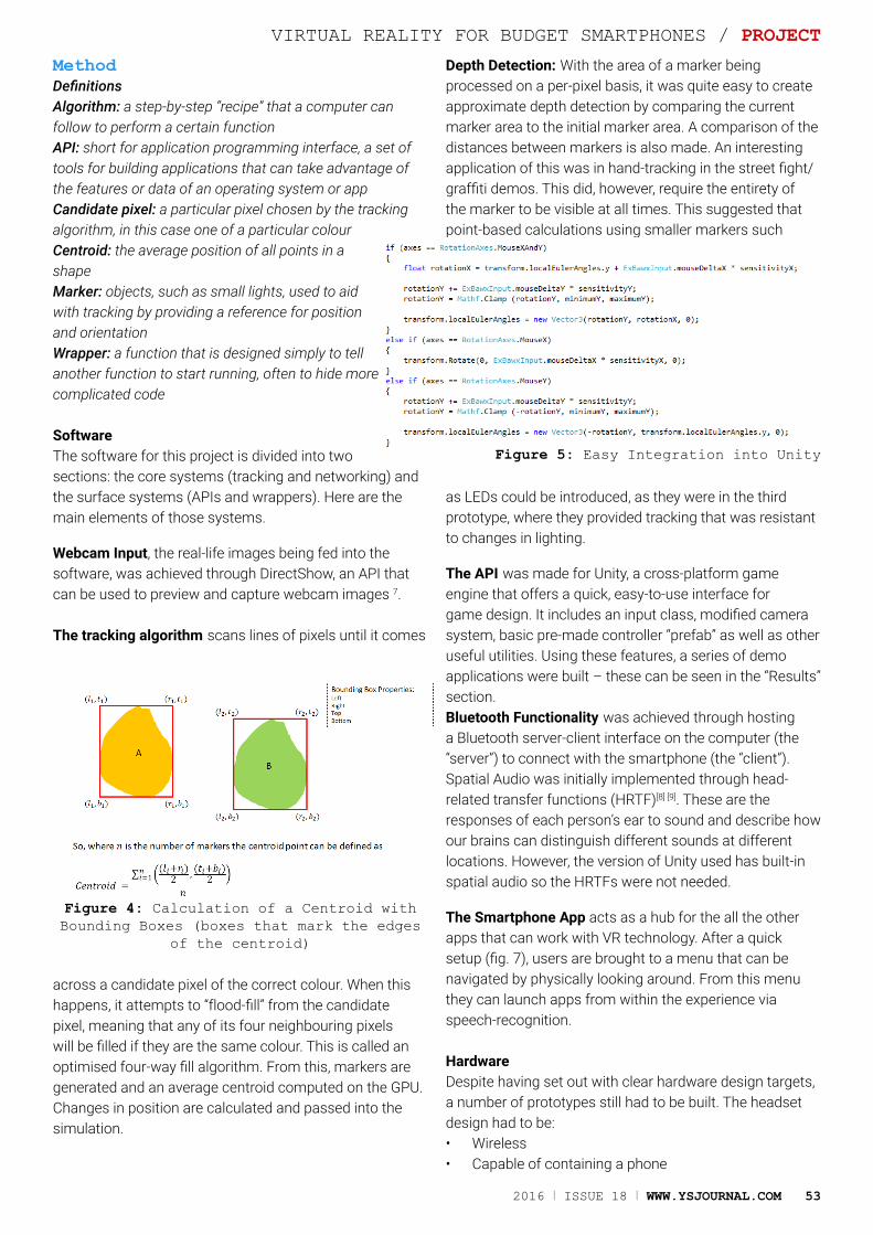
2016 I ISSUE 18 I WWW.YSJOURNAL.COM 53
MethodDefinitionsAlgorithm: a step-by-step “recipe” that a computer can follow to perform a certain functionAPI: short for application programming interface, a set of tools for building applications that can take advantage of the features or data of an operating system or appCandidate pixel: a particular pixel chosen by the tracking algorithm, in this case one of a particular colourCentroid: the average position of all points in a shapeMarker: objects, such as small lights, used to aid with tracking by providing a reference for position and orientationWrapper: a function that is designed simply to tell another function to start running, often to hide more complicated code
SoftwareThe software for this project is divided into two sections: the core systems (tracking and networking) and the surface systems (APIs and wrappers). Here are the main elements of those systems.
Webcam Input, the real-life images being fed into the software, was achieved through DirectShow, an API that can be used to preview and capture webcam images 7.
The tracking algorithm scans lines of pixels until it comes
across a candidate pixel of the correct colour. When this happens, it attempts to “flood-fill” from the candidate pixel, meaning that any of its four neighbouring pixels will be filled if they are the same colour. This is called an optimised four-way fill algorithm. From this, markers are generated and an average centroid computed on the GPU. Changes in position are calculated and passed into the simulation.
Depth Detection: With the area of a marker being processed on a per-pixel basis, it was quite easy to create approximate depth detection by comparing the current marker area to the initial marker area. A comparison of the distances between markers is also made. An interesting application of this was in hand-tracking in the street fight/graffiti demos. This did, however, require the entirety of the marker to be visible at all times. This suggested that point-based calculations using smaller markers such
as LEDs could be introduced, as they were in the third prototype, where they provided tracking that was resistant to changes in lighting.
The API was made for Unity, a cross-platform game engine that offers a quick, easy-to-use interface for game design. It includes an input class, modified camera system, basic pre-made controller “prefab” as well as other useful utilities. Using these features, a series of demo applications were built – these can be seen in the “Results” section.Bluetooth Functionality was achieved through hosting a Bluetooth server-client interface on the computer (the “server”) to connect with the smartphone (the “client”).Spatial Audio was initially implemented through head-related transfer functions (HRTF)[8] [9]. These are the responses of each person’s ear to sound and describe how our brains can distinguish different sounds at different locations. However, the version of Unity used has built-in spatial audio so the HRTFs were not needed.
The Smartphone App acts as a hub for the all the other apps that can work with VR technology. After a quick setup (fig. 7), users are brought to a menu that can be navigated by physically looking around. From this menu they can launch apps from within the experience via speech-recognition.
HardwareDespite having set out with clear hardware design targets, a number of prototypes still had to be built. The headset design had to be:• Wireless• Capable of containing a phone
VIRTUAL REALITY FOR BUDGET SMARTPHONES / PROJECT
Figure 4: Calculation of a Centroid with Bounding Boxes (boxes that mark the edges
of the centroid)
Figure 5: Easy Integration into Unity
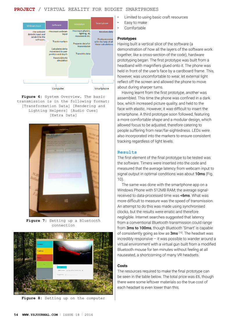
54 WWW.YSJOURNAL.COM I ISSUE 18 I 2016
• Limited to using basic craft resources• Easy to make• Comfortable
PrototypesHaving built a vertical slice of the software (a demonstration of how all the layers of the software work together, like a cross-section of the code), hardware prototyping began. The first prototype was built from a headband with magnifiers glued onto it. The phone was held in front of the user’s face by a cardboard frame. This, however, was uncomfortable to wear, let external light reflect off the screen and allowed the phone to move about during sharper turns.
Having learnt from the first prototype, another was assembled. This time the phone was confined in a dark box, which increased picture quality, and held to the face with elastic. However, it was difficult to insert the smartphone. A third prototype soon followed, featuring a more comfortable shape and a modular design, which allowed focus to be adjusted, therefore catering to people suffering from near/far-sightedness. LEDs were also incorporated into the markers to ensure consistent tracking regardless of light levels.
ResultsThe first element of the final prototype to be tested was the software. Timers were inserted into the code and measured that the average latency from webcam input to signal output in optimal conditions was about 10ms (Fig. 10).
The same was done with the smartphone app on a Windows Phone with 512MB RAM; the average signal-received to data-processed time was <6ms. What was more difficult to measure was the speed of transmission. An attempt to do this was made using synchronised clocks, but the results were erratic and therefore negligible. Internet searches suggested that latency from a conventional Bluetooth transmission could range from 3ms to 100ms, though Bluetooth ‘Smart’ is capable of consistently going as low as 3ms [10]. The headset was incredibly responsive – it was possible to wander around a virtual environment with a virtual gun built from a modified Bluetooth mouse for ten minutes without feeling at all nauseated, a shortcoming of many VR headsets.
CostsThe resources required to make the final prototype can be seen in the table below. The total price was £6, though there were some leftover materials so the true cost of each headset is even lower than this.
PROJECT / VIRTUAL REALITY FOR BUDGET SMARTPHONES
Figure 6: System Overview. The basic transmission is in the following format:
[Transformation Data] [Rendering and Lighting Helpers] [Audio Cues]
[Extra Data]
Figure 7: Setting up a Bluetooth connection
Figure 8: Setting up on the computer
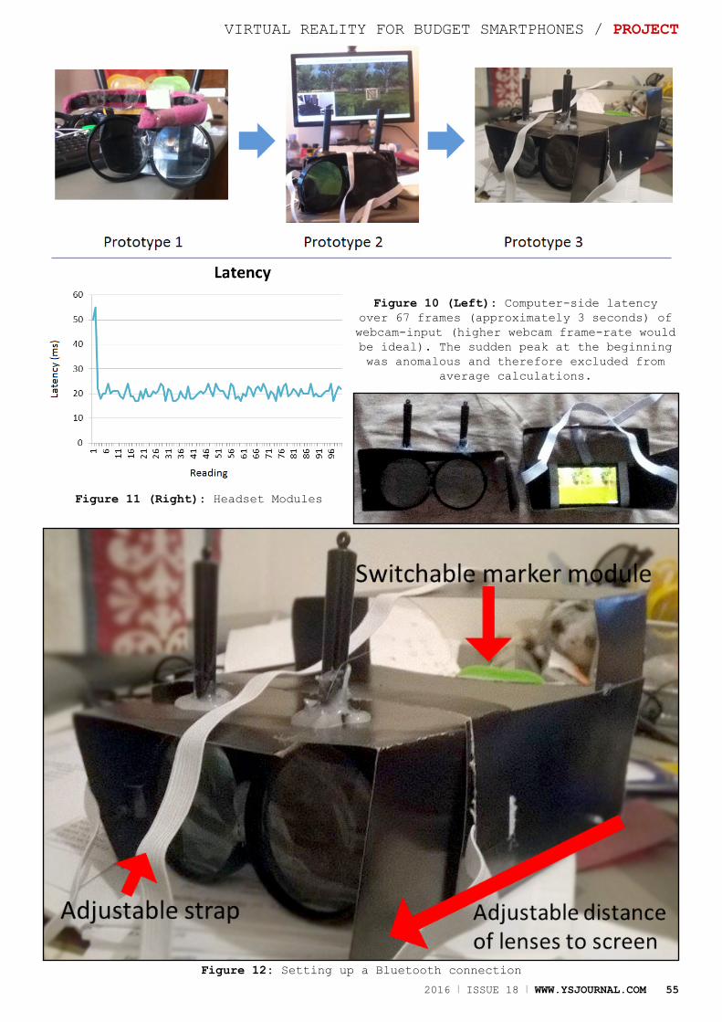
2016 I ISSUE 18 I WWW.YSJOURNAL.COM 55
VIRTUAL REALITY FOR BUDGET SMARTPHONES / PROJECT
Figure 10 (Left): Computer-side latency over 67 frames (approximately 3 seconds) of webcam-input (higher webcam frame-rate would be ideal). The sudden peak at the beginning was anomalous and therefore excluded from
average calculations.
Figure 11 (Right): Headset Modules
Figure 12: Setting up a Bluetooth connection
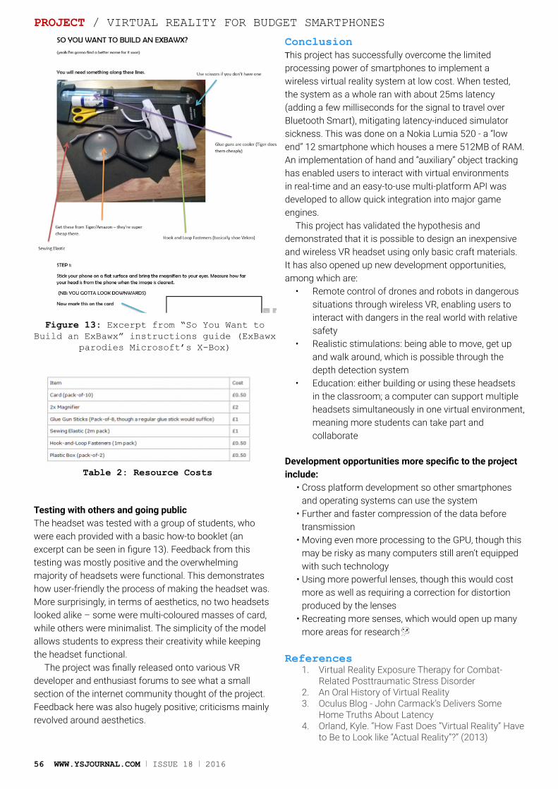
56 WWW.YSJOURNAL.COM I ISSUE 18 I 2016
Testing with others and going publicThe headset was tested with a group of students, who were each provided with a basic how-to booklet (an excerpt can be seen in figure 13). Feedback from this testing was mostly positive and the overwhelming majority of headsets were functional. This demonstrates how user-friendly the process of making the headset was. More surprisingly, in terms of aesthetics, no two headsets looked alike – some were multi-coloured masses of card, while others were minimalist. The simplicity of the model allows students to express their creativity while keeping the headset functional.
The project was finally released onto various VR developer and enthusiast forums to see what a small section of the internet community thought of the project. Feedback here was also hugely positive; criticisms mainly revolved around aesthetics.
ConclusionThis project has successfully overcome the limited processing power of smartphones to implement a wireless virtual reality system at low cost. When tested, the system as a whole ran with about 25ms latency (adding a few milliseconds for the signal to travel over Bluetooth Smart), mitigating latency-induced simulator sickness. This was done on a Nokia Lumia 520 - a “low end” 12 smartphone which houses a mere 512MB of RAM. An implementation of hand and “auxiliary” object tracking has enabled users to interact with virtual environments in real-time and an easy-to-use multi-platform API was developed to allow quick integration into major game engines.
This project has validated the hypothesis and demonstrated that it is possible to design an inexpensive and wireless VR headset using only basic craft materials. It has also opened up new development opportunities, among which are:
• Remote control of drones and robots in dangerous situations through wireless VR, enabling users to interact with dangers in the real world with relative safety
• Realistic stimulations: being able to move, get up and walk around, which is possible through the depth detection system
• Education: either building or using these headsets in the classroom; a computer can support multiple headsets simultaneously in one virtual environment, meaning more students can take part and collaborate
Development opportunities more specific to the project include:
• Cross platform development so other smartphones and operating systems can use the system
• Further and faster compression of the data before transmission
• Moving even more processing to the GPU, though this may be risky as many computers still aren’t equipped with such technology
• Using more powerful lenses, though this would cost more as well as requiring a correction for distortion produced by the lenses
• Recreating more senses, which would open up many more areas for research
References1. Virtual Reality Exposure Therapy for Combat-
Related Posttraumatic Stress Disorder2. An Oral History of Virtual Reality3. Oculus Blog - John Carmack’s Delivers Some
Home Truths About Latency4. Orland, Kyle. “How Fast Does “Virtual Reality” Have
to Be to Look like “Actual Reality”?” (2013)
Figure 13: Excerpt from “So You Want to Build an ExBawx” instructions guide (ExBawx
parodies Microsoft’s X-Box)
PROJECT / VIRTUAL REALITY FOR BUDGET SMARTPHONES
Table 2: Resource Costs
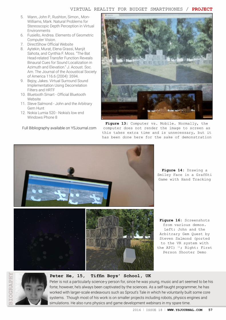
2016 I ISSUE 18 I WWW.YSJOURNAL.COM 57
Peter is not a particularly science-y person for, since he was young, music and art seemed to be his forte; however, he’s always been captivated by the sciences. As a self-taught programmer, he has worked with larger-scale endeavours such as Sprout’s Tale in which he voluntarily built some core systems. Though most of his work is on smaller projects including robots, physics engines and simulations. He also runs physics and game development webinars in my spare time.
Peter He, 15, Tiffin Boys’ School, UK
BIOGRAPHY
Figure 13: Computer vs. Mobile. Normally, the computer does not render the image to screen as this takes extra time and is unnecessary, but it has been done here for the sake of demonstration
Figure 14: Drawing a Smiley Face in a Graffiti Game with Hand Tracking
Figure 16: Screenshots from various demos. Left: John and the
Arbitrary Gem Quest by Steven Salmond (ported to the VR system with
the API) 11; Right: First Person Shooter Demo
VIRTUAL REALITY FOR BUDGET SMARTPHONES / PROJECT
5. Wann, John P., Rushton, Simon., Mon-Williams, Mark. Natural Problems for Stereoscopic Depth Perception in Virtual Environments
6. Fusiello, Andrea. Elements of Geometric Computer Vision.
7. DirectShow Official Website8. Aytekin, Murat, Elena Grassi, Manjit
Sahota, and Cynthia F. Moss. “The Bat Head-related Transfer Function Reveals Binaural Cues for Sound Localization in Azimuth and Elevation.” J. Acoust. Soc. Am. The Journal of the Acoustical Society of America 116.6 (2004): 3594.
9. Bejoy, Jakes. Virtual Surround Sound Implementation Using Decorrelation Filters and HRTF
10. Bluetooth Smart - Official Bluetooth Website
11. Steve Salmond - John and the Arbitrary Gem Hunt
12. Nokia Lumia 520 - Nokia’s low end Windows Phone 8
Full Bibliography available on YSJournal.com

Ammara (17) explores the details of lung cancer and looks at potential treatments of the deadly disease.
Treating Lung Cancer
AbstractIn this article, I will explore the complexity of lung cancer and why it is classed as one of the most deadly cancers. As well as this, I will explain how different types of lung cancer are classified depending on the cells present in tumours and how the classification affects the treatment a patient is given. Furthermore, I will look at the current treatments for lung cancer and how they are used. And last but not least, I will explain what Circulating Tumour Cells (CTCs) are and how they may be able to help scientists diagnose and treat lung cancer in the near future.
Introduction
Treatment of lung cancer has shockingly low success rates. The disease mainly affects people between the ages of sixty and eighty years old.
Young people can develop lung cancer; however, it is a rarity[1]. 30% of people with lung cancer only survive 1 year after diagnosis, 10% survive 5 years or more and only 5% survive 10 years or more after diagnosis. The success rates depend on the stage of lung cancer at diagnosis. Unfortunately, most people are diagnosed in the late stages of their lung cancer[2]. But why is this the case? Why is it so challenging to spot signs of lung cancer in the early stages? In this article, I will explain why it is so difficult to diagnose lung cancer early on and how new advances in treatments using Circulating Tumour Cells could potentially increase the survival rates of lung cancer sufferers.
Definitions• Malignant: A group of cancerous cells which grow and
spread rapidly. • Biopsy: A medical examination carried out by
removing a piece of tissue to discover the presence, extent of a disease or cause.
• Carcinoma: A type of cancer which arises in the epithelial tissue of the skin or the lining of internal organs.
• Microvilli: Tiny hair-like membranes in the body which greatly increase the surface area of a cell.
The 'lowdown' on Lung CancerIn a cigarette, tar is the component that contains a range of substances known as carcinogens. Carcinogens cause mutations in the DNA of cells leading to cancer. This then causes uncontrollable cell growth and eventually forms
REVIEW ARTICLE / TREATING LUNG CANCER
58 WWW.YSJOURNAL.COM I ISSUE 18 I 2016
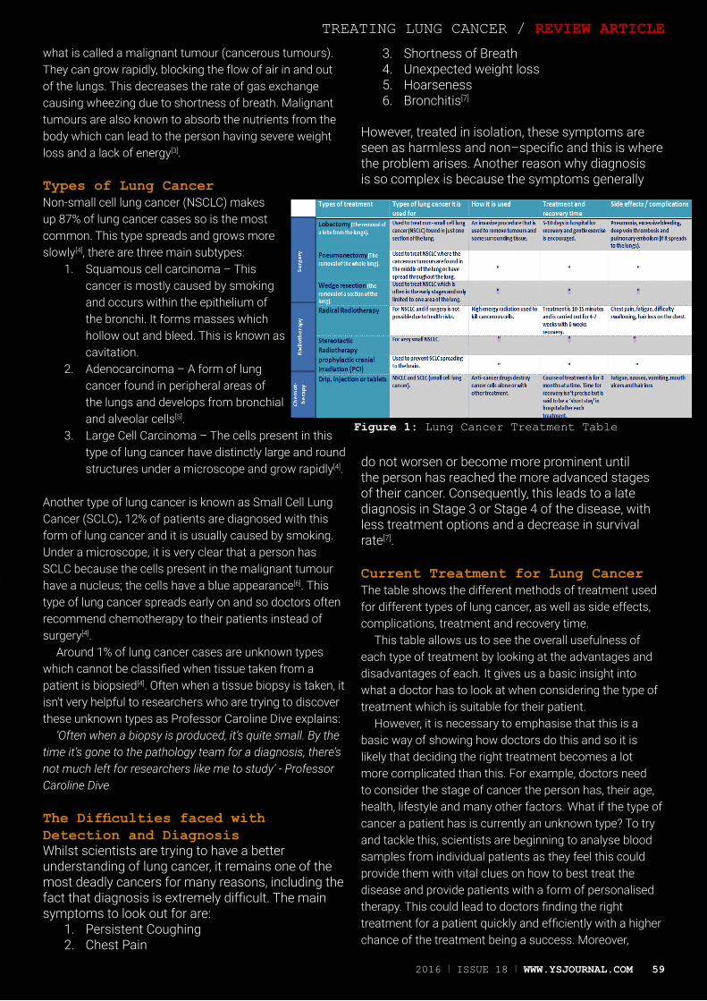
what is called a malignant tumour (cancerous tumours). They can grow rapidly, blocking the flow of air in and out of the lungs. This decreases the rate of gas exchange causing wheezing due to shortness of breath. Malignant tumours are also known to absorb the nutrients from the body which can lead to the person having severe weight loss and a lack of energy[3].
Types of Lung CancerNon-small cell lung cancer (NSCLC) makes up 87% of lung cancer cases so is the most common. This type spreads and grows more slowly[4], there are three main subtypes:
1. Squamous cell carcinoma – This cancer is mostly caused by smoking and occurs within the epithelium of the bronchi. It forms masses which hollow out and bleed. This is known as cavitation.
2. Adenocarcinoma – A form of lung cancer found in peripheral areas of the lungs and develops from bronchial and alveolar cells[5].
3. Large Cell Carcinoma – The cells present in this type of lung cancer have distinctly large and round structures under a microscope and grow rapidly[4].
Another type of lung cancer is known as Small Cell Lung Cancer (SCLC). 12% of patients are diagnosed with this form of lung cancer and it is usually caused by smoking. Under a microscope, it is very clear that a person has SCLC because the cells present in the malignant tumour have a nucleus; the cells have a blue appearance[6]. This type of lung cancer spreads early on and so doctors often recommend chemotherapy to their patients instead of surgery[4].
Around 1% of lung cancer cases are unknown types which cannot be classified when tissue taken from a patient is biopsied[4]. Often when a tissue biopsy is taken, it isn't very helpful to researchers who are trying to discover these unknown types as Professor Caroline Dive explains:
‘Often when a biopsy is produced, it’s quite small. By the time it’s gone to the pathology team for a diagnosis, there’s not much left for researchers like me to study’ - Professor Caroline Dive
The Difficulties faced with Detection and DiagnosisWhilst scientists are trying to have a better understanding of lung cancer, it remains one of the most deadly cancers for many reasons, including the fact that diagnosis is extremely difficult. The main symptoms to look out for are:
1. Persistent Coughing 2. Chest Pain
3. Shortness of Breath4. Unexpected weight loss5. Hoarseness6. Bronchitis[7]
However, treated in isolation, these symptoms are seen as harmless and non–specific and this is where the problem arises. Another reason why diagnosis is so complex is because the symptoms generally
do not worsen or become more prominent until the person has reached the more advanced stages of their cancer. Consequently, this leads to a late diagnosis in Stage 3 or Stage 4 of the disease, with less treatment options and a decrease in survival rate[7].
Current Treatment for Lung CancerThe table shows the different methods of treatment used for different types of lung cancer, as well as side effects, complications, treatment and recovery time.
This table allows us to see the overall usefulness of each type of treatment by looking at the advantages and disadvantages of each. It gives us a basic insight into what a doctor has to look at when considering the type of treatment which is suitable for their patient.
However, it is necessary to emphasise that this is a basic way of showing how doctors do this and so it is likely that deciding the right treatment becomes a lot more complicated than this. For example, doctors need to consider the stage of cancer the person has, their age, health, lifestyle and many other factors. What if the type of cancer a patient has is currently an unknown type? To try and tackle this, scientists are beginning to analyse blood samples from individual patients as they feel this could provide them with vital clues on how to best treat the disease and provide patients with a form of personalised therapy. This could lead to doctors finding the right treatment for a patient quickly and efficiently with a higher chance of the treatment being a success. Moreover,
Figure 1: Lung Cancer Treatment Table
TREATING LUNG CANCER / REVIEW ARTICLE
2016 I ISSUE 18 I WWW.YSJOURNAL.COM 59

60 WWW.YSJOURNAL.COM I ISSUE 18 I 2016
analysing a type of cell in the blood could lead to more lung cancer cases being diagnosed in the early stages.
Circulating Tumour Cells (CTCs)CTCs are cells which have shed from primary tumours into the vascular system and then circulate in the bloodstream. They are known to cause metastasis[8] which is the spread of cancer to other body parts. This causes the cancer to no longer be local, and becomes very difficult to treat. However, from current research, it has been found that CTCs also hold vital information as to how individuals can receive the best treatment for their type of lung cancer[9]. This means that the treatments can be specifically catered to each patient - and the treatment method can be found out just by doing a simple blood test also known as a liquid biopsy (which is a lot more patient friendly). Scientists count the number of CTCs present in a patient’s blood as this can allow them to track how a patient is responding to their treatment[10].
Tracking down CTCsThe majority of us at least once in our lives have attempted to win a toy from a Claw Machine in an arcade – with very little success. This is very similar to trying to track and capture CTCs in a blood sample. In a blood sample there can be as many as 60 million white blood cells – but just 5 CTCs [10]; almost like trying to find a needle in a haystack. However, scientists are experimenting with different ways to efficiently capture these CTCS.
Acoustic Isolation using a Chladni PlateA Chladni Plate can be used to arrange particles in characteristic nodal patterns[11] that respond to changes in sound and frequency. The Chladni plate was created by Ernst Chladni in the 19th century and it wasn’t until recently that scientists began to experiment with different sounds and frequencies to isolate diseased cells such as CTC’s. The nodal patterns are created by vibrations which take the form of Sound Acoustic Waves (SAW)[12]. The different frequencies applied cause the different living cells to separate due to their mechanical properties. This will allow scientists to identify and isolate CTC’s easily so that they can quickly diagnose and choose the right treatment for the patient’s lung cancer.
REVIEW ARTICLE / TREATING LUNG CANCER
Figure 2: Circulating Tumour Cells[9]
Figure 3: Chladni Plate[11]

2016 I ISSUE 18 I WWW.YSJOURNAL.COM 61
Nano-tastic DiagnosisProfessor Yaling Liu is in the process of creating a diagnostic device which can tell doctors what stage of cancer a patient has and if there are any cancerous cells present in the patient’s blood sample. CTCs have a rough surface due to the presence of microvilli on their outer surface. The nano chip has a unique capture path that has been designed to be complementary to the outer surface of the CTCs. This means that the device can capture all the CTCs present in the patient’s blood sample when it is injected into the chip. The nano chip is also made from an extremely cost efficient polymer, polydimethylsiloxane (PDMS), which means that this diagnostic tool could one day be used to find the right treatment for cancer patients[13].
SummaryThis article has shown just how dangerous lung cancer is, due to difficulties with diagnosis and current treatment options. We still do not know what exactly causes some people to get lung cancer and others not to. Some people who may have never smoked or have been exposed to radiation but they are still diagnosed with lung cancer. Fortunately, help is on its way. At this very moment, scientists around the world are working towards creating devices, such as the ones aforementioned, to increase the number of lung cancer survivors. Analysing CTCs from a fluid biopsy will not only potentially lead to early diagnosis, but it can help researchers around the world to understand clearly the biology of lung cancer and the specific reason a patient has the disease. Hopefully in the future, lung cancer will not be such a devastating and deadly disease.
References1. What is lung cancer? Under ‘Who is at risk’? Access
date- 03/06/2015 (https://www.blf.org.uk/Page/What-is-lung-cancer)
2. Statistics and outlook for lung cancer- under ‘Overall outcome’ Access date- 03/06/2015 REFERENCE LINK (http://www.cancerresearchuk.org/about-cancer/type/lung-cancer/treatment/statistics-and-outlook-for-lung-cancer#general)
3. CGP Textbook- OCR AS & A2 Biology. Page 86 ‘Gas Exchange System’ Access date- 25/06/2015
4. Types of lung cancer Access date- 25/06/2015 (http://www.cancerresearchuk.org/about-cancer/type/lung-cancer/about/types-of-lung-cancer)
5. Pearson Textbook- ‘Human Anatomy & Physiology’ The Sixth Edition by Elaine N. Marieb. Page 870 under ‘Lung Cancer’ Access date- 25/06/2015
6. Small-Cell Lung Cancer, Mesothelioma, and Thymoma. Path and physiology section Access date- 27/06/2015 (http://www.cancernetwork.com/cancer-management/small-cell-lung-cancer-mesothelioma-and-thymoma)
7. Why is it difficult to diagnose lung cancer in early stages? Access date- 27/06/2015 (http://www.lcfamerica.org/why-is-it-difficult-to-diagnose-lung-cancer-in-early-stages/#sthash.MqPyRh7v.dfzYbK9p.dpbs)
8. Lung Cancer Complications Access date- 27/06/2015 (http://www.healthline.com/health/lung-cancer-complications#OtherComplications5)
9. Wikapedia- Circulating Tumour Cell Access date- 04/07/2015 (https://en.wikipedia.org/wiki/Circulating_tumour_cell)
10. ‘Every Cell Has A Story’ leaflet -The Royal Society Exhibition 2015 Access date- June 2015
11. Chladni Plate Access date- 04/07/2015 (https://en.wikipedia.org/wiki/Ernst_Chladni#Chladni_figures)Single cell science leaflet- Royal Society, London 2015 Access date- June 2015
12. Yaling Liu on his CTC’s detection Access date- 05/07/2015 (https://www.youtube.com/watch?v=VWddHndyGxo)
TREATING LUNG CANCER / REVIEW ARTICLE
Diagnosis is so complex is because the symptoms generally don’t become more prominent until the person has reached the more advanced stages of their cancer
“
Ammara is 17 years old and is currently studying Biology, Chemistry and French at St Domanics Sixth Form College. She has a very strong interest in science and really enjoys learning about Human Health and Disease. Writing this article has allowed her to further her interest for this sector in science. It has also allowed her to get an insight into how researchers try to understand the nature of certain diseases to produce effective medicines.
Ammara Jones, 17, St Dominic’s Sixth Form College, UK
BIOGRAPHY
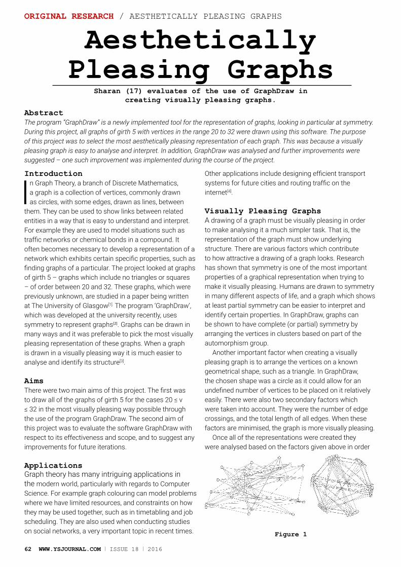
62 WWW.YSJOURNAL.COM I ISSUE 18 I 2016
ORIGINAL RESEARCH / AESTHETICALLY PLEASING GRAPHS
Aesthetically Pleasing Graphs
Sharan (17) evaluates of the use of GraphDraw in creating visually pleasing graphs.
AbstractThe program “GraphDraw” is a newly implemented tool for the representation of graphs, looking in particular at symmetry. During this project, all graphs of girth 5 with vertices in the range 20 to 32 were drawn using this software. The purpose of this project was to select the most aesthetically pleasing representation of each graph. This was because a visually pleasing graph is easy to analyse and interpret. In addition, GraphDraw was analysed and further improvements were suggested – one such improvement was implemented during the course of the project.
Introduction
In Graph Theory, a branch of Discrete Mathematics, a graph is a collection of vertices, commonly drawn as circles, with some edges, drawn as lines, between
them. They can be used to show links between related entities in a way that is easy to understand and interpret. For example they are used to model situations such as traffic networks or chemical bonds in a compound. It often becomes necessary to develop a representation of a network which exhibits certain specific properties, such as finding graphs of a particular. The project looked at graphs of girth 5 – graphs which include no triangles or squares – of order between 20 and 32. These graphs, which were previously unknown, are studied in a paper being written at The University of Glasgow[1]. The program ‘GraphDraw’, which was developed at the university recently, uses symmetry to represent graphs[2]. Graphs can be drawn in many ways and it was preferable to pick the most visually pleasing representation of these graphs. When a graph is drawn in a visually pleasing way it is much easier to analyse and identify its structure[3].
AimsThere were two main aims of this project. The first was to draw all of the graphs of girth 5 for the cases 20 ≤ v ≤ 32 in the most visually pleasing way possible through the use of the program GraphDraw. The second aim of this project was to evaluate the software GraphDraw with respect to its effectiveness and scope, and to suggest any improvements for future iterations.
ApplicationsGraph theory has many intriguing applications in the modern world, particularly with regards to Computer Science. For example graph colouring can model problems where we have limited resources, and constraints on how they may be used together, such as in timetabling and job scheduling. They are also used when conducting studies on social networks, a very important topic in recent times.
Other applications include designing efficient transport systems for future cities and routing traffic on the internet[4].
Visually Pleasing GraphsA drawing of a graph must be visually pleasing in order to make analysing it a much simpler task. That is, the representation of the graph must show underlying structure. There are various factors which contribute to how attractive a drawing of a graph looks. Research has shown that symmetry is one of the most important properties of a graphical representation when trying to make it visually pleasing. Humans are drawn to symmetry in many different aspects of life, and a graph which shows at least partial symmetry can be easier to interpret and identify certain properties. In GraphDraw, graphs can be shown to have complete (or partial) symmetry by arranging the vertices in clusters based on part of the automorphism group.
Another important factor when creating a visually pleasing graph is to arrange the vertices on a known geometrical shape, such as a triangle. In GraphDraw, the chosen shape was a circle as it could allow for an undefined number of vertices to be placed on it relatively easily. There were also two secondary factors which were taken into account. They were the number of edge crossings, and the total length of all edges. When these factors are minimised, the graph is more visually pleasing.
Once all of the representations were created they were analysed based on the factors given above in order
Figure 1
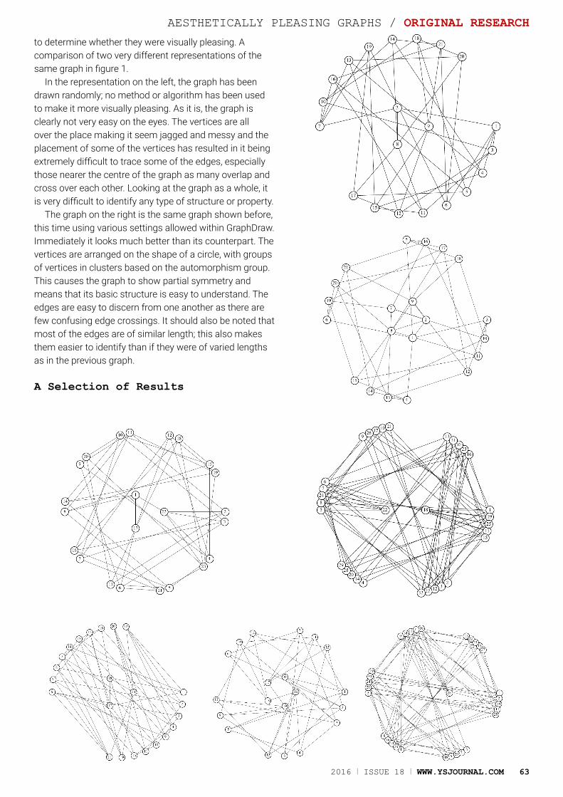
2016 I ISSUE 18 I WWW.YSJOURNAL.COM 63
AESTHETICALLY PLEASING GRAPHS / ORIGINAL RESEARCH
to determine whether they were visually pleasing. A comparison of two very different representations of the same graph in figure 1.
In the representation on the left, the graph has been drawn randomly; no method or algorithm has been used to make it more visually pleasing. As it is, the graph is clearly not very easy on the eyes. The vertices are all over the place making it seem jagged and messy and the placement of some of the vertices has resulted in it being extremely difficult to trace some of the edges, especially those nearer the centre of the graph as many overlap and cross over each other. Looking at the graph as a whole, it is very difficult to identify any type of structure or property.
The graph on the right is the same graph shown before, this time using various settings allowed within GraphDraw. Immediately it looks much better than its counterpart. The vertices are arranged on the shape of a circle, with groups of vertices in clusters based on the automorphism group. This causes the graph to show partial symmetry and means that its basic structure is easy to understand. The edges are easy to discern from one another as there are few confusing edge crossings. It should also be noted that most of the edges are of similar length; this also makes them easier to identify than if they were of varied lengths as in the previous graph.
A Selection of Results
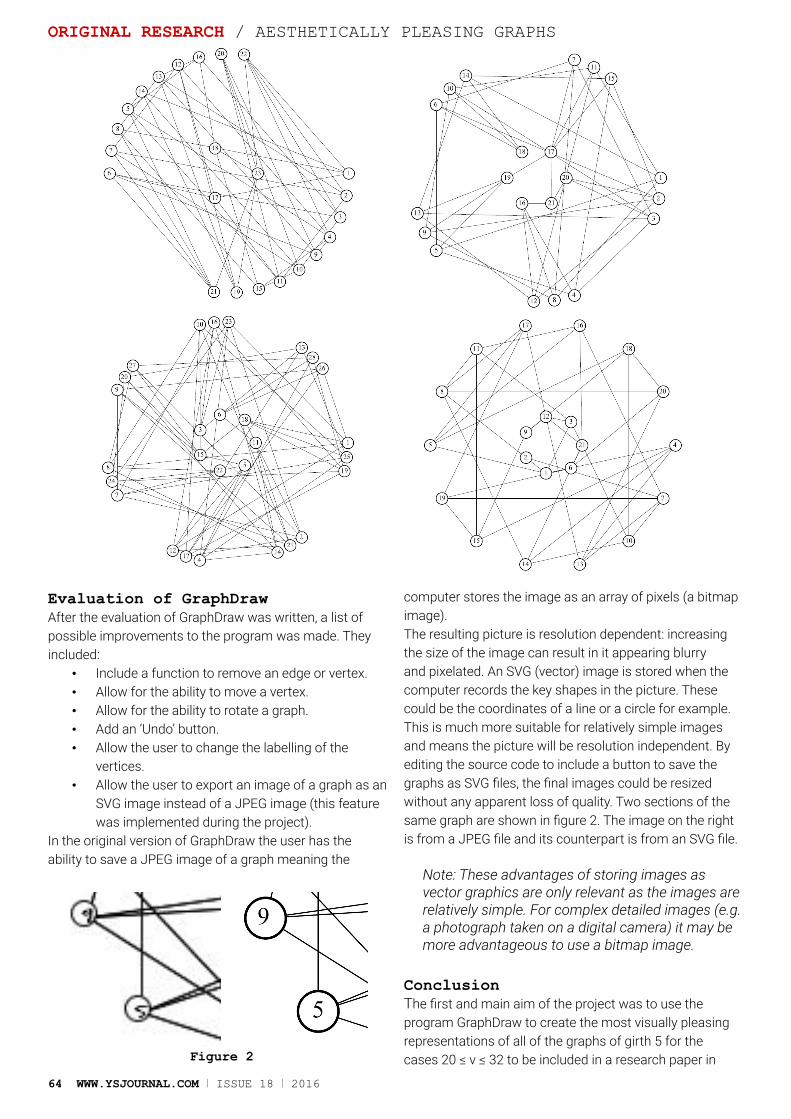
64 WWW.YSJOURNAL.COM I ISSUE 18 I 2016
Evaluation of GraphDrawAfter the evaluation of GraphDraw was written, a list of possible improvements to the program was made. They included:
• Include a function to remove an edge or vertex. • Allow for the ability to move a vertex. • Allow for the ability to rotate a graph. • Add an ‘Undo’ button. • Allow the user to change the labelling of the
vertices. • Allow the user to export an image of a graph as an
SVG image instead of a JPEG image (this feature was implemented during the project).
In the original version of GraphDraw the user has the ability to save a JPEG image of a graph meaning the
computer stores the image as an array of pixels (a bitmap image). The resulting picture is resolution dependent: increasing the size of the image can result in it appearing blurry and pixelated. An SVG (vector) image is stored when the computer records the key shapes in the picture. These could be the coordinates of a line or a circle for example. This is much more suitable for relatively simple images and means the picture will be resolution independent. By editing the source code to include a button to save the graphs as SVG files, the final images could be resized without any apparent loss of quality. Two sections of the same graph are shown in figure 2. The image on the right is from a JPEG file and its counterpart is from an SVG file.
Note: These advantages of storing images as vector graphics are only relevant as the images are relatively simple. For complex detailed images (e.g. a photograph taken on a digital camera) it may be more advantageous to use a bitmap image.
ConclusionThe first and main aim of the project was to use the program GraphDraw to create the most visually pleasing representations of all of the graphs of girth 5 for the cases 20 ≤ v ≤ 32 to be included in a research paper in Figure 2
ORIGINAL RESEARCH / AESTHETICALLY PLEASING GRAPHS
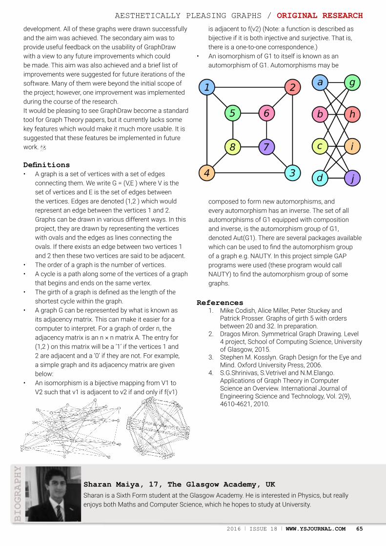
2016 I ISSUE 18 I WWW.YSJOURNAL.COM 65
BIOGRAPHY
Sharan is a Sixth Form student at the Glasgow Academy. He is interested in Physics, but really enjoys both Maths and Computer Science, which he hopes to study at University.
Sharan Maiya, 17, The Glasgow Academy, UK
development. All of these graphs were drawn successfully and the aim was achieved. The secondary aim was to provide useful feedback on the usability of GraphDraw with a view to any future improvements which could be made. This aim was also achieved and a brief list of improvements were suggested for future iterations of the software. Many of them were beyond the initial scope of the project; however, one improvement was implemented during the course of the research.It would be pleasing to see GraphDraw become a standard tool for Graph Theory papers, but it currently lacks some key features which would make it much more usable. It is suggested that these features be implemented in future work.
Definitions• A graph is a set of vertices with a set of edges
connecting them. We write G = (V,E ) where V is the set of vertices and E is the set of edges between the vertices. Edges are denoted (1,2 ) which would represent an edge between the vertices 1 and 2. Graphs can be drawn in various different ways. In this project, they are drawn by representing the vertices with ovals and the edges as lines connecting the ovals. If there exists an edge between two vertices 1 and 2 then these two vertices are said to be adjacent.
• The order of a graph is the number of vertices. • A cycle is a path along some of the vertices of a graph
that begins and ends on the same vertex.• The girth of a graph is defined as the length of the
shortest cycle within the graph. • A graph G can be represented by what is known as
its adjacency matrix. This can make it easier for a computer to interpret. For a graph of order n, the adjacency matrix is an n × n matrix A. The entry for (1,2 ) on this matrix will be a ‘1’ if the vertices 1 and 2 are adjacent and a ‘0’ if they are not. For example, a simple graph and its adjacency matrix are given below:
• An isomorphism is a bijective mapping from V1 to V2 such that v1 is adjacent to v2 if and only if f(v1)
is adjacent to f(v2) (Note: a function is described as bijective if it is both injective and surjective. That is, there is a one-to-one correspondence.)
• An isomorphism of G1 to itself is known as an automorphism of G1. Automorphisms may be
composed to form new automorphisms, and every automorphism has an inverse. The set of all automorphisms of G1 equipped with composition and inverse, is the automorphism group of G1, denoted Aut(G1). There are several packages available which can be used to find the automorphism group of a graph e.g. NAUTY. In this project simple GAP programs were used (these program would call NAUTY) to find the automorphism group of some graphs.
References1. Mike Codish, Alice Miller, Peter Stuckey and
Patrick Prosser. Graphs of girth 5 with orders between 20 and 32. In preparation.
2. Dragos Miron. Symmetrical Graph Drawing. Level 4 project, School of Computing Science, University of Glasgow, 2015.
3. Stephen M. Kosslyn. Graph Design for the Eye and Mind. Oxford University Press, 2006.
4. S.G.Shrinivas, S.Vetrivel and N.M.Elango. Applications of Graph Theory in Computer Science an Overview. International Journal of Engineering Science and Technology, Vol. 2(9), 4610-4621, 2010.
AESTHETICALLY PLEASING GRAPHS / ORIGINAL RESEARCH

66 WWW.YSJOURNAL.COM I ISSUE 18 I 2016
International Advisory Board The IAB is a team of experts who advise the editors of the journal.
Team Leader: Christina Astin, UKEmail:[email protected]
Team Members:Sir Harry Kroto, FRS, UK/USA Baroness Susan Greenfield, UK Ghazwan Butrous, UKAnna Grigoryan, USA/ArmeniaThijs Kouwenhoven, ChinaDon Eliseo Lucero-Prisno III, UKPaul Soderberg, USALee Riley, USACorky Valenti, USAVince Bennett, USAMike Bennett, USATony Grady, USAIan Yorston, UKCharlie Barclay, UKJoanne Manaster, USAAlom Shaha, UKArmen Soghoyan, ArmeniaMark Orders, UKLinda Crouch, UKJohn Boswell, USASam Morris, UKDebbie Nsefik, UKProfessor Clive Coen, UKAnnette Smith, UKEsther Marin, SpainMalcolm Morgan, UKSteven Simpson, UKSarah De Souza, UKSir Martyn Poliakoff, UKDr. Claire McNulty, UKRod Edwards, UK
Young Advisory BoardSteven Chambers, UKFiona Jenkinson, UKTobias Nørbo, DenmarkArjen Dijksman, FranceLorna Quandt, USAJonathan Rogers, UKLara Compston-Garnett, UKOtana Jakpor, USAPamela Barraza Flores, MexicoCleodie Swire, UKMuna Oli, USACourtney Williams, UK
YOUNG SCIENTISTS
All rights reserved. No part of this publication may be reproduced, or transmitted, in any form or by any means, electronic or mechanical, including photocopy,
recording, or any information storage and retrieval system, without permission in writing from the editor. The Young Scientists Journal and/or its published
cannot be held responsible for errors or for any consequences arising from the use of the information contained in this journal. The appearance of advertising or product information in the various sections in the journal does not constitute and endorsement or approval by the journal and/or its publisher of the quality or value of the said product or of claims made for it by its manufacturer, advertisers have
no prior knowlege of content. The journal is printed on acid free paper.
The Young Scientists Journal is the world’s only peer review science journal run by young scientists for young scientists. The journal publishes research
papers and review articles on Science, Technology, Engineering and Mathematics (STEM) both online and in print, written and edited exclusively by 12-20 year olds across the globe. 18 issues have been published since it was founded in 2006 at The King’s School Canterbury by Christina Astin and Ghazwan Butrous.
We are establishing a number of Young Scientists Journal hubs based at schools around the world where groups of students can work together on the journal. Contact Christina Astin if you would like to find out more about getting
your school involved.
Young Scientists Journal is proudly published by
The Butrous Foundation in Canterbury, UK
ISSN 0974-6102 (Print)ISSN 0975-2145 (Online)
Printed by
Hubs: YSJournal Goes Local
2016 I ISSUE 18 I WWW.YSJOURNAL.COM 67
ADVERTISEMENT
68 WWW.YSJOURNAL.COM I ISSUE 18 I 2016
YOUNG SCIENTISTSwww.ysjournal.com/YSJournal @YSJournal@ysjournal
INSPIRING AND NURTURING THE SCIENTISTS OF THE FUTURETHE WORLD’S PEER REVIEW SCIENCE JOURNAL WRITTEN & EDITED BY 12-20 YEAR OLDS
Partner: Publisher: Supported By:
FASTER. FURTHER. FREER.WWW.COTY.COM/COMPANY/CAREERS