Embed Size (px)
Citation preview
What is an animal?

Animals are multicellular, heterotrophic eukaryotes with tissues that develop from embryonic layers.
How do animals differ
from plants?
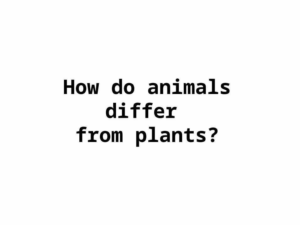
Animals lack cell walls and havehave collagen as their mostabundant protein.
Animals have nerve and muscle cells.
Animals have three distinct cell junc-tions: tight junctions, desmosomes, and gap junctions.
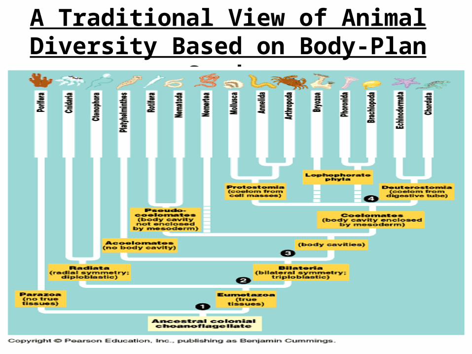
A Traditional View of Animal Diversity Based on Body-Plan Grades
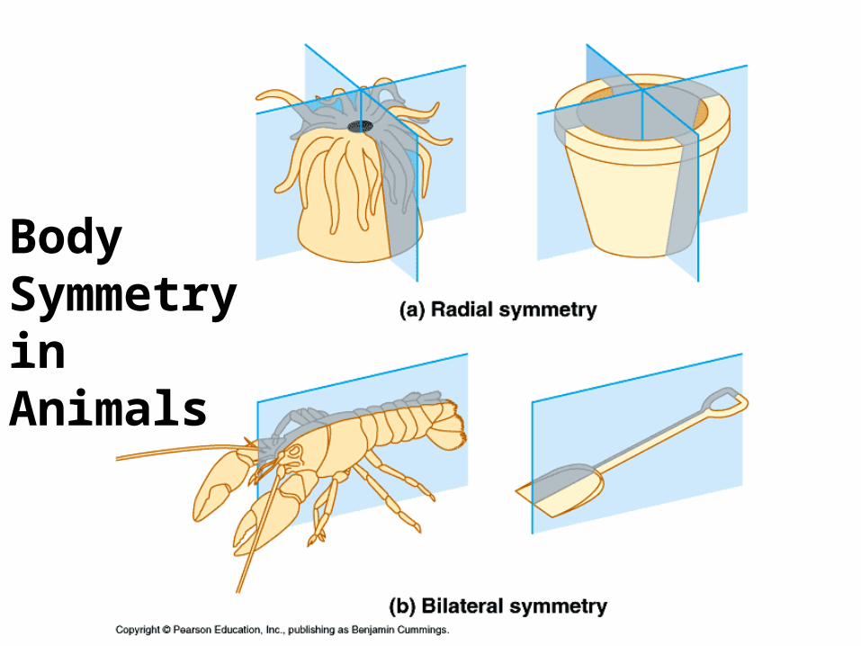
Body Symmetry in Animals

Body Plans of the Bilateria
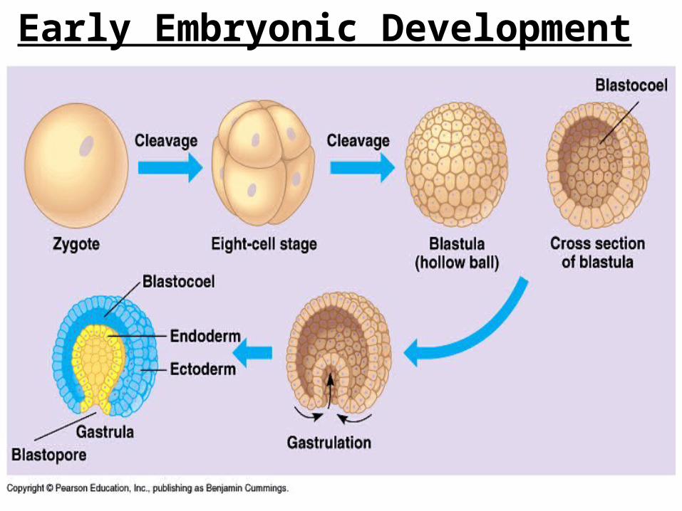
Early Embryonic Development
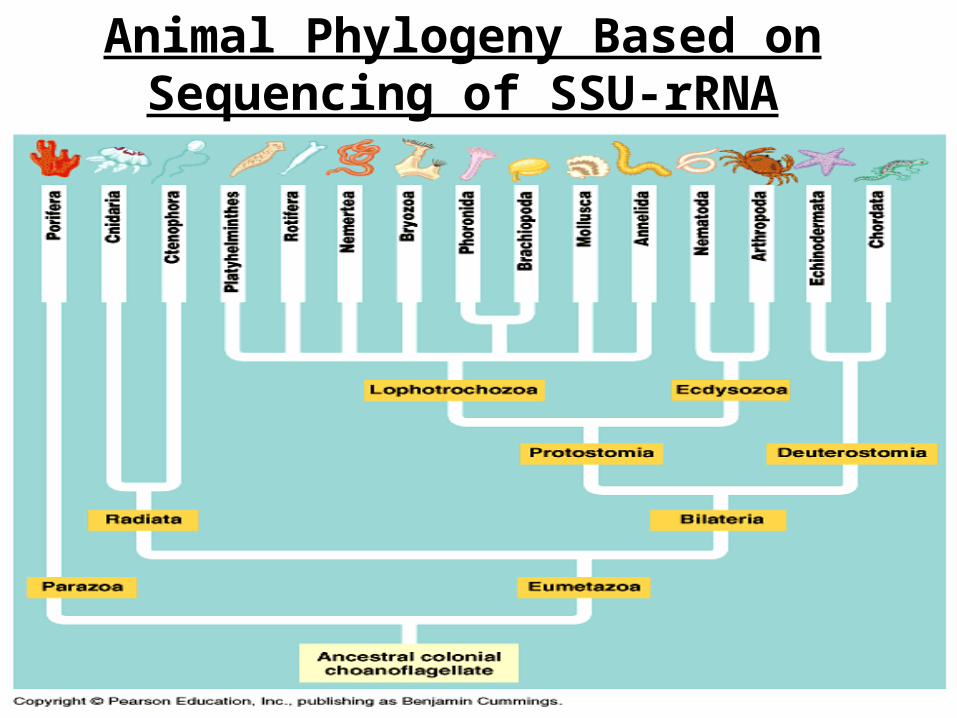
Animal Phylogeny Based on Sequencing of SSU-rRNA
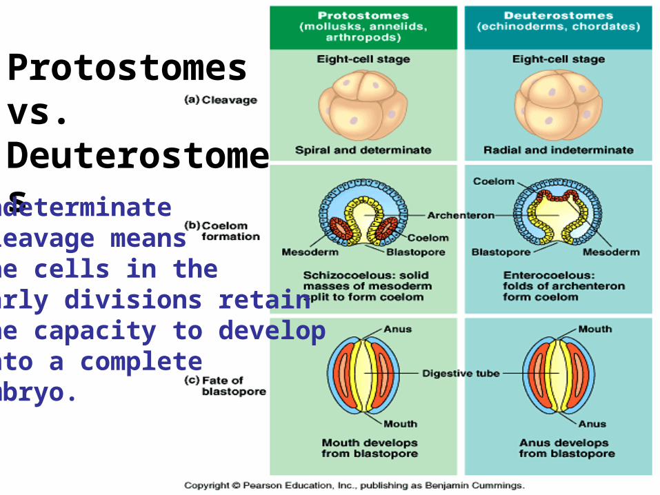
Protostomesvs. DeuterostomesIndeterminatecleavage meansthe cells in the early divisions retainthe capacity to developinto a complete embryo.
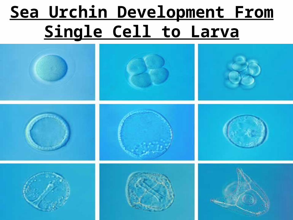
Sea Urchin Development From Single Cell to Larva

The Acrosomal and Cortical Reactions During Sea Urchin Fertilization
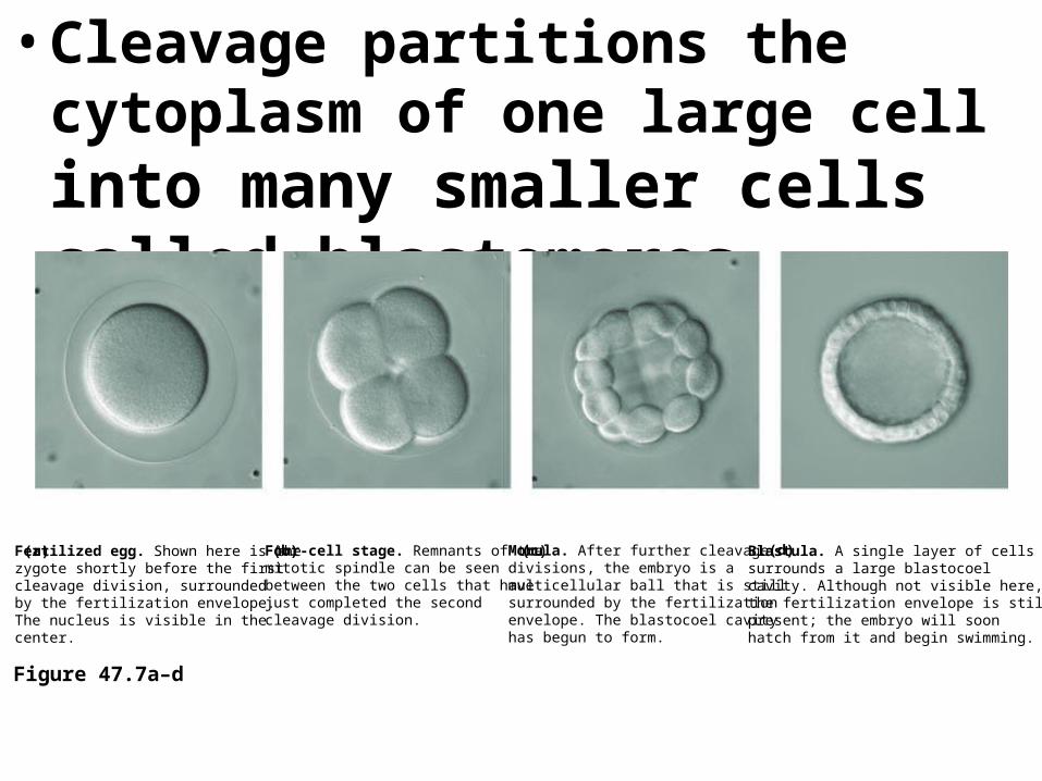
• Cleavage partitions the cytoplasm of one large cell into many smaller cells called blastomeres
Figure 47.7a–d
Fertilized egg. Shown here is thezygote shortly before the first cleavage division, surrounded by the fertilization envelope. The nucleus is visible in the center.
(a) Four-cell stage. Remnants of the mitotic spindle can be seen between the two cells that have just completed the second cleavage division.
(b) Morula. After further cleavage divisions, the embryo is a multicellular ball that is stillsurrounded by the fertilization envelope. The blastocoel cavityhas begun to form.
(c) Blastula. A single layer of cells surrounds a large blastocoel cavity. Although not visible here, the fertilization envelope is still present; the embryo will soon hatch from it and begin swimming.
(d)
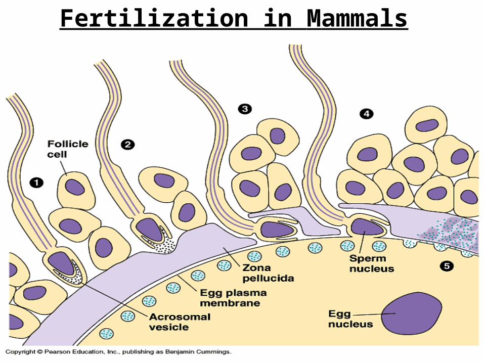
Fertilization in Mammals
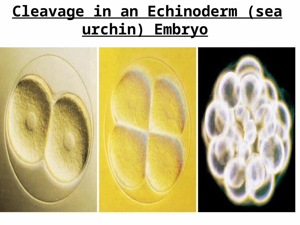
Cleavage in an Echinoderm (sea urchin) Embryo
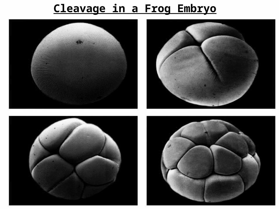
Cleavage in a Frog Embryo

Cross Section of a Frog Blastula
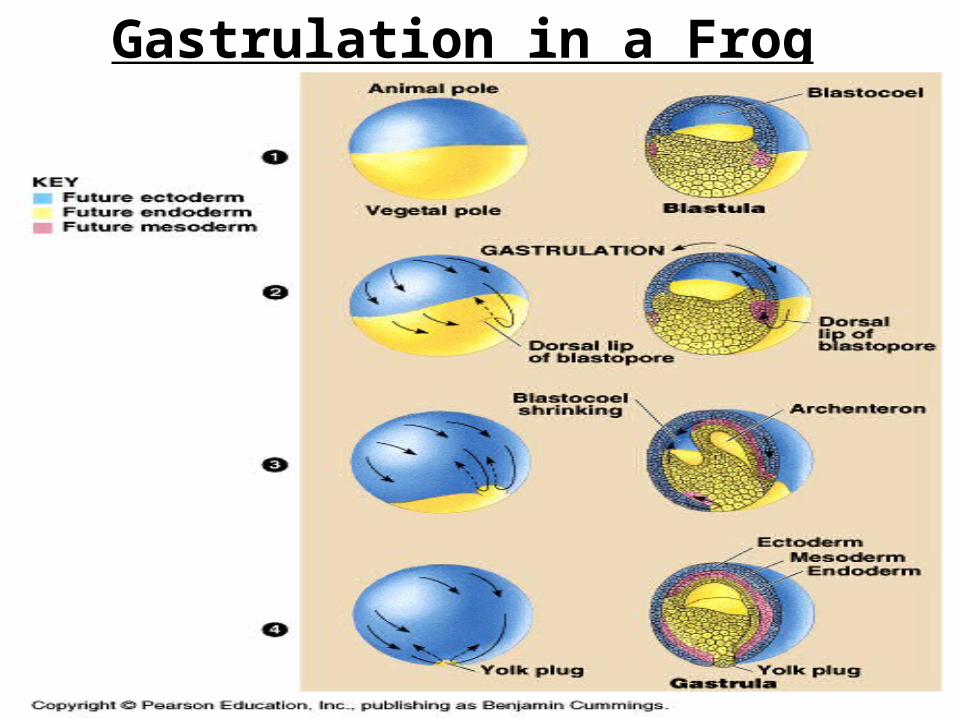
Gastrulation in a Frog Embryo

• The three layers produced by gastrulation are called embryonic germ layers
• The ectoderm forms the outer layer of the gastrula
• The endoderm lines the embryonic digestive tract
• The mesoderm partly fills the space between the endoderm and ectoderm
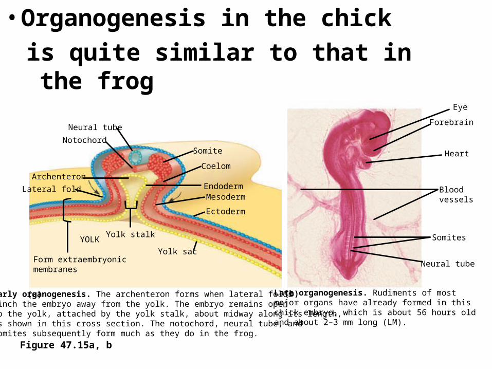
• Organogenesis in the chick
is quite similar to that in the frog
Figure 47.15a, b
Neural tube
Notochord
Archenteron
Lateral fold
Form extraembryonicmembranes
YOLKYolk stalk
Somite
Coelom
EndodermMesoderm
Ectoderm
Yolk sac
Eye
Forebrain
Heart
Blood vessels
Somites
Neural tube
Early organogenesis. The archenteron forms when lateral folds pinch the embryo away from the yolk. The embryo remains opento the yolk, attached by the yolk stalk, about midway along its length,as shown in this cross section. The notochord, neural tube, and somites subsequently form much as they do in the frog.
(a) Late organogenesis. Rudiments of most major organs have already formed in this chick embryo, which is about 56 hours old and about 2–3 mm long (LM).
(b)
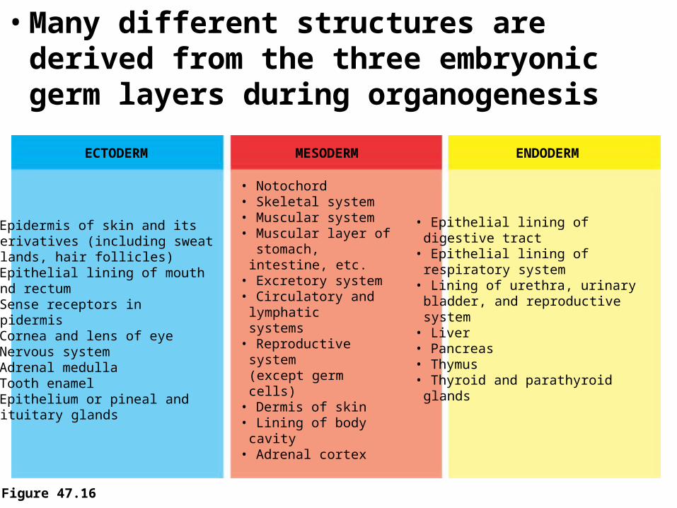
• Many different structures are derived from the three embryonic germ layers during organogenesis
Figure 47.16
ECTODERM MESODERM ENDODERM
• Epidermis of skin and itsderivatives (including sweatglands, hair follicles)
• Epithelial lining of mouthand rectum
• Sense receptors inepidermis
• Cornea and lens of eye• Nervous system• Adrenal medulla• Tooth enamel• Epithelium or pineal and
pituitary glands
• Notochord• Skeletal system• Muscular system• Muscular layer of stomach, intestine, etc.• Excretory system• Circulatory and
lymphaticsystems
• Reproductive system(except germ cells)
• Dermis of skin• Lining of body cavity• Adrenal cortex
• Epithelial lining ofdigestive tract
• Epithelial lining ofrespiratory system
• Lining of urethra, urinarybladder, and reproductivesystem
• Liver• Pancreas• Thymus• Thyroid and parathyroid
glands