Embed Size (px)
Citation preview
© 2012 Pearson Education, Inc.
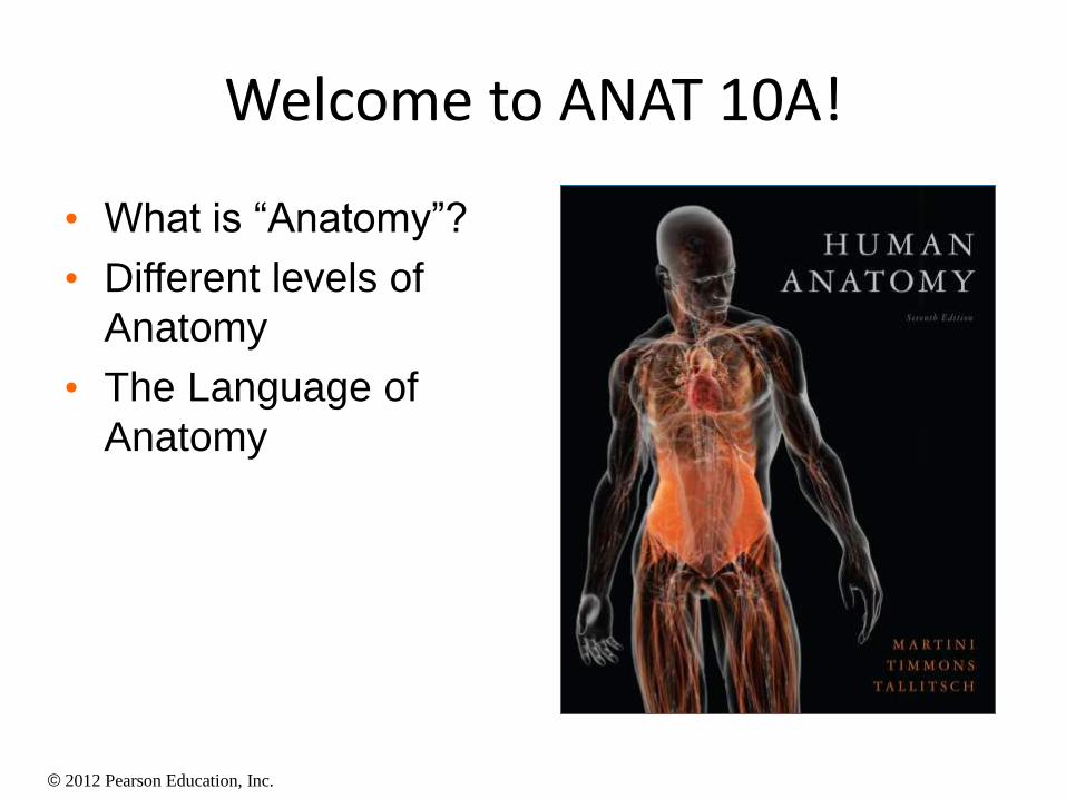
Welcome to ANAT 10A!
• What is “Anatomy”?
• Different levels of
Anatomy
• The Language of
Anatomy
© 2012 Pearson Education, Inc.
Introduction
• “Anatomy” means to dissect: (ANAT 10A)
• The study of internal & external body
structures
• The study of the relationship between body
parts
• i.e. identify structure and location of an organ
• Physiology (ANAT 10B)
• The study of how the body functions & the
mechanisms in the body
© 2012 Pearson Education, Inc.
• Anatomical terminology
• Based on ancient Greek or Latin
• Provides standard nomenclature worldwide
• Branches of anatomy
• Microscopic anatomy (histology)
• Gross/macroscopic anatomy
• Other perspectives
Introduction
© 2012 Pearson Education, Inc.
Microscopic Anatomy
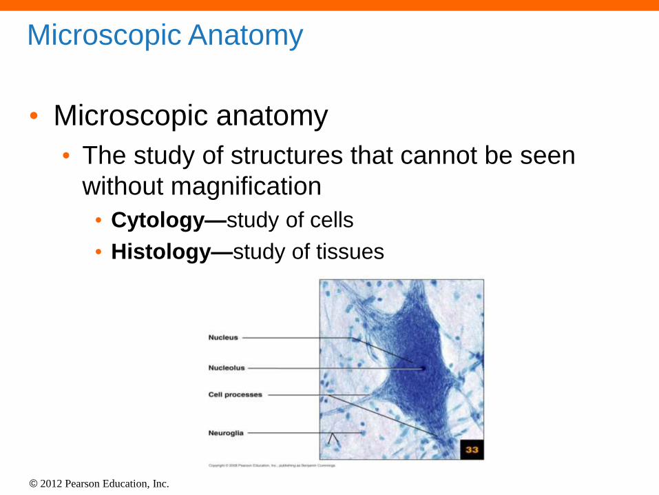
• Microscopic anatomy • The study of structures that cannot be seen
without magnification
• Cytology—study of cells
• Histology—study of tissues
© 2012 Pearson Education, Inc.
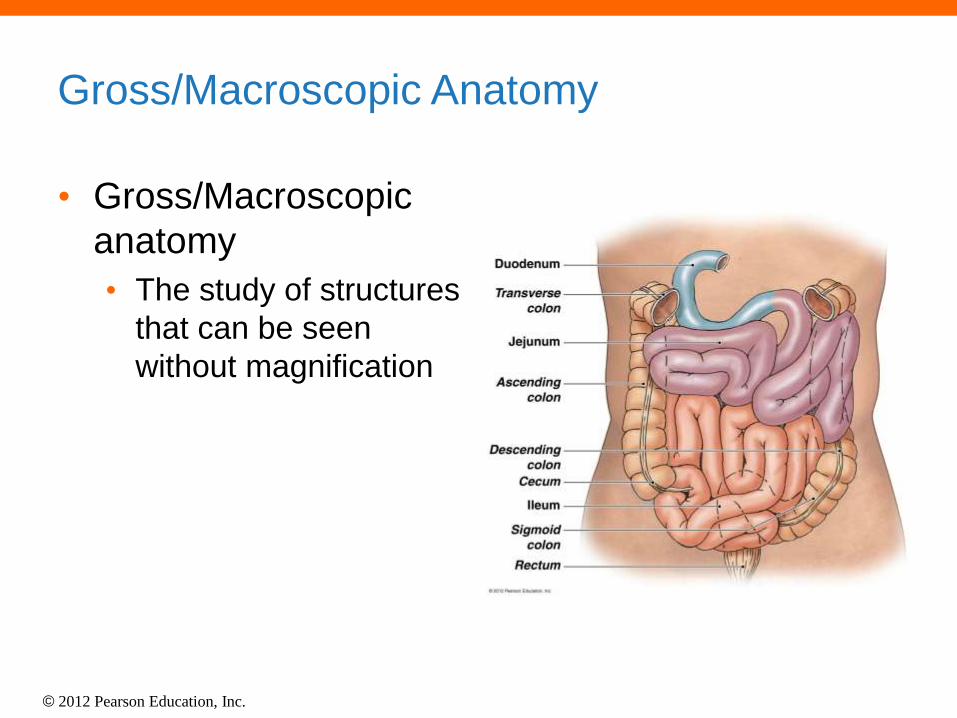
Gross/Macroscopic Anatomy
• Gross/Macroscopic
anatomy
• The study of structures
that can be seen
without magnification
© 2012 Pearson Education, Inc.
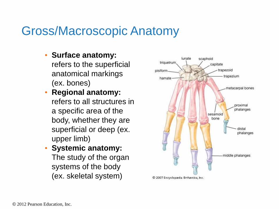
• Surface anatomy:
refers to the superficial
anatomical markings
(ex. bones)
• Regional anatomy:
refers to all structures in
a specific area of the
body, whether they are
superficial or deep (ex.
upper limb)
• Systemic anatomy:
The study of the organ
systems of the body
(ex. skeletal system)
Gross/Macroscopic Anatomy
© 2012 Pearson Education, Inc.
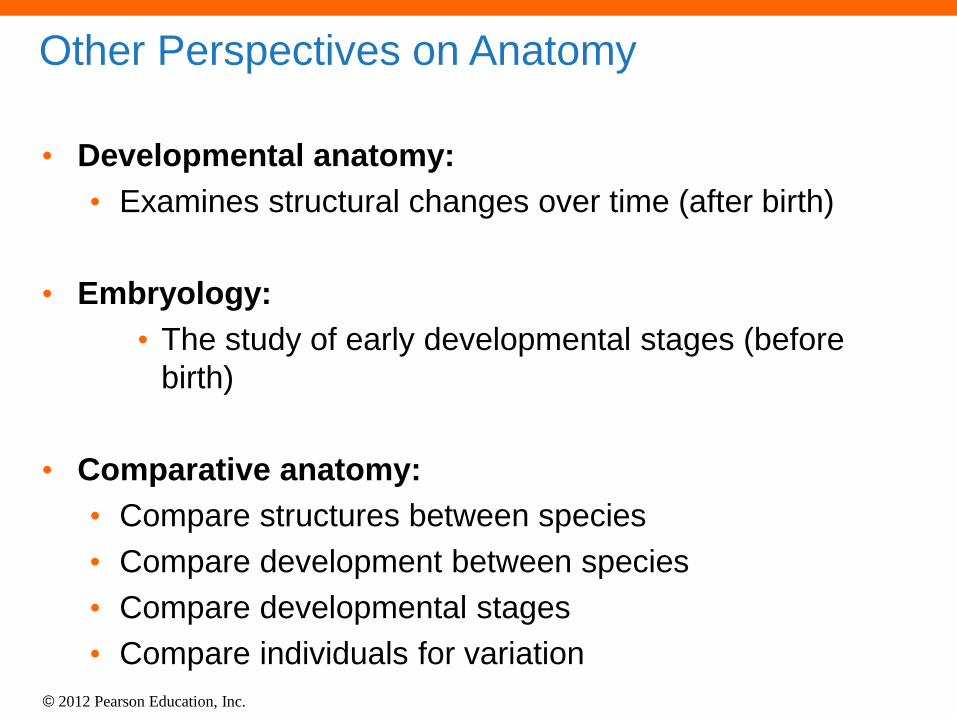
Other Perspectives on Anatomy
• Developmental anatomy:
• Examines structural changes over time (after birth)
• Embryology:
• The study of early developmental stages (before
birth)
• Comparative anatomy:
• Compare structures between species
• Compare development between species
• Compare developmental stages
• Compare individuals for variation
© 2012 Pearson Education, Inc.
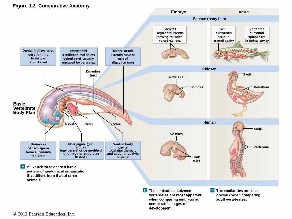
Figure 1.2 Comparative Anatomy
Dorsal, hollow nerve
cord forming
brain and
spinal cord
Notochord
a stiffened rod below
spinal cord, usually
replaced by vertebrae
Muscular tail
extends beyond
exit of
digestive tract
Digestive
tract
Mouth Heart Anus
Braincase
of cartilage or
bone surrounds
the brain
Pharyngeal (gill) arches
may persist or be modified to form other structures
in adult
Ventral body cavity
contains thoracic and abdominopelvic
organs
Somites
segmental blocks
forming muscles,
vertebrae, etc.
Skull
surrounds
brain in
cranail cavity
Limb bud
Somites
Somites
Limb
buds
Embryo
Salmon (bony fish)
Chicken
Human
Adult
Vertebrae
surround
spinal cord
in spinal cavity
Skull
Skull
Vertebrae
Vertebrae
The similarities between
vertebrates are most apparent
when comparing embryos at
comparable stages of
development.
The similarities are less
obvious when comparing
adult vertebrates.
All vertebrates share a basic
pattern of anatomical organization
that differs from that of other
animals.
Basic Vertebrate Body Plan
© 2012 Pearson Education, Inc.
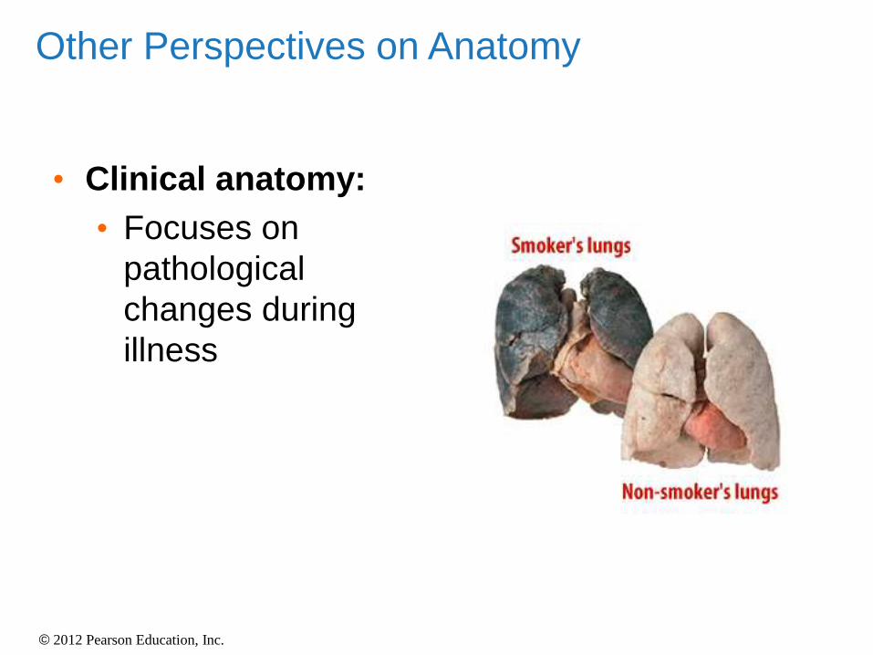
• Clinical anatomy:
• Focuses on
pathological
changes during
illness
Other Perspectives on Anatomy
© 2012 Pearson Education, Inc.
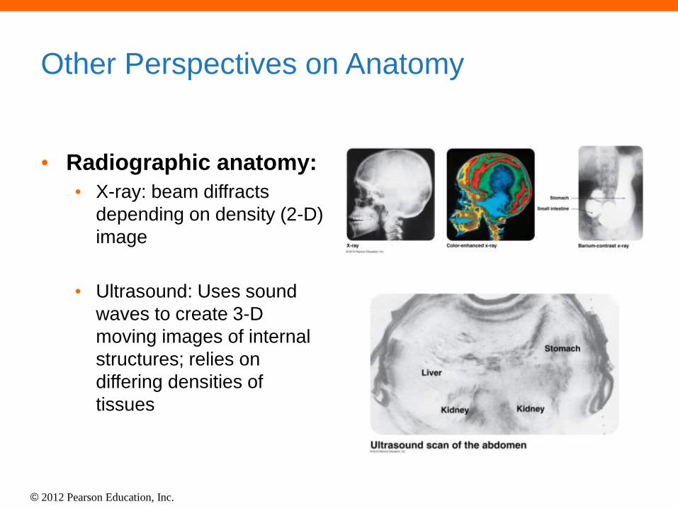
Other Perspectives on Anatomy
• Radiographic anatomy:
• X-ray: beam diffracts
depending on density (2-D)
image
• Ultrasound: Uses sound
waves to create 3-D
moving images of internal
structures; relies on
differing densities of
tissues
© 2012 Pearson Education, Inc.
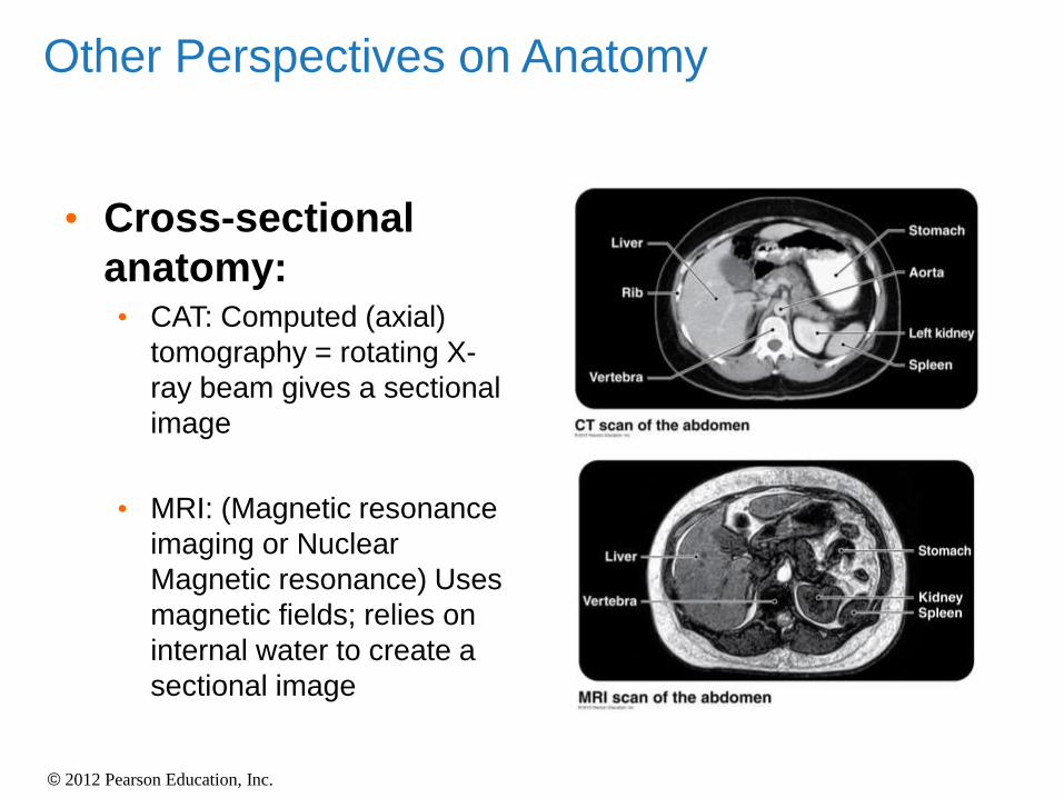
• Cross-sectional
anatomy: • CAT: Computed (axial)
tomography = rotating X-
ray beam gives a sectional
image
• MRI: (Magnetic resonance
imaging or Nuclear
Magnetic resonance) Uses
magnetic fields; relies on
internal water to create a
sectional image
Other Perspectives on Anatomy
© 2012 Pearson Education, Inc.
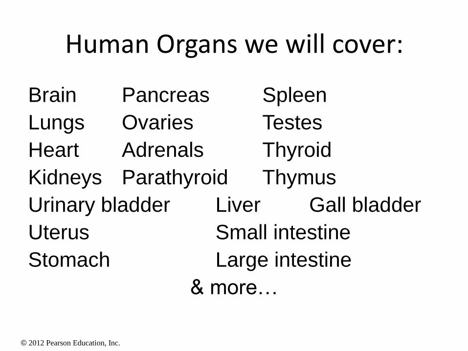
Human Organs we will cover:
Brain Pancreas Spleen
Lungs Ovaries Testes
Heart Adrenals Thyroid
Kidneys Parathyroid Thymus
Urinary bladder Liver Gall bladder
Uterus Small intestine
Stomach Large intestine
& more…
© 2012 Pearson Education, Inc.
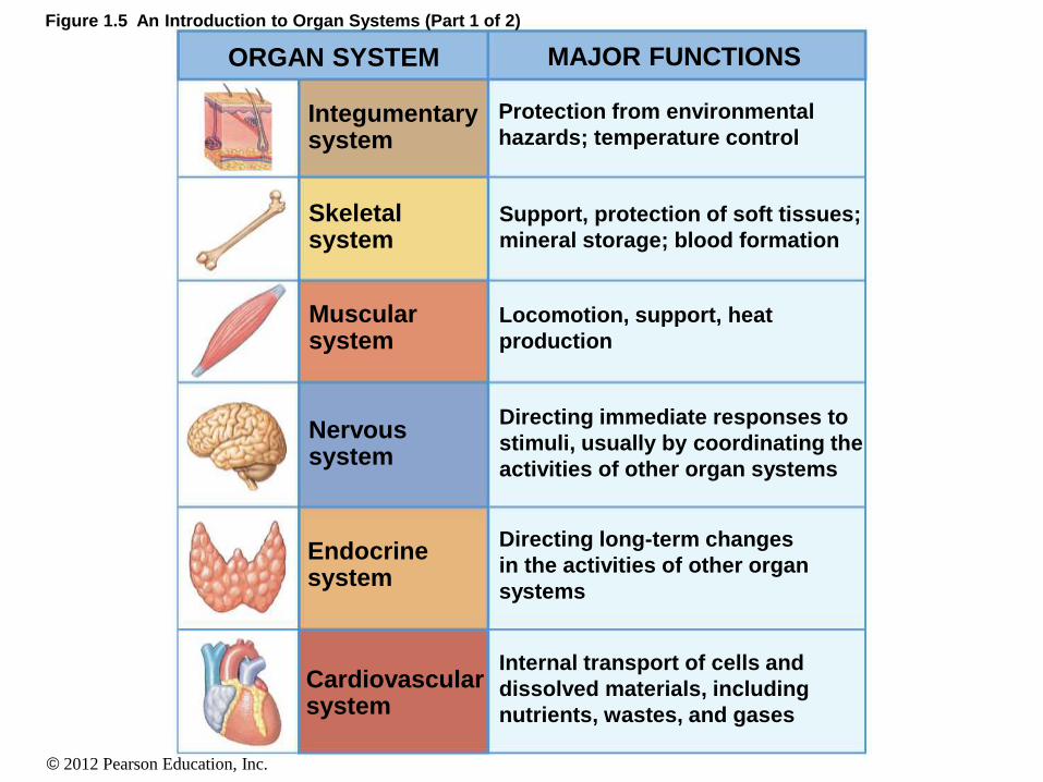
Figure 1.5 An Introduction to Organ Systems (Part 1 of 2)
ORGAN SYSTEM MAJOR FUNCTIONS
Integumentary system
Skeletal system
Muscular system
Nervous system
Endocrine system
Cardiovascular system
Protection from environmental
hazards; temperature control
Support, protection of soft tissues;
mineral storage; blood formation
Locomotion, support, heat
production
Directing immediate responses to
stimuli, usually by coordinating the
activities of other organ systems
Directing long-term changes
in the activities of other organ
systems
Internal transport of cells and
dissolved materials, including
nutrients, wastes, and gases
© 2012 Pearson Education, Inc.
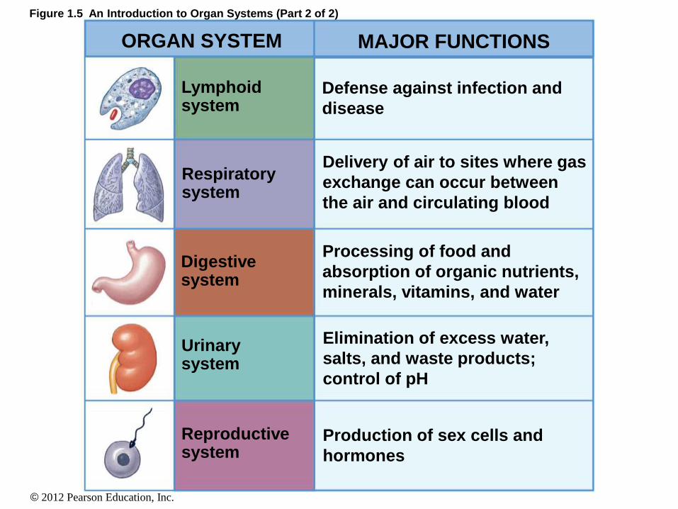
Figure 1.5 An Introduction to Organ Systems (Part 2 of 2)
ORGAN SYSTEM MAJOR FUNCTIONS
Lymphoid system
Defense against infection and
disease
Respiratory system
Digestive system
Urinary system
Reproductive system
Delivery of air to sites where gas
exchange can occur between
the air and circulating blood
Processing of food and
absorption of organic nutrients,
minerals, vitamins, and water
Elimination of excess water,
salts, and waste products;
control of pH
Production of sex cells and
hormones
© 2012 Pearson Education, Inc.
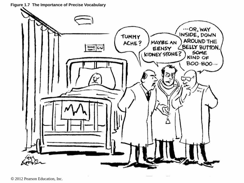
Figure 1.7 The Importance of Precise Vocabulary
© 2012 Pearson Education, Inc.
The Language of Anatomy
• Superficial Anatomy
• Using the proper terms to identify the
structures of the body helps physicians
communicate with each other and the patient
• The terms are typically derived from Latin or
Greek
• Latin or Greek is used because they are descriptive
languages
© 2012 Pearson Education, Inc.
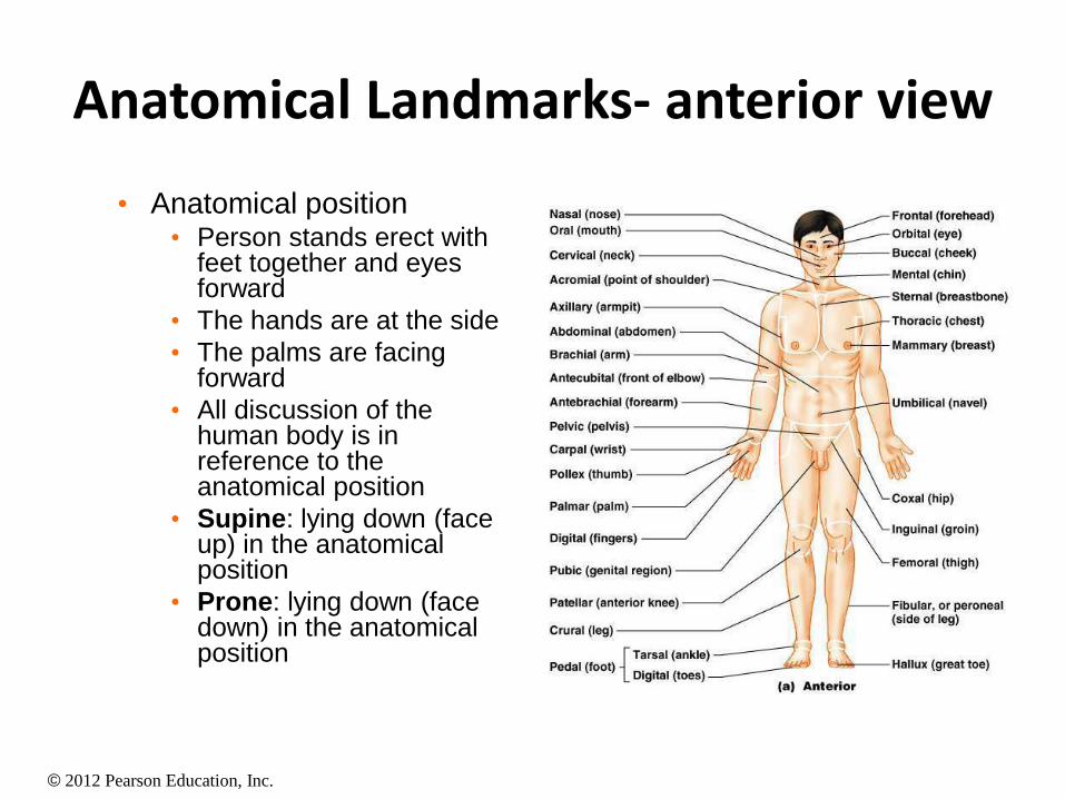
Anatomical Landmarks- anterior view
• Anatomical position • Person stands erect with
feet together and eyes forward
• The hands are at the side
• The palms are facing forward
• All discussion of the human body is in reference to the anatomical position
• Supine: lying down (face up) in the anatomical position
• Prone: lying down (face down) in the anatomical position
© 2012 Pearson Education, Inc.
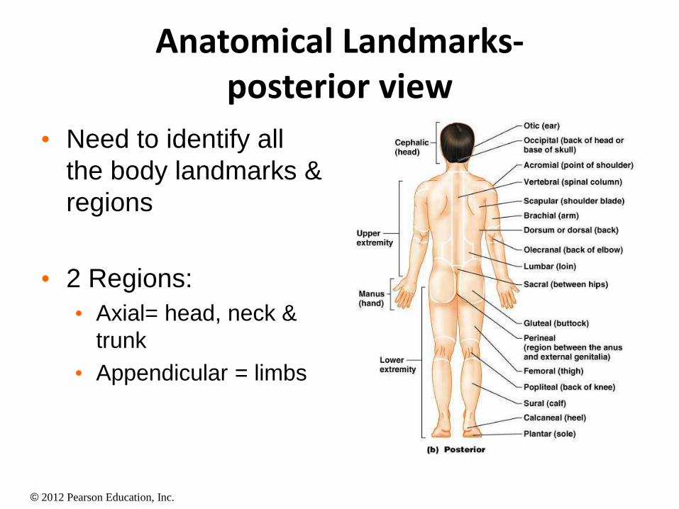
Anatomical Landmarks- posterior view
• Need to identify all
the body landmarks &
regions
• 2 Regions:
• Axial= head, neck &
trunk
• Appendicular = limbs
© 2012 Pearson Education, Inc.
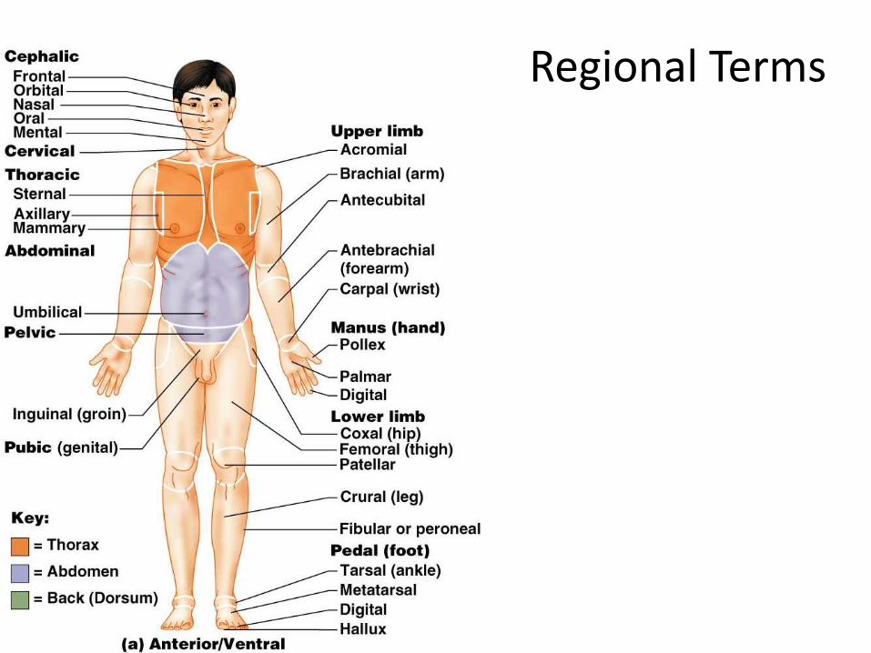
Regional Terms
© 2012 Pearson Education, Inc.
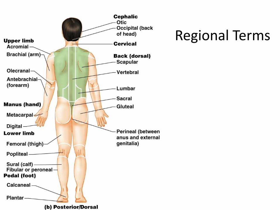
Regional Terms
© 2012 Pearson Education, Inc.
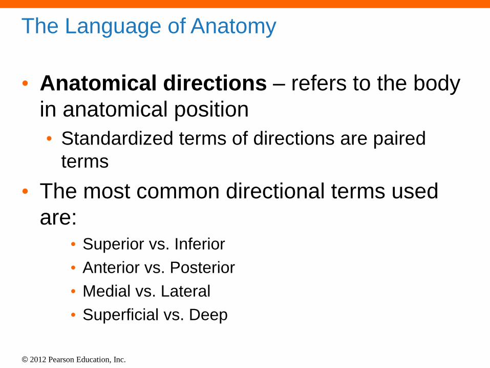
The Language of Anatomy
• Anatomical directions – refers to the body
in anatomical position
• Standardized terms of directions are paired
terms
• The most common directional terms used
are: • Superior vs. Inferior
• Anterior vs. Posterior
• Medial vs. Lateral
• Superficial vs. Deep
© 2012 Pearson Education, Inc.
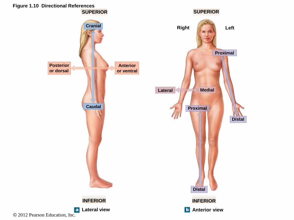
Figure 1.10 Directional References
SUPERIOR SUPERIOR
INFERIOR INFERIOR
Anterior view Lateral view
Right Left
Proximal
Lateral Medial
Distal
Distal
Proximal
Posterior
or dorsal Anterior
or ventral
Cranial
Caudal
© 2012 Pearson Education, Inc.
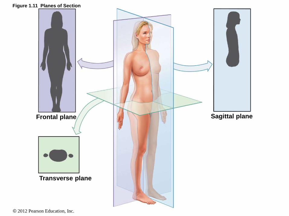
The Language of Anatomy
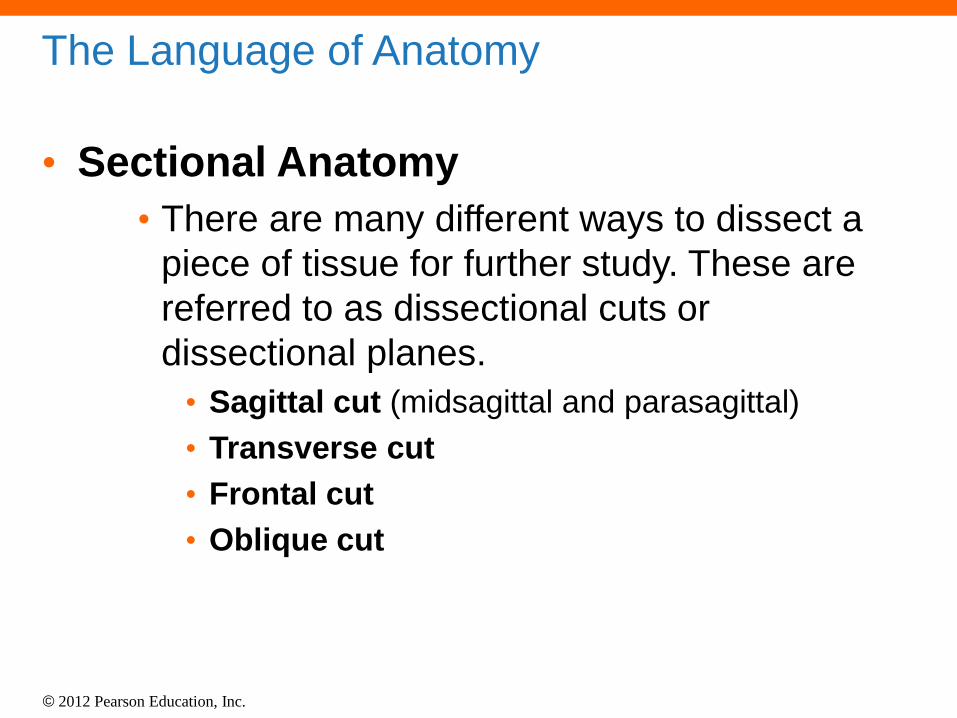
• Sectional Anatomy
• There are many different ways to dissect a
piece of tissue for further study. These are
referred to as dissectional cuts or
dissectional planes.
• Sagittal cut (midsagittal and parasagittal)
• Transverse cut
• Frontal cut
• Oblique cut
© 2012 Pearson Education, Inc.
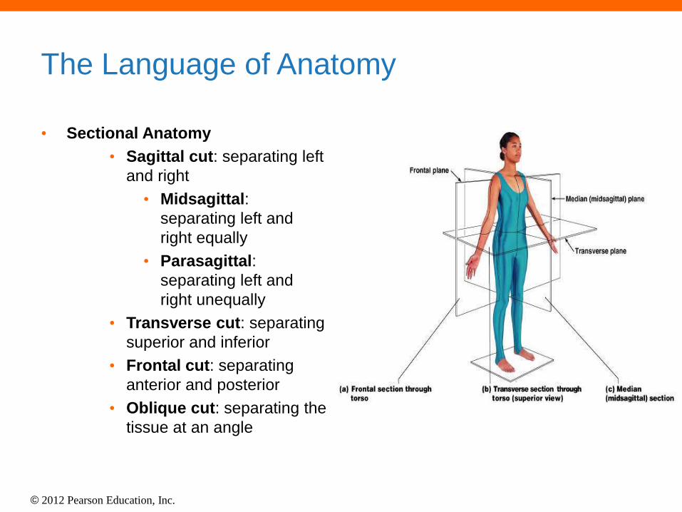
The Language of Anatomy
• Sectional Anatomy
• Sagittal cut: separating left
and right
• Midsagittal:
separating left and
right equally
• Parasagittal:
separating left and
right unequally
• Transverse cut: separating
superior and inferior
• Frontal cut: separating
anterior and posterior
• Oblique cut: separating the
tissue at an angle
© 2012 Pearson Education, Inc.
Figure 1.11 Planes of Section
Frontal plane
Transverse plane
Sagittal plane
© 2012 Pearson Education, Inc.
The Language of Anatomy
• Abdominopelvic quadrants and regions
• Anatomists and clinicians use specialized
regional terms to indicate a specific area of
concern within the abdomen or the pelvic
regions of the body.
• The abdomen and pelvic regions can be subdivided
into four regions (abdominopelvic quadrants)
• The abdomen and pelvic regions can be subdivided
into nine regions (abdominopelvic regions)
© 2012 Pearson Education, Inc.
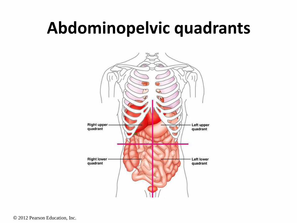
Abdominopelvic quadrants
© 2012 Pearson Education, Inc.
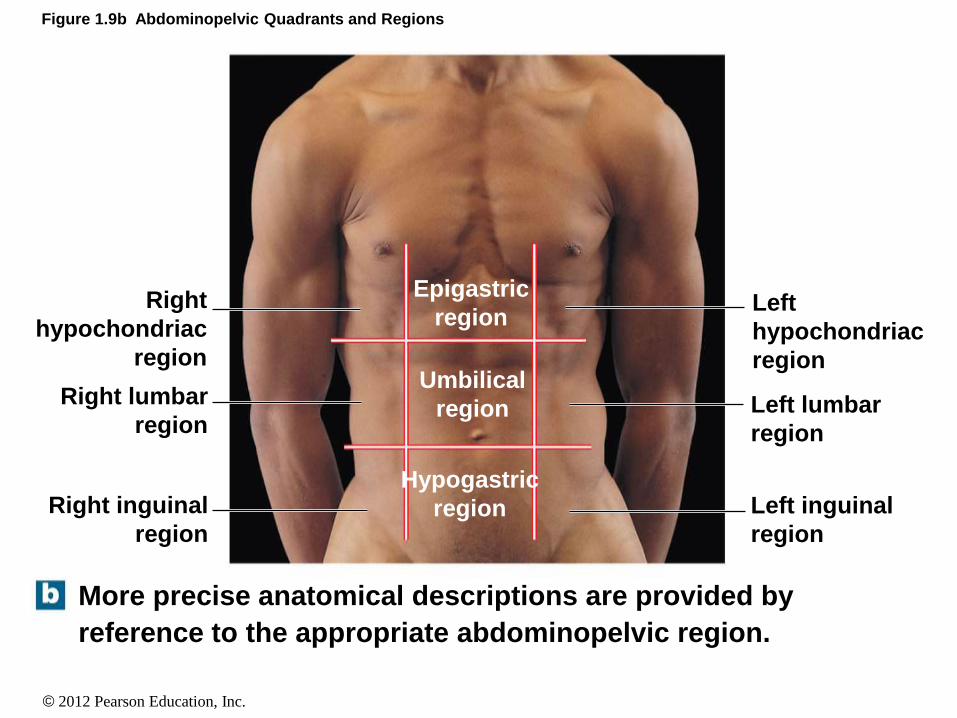
Figure 1.9b Abdominopelvic Quadrants and Regions
More precise anatomical descriptions are provided by
reference to the appropriate abdominopelvic region.
Left
hypochondriac
region
Left lumbar
region
Left inguinal
region
Right inguinal
region
Right lumbar
region
Right
hypochondriac
region
Epigastric
region
Umbilical
region
Hypogastric
region
© 2012 Pearson Education, Inc.
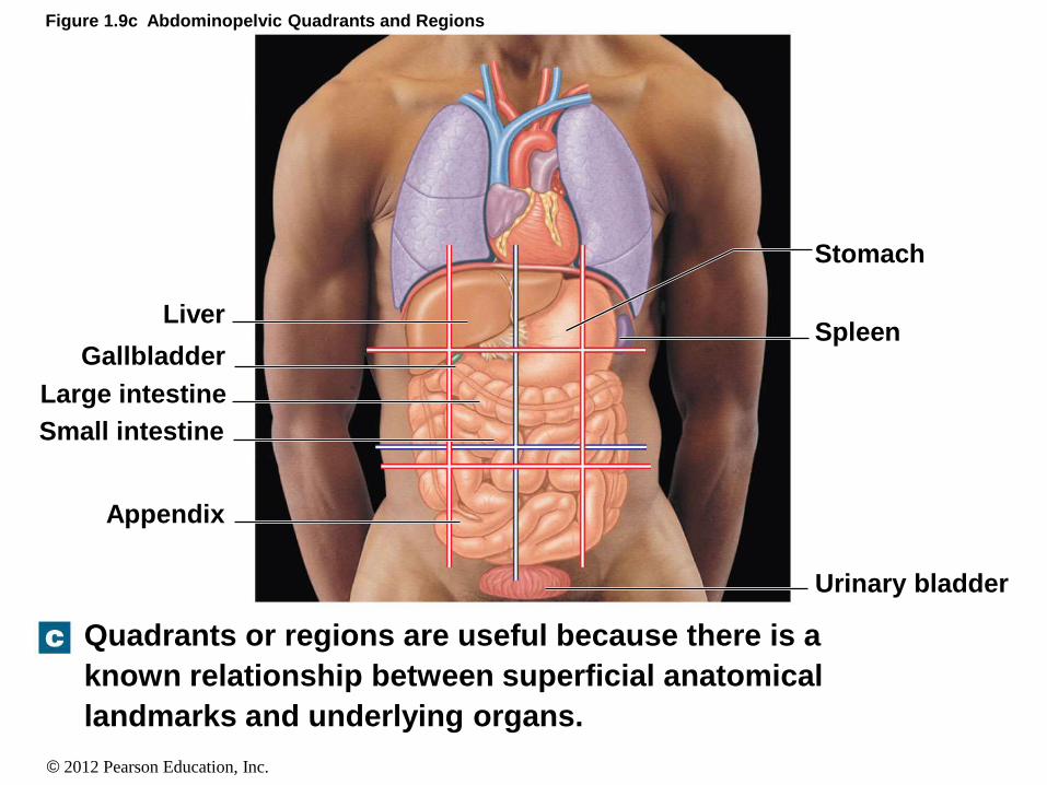
Figure 1.9c Abdominopelvic Quadrants and Regions
Quadrants or regions are useful because there is a
known relationship between superficial anatomical
landmarks and underlying organs.
Stomach
Spleen
Urinary bladder
Liver
Gallbladder
Large intestine
Small intestine
Appendix
© 2012 Pearson Education, Inc.
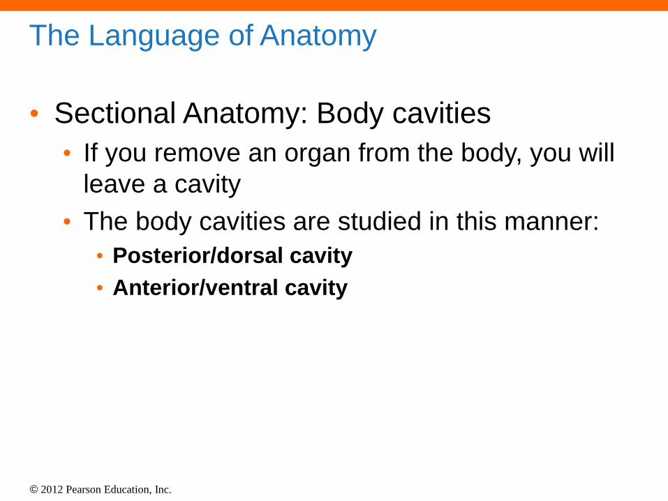
The Language of Anatomy
• Sectional Anatomy: Body cavities
• If you remove an organ from the body, you will
leave a cavity
• The body cavities are studied in this manner:
• Posterior/dorsal cavity
• Anterior/ventral cavity
© 2012 Pearson Education, Inc.
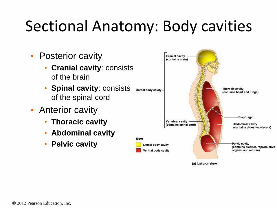
Sectional Anatomy: Body cavities
• Posterior cavity
• Cranial cavity: consists
of the brain
• Spinal cavity: consists
of the spinal cord
• Anterior cavity
• Thoracic cavity
• Abdominal cavity
• Pelvic cavity
© 2012 Pearson Education, Inc.
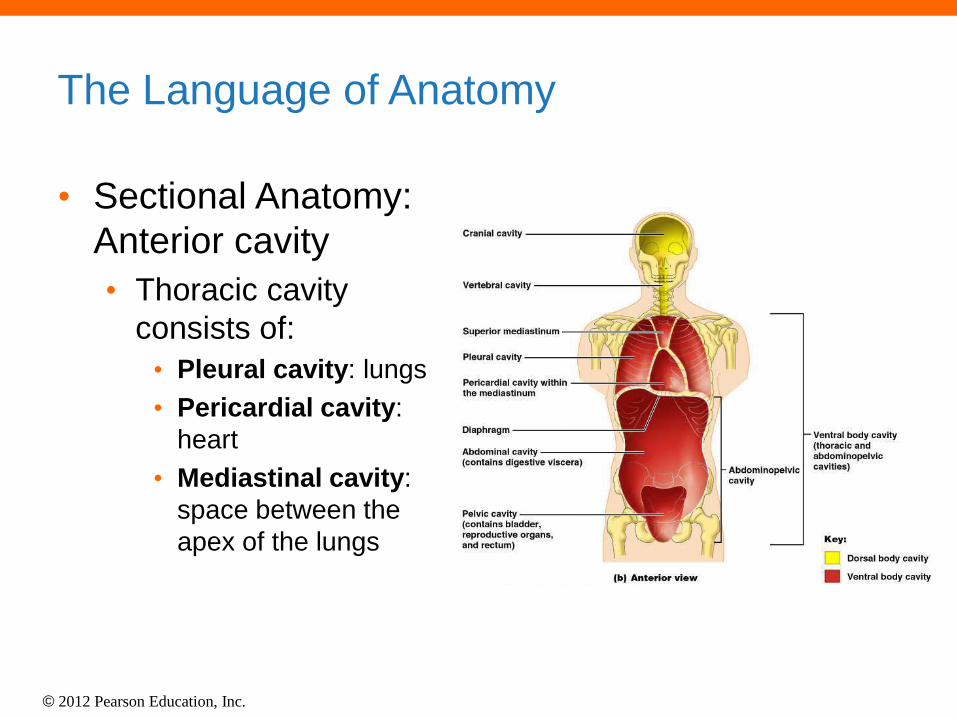
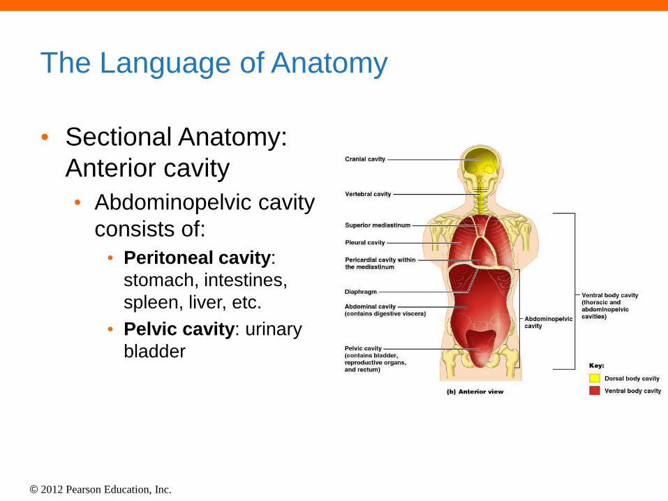
The Language of Anatomy
• Sectional Anatomy:
Anterior cavity
• Thoracic cavity
consists of:
• Pleural cavity: lungs
• Pericardial cavity:
heart
• Mediastinal cavity:
space between the
apex of the lungs
© 2012 Pearson Education, Inc.
The Language of Anatomy
• Sectional Anatomy:
Anterior cavity
• Abdominopelvic cavity
consists of:
• Peritoneal cavity:
stomach, intestines,
spleen, liver, etc.
• Pelvic cavity: urinary
bladder
© 2012 Pearson Education, Inc.
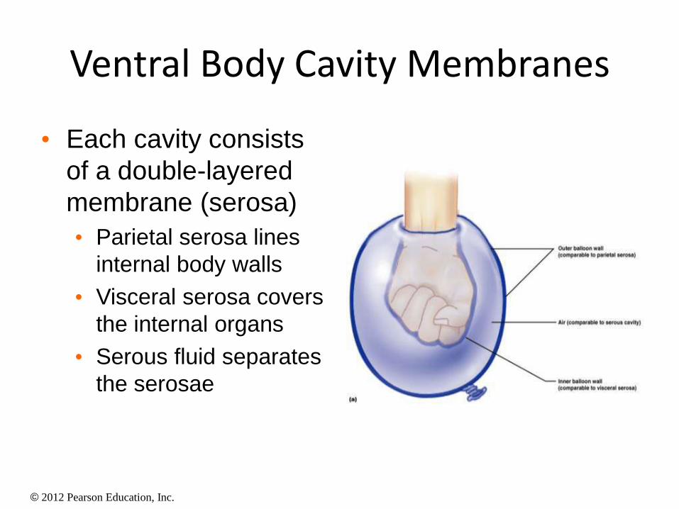
Ventral Body Cavity Membranes
• Each cavity consists
of a double-layered
membrane (serosa)
• Parietal serosa lines
internal body walls
• Visceral serosa covers
the internal organs
• Serous fluid separates
the serosae
© 2012 Pearson Education, Inc.
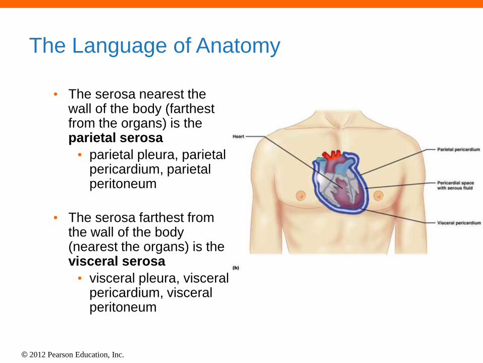
The Language of Anatomy
• The serosa nearest the wall of the body (farthest from the organs) is the parietal serosa
• parietal pleura, parietal pericardium, parietal peritoneum
• The serosa farthest from the wall of the body (nearest the organs) is the visceral serosa
• visceral pleura, visceral pericardium, visceral peritoneum
© 2012 Pearson Education, Inc.
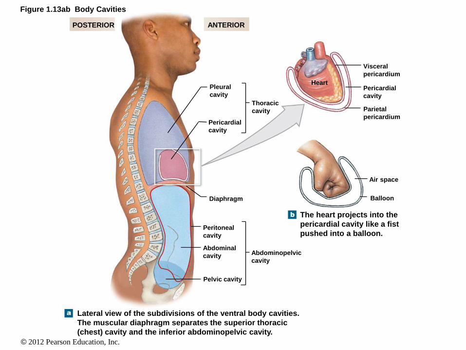
Figure 1.13ab Body Cavities
POSTERIOR ANTERIOR
Pleural
cavity
Thoracic
cavity
Pericardial
cavity
Diaphragm
Peritoneal
cavity
Abdominal
cavity
Pelvic cavity
Abdominopelvic
cavity
Lateral view of the subdivisions of the ventral body cavities.
The muscular diaphragm separates the superior thoracic
(chest) cavity and the inferior abdominopelvic cavity.
The heart projects into the
pericardial cavity like a fist
pushed into a balloon.
Heart
Visceral
pericardium
Pericardial
cavity
Parietal
pericardium
Air space
Balloon