Embed Size (px)
Citation preview
Chapter 3 Cell Structure and
Function
Copyright © The McGraw-Hill Companies, Inc. Permission required for reproduction or display.
3.1 The Cellular Level of
Organization
• The cell is the structural and functional unit
of an organism.
• Smallest structure capable of performing
all the functions necessary for life.
3.1 The Cellular Level of
Organization
• Prokaryotic cells lack membrane-enclosed
structures.
• Eukaryotic cells possess membrane-
enclosed structures.
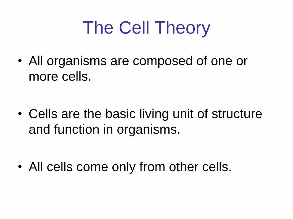
The Cell Theory
• All organisms are composed of one or
more cells.
• Cells are the basic living unit of structure
and function in organisms.
• All cells come only from other cells.
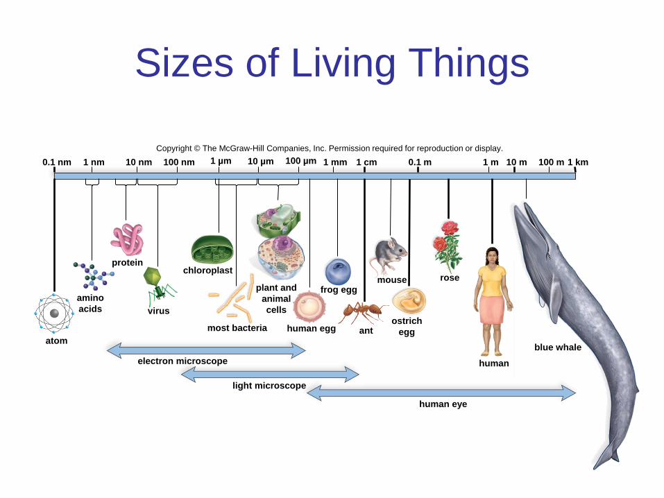
Sizes of Living Things
10 m 1 m 0.1 m 1 cm 1 mm 100 µm 10 µm 100 nm 10 nm 1 nm 0.1 nm
mouse frog egg
human egg most bacteria
virus
protein
atom ant
electron microscope
light microscope
human eye
human
blue whale
chloroplast rose
1 km 100 m 1 µm
amino
acids
plant and
animal
cells
ostrich
egg
Copyright © The McGraw-Hill Companies, Inc. Permission required for reproduction or display.
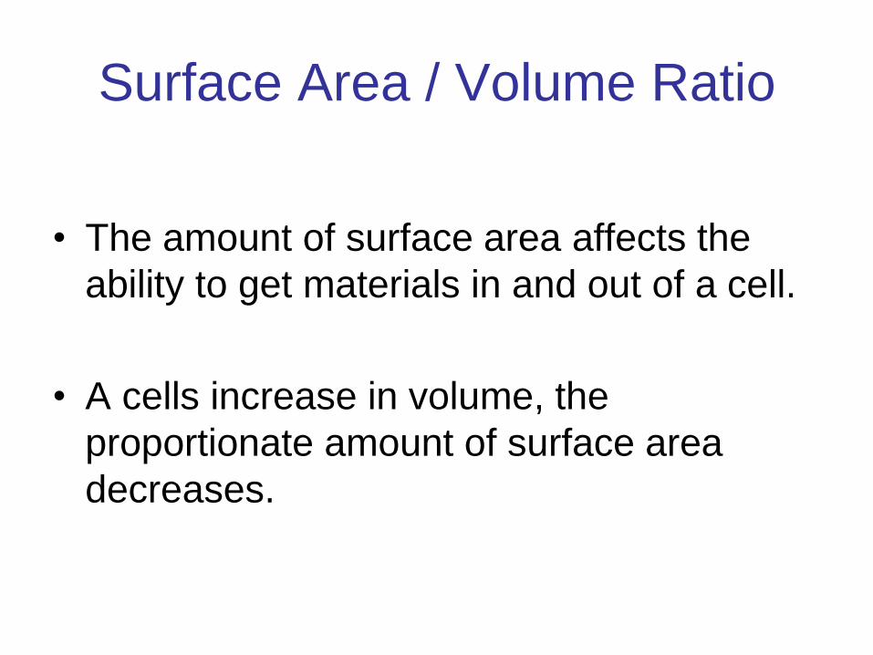
Surface Area / Volume Ratio
• The amount of surface area affects the
ability to get materials in and out of a cell.
• A cells increase in volume, the
proportionate amount of surface area
decreases.
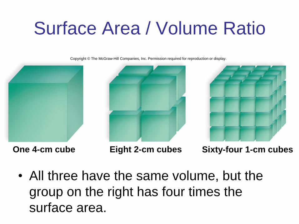
Surface Area / Volume Ratio
• All three have the same volume, but the
group on the right has four times the
surface area.
One 4-cm cube Eight 2-cm cubes Sixty-four 1-cm cubes
Copyright © The McGraw-Hill Companies, Inc. Permission required for reproduction or display.
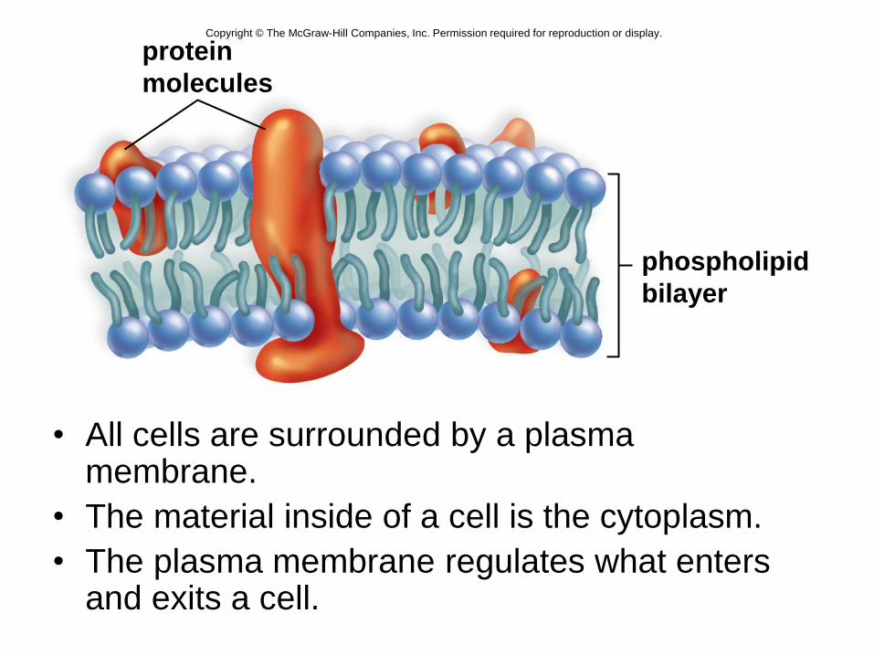
• All cells are surrounded by a plasma membrane.
• The material inside of a cell is the cytoplasm.
• The plasma membrane regulates what enters and exits a cell.
protein
molecules
phospholipid
bilayer
Copyright © The McGraw-Hill Companies, Inc. Permission required for reproduction or display.
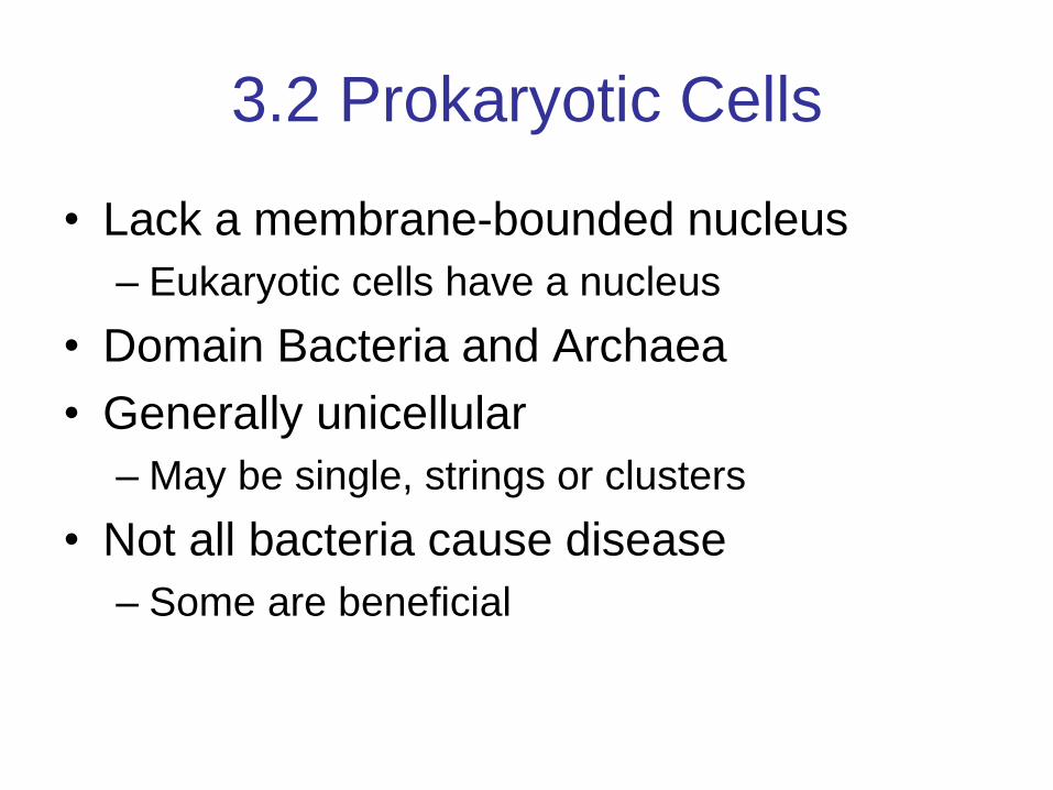
3.2 Prokaryotic Cells
• Lack a membrane-bounded nucleus
– Eukaryotic cells have a nucleus
• Domain Bacteria and Archaea
• Generally unicellular
– May be single, strings or clusters
• Not all bacteria cause disease
– Some are beneficial
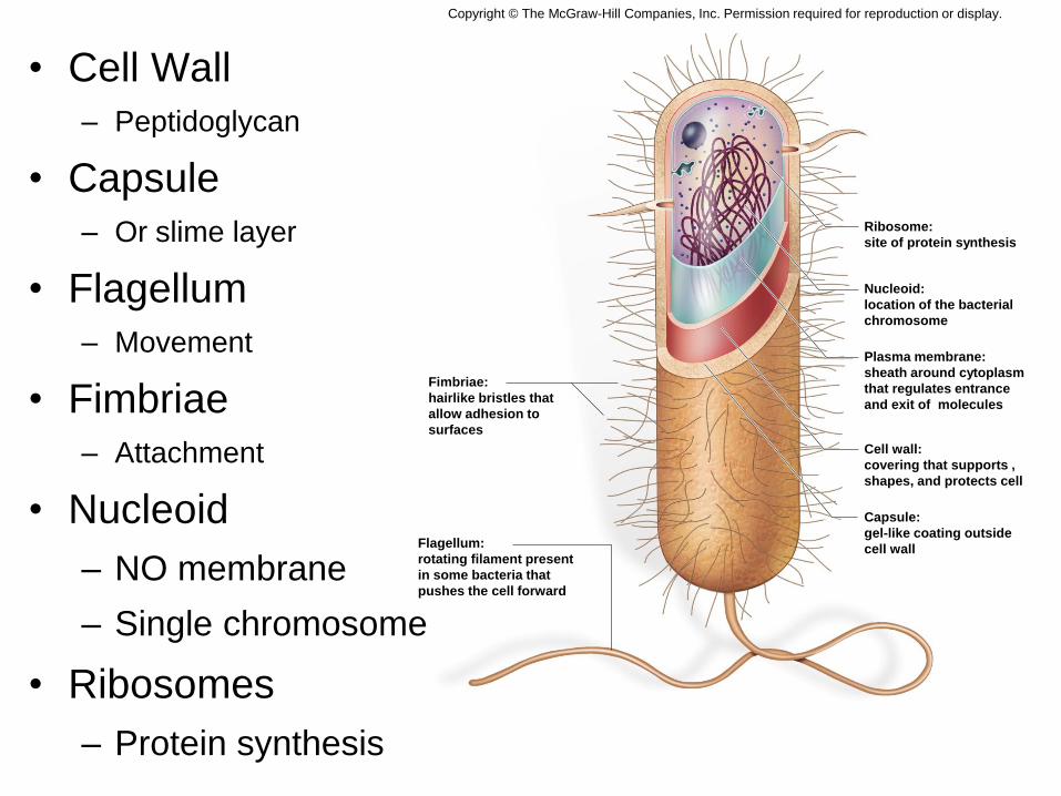
• Cell Wall
– Peptidoglycan
• Capsule
– Or slime layer
• Flagellum
– Movement
• Fimbriae
– Attachment
• Nucleoid
– NO membrane
– Single chromosome
• Ribosomes
– Protein synthesis
Fimbriae:
hairlike bristles that
allow adhesion to
surfaces
Flagellum:
rotating filament present
in some bacteria that
pushes the cell forward
Ribosome:
site of protein synthesis
Nucleoid:
location of the bacterial
chromosome
Plasma membrane:
sheath around cytoplasm
that regulates entrance
and exit of molecules
Cell wall:
covering that supports ,
shapes, and protects cell
Capsule:
gel-like coating outside
cell wall
Copyright © The McGraw-Hill Companies, Inc. Permission required for reproduction or display.
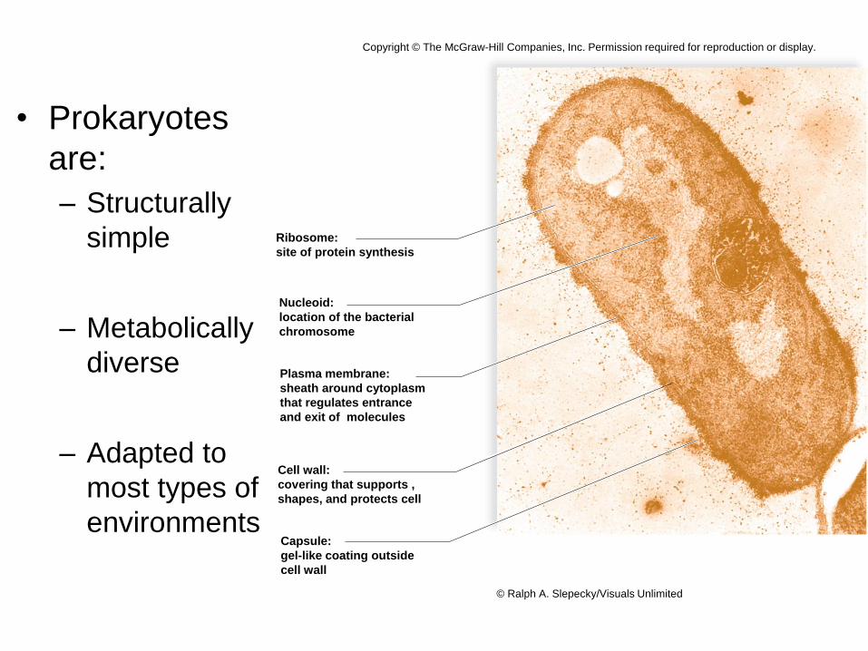
• Prokaryotes
are:
– Structurally
simple
– Metabolically
diverse
– Adapted to
most types of
environments
Ribosome:
site of protein synthesis
Nucleoid:
location of the bacterial
chromosome
Plasma membrane:
sheath around cytoplasm
that regulates entrance
and exit of molecules
Cell wall:
covering that supports ,
shapes, and protects cell
Capsule:
gel-like coating outside
cell wall
Copyright © The McGraw-Hill Companies, Inc. Permission required for reproduction or display.
© Ralph A. Slepecky/Visuals Unlimited
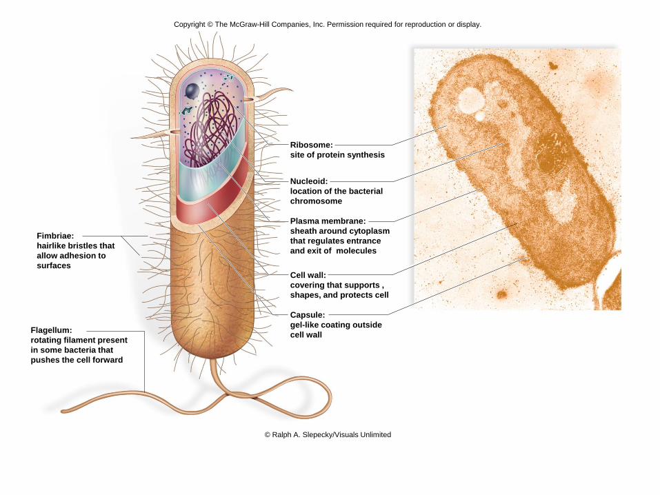
Fimbriae:
hairlike bristles that
allow adhesion to
surfaces
Flagellum:
rotating filament present
in some bacteria that
pushes the cell forward
Ribosome:
site of protein synthesis
Nucleoid:
location of the bacterial
chromosome
Plasma membrane:
sheath around cytoplasm
that regulates entrance
and exit of molecules
Cell wall:
covering that supports ,
shapes, and protects cell
Capsule:
gel-like coating outside
cell wall
Copyright © The McGraw-Hill Companies, Inc. Permission required for reproduction or display.
© Ralph A. Slepecky/Visuals Unlimited
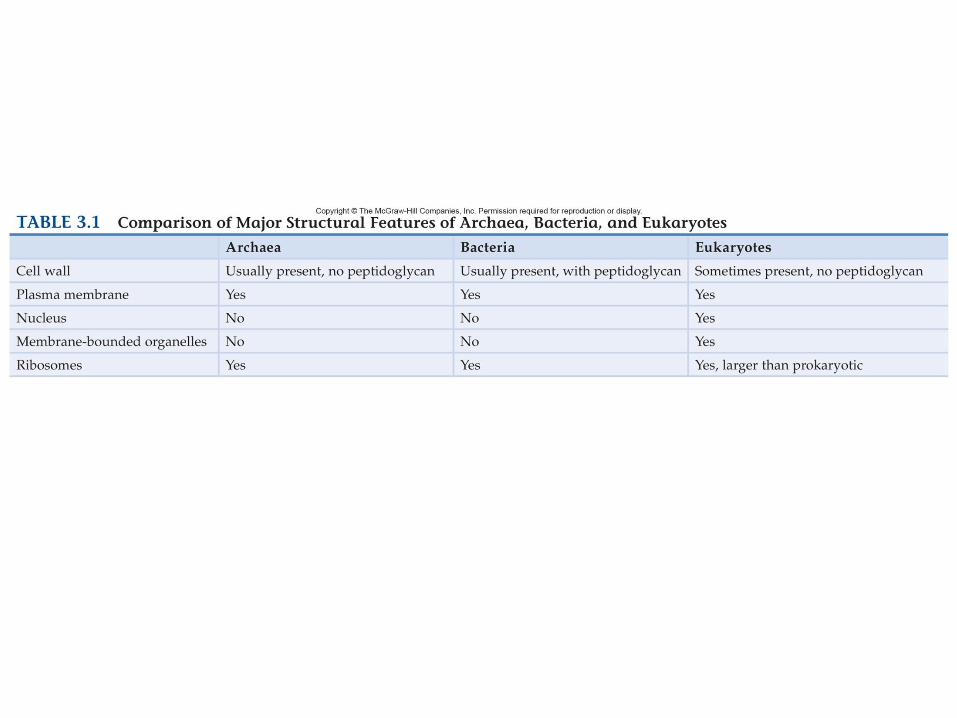
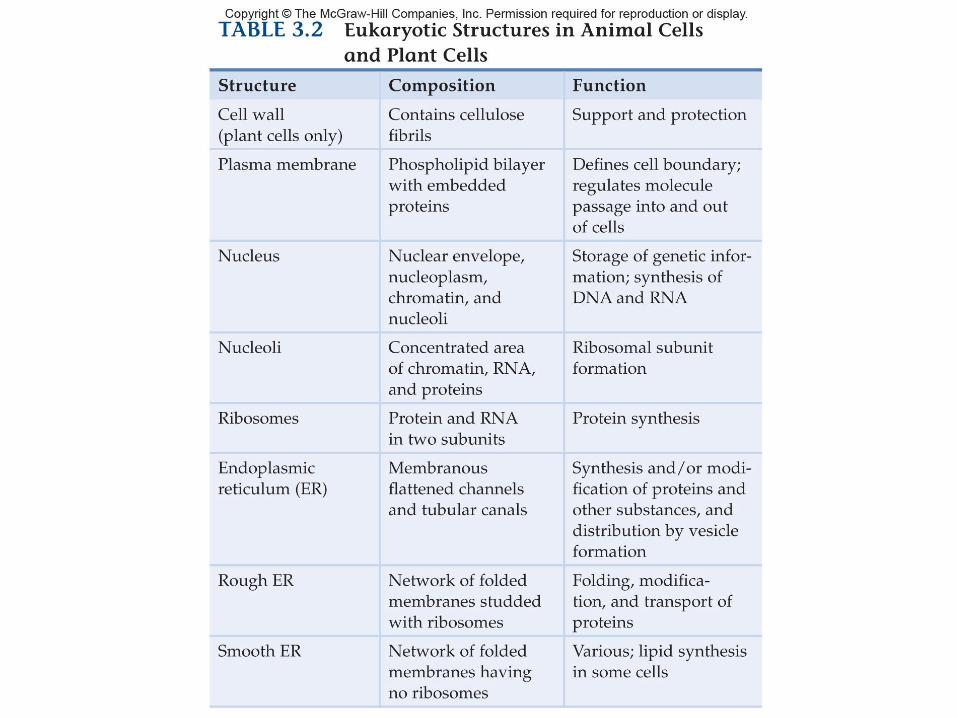
3.3 Eukaryotic Cells
• Eukaryotic cells:
– Are structurally complex
– Have a nucleus
– Possess membrane-bound organelles
– Animals, plants, fungi and protists
• Cell walls
– Some eukaryotic cells have cell walls.
– Plant cells may have a primary and secondary
cell wall.
• Cellulose a constituent of primary cell wall.
– Also found in algae (protist) cell walls
• Lignin found in secondary cell walls.
– Fungi cell walls
• Some cellulose
• Some chitin (also found in insect exoskeletons)
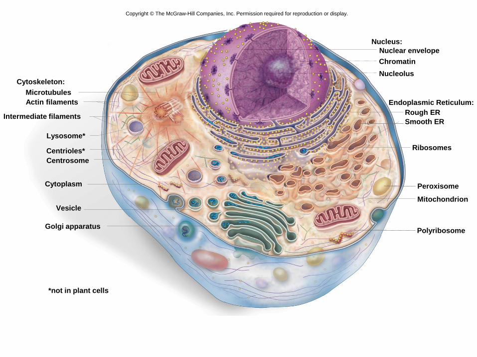
Cytoskeleton:
Actin filaments
Nucleus:
Rough ER
Ribosomes
Golgi apparatus
Centrioles*
Cytoplasm Peroxisome
*not in plant cells
Intermediate filaments Smooth ER
Endoplasmic Reticulum:
Microtubules
Centrosome
Mitochondrion
Polyribosome
Nucleolus
Chromatin
Nuclear envelope
Lysosome*
Vesicle
Copyright © The McGraw-Hill Companies, Inc. Permission required for reproduction or display.
Copyright © The McGraw-Hill Companies, Inc. Permission required for reproduction or display.
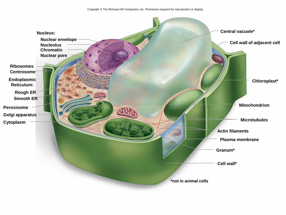
Central vacuole*
Smooth ER
Cytoplasm
*not in animal cells
Cell wall*
Cell wall of adjacent cell
Chloroplast*
Mitochondrion
Microtubules
Plasma membrane
Actin filaments
Granum*
Ribosomes
Rough ER
Endoplasmic
Reticulum:
Centrosome
Nucleus:
Nuclear envelope
Chromatin
Nuclear pore
Golgi apparatus
Peroxisome
Nucleolus
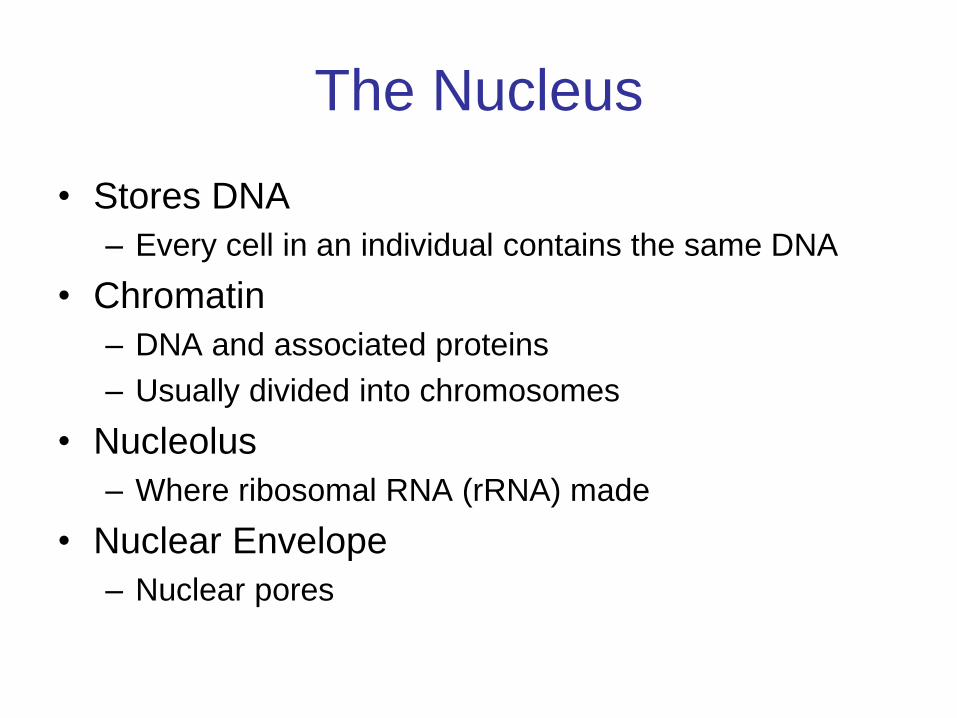
The Nucleus
• Stores DNA
– Every cell in an individual contains the same DNA
• Chromatin
– DNA and associated proteins
– Usually divided into chromosomes
• Nucleolus
– Where ribosomal RNA (rRNA) made
• Nuclear Envelope
– Nuclear pores

nuclear pore
Nuclear envelope:
inner membrane
outer membrane chromatin
nucleoplasm
nucleolus
phospholipid
nuclear
pore
nuclear
envelope
Copyright © The McGraw-Hill Companies, Inc. Permission required for reproduction or display.
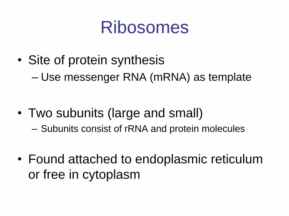
Ribosomes
• Site of protein synthesis
– Use messenger RNA (mRNA) as template
• Two subunits (large and small) – Subunits consist of rRNA and protein molecules
• Found attached to endoplasmic reticulum
or free in cytoplasm
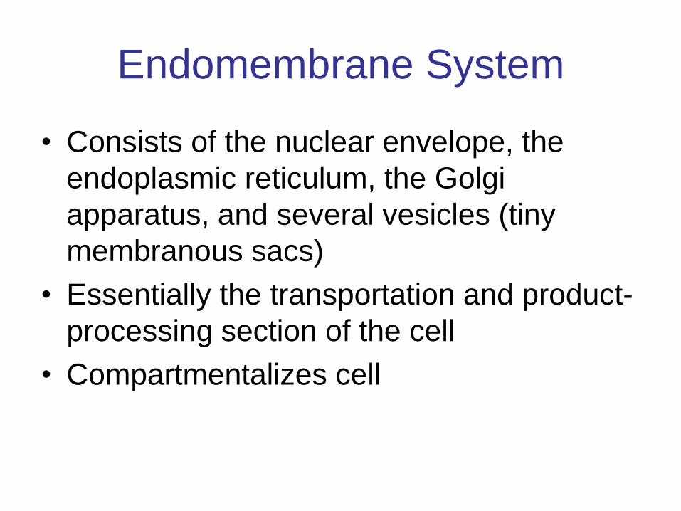
Endomembrane System
• Consists of the nuclear envelope, the
endoplasmic reticulum, the Golgi
apparatus, and several vesicles (tiny
membranous sacs)
• Essentially the transportation and product-
processing section of the cell
• Compartmentalizes cell

Endoplasmic Reticulum
• Membranous channels and saccules (flattened
vesicles)
nuclear envelope ribosomes
0.08 µm
rough
endoplasmic
reticulum
smooth
endoplasmic
reticulum
Copyright © The McGraw-Hill Companies, Inc. Permission required for reproduction or display.
© R. Bolender & D. Fawcett/Visuals Unlimited
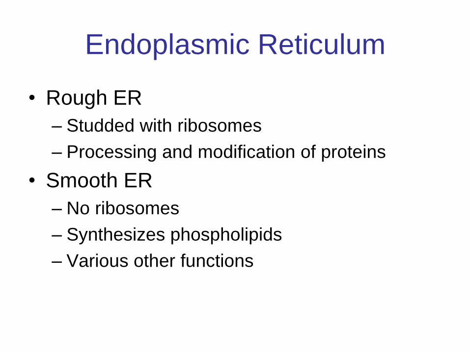
Endoplasmic Reticulum
• Rough ER
– Studded with ribosomes
– Processing and modification of proteins
• Smooth ER
– No ribosomes
– Synthesizes phospholipids
– Various other functions
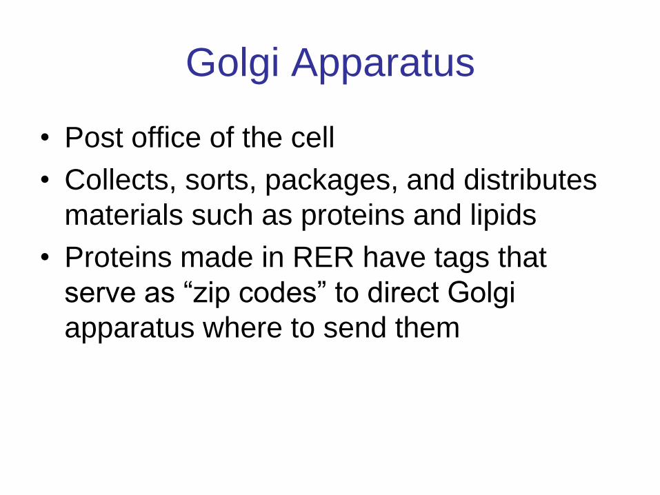
Golgi Apparatus
• Post office of the cell
• Collects, sorts, packages, and distributes
materials such as proteins and lipids
• Proteins made in RER have tags that
serve as “zip codes” to direct Golgi
apparatus where to send them
Copyright © The McGraw-Hill Companies, Inc. Permission required for reproduction or display.
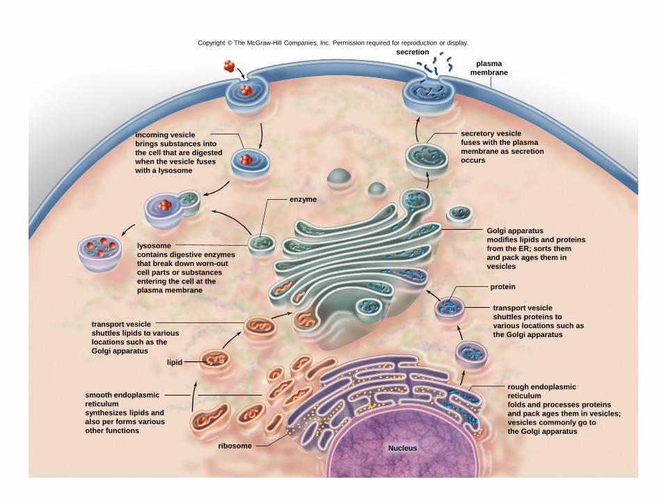
plasma
membrane
secretion
enzyme
lysosome
contains digestive enzymes
that break down worn-out
cell parts or substances
entering the cell at the
plasma membrane
secretory vesicle
fuses with the plasma
membrane as secretion
occurs
Golgi apparatus
modifies lipids and proteins
from the ER; sorts them
and pack ages them in
vesicles
transport vesicle
shuttles lipids to various
locations such as the
Golgi apparatus
lipid
transport vesicle
shuttles proteins to
various locations such as
the Golgi apparatus
protein
ribosome
rough endoplasmic
reticulum
folds and processes proteins
and pack ages them in vesicles;
vesicles commonly go to
the Golgi apparatus
Nucleus
smooth endoplasmic
reticulum
synthesizes lipids and
also per forms various
other functions
incoming vesicle
brings substances into
the cell that are digested
when the vesicle fuses
with a lysosome
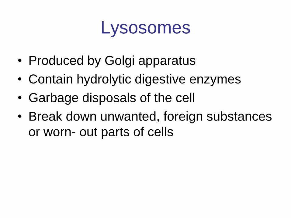
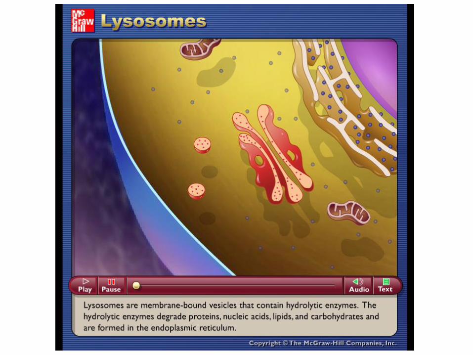
Lysosomes
• Produced by Golgi apparatus
• Contain hydrolytic digestive enzymes
• Garbage disposals of the cell
• Break down unwanted, foreign substances
or worn- out parts of cells
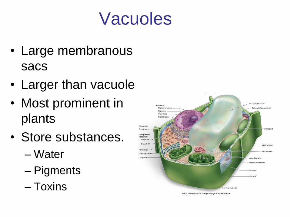
Vacuoles
• Large membranous
sacs
• Larger than vacuole
• Most prominent in
plants
• Store substances.
– Water
– Pigments
– Toxins
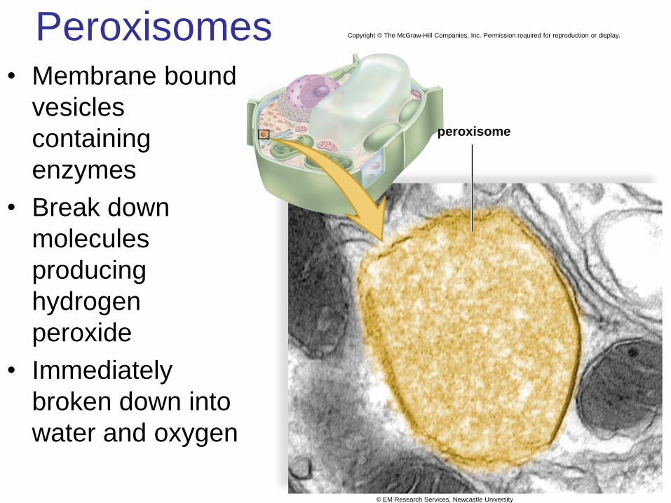
Peroxisomes • Membrane bound
vesicles
containing
enzymes
• Break down
molecules
producing
hydrogen
peroxide
• Immediately
broken down into
water and oxygen
Copyright © The McGraw-Hill Companies, Inc. Permission required for reproduction or display.
peroxisome
© EM Research Services, Newcastle University
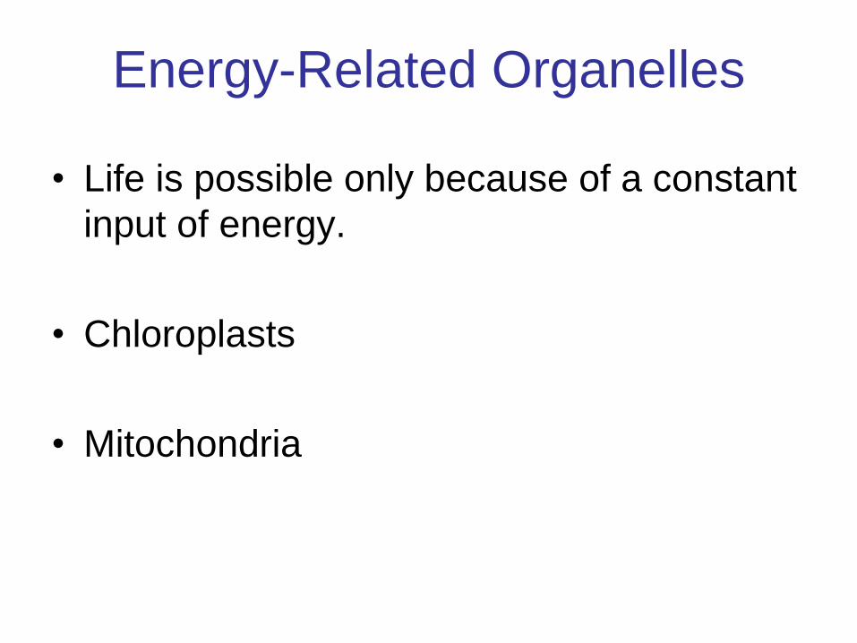
• Life is possible only because of a constant
input of energy.
• Chloroplasts
• Mitochondria
Energy-Related Organelles
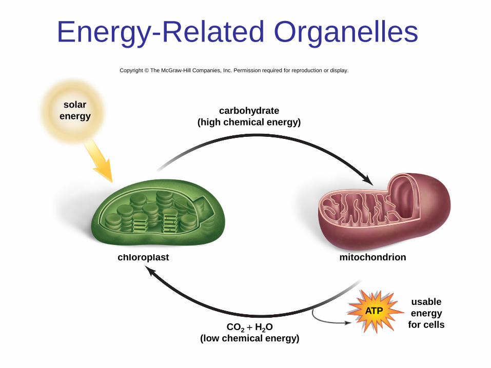
Energy-Related Organelles Copyright © The McGraw-Hill Companies, Inc. Permission required for reproduction or display.
carbohydrate
(high chemical energy)
chloroplast mitochondrion
usable
energy
for cells CO2 + H2O
(low chemical energy)
solar
energy
ATP
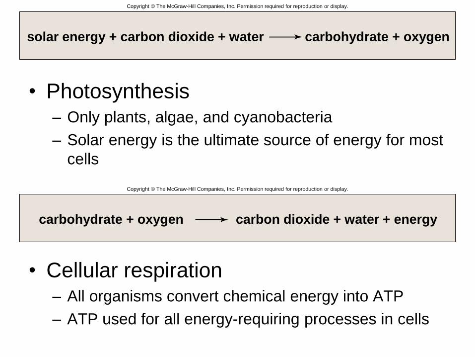
• Photosynthesis – Only plants, algae, and cyanobacteria
– Solar energy is the ultimate source of energy for most
cells
• Cellular respiration – All organisms convert chemical energy into ATP
– ATP used for all energy-requiring processes in cells
Copyright © The McGraw-Hill Companies, Inc. Permission required for reproduction or display.
solar energy + carbon dioxide + water carbohydrate + oxygen
carbohydrate + oxygen carbon dioxide + water + energy
Copyright © The McGraw-Hill Companies, Inc. Permission required for reproduction or display.
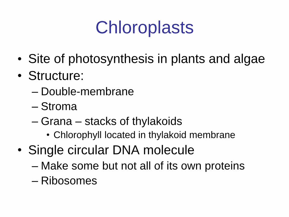
Chloroplasts
• Site of photosynthesis in plants and algae
• Structure:
– Double-membrane
– Stroma
– Grana – stacks of thylakoids • Chlorophyll located in thylakoid membrane
• Single circular DNA molecule
– Make some but not all of its own proteins
– Ribosomes

Copyright © The McGraw-Hill Companies, Inc. Permission required for reproduction or display.
grana thylakoid stroma
a.
b.
500 nm
double
membrane
outer
membrane
inner
membrane
thylakoid
space
Courtesy Herbert W. Israel, Cornell University
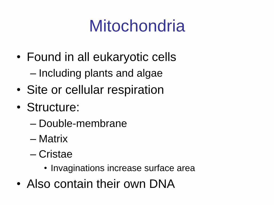
Mitochondria
• Found in all eukaryotic cells
– Including plants and algae
• Site or cellular respiration
• Structure:
– Double-membrane
– Matrix
– Cristae
• Invaginations increase surface area
• Also contain their own DNA
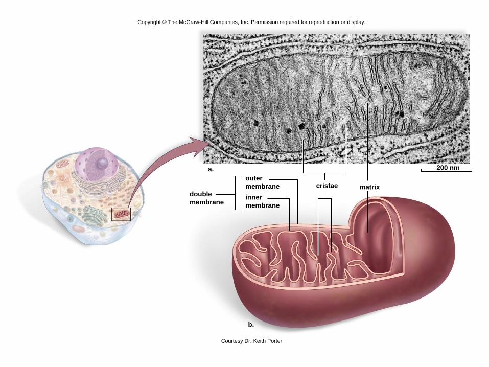
Copyright © The McGraw-Hill Companies, Inc. Permission required for reproduction or display.
cristae matrix
a.
b.
200 nm
double
membrane
outer
membrane
inner
membrane
Courtesy Dr. Keith Porter
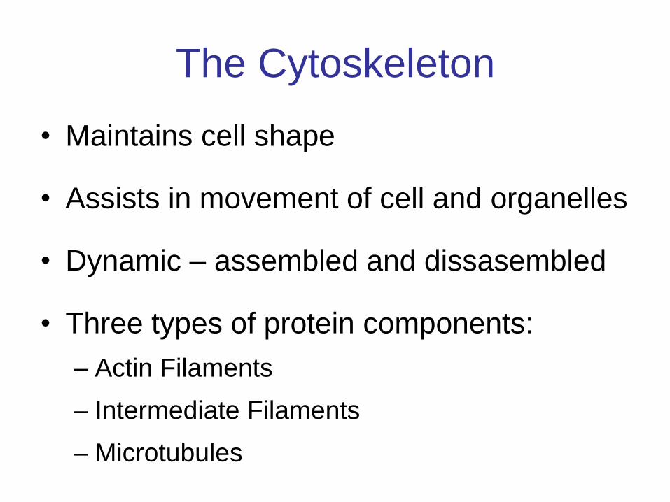
The Cytoskeleton
• Maintains cell shape
• Assists in movement of cell and organelles
• Dynamic – assembled and dissasembled
• Three types of protein components:
– Actin Filaments
– Intermediate Filaments
– Microtubules
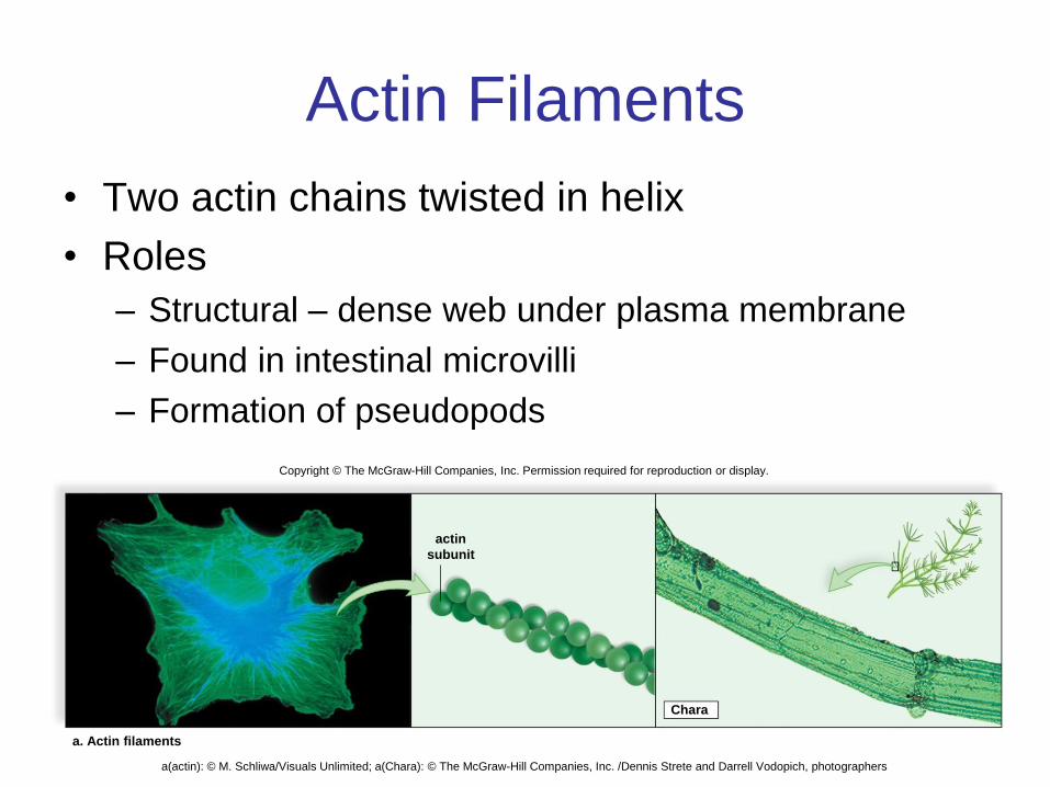
Actin Filaments
• Two actin chains twisted in helix
• Roles
– Structural – dense web under plasma membrane
– Found in intestinal microvilli
– Formation of pseudopods
Copyright © The McGraw-Hill Companies, Inc. Permission required for reproduction or display.
a. Actin filaments
actin
subunit
Chara
a(actin): © M. Schliwa/Visuals Unlimited; a(Chara): © The McGraw-Hill Companies, Inc. /Dennis Strete and Darrell Vodopich, photographers
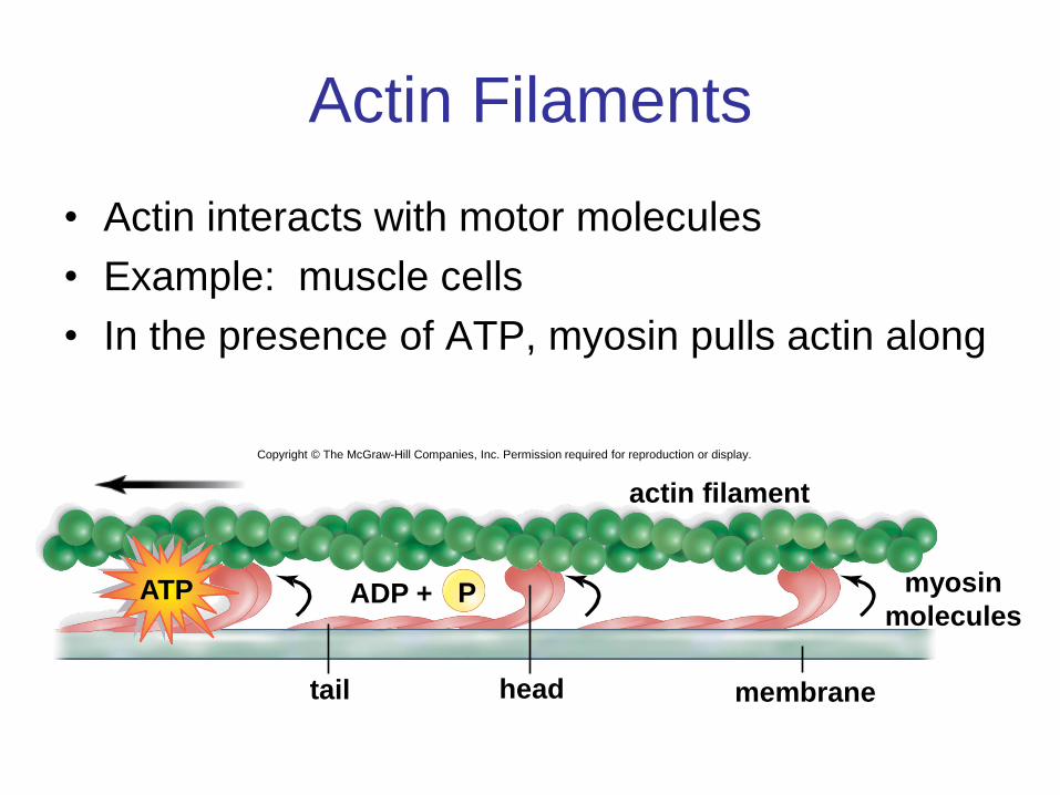
Actin Filaments
• Actin interacts with motor molecules
• Example: muscle cells
• In the presence of ATP, myosin pulls actin along
Copyright © The McGraw-Hill Companies, Inc. Permission required for reproduction or display.
tail head
P ADP +
actin filament
membrane
myosin
molecules ATP
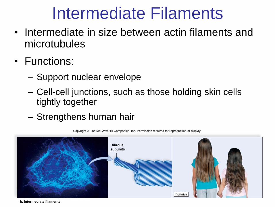
Intermediate Filaments • Intermediate in size between actin filaments and
microtubules
• Functions:
– Support nuclear envelope
– Cell-cell junctions, such as those holding skin cells tightly together
– Strengthens human hair
human
fibrous
subunits
b. Intermediate filaments
Copyright © The McGraw-Hill Companies, Inc. Permission required for reproduction or display.
b(intermediate): © K.G. Murti/Visuals Unlimited; b(humans): © Amos Morgan/Getty RF
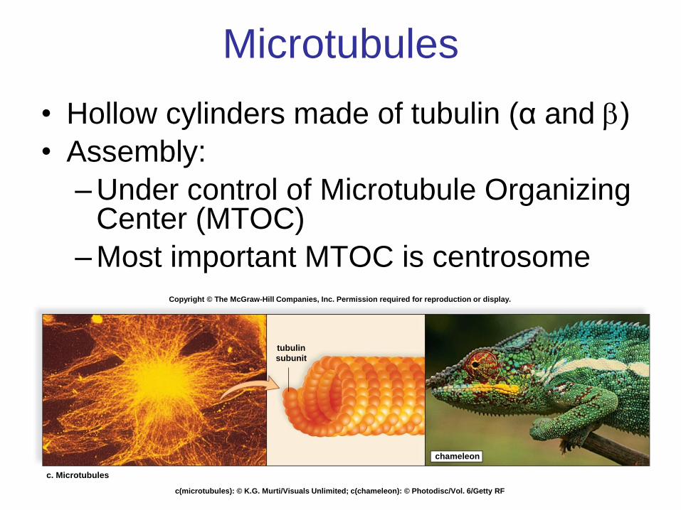
Microtubules
• Hollow cylinders made of tubulin (α and )
• Assembly:
– Under control of Microtubule Organizing Center (MTOC)
– Most important MTOC is centrosome
c. Microtubules
tubulin
subunit
chameleon
Copyright © The McGraw-Hill Companies, Inc. Permission required for reproduction or display.
c(microtubules): © K.G. Murti/Visuals Unlimited; c(chameleon): © Photodisc/Vol. 6/Getty RF
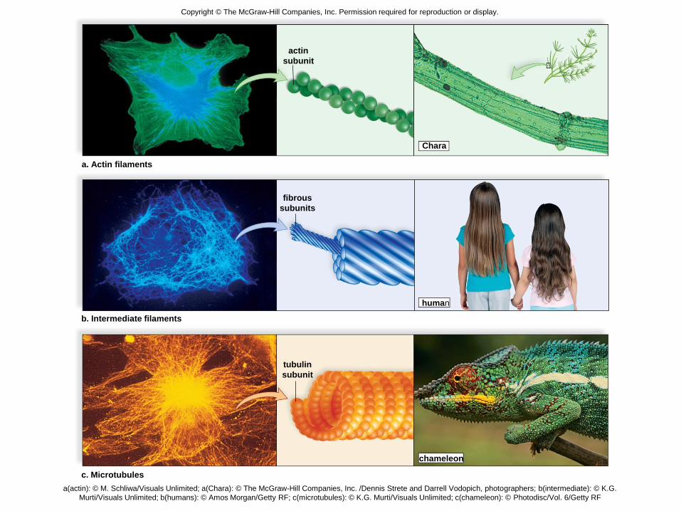
a. Actin filaments
human
actin
subunit
Chara
Copyright © The McGraw-Hill Companies, Inc. Permission required for reproduction or display.
fibrous
subunits
b. Intermediate filaments
c. Microtubules
tubulin
subunit
chameleon
a(actin): © M. Schliwa/Visuals Unlimited; a(Chara): © The McGraw-Hill Companies, Inc. /Dennis Strete and Darrell Vodopich, photographers; b(intermediate): © K.G.
Murti/Visuals Unlimited; b(humans): © Amos Morgan/Getty RF; c(microtubules): © K.G. Murti/Visuals Unlimited; c(chameleon): © Photodisc/Vol. 6/Getty RF
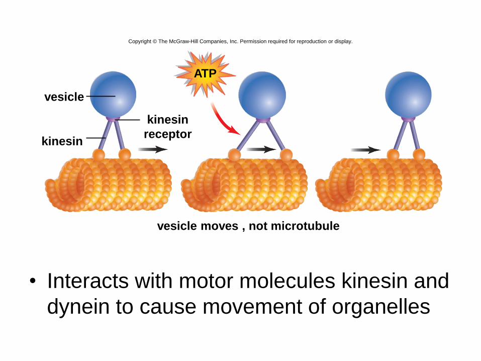
• Interacts with motor molecules kinesin and
dynein to cause movement of organelles
Copyright © The McGraw-Hill Companies, Inc. Permission required for reproduction or display.
vesicle moves , not microtubule
ATP
kinesin
vesicle
kinesin
receptor
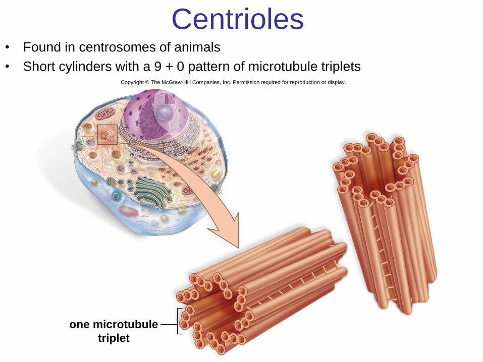
Centrioles • Found in centrosomes of animals
• Short cylinders with a 9 + 0 pattern of microtubule triplets
one microtubule
triplet
Copyright © The McGraw-Hill Companies, Inc. Permission required for reproduction or display.
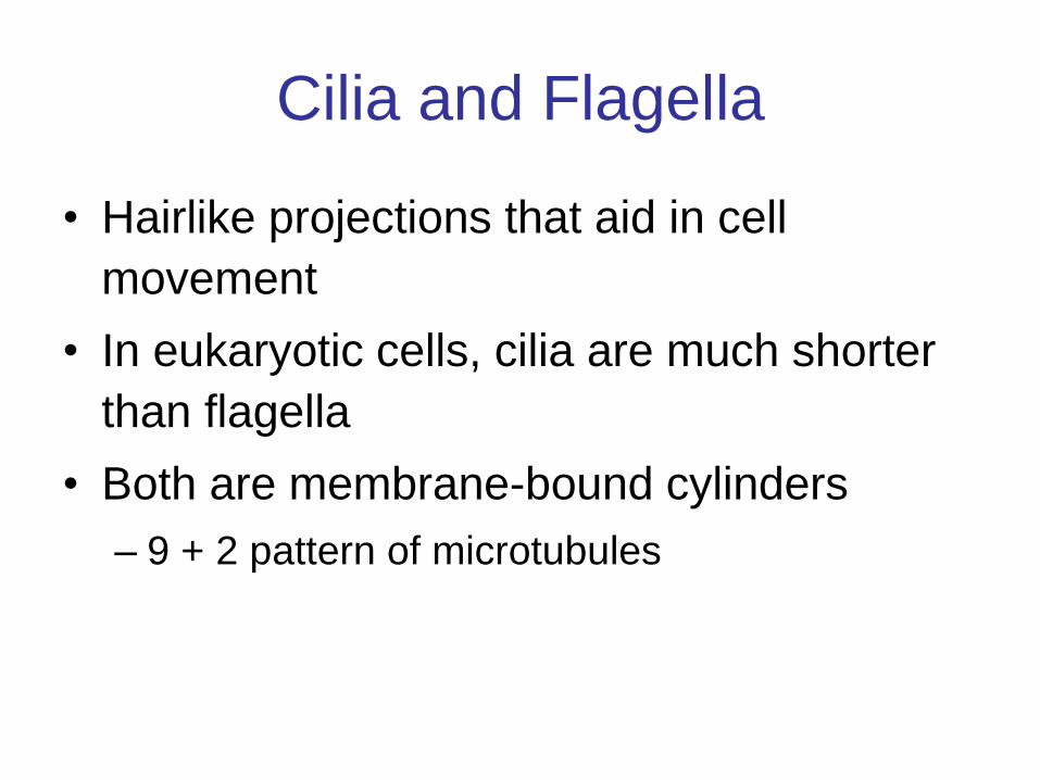
Cilia and Flagella
• Hairlike projections that aid in cell
movement
• In eukaryotic cells, cilia are much shorter
than flagella
• Both are membrane-bound cylinders
– 9 + 2 pattern of microtubules
Copyright © The McGraw-Hill Companies, Inc. Permission required for reproduction or display.
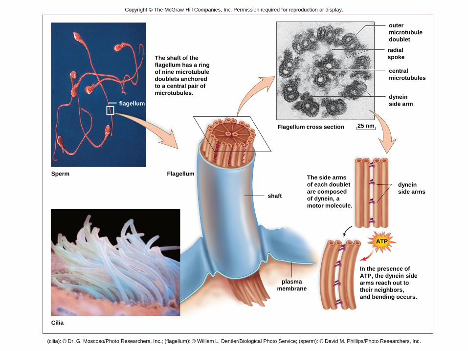
Flagellum
shaft
Sperm
Cilia
flagellum
Flagellum cross section 25 nm
The shaft of the
flagellum has a ring
of nine microtubule
doublets anchored
to a central pair of
microtubules.
In the presence of
ATP, the dynein side
arms reach out to
their neighbors,
and bending occurs.
ATP
dynein
side arms
The side arms
of each doublet
are composed
of dynein, a
motor molecule.
dynein
side arm
central
microtubules
radial
spoke
outer
microtubule
doublet
plasma
membrane
(cilia): © Dr. G. Moscoso/Photo Researchers, Inc.; (flagellum): © William L. Dentler/Biological Photo Service; (sperm): © David M. Phillips/Photo Researchers, Inc.
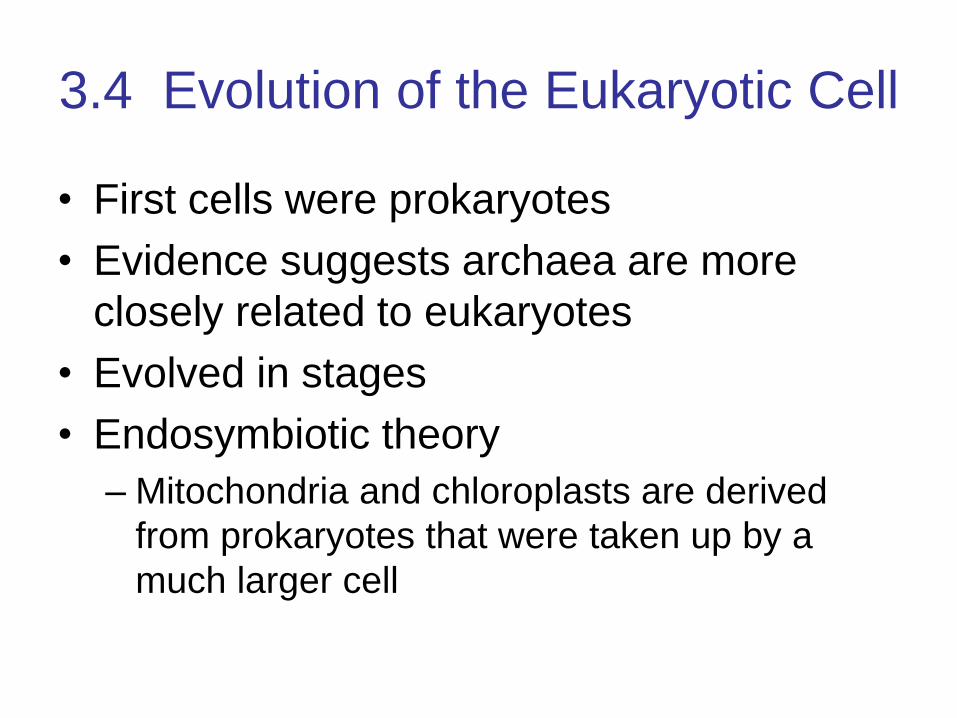
3.4 Evolution of the Eukaryotic Cell
• First cells were prokaryotes
• Evidence suggests archaea are more
closely related to eukaryotes
• Evolved in stages
• Endosymbiotic theory
– Mitochondria and chloroplasts are derived
from prokaryotes that were taken up by a
much larger cell

Copyright © The McGraw-Hill Companies, Inc. Permission required for reproduction or display.
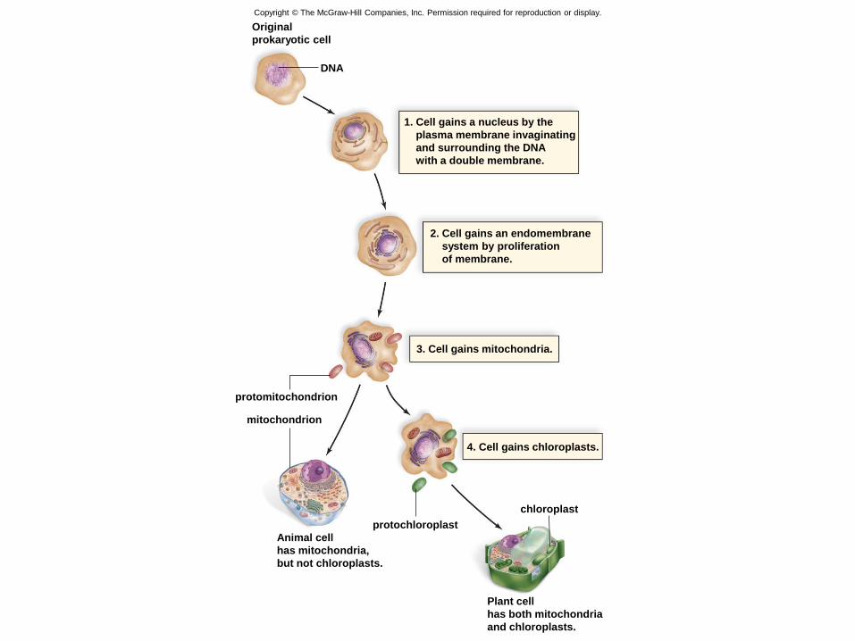
DNA
Original
prokaryotic cell
1. Cell gains a nucleus by the
plasma membrane invaginating
and surrounding the DNA
with a double membrane.
2. Cell gains an endomembrane
system by proliferation
of membrane.
Copyright © The McGraw-Hill Companies, Inc. Permission required for reproduction or display.
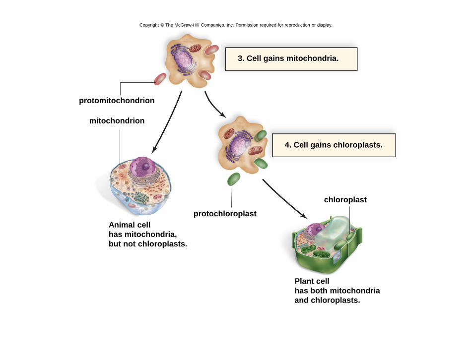
3. Cell gains mitochondria.
protomitochondrion
mitochondrion
Plant cell
has both mitochondria
and chloroplasts.
Animal cell
has mitochondria,
but not chloroplasts.
protochloroplast
chloroplast
4. Cell gains chloroplasts.
Copyright © The McGraw-Hill Companies, Inc. Permission required for reproduction or display.
DNA
Original
prokaryotic cell
1. Cell gains a nucleus by the
plasma membrane invaginating
and surrounding the DNA
with a double membrane.
2. Cell gains an endomembrane
system by proliferation
of membrane.
3. Cell gains mitochondria.
protomitochondrion
mitochondrion
Plant cell
has both mitochondria
and chloroplasts.
Animal cell
has mitochondria,
but not chloroplasts.
protochloroplast
chloroplast
4. Cell gains chloroplasts.
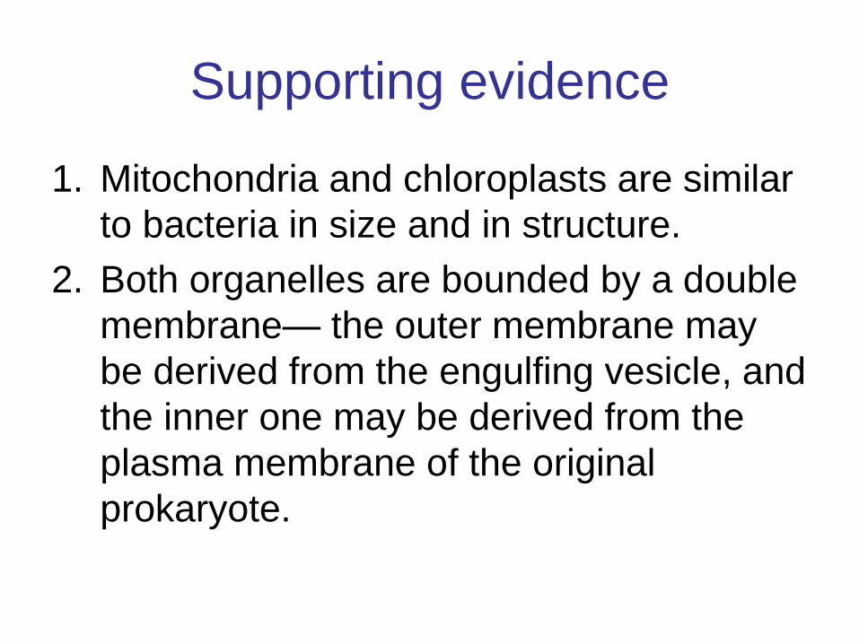
Supporting evidence
1. Mitochondria and chloroplasts are similar
to bacteria in size and in structure.
2. Both organelles are bounded by a double
membrane— the outer membrane may
be derived from the engulfing vesicle, and
the inner one may be derived from the
plasma membrane of the original
prokaryote.
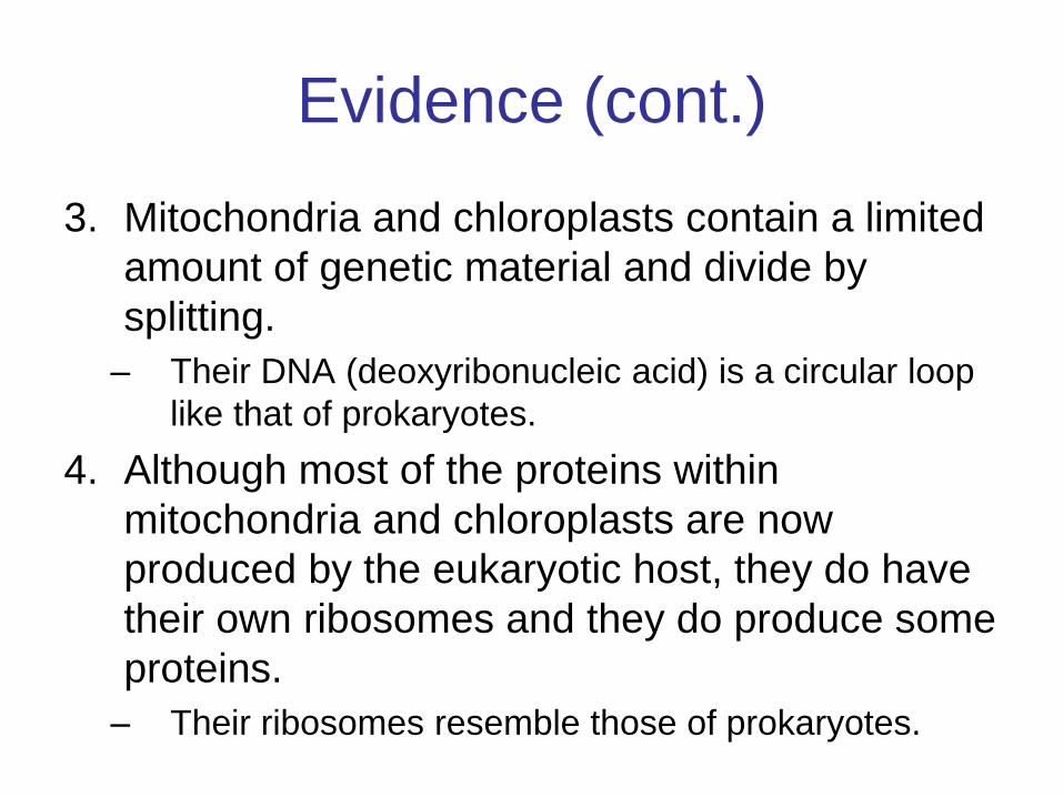
Evidence (cont.)
3. Mitochondria and chloroplasts contain a limited
amount of genetic material and divide by
splitting.
– Their DNA (deoxyribonucleic acid) is a circular loop
like that of prokaryotes.
4. Although most of the proteins within
mitochondria and chloroplasts are now
produced by the eukaryotic host, they do have
their own ribosomes and they do produce some
proteins.
– Their ribosomes resemble those of prokaryotes.
Evidence (cont.)
5. The RNA (ribonucleic acid) base
sequence of the ribosomes in
chloroplasts and mitochondria also
suggests a prokaryotic origin of these
organelles.


![[PPT]PowerPoint Presentation - Department of Molecular & …mcb.berkeley.edu/courses/mcb130L/Originals/Lecture_3.ppt · Web viewTitle PowerPoint Presentation Author Laurent Coscoy](https://img.pdfslide.us/doc/110x75/5ada75737f8b9afc0f8c8abb/pptpowerpoint-presentation-department-of-molecular-mcb-viewtitle-powerpoint.jpg)