Embed Size (px)
Citation preview

Wedel, M. J., & Taylor, M. P. (2013). Caudal pneumaticity and pneumatichiatuses in the sauropod dinosaurs Giraffatitan and Apatosaurus. PLoS ONE,8(10), [e78213]. DOI: 10.1371/journal.pone.0078213
Publisher's PDF, also known as Version of record
Link to published version (if available):10.1371/journal.pone.0078213
Link to publication record in Explore Bristol ResearchPDF-document
CC By
University of Bristol - Explore Bristol ResearchGeneral rights
This document is made available in accordance with publisher policies. Please cite only the publishedversion using the reference above. Full terms of use are available:http://www.bristol.ac.uk/pure/about/ebr-terms

Caudal Pneumaticity and Pneumatic Hiatuses in theSauropod Dinosaurs Giraffatitan and ApatosaurusMathew J. Wedel1*, Michael P. Taylor2*
1 College of Osteopathic Medicine of the Pacific and College of Podiatric Medicine, Western University of Health Sciences, Pomona, California, United States of America,
2 Department of Earth Sciences, University of Bristol, United Kingdom
Abstract
Skeletal pneumaticity is found in the presacral vertebrae of most sauropod dinosaurs, but pneumaticity is much lesscommon in the vertebrae of the tail. We describe previously unrecognized pneumatic fossae in the mid-caudal vertebrae ofspecimens of Giraffatitan and Apatosaurus. In both taxa, the most distal pneumatic vertebrae are separated from otherpneumatic vertebrae by sequences of three to seven apneumatic vertebrae. Caudal pneumaticity is not prominent in mostindividuals of either of these taxa, and its unpredictable development means that it may be more widespread thanpreviously recognised within Sauropoda and elsewhere in Saurischia. The erratic patterns of caudal pneumatization inGiraffatitan and Apatosaurus, including the pneumatic hiatuses, show that pneumatic diverticula were more broadlydistributed in the bodies of the living animals than are their traces in the skeleton. Together with recently publishedevidence of cryptic diverticula—those that leave few or no skeletal traces—in basal sauropodomorphs and in pterosaurs,this is further evidence that pneumatic diverticula were widespread in ornithodirans, both across phylogeny andthroughout anatomy.
Citation: Wedel MJ, Taylor MP (2013) Caudal Pneumaticity and Pneumatic Hiatuses in the Sauropod Dinosaurs Giraffatitan and Apatosaurus. PLoS ONE 8(10):e78213. doi:10.1371/journal.pone.0078213
Editor: Peter Dodson, University of Pennsylvania, United States of America
Received April 16, 2013; Accepted September 12, 2013; Published October 30, 2013
Copyright: � 2013 Wedel and Taylor. This is an open-access article distributed under the terms of the Creative Commons Attribution License, which permitsunrestricted use, distribution, and reproduction in any medium, provided the original author and source are credited.
Funding: Research for this study was conducted on a field trip sponsored by DFG Research Unit 533: Biology of the Sauropod Dinosaurs; DFG 533 also supportedour travel to Germany. The authors thank Martin Sander (University of Bonn) and the organisers and participants of the field trip. The Field Museum of NaturalHistory supported our travel to Chicago. Research at the Carnegie Museum was supported by a grant from the Jurassic Foundation. The funders had no role instudy design, data collection and analysis, decision to publish, or preparation of the manuscript.
Competing Interests: The authors have declared that no competing interests exist.
* E-mail: [email protected] (MJW); [email protected] (MPT)
Introduction
Postcranial skeletal pneumaticity (PSP) is the modification of the
postcranial skeleton by pneumatic diverticula of the respiratory
system. It is widespread in saurischian dinosaurs including birds,
other theropods, and sauropods, and it is also present in
pterosaurs. PSP in archosaurs is of interest as a morphogenetic
system and source of phylogenetic information [1–3], for its effect
in lightening the skeleton [4–8], as the skeletal footprint of the
lungs and air sacs [9–17], and as the osteological correlate of a
system of pneumatic diverticula, which developed from the lungs
and air sacs and may have had important non-respiratory
functions [18,19]. The extent of PSP varied greatly among
sauropod taxa, among individuals and among regions of the
skeleton. Cervical vertebrae are pneumatic in basal eusauropods;
cervical, dorsal and sacral vertebrae are pneumatic in mamench-
isaurids and most neosauropods; and all of these plus caudal
vertebrae are extensively pneumatic in diplodocines and in some
titanosaurians [1,4,12,20]. Cervical and dorsal ribs are pneumatic
in many, maybe most, titanosauriforms (e.g., [21]: p. 239; [22]: p.
52) and some diplodocids (e.g., [23]: figs. 9–10; 24: p. 212; [25]: p.
534). Pectoral girdle elements are pneumatic in some derived
titanosaurs [20], and pneumatization of pelvic girdle elements
apparently evolved independently in rebbachisaurid diplodocoids
[26–27] and somphospondylan macronarians ([20], [28]: p. 233).
Most of the elements listed above are also pneumatized in at least
some pterosaurs [7], non-avian theropods [13,15], and birds
[6,13,14,29], although caudal pneumaticity has not yet been
demonstrated in pterosaurs, and ischial pneumaticity is not yet
known in non-avian theropods [27]. The acquisition of PSP in
parallel in so many ornithodiran lineages suggests that a
diverticular lung and air sac system may be primitive for
Ornithodira as a whole [12,15–17].
To date, caudal pneumaticity has received less attention than
pneumaticity in other parts of the skeleton (but see [30]), but it is
of particular interest because of its possible independent origins
and parallel evolution in diplodocoids and macronarians. Here
we describe complex patterns of caudal pneumaticity in
Giraffatitan brancai (formerly assigned to the genus Brachiosaurus;
see [31]) and Apatosaurus, and discuss the functional and
phylogenetic implications.
Institutional AbbreviationsAMNH, American Museum of Natural History, New York
City, New York, USA; CM, Carnegie Museum of Natural
History, Pittsburgh, Pennsylvania, USA; DMNH, Denver Muse-
um of Natural History, Denver, Colorado, USA; FMNH, Field
Museum of Natural History, Chicago, Illinois, USA; HMN,
Humbolt Museum fur Naturkunde, Berlin, Germany; KLR,
Henan Geological Museum, Zhengzhou, China; LACM, Natural
History Museum of Los Angeles County, Los Angeles, California,
USA; MAL, Malawi Department of Antiquities Collection,
Lilongwe and Nguludi, Malawi; MB.R., Museum fur Naturkunde
Berlin, Berlin, Germany; MCS, Museo de Cinco Saltos, Rıo
PLOS ONE | www.plosone.org 1 October 2013 | Volume 8 | Issue 10 | e78213

Negro Province, Argentina; MCT, Collection of the Earth Science
Museum of the National Department of Mineral Production, Rıo
de Janeiro; MIWG, Museum of Isle of Wight Geology, Sandown,
Isle of Wight, United Kingdom; ML, Museu da Lourinha,
Portugal; MN, Museu Nacional, Rio de Janeiro, Brazil; MPCA-Pv, Coleccion de Paleovertebrados de la Museum Provincial de
Cipolletti ‘‘Carlos Ameghino’’, Cipolletti, Rıo Negro Province,
Argentina; MPS, Museo de Dinosaurios e Paleontologıa, Salas de
los Infantes, Burgos, Spain; MUCPv, Museo de Geologıa y
Paleontologıa de la Universidad Nacional del Comahue, Neu-
quen, Argentina; NHM, Natural History Museum, London,
United Kingdom; NMST, National Science Museum, Tokyo,
Japan; OMNH, Oklahoma Museum of Natural History, Norman,
Oklahoma, USA; ONM, Office National Des Mines, Service
Patrimoine Geologique, Tunis, Tunisia; PVL, Coleccion de
Paleontologıa de Vertebrados de la Fundacion Instituto Miguel
Lillo, Tucuman, Argentina; UNPSJB, Universidad Nacional de la
Patagonia San Juan Bosco, Comodoro Rivadavia, Argentina;
USNM, National Museum of Natural History, Smithsonian
Institution, Washington, D.C., USA; WDC, Wyoming Dinosaur
Center, Thermopolis, Wyoming, USA; YPM, Yale Peabody
Museum, New Haven, Connecticut, USA.
Results and Discussion
Overview of pneumatic featuresThe interaction of pneumatic epithelium and bone tissue
produces a spectrum of osteological features, including pneumatic
tracks, fossae, foramina, and internal chambers of various shapes
and sizes [1,4,9,10,14,32](Figure 1). Not all of these features are
diagnostic for pneumaticity in isolation. Pneumatic fossae are
particularly problematic: fossae on the surface of vertebrae can be
associated with numerous soft tissues, including cartilage, adipose
tissue, muscles, and pneumatic diverticula [14]. Although
distinctly emarginated and sharply lipped fossae are usually
inferred to represent pneumatic invasion [9], apneumatic fossae
sometimes have distinct margins and pneumatic fossae sometimes
do not [16,17,32]. It is worth noting that vertebral fossae are
present in numerous basal and pseudosuchian archosarus
[16,17,33] and in some synapsids (see discussion in [15]: p. 172),
and although it is possible that some of these were pneumatic, it is
unlikely that all of them were.
In equivocal cases, the diagnosis of a fossa as pneumatic may be
strengthened by the presence of other pneumatic features on the
same bone [4]. Unequivocally pneumatic fossae (e.g. those
containing pneumatic foramina) often have multiple subfossae
[17,34], which may represent the resorption of adjacent cortical
bone by a complex diverticulum that consists of multiple tubes or
sacs, such as the complex diverticula of some birds ([11]: fig. 2).
Apneumatic fossae usually have no margins or only weakly
developed margins; the only strongly emarginated apneumatic
fossae are muscle attachments that are easily identified by their
location and texture, such as the temporal fossae of the human
skull and the muscle attachment fossae on the ilia of birds. PSP in
saurischians is typically variable: the presence and form of
pneumatic features varies among individuals, serially along the
vertebral column, and even on the left and right sides of a single
vertebra (e.g., [35]: p. 1552).
Although fossae are less diagnostic for PSP than more invasive
foramina and internal chambers, the differences between pneu-
matic and apneumatic fossae listed above can be used to develop a
profile for distinguishing the two ([9,17]; see also [14]: fig. 12). In
descending order of usefulness, pneumatic fossae are expected to
(1) occur together with other correlates of PSP, (2) have a scalloped
texture or subfossae, (3) occur on bone surfaces not occupied by
muscle attachments, or in the same locations as pneumatic
foramina in related taxa, and (4) vary in expression among
individuals, serially along the axial skeleton, and from left to right
in single vertebra. There is no reason to assume that putatively
pneumatic fossae were originally occupied by some other soft
tissue (e.g., muscle, cartilage, or adipose tissue) which was then
replaced by pneumatic diverticula that produced more diagnostic
bony traces [17], especially given the mounting evidence that a
diverticular lung was present in the ancestral saurischian and
possibly in the ancestral ornithodiran [12,15–17]. Nevertheless, it
is often difficult to tell which fossae may have been pneumatic,
especially in basal taxa or those in which the presence of PSP is
unexpected or not well established [16].
Caudal pneumaticity in OrnithodiraThe phylogenetic distribution of caudal pneumaticity in
sauropods and in ornithodirans more generally is complex
(Figure 2). To date, there are no reports of caudal pneumaticity
in pterosaurs. There are several possible explanations for this.
Although the presence of PSP in pterosaurs has been widely
acknowledged since the mid-1800s (e.g., [36]), and although it has
received more attention in recent years (e.g., [7,37]), there has still
been less work on pneumaticity in pterosaurs than in sauropods or
theropods. So possibly caudal pneumaticity is present in pterosaurs
but hasn’t been recognized yet. Caudal vertebrae in pterosaurs are
Figure 1. Caudal pneumaticity varies among sauropods. In thediplodocid Tornieria, the first 15–20 caudal vertebrae have neural archlaminae and fossae, and lateral pneumatic foramina opening into largeinternal chambers. Images traced from Remes ([51]: fig. 31 [lateral view])and Janensch ([72]: fig. 7 [cross-section]); the two views are fromdifferent vertebrae. In the basal titanosaurian Malawisaurus, caudalpneumaticity is restricted to a handful of proximal caudal vertebrae, inwhich the neural arches are honeycombed with pneumatic chambersbut the vertebral centra are solid. Images traced from Wedel ([12]: fig.2A [lateral view] and 2C [cross-section]). In the derived titanosaurianSaltasaurus, the first 20–25 caudal vertebrae have large external fossaebut small external foramina, and both the neural arches and centra arehoneycombed with chambers. Images traced from Powell ([59]: plate 53[lateral view]) and Cerda et al [20]: fig. 4F [cross-section]); the two viewsare from different vertebrae.doi:10.1371/journal.pone.0078213.g001
Caudal Pneumaticity in Sauropod Dinosaurs
PLOS ONE | www.plosone.org 2 October 2013 | Volume 8 | Issue 10 | e78213

small and at small scale it can be difficult to distinguish pneumatic
and vascular foramina, and to tell pneumatic chambers from
marrow-filled trabecular bone ([16]: p. 18). It does not help that
the pterosaurs with long tails were mostly small-bodied, whereas
the large-bodied pterodactyloids had tiny tails. The absolutely
small tails of pterosaurs may have created little demand or
opportunity for pneumatization, and if any pneumatic traces are
present in pterosaur tails they would be difficult to diagnose.
Caudal pneumaticity is uncommon in non-avian theropods.
The most comprehensive survey to date is that of Benson et al [15],
who found caudal pneumaticity in only 12 of the 159 taxa they
surveyed. Note, however, that 67 taxa could not be scored, so
caudal pneumaticity could be positively ruled out in only half of
the sampled taxa (80 out of 159). Only the proximal caudals, if
any, are pneumatic in megalosaurids (Torvosaurus) and therizino-
sauroids (Nothronychus, Neimongosaurus); proximal and middle
caudals are pneumatic in some allosauroids (Aerosteon, Megaraptor,
Carcharodontosaurus); and proximal, middle, and distal caudals are
pneumatic in some—but not all—oviraptorosaurs (Chirostenotes,
Citipati, Khaan; see fig. 4, table 4, and appendix S1 in [15]). In
contrast, caudal pneumaticity is fairly common in extant birds, at
least in medium-to-large-bodied taxa: O’Connor ([6]: table 2)
found caudal pneumaticity in at least some members of 6 out of 10
higher-level clades (mostly corresponding to traditional Linnean
orders). In addition to the volant taxa surveyed by O’Connor [6],
the large ratites (ostriches, emus, cassowaries, and rheas) all have
pneumatic caudals (pers. obs., Figure 3).
In general, caudal pneumaticity is common in neosauropods
and rare or absent in non-neosauropod sauropodomorphs
(Table 1). A proximal caudal of ‘Bothriospondylus madagascarensis’,
NHM 2599, has fossae on the lateral sides of the centrum, but
lacks large pneumatic foramina or internal pneumatic chambers
[38]. The phylogenetic position of the ‘B. madagascarensis’ material
is uncertain and it may not all pertain to the same taxon [38].
Mannion [38] suggested that it might best be regarded as a non-
neosauropod eusauropod, at least until more complete and
diagnostic material comes to light. If NHM 2599 does belong to
a eusauropod, it is probably the best documented case of caudal
pneumaticity in a non-neosauropod sauropodomorph. Caudal
pneumaticity has not been reported in the Mamenchisauridae, a
clade which otherwise shows some derived pneumatic features,
including complex pneumatic chambers in the cervical vertebrae
[39].
The first caudal vertebra of Haplocanthosaurus CM 879, has
pneumatic fossae on both the centrum and the neural arch ([40]:
plate 2; [12]: figs. 7 and 9). The phylogenetic position of
Haplocanthosaurus is uncertain; it has been recovered as a basal
diplodocoid [41], a basal macronarian [22,42], and a non-
neosauropod close to the origin of Neosauropoda [43] in different
analyses, although recent analyses tend to support a position
within Diplodocoidea [25,44]. Here we regard it as a neosauropod
of uncertain affinities (Figure 2); moving it into either
Diplodocoidea or Macronaria would have no great effect on the
phylogenetic distribution of caudal pneumaticity in sauropods. In
more derived diplodocoids, caudal pneumaticity is present in
rebbachisaurids and diplodocids but apparently absent in
dicraeosaurids (see [45]). In rebbachisaurids the neural arches
and transverse processes of the proximal caudals often have
pronounced laminae and deep, irregular fossae characteristic of
pneumaticity ([46]: figs. 1-3; [47]), and pneumatic foramina
leading to large internal chambers are present in at least the
proximal caudals of the rebbachisaurid Tataouinea (the middle and
distal caudals are as yet unknown) [27]. The same is true in
diplodocids, and in diplodocines such as Diplodocus, Barosaurus, and
Tornieria, these pneumatic foramina persist down to caudal 15 or
20 (48: fig. 13; [49]: p. 35 and plate 9; [50]: p. 54 and fig. 2.6; [51]:
fig. 3). Although some authors have reported pneumatic features in
the most proximal caudal vertebrae of Apatosaurus (e.g., [52,53]),
pneumatic features have not previously been observed further
back than the fifth caudal vertebra; below we report isolated
pneumatic fossae more distally in the tail.
Pneumaticity is absent in the caudal vertebrae of Camarasaurus
(see [54]: plates 74–77) but caudal pneumaticity is otherwise
prevalent in Macronaria. Pneumatic fossae have been reported in
the caudals of the brachiosaurids Cedarosaurus [55] and Venenosaurus
[56], and Janensch [57] briefly mentioned fossae in proximal
caudal vertebrae in three specimens of Giraffatitan (discussed in
more detail below). Below, we describe additional pneumatic
fossae distributed unevenly through the tail in another specimen of
Giraffatitan. Caudal pneumaticity is also widespread in Titano-
sauria ([30]; Table 1), with Opisthocoelicaudia being one of the few
titanosaurs that appears to lack caudal pneumaticity (see [58]:
plates 4–5). Caudal pneumaticity reached its apex among
sauropods in the saltasaurines Rocasaurus, Neuquensaurus, and
Saltasaurus, as did appendicular pneumaticity [20]. Known salt-
asaurines are uniformly small, with femur lengths well under one
meter [59–61]—compare to femur lengths of 1–1.2 meters in
dicraeosaurids and 1.5–2.0 meters in most other neosauropods
([62]: table 1). It is not yet clear why PSP, which is suspected to
have been a key innovation in facilitating the evolution of large
body size in sauropods [63], achieved its maximum expression in
these small-bodied taxa.
Caudal pneumaticity in GiraffatitanCaudal vertebrae of Giraffatitan personally examined by us in
this study are listed in Table 2, and described below.
MB.R.5000 (‘Fund no’, Figures 4 and 5). The mounted
skeleton of Giraffatitan brancai at the Humboldt Museum fur
Naturkunde Berlin consists primarily of elements of the para-
lectotype, MB.R.2181 (formerly cataloged as HMN SII), but
missing parts of the skeleton were provided from the remains of
other similarly sized individuals [64]. The tail of the mounted
skeleton, MB.R.5000 (formerly HMN ‘Fund no’), consists of the
second to fifty-first caudal vertebrae, ‘‘not articulated, with the
exception of a few at the end, but altogether relatively in
sequence’’ ([57]: p. 64, plate IV; Figure 6). The first caudal
vertebra was not recovered, and it is modeled in plaster in the
mounted skeleton. The preserved caudals are discussed in groups
of serially adjacent vertebrae based on pneumatic characters.
MB.R.5000 (‘Fund no’): Caudal vertebrae 2–7. All of
these vertebrae have fossae on the right side of the centrum, and
all but Ca4 and Ca7 also on the left. The fossae of these vertebrae
are all located ventral to the transverse processes on the
dorsolateral faces of the centra. Some of the fossae are
multipartite; that is, divided into subfossae by bony septa. Fossae
are absent from the neural arches and spines. Caudals 4 and 7
have fossae only on the right side of the centrum: similar
asymmetry in the expression of pneumatic fossae is present in the
sacrum of the CM 879 specimen of Haplocanthosaurus [12].
MB.R.5000 (‘Fund no’): Caudal vertebrae 8–10. Although
these vertebrae present a series of intermediate forms relative to
the vertebrae anterior and posterior to them, and all are deeply
waisted, they have no apparent pneumatic features on their centra,
neural arches, or neural spines. As there are obvious traces of
pneumaticity in caudal vertebrae 11–15 (see below), pneumatic
diverticula must have passed by these vertebrae and may even
have been in contact with the bone, but they left no macroscopic
traces. It is possible that correlates of PSP might be found in the
Caudal Pneumaticity in Sauropod Dinosaurs
PLOS ONE | www.plosone.org 3 October 2013 | Volume 8 | Issue 10 | e78213

bone microtexture or histology of these vertebrae, but such
correlates have not been identified to date in any vertebrae so
resolution of this question must wait. This block of three vertebrae
is bounded anteriorly and posteriorly by pneumatic vertebrae and
thus constitutes a pneumatic hiatus [11,12]; the implications of this
hiatus are explored below.
Figure 2. The phylogenetic distribution of caudal pneumaticity in sauropods and other dinosaurs is complex. Boxes representproximal, middle, and distal caudal vertebrae, arbitrarily defined for sauropods as caudals 1–10, 11–20, and 21 on, respectively; blue boxes indicatethat pneumaticity is present in that part of the tail. Pneumaticity data for theropods come from Benson et al [15]—note that although Theropoda iscollapsed to a single node in this figure, caudal pneumaticity is not primitive for the clade, but evolved independently several times in both non-aviantheropods and birds [6,15,29]. Data from sauropods come from the sources listed in Table 1. The figure also shows the phylogenetic framework weuse in this paper. The phylogenetic framework is drawn from Whitlock [44] for diplodocoids, Mannion et al [30] for basal macronarians andXianshanosaurus, Calvo et al [96] for most titanosaurs, and Campos et al [93] for Trigonosaurus. Basal sauropodomorphs are a grade, not a clade, butthey are listed together here for convenience since they all lack caudal pneumaticity.doi:10.1371/journal.pone.0078213.g002
Caudal Pneumaticity in Sauropod Dinosaurs
PLOS ONE | www.plosone.org 4 October 2013 | Volume 8 | Issue 10 | e78213

MB.R.5000 (‘Fund no’): Caudal vertebrae 11–15. All of
these vertebrae have pneumatic fossae, and the distribution and
morphology of these fossae is considerably more complex than in
caudals 2–7. The most obvious difference between these ranges is
that those in the posterior range have pneumatic fossae on both
the centrum and neural arch, whereas more anteriorly fossae are
present only on the centrum. Caudal vertebra 11 has fossae on
both sides of the neural arch, and these fossae are weakly
subdivided by bony septa. No fossae are apparent on either side of
the centrum. Caudal vertebra 12 has the most complex pneumatic
features of any vertebra in the entire tail, with multipartite fossae
on both sides of the centrum and both sides of the neural arch.
Caudal vertebra 13 has a very large fossa on the right side of the
centrum, which in its size and form approximates the large
pneumatic fossae or ‘‘pleurocoels’’ in the dorsal vertebrae of more
basal taxa like Haplocanthosaurus. A small subdivided fossa is also
present on the right side of the neural spine. Pneumatic features
are absent from both the centrum and neural arch on the left side.
Caudal 13 is therefore similar to caudals 4 and 7 in having
pneumatic features present only on the right side. Caudal 14 has
large pneumatic fossae on both sides of the centrum, and a smaller
multipartite fossa on the right side of the neural arch. Caudal 15
has a pair of pneumatic fossae on the left side of the centrum, but
no fossae on the neural arch or anywhere on the right side of the
vertebra. This is the first vertebra in the series in which PSP is
present only on the left side; all of the previous vertebrae that are
unilaterally apneumatic (caudals 4, 7 and 13) have their fossae on
the right side.
MB.R.5000 (‘Fund no’): Caudal vertebrae 16–18. These
three vertebrae, like caudals 8–10, are deeply waisted but lack
distinct fossae. They constitute a second bilateral pneumatic
hiatus.
MB.R.5000 (‘Fund no’): Caudal vertebrae 19–24. These
six vertebrae again present a complex suite of pneumatic features.
Caudals 19, 21, and 23 have pneumatic fossae only on the left side,
like caudal 15, whereas caudals 20, 22, and 24 have pneumatic
fossae on both sides of the centrum. Caudal 22 has a multipartite
fossa on the right side, on the border between the centrum and
neural arch; fossae are otherwise absent from the neural arches
and spines of all six vertebrae. In contrast, pneumatic fossae on the
centra of these six vertebrae are better defined than in almost all of
the preceding vertebrae, with the fossae of caudals 20, 22, and 24
being particularly large, deep, and well subdivided.
MB.R.5000 (‘Fund no’): Caudal vertebrae 25–51. No
obvious pneumatic features are present on any of these vertebrae.
The vertebrae that make up the last 26 cm of the tail (i.e. from
caudal 52 on) were not recovered and are reconstructed in plaster
in the mounted skeleton ([64]: p. 98). We assume that the missing
vertebrae were also apneumatic, based on the absence of
pneumaticity in the preceding 27 vertebrae and in the distal tails
of all other known non-avian saurischians.
MB.R.2921 (‘Fund Aa’, Figure 7). MB.R.2921 (‘Fund Aa’)
consists of the first 18 caudal vertebrae and their chevrons, found
in an articulated sequence behind the last sacral vertebra ([57]: p.
60). Regarding possible pneumatic features, Janensch ([57]: p. 61)
wrote, ‘‘Pleurocentral excavations are absent; only under the root
of the transverse process of the second is an elongated, about four
centimeter long depression clearly developed, particularly on the
right.’’ We have confirmed that small fossae are present on both
sides of the centrum in the second caudal, and that they are absent
from the first caudal. These fossae are similar to those found in the
first pneumatic block (caudals 2–7) of MB.R.5000 (‘Fund no’; see
above). Fossae are absent on the neural arch of the second caudal,
and in all the other caudal vertebrae that make up the specimen.
The first caudal vertebra of MB.R.2921 (‘Fund Aa’) therefore
constitutes another (short) pneumatic hiatus.
MB.R.3736 (‘Fund D’). MB.R.3736 (‘Fund D’) includes 31
caudal vertebrae, of which caudals 1–23 were found in articula-
tion, with the rest associated. According to Janensch ([57] p. 63),
‘‘As in Aa [MB.R.2921], a short and narrow cavity is present
below the transverse process of only the second vertebra.’’ We
confirmed that fossae are present on both sides of the centrum in
caudal 2 but absent in caudals 1 and 3. This specimen therefore
also contains a pneumatic hiatus.
Caudal vertebrae from the Gl quarry. Janensch ([57]: p.
66) reported: ‘‘The site Gl in the Middle Saurian Marl has yielded
weathered remains of Brachiosaurus [ = Giraffatitan], portions of
extremity bones, and centra from various regions of the tail.
Among 15 complete and 6 half centra, one (Gl 4), with ample 25-
cm-high posterior end surfaces, distinguishes itself as the second
caudal vertebra by its extraordinarily wide ventral surface. It
possesses, in accordance with tails Aa and D [MB.R.2921 and
3736], a small lateral depression that is, however, much more
clearly formed.’’ We were unable to locate this vertebra but the
distribution of pneumaticity described by Janensch is consistent
with MB.R.2921 (‘Fund Aa’) and MB.R.3736 (‘Fund D’).
Summary of caudal pneumaticity in GiraffatitanPatterns of PSP along the tail. The pattern of pneumati-
zation along the MB.R.5000 (‘Fund no’) tail is more complex than
in any other known dinosaur (Figure 8). PSP varies serially along
the tail, from the left to the right side in many of the vertebrae,
between the centra and neural arches, and in complex combina-
tions of all three parameters. Proceeding serially from the first
preserved vertebrae (caudal 2), there is a block of six pneumatic
vertebrae, followed by a bilateral pneumatic hiatus of three
vertebrae, then a block of five pneumatic vertebrae, then a second
bilateral pneumatic hiatus of three vertebrae, a final block of six
pneumatic vertebrae, and finally the apneumatic remainder of the
tail. Caudals 2–24 may be considered the total pneumatic domain
of the tail, in which skeletal pneumaticity is often but not always
Figure 3. The caudal vertebrae of ostriches are highlypneumatic. This mid-caudal vertebra of an ostrich (Struthio camelus),LACM Bj342, is shown in dorsal view (top), anterior, left lateral, andposterior views (middle, left to right), and ventral view (bottom). Thevertebra is approximately 5cm wide across the transverse processes.Note the pneumatic foramina on the dorsal, ventral, and lateral sides ofthe vertebra.doi:10.1371/journal.pone.0078213.g003
Caudal Pneumaticity in Sauropod Dinosaurs
PLOS ONE | www.plosone.org 5 October 2013 | Volume 8 | Issue 10 | e78213

present. Asymmetrically pneumatic vertebrae in the anterior half
of the domain are apneumatic on the left but never on the right,
whereas in the posterior half they are apneumatic on the right but
never on the left. The last vertebra that is pneumatic only on the
right is caudal 13, and the first vertebra that is pneumatic only on
the left is caudal 15, so the switch between these two regions of
asymmetric pneumatization occurs in the middle of the second
block of pneumatic vertebrae rather than at one of the pneumatic
hiatuses.
The a priori expectation based on caudal pneumatization in
diplodocids [48–50,65] is that PSP would be best developed in the
anterior caudals and pneumatic features would diminish mono-
tonically in successively posterior vertebrae. However, this is not
the case in MB.R.5000 (‘Fund no’). Except for a fossa in caudal 22
that encroaches on the right side of the neural arch, pneumaticity
of the neural elements is found only in four adjacent vertebrae
(caudals 11–14) in the second pneumatic block. Furthermore,
Table 1. Most posterior pneumatic caudal vertebra in several sauropods.
Clade Genus Specimen Caudal #a Reference
Eusauropoda ‘Bothriospondylus’ NHM 2599 proximal [38]
Neosauropoda Haplocanthosaurus CM 879 1 [12]
Neosauropoda incertae sedis PMU R263 proximal [87]
Rebbachisauridaeb Demandasaurus MPS-RV II-15 proximal [47]
Limaysaurus MUCPv 205 proximal [46]: fig. 3
Tataouinea ONM DT 1-36 proximal [27]
Rebbachisauridae incertae sedis MIWG 5384 proximal [46]: figs. 1-2
Rebbachisauridae incertae sedis NHM R36636 proximal [88]
Diplodocidae Apatosaurus AMNH 222 proximal [74]
AMNH 460 5 [53]: 188
CM 3018 3 pers. obs.
FMNH P25112 5 [53]: 189
OMNH 1436 proximal pers. obs.
YPM 1980 13 pers. obs.
?Apatosaurus AMNH 860 proximal pers. obs.
Dinheirosaurus ML 414 proximal [89]
Supersaurus WDC DMJ-021 proximal [25]
Barosaurus AMNH 6341 14 pers. obs.
YPM 429 17 or 19 [50,90]
Diplodocus AMNH 223 18 [48]
DMNH 1494 16 pers. obs.
USNM 10865 19 [65]
Tornieria MB.R.2956.13 middle [51]
Brachiosauridae Giraffatitan MB.R.2181 24 pers. obs.
MB.R.2921 2 pers. obs.
MB.R.3736 2 pers. obs.
‘Fund G1’ 2 [57]
Cedarosaurus DMNH 39045 proximal [55]
Venenosaurus DMNH 40932 middle [56]
Titanosauria Malawisaurus MAL-200 proximal [12]
Gondwanatitan MN 4111-V ?3 [91]
Aeolosaurus UNPSJB PV 959 proximal [92]
Trigonosaurus MCT 1719-R ?2 [93]
Xianshanosaurus KLR-07-62-06 proximal [94]
Alamosaurus (unspecified) proximal [95]
Rocasaurus MPCV-Pv 58 middle [20]
Neuquensaurus MCS-5 middle [20]
Saltasaurus PVL 4017-28 distal [20]
aIn several specimens the precise serial position is unknown; in these cases the approximate location in the tail is given as proximal (caudals 1–10), middle (caudals 11–20), or distal (caudals 21 and higher).bFor more discussion on caudal pneumaticity in rebbachisaurids, see [46] and [88].doi:10.1371/journal.pone.0078213.t001
Caudal Pneumaticity in Sauropod Dinosaurs
PLOS ONE | www.plosone.org 6 October 2013 | Volume 8 | Issue 10 | e78213

fossae on the lateral sides of the centra are best developed in the
most posterior pneumatic block, caudals 19–24.
The combination of an apneumatic first caudal and pneumatic
second caudal is found in at least two specimens, MB.R.2921
(‘Fund Aa’) and MB.R.3736 (‘Fund D’). Janensch described a
similar pattern in the vertebrae from the G1 quarry [57], although
we were unable to relocate the presumed second caudal with the
pneumatic fossae. Although the first caudal of MB.R.5000 (‘Fund
no’) is missing, the preserved material is consistent with the same
pattern. It will be interesting to see if this pattern holds as the
skeletons of more brachiosaurs are discovered in the future.
The differing extent of caudal pneumatization between
MB.R.5000 (‘Fund no’) on one hand and MB.R.2921 (‘Fund
Aa’) and MB.R.3736 (‘Fund D’) on the other is striking. With so
few samples, the cause of the difference is unclear; it could
represent ontogenetic or phylogenetic changes or intraspecific
variation. MB.R.5000 (‘Fund no’) represents a slightly larger
individual than either of the other specimens, and it might have
been more mature. However, it would be unusual to have such a
large change in the pneumatic domain so late in ontogeny. Taylor
[31,66] has argued on the basis of Migeod’s specimen [67] that
Figure 5. Giraffatitan brancai tail MB.R.5000 (‘Fund no’) in leftlateral view. Shading conventions follow Figure 4, with light bluevertebrae having pneumatic fossae only the left side.doi:10.1371/journal.pone.0078213.g005
Table 2. Caudal vertebrae of Giraffatitan in the Museum fur Naturkunde Berlin personally examined by us in this study.
Specimen Field # Caudal # Pneumatic? Fossae and Foramina
MB.R.5000a no 2–51 Yes scattered fossae to Ca24
MB.R.2921 Aa 1–18 Yes fossae only on Ca2
MB.R.3736 D 1–31 Yes fossae only on Ca2
MB.R.3748 dd middle caudal No
MB.R.3786 St 10 middle caudal No
MB.R.3787 St 274 middle caudal No
MB.R.4029b P proximal centrum No
uncatalogued G1 proximal series Yes fossae reported in Ca2 by [57]c
MB.R.3450d ? proximal centrum No
MB.R.4030 ? middle caudal No
MB.R.4038 ? proximal centrum No
MB.R.4041 ? proximal centrum No neurovascular foramina only
aMB.R.5000 (‘Fund no’) is incorporated into the famous mounted skeleton with MB.R.2181.bMB.R.4029 may pertain to Janenschia rather than Giraffatitan, but as it shows no evidence of pneumaticity it does affect our findings.cWe were unable to locate the pneumatic vertebra from site G1 reported by [57], although we did examine several apneumatic vertebrae from the site. We were alsounable to locate the vertebrae from site Y.dMB.R.3450 might be part of the caudal series from site G1.doi:10.1371/journal.pone.0078213.t002
Figure 4. Giraffatitan brancai tail MB.R.5000 (‘Fund no’) in rightlateral view. Dark blue vertebrae have pneumatic fossae on bothsides, light blue vertebrae have pneumatic fossae only on the right side,and white vertebrae have no pneumatic fossae on either side. The firstcaudal vertebra (hatched) was not recovered and is reconstructed inplaster.doi:10.1371/journal.pone.0078213.g004
Caudal Pneumaticity in Sauropod Dinosaurs
PLOS ONE | www.plosone.org 7 October 2013 | Volume 8 | Issue 10 | e78213

there is more than one brachiosaurid taxon present in the
Tendaguru Formation. It is possible that the variation in caudal
pneumaticity between MB.R.5000 (‘Fund no’) and the other
Tendaguru brachiosaur specimens carries a phylogenetic signal.
For now, though, we assume that all the Tendaguru brachiosaur
tails belong to Giraffatitan. Pneumatic diverticula show high levels
of intraspecific variation in many clades and in different parts of
the body (e.g., [68–70]), and the seemingly erratic patterns of
PSP discussed here could simply represent variation within a
population. At least, intraspecific variation is the closest to a null
hypothesis among these alternatives.
Comparisons to other sauropods. Giraffatitan MB.R.5000
(‘Fund no’) is remarkable in having PSP farther posteriorly in its
vertebral column than almost any other known sauropod, out to
caudal 24. The only other taxa with PSP so far down the tail are
saltasaurine titanosaurs: Cerda et al ([20]: fig. 4) illustrate
pneumaticity down to caudal 25 in Saltasaurus. Furthermore,
Giraffatitan MB.R.5000 (‘Fund no’) has a much larger proportion of
its tail pneumatised than the diplodocines. Janensch ([64])
reconstructed Giraffatitan with only 55 caudal vertebrae, whereas
diplodocines have long caudal series of up to 80 vertebrae ([24]: p.
204). Diplodocines therefore pneumatised only the anterior one
quarter of the caudal vertebrae, whereas in Giraffatitan PSP is
found almost halfway down the caudal series. The situation in
saltasaurines is unclear; although rod-like distal caudals were
present in some saltasaurines [71], none have been found
associated with the same skeletons that preserve extensive caudal
pneumaticity. Cerda et al ([20]: fig. 4) illustrate between 40 and 50
caudal vertebrae in Saltasaurus, in which case PSP was present in
50–60% of the caudal vertebrae.
Figure 8. Patterns of caudal pneumaticity in Giraffatitan andApatosaurus are complex and frequently include pneumatichiatuses. Shading conventions follow Figure 4. The intermittentunilateral and bilateral pneumatic hiatuses (i.e., gaps in pneumatization)in Giraffatitan MB.R.5000 (‘Fund no’) contrast sharply with the veryrestricted pneumaticity in MB.R.2921 (‘Fund Aa’) and the isolatedpneumatic features in Apatosaurus YPM 1980. YPM 1980 has the longestpneumatic hiatuses, unilaterally and bilaterally, that we have found todate in any dinosaur.doi:10.1371/journal.pone.0078213.g008
Figure 6. The ‘Fund no’ quarry at Tendaguru preserved a tail ofGiraffatitan with the vertebrae roughly in order. The series ofcaudal vertebrae catalogued as MB.R.5000 and incorporated in thefamous mounted skeleton of Giraffatitan are visible near the bottom ofthe photo. The photo appears courtesy of the Museum fur NaturkundeBerlin.doi:10.1371/journal.pone.0078213.g006
Figure 7. Pneumatic fossae are present only in the secondcaudal vertebra in several specimens of Giraffatitan. Caudalvertebra 2 from the MB.R.2921 (‘Fund Aa’) is shown here in right lateral(left) and left lateral (right) views. Small pneumatic fossae (f) are presenton both sides of the centrum, but absent in the rest of the tail. Thesame pattern of pneumaticity is present in MB.R.3736 (‘Fund D’) and,according to Janensch [57], in the caudal series from the ‘Fund G1’quarry.doi:10.1371/journal.pone.0078213.g007
Caudal Pneumaticity in Sauropod Dinosaurs
PLOS ONE | www.plosone.org 8 October 2013 | Volume 8 | Issue 10 | e78213

That Janensch did not mention the numerous pneumatic
features in MB.R.5000 (‘Fund no’) is puzzling, given his extensive
discussions of PSP elsewhere [57,72]. From his writing he seems to
have considered the anterior and middle caudal vertebrae to be
best represented by MB.R.2921 (‘Fund Aa’) and MB.R.3736
(‘Fund D’), respectively, and he valued MB.R.5000 (‘Fund no’)
mainly as a source of information about the morphology of distal
caudal vertebrae, which were not preserved in the other specimens
and which lack pneumatic fossae.
Caudal pneumaticity in ApatosaurusAlthough the caudal vertebrae of Apatosaurus have been scored
as lacking pneumatic fossae or foramina in phylogenetic analyses
(e.g., [41]: character 119; [42]: character 181; [73]: character 170),
caudal pneumatic features have been documented in the literature
for several specimens.
In his description of the ‘‘Brontosaurus’’ (now Apatosaurus) excelsus
holotype YPM 1980, the earliest adequate description of any
Apatosaurus material, Marsh ([52]: p. 417) wrote that ‘‘the first three
caudals are lightened by excavations in their sides’’, and expanded
on this saying that ‘‘the three vertebrae next behind the sacrum
[meaning caudals 1–3] have moderate sized cavities between the
base of the neural arch and the transverse processes. These shallow
pockets extend into the base of the processes’’ ([52]: p. 420).
Riggs ([53]: p. 188) observed of AMNH 460 that ‘‘the number
of anterior [caudal] vertebrae having lateral cavities in the centra
is five in the Museum specimen’’ and noted that in the first caudal
of his own specimen FMNH P25112 ‘‘the interior of the centrum
contains numerous small cavities, the pedicles are hollow […] the
prezygapophyses […] are excavated at their bases by deep lateral
fossae’’. He further observed that in the first caudal, ‘‘two sets of
cavities occur in the centra of the anterior caudal vertebrae, the
first above and the second below […] the root of the caudal rib.
[…] The lateral cavities in the centra persist as far back as caudal
V in this specimen’’ ([53]: p. 189). We have confirmed these
observations (Figure 9). Riggs ([53]: p. 189) was also first to note
the unpredictable distribution of pneumatic features in the tail:
‘‘these cavities cannot be regarded as constant characteristics, as
they are sometimes present on one side and absent on the other.’’
AMNH 222 includes some dorsal, sacral, and caudal vertebrae,
originally considered to belong to Camarasaurus [74] but since 1900
universally regarded as pertaining to Apatosaurus, and in fact
incorporated into the mounted skeleton of Apatosaurus at the
AMNH ([75]: 70; [76]: 375). The proximal caudal vertebrae have
complex pneumatic fossae on the neural spines ([74]: fig. 5) and
transverse processes ([74]: figs. 3 and 4), and the third caudal
vertebra has a prominent pneumatic fossa on the left side of the
centrum ([74]: fig. 5).
Gilmore ([24]: p. 203–209), in his detailed discussion of the
caudal vertebrae of the Apatosaurus louisae holotype CM 3018,
surprisingly did not describe any pneumatic features. However,
our personal observations show that pneumatic fossae are present
on the first three caudals.
Upchurch et al [77] reported no caudal pneumaticity in
Apatosaurus ajax NMST-PV 20375, and wrote, ‘‘All caudal centra
are solid with no lateral depressions or pleurocoels’’ ([77]: p. 42).
Shallow lateral depressions are illustrated in the anterior caudals
([77]: pl. 5), but these may represent waisting of the vertebrae
rather than pneumatic invasion of the bone (see [32]: pp. 212–213
for further discussion of waisting versus pneumatization).
YPM 1980. In our own examination of the mounted
Apatosaurus excelsus skeleton YPM 1980, we have been unable to
locate the lateral excavations described by Marsh. This is
surprising because, although many elements of this skeleton were
over-enthusiastically ‘‘restored’’ with plaster, obscuring genuine
osteological features, the caudal centra after the first are an
exception to this, and the bone of the vertebrae, particularly on the
right side, is in good condition. The centra of the first dozen or so
caudals do feature irregularly positioned lateral foramina (pers.
obs., [76]: plates 33–35), but these are very small – less than 1 cm
in diameter – and are almost certainly neurovascular rather than
pneumatic. It seems unlikely that Marsh was referring to these,
especially as they persist long after the first three caudals, but no
other features of the bone can be interpreted as matching his
description. Much more convincing, however, are two isolated
lateral fossae: one on the left side of caudal 9, the other on the right
side of caudal 13 (Figure 10). Both of these are much larger than
the aforementioned foramina – about 6 cm across – and have
distinct lips. There is absolutely no trace of similar fossae in any of
the other caudals, so these fossae represent a bilateral pneumatic
hiatus of at least seven vertebrae (since caudal 1 is extensively
reconstructed and may have had pneumatic fossae that cannot be
observed) and a unilateral hiatus (on the right side) of at least
eleven vertebrae.
Implications for the development of PSP and itsrecognition in fossil taxa
Two characteristics of the caudal pneumaticity in Giraffatitan
and Apatosaurus deserve special comment. The first is that the
development of pneumatic fossae varies strongly among individ-
uals. MB.R.5000 (‘Fund no’) has numerous distinct, multipartite
fossae scattered on the anterior and middle caudal vertebrae,
whereas in MB.R.2921 (‘Fund Aa’), MB.R.3736 (‘Fund D’), and
the vertebrae from the G1 quarry, caudal pneumaticity is limited
to small fossae on the lateral faces of the second caudal centrum.
Similarly, YPM 1980 has pneumatic fossae much farther down the
tail than in any other known specimen of Apatosaurus. The
variability of pneumatic traces within the single individuals
Giraffatitan MB.R.5000 (‘Fund no’) and Apatosaurus YPM 1980 is
also surprising. PSP is not expressed consistently down the tail, and
vertebrae with pneumatic fossae are separated by blocks of
vertebrae with no traces of pneumaticity. This inter- and intra-
individual variation has several important implications:
Figure 9. Pneumatic fossae are present in the proximal caudalvertebrae in many specimens of Apatosaurus. Here the first part ofthe tail of FMNH P25112, the mounted Apatosaurus skeleton in Chicago,is shown in left lateral view.doi:10.1371/journal.pone.0078213.g009
Caudal Pneumaticity in Sauropod Dinosaurs
PLOS ONE | www.plosone.org 9 October 2013 | Volume 8 | Issue 10 | e78213

Pneumatic diverticula were more widespread than their
skeletal traces directly indicate. This is not a new insight: in
extant birds pneumatic diverticula pass under the skin, in between
the muscles, and among the viscera), and only a few of these
diverticula leave traces on the skeleton [78]. But it presents a
particular problem for paleobiologists because in most cases
skeletal evidence is all that we have to work with. Pneumatic
hiatuses are present in several articulated caudal series of
Giraffatitan. The apneumatic first caudal vertebrae of MB.R.2921
(‘Fund Aa’) and MB.R.3736 (‘Fund D’) represent pneumatic
hiatuses of one vertebra each, similar to the pneumatic hiatus in
the fifth sacral of Haplocanthosaurus CM 879 [12]. In MB.R.5000
(‘Fund no’) the pneumatic caudal vertebrae are interrupted by two
bilateral pneumatic hiatuses each of three vertebrae. The tail of
Apatosaurus YPM 1980 has the longest pneumatic hiatus we have
found to date—at least seven vertebrae bilaterally, and at least
eleven vertebrae unilaterally. Presumably the tails of these
sauropods were pneumatized by diverticula of abdominal air sacs
which spread distally along the tail during development. Caudal
pneumatic hiatuses show that pneumatic diverticula are capable of
‘‘leapfrogging’’ over single vertebrae and even sequences of
multiple vertebrae without leaving any diagnostic skeletal traces.
As mentioned above, pneumatic diverticula that leave no traces
on the skeleton are common in birds. Within non-avian
ornithodirans, pneumatization of distal forelimb elements in
pterosaurs suggests the presence of a system of subcutaneous
diverticula [7]. We refer to diverticula that do not leave diagnostic
skeletal traces as ‘cryptic’ diverticula. The presence of long
pneumatic hiatuses in Giraffatitan and Apatosaurus, the evidence for
subcutaneous diverticula in pterosaurs, and the numerous non-
skeletal diverticula of birds suggest that cryptic diverticula are a
general feature of ornithodiran respiratory systems. Therefore
skeletal traces of pneumaticity provide only a lower bound on the
extent of the diverticular system, which is often much more
extensive and complex in extant birds, and may have been equally
extensive and complex in extinct ornithodirans.
Asymmetry of inference. Pneumatization of a single
element is enough to establish the presence of pneumatic
diverticula in a particular region of the body, but even a long
string of apneumatic elements does not necessarily indicate that
diverticula are absent – as seen with the seven-vertebra bilateral
hiatus in the tail of Apatosaurus YPM 1980. This asymmetry of
evidence and inference is particularly troubling in the case of
caudal pneumaticity. As the number of specimens of a taxon
without caudal pneumaticity mounts, the likelihood that caudal
pneumaticity is absent in the taxon increases, but it can never be
truly ruled out because only a single counterexample is needed to
demonstrate its presence. The absence of caudal pneumaticity in
the many well-described specimens of Camarasaurus probably
represents a genuine absence (see, e.g., [54]). The same cannot
be said for Brachiosaurus altithorax, for which the only known caudal
vertebrae are the two most anterior caudals of the holotype
individual. As Giraffatitan demonstrates, Brachiosaurus could have
invasive caudal pneumaticity that was expressed farther down the
tail or in another individual. This seems particularly possible given
that Riggs ([21]: p. 235) described a pneumatic hiatus in the
sacrum of the Brachiosaurus holotype FMNH P25107, in which
pneumatic cavities are apparently absent from the second sacral
vertebra but present in the first, third and fourth (we have been
unable to confirm the presence of this hiatus because the size and
fragility of the specimen prevent close examination of the sacral
centra).
Pneumatic hiatuses do not always indicate separate
sources of pneumatization. Pneumatic hiatuses (sensu [11])
are less informative than previously supposed. In birds, the only
sources of vertebral diverticula posterior to the middle of the
dorsal series are the abdominal air sacs, and this was probably true
for non-avian saurischians as well ([13,14], contra [79,80]). The
caudal vertebral diverticula of Giraffatitan are therefore inferred to
have originated from abdominal air sacs. However, the tail of
MB.R.5000 (‘Fund no’) shows that the caudal vertebral diverticula
were able to leapfrog over sequences of several vertebrae without
leaving any distinct or diagnostic traces, so pneumatic hiatuses do
not always indicate that the vertebrae before and behind them
were pneumatised by different sources of diverticula. This
possibility was recognised by Wedel ([12]: p. 619), but its
likelihood was underestimated. The utility of pneumatic hiatuses
in determining which air-sacs were the sources of pneumatising
diverticula is further undermined by the observation that in
juvenile chickens, the middle cervical vertebrae are the first to be
completely pneumatised ([12]: fig. 3; [81]). This pneumatization is
by diverticula of the cervical air-sacs, and those diverticula leave
no osteological traces on the more posterior cervicals that they are
also adjacent to: in effect the posterior part of the neck is a
cervicodorsal pneumatic hiatus (sensu [12]). The same was
presumably true in Pantydraco, which probably also had pneumatic
middle cervicals [32,82].
This does not mean that pneumatic hiatuses are never produced
by multiple sources of diverticula: some of the pneumatic hiatuses
of chickens certainly are. (Compare patterns of vertebral
pneumatisation in [68]: fig. 1 with mapping of pneumatization
domains to air sacs reported by [13,14]; also see pp. 8-9 and
figure 4 in [12].) However, there is currently no way to distinguish
hiatuses produced by multiple sources of diverticula from those
produced by leapfrogging diverticula, as in Giraffatitan and
Apatosaurus.
Figure 10. An isolated pneumatic fossa is present on the rightside of caudal vertebra 13 in Apatosaurus excelsus holotypeYPM 1980. The front of the vertebra and the fossa are reconstructed,but enough of the original fossil is visible to show that the feature isgenuine.doi:10.1371/journal.pone.0078213.g010
Caudal Pneumaticity in Sauropod Dinosaurs
PLOS ONE | www.plosone.org 10 October 2013 | Volume 8 | Issue 10 | e78213

Pneumatization through ontogeny. It may not be safe to
assume that pneumatization of the postcranial skeleton in
sauropods is completed in early ontogeny, as it is in the few
extant birds in which it has been studied [81,83]. The restriction of
PSP to the second caudal vertebra in all Giraffatitan specimens
other than MB.R.5000 (‘Fund no’) – assuming they really are all
Giraffatitan, and not another, as-yet unrecognised taxon – implies
that pneumatization of the rest of the tail may have progressed
piecemeal throughout ontogeny, and there is no reason to assume
that the mounted tail represents the culmination of caudal
pneumatization. It is likely that this animal was about the same
size as the one represented by MB.R.2181 (HMN SII), from which
most of the rest of the mounted skeleton is drawn ([64]: p. 98).
However, MB.R.2181 (HMN SII) was probably not fully mature
when it died: the suture between the scapula and coracoid is still
open, and the individual represented by the fibula MB.R.2688
(HMN XV2) is about 13% larger in linear dimensions. It is
possible that fully mature individuals of Giraffatitan might have
caudal pneumaticity as continuous and invasive as that of
diplodocines but extending further down the tail.
Morphogenetic rules of postcranial pneumatiza-
tion. Benson et al ([15]: p. 180) identified two morphogenetic
rules that appear to govern posterior dorsal and sacral pneuma-
ticity in non-avian theropods. The first is the ‘‘neural arch first’’
rule for posterior expansions of pneumaticity beyond the anterior
dorsals. In posterior dorsal and sacral vertebrae of non-avian
theropods, if pneumaticity is present, it is always present in the
neural arches. The centra may also be pneumatic, but only
alongside the arches; one never finds a pneumatic centrum and an
apneumatic arch. This is contrast to the ‘‘centrum-first’’ pattern of
pneumatic invasion in the cervical vertebrae.
It is not clear if the ‘‘neural arch first rule’’ applies to caudal
vertebrae in theropods; Benson et al [15] only discussed this rule in
the context of dorsal and sacral vertebrae. Using character
optimization, Fanti et al [27] found that the ‘‘neural arch first’’ rule
held for caudal pneumatization in rebbachisaurid sauropods. They
interpreted the rule as also applying to theropod caudal vertebrae,
and on that basis they proposed that the ‘‘neural arch first’’
pneumatization pattern was synapomorphic for Saurischia ([27]:
p. 6).
The second morphogenetic pattern identified by Benson et al
[15] is the ‘‘no gaps’’ rule, which simply means that there are no
gaps in the pneumatization of the vertebral column. The most
anterior and posterior pneumatic vertebrae in the entire vertebral
column are connected by an unbroken chain of pneumatic
vertebrae.
As we discuss above, caudal pneumaticity in Giraffatitan and
Apatosaurus breaks both the ‘‘neural arch first’’ and ‘‘no gaps’’ rules.
Regarding the ‘‘neural arch first’’ rule, fossae are occasionally
present on the centra but absent on the neural arches in Giraffatitan
(e.g., the second caudal vertebrae of MB.R.2921 and MB.R.3736,
and proximal caudals of MB.R.5000) and Apatosaurus (e.g., caudals
9 and 13 of YPM 1980). The same is true of the most distal
pneumatic vertebrae in Diplodocus (e.g., caudal 18 in AMNH 223,
[48]: fig. 13, and caudals 15–19 in USNM 10865, [65]: fig. 3). The
situation in some of the mid-caudals in Giraffatitan MB.R.5000 is
less clear, since the fossae straddle the base of the neural arch and
the dorsal part of the lateral centrum. As it stands, ‘‘neural arch
first’’ pneumatization of caudal appears to hold in rebbachisaurids
[27] but not diplodocines or brachiosaurids, and its status in
theropods is unclear. Fanti et al [27] proposed ‘‘neural arch first’’
caudal pneumatization as a synapomorphy of Saurischia but that
is not supported by this work. Even determining which pattern
(‘‘arch first’’ or ‘‘centrum first’’) dominates in Sauropoda will
require more work.
The ‘‘no gaps’’ rule proposed for non-avian theropods by
Benson et al [15] does not hold for sauropods. The pneumatic
hiatuses described above in both Giraffatitan and Apatosaurus break
this rule, as do those previously described in Haplocanthosaurus [12]
and Brachiosaurus ([21]: p. 235). A pneumatic hiatus may also be
present in the basal sauropod Tazoudasaurus and in several other
basal sauropodomorphs and basal sauropods ([17]: p. 95 and fig.
12). What is most interesting about this apparent pattern is that the
very thorough survey of Benson et al [15] found no exceptions to
the ‘‘no gaps’’ rule among non-avian theropods, but pneumatic
hiatuses are present in sauropods and birds [12], which bracket
non-avian theropods both phylogenetically and in terms of body
size. Clearly more comparative work is needed to elucidate the
evolutionary, ecological, and developmental drivers of skeletal
pneumatization across Archosauria—the analyses of O’Connor
[6,29], Benson et al [15], and Smith [3] are welcome advances, but
there are plenty of mysteries left to solve.
Functional ImplicationsIn the specimens of Giraffatitan and Apatosaurus discussed herein,
PSP does not invade the caudal vertebrae to a significant extent.
Reduction of the mass of the vertebrae by pneumatization would
have been negligible, a characteristic shared with PSP in early
saurischians like Coelophysis and Pantydraco [32]. This is in sharp
contrast to the presacral and sacral vertebrae in Giraffatitan and
Apatosaurus, which were more than 60% air by volume and as
lightly built, on average, as the pneumatic long bones of birds
[4,8].
The first postcranial bones to be pneumatised, both ontogenet-
ically in birds and evolutionarily in saurischians, are vertebrae that
are not adjacent to the lungs or air sacs, implying that diverticula
evolved, and develop, before they interact with the skeleton ([12]:
fig. 3; [32]: text-fig. 2). Furthermore, many of the diverticula of
extant birds do not pneumatize the skeleton at any point in
ontogeny (i.e., all visceral and most intermuscular and subcuta-
neous diverticula; [78]). These observations suggest that pneu-
matic diverticula did not evolve to pneumatize the skeleton.
(Numerous other possible functions for diverticula are reviewed by
Witmer [84].) The very limited resorption of bone during
pneumatization in basal saurischians further implies that neither
did PSP initially evolve to lighten the skeleton, but it was later
exapted for that purpose in lineages where weight loss was
important due to great size (sauropods) or flight (birds). Now we
find that even in Giraffatitan and Apatosaurus, both large neosaur-
opods with extensive pneumatization of the presacral and sacral
vertebrae, caudal pneumaticity contributed very little to lightening
the skeleton. The model of diverticula as ‘‘opportunistic
pneumatizing machines’’ ([84]: p. 64) is consistent with many
aspects of the development and evolution of skeletal pneumaticity
in amniotes. However, it does not explain why presacral and sacral
pneumatization in Giraffatitan and Apatosaurus is so aggressive,
whereas caudal pneumatization in the same taxa and the same
individuals is so minimal and erratic. This is particularly surprising
in light of the fact that, while the torso’s mass is suspended between
the fore- and hind-limb girdles, the tail is cantilevered, and so its
mass induces a large bending moment. It is unlikely that
mechanical demands would permit extensive pneumatization of
the long, cantilevered neck but prevent pneumatization of the
similarly cantilevered tail, which in Giraffatitan accounted for only
about a third as much volume as the neck ([31]: table 4). The tail
of Apatosaurus was proportionally much larger, but extensive
pneumatization of the tail in the closely related diplodocines
Caudal Pneumaticity in Sauropod Dinosaurs
PLOS ONE | www.plosone.org 11 October 2013 | Volume 8 | Issue 10 | e78213
(Diplodocus, Barosaurus, and Tornieria), which also had proportionally
large tails, suggests that mechanical factors alone are insufficient to
explain the very limited caudal pneumatization in Apatosaurus.
We hypothesize that in its earliest evolutionary stages, in any
part of the body and in any taxon, skeletal pneumaticity has no
selective value. In those early stages it confers no disadvantages but
does not affect the skeleton enough, through lightening or
remodeling individual bones, to offer a selective advantage. It
may therefore be invisible to natural selection and free to evolve
neutrally (sensu [85]). Skeletal pneumaticity can only be favored in
those cases where, by chance, it lightens the skeleton enough to
become visible to selection. The very limited mass reduction from
caudal pneumatization in Giraffatitan and Apatosaurus suggests that
this process of neutral evolution eventually leading, in some cases,
to extensive and exaptive skeletal remodeling took place repeatedly
in different parts of the body in sauropods. An alternative
possibility is that caudal pneumatization was limited by some as-
yet-unknown aspect of the developmental program. Cranial
skeletal pneumaticity is widespread in extant mammals and
archosaurs, and PSP in birds, but the levels of control of the
pneumatization process are poorly known. Therefore, neither of
these hypotheses can be falsified on the basis of current knowledge,
but both could conceivably be tested in extant animals.
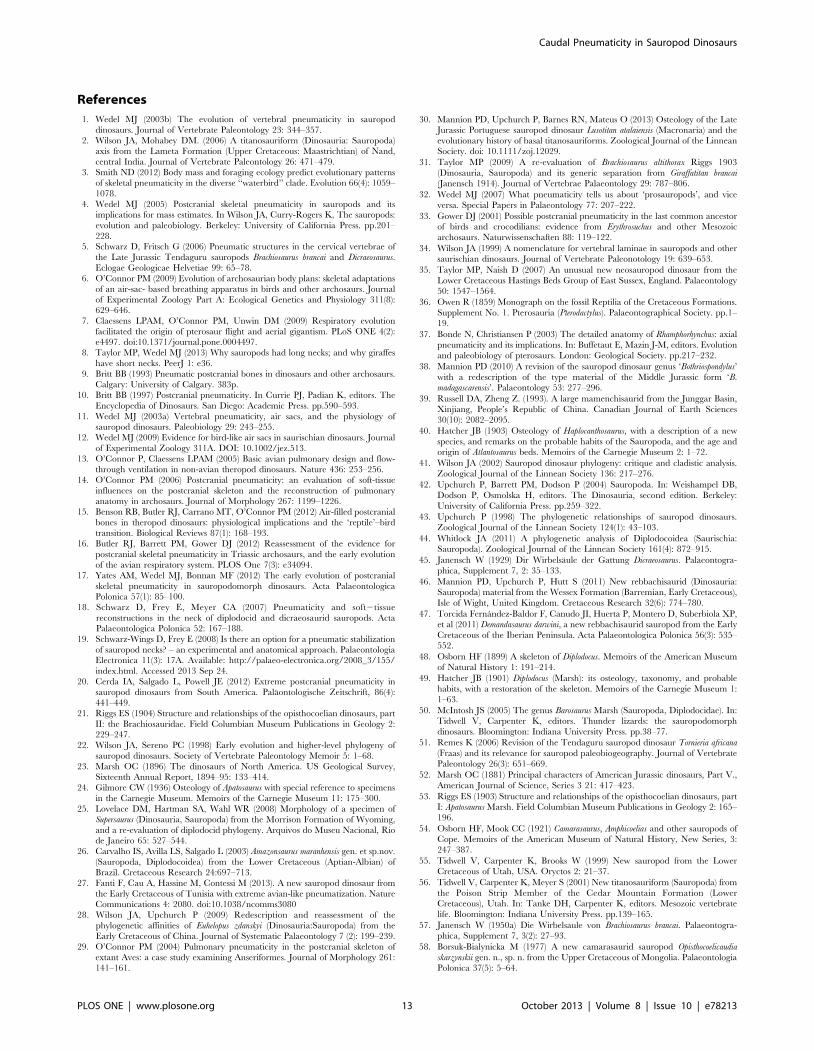
Conclusions
Although it has not been previously recognised, caudal
pneumaticity was present in Apatosaurus and Giraffatitan. Pneumatic
fossae in the mid-caudal vertebrae of these animals were not
detected for decades following their initial descriptions, despite the
fact that two of the most important specimens were on display for
most of the twentieth century. Furthermore, the pattern of caudal
pneumatization in both taxa appears to have been erratic,
although this may be at least partly caused by incomplete
ontogenetic sampling. Taken together, these facts suggest that
caudal pneumaticity, or at least the capacity to develop it, may be
more widely distributed in sauropods (and possibly theropods)
than is currently appreciated. We predict that more examples of
caudal pneumaticity in otherwise well-known taxa will be
discovered in the future.
The discovery of long pneumatic hiatuses in the tails of
Giraffatitan and Apatosaurus complicates our understanding of the
development and evolution of PSP in extinct archosaurs, and
undermines the utility of hiatuses for identifying the air-sac systems
responsible for pneumatization. On one hand, the presence of
multiple pneumatic hiatuses within the inferred domain of a single
pair of air sacs shows that such hiatuses can be produced by
leapfrogging diverticula and do not always indicate pneumatiza-
tion from multiple sources as originally proposed by Wedel [11].
The pneumatic hiatus reported in Haplocanthosaurus [12] seems
likely to have been produced by diverticula that simply affected
adjacent vertebrae inconsistently. If more pneumatic hiatuses are
discovered in extinct ornithodirans, criteria will be needed to
distinguish those caused by multiple sources of diverticula from
those caused by ‘‘leapfrogging’’ diverticula. Until such criteria are
established, the inference that pneumatic hiatuses always indicate
multiple air sacs is falsified. However, the case for an essentially
avian air sac system in pterosaurs and saurischians is also based on
several other lines of evidence [7,12], and remains robust.
The other major implication of the pneumatic hiatuses in
Giraffatitan and Apatosaurus is that pneumatic diverticula were even
more widespread in sauropods than previously thought. This
should not be surprising, given the many visceral, intermuscular,
and subcutaneous diverticula of extant birds that leave no skeletal
traces. The anatomical breadth of diverticular systems in
saurischians and pterosaurs is also underscored by distal forelimb
pneumaticity in pterosaurs [7].
A common discovery pattern for PSP in pterosaurs and
saurischians has been emerging over the past few years: the more
we look, the more we find. Compelling evidence of PSP is now
known in early representatives of both clades, and patterns of
pneumatization in derived pterosaurs, sauropods, and non-avian
theropods are diagnostic for the air sacs required for flow-through
lung ventilation [7,12–15]. The discovery of more pneumaticity in
pterosaurs, sauropodomorphs, and non-avian theropods empha-
sises how strange is the absence of reported pneumaticity in
ornithischians ([16]: p. 19; the putative pneumatic foramen in a
dorsal rib of the iguanodont Delapparentia [86] is not convincing). If,
as seems increasingly likely, an air sac system is primitive for
Ornithodira, why did ornithischians never discover PSP (in a
developmental sense)? And if an air sac system is not primitive for
Ornithodira, why did the three other major lineages evolve PSP so
soon after their divergence from one another and from
Ornithischia?
It is possible that ornithischians did have pneumatic diverticula,
but that—following the hypothesis of initially neutral evolution
described above—these diverticula did not impact the skeleton
enough to become visible to selection. This is a complex scenario
that will be difficult to test, since we currently have no way of
identifying pneumatic diverticula in fossil taxa other than by their
skeletal traces. In basal sauropodomorphs, potentially pneumatic
fossae can be difficult to assess because the recesses ventral to the
diapophyses are often obscured by sediment, even in apparently
well-prepared specimens ([16]: p. 16; [17]: 95). Largely because of
this difficulty, PSP went unrecognized in basal sauropodomorphs
until very recently. By analogy, we think it is at least possible that
pneumatic fossae in ornithischians, if present, may have escaped
detection. We therefore encourage paleobiologists to keep an eye
out for even rudimentary indications of PSP in ornithischians.
Acknowledgments
For curatorial assistance we thank Mark Norell and Carl Mehling
(American Museum of Natural History), Matt Lamanna, David Berman,
and Amy Henrici (Carnegie Museum), Pete Makovicky, Nate Smith, and
William Simpson (Field Museum of Natural History), Kenneth Campbell
and Kimball Garrett (Natural History Museum of Los Angeles County),
Richard Cifelli, Nicholas Czaplewski, Kyle Davies, and Jennifer Larson
(Oklahoma Museum of Natural History), Daniel Brinkman and Marilyn
Fox (Yale Peabody Museum), and especially Daniela Schwarz-Wings,
Oliver Wings, and Heinrich Mallison (Museum fur Naturkunde Berlin). All
our quotations of Janensch [57,64] were made by Gerhard Maier, and
are available at the Polyglot Paleontologist website (http://www.paleoglot.
org/); we thank Gerhard for his effort. The photograph of the ‘Fund no’
quarry used in Figure 6 appears courtesy of Daniela Schwarz-Wings at the
Museum fur Naturkunde Berlin. Pat O’Connor and John Whitlock
reviewed an earlier version of this manuscript and made many helpful
comments, for which we are grateful. We also thank Daniela Schwarz-
Wings and Roger Benson for thoughtful review comments.
Author Contributions
Conceived and designed the experiments: MJW MPT. Performed the
experiments: MJW MPT. Analyzed the data: MJW MPT. Contributed
reagents/materials/analysis tools: MJW MPT. Wrote the paper: MJW
MPT.
Caudal Pneumaticity in Sauropod Dinosaurs
PLOS ONE | www.plosone.org 12 October 2013 | Volume 8 | Issue 10 | e78213
References
1. Wedel MJ (2003b) The evolution of vertebral pneumaticity in sauropod
dinosaurs. Journal of Vertebrate Paleontology 23: 344–357.
2. Wilson JA, Mohabey DM. (2006) A titanosauriform (Dinosauria: Sauropoda)axis from the Lameta Formation (Upper Cretaceous: Maastrichtian) of Nand,
central India. Journal of Vertebrate Paleontology 26: 471–479.
3. Smith ND (2012) Body mass and foraging ecology predict evolutionary patternsof skeletal pneumaticity in the diverse ‘‘waterbird’’ clade. Evolution 66(4): 1059–
1078.
4. Wedel MJ (2005) Postcranial skeletal pneumaticity in sauropods and itsimplications for mass estimates. In Wilson JA, Curry-Rogers K, The sauropods:
evolution and paleobiology. Berkeley: University of California Press. pp.201–
228.5. Schwarz D, Fritsch G (2006) Pneumatic structures in the cervical vertebrae of
the Late Jurassic Tendaguru sauropods Brachiosaurus brancai and Dicraeosaurus.
Eclogae Geologicae Helvetiae 99: 65–78.
6. O’Connor PM (2009) Evolution of archosaurian body plans: skeletal adaptationsof an air-sac- based breathing apparatus in birds and other archosaurs. Journal
of Experimental Zoology Part A: Ecological Genetics and Physiology 311(8):629–646.
7. Claessens LPAM, O’Connor PM, Unwin DM (2009) Respiratory evolution
facilitated the origin of pterosaur flight and aerial gigantism. PLoS ONE 4(2):e4497. doi:10.1371/journal.pone.0004497.
8. Taylor MP, Wedel MJ (2013) Why sauropods had long necks; and why giraffes
have short necks. PeerJ 1: e36.
9. Britt BB (1993) Pneumatic postcranial bones in dinosaurs and other archosaurs.Calgary: University of Calgary. 383p.
10. Britt BB (1997) Postcranial pneumaticity. In Currie PJ, Padian K, editors. The
Encyclopedia of Dinosaurs. San Diego: Academic Press. pp.590–593.
11. Wedel MJ (2003a) Vertebral pneumaticity, air sacs, and the physiology ofsauropod dinosaurs. Paleobiology 29: 243–255.
12. Wedel MJ (2009) Evidence for bird-like air sacs in saurischian dinosaurs. Journal
of Experimental Zoology 311A. DOI: 10.1002/jez.513.
13. O’Connor P, Claessens LPAM (2005) Basic avian pulmonary design and flow-through ventilation in non-avian theropod dinosaurs. Nature 436: 253–256.
14. O’Connor PM (2006) Postcranial pneumaticity: an evaluation of soft-tissue
influences on the postcranial skeleton and the reconstruction of pulmonaryanatomy in archosaurs. Journal of Morphology 267: 1199–1226.
15. Benson RB, Butler RJ, Carrano MT, O’Connor PM (2012) Air-filled postcranial
bones in theropod dinosaurs: physiological implications and the ‘reptile’–birdtransition. Biological Reviews 87(1): 168–193.
16. Butler RJ, Barrett PM, Gower DJ (2012) Reassessment of the evidence for
postcranial skeletal pneumaticity in Triassic archosaurs, and the early evolutionof the avian respiratory system. PLOS One 7(3): e34094.
17. Yates AM, Wedel MJ, Bonnan MF (2012) The early evolution of postcranial
skeletal pneumaticity in sauropodomorph dinosaurs. Acta PalaeontologicaPolonica 57(1): 85–100.
18. Schwarz D, Frey E, Meyer CA (2007) Pneumaticity and soft2tissue
reconstructions in the neck of diplodocid and dicraeosaurid sauropods. ActaPalaeontologica Polonica 52: 167–188.
19. Schwarz-Wings D, Frey E (2008) Is there an option for a pneumatic stabilization
of sauropod necks? – an experimental and anatomical approach. PalaeontologiaElectronica 11(3): 17A. Available: http://palaeo-electronica.org/2008_3/155/
index.html. Accessed 2013 Sep 24.
20. Cerda IA, Salgado L, Powell JE (2012) Extreme postcranial pneumaticity in
sauropod dinosaurs from South America. Palaontologische Zeitschrift, 86(4):441–449.
21. Riggs ES (1904) Structure and relationships of the opisthocoelian dinosaurs, part
II: the Brachiosauridae. Field Columbian Museum Publications in Geology 2:229–247.
22. Wilson JA, Sereno PC (1998) Early evolution and higher-level phylogeny of
sauropod dinosaurs. Society of Vertebrate Paleontology Memoir 5: 1–68.
23. Marsh OC (1896) The dinosaurs of North America. US Geological Survey,Sixteenth Annual Report, 1894–95: 133–414.
24. Gilmore CW (1936) Osteology of Apatosaurus with special reference to specimens
in the Carnegie Museum. Memoirs of the Carnegie Museum 11: 175–300.25. Lovelace DM, Hartman SA, Wahl WR (2008) Morphology of a specimen of
Supersaurus (Dinosauria, Sauropoda) from the Morrison Formation of Wyoming,
and a re-evaluation of diplodocid phylogeny. Arquivos do Museu Nacional, Riode Janeiro 65: 527–544.
26. Carvalho IS, Avilla LS, Salgado L (2003) Amazonsaurus maranhensis gen. et sp.nov.
(Sauropoda, Diplodocoidea) from the Lower Cretaceous (Aptian-Albian) ofBrazil. Cretaceous Research 24:697–713.
27. Fanti F, Cau A, Hassine M, Contessi M (2013). A new sauropod dinosaur from
the Early Cretaceous of Tunisia with extreme avian-like pneumatization. NatureCommunications 4: 2080. doi:10.1038/ncomms3080
28. Wilson JA, Upchurch P (2009) Redescription and reassessment of the
phylogenetic affinities of Euhelopus zdanskyi (Dinosauria:Sauropoda) from theEarly Cretaceous of China. Journal of Systematic Palaeontology 7 (2): 199–239.
29. O’Connor PM (2004) Pulmonary pneumaticity in the postcranial skeleton of
extant Aves: a case study examining Anseriformes. Journal of Morphology 261:141–161.
30. Mannion PD, Upchurch P, Barnes RN, Mateus O (2013) Osteology of the LateJurassic Portuguese sauropod dinosaur Lusotitan atalaiensis (Macronaria) and the
evolutionary history of basal titanosauriforms. Zoological Journal of the LinneanSociety. doi: 10.1111/zoj.12029.
31. Taylor MP (2009) A re-evaluation of Brachiosaurus altithorax Riggs 1903
(Dinosauria, Sauropoda) and its generic separation from Giraffatitan brancai
(Janensch 1914). Journal of Vertebrae Palaeontology 29: 787–806.
32. Wedel MJ (2007) What pneumaticity tells us about ‘prosauropods’, and vice
versa. Special Papers in Palaeontology 77: 207–222.
33. Gower DJ (2001) Possible postcranial pneumaticity in the last common ancestorof birds and crocodilians: evidence from Erythrosuchus and other Mesozoic
archosaurs. Naturwissenschaften 88: 119–122.
34. Wilson JA (1999) A nomenclature for vertebral laminae in sauropods and othersaurischian dinosaurs. Journal of Vertebrate Paleonotology 19: 639–653.
35. Taylor MP, Naish D (2007) An unusual new neosauropod dinosaur from the
Lower Cretaceous Hastings Beds Group of East Sussex, England. Palaeontology50: 1547–1564.
36. Owen R (1859) Monograph on the fossil Reptilia of the Cretaceous Formations.
Supplement No. 1. Pterosauria (Pterodactylus). Palaeontographical Society. pp.1–19.
37. Bonde N, Christiansen P (2003) The detailed anatomy of Rhamphorhynchus: axial
pneumaticity and its implications. In: Buffetaut E, Mazin J-M, editors. Evolutionand paleobiology of pterosaurs. London: Geological Society. pp.217–232.
38. Mannion PD (2010) A revision of the sauropod dinosaur genus ‘Bothriospondylus’
with a redescription of the type material of the Middle Jurassic form ‘B.
madagascarensis’. Palaeontology 53: 277–296.
39. Russell DA, Zheng Z. (1993). A large mamenchisaurid from the Junggar Basin,
Xinjiang, People’s Republic of China. Canadian Journal of Earth Sciences30(10): 2082–2095.
40. Hatcher JB (1903) Osteology of Haplocanthosaurus, with a description of a new
species, and remarks on the probable habits of the Sauropoda, and the age andorigin of Atlantosaurus beds. Memoirs of the Carnegie Museum 2: 1–72.
41. Wilson JA (2002) Sauropod dinosaur phylogeny: critique and cladistic analysis.
Zoological Journal of the Linnean Society 136: 217–276.
42. Upchurch P, Barrett PM, Dodson P (2004) Sauropoda. In: Weishampel DB,
Dodson P, Osmolska H, editors. The Dinosauria, second edition. Berkeley:
University of California Press. pp.259–322.
43. Upchurch P (1998) The phylogenetic relationships of sauropod dinosaurs.
Zoological Journal of the Linnean Society 124(1): 43–103.
44. Whitlock JA (2011) A phylogenetic analysis of Diplodocoidea (Saurischia:Sauropoda). Zoological Journal of the Linnean Society 161(4): 872–915.
45. Janensch W (1929) Dir Wirbelsaule der Gattung Dicraeosaurus. Palaeontogra-
phica, Supplement 7, 2: 35–133.
46. Mannion PD, Upchurch P, Hutt S (2011) New rebbachisaurid (Dinosauria:Sauropoda) material from the Wessex Formation (Barremian, Early Cretaceous),
Isle of Wight, United Kingdom. Cretaceous Research 32(6): 774–780.
47. Torcida Fernandez-Baldor F, Canudo JI, Huerta P, Montero D, Suberbiola XP,et al (2011) Demandasaurus darwini, a new rebbachisaurid sauropod from the Early
Cretaceous of the Iberian Peninsula. Acta Palaeontologica Polonica 56(3): 535–552.
48. Osborn HF (1899) A skeleton of Diplodocus. Memoirs of the American Museum
of Natural History 1: 191–214.
49. Hatcher JB (1901) Diplodocus (Marsh): its osteology, taxonomy, and probable
habits, with a restoration of the skeleton. Memoirs of the Carnegie Museum 1:
1–63.
50. McIntosh JS (2005) The genus Barosaurus Marsh (Sauropoda, Diplodocidae). In:
Tidwell V, Carpenter K, editors. Thunder lizards: the sauropodomorph
dinosaurs. Bloomington: Indiana University Press. pp.38–77.
51. Remes K (2006) Revision of the Tendaguru sauropod dinosaur Tornieria africana
(Fraas) and its relevance for sauropod paleobiogeography. Journal of Vertebrate
Paleontology 26(3): 651–669.
52. Marsh OC (1881) Principal characters of American Jurassic dinosaurs, Part V.,
American Journal of Science, Series 3 21: 417–423.
53. Riggs ES (1903) Structure and relationships of the opisthocoelian dinosaurs, partI: Apatosaurus Marsh. Field Columbian Museum Publications in Geology 2: 165–
196.
54. Osborn HF, Mook CC (1921) Camarasaurus, Amphicoelias and other sauropods ofCope. Memoirs of the American Museum of Natural History, New Series, 3:
247–387.
55. Tidwell V, Carpenter K, Brooks W (1999) New sauropod from the LowerCretaceous of Utah, USA. Oryctos 2: 21–37.
56. Tidwell V, Carpenter K, Meyer S (2001) New titanosauriform (Sauropoda) from
the Poison Strip Member of the Cedar Mountain Formation (LowerCretaceous), Utah. In: Tanke DH, Carpenter K, editors. Mesozoic vertebrate
life. Bloomington: Indiana University Press. pp.139–165.
57. Janensch W (1950a) Die Wirbelsaule von Brachiosaurus brancai. Palaeontogra-phica, Supplement 7, 3(2): 27–93.
58. Borsuk-Bialynicka M (1977) A new camarasaurid sauropod Opisthocoelicaudia
skarzynskii gen. n., sp. n. from the Upper Cretaceous of Mongolia. PalaeontologiaPolonica 37(5): 5–64.
Caudal Pneumaticity in Sauropod Dinosaurs
PLOS ONE | www.plosone.org 13 October 2013 | Volume 8 | Issue 10 | e78213

59. Powell JE (2003) Revision of South American titanosaurid dinosaurs:
palaeobiological, palaeobiogeographical and phylogenetic aspects. Records of
the Queen Victoria Museum 111: 1–173.
60. Martinelli A, Forasiepi AM (2004) Late Cretaceous vertebrates from Bajo de
Santa Rosa (Allen Formation), Rio Negro province, Argentina, with the
description of a new sauropod dinosaur (Titanosauridae). Revista del Museo
Argentino de Ciencias Naturales 6(2): 257–305.
61. Otero A (2010) The appendicular skeleton of Neuquensaurus, a Late Cretaceous
saltasaurine sauropod from Patagonia, Argentina. Acta Palaeontologica Polonica
55 (3): 399–426.
62. Mazzetta GV, Christiansen P, Farina RA (2004) Giants and bizarres: body sizeof some southern South American Cretaceous dinosaurs. Historical Biology
2004: 1–13.
63. Sander PM, Christian A, Clauss M, Fechner R, Gee TC, et al (2010) Biology of
the sauropod dinosaurs: the evolution of gigantism. Biological Reviews 86: 117–155. DOI: 10.1111/j.1469-185X.2010.00137.x.
64. Janensch W (1950b) Die Skelettrekonstruktion von Brachiosaurus brancai.
Palaeontographica, Supplement 7, 3(2): 95–102.
65. Gilmore CW (1932) On a newly mounted skeleton of Diplodocus in the United
States National Museum. Proceedings of the United States National Museum
81(18): 1–21.
66. Taylor MP (2005) Sweet seventy-five and never been kissed: the Natural HistoryMuseum’s Tendaguru brachiosaur. In: Barrett PM,editors. 53rd Symposium of
Vertebrae Palaeontology and Comparative Anatomy, Abstracts. London: The
Natural History Museum. p.25.
67. Migeod FWH (1931) British Museum East Africa Expedition: Account of thework done in 1930. Natural History Magazine 3: 87–103.
68. King AS (1957) The aerated bones of Gallus domesticus. Cells Tissues Organs
31(2): 220–230.
69. Cranford TW, Amundin M, Norris KS (1996) Functional morphology and
homology in the odontocete nasal complex: implications for sound generation.
Journal of Morphology 228(3): 223–285.
70. Weiglein AH (1999). Development of the paranasal sinuses in humans. In:Koppe T, Nagai H, Alt KW, editors. The paranasal sinuses of higher primates.
Chicago: Quintessence. pp.35–50.
71. Wilson JA, Martinez RN, Alcober O (1999). Distal tail segment of a titanosaur
(Dinosauria: Sauropoda) from the Upper Cretaceous of Mendoza, Argentina.Journal of Vertebrate Paleontology 19(3): 591–594.
72. Janensch W (1947) Pneumatizitat bei Wirbeln von Sauropoden und anderen
Saurischien. Palaeontographica, Supplement 7, 3(1): 1–25.
73. Harris JD (2006) The significance of Suuwassea emiliae (Dinosauria: Sauropoda)
forflagellicaudatan intrarelationships and evolution. Journal of Systematic
Palaeontology 4: 185–198.
74. Osborn HF (1898) Additional characters of the great herbivorous dinosaurCamarasaurus. Bulletin of the American Museum of Natural History 10: 219–233.
75. Matthew WD (1905) The mounted skeleton of Brontosaurus. American Museum
Journal 5: 62–70.
76. Ostrom JH, McIntosh JS (1966) Marsh’s dinosaurs: the collections from Como
Bluff. New Haven: Yale University Press. 388p.
77. Upchurch P, Tomida Y, Barrett PM (2004b) A new specimen of Apatosaurus ajax
(Sauropoda: Diplodocidae) from the Morrison Formation (Upper Jurassic) ofWyoming, USA. National Science Museum Monographs (Tokyo) 26: 1–107.
78. Duncker HR. (1971) The lung air sac system of birds. Advances in Anatomy,
Embryology, and Cell Biology 45: 1–171.
79. Ruben JA, Jones TD, Geist NR, Hillenius WJ (1997) Lung structure and
ventilation in theropod dinosaurs and early birds. Science 278: 1267–1270.80. Sereno PC, Martinez RN, Wilson JA, Varricchio DJ, Alcober OA, et al (2008)
Evidence for avian intrathoracic air sacs in a new predatory dinosaur from
Argentina. PLoS ONE 3(9): e3303. doi:10.1371/journal.pone.000330381. Hogg DA (1984) The development of pneumatisation in the skeleton of the adult
domestic fowl. Journal of Anatomy 139: 105–113.82. Yates AM (2003) A new species of the primitive dinosaur Thecodontosaurus
(Saurischia: Sauropodomorpha) and its implications for the systematics of early
dinosaurs. Journal of Systematic Palaeontology 1: 1–42.83. Cover MS (1953) Gross and microscopic anatomy of the respiratory system of
the turkey. III. The air sacs. American Journal of Veterinary Research 14: 239–245.
84. Witmer LM (1997) The evolution of the antorbital cavity of archosaurs: a studyin soft-tissue reconstruction in the fossil record with an analysis of the function of
pneumaticity. Society of Vertebrate Paleontology Memoir 3: 1–73.
85. Kimura M (1968) Evolutionary rate at the molecular level. Nature 217: 624–626.
86. Ruiz-Omenaca JI (2011) Delapparentia turolensis nov. gen et sp., un nuevodinosaurio iguanodontoideo (Ornithischia: Ornithopoda) en el Cretacico
Inferior de Galve. Estudios Geologicos 67 (1): 83–110. doi:10.3989/
egeol.40276.12487. Upchurch P, Mannion PD (2009) The first diplodocid from Asia and its
implications for the evolutionary history of sauropod dinosaurs. Palaeontology52(6): 1195–1207.
88. Mannion PD, Barrett P (2013) Additions to the sauropod dinosaur fauna of theCenomanian (early Late Cretaceous) Kem Kem beds of Morocco: Palaeobio-
geographical implications of the mid-Cretaceous African sauropod fossil record.
Cretaceous Research 45: 49–59.89. Mannion PD, Upchurch P, Mateus O, Barnes RN, Jones MEH (2011) New
information on the anatomy and systematic position of Dinheirosaurus lourinhanensis
(Sauropoda: Diplodocoidea) from the Late Jurassic of Portugal, with a review of
European diplodocoids. Journal of Systematic Palaeontology 10(3): 521–551.
90. Lull RS (1919) The sauropod dinosaur Barosaurus Marsh. Memoirs of theConnecticut Academy of Arts and Sciences 6: 1–42.
91. Kellner AWA, Azevedo SD (1999) A new sauropod dinosaur (Titanosauria) fromthe Late Cretaceous of Brazil. National Science Museum Monographs, 15: 111–
142.92. Casal G, Martınez R, Luna M, Sciutto JC, Lamanna M (2007) Aeolosaurus
colhuehuapensis sp. nov.(Sauropoda, Titanosauria) de la Formacion Bajo Barreal,
Cretacico Superior de Argentina. Revista Brasileira de Paleontologia, 10(1): 53–62.
93. Campos DD, Kellner AWA, Bertini RJ, Santucci RM (2005) On a titanosaurid(Dinosauria, Sauropoda) vertebral column from the Bauru group, Late
Cretaceous of Brazil. Arquivos do Museu Nacional, Rio de Janeiro 63(3):
565–593.94. Lu J, Xu L, Jiang X, Jia S, Li M, et al (2009) A preliminary report on the new
dinosaurian fauna from the Cretaceous of the Ruyang Basin, Henan Province ofcentral China. Journal of the Paleontological Society of Korea 25: 43–56.
95. Fronimos J (2011) Patterns of postcranial pneumaticity in the Late Cretaceoustitanosaur Alamosaurus sanjuanensis. Journal of Vertebrate Paleontology (Supple-
ment 2) 31: 112–113A.
96. Calvo JO, Porfiri JD, Gonzalez-Riga BJ, Kellner AWA (2007) A new Cretaceousterrestrial ecosystem from Gondwana with the description of a new sauropod
dinosaur. Anais da Academia Brasileira de Ciencias 79(3): 529–541.
Caudal Pneumaticity in Sauropod Dinosaurs
PLOS ONE | www.plosone.org 14 October 2013 | Volume 8 | Issue 10 | e78213