Embed Size (px)
Citation preview
Fungal Systematics and Evolution is licensed under a Creative Commons Attribution-NonCommercial-ShareAlike 4.0 International License
© 2018 Westerdijk Fungal Biodiversity Institute 23
Editor-in-ChiefProf. dr P.W. Crous, Westerdijk Fungal Biodiversity Institute, P.O. Box 85167, 3508 AD Utrecht, The Netherlands.E-mail:[email protected]
Fungal Systematics and EvolutionVOLUME 1JUNE 2018
PAGES 23–39
doi.org/10.3114/fuse.2018.01.03
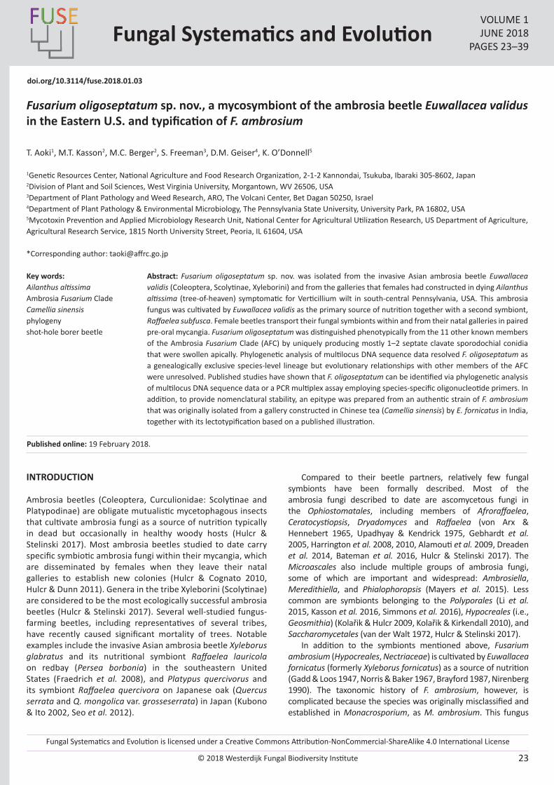
Fusarium oligoseptatum sp. nov., a mycosymbiont of the ambrosia beetle Euwallacea validus in the Eastern U.S. and typification of F. ambrosium
T. Aoki1, M.T. Kasson2, M.C. Berger2, S. Freeman3, D.M. Geiser4, K. O’Donnell5
1Genetic Resources Center, National Agriculture and Food Research Organization, 2-1-2 Kannondai, Tsukuba, Ibaraki 305-8602, Japan2Division of Plant and Soil Sciences, West Virginia University, Morgantown, WV 26506, USA3Department of Plant Pathology and Weed Research, ARO, The Volcani Center, Bet Dagan 50250, Israel4Department of Plant Pathology & Environmental Microbiology, The Pennsylvania State University, University Park, PA 16802, USA5Mycotoxin Prevention and Applied Microbiology Research Unit, National Center for Agricultural Utilization Research, US Department of Agriculture, Agricultural Research Service, 1815 North University Street, Peoria, IL 61604, USA
*Corresponding author: [email protected]
Abstract: Fusarium oligoseptatum sp. nov. was isolated from the invasive Asian ambrosia beetle Euwallacea validis (Coleoptera, Scolytinae, Xyleborini) and from the galleries that females had constructed in dying Ailanthus altissima (tree-of-heaven) symptomatic for Verticillium wilt in south-central Pennsylvania, USA. This ambrosia fungus was cultivated by Euwallacea validis as the primary source of nutrition together with a second symbiont, Raffaelea subfusca. Female beetles transport their fungal symbionts within and from their natal galleries in paired pre-oral mycangia. Fusarium oligoseptatum was distinguished phenotypically from the 11 other known members of the Ambrosia Fusarium Clade (AFC) by uniquely producing mostly 1–2 septate clavate sporodochial conidia that were swollen apically. Phylogenetic analysis of multilocus DNA sequence data resolved F. oligoseptatum as a genealogically exclusive species-level lineage but evolutionary relationships with other members of the AFC were unresolved. Published studies have shown that F. oligoseptatum can be identified via phylogenetic analysis of multilocus DNA sequence data or a PCR multiplex assay employing species-specific oligonucleotide primers. In addition, to provide nomenclatural stability, an epitype was prepared from an authentic strain of F. ambrosium that was originally isolated from a gallery constructed in Chinese tea (Camellia sinensis) by E. fornicatus in India, together with its lectotypification based on a published illustration.
Key words: Ailanthus altissimaAmbrosia Fusarium CladeCamellia sinensisphylogenyshot-hole borer beetle
Published online: 19 February 2018.
INTRODUCTION
Ambrosia beetles (Coleoptera, Curculionidae: Scolytinae and Platypodinae) are obligate mutualistic mycetophagous insects that cultivate ambrosia fungi as a source of nutrition typically in dead but occasionally in healthy woody hosts (Hulcr & Stelinski 2017). Most ambrosia beetles studied to date carry specific symbiotic ambrosia fungi within their mycangia, which are disseminated by females when they leave their natal galleries to establish new colonies (Hulcr & Cognato 2010, Hulcr & Dunn 2011). Genera in the tribe Xyleborini (Scolytinae) are considered to be the most ecologically successful ambrosia beetles (Hulcr & Stelinski 2017). Several well-studied fungus-farming beetles, including representatives of several tribes, have recently caused significant mortality of trees. Notable examples include the invasive Asian ambrosia beetle Xyleborus glabratus and its nutritional symbiont Raffaelea lauricola on redbay (Persea borbonia) in the southeastern United States (Fraedrich et al. 2008), and Platypus quercivorus and its symbiont Raffaelea quercivora on Japanese oak (Quercus serrata and Q. mongolica var. grosseserrata) in Japan (Kubono & Ito 2002, Seo et al. 2012).
Compared to their beetle partners, relatively few fungal symbionts have been formally described. Most of the ambrosia fungi described to date are ascomycetous fungi in the Ophiostomatales, including members of Afroraffaelea, Ceratocystiopsis, Dryadomyces and Raffaelea (von Arx & Hennebert 1965, Upadhyay & Kendrick 1975, Gebhardt et al. 2005, Harrington et al. 2008, 2010, Alamouti et al. 2009, Dreaden et al. 2014, Bateman et al. 2016, Hulcr & Stelinski 2017). The Microascales also include multiple groups of ambrosia fungi, some of which are important and widespread: Ambrosiella, Meredithiella, and Phialophoropsis (Mayers et al. 2015). Less common are symbionts belonging to the Polyporales (Li et al. 2015, Kasson et al. 2016, Simmons et al. 2016), Hypocreales (i.e., Geosmithia) (Kolařik & Hulcr 2009, Kolařik & Kirkendall 2010), and Saccharomycetales (van der Walt 1972, Hulcr & Stelinski 2017).
In addition to the symbionts mentioned above, Fusarium ambrosium (Hypocreales, Nectriaceae) is cultivated by Euwallacea fornicatus (formerly Xyleborus fornicatus) as a source of nutrition (Gadd & Loos 1947, Norris & Baker 1967, Brayford 1987, Nirenberg 1990). The taxonomic history of F. ambrosium, however, is complicated because the species was originally misclassified and established in Monacrosporium, as M. ambrosium. This fungus

© 2018 Westerdijk Fungal Biodiversity Institute
Aoki et al.
Editor-in-ChiefProf. dr P.W. Crous, Westerdijk Fungal Biodiversity Institute, P.O. Box 85167, 3508 AD Utrecht, The Netherlands.E-mail:[email protected]
24
was isolated originally and described from galleries of the tea shot-hole borer, E. fornicatus, in Camellia sinensis (Chinese tea) and Ricinus communis (caster-oil tree) stems in Sri Lanka (Gadd & Loos 1947). Subsequently, F. bugnicourtii was described based on collections from galleries in Chinese tea in India, borer-damaged Hevea brasiliensis (rubber tree) and Theoborma cacao (cacao) in Sabah, Malaysia (Brayford 1987). Nirenberg (1990) synonymized F. bugnicourtii with M. ambrosium and recombined the latter as F. ambrosium based on nomenclatural priority. Brayford (1987) considered F. bugnicourtii to be conspecific with F. tumidum var. coeruleum (Bugnicourt 1939), but distinct from F. tumidum. Although the type of F. tumidum var. coeruleum based on a collection from H. brasiliensis appears to be phylogenetically distinct from F. bugnicourtii, the holotype of F. bugnicourtii selected by Brayford (IMI 296597 = NRRL 20438) is conspecific with F. ambrosium (Kasson et al. 2013).
Kasson et al. (2013) conducted an extensive multilocus molecular phylogenetic study on the ambrosial fusaria, based on isolates from beetles, their galleries, or from trees showing extensive borer damage and dieback. These included Camellia sinensis, Persea americana (avocado), Ailanthus altissima (tree-of-heaven), Acer negundo (box elder), and Hevea brasiliensis from natural and cultivated ecosystems, and avocado in the United States, Israel and Australia. Seven different Fusarium species lineages were reported to be associated with Euwallacea ambrosia beetles within the Ambrosia Fusarium Clade (AFC) and one other species (i.e., Fusarium sp. AF-9) with Xyleborus ferrugineus in Costa Rica. The monophyletic AFC is nested within Clade 3 of the F. solani species complex (FSSC; O’Donnell 2000), which contains 60 plus phylogenetic species based on genealogical concordance phylogenetic species recognition (GCPSR; Taylor et al. 2000). The AFC comprises two strongly supported clades: the four species within Clade A typically produce curved fusiform septate macroconidia, which are typical of Fusarium, whereas nine of the 10 species within Clade B produce clavate macroconidia (Kasson et al. 2013, Aoki et al. unpubl.), described as ‘dolphin-shaped’ by Brayford (1987). O’Donnell et al. (2015) conducted a multilocus phylogenetic analysis of the AFC and Euwallacea and found evidence of repeated host shifts rather than strict co-evolution of this mutualism.
Freeman et al. (2013) described a new species, F. euwallaceae, based on isolates corresponding to the ambrosia species symbiotic with the Euwallacea sp. #1 sensu O’Donnell et al. (2015), which causes serious damage to avocado production in Israel and California, USA (Mendel et al. 2012, Eskalen et al. 2013). Fusarium euwallaceae is closely related morphologically to F. ambrosium, but it can be distinguished from the latter by the abundant production of bluish to brownish sporodochial conidia that form greenish masses on PDA after 1 mo in culture, together with hyaline conidia. To date only three of the 16 species within the AFC have been described formally (Kasson et al. 2013, O’Donnell et al. 2015, Na et al. 2018). Similar to E. validus, Euwallacea sp. #1 also carries additional symbiotic fungi, Graphium euwallaceae and Paracremonium pembeum (Freeman et al. 2016, Lynch et al. 2015). Recently, PCR multiplexes were developed to discriminate Fusarium symbionts of invasive Euwallacea ambrosia beetles that inflict damage on numerous tree species throughout the United States, including F. euwallaceae and F. kuroshium along with four unnamed AFC species-level lineages: AF-3, AF-4, AF-6 and AF-8 (Short et al. 2017). One of the undescribed species, which was informally referred to as Fusarium sp. AF-4, is cultivated by the ambrosia
beetle E. validus primarily in Verticillium wilt-stressed and dying stands of A. altissima, as well as from Verticillium wilt-stressed Acer pensylvanicum (striped maple), Aralia spinosa (devils walkingstick) and Rhus typhina (staghorn sumac) in south-central Pennsylvania, USA (Schall & Davis 2009, Kasson et al. 2013, 2015). In the present study, this species is described as F. oligoseptatum sp. nov. based on a comparison with F. ambrosium (AF-1) and F. euwallaceae (AF-2) (Kasson et al. 2013, Freeman et al. 2013). In addition, because type material for F. ambrosium was not designated (Gadd & Loos 1947), or appears to have been lost (Nirenberg 1990), a line-drawing of a clavate conidium of the species from Gadd & Loos (1947) was selected as the lectotype. Furthermore, an epitype was prepared from an authentic strain of this species to stabilize its taxonomy, according to the International Code of Nomenclature for algae, fungi and plants (ICN, the Melbourne Code; McNeill et al. 2012).
MATERIALS AND METHODS
Fungal isolates and type specimens
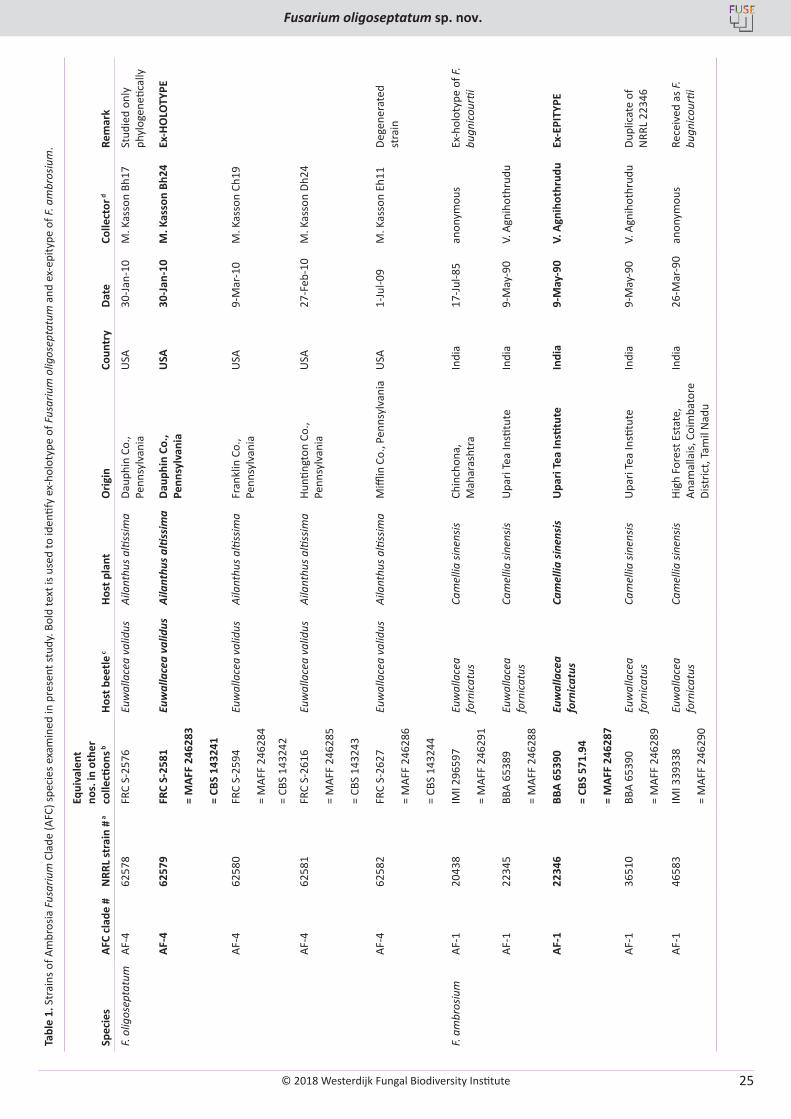
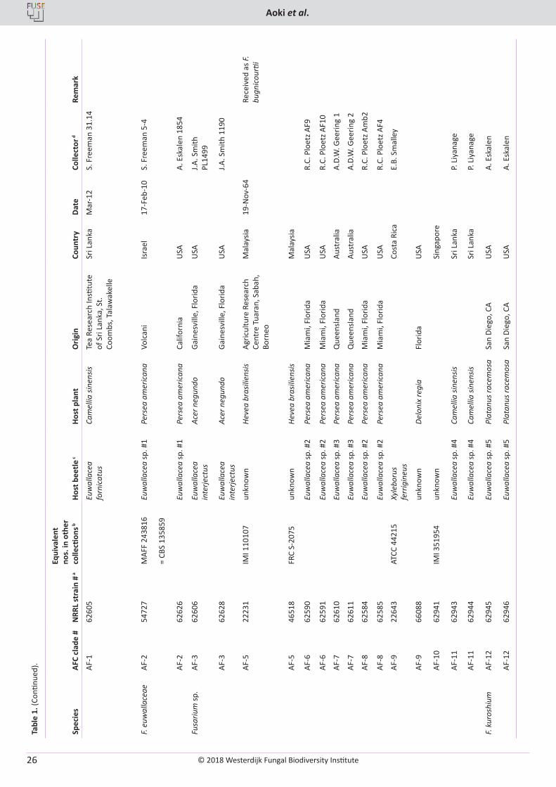
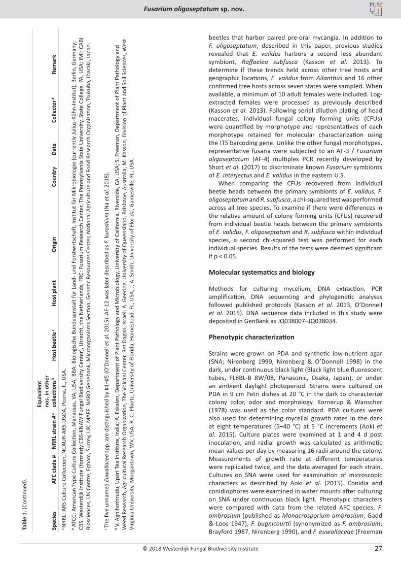
Fusarium strains examined in this study (Table 1) are stored in the Agriculture Research Service Culture Collection (NRRL), National Center for Agricultural Utilization Research (NCAUR), U.S. Department of Agriculture in Peoria, Illinois, USA. These strains were originally isolated from Euwallacea ambrosia beetles and their galleries, or from host trees showing extensive borer damage (Kasson et al. 2013). The Pennsylvanian strains of F. oligoseptatum were isolated from E. validus ambrosia beetles that had colonized A. altissima. Beetles were surface disinfested for 15 s in 70 % ethanol and then washed three times in sterile deionized water. Whole beetles or their heads were macerated using sterile Tenbroek homogenizers (Pyrex, Corning, NY), or pellet pestles (Fisher Scientific, Hampton, NH), suspensions were diluted 1:10 and 1:100, and then spread evenly over half-strength Potato Dextrose Agar (PDA, BD-DifcoTM, Thermo Fisher Scientific, Waltham, MA) amended with 100 ppm streptomycin sulfate (Sigma-Aldrich, St. Louis, MO) as described in Kasson et al. (2013). Other related ambrosia fusaria or close relatives within the F. solani species complex (O’Donnell et al. 2008) were obtained from culture collections (Table 1). Isolates used in this study are available upon request from NRRL (http://nrrl.ncaur.usda.gov/cgi-bin/usda/), NARO Genebank, Microorganisms Section (MAFF), Genetic Resources Center, National Agriculture and Food Research Organization, Tsukuba, Ibaraki, Japan (http://www.gene.affrc.go.jp/about-micro_en.php), and the Westerdijk Institute (formerly CBS-KNAW Fungal Biodiversity Center), Utrecht, the Netherlands (http://www.westerdijkinstitute.nl/). Isolates of four novel Taiwanese AFC species discovered very recently, i.e. AF-13 to AF-16 (Na et al. 2018), were not included in this study.
Holotype and epitype specimens newly prepared from the selected strains were deposited in BPI, US National Fungus Collection (https://nt.ars-grin.gov/fungaldatabases/specimens/specimens.cfm).
Incidence of Fusarium oligoseptatum and other fungi from Euwallacea validus mycangia across recently confirmed tree hosts
Mycangial fungal communities were characterized as previously described by Kasson et al. (2013) for adult female
© 2018 Westerdijk Fungal Biodiversity Institute
Fusarium oligoseptatum sp. nov.
Editor-in-ChiefProf. dr P.W. Crous, Westerdijk Fungal Biodiversity Institute, P.O. Box 85167, 3508 AD Utrecht, The Netherlands.E-mail:[email protected]
25
Tabl
e 1.
Str
ains
of A
mbr
osia
Fus
ariu
m C
lade
(AFC
) spe
cies
exa
min
ed in
pre
sent
stud
y. B
old
text
is u
sed
to id
entif
y ex
-hol
otyp
e of
Fus
ariu
m o
ligos
epta
tum
and
ex-
epity
pe o
f F. a
mbr
osiu
m.
AFC
clad
e #
NRR
L st
rain
# a
Equi
vale
nt
nos.
in o
ther
co
llecti
ons b
Spec
ies
Host
bee
tle c
Host
pla
ntO
rigin
Coun
try
Date
Colle
ctor
dRe
mar
k
F. ol
igos
epta
tum
AF-4
6257
8FR
C S-
2576
Euw
alla
cea
valid
usAi
lant
hus a
ltiss
ima
Daup
hin
Co.,
Penn
sylv
ania
USA
30-Ja
n-10
M. K
asso
n Bh
17St
udie
d on
ly
phyl
ogen
etica
lly
AF-4
6257
9FR
C S-
2581
Euw
alla
cea
valid
usAi
lant
hus a
ltiss
ima
Daup
hin
Co.,
Penn
sylv
ania
USA
30-J
an-1
0M
. Kas
son
Bh24
Ex-H
OLO
TYPE
= M
AFF
2462
83
= CB
S 14
3241
AF-4
6258
0FR
C S-
2594
Euw
alla
cea
valid
usAi
lant
hus a
ltiss
ima
Fran
klin
Co.
, Pe
nnsy
lvan
iaU
SA9-
Mar
-10
M. K
asso
n Ch
19
= M
AFF
2462
84
= CB
S 14
3242
AF-4
6258
1FR
C S-
2616
Euw
alla
cea
valid
usAi
lant
hus a
ltiss
ima
Hunti
ngto
n Co
., Pe
nnsy
lvan
iaU
SA27
-Feb
-10
M. K
asso
n Dh
24
= M
AFF
2462
85
= CB
S 14
3243
AF-4
6258
2FR
C S-
2627
Euw
alla
cea
valid
usAi
lant
hus a
ltiss
ima
Miffl
in C
o., P
enns
ylva
nia
USA
1-Ju
l-09
M. K
asso
n Eh
11De
gene
rate
d st
rain
= M
AFF
2462
86
= CB
S 14
3244
F. am
bros
ium
AF-1
2043
8IM
I 296
597
Euw
alla
cea
forn
icat
usCa
mel
lia si
nens
isCh
inch
ona,
M
ahar
asht
raIn
dia
17-Ju
l-85
anon
ymou
sEx
-hol
otyp
e of
F.
bugn
icou
rtii
= M
AFF
2462
91
AF-1
2234
5BB
A 65
389
Euw
alla
cea
forn
icat
usCa
mel
lia si
nens
isU
pari
Tea
Insti
tute
Indi
a9-
May
-90
V. A
gnih
othr
udu
= M
AFF
2462
88
AF-1
2234
6BB
A 65
390
Euw
alla
cea
forn
icat
usCa
mel
lia si
nens
isU
pari
Tea
Insti
tute
Indi
a9-
May
-90
V. A
gnih
othr
udu
Ex-E
PITY
PE
= CB
S 57
1.94
= M
AFF
2462
87
AF-1
3651
0BB
A 65
390
Euw
alla
cea
forn
icat
usCa
mel
lia si
nens
isU
pari
Tea
Insti
tute
Indi
a9-
May
-90
V. A
gnih
othr
udu
Dupl
icat
e of
N
RRL
2234
6=
MAF
F 24
6289
AF-1
4658
3IM
I 339
338
Euw
alla
cea
forn
icat
usCa
mel
lia si
nens
isHi
gh F
ores
t Est
ate,
An
amal
lais,
Coi
mba
tore
Di
stric
t, Ta
mil
Nad
u
Indi
a26
-Mar
-90
anon
ymou
sRe
ceiv
ed a
s F.
bugn
icou
rtii
= M
AFF
2462
90
© 2018 Westerdijk Fungal Biodiversity Institute
Aoki et al.
Editor-in-ChiefProf. dr P.W. Crous, Westerdijk Fungal Biodiversity Institute, P.O. Box 85167, 3508 AD Utrecht, The Netherlands.E-mail:[email protected]
26
Tabl
e 1.
(Con
tinue
d). AFC
clad
e #
NRR
L st
rain
# a
Equi
vale
nt
nos.
in o
ther
co
llecti
ons b
Spec
ies
Host
bee
tle c
Host
pla
ntO
rigin
Coun
try
Date
Colle
ctor
dRe
mar
k
AF-1
6260
5Eu
wal
lace
a fo
rnic
atus
Cam
ellia
sine
nsis
Tea
Rese
arch
Insti
tute
of
Sri
Lank
a, S
t. Co
ombs
, Tal
awak
elle
Sri L
anka
Mar
-12
S. F
reem
an 3
1.14
F. eu
wal
lace
aeAF
-254
727
MAF
F 24
3816
Euw
alla
cea
sp. #
1Pe
rsea
am
eric
ana
Volc
ani
Isra
el17
-Feb
-10
S. F
reem
an 5
-4
= CB
S 13
5859
AF-2
6262
6Eu
wal
lace
a sp
. #1
Pers
ea a
mer
ican
aCa
lifor
nia
USA
A. E
skal
en 1
854
Fusa
rium
sp.
AF-3
6260
6Eu
wal
lace
a in
terje
ctus
Acer
neg
undo
Gain
esvi
lle, F
lorid
aU
SAJ.A
. Sm
ith
PL14
99
AF-3
6262
8Eu
wal
lace
a in
terje
ctus
Ac
er n
egun
doGa
ines
ville
, Flo
rida
USA
J.A. S
mith
119
0
AF-5
2223
1IM
I 110
107
unkn
own
Heve
a br
asili
ensis
Agric
ultu
re R
esea
rch
Cent
re T
uara
n, S
abah
, Bo
rneo
Mal
aysia
19-N
ov-6
4Re
ceiv
ed a
s F.
bugn
icou
rtii
AF-5
4651
8FR
C S-
2075
unkn
own
Heve
a br
asili
ensis
Mal
aysia
AF-6
6259
0Eu
wal
lace
a sp
. #2
Pers
ea a
mer
ican
aM
iam
i, Fl
orid
aU
SAR.
C. P
loet
z AF9
AF-6
6259
1Eu
wal
lace
a sp
. #2
Pers
ea a
mer
ican
aM
iam
i, Fl
orid
aU
SAR.
C. P
loet
z AF1
0
AF-7
6261
0Eu
wal
lace
a sp
. #3
Pers
ea a
mer
ican
aQ
ueen
sland
Aust
ralia
A.D.
W. G
eerin
g 1
AF-7
6261
1Eu
wal
lace
a sp
. #3
Pers
ea a
mer
ican
aQ
ueen
sland
Aust
ralia
A.D.
W. G
eerin
g 2
AF-8
6258
4Eu
wal
lace
a sp
. #2
Pers
ea a
mer
ican
aM
iam
i, Fl
orid
aU
SAR.
C. P
loet
z Am
b2
AF-8
6258
5Eu
wal
lace
a sp
. #2
Pers
ea a
mer
ican
aM
iam
i, Fl
orid
aU
SAR.
C. P
loet
z AF4
AF-9
2264
3AT
CC 4
4215
Xyle
boru
s fe
rrig
ineu
sCo
sta
Rica
E.B.
Sm
alle
y
AF-9
6608
8un
know
nDe
loni
x re
gia
Flor
ida
USA
AF-1
062
941
IMI 3
5195
4un
know
nSi
ngap
ore
AF-1
162
943
Euw
alla
cea
sp. #
4Ca
mel
lia si
nens
isSr
i Lan
kaP.
Liy
anag
e
AF-1
162
944
Euw
alla
cea
sp. #
4Ca
mel
lia si
nens
isSr
i Lan
kaP.
Liy
anag
e
F. ku
rosh
ium
AF-1
262
945
Euw
alla
cea
sp. #
5Pl
atan
us ra
cem
osa
San
Dieg
o, C
AU
SAA.
Esk
alen
AF-1
262
946
Euw
alla
cea
sp. #
5Pl
atan
us ra
cem
osa
San
Dieg
o, C
AU
SAA.
Esk
alen
© 2018 Westerdijk Fungal Biodiversity Institute
Fusarium oligoseptatum sp. nov.
Editor-in-ChiefProf. dr P.W. Crous, Westerdijk Fungal Biodiversity Institute, P.O. Box 85167, 3508 AD Utrecht, The Netherlands.E-mail:[email protected]
27
Tabl
e 1.
(Con
tinue
d). AFC
clad
e #
NRR
L st
rain
# a
Equi
vale
nt
nos.
in o
ther
co
llecti
ons b
Spec
ies
Host
bee
tle c
Host
pla
ntO
rigin
Coun
try
Date
Colle
ctor
dRe
mar
ka N
RRL:
ARS
Cul
ture
Col
lecti
on, N
CAU
R-AR
S-U
SDA,
Peo
ria, I
L, U
SA.
b ATC
C: A
mer
ican
Typ
e Cu
lture
Col
lecti
on, M
anas
sas,
VA,
USA
; BBA
: Bio
logi
sche
Bun
desa
nsta
lt fü
r Lan
d- u
nd F
orst
wirt
scha
ft, In
stitu
t für
Mik
robi
olog
ie (c
urre
ntly
Juliu
s-Kü
hn-In
stitu
t), B
erlin
, Ger
man
y;
CBS:
Wes
terd
ijk In
stitu
te (f
orm
erly
CBS
-KN
AW F
unga
l Bio
dive
rsity
Cen
ter)
, Utr
echt
, the
Net
herla
nds;
FRC
: Fus
ariu
m R
esea
rch
Cent
er, T
he P
enns
ylva
nia
Stat
e U
nive
rsity
, Sta
te C
olle
ge, P
A, U
SA; I
MI:
CABI
Bi
osci
ence
s, U
K Ce
ntre
, Egh
am, S
urre
y, U
K; M
AFF:
NAR
O G
eneb
ank,
Mic
roor
gani
sms S
ectio
n, G
eneti
c Re
sour
ces C
ente
r, N
ation
al A
gric
ultu
re a
nd F
ood
Rese
arch
Org
aniza
tion,
Tsu
kuba
, Iba
raki
, Jap
an.
c Th
e fiv
e un
nam
ed E
uwal
lace
a sp
p. a
re d
isting
uish
ed b
y #1
‒#5
(O’D
onne
ll et
al.
2015
). AF
-12
was
late
r des
crib
ed a
s F. k
uros
hium
(Na
et a
l. 20
18).
d V. A
gnih
othr
udu,
Upa
ri Te
a In
stitu
te, I
ndia
; A. E
skal
en, D
epar
tmen
t of P
lant
Pat
holo
gy a
nd M
icro
biol
ogy,
Uni
vers
ity o
f Cal
iforn
ia, R
iver
side,
CA,
USA
; S. F
reem
an, D
epar
tmen
t of P
lant
Pat
holo
gy a
nd
Wee
d Re
sear
ch, A
gric
ultu
ral R
esea
rch
Org
aniza
tion,
The
Vol
cani
Cen
ter,
Bet D
agan
, Isr
ael;
A. G
eerin
g, U
nive
rsity
of Q
ueen
sland
, Bris
bane
, Aus
tral
ia; M
. Kas
son,
Div
ision
of P
lant
and
Soi
l Sci
ence
s, W
est
Virg
inia
Uni
vers
ity, M
orga
ntow
n, W
V, U
SA; R
. C. P
loet
z, U
nive
rsity
of F
lorid
a, H
omes
tead
, FL,
USA
; J. A
. Sm
ith, U
nive
rsity
of F
lorid
a, G
aine
svill
e, F
L, U
SA.
beetles that harbor paired pre-oral mycangia. In addition to F. oligoseptatum, described in this paper, previous studies revealed that E. validus harbors a second less abundant symbiont, Raffaelea subfusca (Kasson et al. 2013). To determine if these trends held across other tree hosts and geographic locations, E. validus from Ailanthus and 16 other confirmed tree hosts across seven states were sampled. When available, a minimum of 10 adult females were included. Log-extracted females were processed as previously described (Kasson et al. 2013). Following serial dilution plating of head macerates, individual fungal colony forming units (CFUs) were quantified by morphotype and representatives of each morphotype retained for molecular characterization using the ITS barcoding gene. Unlike the other fungal morphotypes, representative fusaria were subjected to an AF-3 / Fusarium oligoseptatum (AF-4) multiplex PCR recently developed by Short et al. (2017) to discriminate known Fusarium symbionts of E. interjectus and E. validus in the eastern U.S.
When comparing the CFUs recovered from individual beetle heads between the primary symbionts of E. validus, F. oligoseptatum and R. subfusca, a chi-squared test was performed across all tree species. To examine if there were differences in the relative amount of colony forming units (CFUs) recovered from individual beetle heads between the primary symbionts of E. validus, F. oligoseptatum and R. subfusca within individual species, a second chi-squared test was performed for each individual species. Results of the tests were deemed significant if p < 0.05.
Molecular systematics and biology
Methods for culturing mycelium, DNA extraction, PCR amplification, DNA sequencing and phylogenetic analyses followed published protocols (Kasson et al. 2013, O’Donnell et al. 2015). DNA sequence data included in this study were deposited in GenBank as JQ038007–JQ038034.
Phenotypic characterization
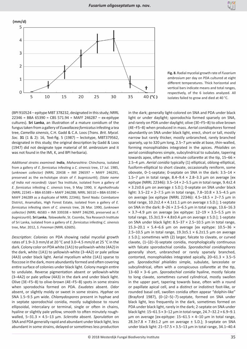
Strains were grown on PDA and synthetic low-nutrient agar (SNA; Nirenberg 1990, Nirenberg & O’Donnell 1998) in the dark, under continuous black light (Black light blue fluorescent tubes, FL8BL-B 8W/08, Panasonic, Osaka, Japan), or under an ambient daylight photoperiod. Strains were cultured on PDA in 9 cm Petri dishes at 20 °C in the dark to characterize colony color, odor and morphology. Kornerup & Wanscher (1978) was used as the color standard. PDA cultures were also used for determining mycelial growth rates in the dark at eight temperatures (5–40 °C) at 5 °C increments (Aoki et al. 2015). Culture plates were examined at 1 and 4 d post inoculation, and radial growth was calculated as arithmetic mean values per day by measuring 16 radii around the colony. Measurements of growth rate at different temperatures were replicated twice, and the data averaged for each strain. Cultures on SNA were used for examination of microscopic characters as described by Aoki et al. (2015). Conidia and conidiophores were examined in water mounts after culturing on SNA under continuous black light. Phenotypic characters were compared with data from the related AFC species, F. ambrosium (published as Monacrosporium ambrosium; Gadd & Loos 1947), F. bugnicourtii (synonymized as F. ambrosium; Brayford 1987, Nirenberg 1990), and F. euwallaceae (Freeman
© 2018 Westerdijk Fungal Biodiversity Institute
Aoki et al.
Editor-in-ChiefProf. dr P.W. Crous, Westerdijk Fungal Biodiversity Institute, P.O. Box 85167, 3508 AD Utrecht, The Netherlands.E-mail:[email protected]
28
et al. 2013). To compare the number of conidial septa in strains of F. oligoseptatum and F. ambrosium, they were incubated on SNA at 25 °C under continuous black light for one to two weeks and the number septa in the clavate sporodochial conidia were counted.
RESULTS
Incidence of Fusarium oligoseptatum and other fungi from Euwallacea validus mycangia across recently confirmed tree hosts
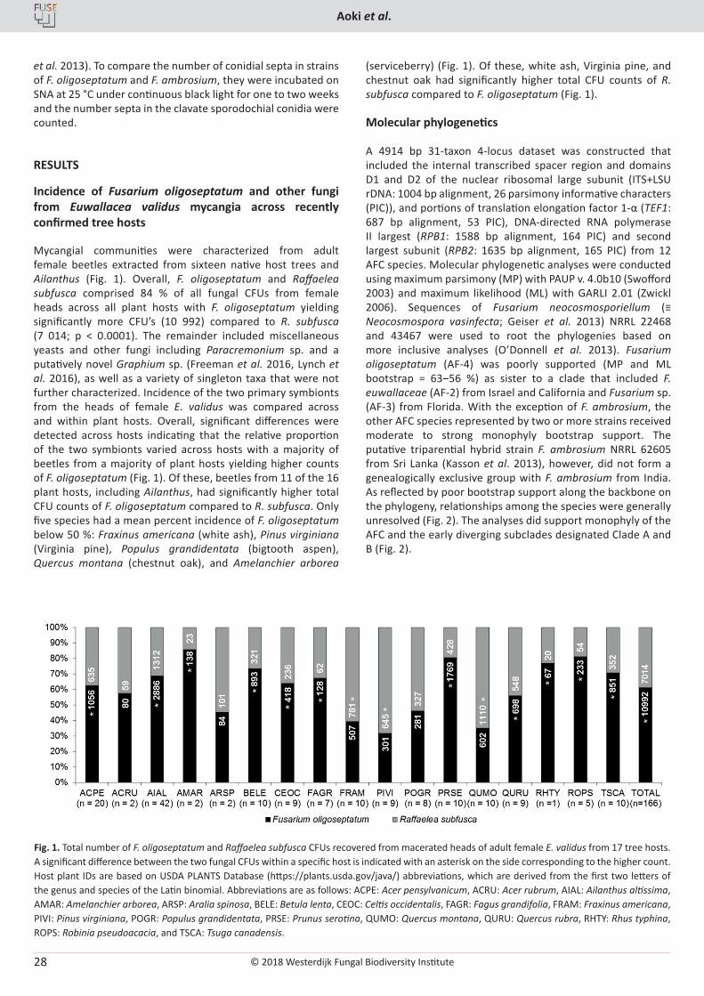
Mycangial communities were characterized from adult female beetles extracted from sixteen native host trees and Ailanthus (Fig. 1). Overall, F. oligoseptatum and Raffaelea subfusca comprised 84 % of all fungal CFUs from female heads across all plant hosts with F. oligoseptatum yielding significantly more CFU’s (10 992) compared to R. subfusca (7 014; p < 0.0001). The remainder included miscellaneous yeasts and other fungi including Paracremonium sp. and a putatively novel Graphium sp. (Freeman et al. 2016, Lynch et al. 2016), as well as a variety of singleton taxa that were not further characterized. Incidence of the two primary symbionts from the heads of female E. validus was compared across and within plant hosts. Overall, significant differences were detected across hosts indicating that the relative proportion of the two symbionts varied across hosts with a majority of beetles from a majority of plant hosts yielding higher counts of F. oligoseptatum (Fig. 1). Of these, beetles from 11 of the 16 plant hosts, including Ailanthus, had significantly higher total CFU counts of F. oligoseptatum compared to R. subfusca. Only five species had a mean percent incidence of F. oligoseptatum below 50 %: Fraxinus americana (white ash), Pinus virginiana (Virginia pine), Populus grandidentata (bigtooth aspen), Quercus montana (chestnut oak), and Amelanchier arborea
(serviceberry) (Fig. 1). Of these, white ash, Virginia pine, and chestnut oak had significantly higher total CFU counts of R. subfusca compared to F. oligoseptatum (Fig. 1).
Molecular phylogenetics
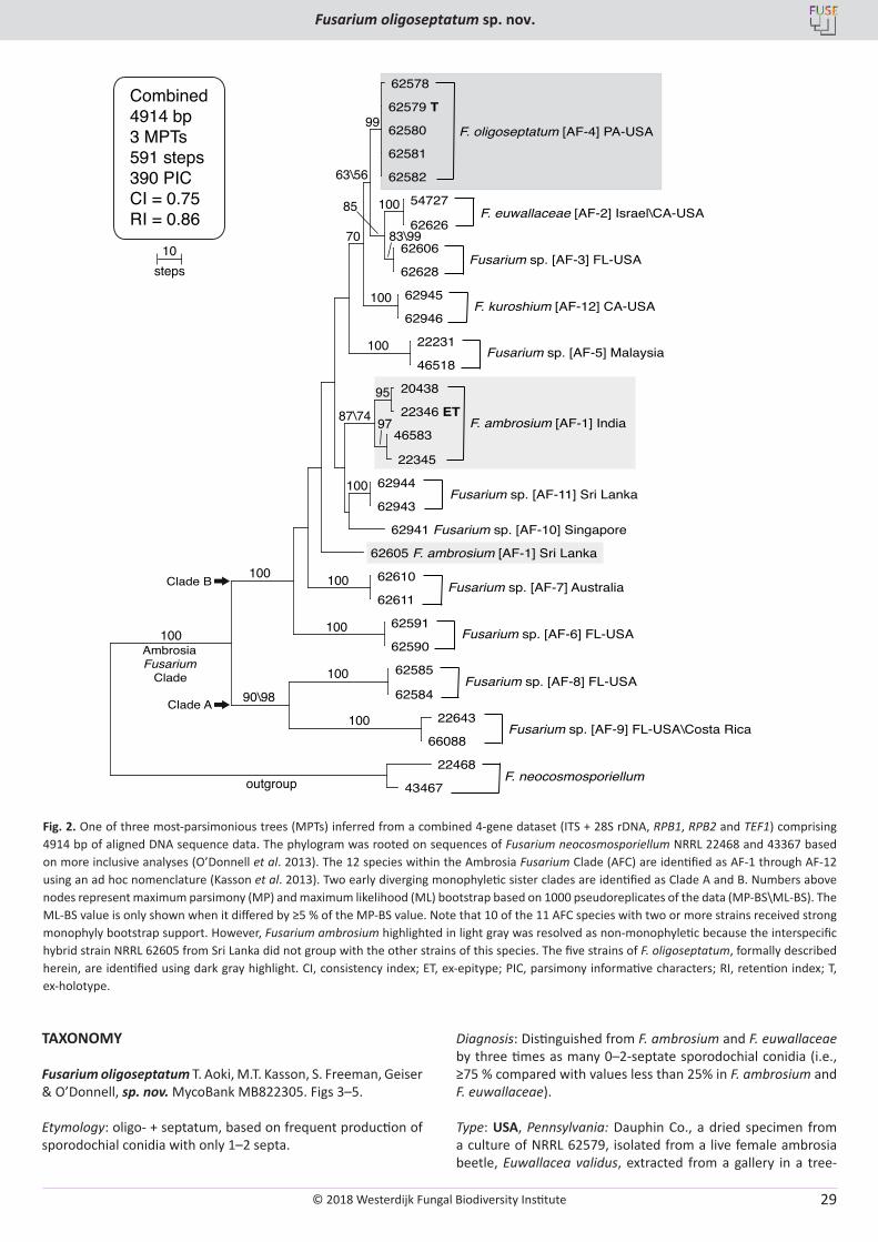
A 4914 bp 31-taxon 4-locus dataset was constructed that included the internal transcribed spacer region and domains D1 and D2 of the nuclear ribosomal large subunit (ITS+LSU rDNA: 1004 bp alignment, 26 parsimony informative characters (PIC)), and portions of translation elongation factor 1-α (TEF1: 687 bp alignment, 53 PIC), DNA-directed RNA polymerase II largest (RPB1: 1588 bp alignment, 164 PIC) and second largest subunit (RPB2: 1635 bp alignment, 165 PIC) from 12 AFC species. Molecular phylogenetic analyses were conducted using maximum parsimony (MP) with PAUP v. 4.0b10 (Swofford 2003) and maximum likelihood (ML) with GARLI 2.01 (Zwickl 2006). Sequences of Fusarium neocosmosporiellum (≡ Neocosmospora vasinfecta; Geiser et al. 2013) NRRL 22468 and 43467 were used to root the phylogenies based on more inclusive analyses (O’Donnell et al. 2013). Fusarium oligoseptatum (AF-4) was poorly supported (MP and ML bootstrap = 63‒56 %) as sister to a clade that included F. euwallaceae (AF-2) from Israel and California and Fusarium sp. (AF-3) from Florida. With the exception of F. ambrosium, the other AFC species represented by two or more strains received moderate to strong monophyly bootstrap support. The putative triparential hybrid strain F. ambrosium NRRL 62605 from Sri Lanka (Kasson et al. 2013), however, did not form a genealogically exclusive group with F. ambrosium from India. As reflected by poor bootstrap support along the backbone on the phylogeny, relationships among the species were generally unresolved (Fig. 2). The analyses did support monophyly of the AFC and the early diverging subclades designated Clade A and B (Fig. 2).
Fig. 1. Total number of F. oligoseptatum and Raffaelea subfusca CFUs recovered from macerated heads of adult female E. validus from 17 tree hosts. A significant difference between the two fungal CFUs within a specific host is indicated with an asterisk on the side corresponding to the higher count. Host plant IDs are based on USDA PLANTS Database (https://plants.usda.gov/java/) abbreviations, which are derived from the first two letters of the genus and species of the Latin binomial. Abbreviations are as follows: ACPE: Acer pensylvanicum, ACRU: Acer rubrum, AIAL: Ailanthus altissima, AMAR: Amelanchier arborea, ARSP: Aralia spinosa, BELE: Betula lenta, CEOC: Celtis occidentalis, FAGR: Fagus grandifolia, FRAM: Fraxinus americana, PIVI: Pinus virginiana, POGR: Populus grandidentata, PRSE: Prunus serotina, QUMO: Quercus montana, QURU: Quercus rubra, RHTY: Rhus typhina, ROPS: Robinia pseudoacacia, and TSCA: Tsuga canadensis.
© 2018 Westerdijk Fungal Biodiversity Institute
Fusarium oligoseptatum sp. nov.
Editor-in-ChiefProf. dr P.W. Crous, Westerdijk Fungal Biodiversity Institute, P.O. Box 85167, 3508 AD Utrecht, The Netherlands.E-mail:[email protected]
29
TAXONOMY
Fusarium oligoseptatum T. Aoki, M.T. Kasson, S. Freeman, Geiser & O’Donnell, sp. nov. MycoBank MB822305. Figs 3–5.
Etymology: oligo- + septatum, based on frequent production of sporodochial conidia with only 1–2 septa.
Diagnosis: Distinguished from F. ambrosium and F. euwallaceae by three times as many 0–2-septate sporodochial conidia (i.e., ≥75 % compared with values less than 25% in F. ambrosium and F. euwallaceae).
Type: USA, Pennsylvania: Dauphin Co., a dried specimen from a culture of NRRL 62579, isolated from a live female ambrosia beetle, Euwallacea validus, extracted from a gallery in a tree-
Fusarium sp. [AF-3] FL-USA
62578
62579 T
62580
62581
62582
54727
62626
62606
62628
62945
62946
22231
46518
20438
22346 ET
46583
22345
62944
62943
62941 Fusarium sp. [AF-10] Singapore
62605 F. ambrosium [AF-1] Sri Lanka
62610
62611
62591
62590
62585
62584
22643
66088
22468
43467
F. oligoseptatum [AF-4] PA-USA
F. ambrosium [AF-1] India
Fusarium sp. [AF-11] Sri Lanka
F. euwallaceae [AF-2] Israel\CA-USA
F. kuroshium [AF-12] CA-USA
Fusarium sp. [AF-5] Malaysia
Fusarium sp. [AF-7] Australia
Fusarium sp. [AF-6] FL-USA
Fusarium sp. [AF-8] FL-USA
Fusarium sp. [AF-9] FL-USA\Costa Rica
F. neocosmosporiellum
Combined4914 bp3 MPTs591 steps390 PICCI = 0.75RI = 0.86
99
63\56
100
100
100
100
100
100
100
100
100
100
87\74
83\9970
90\98
85
95
97
outgroup
10steps
Ambrosia Fusarium Clade
Clade B
Clade A
Fig. 2.
Fig. 2. One of three most-parsimonious trees (MPTs) inferred from a combined 4-gene dataset (ITS + 28S rDNA, RPB1, RPB2 and TEF1) comprising 4914 bp of aligned DNA sequence data. The phylogram was rooted on sequences of Fusarium neocosmosporiellum NRRL 22468 and 43367 based on more inclusive analyses (O’Donnell et al. 2013). The 12 species within the Ambrosia Fusarium Clade (AFC) are identified as AF-1 through AF-12 using an ad hoc nomenclature (Kasson et al. 2013). Two early diverging monophyletic sister clades are identified as Clade A and B. Numbers above nodes represent maximum parsimony (MP) and maximum likelihood (ML) bootstrap based on 1000 pseudoreplicates of the data (MP-BS\ML-BS). The ML-BS value is only shown when it differed by ≥5 % of the MP-BS value. Note that 10 of the 11 AFC species with two or more strains received strong monophyly bootstrap support. However, Fusarium ambrosium highlighted in light gray was resolved as non-monophyletic because the interspecific hybrid strain NRRL 62605 from Sri Lanka did not group with the other strains of this species. The five strains of F. oligoseptatum, formally described herein, are identified using dark gray highlight. CI, consistency index; ET, ex-epitype; PIC, parsimony informative characters; RI, retention index; T, ex-holotype.
© 2018 Westerdijk Fungal Biodiversity Institute
Aoki et al.
Editor-in-ChiefProf. dr P.W. Crous, Westerdijk Fungal Biodiversity Institute, P.O. Box 85167, 3508 AD Utrecht, The Netherlands.E-mail:[email protected]
30
of-heaven, Ailanthus altissima, 30 Jan. 2010, Matthew T. Kasson (Kasson Bh24) (BPI 910525 – holotype, designated in this study; NRRL 62579 = FRC S-2581 = MAFF 246283 = CBS 143241 – ex-holotype cultures).
Additional strains examined: USA: Pennsylvania: Franklin Co., isolated from a live E. validus female infesting an A. altissima tree, 9 Mar. 2010, Matthew T. Kasson (Kasson Ch19) (NRRL 62580 = FRC S-2594 = MAFF 246284 = CBS 143242); Pennsylvania: Huntingdon Co., isolated from a live E. validus female infesting an A. altissima tree, 27 Feb. 2010, Matthew T. Kasson (Kasson Dh24) (NRRL 62581 = FRC S-2616 = MAFF 246285 = CBS 143243; Pennsylvania: Mifflin Co., isolated from a live E. validus female infesting an A. altissima tree, 1 July 2009, Matthew T. Kasson (Kasson Eh11) (NRRL 62582 = FRC S-2627 = MAFF 246286 = CBS 143244; morphologically degenerated strain).
Description: Colonies on PDA showing radial mycelial growth rates of 2.2–3.6 mm/d at 20 °C and 3.3–4.6 mm/d at 25 °C in the dark. Colony color on PDA white (1A1) to yellowish-white (4A2) or orange white (5A2) in the dark, white (1A1) to yellowish-white (3–4A2) or pale yellow (3–4A3) under black light. Aerial mycelium white (1A1), sparsely formed or floccose in the dark,
more abundantly formed and covering entire surface of colonies under black light. Colony margin entire to undulate. Reverse pigmentation absent or yellowish-white (3–4A2) or pale yellow (3–4A3) in the dark and under black light. Exudates absent. Odor absent, or slightly moldy or sweet in some strains. Hyphae on SNA 1.5–7.5 µm wide. Chlamydospores present but formation delayed in hyphae and in septate sporodochial conidia, mostly subglobose to round ellipsoidal, intercalary or terminal, mostly single, sometimes in chains, ordinary hyaline to very pale-yellow, wall smooth or often minutely roughened, 6–23.5 × 4.5–9 µm. Sclerotia absent. Sporulation on SNA and PDA generally rapid and abundant under black light, delayed in the dark, sometimes less sporulation on PDA in the dark; light-colored on SNA and PDA under black light or under daylight; sporodochia formed sparsely on SNA, rare on PDA. Aerial conidiophores formed abundantly on SNA under black light, less frequently in the dark, erect, short or tall and narrow, mostly unbranched, rarely branched sparsely, up to 130 µm long, 3–5.5 µm wide at base, thin-walled, forming monophialides integrated in the apices. Phialides on aerial conidiophores simple, subcylindrical to subulate, tapering towards apex, often with a minute collarette at the tip, 10–62.5 × 2.5–5.5 µm. Aerial conidia mostly (1) elliptical, oblong-elliptical,
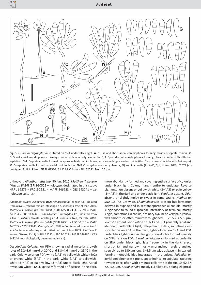
Fig. 3. Fusarium oligoseptatum cultured on SNA under black light. A, B. Tall and short aerial conidiophores forming mostly 0-septate conidia. C, D. Short aerial conidiophores forming conidia with relatively few septa. E, F. Sporodochial conidiophores forming clavate conidia with different septation. G‒L. Septate conidia formed on sporodochial conidiophores, with some large clavate conidia (G‒I: Short clavate conidia with 1‒2 septa). M. 0-septate conidia formed on aerial conidiophores. N‒P. Chlamydospores in hyphae (N, O) and in conidia (P). A‒D, G, J, N from NRRL 62579 (ex-holotype); E, H, L, P from NRRL 62580; F, I, K, M, O from NRRL 62581. Bar = 25 µm.
© 2018 Westerdijk Fungal Biodiversity Institute
Fusarium oligoseptatum sp. nov.
Editor-in-ChiefProf. dr P.W. Crous, Westerdijk Fungal Biodiversity Institute, P.O. Box 85167, 3508 AD Utrecht, The Netherlands.E-mail:[email protected]
31
fusiform-elliptical to short clavate, occasionally reniform, some obovate to subglobose, 0–1(–2)-septate; 0-septate on SNA in the dark: 3–13 × 2–5.5 µm in total range, 5.3–8.5 × 2.8–3.9 µm on average [ex type (NRRL 62579): 3.5–12 × 2–4 µm in total range, 6.9±2.0 × 2.8±0.5 µm on average ± S.D.]; 0-septate on SNA under black light: 3–17 × 2–6.5 µm in total range, 6.0–9.0 × 2.8–3.8
µm on average [ex type (NRRL 62579): 4–17 × 2.5–6.5 µm in total range, 9.0±2.8 × 3.8±0.9 µm on average ± S.D.]; 1-septate on SNA under black light: 7.5–26 × 2.5–8 µm in total range, 14.5–15.3 × 4.6–4.9 µm on average [ex type: 10.5–21.5 × 2.5–6 µm in total range, 15.1±2.7 × 4.6±0.7 µm on average ± S.D.]; sometimes with (2) larger, falcate to clavate, or curved cylindrical,
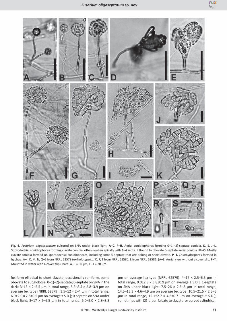
Fig. 4. Fusarium oligoseptatum cultured on SNA under black light. A‒C, F‒H. Aerial conidiophores forming 0‒1(‒2)-septate conidia. D, E, J‒L. Sporodochial conidiophores forming clavate conidia, often swollen apically with 1‒4 septa. I. Round to obovate 0-septate aerial conidia. M‒O. Mostly clavate conidia formed on sporodochial conidiophores, including some 0-septate that are oblong or short-clavate. P‒T. Chlamydospores formed in hyphae. A‒I, K, M, N, Q‒S from NRRL 62579 (ex-holotype); J, O, P, T from NRRL 62580; L from NRRL 62581. (A‒E: Aerial view without a cover slip; F‒T: Mounted in water with a cover slip). Bars: A‒E = 50 μm, F‒T = 20 μm.
© 2018 Westerdijk Fungal Biodiversity Institute
Aoki et al.
Editor-in-ChiefProf. dr P.W. Crous, Westerdijk Fungal Biodiversity Institute, P.O. Box 85167, 3508 AD Utrecht, The Netherlands.E-mail:[email protected]
32
(1–)2(–3)-septate conidia, morphologically continuous with falcate sporodochial conidia. Sporodochial conidiophores generally shorter and thicker than aerial conidiophores, unbranched or sometimes sparsely branched, contorted, forming monophialides integrated apically, 20–145 × 3–6 µm, or sometimes adelophialides. Sporodochial phialides simple, subulate, lanceolate or subcylindrical, often with a conspicuous collarette at the tip, 9.5–44 × 2.5–5.5 µm. Sporodochial conidia hyaline, mostly falcate to long clavate, sometimes curved cylindrical, often swollen slightly or conspicuously in their upper part, tapering towards base, often with a rounded or papillate apical cell, and an indistinct foot-like or rounded basal cell, (0–)1–3(–5)-septate; swollen conidia sometimes ‘dolphin-like’ (Brayford 1987) or comma-shaped when 1- to 2-septate, formed on SNA frequently under black light, less frequently in the dark, very rarely formed on PDA under black light; 1-septate on SNA under black light: 11–32.5 × 4–10 µm in total range, 18.2–20.1 × 6.1–7.2 µm on average [ex type: 13.5–30 × 4.5–8.5 µm in total range, 19.3±3.9 × 6.2±1.0 µm on average ± S.D.]; 2-septate on SNA under black light: 15.5–39.5 × 5.5–12 µm in total range, 24.8–26.5 × 7.4–8.0 µm on average [ex type: 22–34 × 5.5–12 µm in total range, 26.1±3.2 × 7.4±1.2 µm on average ± S.D.]; 3-septate on SNA under black light: 20–60 × 5.5–12.5 µm in total range, 31.4–35.7 × 8.3–8.7 µm on average [ex type: 23–60 × 5.5–12.5 µm in total range, 35.7±7.2 × 8.7±1.3 µm on average ± S.D.]; 4-septate on SNA under black light: 28.5–67.5 × 7–11 µm in total range, 38.7–47.9 × 8.8–8.9 µm on average [ex type: 28.5–67.5 × 7–10 µm in total range, 47.9 × 8.9 µm on average]. Together with multiseptate sporodochial conidia, often forming (0-)1(-2)-septate, oblong to naviculate or short-clavate, straight or curved conidia with a rounded apex and truncate base.
Clade-based diagnosis: Distinguished by phylogenetic analysis of multilocus DNA sequence data (Kasson et al. 2013, O’Donnell et al. 2015).
Substrates or hosts: All ex-holotype and authentic strains were isolated from E. validus in the galleries of A. altissima in Pennsylvania (PA), USA. Fusarium oligoseptatum has also been confirmed using multilocus sequence typing from Ohio (OH), Virginia (VA) and Maryland (MD) (O’Donnell et al. 2015), and from Tennessee (TN) and (West Virginia) WV using the AF-3 / F. oligoseptatum (AF-4) multiplex PCR assay (Short et al. 2017). Currently known from 16 additional plant hosts, all of which
have been confirmed molecularly as F. oligoseptatum using AF-3 / F. oligoseptatum (AF-4) multiplex PCR (Short et al. 2017): Acer pensylvanicum (PA, USA), Acer rubrum (PA, USA), Amelanchier arborea (VA, USA), Aralia spinosa (PA, USA), Betula lenta (PA, USA), Celtis occidentalis (WV, USA), Fagus grandifolia (OH, USA), Fraxinus americana (WV, USA), Populus grandidentata (PA, USA), Prunus serotina (GA, USA), Quercus montana (PA, USA), Quercus rubra (PA, USA), Rhus typhina (PA, USA), Robinia pseudoacacia (PA, USA), Tsuga canadensis (OH, USA), and Pinus virginiana (VA, USA).
Distribution: Presently confirmed from GA (Georgia), MD, OH, PA, TN, VA, and WV, USA.
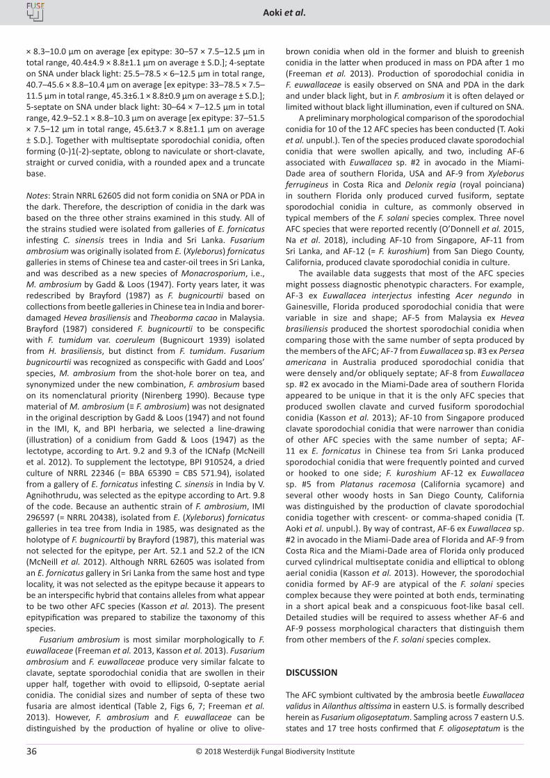
Notes: Morphological data on sporodochial conidia was based mainly on NRRL 62579, 62580 and 62581. Strain NRRL 62582 appears degenerated and produced only 1-septate sporodochial conidia after 1 mo on SNA under continuous black light. Strains of this species were all isolated from female E. validus ambrosia beetles infesting A. altissima that were collected in different counties in Pennsylvania, USA. The most distinctive morphological feature of this fungus is the frequent production of sporodochial conidia with 1–2 septa (Table 2, Figs. 3E–I, 4E, J–O). This species formed sporodochial conidia with more than two septa, but the percentage of 0–2-septate conidia (76.5–81 %) was much higher than observed in F. ambrosium (3.7–24.5 %) and F. euwallaceae (Freeman et al. 2013), where more than 75 % of the conidia were 3–5-septate. Cultures appear whitish to yellowish-white when aerial mycelium is sparse on PDA.
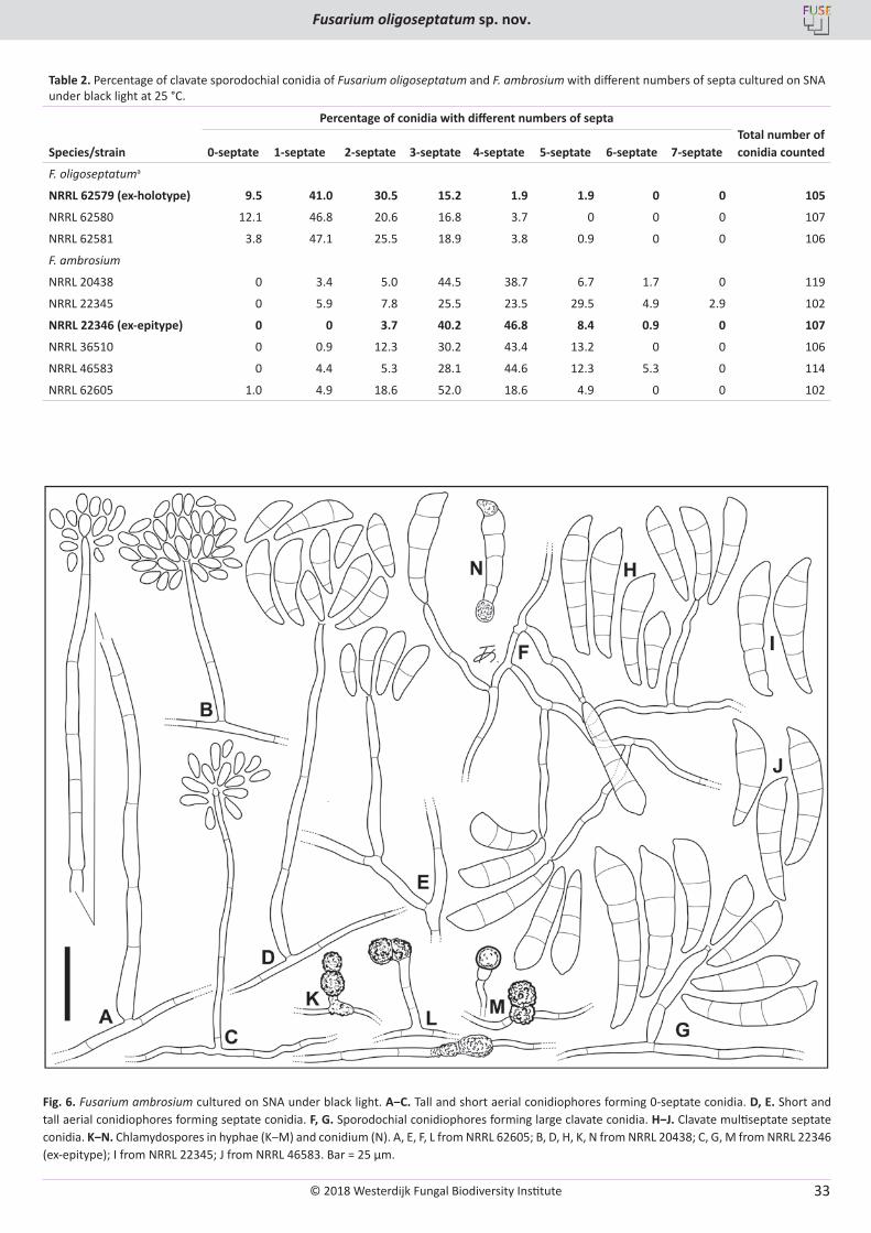
Fusarium ambrosium (Gadd & Loos) Agnihothr. & Nirenberg, Stud. Mycol. 32: 98. 1990. MycoBank MB130225. Figs 6–8.Basionym: Monacrosporium ambrosium Gadd & Loos, Trans. Brit. Mycol. Soc. 31(1 & 2): 13. 1947. MB288427. Synonyms: Dactylella ambrosia (Gadd & Loos) K.Q. Zhang, Xing Z. Liu & L. Cao, Mycosystema 7: 112. 1995. MB447506.Neocosmospora ambrosia (Gadd & Loos) L. Lombard & Crous, Stud. Mycol. 80: 227. 2015. MB810957.Fusarium bugnicourtii Brayford, Trans. Brit. Mycol. Soc. 89 (3): 350. 1987. MB133337.
Type: India, Upari Tea Institute, a dried specimen from culture of NRRL 22346, isolated from a gallery of Euwallacea fornicatus infesting a tea tree, Camellia sinensis, 9 May 1990, V. Agnihothrudu
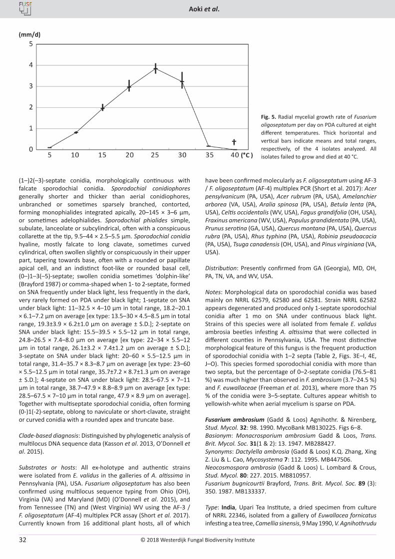
Fig. 5. Radial mycelial growth rate of Fusarium oligoseptatum per day on PDA cultured at eight different temperatures. Thick horizontal and vertical bars indicate means and total ranges, respectively, of the 4 isolates analyzed. All isolates failed to grow and died at 40 °C.
© 2018 Westerdijk Fungal Biodiversity Institute
Fusarium oligoseptatum sp. nov.
Editor-in-ChiefProf. dr P.W. Crous, Westerdijk Fungal Biodiversity Institute, P.O. Box 85167, 3508 AD Utrecht, The Netherlands.E-mail:[email protected]
33
Table 2. Percentage of clavate sporodochial conidia of Fusarium oligoseptatum and F. ambrosium with different numbers of septa cultured on SNA under black light at 25 °C.
Percentage of conidia with different numbers of septa Total number of conidia countedSpecies/strain 0-septate 1-septate 2-septate 3-septate 4-septate 5-septate 6-septate 7-septate
F. oligoseptatuma
NRRL 62579 (ex-holotype) 9.5 41.0 30.5 15.2 1.9 1.9 0 0 105
NRRL 62580 12.1 46.8 20.6 16.8 3.7 0 0 0 107
NRRL 62581 3.8 47.1 25.5 18.9 3.8 0.9 0 0 106
F. ambrosium
NRRL 20438 0 3.4 5.0 44.5 38.7 6.7 1.7 0 119
NRRL 22345 0 5.9 7.8 25.5 23.5 29.5 4.9 2.9 102
NRRL 22346 (ex-epitype) 0 0 3.7 40.2 46.8 8.4 0.9 0 107
NRRL 36510 0 0.9 12.3 30.2 43.4 13.2 0 0 106
NRRL 46583 0 4.4 5.3 28.1 44.6 12.3 5.3 0 114
NRRL 62605 1.0 4.9 18.6 52.0 18.6 4.9 0 0 102
Fig. 6. Fusarium ambrosium cultured on SNA under black light. A‒C. Tall and short aerial conidiophores forming 0-septate conidia. D, E. Short and tall aerial conidiophores forming septate conidia. F, G. Sporodochial conidiophores forming large clavate conidia. H‒J. Clavate multiseptate septate conidia. K‒N. Chlamydospores in hyphae (K‒M) and conidium (N). A, E, F, L from NRRL 62605; B, D, H, K, N from NRRL 20438; C, G, M from NRRL 22346 (ex-epitype); I from NRRL 22345; J from NRRL 46583. Bar = 25 µm.
© 2018 Westerdijk Fungal Biodiversity Institute
Aoki et al.
Editor-in-ChiefProf. dr P.W. Crous, Westerdijk Fungal Biodiversity Institute, P.O. Box 85167, 3508 AD Utrecht, The Netherlands.E-mail:[email protected]
34
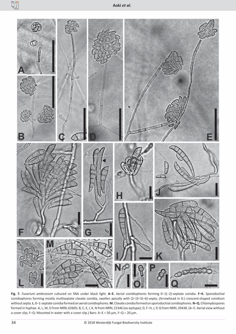
Fig. 7. Fusarium ambrosium cultured on SNA under black light. A‒E. Aerial conidiophores forming 0‒1(‒2)-septate conidia. F‒K. Sporodochial conidiophores forming mostly multiseptate clavate conidia, swollen apically with (2‒)3‒5(‒6)-septa; (Arrowhead in G:) crescent-shaped conidium without septa. L. 0‒1-septate conidia formed on aerial conidiophores. M. Clavate conidia formed on sporodochial conidiophores. N‒Q. Chlamydospores formed in hyphae. A, L, M, O from NRRL 62605; B, C, E, I, K, N from NRRL 22346 (ex-epitype); D, F‒H, J, P, Q from NRRL 20438. (A‒E: Aerial view without a cover slip; F‒Q: Mounted in water with a cover slip.) Bars: A‒E = 50 µm, F‒Q = 20 µm.
© 2018 Westerdijk Fungal Biodiversity Institute
Fusarium oligoseptatum sp. nov.
Editor-in-ChiefProf. dr P.W. Crous, Westerdijk Fungal Biodiversity Institute, P.O. Box 85167, 3508 AD Utrecht, The Netherlands.E-mail:[email protected]
35
(BPI 910524 – epitype MBT 378232, designated in this study; NRRL 22346 = BBA 65390 = CBS 571.94 = MAFF 246287 – ex-epitype cultures). Sri Lanka, an illustration of a mature conidium of the fungus taken from a gallery of Euwallacea fornicatus infesting a tea tree, Camellia sinensis, C.H. Gadd & C.A. Loos (Trans. Brit. Mycol. Soc. 31 (1 & 2): 16, Text-fig. 5 (1987) – lectotype, MBT379562, designated in this study; the original description by Gadd & Loos (1947) did not designate type material of M. ambrosium and it was not found in the IMI, K, and BPI herbaria).
Additional strains examined: India, Maharashtra: Chinchona, isolated from a gallery of E. fornicatus infesting a C. sinensis tree, 17 Jul. 1985, (unknown collector) (NRRL 20438 = IMI 296597 = MAFF 246291, preserved as the ex-holotype strain of F. bugnicourtii); (State name of India not recorded): Upari Tea Institute, isolated from a gallery of E. fornicatus infesting C. sinensis tree, 9 May 1990, V. Agnihothrudu (NRRL 22345 = BBA 65389 = MAFF 246288; NRRL 36510 = BBA 65390 = MAFF 246289 as a duplicate of NRRL 22346); Tamil Nadu: Coimbatore District, Anamallais, High Forest Estate, isolated from a gallery of E. fornicatus infesting stem of C. sinensis tree, 26 Mar. 1990, (unknown collector) (NRRL 46583 = IMI 339338 = MAFF 246290, preserved as F. bugnicourtii). Sri Lanka, Talawakelle, St. Coombs, Tea Research Institute of Sri Lanka, isolated from a gallery of E. fornicatus infesting C. sinensis tree, Mar. 2012, S. Freeman (NRRL 62605).
Description: Colonies on PDA showing radial mycelial growth rates of 1.9–3.3 mm/d at 20 °C and 3.0–4.5 mm/d at 25 °C in the dark. Colony color on PDA white (1A1) to yellowish-white (4A2) in the dark, white (1A1) to yellowish-white (3–4A2) or pale yellow (4A3) under black light. Aerial mycelium white (1A1) sparse to floccose in the dark, more abundantly formed and often covering entire surface of colonies under black light. Colony margin entire to undulate. Reverse pigmentation absent or yellowish-white (3–4A2) or pale yellow (4A3) in the dark and under black light. Olive (3E–F5–8) to olive-brown (4E–F5–8) spots in some strains when sporodochia formed on PDA. Exudates absent. Odor absent, or slightly moldy or sweet in some strains. Hyphae on SNA 1.5–9.5 µm wide. Chlamydospores present in hyphae and in septate sporodochial conidia, mostly subglobose to round ellipsoidal, intercalary or terminal, single or often in chains, hyaline or slightly pale yellow, smooth to often minutely rough-walled, 5–31.3 × 4.5–13 µm. Sclerotia absent. Sporulation on SNA and PDA generally rapid and abundant under black light, less abundant in some strains, delayed or sometimes less production
in the dark; generally light-colored on SNA and PDA under black light or under daylight; sporodochia formed sparsely on SNA, and rarely on PDA under daylight; olive (3E–F5–8) to olive brown (4E–F5–8) when produced in mass. Aerial conidiophores formed abundantly on SNA under black light, erect, short or tall, mostly narrow but rarely thicker, mostly unbranched, rarely branched sparsely, up to 320 µm long, 2.5–7 µm wide at base, thin-walled, forming monophialides integrated in the apices. Phialides on aerial conidiophores simple, subcylindrical to subulate, tapering towards apex, often with a minute collarette at the tip, 15–66 × 2.5–4 µm. Aerial conidia typically (1) elliptical, oblong-elliptical, fusiform-elliptical to short clavate, occasionally reniform, some obovate, 0–1-septate; 0-septate on SNA in the dark: 3.5–14 × 1.5–7 µm in total range, 8.4–9.4 × 2.8–3.3 µm on average [ex epitype (NRRL 22346): 3.5–14 × 2–5.5 µm in total range, 9.4±2.4 × 3.2±0.6 µm on average ± S.D.]; 0-septate on SNA under black light: 3.5–22 × 2–7.5 µm in total range, 7.8–10.8 × 3.5–4.5 µm on average [ex epitype (NRRL 22346): 4.5–18.5 × 2–7.5 µm in total range, 10.2±2.4 × 4.1±1.1 µm on average ± S.D.]; 1-septate on SNA in the dark: 8–26 × 2.5–6.5 µm in total range, 13.6–16.3 × 3.7–4.9 µm on average [ex epitype: 12–19 × 3.5–5.5 µm in total range, 15.3±1.9 × 4.8±0.4 µm on average ± S.D.]; 1-septate on SNA under black light: 8.5–37 × 2.5–10.5 µm in total range, 15.3–20.1 × 5.4–6.6 µm on average [ex epitype: 10.5–36 × 2.5–10.5 µm in total range, 19.3±5.1 × 6.2±1.5 µm on average ± S.D.]; sometimes with (2) larger, falcate to clavate, or curved clavate, (1–)2(–3)-septate conidia, morphologically continuous with falcate sporodochial conidia. Sporodochial conidiophores generally short, unbranched or rarely sparsely branched, contorted, monophialides integrated apically, 20–61.3 × 3.5–5 µm. Sporodochial phialides simple, subulate, lanceolate or subcylindrical, often with a conspicuous collarette at the tip, 13–60 × 3–6 µm. Sporodochial conidia hyaline, mostly falcate to long clavate, sometimes curved cylindrical, mostly swollen in the upper part, tapering towards base, often with a round or papillate apical cell, and a distinct or indistinct foot-like, or rounded basal cell, swollen conidia often appear “dolphin-like” (Brayford 1987), (0–)2–5(–7)-septate, formed on SNA under black light, less frequently in the dark, sometimes formed on PDA under black light, rarely in the dark; 2-septate on SNA under black light: 15–61.5 × 3–12 µm in total range, 24.7–32.2 × 6.9–9.1 µm on average [ex-epitype: 15–61.5 × 4–10 µm in total range, 28.3±7.8 × 7.8±1.2 µm on average ± S.D.]; 3-septate on SNA under black light: 21–57.5 × 3.5–13 µm in total range, 34.1–40.4
Fig. 8. Radial mycelial growth rate of Fusarium ambrosium per day on PDA cultured at eight different temperatures. Thick horizontal and vertical bars indicate means and total ranges, respectively, of the 6 isolates analyzed. All isolates failed to grow and died at 40 °C.
© 2018 Westerdijk Fungal Biodiversity Institute
Aoki et al.
Editor-in-ChiefProf. dr P.W. Crous, Westerdijk Fungal Biodiversity Institute, P.O. Box 85167, 3508 AD Utrecht, The Netherlands.E-mail:[email protected]
36
× 8.3–10.0 µm on average [ex epitype: 30–57 × 7.5–12.5 µm in total range, 40.4±4.9 × 8.8±1.1 µm on average ± S.D.]; 4-septate on SNA under black light: 25.5–78.5 × 6–12.5 µm in total range, 40.7–45.6 × 8.8–10.4 µm on average [ex epitype: 33–78.5 × 7.5–11.5 µm in total range, 45.3±6.1 × 8.8±0.9 µm on average ± S.D.]; 5-septate on SNA under black light: 30–64 × 7–12.5 µm in total range, 42.9–52.1 × 8.8–10.3 µm on average [ex epitype: 37–51.5 × 7.5–12 µm in total range, 45.6±3.7 × 8.8±1.1 µm on average ± S.D.]. Together with multiseptate sporodochial conidia, often forming (0-)1(-2)-septate, oblong to naviculate or short-clavate, straight or curved conidia, with a rounded apex and a truncate base.
Notes: Strain NRRL 62605 did not form conidia on SNA or PDA in the dark. Therefore, the description of conidia in the dark was based on the three other strains examined in this study. All of the strains studied were isolated from galleries of E. fornicatus infesting C. sinensis trees in India and Sri Lanka. Fusarium ambrosium was originally isolated from E. (Xyleborus) fornicatus galleries in stems of Chinese tea and caster-oil trees in Sri Lanka, and was described as a new species of Monacrosporium, i.e., M. ambrosium by Gadd & Loos (1947). Forty years later, it was redescribed by Brayford (1987) as F. bugnicourtii based on collections from beetle galleries in Chinese tea in India and borer-damaged Hevea brasiliensis and Theoborma cacao in Malaysia. Brayford (1987) considered F. bugnicourtii to be conspecific with F. tumidum var. coeruleum (Bugnicourt 1939) isolated from H. brasiliensis, but distinct from F. tumidum. Fusarium bugnicourtii was recognized as conspecific with Gadd and Loos’ species, M. ambrosium from the shot-hole borer on tea, and synonymized under the new combination, F. ambrosium based on its nomenclatural priority (Nirenberg 1990). Because type material of M. ambrosium (≡ F. ambrosium) was not designated in the original description by Gadd & Loos (1947) and not found in the IMI, K, and BPI herbaria, we selected a line-drawing (illustration) of a conidium from Gadd & Loos (1947) as the lectotype, according to Art. 9.2 and 9.3 of the ICNafp (McNeill et al. 2012). To supplement the lectotype, BPI 910524, a dried culture of NRRL 22346 (= BBA 65390 = CBS 571.94), isolated from a gallery of E. fornicatus infesting C. sinensis in India by V. Agnihothrudu, was selected as the epitype according to Art. 9.8 of the code. Because an authentic strain of F. ambrosium, IMI 296597 (= NRRL 20438), isolated from E. (Xyleborus) fornicatus galleries in tea tree from India in 1985, was designated as the holotype of F. bugnicourtii by Brayford (1987), this material was not selected for the epitype, per Art. 52.1 and 52.2 of the ICN (McNeill et al. 2012). Although NRRL 62605 was isolated from an E. fornicatus gallery in Sri Lanka from the same host and type locality, it was not selected as the epitype because it appears to be an interspecific hybrid that contains alleles from what appear to be two other AFC species (Kasson et al. 2013). The present epitypification was prepared to stabilize the taxonomy of this species.
Fusarium ambrosium is most similar morphologically to F. euwallaceae (Freeman et al. 2013, Kasson et al. 2013). Fusarium ambrosium and F. euwallaceae produce very similar falcate to clavate, septate sporodochial conidia that are swollen in their upper half, together with ovoid to ellipsoid, 0-septate aerial conidia. The conidial sizes and number of septa of these two fusaria are almost identical (Table 2, Figs 6, 7; Freeman et al. 2013). However, F. ambrosium and F. euwallaceae can be distinguished by the production of hyaline or olive to olive-
brown conidia when old in the former and bluish to greenish conidia in the latter when produced in mass on PDA after 1 mo (Freeman et al. 2013). Production of sporodochial conidia in F. euwallaceae is easily observed on SNA and PDA in the dark and under black light, but in F. ambrosium it is often delayed or limited without black light illumination, even if cultured on SNA.
A preliminary morphological comparison of the sporodochial conidia for 10 of the 12 AFC species has been conducted (T. Aoki et al. unpubl.). Ten of the species produced clavate sporodochial conidia that were swollen apically, and two, including AF-6 associated with Euwallacea sp. #2 in avocado in the Miami-Dade area of southern Florida, USA and AF-9 from Xyleborus ferrugineus in Costa Rica and Delonix regia (royal poinciana) in southern Florida only produced curved fusiform, septate sporodochial conidia in culture, as commonly observed in typical members of the F. solani species complex. Three novel AFC species that were reported recently (O’Donnell et al. 2015, Na et al. 2018), including AF-10 from Singapore, AF-11 from Sri Lanka, and AF-12 (= F. kuroshium) from San Diego County, California, produced clavate sporodochial conidia in culture.
The available data suggests that most of the AFC species might possess diagnostic phenotypic characters. For example, AF-3 ex Euwallacea interjectus infesting Acer negundo in Gainesville, Florida produced sporodochial conidia that were variable in size and shape; AF-5 from Malaysia ex Hevea brasiliensis produced the shortest sporodochial conidia when comparing those with the same number of septa produced by the members of the AFC; AF-7 from Euwallacea sp. #3 ex Persea americana in Australia produced sporodochial conidia that were densely and/or obliquely septate; AF-8 from Euwallacea sp. #2 ex avocado in the Miami-Dade area of southern Florida appeared to be unique in that it is the only AFC species that produced swollen clavate and curved fusiform sporodochial conidia (Kasson et al. 2013); AF-10 from Singapore produced clavate sporodochial conidia that were narrower than conidia of other AFC species with the same number of septa; AF-11 ex E. fornicatus in Chinese tea from Sri Lanka produced sporodochial conidia that were frequently pointed and curved or hooked to one side; F. kuroshium AF-12 ex Euwallacea sp. #5 from Platanus racemosa (California sycamore) and several other woody hosts in San Diego County, California was distinguished by the production of clavate sporodochial conidia together with crescent- or comma-shaped conidia (T. Aoki et al. unpubl.). By way of contrast, AF-6 ex Euwallacea sp. #2 in avocado in the Miami-Dade area of Florida and AF-9 from Costa Rica and the Miami-Dade area of Florida only produced curved cylindrical multiseptate conidia and elliptical to oblong aerial conidia (Kasson et al. 2013). However, the sporodochial conidia formed by AF-9 are atypical of the F. solani species complex because they were pointed at both ends, terminating in a short apical beak and a conspicuous foot-like basal cell. Detailed studies will be required to assess whether AF-6 and AF-9 possess morphological characters that distinguish them from other members of the F. solani species complex.
DISCUSSION
The AFC symbiont cultivated by the ambrosia beetle Euwallacea validus in Ailanthus altissima in eastern U.S. is formally described herein as Fusarium oligoseptatum. Sampling across 7 eastern U.S. states and 17 tree hosts confirmed that F. oligoseptatum is the
© 2018 Westerdijk Fungal Biodiversity Institute
Fusarium oligoseptatum sp. nov.
Editor-in-ChiefProf. dr P.W. Crous, Westerdijk Fungal Biodiversity Institute, P.O. Box 85167, 3508 AD Utrecht, The Netherlands.E-mail:[email protected]
37
primary symbiont of E. validus and dominant, regardless of plant host with few exceptions. This species can be distinguished from the 11 other known AFC species by producing significantly more 0–2-septate clavate sporodochial conidia that are swollen apically and via multilocus molecular phylogenetics where it was strongly supported as a genealogically exclusive species-level lineage in the analyses reported here and in previous studies (Kasson et al. 2013, O’Donnell et al. 2015, Na et al. 2018). Fusarium oligoseptatum was strongly supported as a reciprocally monophyletic sister to F. euwallaceae + Fusarium sp. (AF-3) in Kasson et al. (2013), but the sister group relationship of F. oligoseptatum was unresolved in analyses that included the closely related F. kuroshium (AF-12) from San Diego, California (O’Donnell et al. 2015, and present study). Efforts to develop a robust hypothesis of evolutionary relationships among these four AFC species, which are estimated to have shared a most recent common ancestor approximately 1.6 Mya (O’Donnell et al. 2015), might benefit from the comparative phylogenomic analyses that are currently underway (Stajich et al., pers. comm.).
Herein, an epitype of F. ambrosium was designated based on material originally isolated from a gallery of E. fornicatus infesting Chinese tea in India to provide nomenclatural stability for this species. AFC species have been collected in eight different countries, including Sri Lanka (F. ambrosium AF-1 and Fusarium sp. AF-11), India (F. ambrosium AF-1), Malaysia (Fusarium sp. AF-5), Singapore (Fusarium sp. AF-10), Australia (Fusarium sp. AF-7), Israel (F. euwallaceae AF-2), Costa Rica (Fusarium sp. AF-9) and the United States (F. euwallaceae AF-2, Fusarium sp. AF-3, F. oligoseptatum AF-4, Fusarium spp. AF-6, AF-8, AF-9 and F. kuroshium AF-12)(Brayford 1987, Nirenberg 1990, Freeman et al. 2013, Kasson et al. 2013, O’Donnell et al. 2015, Short et al. 2017, Na et al. 2018). To date only three species within the AFC have been described formally, i.e., F. ambrosium (AF-1; Gadd & Loos 1947, Nirenberg 1990), F. euwallaceae (AF-2; Freeman et al. 2013) and F. oligoseptatum (AF-4; in this study). Although nine of the AFC species are currently unnamed, the prospects for naming them are excellent because most of them appear to possess unique phenotypic/morphological features. Delimitations of such features may, in time, help to uncover the mechanisms underlying the production of clavate conidia, a posited adaptation for the Euwallacea − Fusarium symbiosis (Kasson et al. 2013). Indeed, analogous adaptations in agaricalean fungi (i.e., gongylidia) farmed by higher and occasionally lower attine ants (Schultz & Brady 2008, Masiulionis et al. 2014) also appear to exhibit variation among closely related lineages. However, quality of the substrate, pH, and temperature have also been shown to affect the growth and size of gongylidia in some higher attine ant cultivars when cultivated under lab conditions (Powell & Stradling 1986).
It remains unclear whether F. ambrosium, F. euwallaceae, or F. oligoseptatum are each farmed by a single Euwallacea species, including within their native range, where evidence of hybridization and co-cultivation with other closely related AFC members have been reported (O’Donnell et al. 2015). However, it has been shown that F. euwallaceae from avocado is obligately required for the survival and development of Euwallacea sp. #1 sensu O’Donnell et al. (2015) currently occurring in Israel, whereas F. ambrosium does not support development of this beetle species (Freeman et al. 2012). Likewise, specificity exists for F. ambrosium and its beetle host. Future studies focused on vector specificity could help clarify the threats these beetle-fungus consortia pose to our
native ecosystems. This is especially important given that some AFC members such as F. euwallaceae have caused significant damage to orchard, landscape and forest trees and threaten avocado production worldwide (Mendel et al. 2012, Eskalen et al. 2013, Kasson et al. 2013), while other AFC members such as F. oligoseptatum appear to be quite innocuous when challenged against numerous plant species (Berger 2017).
The FSSC includes over 60 species (Zhang et al. 2006, O’Donnell et al. 2008, Short et al. 2013), a majority of which lack formal Latin binomials thus making it difficult to link specific plant diseases with specific phylogenetic species within the FSSC, including the AFC (Montecchio et al. 2015). The designation of formal Latin binomials for a majority of phylogenetic species within the FSSC coupled with recent abolishment of the dual system of fungal nomenclature will likely reduce confusion surrounding molecular identification of taxa within this large species complex. Nevertheless, the use of multilocus phylogenetic studies will remain the gold standard to discriminate closely related members in the FSSC.
Another avenue to further resolve these closely related phylogenetic species is to examine functional differences among closely related AFC. A recent study by Kasson et al. (2016) assessed the enzyme activity and wood degrading capacity of F. oligoseptatum and R. subfusca, the two known symbionts of E. validus in the eastern U.S. Polyphenol oxidase production was detected from F. oligoseptatum but not R. subfusca. An earlier study by Norris (1980) on AFC member Fusarium sp. AF-9 revealed this fungus was capable of degrading lignin. Further enzymatic studies among closely related AFC may compliment morphological and phylogenetic studies within the Euwallacea − Fusarium mutualism, revealing significant differences in enzyme activity. This is particularly important given recent studies by Aylward et al. (2015) that showed a diverse but consistent set of enzymes present in gongylidia, which are essential for initial degradation of plant substrates in the leaf-cutter ant-Leucoagaricus mutualism.
The results of this study suggest that many of the unnamed AFC species like F. oligoseptatum possess unique phenotypic/morphological features, which will facilitate formal description of these economically important pathogens. Phenotypic/morphological studies on the four additional AFC species from Tawain (Na et al. 2018) are, therefore, also fully expected. Our ongoing research is focused on advancing the systematics of the AFC to promote accurate communication within the global scientific community.
ACKNOWLEDGMENTS
We are pleased to acknowledge the skilled technical assistance of Gail Doehring, Stacy Sink and Nathane Orwig in generating the DNA sequence data reported in this study. Dr. Konstanze Bensch, MycoBank, is thanked for her helpful advice concerning typification of F. ambrosium.
DISCLAMER
Mention of trade names or commercial products in this publication is solely for the purpose of providing specific information and does not imply recommendation or endorsement by the U.S. Department of Agriculture. USDA is an equal opportunity provider and employer.
© 2018 Westerdijk Fungal Biodiversity Institute
Aoki et al.
Editor-in-ChiefProf. dr P.W. Crous, Westerdijk Fungal Biodiversity Institute, P.O. Box 85167, 3508 AD Utrecht, The Netherlands.E-mail:[email protected]
38
REFERENCES
Alamouti S, Tsui C, Breuil C (2009). Multigene phylogeny of filamentous ambrosia fungi associated with ambrosia and bark beetles. Mycological Research 113: 822–835.
Aoki T, Vaughan MM, McCormick SP, et al. (2015). Fusarium dactylidis sp. nov., a novel nivalenol toxin-producing species sister to F. pseudograminearum isolated from orchard grass (Dactylis glomerata) in Oregon and New Zealand. Mycologia 107: 409–418.
Aylward FO, Khadempour L, Tremmel DM, et al. (2015). Enrichment and broad representation of plant biomass-degrading enzymes in the specialized hyphal swellings of Leucoagaricus gongylophorus, the fungal symbiont of leaf-cutter ants. PloS ONE 10: e0134752.
Bateman CC, Huang Y-T, Simmons D, et al. (2016). Ambrosia beetle Premnobius cavipennis (Scolytinae: Ipini) carries highly divergent ascomycotan ambrosia fungus, Afroraffaelea ambrosiae gen. nov. et sp. nov. (Ophiostomatales). Fungal Ecology 25: 41–49.
Berger MC (2017). Interactions between Euwallacea ambrosia beetles, their fungal symbionts and the native trees - They attack in the Eastern United States (Master Thesis, West Virginia University).
Brayford D (1987). Fusarium bugincourtii sp. nov., and its relationship to F. tumidum and F. tumidum var. coeruleum. Transactions of the British Mycological Society 89: 347–351.
Bugnicourt F (1939). Les Fusarium et Cylindrocarpon de l’Indochine. Encyclopedie Mycologique 11: 83–87 + plate II.
Dreaden TJ, Davis JM, de Beer ZW, et al. (2014). Phylogeny of ambrosia beetle symbionts in the genus Raffaelea. Fungal Biology 118: 970–978.
Eskalen A, Stouthamer R, Lynch SC, et al. (2013). Host range of Fusarium dieback and its ambrosia beetle (Coleoptera: Scolytinae) vector in southern California. Plant Disease 97: 938–951.
Fraedrich SW, Harrington TC, Rabaglia RJ, et al. (2008). A fungal symbiont of the redbay ambrosia beetle causes a lethal wilt in redbay and other Lauraceae in the southeastern United States. Plant Disease 92: 215–224.
Freeman S, Protasov A, Sharon M, et al. (2012). Obligate feed requirement of Fusarium sp. nov., an avocado wilting agent, by the ambrosia beetle Euwallacea aff. fornicata. Symbiosis 58: 245–251.
Freeman S, Sharon M, Dori-Bachash M, et al. (2016). Symbiotic association of three fungal species throughout the life cycle of the ambrosia beetle Euwallacea nr. fornicatus. Symbiosis 68: 115–128.
Freeman S, Sharon M, Maymon M, et al. (2013). Fusarium euwallaceae sp. nov. - a symbiotic fungus of Euwallacea sp., an invasive ambrosia beetle in Israel and California. Mycologia 105: 1595–1606.
Gadd CH, Loos CA (1947). The ambrosia fungus of Xyleborus fornicatus Eich. Transactions of the British Mycological Society 30: 13–18 + plate I.
Gebhardt H, Weiss M, Oberwinkler F (2005). Dryadomyces amasae: a nutritional fungus associated with ambrosia beetles of the genus Amasa (Coleoptera: Curculionidae, Scolytinae). Mycological Research 109: 687–696.
Geiser DM, Aoki T, Bacon CW, et al. (2013). One fungus, one name: defining the genus Fusarium in a scientifically robust way that preserves longstanding use. Phytopathology 103: 400–408.
Harrington TC, Aghayeva DN, Fraedrich SW (2010). New combinations in Raffaelea, Ambrosiella, and Hyalorhinocladiella, and four new species from the Redbay ambrosia beetle, Xyleborus glabratus. Mycotaxon 111: 337–361.
Harrington TC, Fraedrich SW, Aghayeva DN (2008). Raffaelea lauricola, a new ambrosia beetle symbiont and pathogen on the Lauraceae. Mycotaxon 104: 399–404.
Hulcr J, Cognato AI (2010). Repeated evolution of crop theft in fungus-
farming ambrosia beetles. Evolution 64: 3205–3212. Hulcr J, Dunn RR (2011). The sudden emergence of pathogenicity
in insect-fungus symbioses threatens naive forest ecosystems. Proceeding of the Royal Society B 278: 2866–2873.
Hulcr J, Stelinski LL (2017). The ambrosia symbiosis: from evolutionary ecology to practical management. Annual Review of Entomology 62: 285–303.
Kasson MT, O’Donnell K, Rooney AP, et al. (2013). An inordinate fondness for Fusarium: phylogenetic diversity of fusaria cultivated by ambrosia beetles in the genus Euwallacea on avocado and other plant hosts. Fungal Genetics and Biology 56: 147–157.
Kasson MT, O’Neal ES, Davis DD (2015). Expanded host range testing for Verticillium nonalfalfae: potential biocontrol agent against the invasive Ailanthus altissima. Plant Disease 99: 823–835.
Kasson MT, Wickert KL, Stauder CM, et al. (2016). Mutualism with aggressive wood-degrading Flavodon ambrosius (Polyporales) facilitates niche expansion and communal social structure in Ambrosiophilus ambrosia beetles. Fungal Ecology 23: 86–96.
Kolařik M, Hulcr J (2009). Mycobiota associated with the ambrosia beetle Scolytodes unipunctatus (Coleoptera: Curculionidae, Scolytinae). Mycological Research 113: 44–60.
Kolařik M, Kirkendall LR (2010). Evidence for a new lineage of primary ambrosia fungi in Geosmithia Pitt (Ascomycota: Hypocreales). Fungal Biology 114: 676–689.
Kornerup A, Wanscher JH (1978). Methuen handbook of colour. London; Eyre Methuen.
Kubono T, Ito S (2002). Raffaelea quercivora sp. nov. associated with mass mortality of Japanese oak, and the ambrosia beetle (Platypus quercivorus). Mycoscience 43: 255–260.
Li Y, Simmons DR, Bateman CC, et al. (2015). New fungus-insect symbiosis: culturing, molecular, and histological methods determine saprophytic Polyporales mutualists of Ambrosiodmus ambrosia beetles. PLoS ONE 10: e0137689.
Lynch SC, Twizeyimana M, Mayorquin JS, et al. (2016). Identification, pathogenicity and abundance of Paracremonium pembeum sp. nov. and Graphium euwallaceae sp. nov.—two newly discovered mycangial associates of the polyphagous shot hole borer (Euwallacea sp.) in California. Mycologia 108: 313–329.
Masiulionis VE, Rabeling C, Henrik H, et al. (2014). A Brazilian population of the asexual fungus-growing ant Mycocepurus smithii (Formicidae, Myrmicinae, Attini) cultivates fungal symbionts with gongylidia-like structures. PloS ONE 9: e103800.
Mayers CG, McNew DL, Harrington TC, et al. (2015). Three genera in the Ceratocystidaceae are the respective symbionts of three independent lineages of ambrosia beetles with large, complex mycangia. Fungal Biology 119: 1075–1092.
McNeill J, Barrie FR, Buck WR, et al. (eds.) (2012). International Code of Nomenclature for algae, fungi, and plants (Melbourne Code). [Regnum Vegetabile no. 154.] Königstein: Koeltz Scientific Books.
Mendel Z, Protasov A, Sharon M, et al. (2012). An Asian ambrosia beetle Euwallacea fornicatus and its novel symbiotic fungus Fusarium sp. pose a serious threat to the Israeli avocado industry. Phytoparasitica 40: 235–238.
Montecchio L, Faccoli M, Short DPG, et al. (2015). First report of Fusarium solani phylogenetic species 25 associated with early stages of Thousand Cankers Disease on Juglans nigra and Juglans regia in Italy. Plant Disease 99: 1183.
Na F, Carrillo JD, Mayorquin JS, et al. (2017). Two novel fungal symbionts Fusarium kuroshium sp. nov. and Graphium kuroshium sp. nov. of Kuroshio shot hole borer (Euwallacea sp. nr. fornicatus) cause Fusarium dieback on woody host species in California. Plant Disease (ja). (in press) https://doi.org/10.1094/PDIS-07-17-1042-RE
© 2018 Westerdijk Fungal Biodiversity Institute
Fusarium oligoseptatum sp. nov.
Editor-in-ChiefProf. dr P.W. Crous, Westerdijk Fungal Biodiversity Institute, P.O. Box 85167, 3508 AD Utrecht, The Netherlands.E-mail:[email protected]
39
Nirenberg HI (1990). Recent advances in Fusarium taxonomy. Studies in Mycology 32: 91–101.
Nirenberg HI, O’Donnell K (1998). New Fusarium species and combinations within the Gibberella fujikuroi species complex. Mycologia 90: 434–458.
Norris DM (1980). Degradation of 14C-labeled lignins and 14C-labeled aromatic acids by Fusarium solani. Applied and Environmental Microbiology 40: 376–380.
Norris DM, Baker JK (1967). Symbiosis: effects of a mutualistic fungus upon the growth and reproduction of Xyleborus ferrugineus. Science 156: 1120–1122.
O’Donnell K (2000). Molecular phylogeny of the Nectria haematococca–Fusarium solani species complex. Mycologia 92: 919–938.
O’Donnell K, Rooney AP, Proctor RH, et al. (2013). Phylogenetic analyses of RPB1 and RPB2 support a middle Cretaceous origin for a clade comprising all agriculturally and medically important fusaria. Fungal Genetics and Biology 52: 20–31.
O’Donnell K, Sink S, Libeskind-Hadas R, et al. (2015). Discordant phylogenies suggest repeated host shifts in the Fusarium-Euwallacea ambrosia beetle mutualism. Fungal Genetics and Biology 82: 277–290.
O’Donnell K, Sutton DA, Fothergill A, et al. (2008). Molecular phylogenetic diversity, multilocus haplotype nomenclature, and in vitro antifungal resistance within the Fusarium solani species complex. Journal of Clinical Microbiology 46: 2477–2490.
Powell RJ, Stradling DJ (1986). Factors influencing the growth of Attamyces bromatificus, a symbiont of attine ants. Transactions of the British Mycological Society 87: 205–213.
Schall MJ, Davis DD (2009). Ailanthus altissima wilt and mortality: Etiology. Plant Disease 93: 747–751.
Schultz TR, Brady SG (2008). Major evolutionary transitions in ant agriculture. Proceedings of the National Academy of Science of the United States of America 105: 5435–5440.
Seo MY, Matsuda Y, Nakashima C, et al. (2012). Taxonomic reevaluation of Raffaelea quercivora isolates collected from mass mortality of oak trees in Japan. Mycoscience 53: 211–219.
Short DPG, O’Donnell K, Stajich JE, et al. (2017). PCR multiplexes discriminate Fusarium symbionts of invasive Euwallacea ambrosia beetles that inflict damage on numerous tree species throughout the United States. Plant Disease 101: 233–240.
Simmons DR, Li Y, Bateman CC, et al. (2016). Flavodon ambrosius sp. nov., a basidiomycetous mycosymbiont of Ambrosiodmus ambrosia beetles. Mycotaxon 131: 277–285.
Swofford DL (2003). PAUP*: Phylogenetic analysis using parsimony (and other methods), version 4.0b10. Sunderland, MA: Sinauer Associates.
Upadhyay HP, Kendrick WB (1975). Prodromus for a revision of Ceratocystis (Microascales, Ascomycetes) and its conidial states. Mycologia 67: 798–805.
van der Walt JP (1972). The yeast genus Ambrosiozyma gen. nov. (Ascomycetes). Mycopathologia et Mycologia Applicata 46: 305–316.
von Arx JA, Hennebert GL (1965). Deux champignons ambrosia. Mycopathologia et Mycologia Applicata 25: 309–315.
Zhang N, O’Donnell K, Sutton DA, et al. (2006). Members of the Fusarium solani species complex that cause infections in both humans and plants are common in the environment. Journal of Clinical Microbiology 44: 2186–2190.
Zwickl DJ (2006). Genetic algorithm approaches for the phylogenetic analysis of large biological sequence data sets under the maximum likelihood criterion. Ph.D. dissertation, The University of Texas, Austin.