Embed Size (px)
Citation preview
Listen to this manuscript’s
audio summary by
Editor-in-Chief
Dr. Valentin Fuster on
JACC.org.
J O U R N A L O F T H E A M E R I C A N C O L L E G E O F C A R D I O L O G Y VO L . 7 3 , N O . 6 , 2 0 1 9
ª 2 0 1 9 B Y T H E A M E R I C A N C O L L E G E O F C A R D I O L O G Y F O U N D A T I O N
P U B L I S H E D B Y E L S E V I E R
THE PRESENT AND FUTURE
JACC SCIENTIFIC EXPERT PANEL
Venoarterial ECMO for Adults
JACC Scientific Expert PanelMaya Guglin, MD, PHD,a Mark J. Zucker, MD,b Vanessa M. Bazan, BBA, BS,c Biykem Bozkurt, MD, PHD,d
Aly El Banayosy, MD,e Jerry D. Estep, MD,f John Gurley, MD,a Karl Nelson, MBA, RN,e Rajasekhar Malyala, MD,g
Gurusher S. Panjrath, MD,h Joseph B. Zwischenberger, MD,g Sean P. Pinney, MDi
JACC JOURNAL CME/MOC/ECME
This article has been selected as the month’s JACC CME/MOC/ECME
activity, available online at http://www.acc.org/jacc-journals-cme by
selecting the JACC Journals CME/MOC/ECME tab.
Accreditation and Designation Statement
The American College of Cardiology Foundation (ACCF) is accredited by
the Accreditation Council for Continuing Medical Education to provide
continuing medical education for physicians.
The ACCF designates this Journal-based CME activity for a maximum
of 1 AMA PRA Category 1 Credit(s)�. Physicians should claim only the
credit commensurate with the extent of their participation in the
activity.
Successful completion of this CME activity, which includes participa-
tion in the evaluation component, enables the participant to earn up to
1 Medical Knowledge MOC point in the American Board of Internal
Medicine’s (ABIM) Maintenance of Certification (MOC) program. Par-
ticipants will earn MOC points equivalent to the amount of CME credits
claimed for the activity. It is the CME activity provider’s responsibility
to submit participant completion information to ACCME for the pur-
pose of granting ABIM MOC credit.
Venoarterial ECMO for Adults: JACC Scientific Expert Panel will be
accredited by the European Board for Accreditation in Cardiology
(EBAC) for 1 hour of External CME credits. Each participant should
claim only those hours of credit that have actually been spent in the
educational activity. The Accreditation Council for Continuing
Medical Education (ACCME) and the European Board for Accredita-
tion in Cardiology (EBAC) have recognized each other’s accreditation
systems as substantially equivalent. Apply for credit through the
post-course evaluation. While offering the credits noted above, this
program is not intended to provide extensive training or certification
in the field.
Method of Participation and Receipt of CME/MOC/ECME Certificate
To obtain credit for JACC CME/MOC/ECME, you must:
1. Be an ACC member or JACC subscriber.
ISSN 0735-1097/$36.00
From the aGill Heart Institute, University of Kentucky, Lexington, Kentucky
Beth Israel Medical Center, Rutgers University-New Jersey Medical School,
ington, Kentucky; dMichael E. DeBakey VA Medical Center and Section of Ca
Medicine, Houston, Texas; eAcute Circulatory Support and Advanced Critica
City, Oklahoma; fSection of Heart Failure & Transplantation, Cleveland C
Surgery, University of Kentucky, Lexington, Kentucky; hDivision of Cardiol
University, Washington, DC; and the iDivision of Cardiology, Icahn Schoo
Downloaded for Anonymous User (n/a) at EIRA Region Skåne fromFor personal use only. No other uses without permission. Copyr
2. Carefully read the CME/MOC/ECME-designated article available on-
line and in this issue of the Journal.
3. Answer the post-test questions. A passing score of at least 70% must be
achieved to obtain credit.
4. Complete a brief evaluation.
5. Claim your CME/MOC/ECME credit and receive your certificate electron-
ically by following the instructions given at the conclusion of the activity.
CME/MOC/ECME Objective for This Article: Upon completion of
this activity, the learner should be able to: 1) identify the indications for
venoarterial ECMO; 2) understand the differences between venoarterial
and venovenous ECMO; 3) discuss the hemodynamic changes in patients
on VA ECMO support; 4) understand the need and the options of decom-
pression (venting) of the left ventricle on VA ECMO; 5) know the principles
of building the ECMO team; and 6) recognize common complications of VA
ECMO and know how to manage them.
CME/MOC/ECME Editor Disclosure: JACC CME/MOC/ECME Editor
Ragavendra R. Baliga, MD, FACC, has reported that he has no financial
relationships or interests to disclose.
Author Disclosures: Dr. Zucker has reported that he has served on the
scientific advisory board for and received honoraria from Alnylam and
Pfizer. Dr. Bozkurt has served as a consultant for Lantheus and Bayer. Dr.
Estep has served as a consultant for Abbott; and as a medical advisor for
Medtronic. Dr. Zwischenberger has received licensed patent royalties for
Avalon Elite double lumen cannula (Maquet); has received an industry
grant from Xenios Austria GmBH; served as a partner to WZ Biotech; has
served as a consultant for CytoSorb; has served on and as chair of
Cytosorb Cardiac Advisory Board; and has received grant funding from
the National Institutes of Health. All other authors have reported that
they have no relationships relevant to the contents of this paper to
disclose.
Medium of Participation: Print (article only); online (article and quiz).
CME/MOC/ECME Term of Approval
Issue Date: February 19, 2019
Expiration Date: February 18, 2020
https://doi.org/10.1016/j.jacc.2018.11.038
; bCardiothoracic Transplantation Programs, Newark
Newark, New Jersey; cUniversity of Kentucky, Lex-
rdiology, Department of Medicine, Baylor College of
l Care, INTEGRIS Baptist Medical Center, Oklahoma
linic Foundation, Cleveland, Ohio; gDepartment of
ogy, Department of Medicine, George Washington
l of Medicine at Mount Sinai, New York, New York.
ClinicalKey.com by Elsevier on April 09, 2019.ight ©2019. Elsevier Inc. All rights reserved.
J A C C V O L . 7 3 , N O . 6 , 2 0 1 9 Guglin et al.F E B R U A R Y 1 9 , 2 0 1 9 : 6 9 8 – 7 1 6 Adult Venoarterial ECMO in Cardiology
699
Venoarterial ECMO for Adults
JACC Scientific Expert Panel
Maya Guglin, MD, PHD,a Mark J. Zucker, MD,b Vanessa M. Bazan, BBA, BS,c Biykem Bozkurt, MD, PHD,d
Aly El Banayosy, MD,e Jerry D. Estep, MD,f John Gurley, MD,a Karl Nelson, MBA, RN,e Rajasekhar Malyala, MD,g
Gurusher S. Panjrath, MD,h Joseph B. Zwischenberger, MD,g Sean P. Pinney, MDi
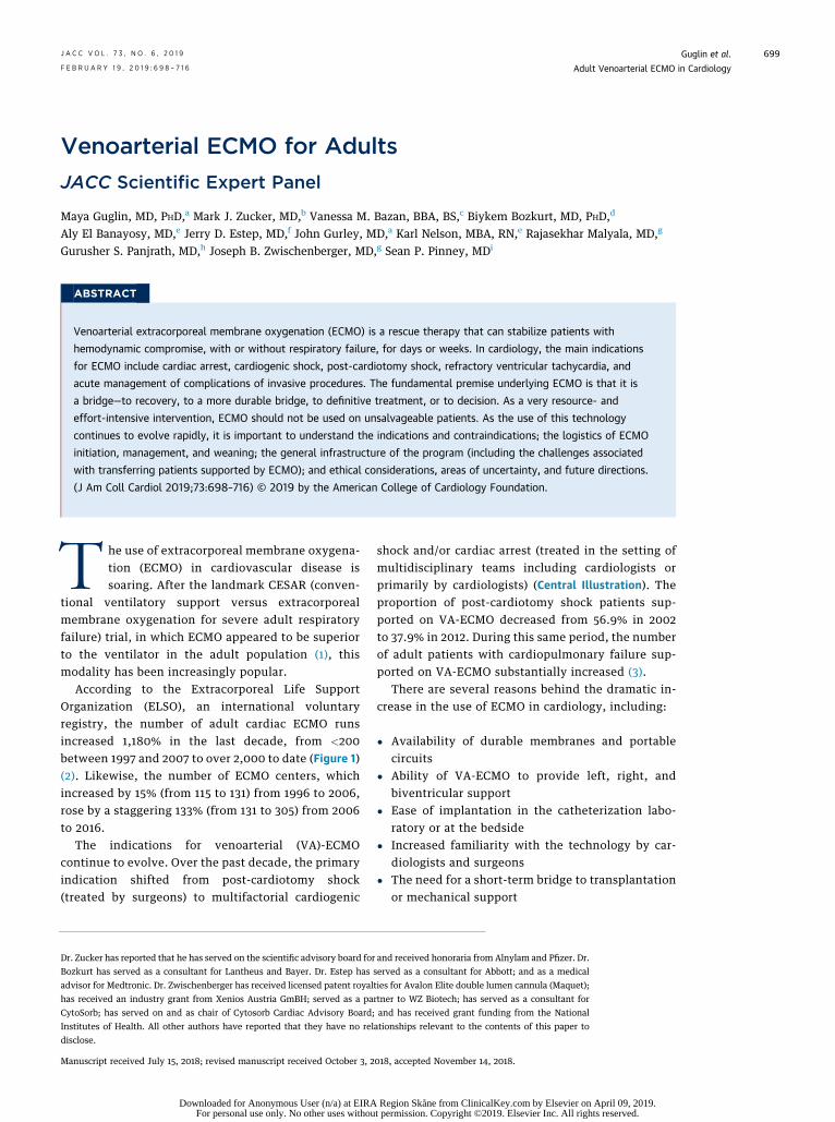
ABSTRACT
Dr
Bo
ad
ha
Cy
Ins
dis
Ma
Venoarterial extracorporeal membrane oxygenation (ECMO) is a rescue therapy that can stabilize patients with
hemodynamic compromise, with or without respiratory failure, for days or weeks. In cardiology, the main indications
for ECMO include cardiac arrest, cardiogenic shock, post-cardiotomy shock, refractory ventricular tachycardia, and
acute management of complications of invasive procedures. The fundamental premise underlying ECMO is that it is
a bridge—to recovery, to a more durable bridge, to definitive treatment, or to decision. As a very resource- and
effort-intensive intervention, ECMO should not be used on unsalvageable patients. As the use of this technology
continues to evolve rapidly, it is important to understand the indications and contraindications; the logistics of ECMO
initiation, management, and weaning; the general infrastructure of the program (including the challenges associated
with transferring patients supported by ECMO); and ethical considerations, areas of uncertainty, and future directions.
(J Am Coll Cardiol 2019;73:698–716) © 2019 by the American College of Cardiology Foundation.
T he use of extracorporeal membrane oxygena-tion (ECMO) in cardiovascular disease issoaring. After the landmark CESAR (conven-
tional ventilatory support versus extracorporealmembrane oxygenation for severe adult respiratoryfailure) trial, in which ECMO appeared to be superiorto the ventilator in the adult population (1), thismodality has been increasingly popular.
According to the Extracorporeal Life SupportOrganization (ELSO), an international voluntaryregistry, the number of adult cardiac ECMO runsincreased 1,180% in the last decade, from <200between 1997 and 2007 to over 2,000 to date (Figure 1)(2). Likewise, the number of ECMO centers, whichincreased by 15% (from 115 to 131) from 1996 to 2006,rose by a staggering 133% (from 131 to 305) from 2006to 2016.
The indications for venoarterial (VA)-ECMOcontinue to evolve. Over the past decade, the primaryindication shifted from post-cardiotomy shock(treated by surgeons) to multifactorial cardiogenic
. Zucker has reported that he has served on the scientific advisory board for
zkurt has served as a consultant for Lantheus and Bayer. Dr. Estep has
visor for Medtronic. Dr. Zwischenberger has received licensed patent royal
s received an industry grant from Xenios Austria GmBH; served as a pa
toSorb; has served on and as chair of Cytosorb Cardiac Advisory Board;
titutes of Health. All other authors have reported that they have no rel
close.
nuscript received July 15, 2018; revised manuscript received October 3, 2
Downloaded for Anonymous User (n/a) at EIRAFor personal use only. No other uses withou
shock and/or cardiac arrest (treated in the setting ofmultidisciplinary teams including cardiologists orprimarily by cardiologists) (Central Illustration). Theproportion of post-cardiotomy shock patients sup-ported on VA-ECMO decreased from 56.9% in 2002to 37.9% in 2012. During this same period, the numberof adult patients with cardiopulmonary failure sup-ported on VA-ECMO substantially increased (3).
There are several reasons behind the dramatic in-crease in the use of ECMO in cardiology, including:
� Availability of durable membranes and portablecircuits
� Ability of VA-ECMO to provide left, right, andbiventricular support
� Ease of implantation in the catheterization labo-ratory or at the bedside
� Increased familiarity with the technology by car-diologists and surgeons
� The need for a short-term bridge to transplantationor mechanical support
and received honoraria from Alnylam and Pfizer. Dr.
served as a consultant for Abbott; and as a medical
ties for Avalon Elite double lumen cannula (Maquet);
rtner to WZ Biotech; has served as a consultant for
and has received grant funding from the National
ationships relevant to the contents of this paper to
018, accepted November 14, 2018.
Region Skåne from ClinicalKey.com by Elsevier on April 09, 2019.t permission. Copyright ©2019. Elsevier Inc. All rights reserved.
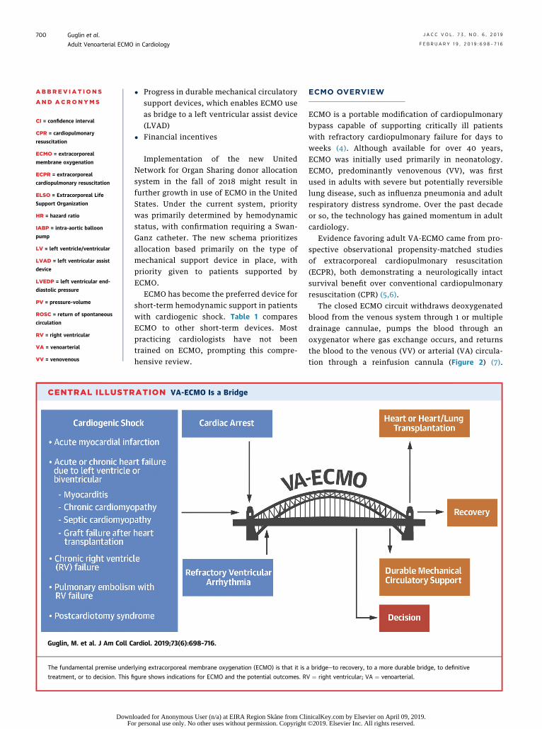
CENTRAL ILLUSTR
Guglin, M. et al. J Am Coll C
The fundamental premise under
treatment, or to decision. This fi
ABBR EV I A T I ON S
AND ACRONYMS
CI = confidence interval
CPR = cardiopulmonary
resuscitation
ECMO = extracorporeal
membrane oxygenation
ECPR = extracorporeal
cardiopulmonary resuscitation
ELSO = Extracorporeal Life
Support Organization
HR = hazard ratio
IABP = intra-aortic balloon
pump
LV = left ventricle/ventricular
LVAD = left ventricular assist
device
LVEDP = left ventricular end-
diastolic pressure
PV = pressure-volume
ROSC = return of spontaneous
circulation
RV = right ventricular
VA = venoarterial
VV = venovenous
Guglin et al. J A C C V O L . 7 3 , N O . 6 , 2 0 1 9
Adult Venoarterial ECMO in Cardiology F E B R U A R Y 1 9 , 2 0 1 9 : 6 9 8 – 7 1 6
700
DownFo
� Progress in durable mechanical circulatorysupport devices, which enables ECMO useas bridge to a left ventricular assist device(LVAD)
� Financial incentives
Implementation of the new UnitedNetwork for Organ Sharing donor allocationsystem in the fall of 2018 might result infurther growth in use of ECMO in the UnitedStates. Under the current system, prioritywas primarily determined by hemodynamicstatus, with confirmation requiring a Swan-Ganz catheter. The new schema prioritizesallocation based primarily on the type ofmechanical support device in place, withpriority given to patients supported byECMO.
ECMO has become the preferred device forshort-term hemodynamic support in patientswith cardiogenic shock. Table 1 comparesECMO to other short-term devices. Mostpracticing cardiologists have not beentrained on ECMO, prompting this compre-hensive review.
ATION VA-ECMO Is a Bridge
ardiol. 2019;73(6):698–716.
lying extracorporeal membrane oxygenation (ECMO) is that it is
gure shows indications for ECMO and the potential outcomes. R
loaded for Anonymous User (n/a) at EIRA Region Skåne from Clir personal use only. No other uses without permission. Copyright
ECMO OVERVIEW
ECMO is a portable modification of cardiopulmonarybypass capable of supporting critically ill patientswith refractory cardiopulmonary failure for days toweeks (4). Although available for over 40 years,ECMO was initially used primarily in neonatology.ECMO, predominantly venovenous (VV), was firstused in adults with severe but potentially reversiblelung disease, such as influenza pneumonia and adultrespiratory distress syndrome. Over the past decadeor so, the technology has gained momentum in adultcardiology.
Evidence favoring adult VA-ECMO came from pro-spective observational propensity-matched studiesof extracorporeal cardiopulmonary resuscitation(ECPR), both demonstrating a neurologically intactsurvival benefit over conventional cardiopulmonaryresuscitation (CPR) (5,6).
The closed ECMO circuit withdraws deoxygenatedblood from the venous system through 1 or multipledrainage cannulae, pumps the blood through anoxygenator where gas exchange occurs, and returnsthe blood to the venous (VV) or arterial (VA) circula-tion through a reinfusion cannula (Figure 2) (7).
a bridge—to recovery, to a more durable bridge, to definitive
V ¼ right ventricular; VA ¼ venoarterial.
nicalKey.com by Elsevier on April 09, 2019. ©2019. Elsevier Inc. All rights reserved.
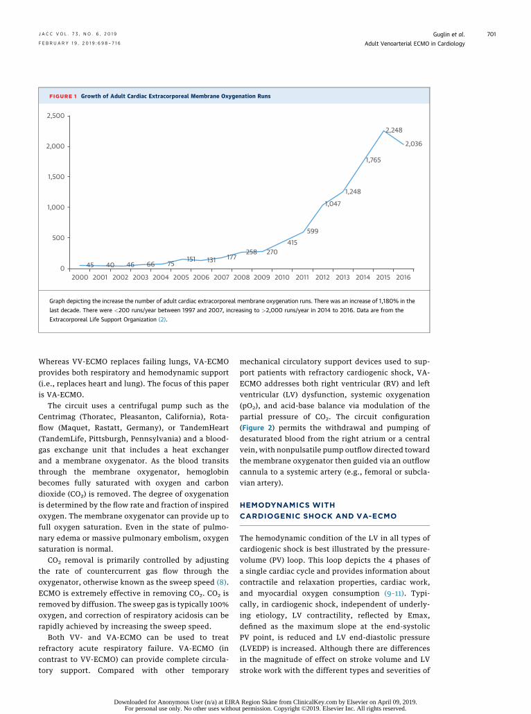
FIGURE 1 Growth of Adult Cardiac Extracorporeal Membrane Oxygenation Runs
2,500
2,000
1,500
1,000
500
02000 2001 2002 2003 2004 2005 2006 2007 2008 2009 2010 2011 2012 2013 2014 2015 2016
45 40 46 66 75151 131 177
258 270415
599
1,047
1,248
1,765
2,248
2,036
Graph depicting the increase the number of adult cardiac extracorporeal membrane oxygenation runs. There was an increase of 1,180% in the
last decade. There were <200 runs/year between 1997 and 2007, increasing to >2,000 runs/year in 2014 to 2016. Data are from the
Extracorporeal Life Support Organization (2).
J A C C V O L . 7 3 , N O . 6 , 2 0 1 9 Guglin et al.F E B R U A R Y 1 9 , 2 0 1 9 : 6 9 8 – 7 1 6 Adult Venoarterial ECMO in Cardiology
701
Whereas VV-ECMO replaces failing lungs, VA-ECMOprovides both respiratory and hemodynamic support(i.e., replaces heart and lung). The focus of this paperis VA-ECMO.
The circuit uses a centrifugal pump such as theCentrimag (Thoratec, Pleasanton, California), Rota-flow (Maquet, Rastatt, Germany), or TandemHeart(TandemLife, Pittsburgh, Pennsylvania) and a blood-gas exchange unit that includes a heat exchangerand a membrane oxygenator. As the blood transitsthrough the membrane oxygenator, hemoglobinbecomes fully saturated with oxygen and carbondioxide (CO2) is removed. The degree of oxygenationis determined by the flow rate and fraction of inspiredoxygen. The membrane oxygenator can provide up tofull oxygen saturation. Even in the state of pulmo-nary edema or massive pulmonary embolism, oxygensaturation is normal.
CO2 removal is primarily controlled by adjustingthe rate of countercurrent gas flow through theoxygenator, otherwise known as the sweep speed (8).ECMO is extremely effective in removing CO2. CO2 isremoved by diffusion. The sweep gas is typically 100%oxygen, and correction of respiratory acidosis can berapidly achieved by increasing the sweep speed.
Both VV- and VA-ECMO can be used to treatrefractory acute respiratory failure. VA-ECMO (incontrast to VV-ECMO) can provide complete circula-tory support. Compared with other temporary
Downloaded for Anonymous User (n/a) at EIRAFor personal use only. No other uses withou
mechanical circulatory support devices used to sup-port patients with refractory cardiogenic shock, VA-ECMO addresses both right ventricular (RV) and leftventricular (LV) dysfunction, systemic oxygenation(pO2), and acid-base balance via modulation of thepartial pressure of CO2. The circuit configuration(Figure 2) permits the withdrawal and pumping ofdesaturated blood from the right atrium or a centralvein, with nonpulsatile pump outflow directed towardthe membrane oxygenator then guided via an outflowcannula to a systemic artery (e.g., femoral or subcla-vian artery).
HEMODYNAMICS WITH
CARDIOGENIC SHOCK AND VA-ECMO
The hemodynamic condition of the LV in all types ofcardiogenic shock is best illustrated by the pressure-volume (PV) loop. This loop depicts the 4 phases ofa single cardiac cycle and provides information aboutcontractile and relaxation properties, cardiac work,and myocardial oxygen consumption (9–11). Typi-cally, in cardiogenic shock, independent of underly-ing etiology, LV contractility, reflected by Emax,defined as the maximum slope at the end-systolicPV point, is reduced and LV end-diastolic pressure(LVEDP) is increased. Although there are differencesin the magnitude of effect on stroke volume and LVstroke work with the different types and severities of
Region Skåne from ClinicalKey.com by Elsevier on April 09, 2019.t permission. Copyright ©2019. Elsevier Inc. All rights reserved.
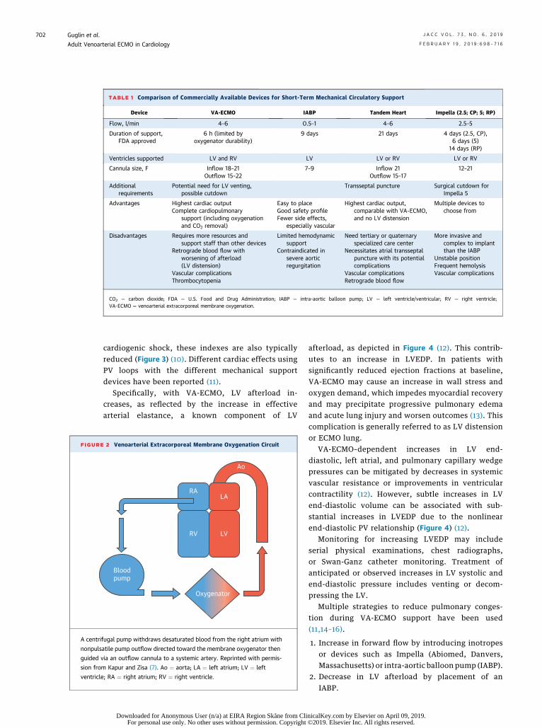
TABLE 1 Comparison of Commercially Available Devices for Short-Term Mechanical Circulatory Support
Device VA-ECMO IABP Tandem Heart Impella (2.5; CP; 5; RP)
Flow, l/min 4–6 0.5–1 4–6 2.5–5
Duration of support,FDA approved
6 h (limited byoxygenator durability)
9 days 21 days 4 days (2.5, CP),6 days (5)
14 days (RP)
Ventricles supported LV and RV LV LV or RV LV or RV
Cannula size, F Inflow 18–21Outflow 15–22
7–9 Inflow 21Outflow 15–17
12–21
Additionalrequirements
Potential need for LV venting,possible cutdown
Transseptal puncture Surgical cutdown forImpella 5
Advantages Highest cardiac outputComplete cardiopulmonary
support (including oxygenationand CO2 removal)
Easy to placeGood safety profileFewer side effects,
especially vascular
Highest cardiac output,comparable with VA-ECMO,and no LV distension
Multiple devices tochoose from
Disadvantages Requires more resources andsupport staff than other devices
Retrograde blood flow withworsening of afterload(LV distension)
Vascular complicationsThrombocytopenia
Limited hemodynamicsupport
Contraindicated insevere aorticregurgitation
Need tertiary or quaternaryspecialized care center
Necessitates atrial transseptalpuncture with its potentialcomplications
Vascular complicationsRetrograde blood flow
More invasive andcomplex to implantthan the IABP
Unstable positionFrequent hemolysisVascular complications
CO2 ¼ carbon dioxide; FDA ¼ U.S. Food and Drug Administration; IABP ¼ intra-aortic balloon pump; LV ¼ left ventricle/ventricular; RV ¼ right ventricle;VA-ECMO ¼ venoarterial extracorporeal membrane oxygenation.
FIGURE
A centri
nonpuls
guided v
sion fro
ventricle
Guglin et al. J A C C V O L . 7 3 , N O . 6 , 2 0 1 9
Adult Venoarterial ECMO in Cardiology F E B R U A R Y 1 9 , 2 0 1 9 : 6 9 8 – 7 1 6
702
cardiogenic shock, these indexes are also typicallyreduced (Figure 3) (10). Different cardiac effects usingPV loops with the different mechanical supportdevices have been reported (11).
Specifically, with VA-ECMO, LV afterload in-creases, as reflected by the increase in effectivearterial elastance, a known component of LV
2 Venoarterial Extracorporeal Membrane Oxygenation Circuit
Oxygenator
RV LV
LA
Ao
RA
Bloodpump
fugal pump withdraws desaturated blood from the right atrium with
atile pump outflow directed toward the membrane oxygenator then
ia an outflow cannula to a systemic artery. Reprinted with permis-
m Kapur and Zisa (7). Ao ¼ aorta; LA ¼ left atrium; LV ¼ left
; RA ¼ right atrium; RV ¼ right ventricle.
Downloaded for Anonymous User (n/a) at EIRA Region Skåne from CliFor personal use only. No other uses without permission. Copyright
afterload, as depicted in Figure 4 (12). This contrib-utes to an increase in LVEDP. In patients withsignificantly reduced ejection fractions at baseline,VA-ECMO may cause an increase in wall stress andoxygen demand, which impedes myocardial recoveryand may precipitate progressive pulmonary edemaand acute lung injury and worsen outcomes (13). Thiscomplication is generally referred to as LV distensionor ECMO lung.
VA-ECMO–dependent increases in LV end-diastolic, left atrial, and pulmonary capillary wedgepressures can be mitigated by decreases in systemicvascular resistance or improvements in ventricularcontractility (12). However, subtle increases in LVend-diastolic volume can be associated with sub-stantial increases in LVEDP due to the nonlinearend-diastolic PV relationship (Figure 4) (12).
Monitoring for increasing LVEDP may includeserial physical examinations, chest radiographs,or Swan-Ganz catheter monitoring. Treatment ofanticipated or observed increases in LV systolic andend-diastolic pressure includes venting or decom-pressing the LV.
Multiple strategies to reduce pulmonary conges-tion during VA-ECMO support have been used(11,14–16).
1. Increase in forward flow by introducing inotropesor devices such as Impella (Abiomed, Danvers,Massachusetts) or intra-aortic balloon pump (IABP).
2. Decrease in LV afterload by placement of anIABP.
nicalKey.com by Elsevier on April 09, 2019. ©2019. Elsevier Inc. All rights reserved.
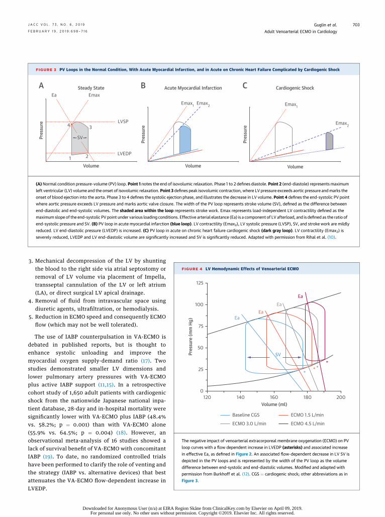
FIGURE 3 PV Loops in the Normal Condition, With Acute Myocardial Infarction, and in Acute on Chronic Heart Failure Complicated by Cardiogenic Shock
VolumeVolume Volume
LVEDP
Pres
sure
Pres
sure
Pres
sure
1 2
3LVSP
EmaxEmax1 Emax2 Emax1
Emax2
EaSteady State Cardiogenic ShockAcute Myocardial Infarction
4
SV
CBA
(A)Normal condition pressure-volume (PV) loop. Point 1 notes the end of isovolumic relaxation. Phase 1 to 2 defines diastole. Point 2 (end-diastole) represents maximum
left ventricular (LV) volume and the onset of isovolumic relaxation. Point 3 defines peak isovolumic contraction, where LV pressure exceeds aortic pressure andmarks the
onset of blood ejection into the aorta. Phase 3 to 4 defines the systolic ejection phase, and illustrates the decrease in LV volume. Point 4 defines the end-systolic PV point
where aortic pressure exceeds LV pressure and marks aortic valve closure. The width of the PV loop represents stroke volume (SV), defined as the difference between
end-diastolic and end-systolic volumes. The shaded area within the loop represents stroke work. Emax represents load-independent LV contractility defined as the
maximum slope of the end-systolic PV point under various loading conditions. Effective arterial elastance (Ea) is a component of LV afterload, and is defined as the ratio of
end-systolic pressure and SV. (B) PV loop in acute myocardial infarction (blue loop). LV contractility (Emax2), LV systolic pressure (LVSP), SV, and stroke work are mildly
reduced. LV end-diastolic pressure (LVEDP) is increased. (C) PV loop in acute on chronic heart failure cardiogenic shock (dark gray loop). LV contractility (Emax2) is
severely reduced, LVEDP and LV end-diastolic volume are significantly increased and SV is significantly reduced. Adapted with permission from Rihal et al. (10).
FIGURE 4 LV Hemodynamic Effects of Venoarterial ECMO
Volume (ml)
125
100
Pres
sure
(mm
Hg)
75
50
25
0120 140 160 180 200
SV
** *
*
EaEa
EaEa
Baseline CGS ECMO 1.5 L/min
ECMO 3.0 L/min ECMO 4.5 L/min
The negative impact of venoarterial extracorporeal membrane oxygenation (ECMO) on PV
loop curves with a flow dependent increase in LVEDP (asterisks) and associated increase
in effective Ea, as defined in Figure 2. An associated flow-dependent decrease in LV SV is
depicted in the PV loops and is represented by the width of the PV loop as the volume
difference between end-systolic and end-diastolic volumes. Modified and adapted with
permission from Burkhoff et al. (12). CGS ¼ cardiogenic shock; other abbreviations as in
Figure 3.
J A C C V O L . 7 3 , N O . 6 , 2 0 1 9 Guglin et al.F E B R U A R Y 1 9 , 2 0 1 9 : 6 9 8 – 7 1 6 Adult Venoarterial ECMO in Cardiology
703
3. Mechanical decompression of the LV by shuntingthe blood to the right side via atrial septostomy orremoval of LV volume via placement of Impella,transseptal cannulation of the LV or left atrium(LA), or direct surgical LV apical drainage.
4. Removal of fluid from intravascular space usingdiuretic agents, ultrafiltration, or hemodialysis.
5. Reduction in ECMO speed and consequently ECMOflow (which may not be well tolerated).
The use of IABP counterpulsation in VA-ECMO isdebated in published reports, but is thought toenhance systolic unloading and improve themyocardial oxygen supply-demand ratio (17). Twostudies demonstrated smaller LV dimensions andlower pulmonary artery pressures with VA-ECMOplus active IABP support (11,15). In a retrospectivecohort study of 1,650 adult patients with cardiogenicshock from the nationwide Japanese national inpa-tient database, 28-day and in-hospital mortality weresignificantly lower with VA-ECMO plus IABP (48.4%vs. 58.2%; p ¼ 0.001) than with VA-ECMO alone(55.9% vs. 64.5%; p ¼ 0.004) (18). However, anobservational meta-analysis of 16 studies showed alack of survival benefit of VA-ECMO with concomitantIABP (19). To date, no randomized controlled trialshave been performed to clarify the role of venting andthe strategy (IABP vs. alternative devices) that bestattenuates the VA-ECMO flow-dependent increase inLVEDP.
Downloaded for Anonymous User (n/a) at EIRA Region Skåne from ClinicalKey.com by Elsevier on April 09, 2019.For personal use only. No other uses without permission. Copyright ©2019. Elsevier Inc. All rights reserved.
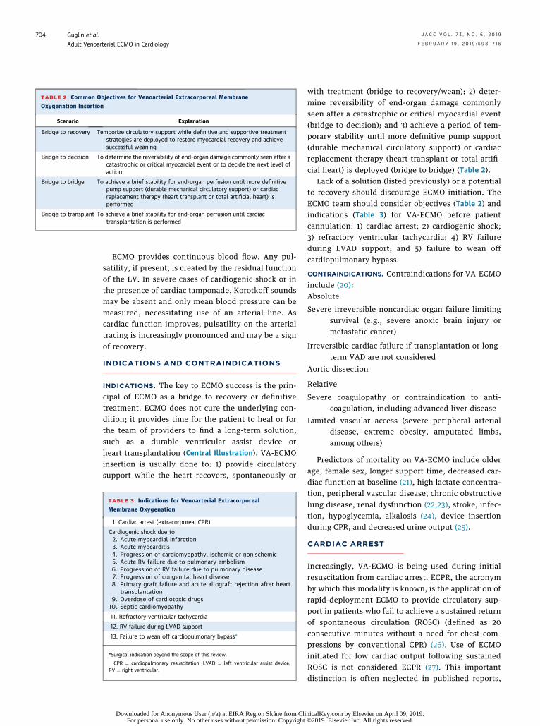
TABLE 2 Common Objectives for Venoarterial Extracorporeal Membrane
Oxygenation Insertion
Scenario Explanation
Bridge to recovery Temporize circulatory support while definitive and supportive treatmentstrategies are deployed to restore myocardial recovery and achievesuccessful weaning
Bridge to decision To determine the reversibility of end-organ damage commonly seen after acatastrophic or critical myocardial event or to decide the next level ofaction
Bridge to bridge To achieve a brief stability for end-organ perfusion until more definitivepump support (durable mechanical circulatory support) or cardiacreplacement therapy (heart transplant or total artificial heart) isperformed
Bridge to transplant To achieve a brief stability for end-organ perfusion until cardiactransplantation is performed
Guglin et al. J A C C V O L . 7 3 , N O . 6 , 2 0 1 9
Adult Venoarterial ECMO in Cardiology F E B R U A R Y 1 9 , 2 0 1 9 : 6 9 8 – 7 1 6
704
ECMO provides continuous blood flow. Any pul-satility, if present, is created by the residual functionof the LV. In severe cases of cardiogenic shock or inthe presence of cardiac tamponade, Korotkoff soundsmay be absent and only mean blood pressure can bemeasured, necessitating use of an arterial line. Ascardiac function improves, pulsatility on the arterialtracing is increasingly pronounced and may be a signof recovery.
INDICATIONS AND CONTRAINDICATIONS
INDICATIONS. The key to ECMO success is the prin-cipal of ECMO as a bridge to recovery or definitivetreatment. ECMO does not cure the underlying con-dition; it provides time for the patient to heal or forthe team of providers to find a long-term solution,such as a durable ventricular assist device orheart transplantation (Central Illustration). VA-ECMOinsertion is usually done to: 1) provide circulatorysupport while the heart recovers, spontaneously or
TABLE 3 Indications for Venoarterial Extracorporeal
Membrane Oxygenation
1. Cardiac arrest (extracorporeal CPR)
Cardiogenic shock due to2. Acute myocardial infarction3. Acute myocarditis4. Progression of cardiomyopathy, ischemic or nonischemic5. Acute RV failure due to pulmonary embolism6. Progression of RV failure due to pulmonary disease7. Progression of congenital heart disease8. Primary graft failure and acute allograft rejection after heart
transplantation9. Overdose of cardiotoxic drugs10. Septic cardiomyopathy
11. Refractory ventricular tachycardia
12. RV failure during LVAD support
13. Failure to wean off cardiopulmonary bypass*
*Surgical indication beyond the scope of this review.
CPR ¼ cardiopulmonary resuscitation; LVAD ¼ left ventricular assist device;RV ¼ right ventricular.
Downloaded for Anonymous User (n/a) at EIRA Region Skåne from CliFor personal use only. No other uses without permission. Copyright
with treatment (bridge to recovery/wean); 2) deter-mine reversibility of end-organ damage commonlyseen after a catastrophic or critical myocardial event(bridge to decision); and 3) achieve a period of tem-porary stability until more definitive pump support(durable mechanical circulatory support) or cardiacreplacement therapy (heart transplant or total artifi-cial heart) is deployed (bridge to bridge) (Table 2).
Lack of a solution (listed previously) or a potentialto recovery should discourage ECMO initiation. TheECMO team should consider objectives (Table 2) andindications (Table 3) for VA-ECMO before patientcannulation: 1) cardiac arrest; 2) cardiogenic shock;3) refractory ventricular tachycardia; 4) RV failureduring LVAD support; and 5) failure to wean offcardiopulmonary bypass.
CONTRAINDICATIONS. Contraindications for VA-ECMOinclude (20):Absolute
Severe irreversible noncardiac organ failure limiting
nicalKey ©2019.
survival (e.g., severe anoxic brain injury ormetastatic cancer)
Irreversible cardiac failure if transplantation or long-
term VAD are not consideredAortic dissection
Relative
Severe coagulopathy or contraindication to anti-
coagulation, including advanced liver diseaseLimited vascular access (severe peripheral arterial
disease, extreme obesity, amputated limbs,among others)Predictors of mortality on VA-ECMO include olderage, female sex, longer support time, decreased car-diac function at baseline (21), high lactate concentra-tion, peripheral vascular disease, chronic obstructivelung disease, renal dysfunction (22,23), stroke, infec-tion, hypoglycemia, alkalosis (24), device insertionduring CPR, and decreased urine output (25).
CARDIAC ARREST
Increasingly, VA-ECMO is being used during initialresuscitation from cardiac arrest. ECPR, the acronymby which this modality is known, is the application ofrapid-deployment ECMO to provide circulatory sup-port in patients who fail to achieve a sustained returnof spontaneous circulation (ROSC) (defined as 20consecutive minutes without a need for chest com-pressions by conventional CPR) (26). Use of ECMOinitiated for low cardiac output following sustainedROSC is not considered ECPR (27). This importantdistinction is often neglected in published reports,
.com by Elsevier on April 09, 2019.Elsevier Inc. All rights reserved.
FIGURE 5 ECPR Time for Patient Selection
0 5 10 15 20 25 min. 35 min. 50 min. 60 min.
No ECLSECLS FlowLow FlowNoFlow
Time to BLS
CPR initiates a Low Flow State Definition of Refractory OHCAUnresponsive to 30 min. of CPRLonger CPR yields worse outcomes.
The most important determinant of outcome.Early, high-quality chest compressions determine the success of all subsequent interventions.Immediate bystander CPR or a no-flow time <5 min.
Cutoff Time for Switch to ECPRCARDIAC ARREST ECLS LimitNo reasonablechance ofacceptableoutcome.
Neuro Recovery LimitAcceptable neuro outcome falls to 2% after 15 minutes of CPR.
Suggested transition to ECLS at 21 minutes of CPR.
The interval from the arrest to the beginning of cardiopulmonary resuscitation (CPR) should be considered a no-flow period (far left), whereas
time on CPR is a low-flow period with suboptimal circulation. The probability of survival with good neurological outcome declines rapidly
with each minute of conventional CPR. When extracorporeal CPR (ECPR) is delayed until refractory cardiac arrest, CPR, survival is extremely
poor (far right). BLS ¼ basic life support; ECLS ¼ extracorporeal life support; OHCA ¼ out-of-hospital cardiac arrest.
J A C C V O L . 7 3 , N O . 6 , 2 0 1 9 Guglin et al.F E B R U A R Y 1 9 , 2 0 1 9 : 6 9 8 – 7 1 6 Adult Venoarterial ECMO in Cardiology
705
making outcome analysis difficult. A recent prospec-tive cohort study of in- and out-of-hospital ECPR inselected patients boasted 54% survival to dischargewith full neurological function (6).
The most important determinant of outcome istime to basic life support. Early chest compressionsinfluence all subsequent interventions. Immediatebystander CPR or a no-flow time <5 min are pre-requisites for ECPR (4). The interval from the arrest tothe beginning of CPR should be considered a no-flowperiod, whereas time on CPR is a low-flow period (28)with suboptimal circulation. The probability of sur-vival with a good neurological outcome declinesrapidly with each minute of conventional CPR. WhenECPR is delayed until refractory cardiac arrest,defined as no response to resuscitation efforts after30 min of conventional CPR, survival is extremelypoor (29).
ECPR should be attempted early after cardiacarrest, rather than after the complete failure oftraditional measures (30). Studies suggest 21 min ofconventional CPR before initiation of extracorporeallife support (VA-ECMO), with ECPR preparation in thefirst 10 min of CPR and cannulation within 20 min ofcollapse (Figure 5) (31).
Although the upper age limit for ECPR varies bycenter, some studies exclude patients >70 to 75 years
Downloaded for Anonymous User (n/a) at EIRAFor personal use only. No other uses withou
of age (32,33). Commonly used inclusion or exclusioncriteria for ECPR are:Inclusion criteria
� Age <70 years� Initial rhythm of ventricular fibrillation or ven-
tricular tachycardia� Witnessed arrest� Bystander CPR within 5 min� Failure to achieve ROSC within 15 min of beginning
CPR (20)
Exclusion criteria
� Asystole as an initial rhythm� Unwitnessed arrest� Total cardiac arrest time >60 min� Pre-existing severe neurological or systemic dis-
ease (including stroke, severe dementia, advancedmalignancy, chronic neuromuscular dystrophy,psychiatric conditions, anoxic brain injury)
� Contraindications to anticoagulation� Acute aortic dissection� Suspicion of shock due to hemorrhage or other
noncardiovascular cause� Known “do not resuscitate” (DNR) status
A decision-making algorithm to initiate ECPR ispresented in Figure 6. In-hospital cardiac arrest may
Region Skåne from ClinicalKey.com by Elsevier on April 09, 2019.t permission. Copyright ©2019. Elsevier Inc. All rights reserved.
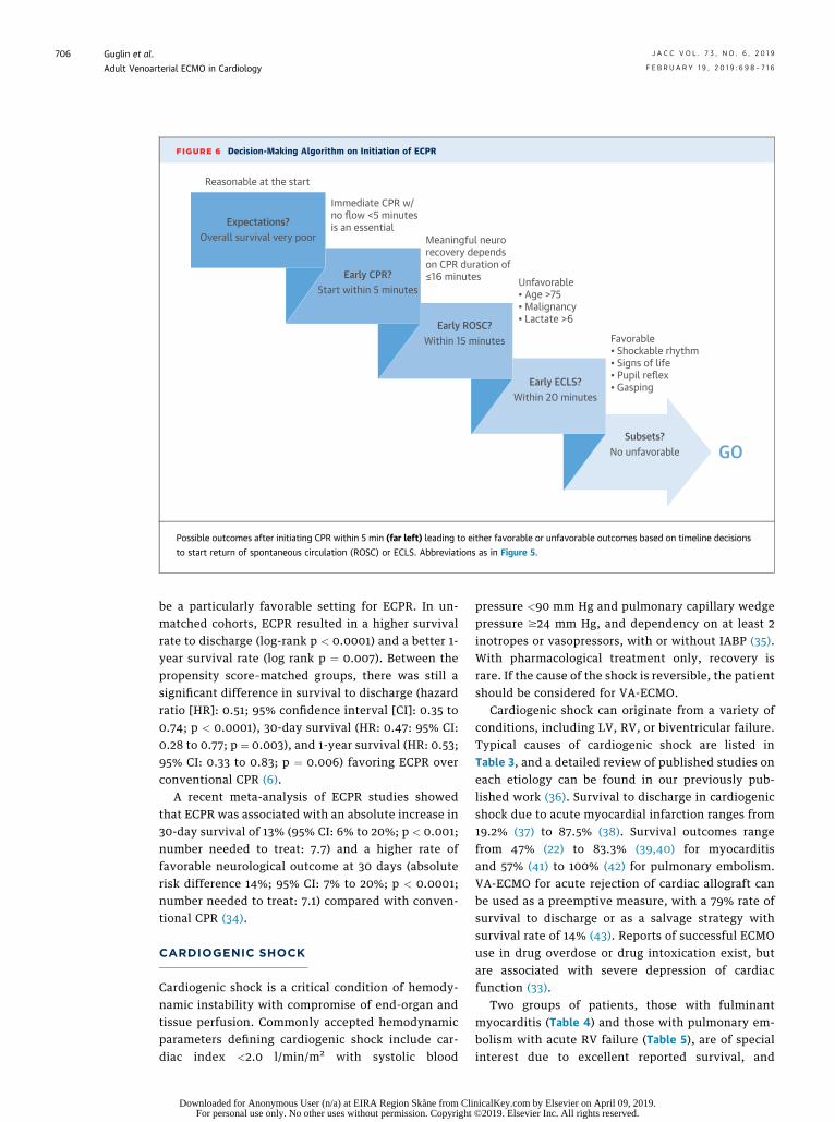
FIGURE 6 Decision-Making Algorithm on Initiation of ECPR
Reasonable at the start
Expectations?Overall survival very poor
Early CPR?Start within 5 minutes
Early ROSC?Within 15 minutes
Early ECLS?Within 20 minutes
Immediate CPR w/no flow <5 minutesis an essential
Meaningful neurorecovery dependson CPR duration of ≤16 minutes Unfavorable
• Age >75• Malignancy• Lactate >6
Favorable• Shockable rhythm• Signs of life• Pupil reflex• Gasping
Subsets?No unfavorable GOGO
Possible outcomes after initiating CPR within 5 min (far left) leading to either favorable or unfavorable outcomes based on timeline decisions
to start return of spontaneous circulation (ROSC) or ECLS. Abbreviations as in Figure 5.
Guglin et al. J A C C V O L . 7 3 , N O . 6 , 2 0 1 9
Adult Venoarterial ECMO in Cardiology F E B R U A R Y 1 9 , 2 0 1 9 : 6 9 8 – 7 1 6
706
be a particularly favorable setting for ECPR. In un-matched cohorts, ECPR resulted in a higher survivalrate to discharge (log-rank p < 0.0001) and a better 1-year survival rate (log rank p ¼ 0.007). Between thepropensity score–matched groups, there was still asignificant difference in survival to discharge (hazardratio [HR]: 0.51; 95% confidence interval [CI]: 0.35 to0.74; p < 0.0001), 30-day survival (HR: 0.47: 95% CI:0.28 to 0.77; p ¼ 0.003), and 1-year survival (HR: 0.53;95% CI: 0.33 to 0.83; p ¼ 0.006) favoring ECPR overconventional CPR (6).
A recent meta-analysis of ECPR studies showedthat ECPR was associated with an absolute increase in30-day survival of 13% (95% CI: 6% to 20%; p < 0.001;number needed to treat: 7.7) and a higher rate offavorable neurological outcome at 30 days (absoluterisk difference 14%; 95% CI: 7% to 20%; p < 0.0001;number needed to treat: 7.1) compared with conven-tional CPR (34).
CARDIOGENIC SHOCK
Cardiogenic shock is a critical condition of hemody-namic instability with compromise of end-organ andtissue perfusion. Commonly accepted hemodynamicparameters defining cardiogenic shock include car-diac index <2.0 l/min/m2 with systolic blood
Downloaded for Anonymous User (n/a) at EIRA Region Skåne from CliFor personal use only. No other uses without permission. Copyright
pressure <90 mm Hg and pulmonary capillary wedgepressure $24 mm Hg, and dependency on at least 2inotropes or vasopressors, with or without IABP (35).With pharmacological treatment only, recovery israre. If the cause of the shock is reversible, the patientshould be considered for VA-ECMO.
Cardiogenic shock can originate from a variety ofconditions, including LV, RV, or biventricular failure.Typical causes of cardiogenic shock are listed inTable 3, and a detailed review of published studies oneach etiology can be found in our previously pub-lished work (36). Survival to discharge in cardiogenicshock due to acute myocardial infarction ranges from19.2% (37) to 87.5% (38). Survival outcomes rangefrom 47% (22) to 83.3% (39,40) for myocarditisand 57% (41) to 100% (42) for pulmonary embolism.VA-ECMO for acute rejection of cardiac allograft canbe used as a preemptive measure, with a 79% rate ofsurvival to discharge or as a salvage strategy withsurvival rate of 14% (43). Reports of successful ECMOuse in drug overdose or drug intoxication exist, butare associated with severe depression of cardiacfunction (33).
Two groups of patients, those with fulminantmyocarditis (Table 4) and those with pulmonary em-bolism with acute RV failure (Table 5), are of specialinterest due to excellent reported survival, and
nicalKey.com by Elsevier on April 09, 2019. ©2019. Elsevier Inc. All rights reserved.
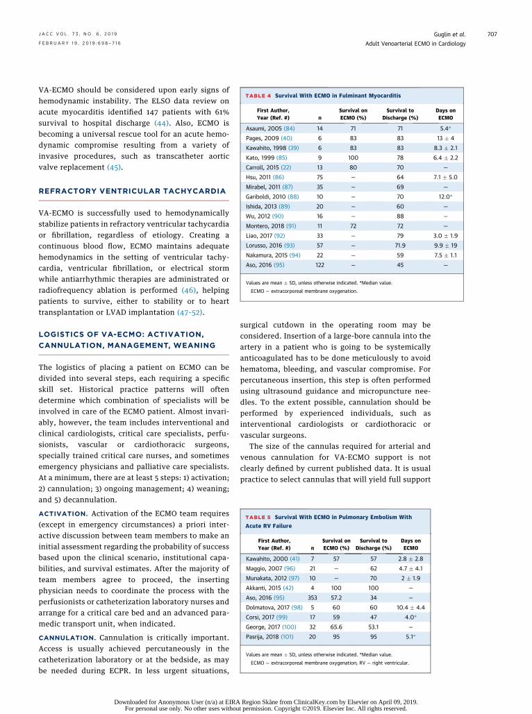
TABLE 4 Survival With ECMO in Fulminant Myocarditis
First Author,Year (Ref. #) n
Survival onECMO (%)
Survival toDischarge (%)
Days onECMO
Asaumi, 2005 (84) 14 71 71 5.4*
Pages, 2009 (40) 6 83 83 13 � 4
Kawahito, 1998 (39) 6 83 83 8.3 � 2.1
Kato, 1999 (85) 9 100 78 6.4 � 2.2
Carroll, 2015 (22) 13 80 70 —
Hsu, 2011 (86) 75 — 64 7.1 � 5.0
Mirabel, 2011 (87) 35 — 69 —
Gariboldi, 2010 (88) 10 — 70 12.0*
Ishida, 2013 (89) 20 — 60 —
Wu, 2012 (90) 16 — 88 —
Montero, 2018 (91) 11 72 72 —
Liao, 2017 (92) 33 — 79 3.0 � 1.9
Lorusso, 2016 (93) 57 — 71.9 9.9 � 19
Nakamura, 2015 (94) 22 — 59 7.5 � 1.1
Aso, 2016 (95) 122 — 45 —
Values are mean � SD, unless otherwise indicated. *Median value.
ECMO ¼ extracorporeal membrane oxygenation.
TABLE 5 Survival With ECMO in Pulmonary Embolism With
Acute RV Failure
First Author,Year (Ref. #) n
Survival onECMO (%)
Survival toDischarge (%)
Days onECMO
Kawahito, 2000 (41) 7 57 57 2.8 � 2.8
Maggio, 2007 (96) 21 — 62 4.7 � 4.1
Munakata, 2012 (97) 10 — 70 2 � 1.9
Akkanti, 2015 (42) 4 100 100 —
Aso, 2016 (95) 353 57.2 34 —
Dolmatova, 2017 (98) 5 60 60 10.4 � 4.4
Corsi, 2017 (99) 17 59 47 4.0*
George, 2017 (100) 32 65.6 53.1 —
Pasrija, 2018 (101) 20 95 95 5.1*
Values are mean � SD, unless otherwise indicated. *Median value.
ECMO ¼ extracorporeal membrane oxygenation; RV ¼ right ventricular.
J A C C V O L . 7 3 , N O . 6 , 2 0 1 9 Guglin et al.F E B R U A R Y 1 9 , 2 0 1 9 : 6 9 8 – 7 1 6 Adult Venoarterial ECMO in Cardiology
707
VA-ECMO should be considered upon early signs ofhemodynamic instability. The ELSO data review onacute myocarditis identified 147 patients with 61%survival to hospital discharge (44). Also, ECMO isbecoming a universal rescue tool for an acute hemo-dynamic compromise resulting from a variety ofinvasive procedures, such as transcatheter aorticvalve replacement (45).
REFRACTORY VENTRICULAR TACHYCARDIA
VA-ECMO is successfully used to hemodynamicallystabilize patients in refractory ventricular tachycardiaor fibrillation, regardless of etiology. Creating acontinuous blood flow, ECMO maintains adequatehemodynamics in the setting of ventricular tachy-cardia, ventricular fibrillation, or electrical stormwhile antiarrhythmic therapies are administrated orradiofrequency ablation is performed (46), helpingpatients to survive, either to stability or to hearttransplantation or LVAD implantation (47–52).
LOGISTICS OF VA-ECMO: ACTIVATION,
CANNULATION, MANAGEMENT, WEANING
The logistics of placing a patient on ECMO can bedivided into several steps, each requiring a specificskill set. Historical practice patterns will oftendetermine which combination of specialists will beinvolved in care of the ECMO patient. Almost invari-ably, however, the team includes interventional andclinical cardiologists, critical care specialists, perfu-sionists, vascular or cardiothoracic surgeons,specially trained critical care nurses, and sometimesemergency physicians and palliative care specialists.At a minimum, there are at least 5 steps: 1) activation;2) cannulation; 3) ongoing management; 4) weaning;and 5) decannulation.
ACTIVATION. Activation of the ECMO team requires(except in emergency circumstances) a priori inter-active discussion between team members to make aninitial assessment regarding the probability of successbased upon the clinical scenario, institutional capa-bilities, and survival estimates. After the majority ofteam members agree to proceed, the insertingphysician needs to coordinate the process with theperfusionists or catheterization laboratory nurses andarrange for a critical care bed and an advanced para-medic transport unit, when indicated.
CANNULATION. Cannulation is critically important.Access is usually achieved percutaneously in thecatheterization laboratory or at the bedside, as maybe needed during ECPR. In less urgent situations,
Downloaded for Anonymous User (n/a) at EIRAFor personal use only. No other uses withou
surgical cutdown in the operating room may beconsidered. Insertion of a large-bore cannula into theartery in a patient who is going to be systemicallyanticoagulated has to be done meticulously to avoidhematoma, bleeding, and vascular compromise. Forpercutaneous insertion, this step is often performedusing ultrasound guidance and micropuncture nee-dles. To the extent possible, cannulation should beperformed by experienced individuals, such asinterventional cardiologists or cardiothoracic orvascular surgeons.
The size of the cannulas required for arterial andvenous cannulation for VA-ECMO support is notclearly defined by current published data. It is usualpractice to select cannulas that will yield full support
Region Skåne from ClinicalKey.com by Elsevier on April 09, 2019.t permission. Copyright ©2019. Elsevier Inc. All rights reserved.
Guglin et al. J A C C V O L . 7 3 , N O . 6 , 2 0 1 9
Adult Venoarterial ECMO in Cardiology F E B R U A R Y 1 9 , 2 0 1 9 : 6 9 8 – 7 1 6
708
for a given patient, that is, to achieve an index of2.2 l/min/m2.
Investigators from the University of Michigan statethat a pressure drop in the circuit should not exceed100 mm Hg across the venous cannula and300 mm Hg across the arterial side. The flow requiredfor full support in an adult is about 60 cm3/kg/min.Not only are the length and diameter of the cannulaimportant, but there are special flow characteristicsfor each cannula, that can be described by a pressureflow curve. A unique number, called a UM number(flow achieved at a pressure drop of 100 mm Hg acrossa cannula) may be used to describe the cannula andits flow characteristics (53).
Cannulation can be performed peripherally orcentrally. Peripheral insertion may be quicker, but itis known to be associated with vascular compromiseof the lower extremity due to the large size of thecannula, particularly in small women or in a highsystemic vascular resistance (“clamped down”) state.It is generally stated that unless the vessel is at least 1to 2 mm larger than the cannula, there is the risk oflimb ischemia. This problem can be addressed byplacing a small 6-F to 8-F antegrade sheath or intro-ducer into the superficial femoral artery at the time ofcannula insertion and diverting a small portion of thearterial return flow down the limb (reperfusioncatheter).
Femoral vein to femoral artery is the most commonapproach. Nonemergent VA-ECMO patients may beconsidered for internal jugular vein to subclavianartery cannulation, termed the “sport model” (54,55).This development in technique moved nonemergentVA-ECMO cannulation sites from the groin (femoral)to the upper body (subclavian) vessels, facilitatingambulation (56,57). Ambulatory ECMO is particularlyimportant in patients awaiting transplantation toreduce the risk of patients deconditioning as theyawait transplantation (58).
Unique patient needs can be accommodated by theaddition of a second venous cannula (including dual-lumen cannulae, such as the Avalon Elite [Getinge,Gothenburg, Sweden] or OriGen [OriGen Biomedical,Inc., Austin, Texas]) to convert between the VV andthe venoarterial-venous hybrid configuration.Venoarterial-venous cannulation is rarely used as aninitial strategy, but may be used for additionaloxygenation. A case series by Biscotti et al. (58)reports 21 patients resuscitated using hybrid ECMOwith a 43% survival rate.
MANAGEMENT. ECMO flow. Usually, an ECMO flowof 50 to 70 ml/kg/min (w4 to 6 l/min) is sufficient forfull markedly decreased and the aorta is filled by
Downloaded for Anonymous User (n/a) at EIRA Region Skåne from CliFor personal use only. No other uses without permission. Copyright
retrograde flow from the arterial ECMO cannula.Therefore, the afterload faced by the LV is increased.This may result in an elevated LVEDP, elevated pul-monary capillary wedge pressure, and ultimatelypulmonary congestion. In this situation, the use ofinotropes may be considered or, in unusual circum-stances, an interatrial balloon septostomy can beperformed to shunt blood from the LA to the rightatrium and reduce the elevated left-sided pressures.Multiple approaches to LV unloading or “venting”are reviewed in the Pathophysiology section.Another disadvantage to VA bypass is that anyparticles, bubbles, or emboli that may be infusedinto the arterial or venous circuit can result in anadverse cerebral event. Finally, the decreased flowthrough the lungs may permit the development ofmicrovascular intrapulmonary thromboses, whichmay further worsen the pulmonary pathology.
Moni tor ing . An ECMO patient should invariablyhave an arterial line and may have a pulmonary arterycatheter. The pulmonary artery catheter is normallyused to monitor mixed venous saturation, althoughsome of the newer pumps (Maquet CardioHelp) docontinuously monitor mixed venous saturation,perhaps obviating the need for the pulmonary arterycatheter. Use of the thermodilution cardiac outputis misleading, as an unknown amount of the coldbolus is drawn into the ECMO circuit, invalidatingthe calculation. A Swan-Ganz catheter should beplaced and used as a tool to monitor pulmonaryarterial pressure as a surrogate of LV distention.Intermittent measurement of lactate might behelpful.
Ant icoagulat ion . Systemic anticoagulation therapyminimizes interaction of blood products with thesurface of the ECMO circuit. Fibrinogen and albuminadsorb to the circuit’s biopolymer components,resulting in platelet aggregation, activation, andconsumption within 1 h of ECMO initiation (59). Theactivated coagulation system may result in throm-bocytopenia, which can be profound, sometimesrequiring platelet transfusions.
The choice of agent used for anticoagulation, aswell as monitoring of the level of anticoagulation,varies from site to site. Heparin is the most commonanticoagulant, used according to a standard weight-based protocol and monitored either by activatedthromboplastin time, goal 50 to 75 s (1.5� to 2.5�baseline) or anti-factor Xa, goal 0.3 to 0.7 IU/ml. Somecenters prefer to follow the activated clotting time,due to its bedside availability and rapid turnaroundtime, goal 180 to 220 s.
nicalKey.com by Elsevier on April 09, 2019. ©2019. Elsevier Inc. All rights reserved.
J A C C V O L . 7 3 , N O . 6 , 2 0 1 9 Guglin et al.F E B R U A R Y 1 9 , 2 0 1 9 : 6 9 8 – 7 1 6 Adult Venoarterial ECMO in Cardiology
709
Direct thrombin inhibitors, such as parenteralbivalirudin and argatroban, represent alternatives toheparin in individuals with heparin-induced throm-bocytopenia. Patients anticoagulated using thesedirect thrombin inhibitors are monitored for activatedpartial thromboplastin times between 50 and 60 s(60).
VENTILATOR MANAGEMENT. Patients on ECMO mayor may not require ventilator support. Because ECMOprovides up to full gas exchange, the conscious pa-tient in cardiogenic shock can breathe spontaneouslywhile on ECMO, regardless of respiratory function.However, ventilator support may be necessary forairway protection in patients requiring sedation, suchas cardiac arrest patients. Most centers use low tidalvolume ventilation (3 to 5 ml/kg) to reduce the risk oflung injury. Protective mechanical ventilation set-tings include a rate of <8/min, positive end-expiratory pressure of 10 to 15 Torr, fraction ofinspired oxygen <0.40, and low tidal volume (61).Hemodia lys i s on ECMO. Continuous VV hemodial-ysis can be done via ECMO circuit or via separatevascular access. Conventional hemodialysis is also anoption.Temperature . An integral heat exchanger isincluded with the membrane oxygenator. Thus, apatient may be warmed or cooled as necessary. Thismay obscure the presence of a “fever” and make therecognition of an infection more difficult. ShouldECMO be used for a patient after an anoxic insult,maintaining a temperature no higher than 36�F for upto 24 h might be of value (62).
WEANING. If a patient improves on ECMO, weaningis the next step. ECMO flow is decreased by approxi-mately 1 l/h over a period of 3 to 4 h, although slowerrates of weaning at 0.5 l every 6 to 24 h have beenreported as well (57). The patient should be able tomaintain a mixed venous saturation >65%, and anarterial saturation of >90% with an ECMOflow <1.5 l/min. A bridge between the arterial andvenous cannulae can be also used to completelyseparate patient circulation from the ECMO circuitwithout decannulation. If there are signs of decom-pensation, the bridge is clamped, and the patient isplaced back on full support (63). Decannulationtypically occurs in the catheterization laboratory or inthe operating room.
COMPLICATIONS
LIMB ISCHEMIA. Historically, limb ischemia occurredin 16.9% of patients supported by peripheral VA-ECMO. Fasciotomy, due to compartment syndrome,was needed in 10.3% of patients, with 4.7% requiring
Downloaded for Anonymous User (n/a) at EIRAFor personal use only. No other uses withou
amputation (54). Currently, the use of prophylacticantegrade perfusion catheters (as described earlier)has probably reduced the incidence of this compli-cation. Per Lamb et al. (64), none of 55 patients with adistal perfusion catheter placed prophylacticallydeveloped limb ischemia, as opposed to 12 of 36 pa-tients without such a catheter. A meta-analysis byJuo et al. (65) reported a relative risk ratio of 0.41with a distal perfusion catheter, so it is stronglyrecommended.
Typical symptoms of limb ischemia include pallor,loss of pulses, and gangrene. Compartment syndromeis rarely seen in profoundly ischemic limbs beforereperfusion, with a distal perfusion catheter. Afterreperfusion, however, the limb may become swollenand the skin taut. If measurement of the compart-ment pressures reveals a value above 20 mm Hg,fasciotomy is usually required. A high index of sus-picion is necessary as are frequent Doppler checksand hourly monitoring. The presence of an elevatedcreatinine phosphokinase or lactate level (althoughnot consistently seen) is usually a late and veryconcerning finding.
STROKE. Ischemic and hemorrhagic stroke occur inapproximately 4% of VA ECMO patients (66). Therate of stroke varies by indication and cannulationtechnique. Ischemic stroke is most common inECPR patients, diagnosed in 7% of successful re-suscitations (66). Cannulation via the carotid arterytriples the risk of ischemic stroke compared withfemoral artery cannulation (67). One in 4 cerebralischemia patients on VA-ECMO survive, whereasonly 1 in 10 cerebral hemorrhage patients survives(68), usually with neurological deficit. Cognitivefunction should be monitored continuously in ECMOpatients.
The cause of stroke is multifactorial, with throm-boembolic events, systemic anticoagulation, andhemodynamic instability thought to contribute.The presence of the circuit adds risk secondary toparticles, bubbles, or emboli, which may be infusedinadvertently into the arterial circuit. Because of thelow-flow state, thrombi can form spontaneously inthe LA and LV.
BLEEDING. Patients on VA-ECMO are typically anti-coagulated and prone to bleeding. For optimal bloodoxygen saturation, hemoglobin should be maintainedbetween 8 and 10 mg/dl. Transfusions may berequired, but they may allosensitize a potentialtransplant candidate and cause transfusion-relatedacute lung injury.
Bleeding is treated by reducing the dosage ofheparin (or direct thrombin inhibitor) or stopping
Region Skåne from ClinicalKey.com by Elsevier on April 09, 2019.t permission. Copyright ©2019. Elsevier Inc. All rights reserved.

Guglin et al. J A C C V O L . 7 3 , N O . 6 , 2 0 1 9
Adult Venoarterial ECMO in Cardiology F E B R U A R Y 1 9 , 2 0 1 9 : 6 9 8 – 7 1 6
710
anticoagulation. A number of reports suggest that it issafe to withdraw anticoagulation for up to 3 days incircumstances of anticoagulation intolerance (69).When modification of anticoagulation is inadequate,either protamine or, more commonly, factor-containing products are needed, and the possibilityof heparin induced thrombocytopenia should beconsidered.
INFECTION. The most likely infectious complicationsof VA-ECMO are bacteremia and sepsis, with longerECMO runs associated with higher infection rates.More than 53% of adults acquire an infection within14 days of ECMO initiation. Mortality in patients withinfectious complications reaches 60%. The steriletechnique during cannulation is of paramountimportance, especially considering the urgent oremergent nature of the procedure.
HARLEQUIN SYNDROME. As the LV recovers, itstarts ejecting blood it receives from the pulmonarycirculation. Forward flow of deoxygenated bloodfrom failing native lungs mixes unpredictably withretrograde flow from the oxygenator, which can resultin inadequate delivery of oxygenated blood into theaortic arch, resulting in upper body and brainhypoxia. This phenomenon, known as Harlequinsyndrome, may result in cyanosis of the upper ex-tremities while the lower extremities appear pink.Saturation monitoring for Harlequin syndrome isperformed at the right hand, forehead, nose, or rightear, and arterial blood gas should be obtained from aright arm arterial line.
ECMO PROGRAM
ECMO is a complex and high-risk therapy that shouldbe managed at experienced centers with appropriatepersonnel and sufficient resources to ensure it isused effectively (70,71). New ECMO programs shouldpartner with larger and more experienced programsto not only learn from their personnel, but to acquiresample care plans, nursing protocols, and policies.Advanced heart failure care programs that offermultiple forms of mechanical circulatory support,heart and lung transplant, and advanced medicaltherapy at high-volume tertiary care centers shouldserve as hub ECMO centers. All centers participatingin such a hub-and-spoke system of care shouldstrictly adhere to written standardized protocolsthat detail criteria for the initiation of ECMOsupport, contraindications, follow-up care, and exitstrategies.
Downloaded for Anonymous User (n/a) at EIRA Region Skåne from CliFor personal use only. No other uses without permission. Copyright
ECMO TEAM STRUCTURE
Patients requiring ECMO support need the highestlevel of intensive care from a multidisciplinary team.Because the initiation of ECMO therapy is time sen-sitive, numerous interventions take place simulta-neously or in rapid succession until the patient’scondition is stabilized. Triage by ECMO experts toconfirm appropriate candidates is necessary to avoidfutile application of the therapy. Core ECMO teamsgenerally consist of trained and dedicated physicians,an ECMO coordinator, nurse practitioners, staffnurses, perfusionists, and respiratory therapists (72).The ELSO guidelines recommend that program di-rectors should be a board-certified critical carespecialist; cardiovascular specialist; a thoracic,vascular, or trauma surgeon; cardiac and critical careanesthesiologist; or other board-certified specialistwith training and experience in ECMO therapy(70,73,74). ECMO specialists should be available 24/7to support the team and complete daily patientmanagement. In some hospitals, emergency roomphysicians become an integral part of the program,fully capable of initiating ECMO (75).
After appropriate patient selection and team noti-fication, a primary ECMO physician directs the team,performs cannulation, and leads multidisciplinarypatient management. Critical care nurses manageintravenous lines, administer medications, monitorpressure and electrocardiogram data, request labora-tory tests, and document care. Specifically, a nurseplaces defibrillator pads on the chest, ensuresadequate intravenous access, prepares heparin, andtypes and crossmatches for red blood cells, freshfrozen plasma, and platelets. The perfusionist pro-vides cannulas, primes and sets up the ECMO circuit,and initiates support after the circuit is completed.After support is initiated, cannula sites are securedwith bands, a sterile dressing is placed, and x-rays aretaken to check for proper cannula locations. Contin-uous ECMO support requires nursing at 1:1 or 1:2 ra-tios, and ECMO physicians should be in theimmediate vicinity and should provide continuousmedical management.
Staffing of the ECMO team varies considerablyamong institutions; case volume and other re-sponsibilities are the primary determinant. Physicianavailability 24 h a day to cannulate and manageongoing cases requires a minimum of 3 physicians,but this number depends largely on their total re-sponsibilities while on duty. If the physician is alsoresponsible for other critically ill patients, post-LVAD
nicalKey.com by Elsevier on April 09, 2019. ©2019. Elsevier Inc. All rights reserved.
J A C C V O L . 7 3 , N O . 6 , 2 0 1 9 Guglin et al.F E B R U A R Y 1 9 , 2 0 1 9 : 6 9 8 – 7 1 6 Adult Venoarterial ECMO in Cardiology
711
or post-transplant cases, the acuity increasessubstantially.
When institutions initiate a new ECMO program, athorough analysis of the potential patient volume,intensive care unit capacity, and staffing should beconducted to assure that an appropriate amount offinancial support, human resources, and spaces areavailable. Hospital administration should becommitted to the support of the program’s initiationcosts, but should also be aware that if the volume ofcases exceeds the estimate, more equipment andpersonnel resources will be needed.
ECMO TRANSPORT PROGRAM
ELSO provides detailed guidelines for the transport ofpatients supported by ECMO (2). The establishment ofadvanced cardiac care systems that are designed withhigh-volume hub hospitals integrated with emer-gency medical systems and community-based spokecenters may affect the outcomes of patients withprofound cardiac or pulmonary failure (71). A hub-and-spoke regional network consists of 3 levels ofcare. Level 1 centers provide all aspects of care,including transplant, durable VADs, and short-termcirculatory support, including ECMO. Level 2 centersprovide cardiac catheterization and surgery, with thecapability of short-term mechanical support. Level 3centers provide resuscitation, with medical therapyfor stabilization (Figure 7) (76). Bringing togetherthese different-level centers to provide ECMO sup-port requires commitment and communication froma variety of health care professionals. Protocolsthat define communications, triage and patient se-lection, patient management, and the transport pro-cess must be in place and strictly adhered to by allparticipating centers so that debate is minimizedduring the triage process. The overall coordination ofthe ECMO system is the responsibility of the Level 1hub hospital.
The mobile ECMO team must be available 24 h aday and be staffed with experienced personneltrained in cannulation and transport of patients,initiation of ECMO support, and patient management.An ECMO specialist at the Level 1 hub center needs tobe available 24 h a day to consult with Level 2 andLevel 3 centers regarding the choice of care for pa-tients at the spoke centers. When ECMO is the treat-ment of choice, rapid implementation of therapy isessential (77). Patients with severe cardiogenic shockor pulmonary failure presenting at Level 3 centersshould be promptly transported to the hub center if
Downloaded for Anonymous User (n/a) at EIRAFor personal use only. No other uses withou
their condition is sufficiently stable. For unstablecases, an ECMO transport team is dispatched toinitiate therapy onsite.
Depending on distance and the size of the metro-politan area, ground ambulance, helicopter, or fixed-wing aircraft may be used. Ideally, the hub hospitalhas helicopter service readily available and can pro-vide timely transport. For transport of a stable patientto the hub hospital, a ground ambulance may be bestif the distance is not excessive (78). In the regionalprogram in Oklahoma, 250 patients from 4 differentstates have been transported by multiple modes oftransportation.
DEVELOPING AREAS AND
FUTURE DIRECTIONS
Many topics surrounding VA-ECMO use in cardiologyare heavily debated. We discuss several of particularimportance.
ETHICS
As with all critically ill patients with high odds of abad outcome, ethical considerations play an impor-tant role in handling ECMO. Ethical considerationspertain to the 3 pillars: 1) patients and their families;2) the health care team; and 3) society in general.
PATIENT AND FAMILY. ECMO is often institutedwithout adequate time for a detailed discussion be-tween the health care team and the patients andtheir families. This can result in unclear perceptionsof goals, poor understanding of limitations, andcases of futility and situations mandating with-drawal. An ECMO-specific consent form should beconsidered by institutions. In situations involvingdisagreement, early involvement of the ethics team,palliative care team, and hospital leadership may bebeneficial (79).
HEALTH CARE TEAM. ECMO care can result in a sig-nificant burden on the health care team. It is notuncommon to observe disagreements among pro-viders on goals of care, duration of support, andmeaningful withdrawal (80). An ethics consultationshould be considered early after initiation of ECMOsupport. Moreover, automatic referral to ethics mayreduce the burden and sense of disagreement be-tween health care team members. Institutions settingup ECMO teams may consider having policy guide-lines to set up a framework for cases of discord be-tween the various stakeholders involved. Themeaning of DNR status in patients on ECMO support
Region Skåne from ClinicalKey.com by Elsevier on April 09, 2019.t permission. Copyright ©2019. Elsevier Inc. All rights reserved.
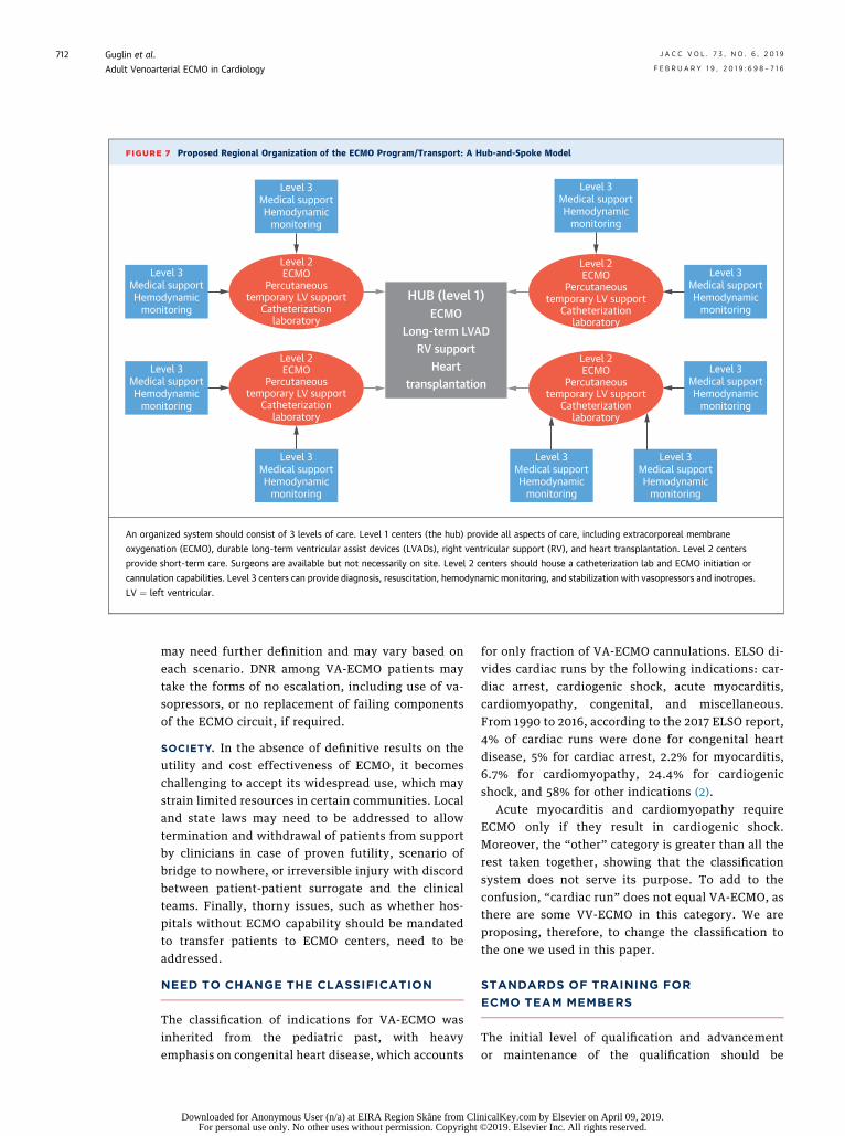
FIGURE 7 Proposed Regional Organization of the ECMO Program/Transport: A Hub-and-Spoke Model
Level 3Medical supportHemodynamic
monitoring
Level 3Medical supportHemodynamic
monitoring
Level 3Medical supportHemodynamic
monitoring
Level 3Medical supportHemodynamic
monitoring
Level 3Medical supportHemodynamic
monitoring
Level 3Medical supportHemodynamic
monitoring
Level 3Medical supportHemodynamic
monitoring
Level 3Medical supportHemodynamic
monitoring
Level 2ECMO
Percutaneoustemporary LV support
Catheterizationlaboratory
Level 2ECMO
Percutaneoustemporary LV support
Catheterizationlaboratory
Level 2ECMO
Percutaneoustemporary LV support
Catheterizationlaboratory
HUB (level 1)ECMO
Long-term LVADRV support
Hearttransplantation
Level 2ECMO
Percutaneoustemporary LV support
Catheterizationlaboratory
Level 3Medical supportHemodynamic
monitoring
An organized system should consist of 3 levels of care. Level 1 centers (the hub) provide all aspects of care, including extracorporeal membrane
oxygenation (ECMO), durable long-term ventricular assist devices (LVADs), right ventricular support (RV), and heart transplantation. Level 2 centers
provide short-term care. Surgeons are available but not necessarily on site. Level 2 centers should house a catheterization lab and ECMO initiation or
cannulation capabilities. Level 3 centers can provide diagnosis, resuscitation, hemodynamic monitoring, and stabilization with vasopressors and inotropes.
LV ¼ left ventricular.
Guglin et al. J A C C V O L . 7 3 , N O . 6 , 2 0 1 9
Adult Venoarterial ECMO in Cardiology F E B R U A R Y 1 9 , 2 0 1 9 : 6 9 8 – 7 1 6
712
may need further definition and may vary based oneach scenario. DNR among VA-ECMO patients maytake the forms of no escalation, including use of va-sopressors, or no replacement of failing componentsof the ECMO circuit, if required.
SOCIETY. In the absence of definitive results on theutility and cost effectiveness of ECMO, it becomeschallenging to accept its widespread use, which maystrain limited resources in certain communities. Localand state laws may need to be addressed to allowtermination and withdrawal of patients from supportby clinicians in case of proven futility, scenario ofbridge to nowhere, or irreversible injury with discordbetween patient-patient surrogate and the clinicalteams. Finally, thorny issues, such as whether hos-pitals without ECMO capability should be mandatedto transfer patients to ECMO centers, need to beaddressed.
NEED TO CHANGE THE CLASSIFICATION
The classification of indications for VA-ECMO wasinherited from the pediatric past, with heavyemphasis on congenital heart disease, which accounts
Downloaded for Anonymous User (n/a) at EIRA Region Skåne from CliFor personal use only. No other uses without permission. Copyright
for only fraction of VA-ECMO cannulations. ELSO di-vides cardiac runs by the following indications: car-diac arrest, cardiogenic shock, acute myocarditis,cardiomyopathy, congenital, and miscellaneous.From 1990 to 2016, according to the 2017 ELSO report,4% of cardiac runs were done for congenital heartdisease, 5% for cardiac arrest, 2.2% for myocarditis,6.7% for cardiomyopathy, 24.4% for cardiogenicshock, and 58% for other indications (2).
Acute myocarditis and cardiomyopathy requireECMO only if they result in cardiogenic shock.Moreover, the “other” category is greater than all therest taken together, showing that the classificationsystem does not serve its purpose. To add to theconfusion, “cardiac run” does not equal VA-ECMO, asthere are some VV-ECMO in this category. We areproposing, therefore, to change the classification tothe one we used in this paper.
STANDARDS OF TRAINING FOR
ECMO TEAM MEMBERS
The initial level of qualification and advancementor maintenance of the qualification should be
nicalKey.com by Elsevier on April 09, 2019. ©2019. Elsevier Inc. All rights reserved.
J A C C V O L . 7 3 , N O . 6 , 2 0 1 9 Guglin et al.F E B R U A R Y 1 9 , 2 0 1 9 : 6 9 8 – 7 1 6 Adult Venoarterial ECMO in Cardiology
713
standardized due to the high demands and specificskills required.
VA-ECMO FOR SEPTIC SHOCK. Sepsis has histori-cally been considered a contraindication to ECMO.The standard explanation has been that sepsis isassociated with multisystem organ failure and poorsurvival, regardless of extracorporeal life support.However, the lack of difference in survival betweenthe septic and nonseptic groups indicated thatinfection does not adversely affect the outcomes onECMO (10). ECMO can be beneficial in selected septicpatients with sepsis-related LV dysfunction.
VA-ECMO USE FOR BETTER PRESERVATION OF
DONOR ORGANS FOR TRANSPLANTATION. Trans-plant organs salvaged by VA-ECMO show exceptionalpromise. Single centers demonstrated that organs(kidney, liver) perfused by VA-ECMO after cardiacdeath have the same transplantation outcomes as dothose procured after brain death, the current goldstandard for posthumous donor harvesting (81).ECMO maintains an organ at body temperature anddelivers perfusate to a mechanically ventilated “lungin a box” or a beating “heart in a box” to allow for
Downloaded for Anonymous User (n/a) at EIRAFor personal use only. No other uses withou
organ improvement and assessment before trans-plantation with promising results (82,83).
CONCLUSIONS
VA ECMO is a powerful tool that needs to be usedjudiciously. As we gain proficiency, the lack of pro-spective randomized studies or a useful classificationsystem and areas of uncertainty create multiplechallenges. Appropriate infrastructure, appropriateskill set of team members, and thorough assessmentof each ECMO candidate, with an evaluation of in-dications and contraindication to ECMO support, arekey steps to ensure optimal outcomes.
ADDRESS FOR CORRESPONDENCE: Dr. Maya E.Guglin, Medical Director, Mechanical CirculatorySupport Program, Division of Cardiovascular Medi-cine, Gill Heart & Vascular Institute, University ofKentucky Albert B. Chandler Hospital, First Floor,Suite G100, 800 Rose Street, Lexington, Ken-tucky 40536. E-mail: [email protected]. Twitter:@MGuglin.
RE F E RENCE S
1. Peek GJ, Mugford M, Tiruvoipati R, et al., CESAtrial collaboration. Efficacy and economic assess-ment of conventional ventilatory support versusextracorporeal membrane oxygenation for severeadult respiratory failure (CESAR): a multicentrerandomised controlled trial. Lancet 2009;374:1351–63.
2. Extracorporeal Life Support Organization.Available at: http://www.elso.org. AccessedDecember 4, 2018.
3. McCarthy FH, McDermott KM, Kini V, et al.Trends in U.S. extracorporeal membrane oxygen-ation use and outcomes: 2002–2012. SeminThorac Cardiovasc Surg 2015;27:81–8.
4. Rajan S, Wissenberg M, Folke F, et al. Associa-tion of bystander cardiopulmonary resuscitationand survival according to ambulance responsetimes after out-of-hospital cardiac arrest. Circu-lation 2016;134:2095–104.
5. Shin TG, Choi JH, Jo IJ, et al. Extracorporealcardiopulmonary resuscitation in patients withinhospital cardiac arrest: a comparison with con-ventional cardiopulmonary resuscitation. Crit CareMed 2011;39:1–7.
6. Chen YS, Lin JW, Yu HY, et al. Cardiopulmonaryresuscitation with assisted extracorporeal life-support versus conventional cardiopulmonaryresuscitation in adults with in-hospital cardiac ar-rest: an observational study and propensity anal-ysis. Lancet 2008;372:554–61.
7. Kapur NK, Zisa DC. Veno-arterial extracorpo-real membrane oxygenation (VA-ECMO) fails tosolve the haemodynamic support equation incardiogenic shock. EuroIntervention 2016;11:1337–9.
8. Schmidt M, Tachon G, Devilliers C, et al.Blood oxygenation and decarboxylation de-terminants during venovenous ECMO for respi-ratory failure in adults. Intensive Care Med 2013;39:838–46.
9. Burkhoff D, Mirsky I, Suga H. Assessment ofsystolic and diastolic ventricular properties viapressure-volume analysis: a guide for clinical,translational, and basic researchers. Am J PhysiolHeart Circ Physiol 2005;289:H501–12.
10. Rihal CS, Naidu SS, Givertz MM, et al. 2015SCAI/ACC/HFSA/STS clinical expert consensusstatement on the use of percutaneous mechanicalcirculatory support devices in cardiovascular care:endorsed by the American Heart Association, theCardiological Society of India, and Sociedad LatinoAmericana de Cardiologia Intervencion; Affirma-tion of Value by the Canadian Association ofInterventional Cardiology-Association Canadiennede Cardiologie d’intervention. J Am Coll Cardiol2015;65:e7–26.
11. Bréchot N, Luyt CE, Schmidt M, et al. Venoar-terial extracorporeal membrane oxygenation sup-port for refractory cardiovascular dysfunctionduring severe bacterial septic shock. Crit Care Med2013;41:1616–26.
Region Skåne from Clint permission. Copyright ©
12. Burkhoff D, Sayer G, Doshi D, Uriel N. Hemo-dynamics of mechanical circulatory support. J AmColl Cardiol 2015;66:2663–74.
13. Boulate D, Luyt CE, Pozzi M, et al. Acute lunginjury after mechanical circulatory supportimplantation in patients on extracorporeallife support: an unrecognized problem. Eur JCardiothorac Surg 2013;44:544–9; discussion549–50.
14. Patel SM, Lipinski J, Al-Kindi SG, et al. Simul-taneous venoarterial extracorporeal membraneoxygenation and percutaneous left ventriculardecompression therapy with impella is associatedwith improved outcomes in refractory cardiogenicshock. ASAIO J 2019;65:21–8.
15. Petroni T, Harrois A, Amour J, et al. Intra-aorticballoon pump effects on macrocirculation andmicrocirculation in cardiogenic shock patientssupported by venoarterial extracorporeal mem-brane oxygenation*. Crit Care Med 2014;42:2075–82.
16. Akanni OJ, Takeda K, Truby LK, et al. EC-VAD:combined use of extracorporeal membraneoxygenation and percutaneous microaxial pumpleft ventricular assist device. ASAIO J 2018 Apr 19[E-pub ahead of print].
17. Annamalai SK, Buiten L, Esposito ML, et al.Acute hemodynamic effects of intra-aortic ballooncounterpulsation pumps in advanced heart failure.J Card Fail 2017;23:606–14.
icalKey.com by Elsevier on April 09, 2019.2019. Elsevier Inc. All rights reserved.
Guglin et al. J A C C V O L . 7 3 , N O . 6 , 2 0 1 9
Adult Venoarterial ECMO in Cardiology F E B R U A R Y 1 9 , 2 0 1 9 : 6 9 8 – 7 1 6
714
18. Aso S, Matsui H, Fushimi K, Yasunaga H. Theeffect of intraaortic balloon pumping undervenoarterial extracorporeal membrane oxygena-tion on mortality of cardiogenic patients: ananalysis using a nationwide inpatient database.Crit Care Med 2016;44:1974–9.
19. Lazzeri C, Bernardo P, Sori A, et al. Venous-arterial extracorporeal membrane oxygenationfor refractory cardiac arrest: a clinical challenge.Eur Heart J Acute Cardiovasc Care 2013;2:118–26.
20. Jaski BE, Ortiz B, Alla KR, et al. A 20-yearexperience with urgent percutaneous cardiopul-monary bypass for salvage of potential survivorsof refractory cardiovascular collapse. J ThoracCardiovasc Surg 2010;139:753–7. e1–2.
21. Murashita T, Eya K, Miyatake T, et al. Outcomeof the perioperative use of percutaneous cardio-pulmonary support for adult cardiac surgery: fac-tors affecting hospital mortality. Artif Organs2004;28:189–95.
22. Carroll BJ, Shah RV, Murthy V, et al. Clinicalfeatures and outcomes in adults with cardiogenicshock supported by extracorporeal membraneoxygenation. Am J Cardiol 2015;116:1624–30.
23. Ariyaratnam P, McLean LA, Cale AR,Loubani M. Extra-corporeal membrane oxygena-tion for the post-cardiotomy patient. Heart FailRev 2014;19:717–25.
24. Lan C, Tsai PR, Chen YS, Ko WJ. Prognosticfactors for adult patients receiving extracorporealmembrane oxygenation as mechanical circulatorysupport–a 14-year experience at a medical center.Artif Organs 2010;34:E59–64.
25. Combes A, Leprince P, Luyt CE, et al. Out-comes and long-term quality-of-life of patientssupported by extracorporeal membrane oxygena-tion for refractory cardiogenic shock. Crit CareMed 2008;36:1404–11.
26. Link MS, Berkow LC, Kudenchuk PJ, et al. Part7: adult advanced cardiovascular life support:2015 American Heart Association guidelines up-date for cardiopulmonary resuscitation and emer-gency cardiovascular care. Circulation 2015;132:S444–64.
27. Thiagarajan RR, Brogan TV, Scheurer MA,Laussen PC, Rycus PT, Bratton SL. Extracorporealmembrane oxygenation to support cardiopulmo-nary resuscitation in adults. Ann Thorac Surg2009;87:778–85.
28. Kagawa E, Inoue I, Kawagoe T, et al. Assess-ment of outcomes and differences between in- andout-of-hospital cardiac arrest patients treated withcardiopulmonary resuscitation using extracorpo-real life support. Resuscitation 2010;81:968–73.
29. Hutin A, Abu-Habsa M, Burns B, et al. EarlyECPR for out-of-hospital cardiac arrest: bestpractice in 2018. Resuscitation 2018;130:44–8.
30. Reynolds JC, Frisch A, Rittenberger JC,Callaway CW. Duration of resuscitation efforts andfunctional outcome after out-of-hospital cardiacarrest: when should we change to novel therapies?Circulation 2013;128:2488–94.
Downloaded for Anonymous User (n/a) at EIRFor personal use only. No other uses witho
31. Kim SJ, Jung JS, Park JH, Park JS, Hong YS,Lee SW. An optimal transition time to extracor-poreal cardiopulmonary resuscitation for predict-ing good neurological outcome in patients without-of-hospital cardiac arrest: a propensity-matched study. Crit Care 2014;18:535.
32. Wengenmayer T, Rombach S, Ramshorn F,et al. Influence of low-flow time on survival afterextracorporeal cardiopulmonary resuscitation(eCPR). Crit Care 2017;21:157.
33. Johnson NJ, Acker M, Hsu CH, et al. Extra-corporeal life support as rescue strategy for out-of-hospital and emergency department cardiacarrest. Resuscitation 2014;85:1527–32.
34. Ouweneel DM, Schotborgh JV, Limpens J,et al. Extracorporeal life support during cardiacarrest and cardiogenic shock: a systematic reviewand meta-analysis. Intensive Care Med 2016;42:1922–34.
35. Pagani FD, Lynch W, Swaniker F, et al. Extra-corporeal life support to left ventricular assistdevice bridge to heart transplant: a strategy tooptimize survival and resource utilization. Circu-lation 1999;100:II206–10.
36. Guglin M, Burchett A. VA-ECMO in cardiogenicshock in adults: indications and outcomes. TheVAD Journal 2016;2:8.
37. Aiba T, Nonogi H, Itoh T, et al. Appropriateindications for the use of a percutaneous cardio-pulmonary support system in cases with cardio-genic shock complicating acute myocardialinfarction. Jpn Circ J 2001;65:145–9.
38. Shawl FA, Domanski MJ, Hernandez TJ,Punja S. Emergency percutaneous cardiopulmo-nary bypass support in cardiogenic shock fromacute myocardial infarction. Am J Cardiol 1989;64:967–70.
39. Kawahito K, Murata S, Yasu T, et al. Usefulnessof extracorporeal membrane oxygenation fortreatment of fulminant myocarditis and circulatorycollapse. Am J Cardiol 1998;82:910–1.
40. Pages ON, Aubert S, Combes A, et al. Para-corporeal pulsatile biventricular assist device versusextracorporalmembraneoxygenation-extracorporallife support in adult fulminant myocarditis. J ThoracCardiovasc Surg 2009;137:194–7.
41. Kawahito K, Murata S, Adachi H, Ino T, Fuse K.Resuscitation and circulatory support usingextracorporeal membrane oxygenation for fulmi-nant pulmonary embolism. Artif Organs 2000;24:427–30.
42. Akkanti B, Nathan S, Centeno EN, et al.Massive pulmonary embolism with hemodynamiccompromise successfully treated with veno-arterial extracorporeal membrane oxygenation.VAD J 2015;1. Article 20.
43. Kittleson MM, Patel JK, Moriguchi JD, et al.Heart transplant recipients supported with extra-corporeal membrane oxygenation: outcomes froma single-center experience. J Heart Lung Trans-plant 2011;30:1250–6.
44. Diddle JW, Almodovar MC, Rajagopal SK,Rycus PT, Thiagarajan RR. Extracorporeal
A Region Skåne from ClinicalKey.com by Elsevierut permission. Copyright ©2019. Elsevier Inc. All r
membrane oxygenation for the support of adultswith acute myocarditis. Crit Care Med 2015;43:1016–25.
45. Banjac I, Petrovic M, Akay MH, et al. Extra-corporeal membrane oxygenation as a proceduralrescue strategy for transcatheter aortic valvereplacement cardiac complications. ASAIO J 2016;62:e1–4.
46. Tsai FC, Wang YC, Huang YK, et al. Extracor-poreal life support to terminate refractory ven-tricular tachycardia. Crit Care Med 2007;35:1673–6.
47. Baratto F, Pappalardo F, Oloriz T, et al. Extra-corporeal membrane oxygenation for hemody-namic support of ventricular tachycardia ablation.Circ Arrhythm Electrophysiol 2016;9:e004492.
48. Enriquez A, Liang J, Gentile J, et al. Outcomesof rescue cardiopulmonary support for periproce-dural acute hemodynamic decompensation in pa-tients undergoing catheter ablation of electricalstorm. Heart Rhythm 2018;15:75–80.
49. Chen CY, Tsai J, Hsu TY, et al. ECMO used in arefractory ventricular tachycardia and ventricularfibrillation patient: a national case-control study.Medicine (Baltimore) 2016;95:e3204.
50. Bhandary SP, Joseph N, Hofmann JP,Saranteas T, Papadimos TJ. Extracorporeal lifesupport for refractory ventricular tachycardia. AnnTransl Med 2017;5:73.
51. Le Pennec-Prigent S, Flecher E, Auffret V, et al.Effectiveness of extracorporeal life support forpatients with cardiogenic shock due to intractablearrhythmic storm. Crit Care Med 2017;45:e281–9.
52. Palaniswamy C, Miller MA, Reddy VY,Dukkipati SR. Hemodynamic support for ventricu-lar tachycardia ablation. Card Electrophysiol Clin2017;9:141–52.
53. Paulsen MJ, Orizondo R, Le D, Rojas-Pena A,Bartlett RH. A simple, standard method to char-acterize pressure/flow performance of vascularaccess cannulas. ASAIO J 2013;59:24–9.
54. Cheng R, Hachamovitch R, Kittleson M, et al.Complications of extracorporeal membraneoxygenation for treatment of cardiogenic shockand cardiac arrest: a meta-analysis of 1,866 adultpatients. Ann Thorac Surg 2014;97:610–6.
55. Wang CH, Chou NK, Becker LB, et al. Improvedoutcome of extracorporeal cardiopulmonaryresuscitation for out-of-hospital cardiac arrest–acomparison with that for extracorporeal rescue forin-hospital cardiac arrest. Resuscitation 2014;85:1219–24.
56. Nolan JP, Soar J, Zideman DA, et al., ERCGuidelines Writing Group. European ResuscitationCouncil guidelines for resuscitation 2010 Section1. Executive summary. Resuscitation 2010;81:1219–76.
57. Stub D, Bernard S, Pellegrino V, et al. Re-fractory cardiac arrest treated with mechanicalCPR, hypothermia, ECMO and early reperfusion(the CHEER trial). Resuscitation 2015;86:88–94.
on April 09, 2019.ights reserved.

J A C C V O L . 7 3 , N O . 6 , 2 0 1 9 Guglin et al.F E B R U A R Y 1 9 , 2 0 1 9 : 6 9 8 – 7 1 6 Adult Venoarterial ECMO in Cardiology
715
58. Biscotti M, Lee A, Basner RC, et al. Hybridconfigurations via percutaneous access for extra-corporeal membrane oxygenation: a single-centerexperience. ASAIO J 2014;60:635–42.
59. Abrams D, Baldwin MR, Champion M, et al.Thrombocytopenia and extracorporeal membraneoxygenation in adults with acute respiratory fail-ure: a cohort study. Intensive Care Med 2016;42:844–52.
60. Bridges B, Ranucci M, Lequier L. Anti-coagulation and disorders of hemostasis. In:Brogan TLL, Lorusso R, MacLaren G, Peek G, edi-tors. Extracorporeal Life Support: The ELSO RedBook. 5th ed. Ann Arbor, MI: Extracorporeal LifeSupport Organization (ELSO), 2017:81–92.
61. Zhang Z, Gu WJ, Chen K, Ni H. Mechanicalventilation during extracorporeal membraneoxygenation in patients with acute severe respi-ratory failure. Can Respir J 2017;2017:1783857.
62. Swol J, Belohlávek J, Haft JW, Ichiba S,Lorusso R, Peek GJ. Conditions and procedures forin-hospital extracorporeal life support (ECLS) incardiopulmonary resuscitation (CPR) of adult pa-tients. Perfusion 2016;31:182–8.
63. Vida VL, Lo Rito M, Padalino MA, Stellin G.Extracorporeal membrane oxygenation: thesimplified weaning bridge. J Thorac CardiovascSurg 2012;143:e27–8.
64. Lamb KM, DiMuzio PJ, Johnson A, et al. Arterialprotocol including prophylactic distal perfusioncatheter decreases limb ischemia complications inpatients undergoing extracorporeal membraneoxygenation. J Vasc Surg 2017;65:1074–9.
65. Juo YY, Skancke M, Sanaiha Y, Mantha A,Jimenez JC, Benharash P. Efficacy of distal perfu-sion cannulae in preventing limb ischemia duringextracorporeal membrane oxygenation: a system-atic review and meta-analysis. Artif Organs 2017;41:E263–73.
66. Nasr DM, Rabinstein AA. Neurologic compli-cations of extracorporeal membrane oxygenation.J Clin Neurol 2015;11:383–9.
67. Di Gennaro J, Farris R, McMullan D. Risk ofstroke by location of arterial cannulation and agein veno-arterial ECMO. Crit Care Med 2013;41:A168–9.
68. Camboni D, Schmid C. Neurologic and pul-monary complications in ECLS. In: Brogan T LL,Lorusso R, MacLaren G, Peek G, editors. Extra-corporeal Life Support: The ELSO Red Book. 5 ed.Ann Arbor, MI: Extracorporeal Life Support Orga-nization (ELSO), 2017:575–81.
69. Chung YS, Cho DY, Sohn DS, et al. Is stoppingheparin safe in patients on extracorporeal mem-brane oxygenation treatment? ASAIO J 2017;63:32–6.
70. Combes A, Brodie D, Bartlett R, et al., Inter-national ECMO Network (ECMONet). Position pa-per for the organization of extracorporealmembrane oxygenation programs for acute respi-ratory failure in adult patients. Am J Respir CritCare Med 2014;190:488–96.
71. van Diepen S, Katz JN, Albert NM, et al.,American Heart Association Council on Clinical
Downloaded for AnonFor personal use o
Cardiology, Council on Cardiovascular and StrokeNursing, Council on Quality of Care and OutcomesResearch; and Mission: Lifeline. Contemporarymanagement of cardiogenic shock: a scientificstatement from the American Heart Association.Circulation 2017;136:e232–68.
72. Salna M, Chicotka S, Biscotti M 3rd, et al.Management of surge in extracorporeal membraneoxygenation transport. Ann Thorac Surg 2018;105:528–34.
73. Gutsche JT, Vernick WJ. Cardiac and criticalcare anesthesiologists may be ideal members ofthe mobile ECMO team. J Cardiothorac VascAnesth 2016;30:1439–40.
74. Lawson DS, Lawson AF, Walczak R, et al. NorthAmerican neonatal extracorporeal membraneoxygenation (ECMO) devices and team roles:2008 survey results of Extracorporeal Life Sup-port Organization (ELSO) centers. J Extra CorporTechnol 2008;40:166–74.
75. Nichol G, Karmy-Jones R, Salerno C, Cantore L,Becker L. Systematic review of percutaneous car-diopulmonary bypass for cardiac arrest or cardio-genic shock states. Resuscitation 2006;70:381–94.
76. Tchantchaleishvili V, Hallinan W, Massey HT.Call for organized statewide networks for man-agement of acute myocardial infarction-relatedcardiogenic shock. JAMA Surg 2015;150:1025–6.
77. Beurtheret S, Mordant P, Paoletti X, et al.Emergency circulatory support in refractorycardiogenic shock patients in remote institutions: apilot study (the cardiac-RESCUE program). EurHeart J 2013;34:112–20.
78. Kaliyev R, Kapyshev T, Goncharov A,Lesbekov T, Pya Y. Maternal and fetal recoveryafter severe respiratory failure: a case report of airtransportation of a pregnant woman on ECMOusing the CentriMag transporter system. ASAIO J2015;61:e33–5.
79. Ramanathan K, Cove ME, Caleb MG, Teoh KL,Maclaren G. Ethical dilemmas of adult ECMO:emerging conceptual challenges. J CardiothoracVasc Anesth 2015;29:229–33.
80. Courtwright AM, Robinson EM, Feins K, et al.Ethics committee consultation and extracorporealmembrane oxygenation. Ann Am Thorac Soc 2016;13:1553–8.
81. Christopher DA, Woodside KJ. Expanding thedonor pool: organ donation after brain death forextracorporeal membrane oxygenation patients.Crit Care Med 2017;45:1790–1.
82. Ardehali A, Esmailian F, Deng M, et al. Ex-vivoperfusion of donor hearts for human heart trans-plantation (PROCEED II): a prospective, open-label, multicentre, randomised non-inferioritytrial. Lancet 2015;385:2577–84.
83. Perez-Villares JM, Lara-Rosales R, Fernandez-Carmona A, Fuentes-Garcia P, Burgos-Fuentes M,Baquedano-Fernandez B. Mobile ECMO team forcontrolled donation after circulatory death. Am JTransplant 2018;18:1293–4.
84. Asaumi Y, Yasuda S, Morii I, et al. Favourableclinical outcome in patients with cardiogenic shock
ymous User (n/a) at EIRA Region Skåne from Clinnly. No other uses without permission. Copyright ©
due to fulminant myocarditis supported bypercutaneous extracorporeal membrane oxygena-tion. Eur Heart J 2005;26:2185–92.
85. Kato S, Morimoto S, Hiramitsu S, Nomura M,Ito T, Hishida H. Use of percutaneous cardiopul-monary support of patients with fulminantmyocarditis and cardiogenic shock for improvingprognosis. Am J Cardiol 1999;83:623–5. A10.
86. Hsu KH, Chi NH, Yu HY, et al. Extracorporealmembranous oxygenation support for acutefulminant myocarditis: analysis of a single center’sexperience. Eur J Cardiothorac Surg 2011;40:682–8.
87. Mirabel M, Luyt CE, Leprince P, et al. Out-comes, long-term quality of life, and psychologicassessment of fulminant myocarditis patientsrescued by mechanical circulatory support. CritCare Med 2011;39:1029–35.
88. Gariboldi V, Grisoli D, Tarmiz A, et al. Mobileextracorporeal membrane oxygenation unit ex-pands cardiac assist surgical programs. Ann ThoracSurg 2010;90:1548–52.
89. Ishida K, Wada H, Sakakura K, et al. Long-termfollow-up on cardiac function following fulminantmyocarditis requiring percutaneous extracorporealcardiopulmonary support. Heart Vessels 2013;28:86–90.
90. Wu MY, Lee MY, Lin CC, Chang YS, Tsai FC,Lin PJ. Resuscitation of non-postcardiotomycardiogenic shock or cardiac arrest with extracor-poreal life support: the role of bridging to inter-vention. Resuscitation 2012;83:976–81.
91. Montero S, Aissaoui N, Tadie JM, et al. Fulmi-nant giant-cell myocarditis on mechanical circu-latory support: management and outcomes of aFrench multicentre cohort. Int J Cardiol 2018;253:105–12.
92. Liao X, Li B, Cheng Z. Extracorporeal mem-brane oxygenation in adult patients with acutefulminant myocarditis: clinical outcomes and riskfactor analysis. Herz 2018;43:728–32.
93. Lorusso R, Centofanti P, Gelsomino S, et al.Venoarterial extracorporeal membrane oxygena-tion for acute fulminant myocarditis in adult pa-tients: a 5-Year multi-institutional experience. AnnThorac Surg 2016;101:919–26.
94. Nakamura T, Ishida K, Taniguchi Y, et al.Prognosis of patients with fulminant myocarditismanaged by peripheral venoarterial extracorpo-real membranous oxygenation support: a retro-spective single-center study. J Intensive Care2015;3:5.
95. Aso S, Matsui H, Fushimi K, Yasunaga H.In-hospital mortality and successful weaningfrom venoarterial extracorporeal membraneoxygenation: analysis of 5,263 patients using anational inpatient database in Japan. Crit Care2016;20:80.
96. Maggio P, Hemmila M, Haft J, Bartlett R.Extracorporeal life support for massive pulmonaryembolism. J Trauma 2007;62:570–6.
97. Munakata R, Yamamoto T, Hosokawa Y, et al.Massive pulmonary embolism requiring extracorporeal
icalKey.com by Elsevier on April 09, 2019.2019. Elsevier Inc. All rights reserved.
Guglin et al. J A C C V O L . 7 3 , N O . 6 , 2 0 1 9
Adult Venoarterial ECMO in Cardiology F E B R U A R Y 1 9 , 2 0 1 9 : 6 9 8 – 7 1 6
716
life support treated with catheter-based in-terventions. Int Heart J 2012;53:370–4.
98. Dolmatova EV, Moazzami K, Cocke TP, et al.Extracorporeal membrane oxygenation inmassive pulmonary embolism. Heart Lung 2017;46:106–9.
99. Corsi F, Lebreton G, Bréchot N, et al.Life-threatening massive pulmonary embolismrescued by venoarterial-extracorporeal membraneoxygenation. Crit Care 2017;21:76.
Downloaded for Anonymous User (n/a) at EIRFor personal use only. No other uses witho
100. George B, Parazino M, Omar HR, et al. Aretrospective comparison of survivors and non-survivors of massive pulmonary embolismreceiving veno-arterial extracorporeal membraneoxygenation support. Resuscitation 2018;122:1–5.
101. Pasrija C, Kon Z. A protocolized approach toveno-arterial extracorporeal membrane oxygena-tion for massive pulmonary embolism. Resuscita-tion 2018;126:e12.
A Region Skåne from ClinicalKey.com by Elsevierut permission. Copyright ©2019. Elsevier Inc. All r
KEY WORDS cardiac arrest, cardiogenicshock, ECLS
Go to http://www.acc.org/jacc-journals-cme to takethe CME/MOC/ECME quizfor this article.
oig
n April 09, 2019.hts reserved.