Embed Size (px)
Citation preview
VV-ECMO
ECMO Physiotherapy NetworkJuly 2016
Veno-Venous ECMO
• Indicated for potentially reversible, life-threatening forms of respiratory failure when adequate heart function is anticipated for the duration of ECMO.
• Venous blood is drained, circulated through oxygenator membrane and returned to the venous circulation
Indications for V-V ECMO• Common• Severe pneumonia• ARDS• Pulmonary contusion• Possible• Alveolar proteinosis• Smoke inhalation• Status asthmaticus• Airway obstruction• Aspiration syndromes
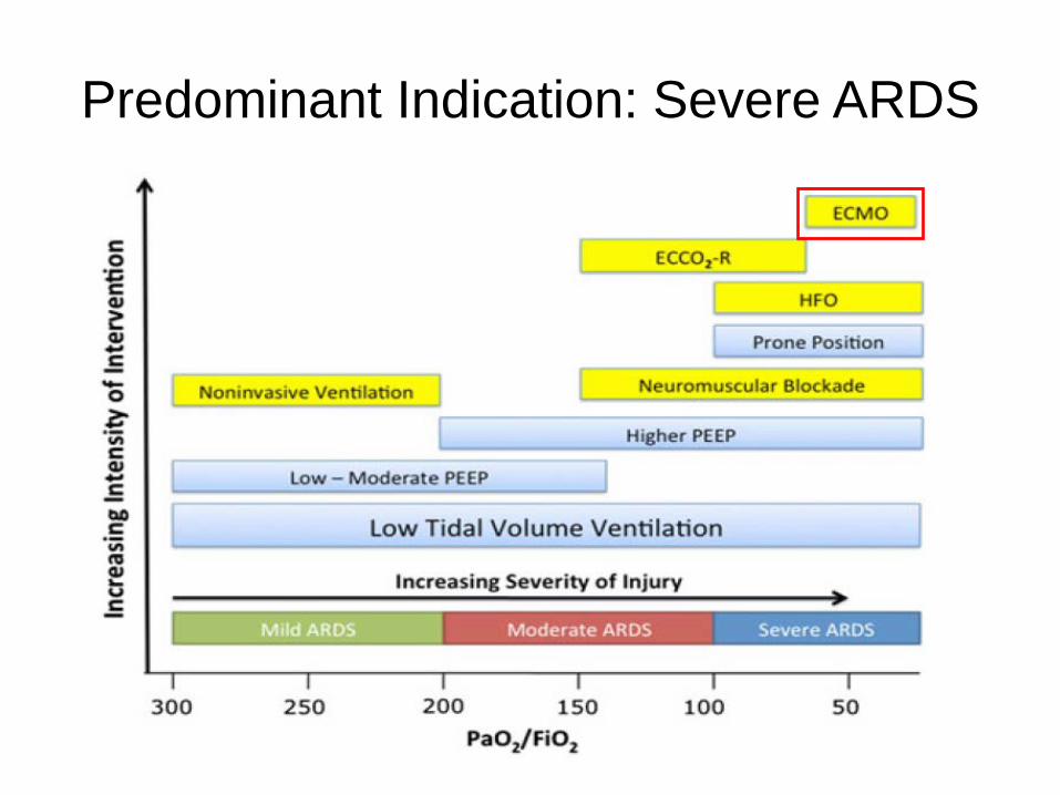
Predominant Indication: Severe ARDS
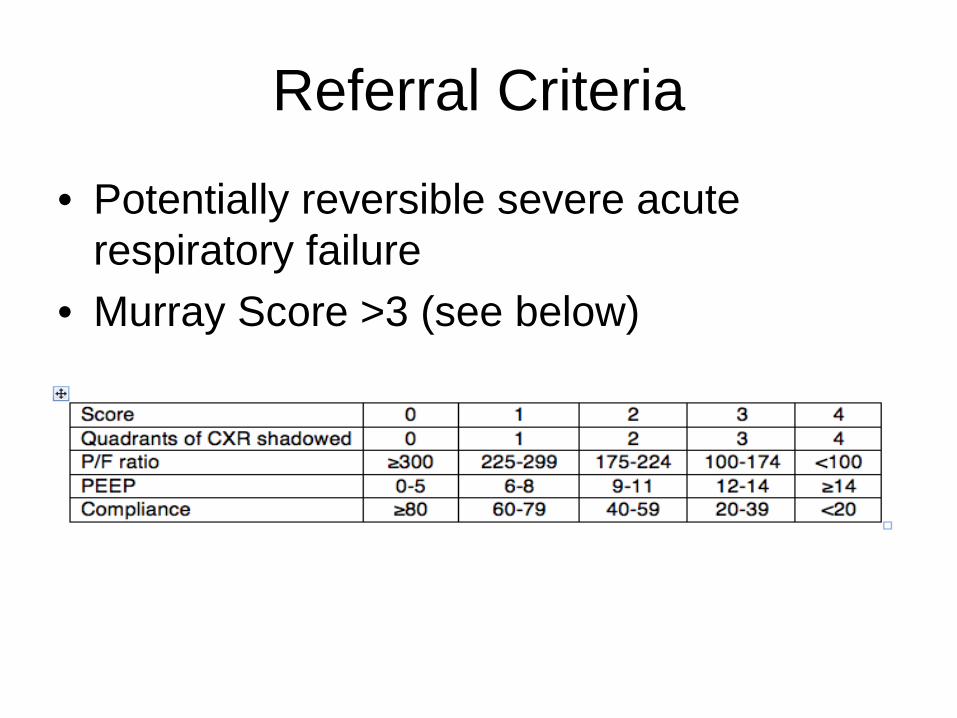
Referral Criteria
• Potentially reversible severe acute respiratory failure
• Murray Score >3 (see below)
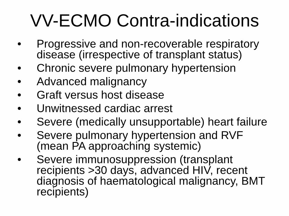
VV-ECMO Contra-indications• Progressive and non-recoverable respiratory
disease (irrespective of transplant status)• Chronic severe pulmonary hypertension • Advanced malignancy• Graft versus host disease• Unwitnessed cardiac arrest• Severe (medically unsupportable) heart failure• Severe pulmonary hypertension and RVF
(mean PA approaching systemic)• Severe immunosuppression (transplant
recipients >30 days, advanced HIV, recent diagnosis of haematological malignancy, BMT recipients)
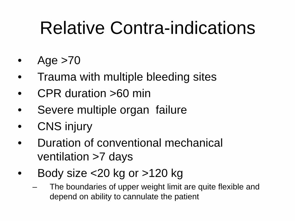
Relative Contra-indications• Age >70• Trauma with multiple bleeding sites• CPR duration >60 min• Severe multiple organ failure• CNS injury• Duration of conventional mechanical
ventilation >7 days • Body size <20 kg or >120 kg
– The boundaries of upper weight limit are quite flexible and depend on ability to cannulate the patient
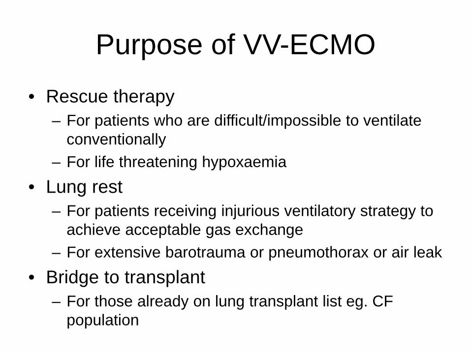
Purpose of VV-ECMO• Rescue therapy
– For patients who are difficult/impossible to ventilate conventionally
– For life threatening hypoxaemia• Lung rest
– For patients receiving injurious ventilatory strategy to achieve acceptable gas exchange
– For extensive barotrauma or pneumothorax or air leak• Bridge to transplant
– For those already on lung transplant list eg. CF population
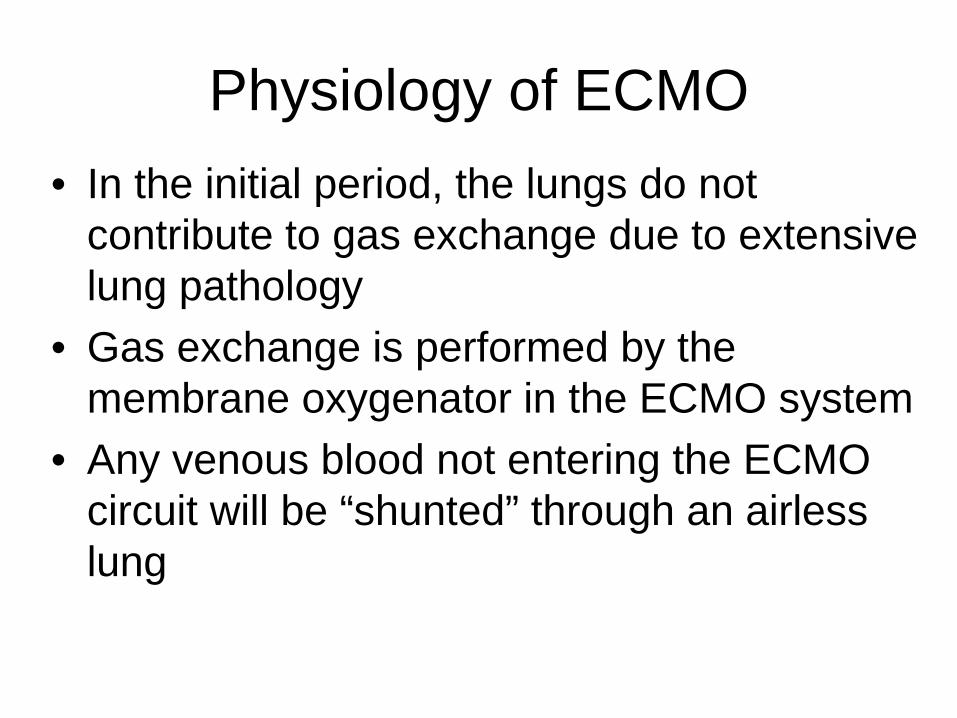
Physiology of ECMO • In the initial period, the lungs do not
contribute to gas exchange due to extensive lung pathology
• Gas exchange is performed by the membrane oxygenator in the ECMO system
• Any venous blood not entering the ECMO circuit will be “shunted” through an airless lung
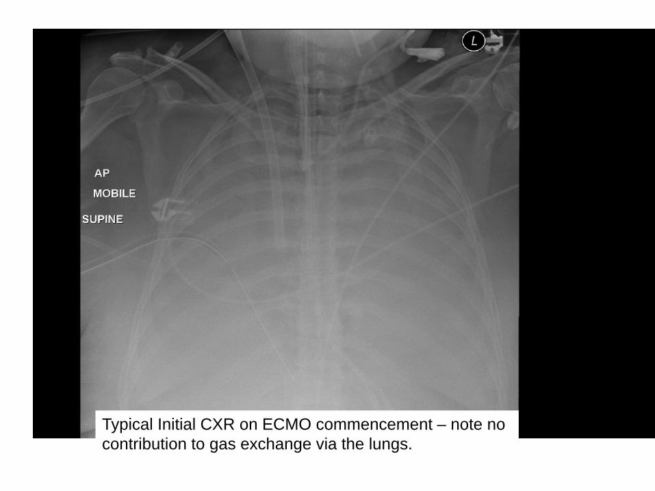
Typical Initial CXR on ECMO commencement – note no contribution to gas exchange via the lungs.
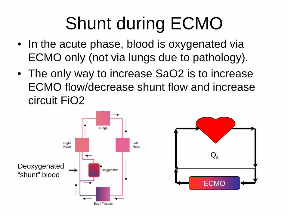
Shunt during ECMO• In the acute phase, blood is oxygenated via
ECMO only (not via lungs due to pathology).• The only way to increase SaO2 is to increase
ECMO flow/decrease shunt flow and increase circuit FiO2
ECMO
Qs
Deoxygenated “shunt” blood
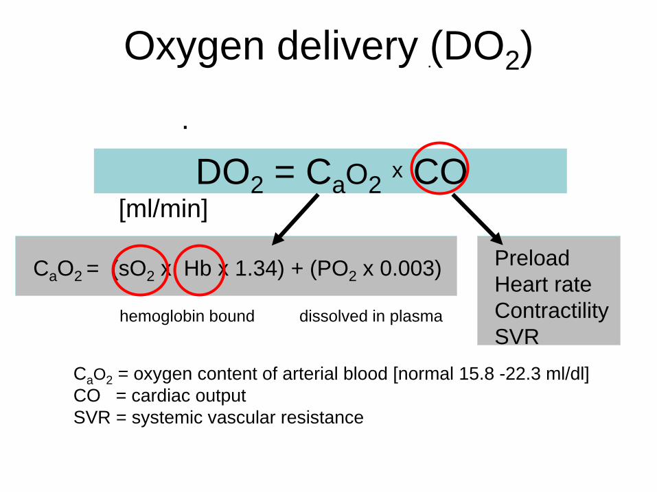
Oxygen delivery (DO2)
DO2 = CaO2 x CO [ml/min]
CaO2 = (sO2 x Hb x 1.34) + (PO2 x 0.003) PreloadHeart rateContractilitySVR
CaO2 = oxygen content of arterial blood [normal 15.8 -22.3 ml/dl]CO = cardiac outputSVR = systemic vascular resistance
.
.
hemoglobin bound dissolved in plasma
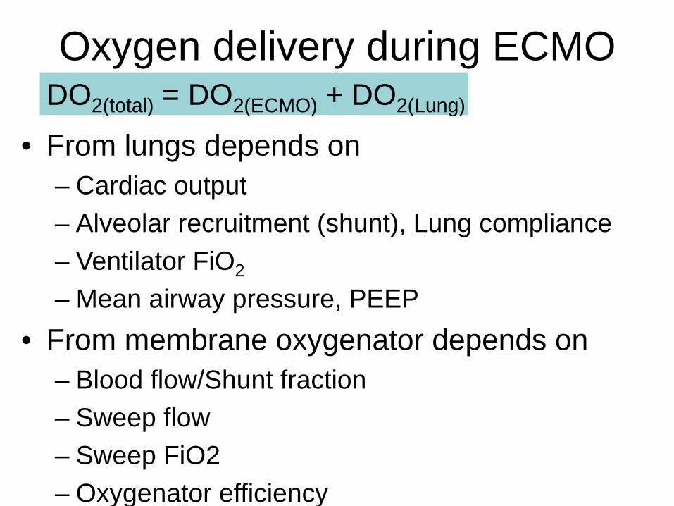
Oxygen delivery during ECMO
• From lungs depends on– Cardiac output– Alveolar recruitment (shunt), Lung compliance– Ventilator FiO2
– Mean airway pressure, PEEP• From membrane oxygenator depends on
– Blood flow/Shunt fraction– Sweep flow– Sweep FiO2– Oxygenator efficiency
DO2(total) = DO2(ECMO) + DO2(Lung)
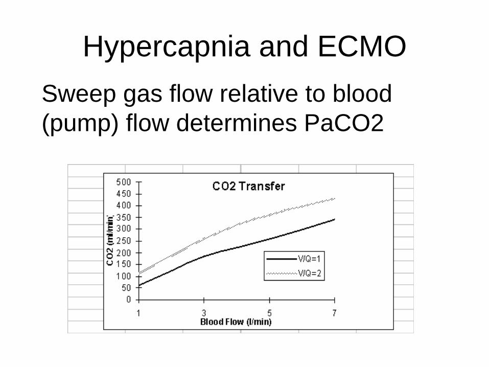
Hypercapnia and ECMOSweep gas flow relative to blood (pump) flow determines PaCO2
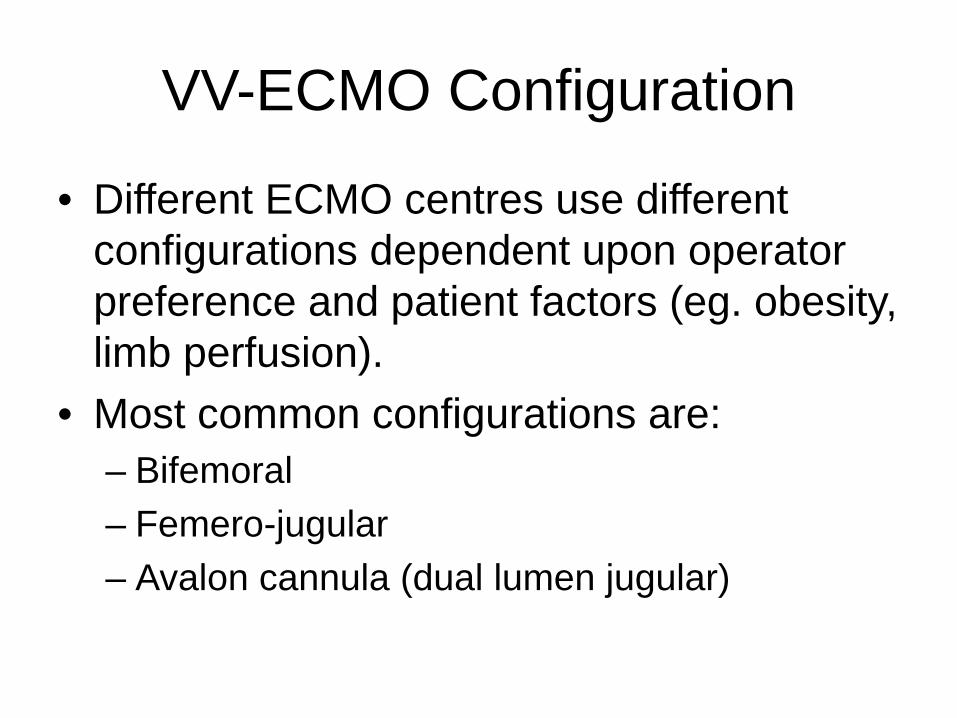
VV-ECMO Configuration
• Different ECMO centres use different configurations dependent upon operator preference and patient factors (eg. obesity, limb perfusion).
• Most common configurations are:– Bifemoral– Femero-jugular– Avalon cannula (dual lumen jugular)
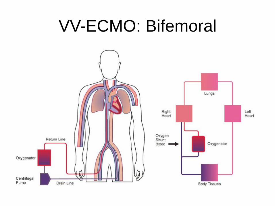
VV-ECMO: Bifemoral

VV-ECMO: Fem/Jug & Avalon
Femoral-Jugular Configuration Avalon Dual (Jugular) Cannulation
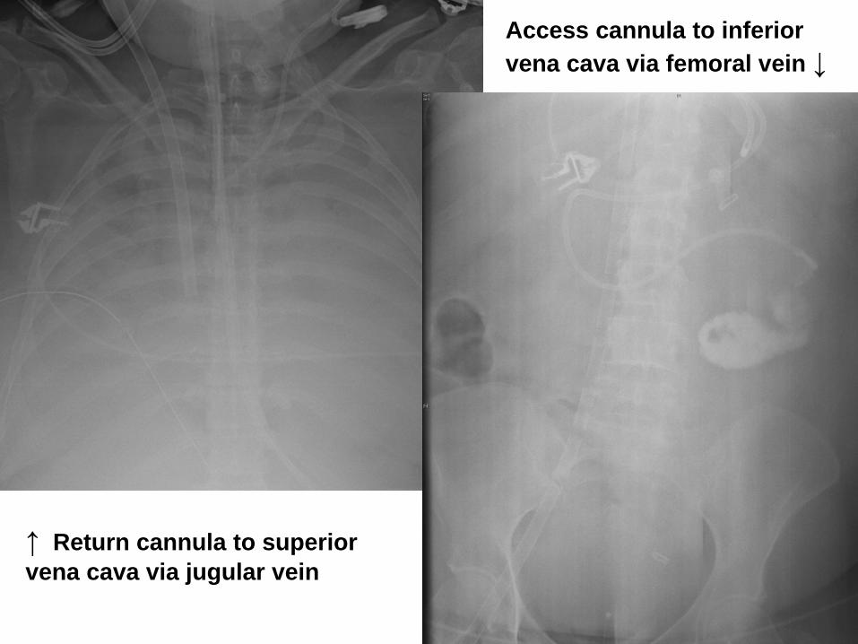
↑ Return cannula to superior vena cava via jugular vein
Access cannula to inferior vena cava via femoral vein ↓
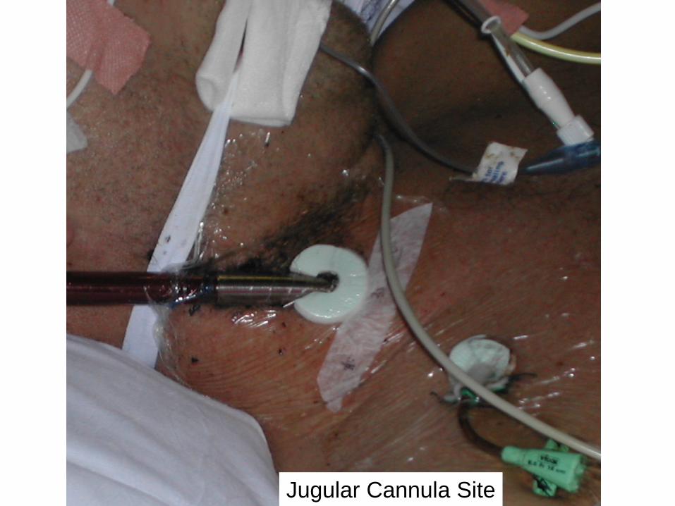
Jugular Cannula Site
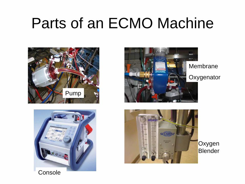
Parts of an ECMO Machine
Pump
Membrane
Oxygenator
Oxygen Blender
Console
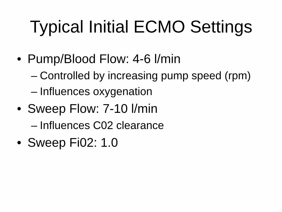
Typical Initial ECMO Settings
• Pump/Blood Flow: 4-6 l/min– Controlled by increasing pump speed (rpm)– Influences oxygenation
• Sweep Flow: 7-10 l/min– Influences C02 clearance
• Sweep Fi02: 1.0
Weaning from ECMO• As lung function recovers, ECMO support can
be reduced• Pump/blood flow usually remains >3 l/min to
prevent clots• Sweep Fi02 remains 1.0• Weaning occurs by reducing Sweep Flow
– This reduces contribution of membrane oxygenator to gas exchange.
• Trail off sweep flow– With blood still circulating via ECMO circuit but no
contribution to gas exchange externally– “Tests” whether patient can manage without ECMO
support
Mechanical Ventilation During VV-ECMO
• Most patients are mechanically ventilated – Ultra-protective ventilation strategy TVs <6 mls/kg– Typical settings 10 PEEP + 10 inspiratory pressure =
peak pressures 20cmH20– Increased sweep flow is used for C02 clearance
rather than increasing minute volume which may be injurious
• Some patients are awake, spontaneously breathing without mechanical ventilation
Physiotherapy Management During VV-ECMO
• See ECMO Physiotherapy Network consensus document.