Embed Size (px)
Citation preview
October 2019 | veinforum.org
NEWSLETTERVEIN SPECIALIST
Want to receive monthly updates? Click here to be added to the email list
INSIDE THIS EDITIONThe Next Conundrum 2
JVS-VL November Issue Preview 4Deep Venous Stenting 6
Iliofemoral Venous Stent 13Venous Stents: Old vs. New 16
Vein Stenting in Europe 17Why Do Venus Stents Fail? 19
Stent Issue
October 2019 | veinforum.org@americanvenousforum @VeinForum 2
INSIDE THIS EDITION
The Next Conundrum 2 JVS-VL November Issue Preview 4
Deep Venous Stenting 6 Iliofemoral Venous Stent 13
Venous Stents: Old vs. New 16Vein Stenting in Europe 17
Why Do Venus Stents Fail? 19
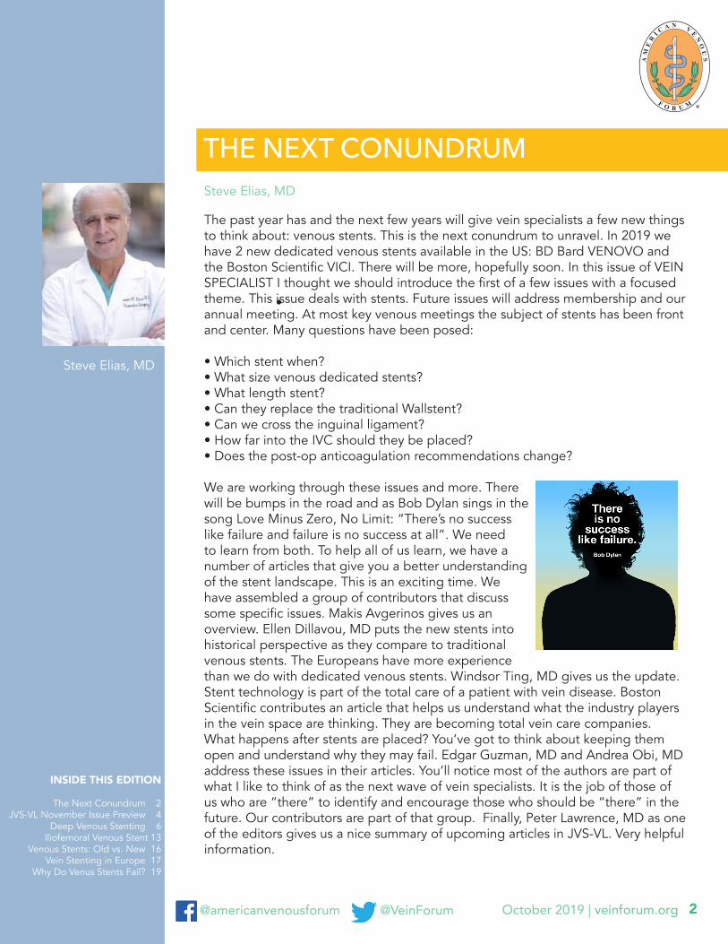
THE NEXT CONUNDRUM
The past year has and the next few years will give vein specialists a few new things to think about: venous stents. This is the next conundrum to unravel. In 2019 we have 2 new dedicated venous stents available in the US: BD Bard VENOVO and the Boston Scientific VICI. There will be more, hopefully soon. In this issue of VEIN SPECIALIST I thought we should introduce the first of a few issues with a focused theme. This issue deals with stents. Future issues will address membership and our annual meeting. At most key venous meetings the subject of stents has been front and center. Many questions have been posed:
• Which stent when?• What size venous dedicated stents?• What length stent?• Can they replace the traditional Wallstent?• Can we cross the inguinal ligament? • How far into the IVC should they be placed?• Does the post-op anticoagulation recommendations change?
We are working through these issues and more. There will be bumps in the road and as Bob Dylan sings in the song Love Minus Zero, No Limit: “There’s no success like failure and failure is no success at all”. We need to learn from both. To help all of us learn, we have a number of articles that give you a better understanding of the stent landscape. This is an exciting time. We have assembled a group of contributors that discuss some specific issues. Makis Avgerinos gives us an overview. Ellen Dillavou, MD puts the new stents into historical perspective as they compare to traditional venous stents. The Europeans have more experience than we do with dedicated venous stents. Windsor Ting, MD gives us the update. Stent technology is part of the total care of a patient with vein disease. Boston Scientific contributes an article that helps us understand what the industry players in the vein space are thinking. They are becoming total vein care companies. What happens after stents are placed? You’ve got to think about keeping them open and understand why they may fail. Edgar Guzman, MD and Andrea Obi, MD address these issues in their articles. You’ll notice most of the authors are part of what I like to think of as the next wave of vein specialists. It is the job of those of us who are “there” to identify and encourage those who should be “there” in the future. Our contributors are part of that group. Finally, Peter Lawrence, MD as one of the editors gives us a nice summary of upcoming articles in JVS-VL. Very helpful information.
Steve Elias, MD
Steve Elias, MD
•
October 2019 | veinforum.org@americanvenousforum @VeinForum 3
INSIDE THIS EDITION
The Next Conundrum 2 JVS-VL November Issue Preview 4
Deep Venous Stenting 6 Iliofemoral Venous Stent 13
Venous Stents: Old vs. New 16Vein Stenting in Europe 17
Why Do Venus Stents Fail? 19
THE NEXT CONUNDRUMSteve Elias, MD
This “Stent Issue” highlights some of the emerging concepts of dedicated venous stents. We are in the infancy of dedicated venous stents. I suspect with more experience we will fine tune our approach. As mentioned earlier, this issue is the first of “themed” issue of VEIN SPECIALIST. Let us know what you think. As we evolve we need your feedback. To that end we are also instituting a commentary section where you can send comments. VEIN SPECIALIST is a work in progress. Nothing is written in stone except perhaps an epitaph chisled on a gravestone. VEIN SPECIALIST doesn’t intend to die. At least not any time soon. We hope you enjoy.
October 2019 | veinforum.org@americanvenousforum @VeinForum 4
INSIDE THIS EDITION
The Next Conundrum 2 JVS-VL November Issue Preview 4
Deep Venous Stenting 6 Iliofemoral Venous Stent 13
Venous Stents: Old vs. New 16Vein Stenting in Europe 17
Why Do Venus Stents Fail? 19
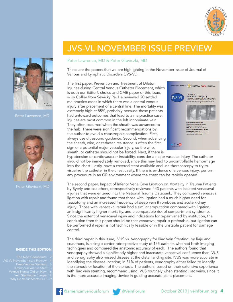
These are the papers that we are highlighting in the November issue of Journal of Venous and Lymphatic Disorders (JVS-VL): The first paper, Prevention and Treatment of Dilator Injuries during Central Venous Catheter Placement, which is both our Editor’s choice and CME paper of this issue, is by Collier from Sewicky Pa. He reviewed 20 settled malpractice cases in which there was a central venous injury after placement of a central line. The mortality was extremely high at 85%, probably because these patients had untoward outcomes that lead to a malpractice case. Injuries are most common in the left innominate vein. They often occurred when the sheath was advanced to the hub. There were significant recommendations by the author to avoid a catastrophic complication. First, always use ultrasound guidance. Second, when advancing the sheath, wire, or catheter, resistance is often the first sign of a potential major vascular injury, so the wire, sheath, or catheter should not be forced. Next, if there is hypotension or cardiovascular instability, consider a major vascular injury. The catheter should not be immediately removed, since this may lead to uncontrollable hemorrhage into the chest. Lastly, have a covered stent available and use thoracoscopy to try to visualize the catheter in the chest cavity. If there is evidence of a venous injury, perform any procedure in an OR environment where the chest can be rapidly opened. The second paper, Impact of Inferior Vena Cava Ligation on Mortality in Trauma Patients, by Byerly and coauthors, retrospectively reviewed 443 patients with isolated venacaval injuries that were entered into the National Trauma Databank. They compared venacaval ligation with repair and found that those with ligation had a much higher need for fasciotomy and an increased frequency of deep vein thrombosis and acute kidney injury. Those with venacaval repair had a similar amputation compared with ligation, an insignificantly higher mortality, and a comparable risk of compartment syndrome. Since the extent of venacaval injury and indications for repair varied by institution, the conclusion from this paper should be that venacaval repair is preferable, but ligation can be performed if repair is not technically feasible or in the unstable patient for damage control. The third paper in this issue, IVUS vs. Venography for Iliac Vein Stenting, by Raju and coauthors, is a single center retrospective study of 155 patients who had both imaging techniques and compared the anatomic accuracy of each. The authors found that venography showed a significantly higher and inaccurate venacaval confluence than IVUS and venography also missed disease at the distal landing site. IVUS was more accurate in identifying the disease location; in 51% of patients, venography either failed to identify the stenosis or location of the stenosis. The authors, based on their extensive experience with iliac vein stenting, recommend using IVUS routinely when stenting iliac veins, since it is the more accurate imaging device in guiding accurate stent placement.
JVS-VL NOVEMBER ISSUE PREVIEW
Peter Lawrence, MD
Peter Lawrence, MD & Peter Gloviczki, MD
Peter Gloviczki, MD

October 2019 | veinforum.org@americanvenousforum @VeinForum 5
INSIDE THIS EDITION
The Next Conundrum 2 JVS-VL November Issue Preview 4
Deep Venous Stenting 6 Iliofemoral Venous Stent 13
Venous Stents: Old vs. New 16Vein Stenting in Europe 17
Why Do Venus Stents Fail? 19
The last highlighted paper of the month, Significant Physician Practice Variability in the Utilization of Endovenous Thermal Ablation in the 2017 Medicare Population is by Hicks and coauthors from Johns Hopkins. They reviewed the Medicare database for endovenous thermal ablations and identified 102,145 patients who underwent the procedure in that year. There was great variation in the number of ablations performed by a physician in a single setting, with a mean of 1.9 for 2462 physicians, but 4.3% did a mean of >3.4 ablations per patient. Those who were outliers had fewer years in practice and had not trained in a vascular residency. This paper adds to the growing body of literature on inappropriate venous care, which is also being addressed by the American Venous Forum ethics committee, who are in the process of developing appropriateness guidelines. We hope you enjoy these papers and the rest in the November issue of the journal.
JVS-VL NOVEMBER ISSUE PREVIEW continued
Peter Lawrence, MD & Peter Gloviczki, MD
AVF 32nd Annual MeetingMarch 3 – 6, 2020
Omni Amelia IslandPlantation
Amelia Island, FL
More Information
WANT YOUR FREE COPY OF JVS-VL?
Join AVF today - you will get access to all editions of the JVS-VL with your membership
Join Today
October 2019 | veinforum.org@americanvenousforum @VeinForum 6
INSIDE THIS EDITION
The Next Conundrum 2 JVS-VL November Issue Preview 4
Deep Venous Stenting 6 Iliofemoral Venous Stent 13
Venous Stents: Old vs. New 16Vein Stenting in Europe 17
Why Do Venus Stents Fail? 19
Deep venous stenting, to treat acute or chronic post-thrombotic obstructions or iliac vein compression, has gained increased attention during the past couple of years. Accumulating data, experience and new technologies have helped us better target populations with acute or chronic deep venous disease that can benefit from an intervention. For long we have been using stents developed for the arterial system that may have not been ideal for the venous system. Arteries are smaller and potentially atherosclerotic with a different hemodynamic load compared to the larger, low flow, externally compressed or scarred veins. The Wallstent (Boston Scientific, Marlborough, Mass) has been our “workhorse” and we learned to adapt our practice and technical maneuvers to accommodate its behavior and shortcomings. Dedicated venous stents have recently been FDA approved and entered the US market. Novel designs point to a need to change our technical principles. This article reviews the characteristics of the novel venous stents, current evidence and some technical considerations relevant to their deployment. It can’t of course be stressed enough that recanalization techniques, principles of balloon pre and post dilatation, intravascular ultrasound imaging and stent landing in healthy venous segments are of outmost importance for a successful procedure but this will not be the focus of this article. Wallstent Endoprosthesis No discussion on venous stenting would be complete without reporting the dominant stent in deep venous practice. It is the good long-term outcomes of the Wallstent that set the stage for the explosion of venous interventions and eventually the need for even better alternatives.1,2 Wallstent is a self-expanding, stainless steel, braided closed cell stent with a wide range of available diameters reaching up to 24mm. Along with the Z-stent (Gianturco stent; Cook Medical, Bloomington, IN) it is the only one large enough to be deployed in the vena cava without employing a double barrel technique. It accommodates well in the pelvic vein curves and being fracture resistant crossing the inguinal ligament is not a concern. It however never received FDA approval for venous use and its shortcomings include high stiffness, lower radial force at its ends and significant foreshortening making deployment inaccurate even in experienced hands. Boston Scientific is in the pro¬cess of applying to the FDA for a venous indication.
PRIME TIME FOR VENOUS STENTING: WHAT DO I NEED TO KNOW?
Efthymios Avgerinos MD, FACS, FEBVS
Efthymios Avgerinos MD, FACS, FEBVS
October 2019 | veinforum.org@americanvenousforum @VeinForum 7
INSIDE THIS EDITION
The Next Conundrum 2 JVS-VL November Issue Preview 4
Deep Venous Stenting 6 Iliofemoral Venous Stent 13
Venous Stents: Old vs. New 16Vein Stenting in Europe 17
Why Do Venus Stents Fail? 19
Technical Considerations To compensate for the lower radial force at its ends, it is recommended that larger diameters are used (16mm or 18mm) and the cranial end is deployed far in the vena cava when treating proximal common iliac vein compression. Deployment of sequential stents typically starts proximally and continues distally with a generous overlap till landing to a disease-free venous segment is achieved. Diameter differences between stents are typically well adjusted with appropriate balloon sizing (at the cost of an unpredictable length) so that even a smaller stent can safely be placed inside a larger stent (provided that the larger stent will not be dilated to its maximum diameter). Extending the Wallstent high into the vena cava has been recognized as a precipitating factor for contralateral iliac vein jailing and thrombosis.3 An alternative technique to overcome this risk was suggested by the Raju group and it includes proximal extension with a Gianturco Z-stent that has higher radial force and large interstices that do not affect contralateral iliac vein outflow.4 Novel Nitinol Stents Acknowledging the weaknesses of the Wallstent, novel dedicated venous stents were designed focusing in having a better balance between flexibility, radial force and accurate deployment. They are all composed of nitinol self-expanding platforms and are non-braided (except for the Blueflow (Plus Medica GmbH & Co. KG, Germany) venous stent that is not available in the United States). With the exception of the Vici stent (Boston Scientific, Marlborough, Mass) all others have an open-cell design matrix. At least seven dedicated venous stents are available in Europe with a CE Mark, while four stents are currently available in the United States either FDA approved (Venovo and Vici) or pending approval (Zilver Vena and Abre). Available investigational device exemption (IDE) trial results do not allow head to head comparison between the stents as populations, lesions and follow up imaging modalities were different. Table 1 summarizes stent characteristics and available trial data.
PRIME TIME FOR VENOUS STENTING: WHAT DO I NEED TO KNOW? continuedEfthymios Avgerinos MD, FACS, FEBVS
Fall Fellows/Residents/ Early Career Course
December 6-7, 2019Sacramento, CA
More Information
October 2019 | veinforum.org@americanvenousforum @VeinForum 8
INSIDE THIS EDITION
The Next Conundrum 2 JVS-VL November Issue Preview 4
Deep Venous Stenting 6 Iliofemoral Venous Stent 13
Venous Stents: Old vs. New 16Vein Stenting in Europe 17
Why Do Venus Stents Fail? 19
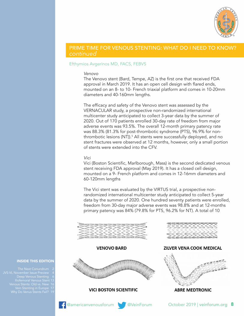
Venovo The Venovo stent (Bard, Tempe, AZ) is the first one that received FDA
approval in March 2019. It has an open cell design with flared ends, mounted on an 8- to 10- French triaxial platform and comes in 10-20mm diameters and 40-160mm lengths. The efficacy and safety of the Venovo stent was assessed by the VERNACULAR study, a prospective non-randomized international multicenter study anticipated to collect 3-year data by the summer of 2020. Out of 170 patients enrolled 30-day rate of freedom from major adverse events was 93.5%. The overall 12-month primary patency rate was 88.3% (81.3% for post-thrombotic syndrome (PTS), 96.9% for non-thrombotic lesions (NT)).5 All stents were successfully deployed, and no stent fractures were observed at 12 months, however, only a small portion of stents were extended into the CFV. Vici Vici (Boston Scientific, Marlborough, Mass) is the second dedicated venous stent receiving FDA approval (May 2019). It has a closed cell design, mounted on a 9- French platform and comes in 12-16mm diameters and 60-120mm lengths The Vici stent was evaluated by the VIRTUS trial, a prospective non-randomized international multicenter study anticipated to collect 5-year data by the summer of 2020. One hundred seventy patients were enrolled, freedom from 30-day major adverse events was 98.8% and at 12-months primary patency was 84% (79.8% for PTS, 96.2% for NT). A total of 10
PRIME TIME FOR VENOUS STENTING: WHAT DO I NEED TO KNOW? continuedEfthymios Avgerinos MD, FACS, FEBVS

October 2019 | veinforum.org@americanvenousforum @VeinForum 9
INSIDE THIS EDITION
The Next Conundrum 2 JVS-VL November Issue Preview 4
Deep Venous Stenting 6 Iliofemoral Venous Stent 13
Venous Stents: Old vs. New 16Vein Stenting in Europe 17
Why Do Venus Stents Fail? 19
patients (5.9%) had stent fractures in this trial, with an overall stent fracture rate of 3.6% (10/281).6 Nine out of 10 fractures occurred in stents that extended into the CFV but had no clinical significance. Notably, this trial enrolled patients with more complex lesions and half of all patients had the stents extending below the inguinal ligament. Zilver Vena Zilver Vena (Cook Medical, Bloomington, IN is anticipated to receive FDA approval early 2020. It has an open cell design, mounted on a 7- French platform and comes in 14-16mm diameters and 60-140mm lengths. The VIVO trial, a prospective non-randomized international multicenter study has completed enrollment of 243 patients (since 2016) and results haven’t been made available yet. The European VIVO-EU followed 35 patients, procedural success (minimum treated lumen diameter ≥ 8 and no perioperative adverse events) was 97.1% and at 12-months primary patency was 87.9%7 Abre The Abre venous stent (Medtronic, Minneapolis, Minnesota, USA) is anticipated to receive FDA approval in 2021. It utilizes an open-cell design with three connection points between the cells intended to enhance flexibility. Strut dimensions are customized for each stent size. It is premounted on a 9- French delivery system with a triaxial shaft design and comes in 10-20mm diameters and 40-150mm lengths. The ABRE multicenter IDE completed enrollment of 200 patients (early 219) and results are to be announced within 2020. Other venous stents (non-available in US) There are five more dedicated venous stents on the market in Europe, not anticipated currently to enter the US market. Optimed (Ettlingen, Germany) has developed 4 different stents designed to accommodate different demands based on the venous segment. The Optimed sinus stent has a hybrid design trying to balance radial force and flexibility, the Optimed sinus-XL has a closed design affording high radial force, the Optimed sinus-XL Flex with an open cell design afford more flexibility and the Optimed sinus-Obliquus has a hybrid skeleton with a closed cell design oblique-shaped central end, an open cell design mid-segment, and an anchor ring at the peripheral end. The closed cell oblique segment allows for increased radial force and crush resistance at the iliocaval stress point while minimizing overlap of the contralateral common iliac vein. The open cell design of the mid- segment provides
PRIME TIME FOR VENOUS STENTING: WHAT DO I NEED TO KNOW? continuedEfthymios Avgerinos MD, FACS, FEBVS
October 2019 | veinforum.org@americanvenousforum @VeinForum 10
INSIDE THIS EDITION
The Next Conundrum 2 JVS-VL November Issue Preview 4
Deep Venous Stenting 6 Iliofemoral Venous Stent 13
Venous Stents: Old vs. New 16Vein Stenting in Europe 17
Why Do Venus Stents Fail? 19
PRIME TIME FOR VENOUS STENTING: WHAT DO I NEED TO KNOW? continued
flexibility and conformity to the stent. The peripheral end anchor helps with stent fixation. A prospective multicenter European study, the TOPOS study, is an efficacy study evaluating the use of the sinus-Obliquus stent in the common iliac vein and the sinus-XL Flex stent or the sinus-Venous stent in the external iliac and common femoral veins. The study is currently recruiting with estimated primary completion date of October 2019. Finally, the Blueflow Venous Stent (Plus Medica GmbH & Co. KG, Germany) utilizes a woven nitinol design particularly suitable for treatment below the inguinal ligament and is promoted as a distal extension stent.
Technical Considerations The novel dedicated venous stents have a predictable deployment, are very flexible and possess a high degree of uniform radial force and crash resistance that makes them favorable against the traditionally used Wallstent. Longer sizes also allow fewer or no overlapping zones. The only potential “unknown” is the real world fracture rate of these stents when deployed below the inguinal ligament, an area Wallstent has performed pretty well with rarely reported fractures. For the US available stents, it is assumed that the open cell designs (Venovo, Zilver Vena, Abre) are more flexible thus safe to deploy below the ligament, but fractures may not be as forgiving as is the case with the closed cell VICI stent (see VIRTUS trial results). The Arnsberg (Germany) group has published large real world series with both the Vici and Venovo stents extending below the inguinal ligament in roughly 20% of cases and no demonstrated fractures within 12 months.8,9 Black et al published two year results of the Vici stent implanted in 52 limbs infrainguinally; 3 had a fracture (2 symptomatic), none thrombosed.10 Similar “challenging lesion” data are not available for the Venovo stent. Irrespective, technical tips used for the Wallstent deployment may not be valid or needed for the novel stents. Oversizing is not as necessary anymore given the higher and uniform radial forces. IVUS based sizing of a healthy segment can guide stent size which will typically be 14-16mm for the common iliac vein, 12-14mm for the external iliac vein and 10-12mm for the common femoral vein. Treating common iliac vein compression syndromes does not require long extension into the vena cava (5mm is typically enough). Finally, when planning to stent low to the common femoral vein level it is reasonable to consider deploying the distal smaller stent first and built proximally with larger stents. Choosing and tailoring available stents to the underlying pathology is yet a difficult task and despite various opinions of experienced users, the truth is available studies cannot be used for comparisons given the different populations enrolled.
Efthymios Avgerinos MD, FACS, FEBVS
October 2019 | veinforum.org@americanvenousforum @VeinForum 11
INSIDE THIS EDITION
The Next Conundrum 2 JVS-VL November Issue Preview 4
Deep Venous Stenting 6 Iliofemoral Venous Stent 13
Venous Stents: Old vs. New 16Vein Stenting in Europe 17
Why Do Venus Stents Fail? 19
PRIME TIME FOR VENOUS STENTING: WHAT DO I NEED TO KNOW? continued
A bench test of available stents indicated Venovo as having higher outward force but lower crush resistance against Vici and Wallstent. Taking flexibility into account, my rule of thumb is to use Vici for predominately proximal compressive lesions and Venovo for predominately external iliac vein or more distal lesions. I would not recommend routine mix and matching due to risks of restenosis at the overlapping zones, yet sometimes it may be unavoidable. Conclusion We’re experiencing exciting times for the previously neglected venous world. Novel stents are promising better outcomes for our patients, still longer-term results of the IDE trials and real-world series are eagerly awaited. We should not, however, forget that stents are only one part of a successful clinical outcome; appropriate patient selection and technique will still remain the cornerstone of success. References 1. Raju S. Best management options for chronic iliac vein stenosis and occlusion. J Vasc
Surg.2013;57:1163-1169.2. Gagne PJ, Gagne N, Kucher T, Thompson M, Bentley D. Long-term clinical outcomes
and technical factors with the Wallstent for treatment of chronic iliofemoral venous obstruction. J Vasc Surg: Venous and Lym Dis 2019;7:45-55
3. Murphy EH, Johns B, Varney E, et al. Deep venous thrombosis associated with caval extension of iliac stents. J Vasc Surg Venous Lymphat Disord. 2017;5:8-17.
4. Raju S, Ward M Jr, Kirk O. A modification of iliac vein stent technique. Ann Vasc Surg. 2014;28:1485-1492.
5. Dake M. 12-month results from the Venovo venous stent trial. Presented at: Vascular Interventional Advances conference; November 5–8, 2018; Las Vegas, Nevada
6. Razavi MK. VIRTUS clinical trial: 12-month data. Presented at: Leipzig Interventional Course; January 22–25, 2019; Leipzig, Germany.
7. O’Sullivan GJ. VIVO-EU results: Prospective European Study of the Zilver Vena venous stent in the treatment of symptomatic iliofemoral venous outflow obstruction. Presented at: Leipzig Interventional Course; January …, 2017; Leipzig, Germany
8. Lichtenberg M, Breuckmann F, Stahlhoff WF, et al. Placement of closed-cell designed venous stents in a mixed cohort of patients with chronic venous outflow obstructions – short-term safety, patency, and clinical outcomes. Vasa. 2018;47:475-481
9. Lichtenberg MKW, de Graaf R, Stahlhoff WF, et al. Venovo venous stent in the treatment of non-thrombotic or post-thrombotic iliac vein lesions – short-term results from the Arnsberg venous registry. Vasa. 2019;48:175-180
10. Black S, Gwozdz A, Karunanithy N, et al. Two year outcome after chronic iliac vein occlusion recanalisation using the Vici venous stent. Eur J Vasc Endovasc Surg. 2018;56:710-718.
Efthymios Avgerinos MD, FACS, FEBVS
October 2019 | veinforum.org@americanvenousforum @VeinForum 12
INSIDE THIS EDITION
The Next Conundrum 2 JVS-VL November Issue Preview 4
Deep Venous Stenting 6 Iliofemoral Venous Stent 13
Venous Stents: Old vs. New 16Vein Stenting in Europe 17
Why Do Venus Stents Fail? 19
PRIME TIME FOR VENOUS STENTING: WHAT DO I NEED TO KNOW? continued
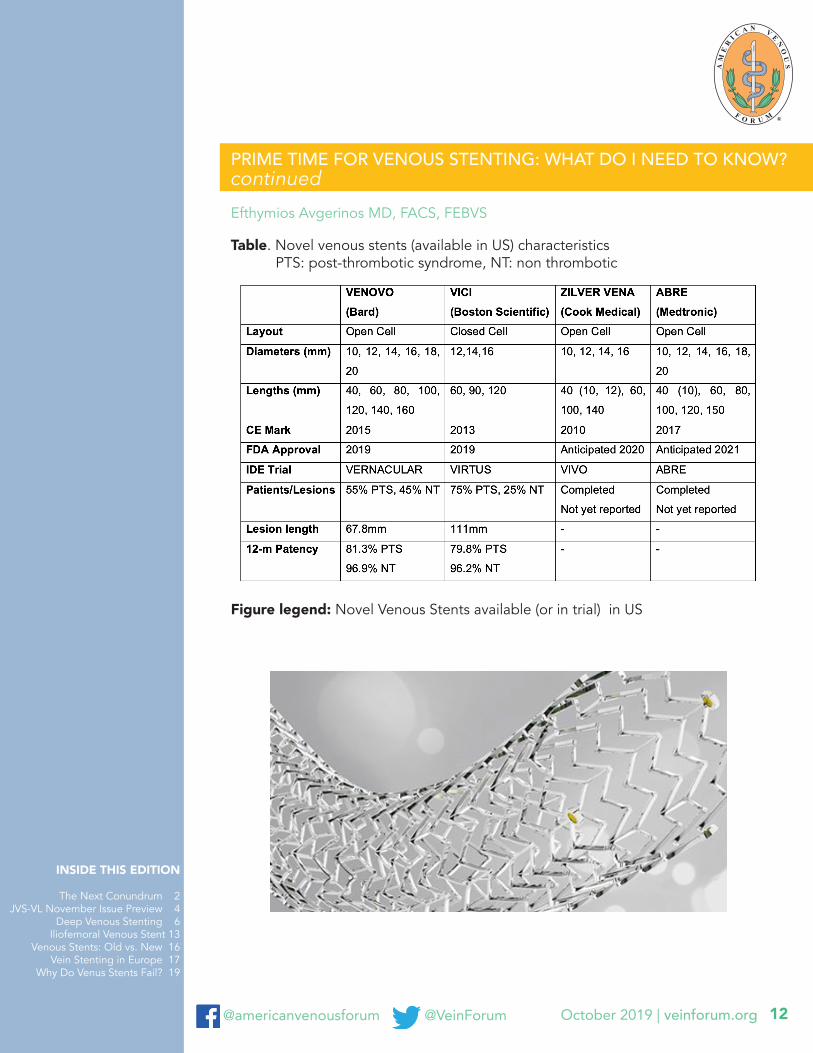
Table. Novel venous stents (available in US) characteristics PTS: post-thrombotic syndrome, NT: non thrombotic
Efthymios Avgerinos MD, FACS, FEBVS
Figure legend: Novel Venous Stents available (or in trial) in US
October 2019 | veinforum.org@americanvenousforum @VeinForum 13
INSIDE THIS EDITION
The Next Conundrum 2 JVS-VL November Issue Preview 4
Deep Venous Stenting 6 Iliofemoral Venous Stent 13
Venous Stents: Old vs. New 16Vein Stenting in Europe 17
Why Do Venus Stents Fail? 19
Edgar Guzman, MD
ILIOFEMORAL VENOUS STENT SURVEILLANCE AND POST PROCEDURAL ANTICOAGULATION
After over twenty years of cumulative experience iliofemoral venous stents have proven to be a safe and reliable option in the treatment of venous outflow obstruction. Through this time the Wallstent has been the main device used. Its excellent flexibility allows safe extension into the femoral vein, well below the inguinal ligament. Further refinements incorporate the use of Gianturco Z stents to address the IVC and proximal CIV1, improving fixation, expansion and reducing the risk of contralateral DVT. While there are no formal guidelines, it is common to obtain routine post procedural images, usually duplex ultrasound, four to twelve weeks after implantation. Long term follow up may be carried out at yearly intervals. This is of greater importance when the indication for stent placement was associated with thrombosis as opposed to compression.2 In a large series spanning the 1997-2007 decade, the incidence of reintervention following iliofemoral venous stenting was 13%. 31% of these reinterventions were prompted by surveillance findings.3 The median time to intervention was fifteen months.
The goal of early surveillance is to identify technical faults such as stent disjunction, angulation and poor expansion. Plain X-Rays can provide additional information. In the future this imaging modality may come to play a more important role if stent fractures prove to be more common with newly available stents.
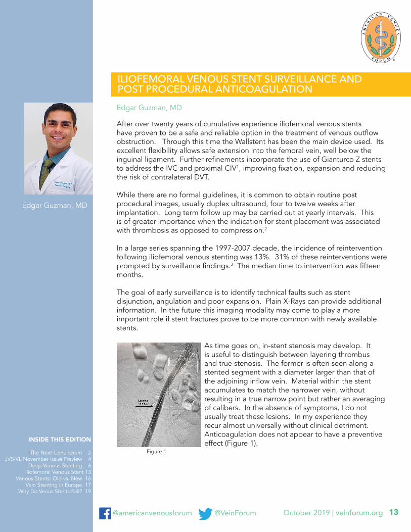
As time goes on, in-stent stenosis may develop. It is useful to distinguish between layering thrombus and true stenosis. The former is often seen along a stented segment with a diameter larger than that of the adjoining inflow vein. Material within the stent accumulates to match the narrower vein, without resulting in a true narrow point but rather an averaging of calibers. In the absence of symptoms, I do not usually treat these lesions. In my experience they recur almost universally without clinical detriment. Anticoagulation does not appear to have a preventive effect (Figure 1).
Figure 1
Edgar Guzman, MD
October 2019 | veinforum.org@americanvenousforum @VeinForum 14
INSIDE THIS EDITION
The Next Conundrum 2 JVS-VL November Issue Preview 4
Deep Venous Stenting 6 Iliofemoral Venous Stent 13
Venous Stents: Old vs. New 16Vein Stenting in Europe 17
Why Do Venus Stents Fail? 19
Edgar Guzman, MD
ILIOFEMORAL VENOUS STENT SURVEILLANCE AND POST PROCEDURAL ANTICOAGULATION continued
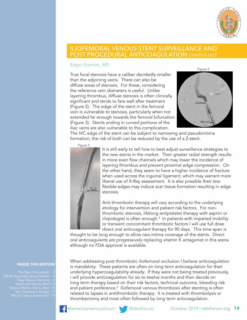
True focal stenosis have a caliber decidedly smaller than the adjoining veins. There can also be diffuse areas of stenosis. For these, considering the reference vein diameters is useful. Unlike layering thrombus, diffuse stenosis is often clinically significant and tends to fare well after treatment (Figure 2). The edge of the stent in the femoral vein is vulnerable to stenosis, particularly when not extended far enough towards the femoral bifurcation (Figure 3). Stents ending in curved portions of the iliac veins are also vulnerable to this complication. The IVC edge of the stent can be subject to narrowing and pseudointima formation; the risk of both can be reduced by the use of a Z-stent.
It is still early to tell how to best adjust surveillance strategies to the new stents in the market. Their greater radial strength results in more even flow channels which may lower the incidence of layering thrombus and prevent proximal edge compression. On the other hand, they seem to have a higher incidence of fracture when used across the inguinal ligament, which may warrant more liberal use of X-Ray assessment. It is also possible their less flexible edges may induce scar tissue formation resulting in edge stenosis.
Anti-thrombotic therapy will vary according to the underlying etiology for intervention and patient risk factors. For non-thrombotic stenosis, lifelong antiplatelet therapy with aspirin or clopidogrel is often enough.4 In patients with impaired mobility or transient concomitant thrombotic factors I will use full dose direct oral anticoagulant therapy for 90 days. This time span is
thought to be long enough to allow neo-intima coverage of the stents. Direct oral anticoagulants are progressively replacing vitamin K antagonist in this arena although no FDA approval is available.
When addressing post thrombotic iliofemoral occlusion I believe anticoagulation is mandatory. These patients are often on long term anticoagulation for their underlying hypercoagulability already. If they were not being treated previously, I will provide anticoagulation for six to twelve months and then decide on long term therapy based on their risk factors, technical outcome, bleeding risk and patient preference.5 Iliofemoral venous thrombosis after stenting is often related to lapses in antithrombotic therapy. It is treated with thrombolysis or thrombectomy and most often followed by long term anticoagulation.
Figure 3
Figure 2

October 2019 | veinforum.org@americanvenousforum @VeinForum 15
INSIDE THIS EDITION
The Next Conundrum 2 JVS-VL November Issue Preview 4
Deep Venous Stenting 6 Iliofemoral Venous Stent 13
Venous Stents: Old vs. New 16Vein Stenting in Europe 17
Why Do Venus Stents Fail? 19
Edgar Guzman, MD
ILIOFEMORAL VENOUS STENT SURVEILLANCE AND POST PROCEDURAL ANTICOAGULATION continued
Stenting for May Thurner syndrome following thrombolysis deserves special mention. Provided the underlying lesion has been treated successfully, anticoagulation will be discontinued after six months.
Given the rapid pace of innovation in the venous space, post intervention surveillance will be more important than ever as the strengths and weaknesses of novel devices and techniques are discovered.
References1. Raju S, Ward M, Kirk O. A modification of iliac vein stent technique. Ann Vasc Surg
2014; 28:1485-1492.2. Abdul-Hagg R, Novak Z, Pearce BJ, Matthews TC, Patterson MA, Jordan WD, Passman
MA.Routine extended follow-up surveillance of iliac vein stents for iliocaval venous obstruction may not be warranted.
3. Raju S, Tackett P, Neglen P. Reinterventions for nonocclusive iliofemoral venous stent malfunctions. J Vasc Surg 2009; 49:511-8.
4. Meissner MH. Indications for platelet aggregation inhibitors after venous stents. Phlebology 2013; 28 (Suppl 1): 91-8.
5. Milinis K, Thapar A, Shalhoub J, Davies AH. Antithrombotic therapy following venous stenting: International Delphi consensus. Eur J Vasc Endovasc Surg 2018; 55:537-44.
October 2019 | veinforum.org@americanvenousforum @VeinForum 16
INSIDE THIS EDITION
The Next Conundrum 2 JVS-VL November Issue Preview 4
Deep Venous Stenting 6 Iliofemoral Venous Stent 13
Venous Stents: Old vs. New 16Vein Stenting in Europe 17
Why Do Venus Stents Fail? 19
VENOUS STENTS: HOW DO THE OLD COMPARE WITH THE NEW? Ellen D. Dillavou, MD
There have recently been exciting developments in the world of venous stenting; the Sinus Venous (Optimed) and Zilver Vena (Cook, Bloomington, IN, USA) are available in Europe, and the Venovo (Bard, Tempe, AZ, USA) and VICI (Boston Scientific, Marlborough, MA, USA) were recently FDA-approved in the US, all specifically designed for deep venous work. Prior to this the most commonly used stent was the Wallstent (Boston Scientific). Deep venous interventionalists have all eagerly been awaiting this day and can now treat post-thrombotic as well as non-occlusive deep venous compression with a dedicated product. None of the new stents underwent a randomized trial against the Wallstent, but instead used historic controls in non-inferiority comparisons. We asked, when looking at the published work with prior, off-label use of stents (standard stents) in the venous system, primarily Wallstents, how do these results compare to trial and early experience with dedicated venous stents?
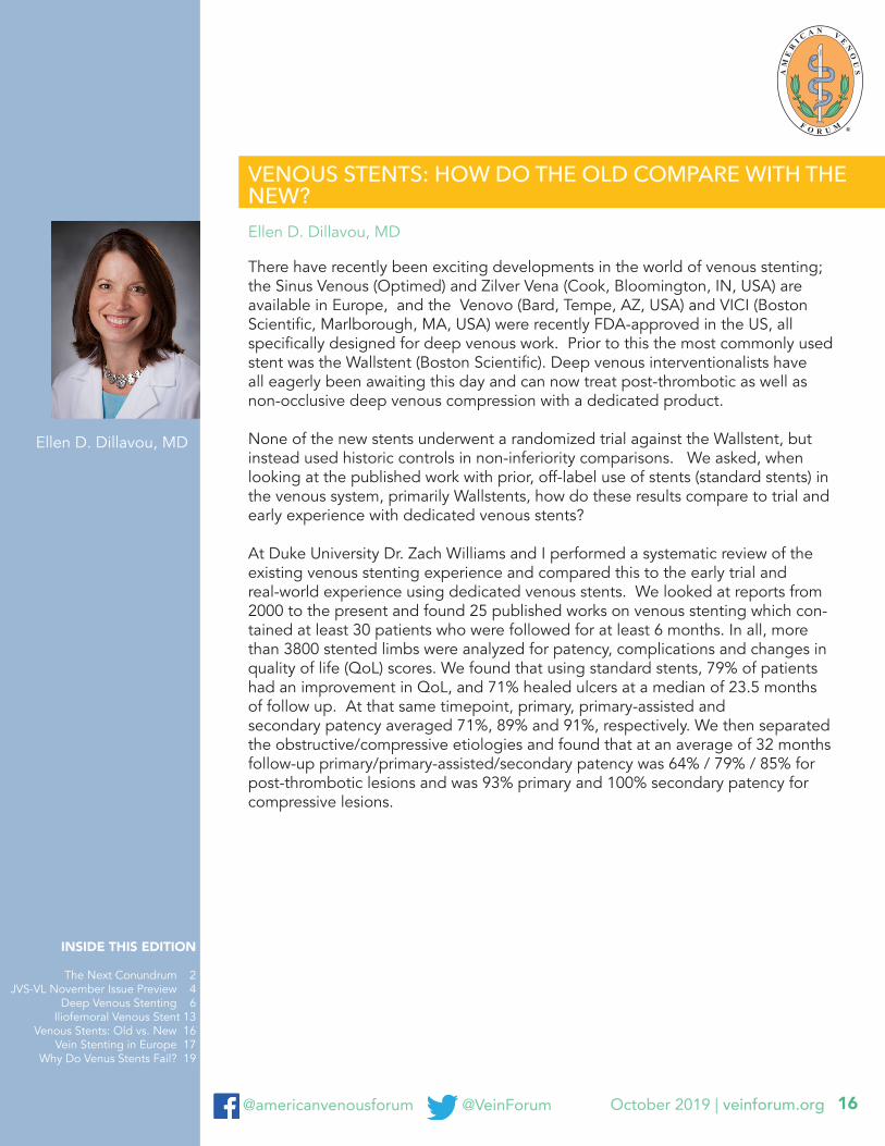
At Duke University Dr. Zach Williams and I performed a systematic review of the existing venous stenting experience and compared this to the early trial and real-world experience using dedicated venous stents. We looked at reports from 2000 to the present and found 25 published works on venous stenting which con-tained at least 30 patients who were followed for at least 6 months. In all, more than 3800 stented limbs were analyzed for patency, complications and changes in quality of life (QoL) scores. We found that using standard stents, 79% of patients had an improvement in QoL, and 71% healed ulcers at a median of 23.5 months of follow up. At that same timepoint, primary, primary-assisted and secondary patency averaged 71%, 89% and 91%, respectively. We then separated the obstructive/compressive etiologies and found that at an average of 32 months follow-up primary/primary-assisted/secondary patency was 64% / 79% / 85% for post-thrombotic lesions and was 93% primary and 100% secondary patency for compressive lesions.
Ellen D. Dillavou, MD

October 2019 | veinforum.org@americanvenousforum @VeinForum 17
INSIDE THIS EDITION
The Next Conundrum 2 JVS-VL November Issue Preview 4
Deep Venous Stenting 6 Iliofemoral Venous Stent 13
Venous Stents: Old vs. New 16Vein Stenting in Europe 17
Why Do Venus Stents Fail? 19
VEIN STENTING IN EUROPE
Two stents (Venovo, Bard; Vici, Boston Scientific Corporation) received FDA approval in 2019 for veins. This is a remarkable milestone in vein stenting, a procedure first reported by Raju, Neglen and colleagues more than a decade before. As I write this, several additional stents are in various stages of clinical trial and FDA certification; these stents will likely receive FDA approval and become commercially available during the next few years. It seems fitting to ask what is the status of vein stents in the European theater?
Europe and U.S. have very different approval processes for drugs & devices. For an implantable device such as a venous stent, the FDA requires the performance of a prospective clinical trial to demonstrate efficacy and safety. In Europe, the manufacturer only has to demonstrate that a new device conforms to specific directives of the European Union. Once approved in one EU country, the device can be marketed in every EU country. When approved, the device is labeled as having the CE mark in Europe, the name CE mark refers to Conformite Europeenne. Since 2010, manufacturers have to undertake post-marketing surveillance and report to a central database in Europe.
There are currently eight vein stents that have received the CE mark and are commercially available in Europe. Many of these stents are familiar to us: Wallstent (Boston Scientific), Venovo (Bard), Abre (Medtronic), Vici (Boston Scientific), and Zilver Vena (Cook). Three other CE mark approved stents that are available primarily in Europe and not as familiar to us: Blueflow (Plus Medica), sinus-Obliquus (Optimed) and sinus-Venous (Opitmed).
Because a prospective clinical trial to demonstrate safety and efficacy is not required for CE mark certification, there have been no separate trials of these eight stents undertaken exclusively in Europe or no published reports of stent trials that I am able to identify. Of note, there are published case reports and case series that included these stents. It is important to point out that many of the prospective vein stent trials in U.S. have included European sites.
While undertaking the research for this report, I spoke to several individuals who are familiar with vein stenting in Europe. While there are more venous stents currently approved and commercially available in Europe, vein stenting is more regionalized in the European Union and just a few hospitals in each country are performing these procedures in significant volume. Unlike the U.S., these cases are performed typically in a hospital setting rather than in an ambulatory facility or as an ambulatory procedure. Furthermore, post thrombotic syndrome is a common indication for vein stenting and patients oftentimes present with severe and advanced venous symptoms.
Windsor Ting, MD
Windsor Ting, MD
October 2019 | veinforum.org@americanvenousforum @VeinForum 18
INSIDE THIS EDITION
The Next Conundrum 2 JVS-VL November Issue Preview 4
Deep Venous Stenting 6 Iliofemoral Venous Stent 13
Venous Stents: Old vs. New 16Vein Stenting in Europe 17
Why Do Venus Stents Fail? 19
VEIN STENTING IN EUROPE continued
If you are interested in additional reading, I recommend a report, Surveying the 2019 Venous Stent Landscape, authored by Erin Murphy that was published in the July issue of Endovascular Today.
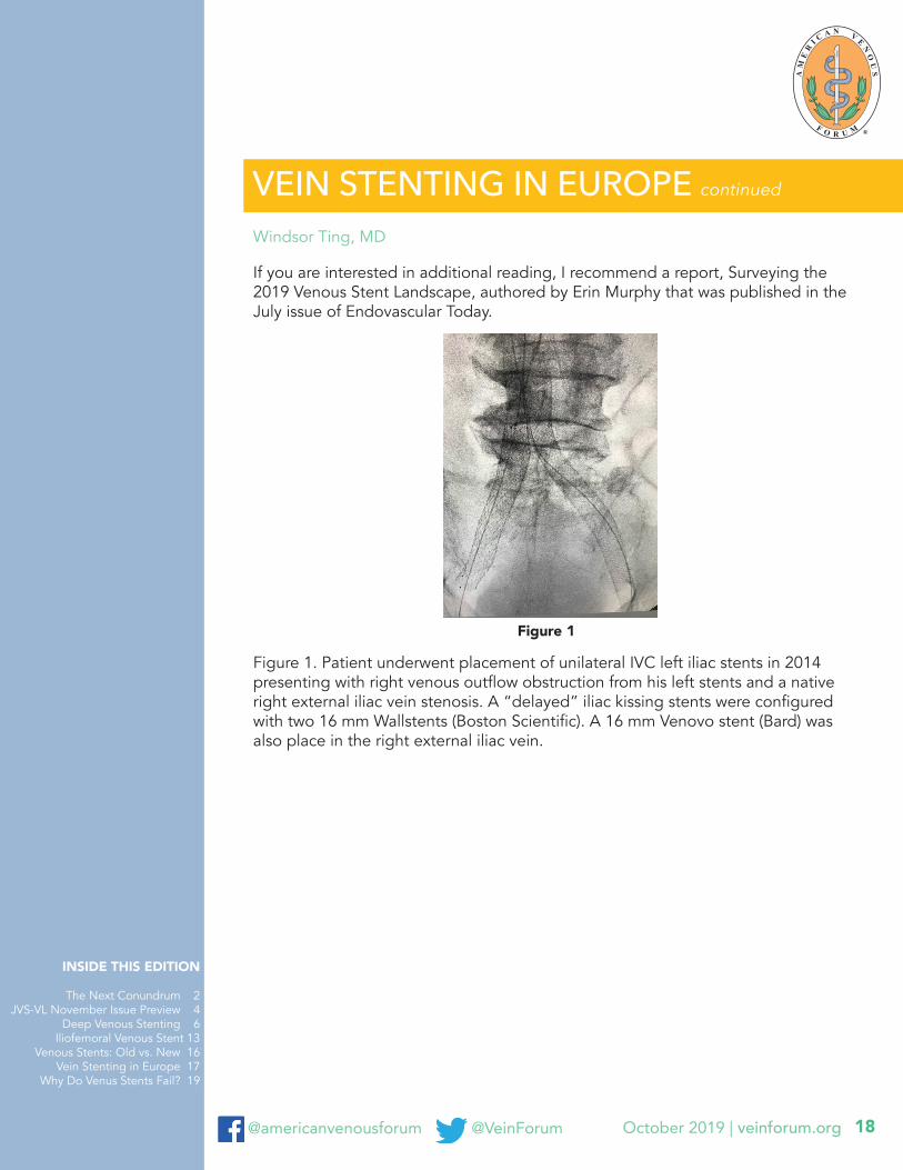
Figure 1. Patient underwent placement of unilateral IVC left iliac stents in 2014 presenting with right venous outflow obstruction from his left stents and a native right external iliac vein stenosis. A “delayed” iliac kissing stents were configured with two 16 mm Wallstents (Boston Scientific). A 16 mm Venovo stent (Bard) was also place in the right external iliac vein.
Windsor Ting, MD
Figure 1
October 2019 | veinforum.org@americanvenousforum @VeinForum 19
INSIDE THIS EDITION
The Next Conundrum 2 JVS-VL November Issue Preview 4
Deep Venous Stenting 6 Iliofemoral Venous Stent 13
Venous Stents: Old vs. New 16Vein Stenting in Europe 17
Why Do Venus Stents Fail? 19
WHY DO VENOUS STENTS FAIL?
The concept of vascular stenting has been around since Nobel Prize winner Alexis Carrel (1873-1944) first implanted glass tubes into the arteries of dogs. The first human stenting was undertaken in France in 1986. Since this time, advances in an arterial stenting, (and particularly coronary stenting), have far outpaced improvements in venous stenting. For many years, the Wallstent (Boston Scientific) was the workhorse of the interventionalist treating central venous stenosis, thrombosis and May-Thurner syndrome. In the course of the last year, the landscape of options for treatment of central venous disease has been completely disrupted by introduction of two new FDA-approved venous stents, begging two essential questions: how have we learned from venous stent failures? And what is the optimal venous stent design?
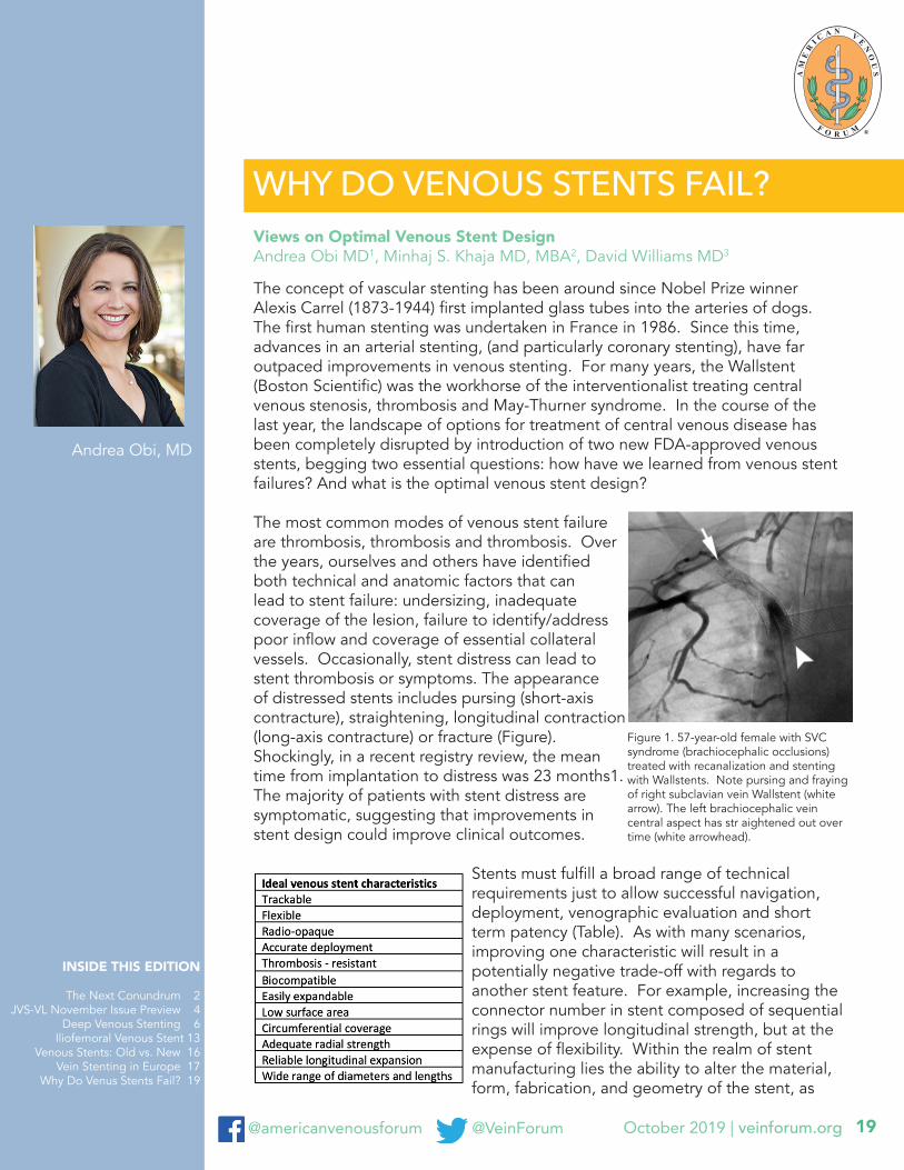
The most common modes of venous stent failure are thrombosis, thrombosis and thrombosis. Over the years, ourselves and others have identified both technical and anatomic factors that can lead to stent failure: undersizing, inadequate coverage of the lesion, failure to identify/address poor inflow and coverage of essential collateral vessels. Occasionally, stent distress can lead to stent thrombosis or symptoms. The appearance of distressed stents includes pursing (short-axis contracture), straightening, longitudinal contraction (long-axis contracture) or fracture (Figure). Shockingly, in a recent registry review, the mean time from implantation to distress was 23 months1. The majority of patients with stent distress are symptomatic, suggesting that improvements in stent design could improve clinical outcomes.
Stents must fulfill a broad range of technical requirements just to allow successful navigation, deployment, venographic evaluation and short term patency (Table). As with many scenarios, improving one characteristic will result in a potentially negative trade-off with regards to another stent feature. For example, increasing the connector number in stent composed of sequential rings will improve longitudinal strength, but at the expense of flexibility. Within the realm of stent manufacturing lies the ability to alter the material, form, fabrication, and geometry of the stent, as
Views on Optimal Venous Stent DesignAndrea Obi MD1, Minhaj S. Khaja MD, MBA2, David Williams MD3
Andrea Obi, MD
Figure 1. 57-year-old female with SVC syndrome (brachiocephalic occlusions) treated with recanalization and stenting with Wallstents. Note pursing and fraying of right subclavian vein Wallstent (white arrow). The left brachiocephalic vein central aspect has str aightened out over time (white arrowhead).

October 2019 | veinforum.org@americanvenousforum @VeinForum 20
INSIDE THIS EDITION
The Next Conundrum 2 JVS-VL November Issue Preview 4
Deep Venous Stenting 6 Iliofemoral Venous Stent 13
Venous Stents: Old vs. New 16Vein Stenting in Europe 17
Why Do Venus Stents Fail? 19
WHY DO VENOUS STENTS FAIL? continued
well as add on additional features such as radio-opaque markers and drug-eluting coatings. The advent of stents specifically designed for the venous systems allows before untested (in the venous system) combinations to be implemented with the goal of optimizing all of the parameters for the compliance, diameter, and pitfalls unique to the central venous system. For example, the Wallstent is comprised of a superalloy (eligiloy: cobalt, chromium, nickel and a small amount of iron), in a wire form, fabricated by braiding in a tubular mesh conformation. In comparison, both of the newer venous stents are composed of nitinol, in tube formation, fabricated via laser cutting. The new stents vary in differences with closed versus open cell design, a determinant of flexibility and foreshortening, thickness and a variety of additional features. The end result is significant variation in radial resistive forces, chronic outward forces and crush resistance amongst FDA approved and IDE venous stents.2
Within the following newsletter, we review two new venous stents entering the U.S. market in 2019. The Venovo venous stent (BD Interventional), a flexible nitinol stent with 3mm flared ends and an open cell design, was granted FDA approval on March 14th 2019, based on the findings of the VERNACULAR trial, a prospective international multicentered single-armed clinical trial. Next to enter the market was the Vici venous stent (Boston Scientific), a laser cut nitinol stent with closed cell design and high scaffold thickness to strut ratio on May 6, 2019, based on the results of the VIRTUS study. Additional venous stents under investigation include the Abre (Medtronic) and Zilver Vena (Cook Medical). With a new armamentarium at hand, the next question to address will be defining the performance in vivo to tailor stent selection to the patient/lesion for the best possible outcome.
Selected References 1. Chick JFB, Abramowitz SD, Osher ML, et al. Radiographic Findings of Distressed
Venous Stents and Inferior Vena Cava Filters: Clinical Implications. AJR Am J Roentgenol 2017;209(5):1150-57. doi: 10.2214/AJR.16.17750
2. Dabir D, Feisst A, Thomas D, et al. Physical Properties of Venous Stents: An Experimental Comparison. Cardiovasc Intervent Radiol 2018;41(6):942-50. doi: 10.1007/s00270-018-1916-1
Views on Optimal Venous Stent DesignAndrea Obi MD1, Minhaj S. Khaja MD, MBA2, David Williams MD3
FEEDBACK?do you have
email us [email protected]
October 2019 | veinforum.org@americanvenousforum @VeinForum 21
INSIDE THIS EDITION
The Next Conundrum 2 JVS-VL November Issue Preview 4
Deep Venous Stenting 6 Iliofemoral Venous Stent 13
Venous Stents: Old vs. New 16Vein Stenting in Europe 17
Why Do Venus Stents Fail? 19
VEIN SPECIALIST PUBLICATIONEDITOR-IN-CHIEF: Steve Elias, MDEXECUTIVE EDITOR: John ForbesPUBLICATION EDITOR: Stacey Meyer
EDITORIAL BOARD:Windsor Ting, MDHaraldur Bjarnason, MDEdgar Guzman, MDAndrea Obi, MDAlessandra Puggioni, MDMaxim Shaydakov, MDEric Hager, MDAnil Hingorani, MD
Want to receive monthly updates?Sign Me Up for VEIN SPECIALISTContact us 847-752-5355 [email protected]
VEIN SPECIALIST welcomes your thoughts and comments. Please send all comments to [email protected].
UPCOMING COURSE
Register Now