Embed Size (px)
Citation preview

Integrative Systems
Locus Coeruleus and Tuberomammillary NucleiAblations Attenuate Hypocretin/OrexinAntagonist-Mediated REM Sleep
Michael D. Schwartz,1 Alexander T. Nguyen,1 Deepti R. Warrier,1 Jeremiah B. Palmerston,1
Alexia M. Thomas,1 Stephen R. Morairty,1 Thomas C. Neylan,2 and Thomas S. Kilduff1
DOI:http://dx.doi.org/10.1523/ENEURO.0018-16.2016
1Biosciences Division, Center for Neuroscience, SRI International, Menlo Park, California 94025, and 2UCSF SanFrancisco VA Medical Center/NCIRE, San Francisco, California 94121
AbstractHypocretin 1 and 2 (Hcrts; also known as orexin A and B), excitatory neuropeptides synthesized in cells locatedin the tuberal hypothalamus, play a central role in the control of arousal. Hcrt inputs to the locus coeruleusnorepinephrine (LC NE) system and the posterior hypothalamic histaminergic tuberomammillary nuclei (TMN HA)are important efferent pathways for Hcrt-induced wakefulness. The LC expresses Hcrt receptor 1 (HcrtR1),whereas HcrtR2 is found in the TMN. Although the dual Hcrt/orexin receptor antagonist almorexant (ALM)decreases wakefulness and increases NREM and REM sleep time, the neural circuitry that mediates these effectsis currently unknown. To test the hypothesis that ALM induces sleep by selectively disfacilitating subcorticalwake-promoting populations, we ablated LC NE neurons (LCx) or TMN HA neurons (TMNx) in rats usingcell-type-specific saporin conjugates and evaluated sleep/wake following treatment with ALM and the GABAA
receptor modulator zolpidem (ZOL). Both LCx and TMNx attenuated the promotion of REM sleep by ALM withoutaffecting ALM-mediated increases in NREM sleep. Thus, eliminating either HcrtR1 signaling in the LC or HcrtR2signaling in the TMN yields similar effects on ALM-induced REM sleep without affecting NREM sleep time. Incontrast, neither lesion altered ZOL efficacy on any measure of sleep–wake regulation. These results contrast withthose of a previous study in which ablation of basal forebrain cholinergic neurons attenuated ALM-inducedincreases in NREM sleep time without affecting REM sleep, indicating that Hcrt neurotransmission influencesdistinct aspects of NREM and REM sleep at different locations in the sleep–wake regulatory network.
Key words: arousal; hypnotics; insomnia; monoamine; orexin; paradoxical sleep
Significance Statement
The hypocretin/orexin (Hcrt) system powerfully regulates arousal in part by excitatory projections towake-promoting cell groups in the posterior hypothalamus and brainstem. Cell-type-specific ablations ofthe locus coeruleus norepinephrine (LC NE) neurons or the tuberomammillary histamine (TMN HA) neuronsdecreased the hypnotic efficacy of the dual Hcrt receptor antagonist Almorexant, while having no effect onsleep promotion by the GABA receptor modulator zolpidem. Lesioning the LC or TMN attenuatedalmorexant-induced REM sleep without affecting NREM sleep time. Lesions exerted similar effects inde-pendently of the Hcrt receptor type expressed in each region, suggesting that the site of action, not just thespecific receptor or receptors targeted, is a key determinant of how Hcrt receptor antagonism facilitatessleep.
New Research
January/February 2016, 3(1) e0018-16.2016 1–17

IntroductionHypocretin-1 and -2 (Hcrts; also known as orexin-A and
-B), excitatory neuropeptides synthesized in neurons lo-cated in the tuberal hypothalamus, are involved in metab-olism, feeding, reward, addiction, and sleep–wake control(Ohno and Sakurai, 2008). Hcrt neurons are wake-active(Estabrooke et al., 2001; Lee et al., 2005). Hcrt adminis-tration (Bourgin et al., 2000; Morairty et al., 2011) oroptogenetic stimulation of Hcrt neurons (Adamantidiset al., 2007; Carter et al., 2010) is wake-promoting. Defi-cient Hcrt signaling underlies narcolepsy (Chemelli et al.,1999; Lin et al., 1999; Thannickal et al., 2000), a sleepdisorder characterized by fragmented sleep, degradedsleep–wake rhythms, and profound dysregulation of REMsleep. Hcrt signaling thus plays a critical role in the orga-nization and consolidation of sleep–wake states.
Hcrt neurons project to several wake-promoting brainpopulations, including the locus coeruleus (LC; Peyronet al., 1998; Chemelli et al., 1999; Horvath et al., 1999). LCactivation desynchronizes cortical activity and precedestransitions to waking, exhibiting a strongly wake-active,REM-silent firing profile (Aston-Jones and Bloom, 1981;Berridge and Foote, 1991; Takahashi et al., 2010). Opto-genetic inhibition or activation of LC norepinephrine (NE)neurons increases or decreases the likelihood of sleep,respectively (Carter et al., 2010). Disruption of NE signal-ing via cell-type-specific LC lesions or knockout (KO) isreported to increase NREM sleep (González et al., 1998;Blanco-Centurion et al., 2004; Ouyang et al., 2004) orblock wakefulness following arousing stimuli (Hunsleyand Palmiter, 2004; Gompf et al., 2010), consistent witha role in maintenance of wakefulness. The LC expressesHcrt receptor 1 (HcrtR1; Marcus et al., 2001) and Hcrt-1/orexin-A infusion into the LC increases LC neuron firingand promotes wakefulness (Hagan et al., 1999; Bourginet al., 2000) in a HcrtR1-dependent manner (Soffin et al.,2002; Choudhary et al., 2014). Conversely, optogeneticLC inactivation blocks transitions to wakefulness follow-ing Hcrt neuron activation (Carter et al., 2012), indicatingthat the LC is important for Hcrt-induced wakefulness.
Hcrt neurons also strongly innervate histaminergic (HA)cells in the tuberomammillary nuclei (TMN) of the posteriorhypothalamus (Peyron et al., 1998; Chemelli et al., 1999).TMN HA neurons express HcrtR2 (Marcus et al., 2001)and are excited by Hcrt peptides (Eriksson et al., 2001).HA is wake-promoting (Chu et al., 2004; Ramesh et al.,2004) and TMN HA neurons, like LC NE neurons, exhibit awake-active, REM-off firing pattern (Takahashi et al.,2006). TMN HA lesions have relatively mild effects onsleep–wake states (Gerashchenko et al., 2004). However,mice unable to synthesize HA exhibit decreased wakeful-ness at lights-off, increased REM sleep time during thelight phase, and short sleep latency in a novel environ-ment (Parmentier et al., 2002; Anaclet et al., 2009). Wakepromotion by Hcrt-1/orexin A is mediated in part throughhistaminergic neurotransmission (Huang et al., 2001).Thus, Hcrt inputs to the LC NE system and the TMN HAsystem are important pathways for Hcrt-induced wakeful-ness.
The dual Hcrt/orexin receptor antagonist (DORA) alm-orexant (ALM) blocks the excitatory effects of the Hcrtpeptides at HcrtR1 and HcrtR2, decreasing wakefulnessand increasing NREM and REM sleep time (Brisbare-Roch et al., 2007; Morairty et al., 2012). In contrast,zolpidem (ZOL; trade name Ambien) induces somnolenceby activating GABAA receptors, thereby causing wide-spread neuronal inhibition (Dang et al., 2011). ALM, butnot ZOL, requires an intact basal forebrain (BF) for max-imal hypnotic efficacy and induces neurochemical eventsassociated with the transition to normal sleep (Vazquez-DeRose et al., 2014). These findings support the hypoth-esis that ALM induces sleep by selectively disfacilitatingsubcortical wake-promoting populations whereas ZOLacts via generalized inhibition throughout the brain. Here,we tested this hypothesis by selectively ablating the LCNE neurons or the TMN HA neurons using cell-type-specific saporin conjugates, and subsequently evaluatingthe efficacy of ALM and ZOL in lesioned and intact rats.We find that eliminating either HcrtR1 signaling in the LCor HcrtR2 signaling in the TMN yields similar effects onALM-induced REM sleep without affecting NREM sleeptime. Because a previous study (Vazquez-DeRose et al.,2014) found the converse effects after ablation of basalforebrain cholinergic neurons, these results support theconcept that Hcrt neurotransmission influences distinctaspects of NREM and REM sleep at different locations inthe sleep—wake regulatory network.
Materials and MethodsAnimals
Male Sprague-Dawley rats (n � 25; 200–250 g; HarlanLaboratories) were housed in light-tight, sound-attenuatedenvironmental chambers under constant temperature (22� 2°C, 50 � 25% relative humidity) on a 12 h dark/lightcycle with food and water ad libitum. All dosing proce-dures were performed under dim red light (�2 lux). Allstudies were conducted in accordance with the Guide forthe Care and Use of Laboratory Animals and were ap-proved by the Institutional Animal Care and Use Commit-tee at SRI International.
Received January 26, 2016; accepted February 12, 2016; First publishedFebruary 28, 2016.1The authors report no conflict of interest.2Author contributions: M.D.S., S.R.M., T.C.N., and T.S.K. designed re-
search; M.D.S., A.T.N., D.R.W., J.B.P., and A.M.T. performed research;M.D.S., A.T.N., J.B.P., and A.M.T. analyzed data; M.D.S. and T.S.K. wrote thepaper.
3This work was supported by the U.S. Army Medical Research AcquisitionActivity award number W81XWH-09-2-0081 and NIH R01 NS077408 to T.S.K.We thank Drs Priyattam J. Shiromani and Carlos Blanco-Centurion for provid-ing Hcrt2-SAP; Tsui-Ming Chen, Alan Wilk, and Dr Simon Fisher for technicalassistance; Dr Ling Jong for synthesis of almorexant; and Drs Sarah Black andGregory Parks for helpful comments on data analysis and the paper.
Correspondence should be addressed to Dr Thomas S. Kilduff, SRI Inter-national, 333 Ravenswood Avenue, Menlo Park, CA 94025. E-mail:[email protected].
DOI:http://dx.doi.org/10.1523/ENEURO.0018-16.2016Copyright © 2016 Schwartz et al.This is an open-access article distributed under the terms of the CreativeCommons Attribution 4.0 International, which permits unrestricted use, distri-bution and reproduction in any medium provided that the original work isproperly attributed.
2 of 17
January/February 2016, 3(1) e0018-16.2016 eNeuro.sfn.org

ChemicalsALM was synthesized by the Medicinal Chemistry Lab-
oratory at SRI International according to previously pub-lished methods (Koberstein et al., 2003, 2005). ZOL waspurchased from IS Chemical. All drugs that were deliveredorally were suspended and sonicated for 1 h in 1.25%hydroxypropyl methyl cellulose with 0.1% dioctyl sodiumsulfosuccinate and 0.25% methylcellulose in sterile water[hereafter referred to as vehicle (VEH)]. All drug solutionswere made on the day of the experiment and seriallydiluted to their final concentrations.
Saporin lesionsUnder isoflurane anesthesia, rats were placed into a
stereotaxic apparatus (Kopf Instruments) and the skullwas exposed. For LC lesions, rats were injected intrac-erebroventricularly with 10 �l of anti-dopamine betahydroxylase-conjugated saporin (n � 8; DBH-SAP; 0.3�g/�l; Advanced Targeting Systems; Wrenn et al., 1996;Wiley and Kline, 2000; Brightwell and Taylor, 2009) orsterile saline (n � 7; hereafter referred to as “Sham” rats)via a 26 gauge stainless steel injection cannula connectedto a 10 �l Nanofil Hamilton syringe and a digitally con-trolled microinjector (World Precision Instruments) at �0.8mm AP and �1.5 mm ML relative to bregma, and 3.3 mmbelow dura. The infusion volume and concentration wereselected based on previously published methods andwere verified in pilot studies. Injections lasted �10 min;the cannula was left in place for 5 min after the injection.For TMN lesions, rats were injected bilaterally with 250–350 nl of Hcrt2-saporin (n � 13; Hcrt2-SAP; 0.228 �g/�l;Advanced Targeting Systems; Gerashchenko et al., 2001,2004) or sterile saline (n � 7) via glass micropipettes (innertip diameter �30–50 �m) using a Picospritzer (ParkerHannifin) at �4.2 or �4.35 mm AP and �0.8 mm MLrelative to bregma, and 9.3 mm below dura. Injectatevolume was measured via precalibrated marks on thebarrel of the pipette. Injections lasted 5 min/side; thepipette was left in place for 5 min after the injection.Following SAP injections, rats were instrumented for EEG/EMG telemetry.
Telemetry surgeryAll rats were surgically implanted with a sterile abdom-
inal transmitter (F40-EET, DSI) for continuous telemetricrecordings of electroencephalograph (EEG), electromyo-graph (EMG), core body temperature (Tb), and locomotoractivity as described previously (Morairty et al., 2008,2012). Briefly, the wires from the transmitter were subcu-taneously channeled rostrally to the head. Two biopoten-tial leads (EEG electrodes) were inserted into drilled holesover the dura (lead 1: �2.0 mm AP, �1.5 mm LM; lead 2:�7.0 mm AP, �2.0 mm LM; all coordinates relative tobregma) and affixed with dental acrylic. Two additionalbiopotential leads (EMG electrodes) were sutured intothe neck musculature and closed with non-absorbablesuture. Both DBH-SAP (Wrenn et al., 1996; Blanco-Centurion et al., 2004; Gompf et al., 2010) and Hcrt2-SAP(Gerashchenko et al., 2004) induce maximal degenerationby 12–14 d postinjection. Accordingly, animals were sin-gly housed after surgery and allowed to recover undis-
turbed in their home cage for 3 weeks to allow sufficienttime for SAP-induced neurodegeneration and sleep–wakebehavior to stabilize prior to any recordings.
Assessment of hypnotic efficacy in saporin-lesionedrats
Rats were kept in their home cages for the duration ofthe study in ventilated, light-tight, and sound-attenuatedchambers in 12 h light/dark cycles. Prior to initiation ofsleep recordings, animals were acclimated to handling for�1 week, and were dosed with VEH once per day for thelast 3 d of the acclimation period. Animals were then leftundisturbed for 2 d after acclimation was complete. Ratswere administered ALM (30, 100, and 300 mg/kg), ZOL(10, 30, and 100 mg/kg), or VEH orally starting at lights-out (ZT 12) with at least 3 d between treatments to allowsufficient time for washout between doses. Drug treat-ments were balanced across lesion condition and treat-ment day, such that every dose was administered on eachtreatment day, and an approximately equal number oflesioned and sham rats received each dose on eachtreatment day. EEG was recorded for 24 h following dos-ing; the first 6 h following dosing was scored and analyzed(from ZT12 to ZT18).
To confirm the extent of lesions, rats were deeply anes-thetized and transcardially perfused with heparinized 0.1 M
phosphate-buffered saline followed by 4% paraformalde-hyde. Brains were removed, postfixed in 4% paraformal-dehyde, and then transferred to 30% sucrose untilsectioning. Brains were sectioned at 40 �m on a freezingmicrotome. Free-floating sections containing the LC(bregma �9.16 mm to �10.30 mm) were incubated with1% H2O2 for 15 min to quench endogenous peroxidaseactivity, followed by: (1) 1 h in blocking buffer containing3% normal donkey serum, (2) overnight in mouse anti-DBH (1:100,000, MAB308, EMD Millipore), (3) 2 h inbiotinylated donkey anti-mouse IgG (1:500; Jackson Im-munoResearch), and (4) 2 h in avidin–biotin complex(Vector Laboratories). DBH was visualized by reactingsections in 0.05% diaminobenzidine tetrahydrochlorideand 0.01% H2O2 to form a brown reaction product. Sec-tions were then mounted, dehydrated and coverslipped.To visualize HA neurons, sections containing the TMN(bregma �3.80 to �4.80 mm) were processed using asimilar protocol that was modified as follows: (1) sectionswere incubated overnight in rabbit anti-adenosine deami-nase (ADA; 1:20,000, ab176, EMD Millipore), followed by(2) 2 h in biotinylated donkey anti-rabbit IgG (1:500; Jack-son ImmunoResearch).
The extent of the LC was delineated by the fourthventricle and other landmarks. The exact number of DBH-positive LC neurons could not be accurately counted inSham rats because of the high density of these cells (Fig.1B); accordingly, Sham rats were only scored for thepresence of DBH-positive cells. In DBH-SAP injected rats,all residual DBH-positive cells in the LC region werecounted. To evaluate the extent of TMN HA neuronal loss,ADA-positive neurons were counted in the dorsal (dTMN),ventral (vTMN), and caudal TMN (cTMN) subregions as
3 of 17
January/February 2016, 3(1) e0018-16.2016 eNeuro.sfn.org

identified previously (Ko et al., 2003; Parks et al., 2016). Allneurons expressing ADA in each subregion were scored.
EEG and EMG analyses and sleep–wakedeterminationsEEG and EMG were recorded via telemetry on a PCrunning Dataquest ART 3.1 (DSI). All recordings weremanually scored off-line by a trained expert in 10 s ep-ochs as Wake, NREM, or REM sleep using NeuroScore2.1 (DSI). Any epochs that contained recording artifactswere tagged and excluded from spectral analyses. Indi-
vidual state data were quantified as time spent in eachstate per 6 h. Latency to NREM and REM onset for eachanimal was calculated from the time of drug injection.Bouts were defined as a minimum of three consecutiveepochs of wake or NREM, and two consecutive epochs ofREM sleep.
Statistical analysesLatency to NREM and REM sleep, total time in each state(wake, NREM, REM), REM–NREM ratio, average boutduration, and total number of bouts for the 6 h following
Figure 1. Characterization of DBH-SAP lesions. A, Schematic showing location of LC NE neurons targeted by DBH-SAP infusions(red) and the more ventrally located A5 noradrenergic neurons (yellow). B, DBH immunostaining of the LC in a Sham-injected ratshows densely-packed NE neurons, which were destroyed following DBH-SAP injections (C). By contrast, A5 neurons were intact inboth Sham (D) and DBH-SAP-injected rats (E). 4v, Forth ventricle; A5, A5 NE group; PRN, pontine reticular nucleus; scp, superiorcerebellar peduncle, SOC, superior olivary complex; SubC, subcoeruleus; VIIn, facial nerve. Scale bar, 200 �m. Adapted fromSwanson (2004).
4 of 17
January/February 2016, 3(1) e0018-16.2016 eNeuro.sfn.org

dosing were analyzed by a two-way ANOVA comparinglesion condition (between-subjects) and drug treatment(within-subjects). Bout architecture was further analyzedusing a three-way mixed model ANOVA comparing theeffects of lesion condition (between-subjects), drug treat-ment (within-subjects) and bout duration (within-subjects)on bout number. To assess fragmentation of arousalstates, we included all sleep–wake bouts without a mini-mum bout length requirement. Significant main effectsand interactions (p � 0.05) were subsequently analyzedwith Bonferroni post hoc tests. In some cases, near-significant trends in the omnibus ANOVA were followedup with planned comparisons (F test), examining the ef-fects of lesion at each drug dose; these planned compar-isons are specified in the results. TMN lesion efficacy wasassessed via a Student’s t-test. All statistical analyseswere run using Statistica (Statsoft), except the t test andaccompanying power analysis (Table 1, row n), whichwere run using R and G�Power, respectively.
ResultsLC lesion evaluationFigure 1A shows the LC area targeted by the DBH-SAPlesions (red), as well as the approximate location of thenearby A5 NE neurons (yellow). In Sham-injected rats, theLC was clearly delineated by densely packed DBH-positive cells and fibers (Fig. 1B). The darkly stainedneuropil and proximity of DBH-positive cells to each othermade it difficult to accurately count individual cells. InDBH-SAP-injected rats, only a few scattered DBH-positive neurons were visible in the LC (Fig. 1C); bilateralcounts in the LC revealed 15.75 � 4.2 DBH-positive cellsin SAP-treated rats, ranging from 2 to 38 neurons remain-ing in individual animals. In contrast, the more ventral A5
neurons were largely or entirely spared following DBH-SAP injections (Sham, Fig. 1D; DBH-SAP, Fig. 1E) aspreviously reported for a similar DBH-SAP dose (Wrennet al., 1996; Brightwell and Taylor, 2009). All DBH-SAP-injected rats were thus considered to have complete LClesions.
LCx attenuates sleep induction by ALMBoth ALM and ZOL (all doses) shortened the latency toNREM sleep compared with VEH in Sham rats, whereasonly ZOL (10 and 100 mg/kg) was effective in LCx rats(interaction: F(6,78) � 2.553, p � 0.026; Fig. 2A,B)a.Thus, LCx attenuated the ALM-induced but not theZOL-induced decrease in NREM latency. Although LClesions shortened NREM sleep latency following VEHfrom �60 to �40 min, the decrease was not significantcompared to VEH-treated Sham rats likely because of thelarge variance in NREM latency among the Sham rats.Importantly, ZOL further decreased NREM latency fromthis baseline in LCx rats, whereas ALM only decreasedNREM latency in Shams (Fig. 2B), indicating that lesionedrats could still respond to hypnotics. REM sleep latencywas significantly increased by ZOL at 100 mg/kg inde-pendently of lesion status (main effect of drug: F(6,78) �12.58, p � 0.001; Fig. 2C,D)b.
LCx attenuates REM sleep increases following ALMBoth ALM (all doses) and ZOL (30, 100 mg/kg) decreasedwake time for the 6 h period following dosing (ZT 12 to ZT18) independent of lesion status (main effect of drug:F(6,78) � 21.532, p � 0.001; Fig. 3A,B)c. NREM sleep timewas increased by LC lesions (main effect of lesion: F(1,13) �5.722, p � 0.033)d and by all doses of ALM and ZOL (maineffect of drug: Fig. 3C,D; F(6,78) � 18.821, p � 0.001)d, with
Table 1. Statistical table
Data structure Type of test Observed powera Normal distribution 2-factor mixed-model ANOVA 0.82b Normal distribution 2-factor mixed-model ANOVA 1.00c Normal distribution 2-factor mixed-model ANOVA 1.00d Normal distribution 2-factor mixed-model ANOVA 0.60 (lesion); 1.00 (drug)e Normal distribution 2-factor mixed-model ANOVA 0.98f Normal distribution 2-factor mixed-model ANOVA 0.99g Normal distribution 2-factor mixed-model ANOVA 0.61 (lesion); 1.00 (drug); 0.73 (interaction)h Normal distribution 2-factor mixed-model ANOVA 0.81i Normal distribution 2-factor mixed-model ANOVA 0.98j Normal distribution 2-factor mixed-model ANOVA 0.79 (lesion); 1.00 (drug)k Normal distribution 3-factor mixed-model ANOVA 1.00l Normal distribution 3-factor mixed-model ANOVA 1.00m Normal distribution 3-factor mixed-model ANOVA 0.98 (drug � lesion); 0.98 (bout � lesion)n Normal distribution Student’s t test 1.00o Normal distribution 2-factor mixed-model ANOVA 1.00p Normal distribution 2-factor mixed-model ANOVA 0.99q Normal distribution 2-factor mixed-model ANOVA 1.00r Normal distribution 2-factor mixed-model ANOVA 1.00s Normal distribution 2-factor mixed-model ANOVA 0.79t Normal distribution 2-factor mixed-model ANOVA 1.00 (drug); 0.72 (interaction)u Normal distribution 2-factor mixed-model ANOVA 1.00v Normal distribution 2-factor mixed-model ANOVA 1.00w Normal distribution 3-factor mixed-model ANOVA 0.88x Normal distribution 2-factor mixed-model ANOVA 0.86
5 of 17
January/February 2016, 3(1) e0018-16.2016 eNeuro.sfn.org

no drug–lesion interaction (Fig. 3C,D). By contrast, ALM(100, 300 mg/kg) increased REM sleep time compared toVEH in both Sham and LCx rats, but this increase wasattenuated in LCx compared with Sham rats at the ALM300 mg/kg dose (interaction: F(6,78 � 4.439, p � 0.001;Fig. 3E,F)e. Similarly, ALM (100, 300 mg/kg) increased theratio of REM to NREM sleep (REM–NREM) compared toVEH in Shams, but not LCx rats (interaction: F(6,78) �5.010, p � 0.001; Fig. 3G)f, such that REM–NREM wassignificantly attenuated in LCx rats compared to Shamsfollowing ALM (100 and 300 mg/kg). Although ZOL did notsignificantly affect REM sleep time (Fig. 3F), ZOL de-creased REM–NREM in both Sham (30 and 100 mg/kg)and LCx rats (100 mg/kg; Fig. 3H). Thus, LCx blockedALM-induced, but not ZOL-induced shortening of NREMsleep latency, attenuated the ALM-mediated increase ofREM sleep time, and increased NREM sleep time inde-pendent of drug effects.
LCx attenuates NREM and REM bout increasesfollowing ALMLC lesions decreased the total number of wake boutscompared with Shams (main effect: F(1,13) � 5.891, p �0.030; Fig. 4A,B)g. ALM (all doses) increased the totalnumber of wake bouts, whereas ZOL did not (main effectof drug: F(6,78) � 29.152, p � 0.001; Fig. 4A,B)g. There wasa borderline drug x lesion interaction (F(6,78) � 2.136; p �0.058)g; planned comparisons revealed that LCx rats hadfewer wake bouts than Shams following VEH and ALM(100 and 300 mg/kg) but not after ZOL (Fig. 4A,B).
A similar effect was observed for the number of bouts ofNREM sleep (interaction: F(6,78) � 2.514, p � 0.028, Fig.4C,D)h; ALM (all doses) significantly increased NREM boutnumber compared with VEH in all rats, but bout numberswere consistently lower in LCx compared with Sham ratsat all doses (Fig. 4C). By contrast, ZOL tended to equalizeNREM bout number between lesion conditions, especiallyat the highest dose (100 mg/kg; Fig. 4D).
ANOVA for the number of REM bouts revealed a signif-icant drug x lesion interaction (F(6,78) � 4.519; p � 0.001;Fig. 4E,F)i. ALM increased the number of REM boutscompared with VEH in both Sham (all doses) and LCx rats(100 and 300 mg/kg); however, LCx attenuated the ALM-induced increase at 300 mg/kg, with a borderline effect atthe 100 mg/kg dose (Bonferroni, p�0.055). ZOL did notaffect REM bout number in either Sham or LCx rats.
The mean duration of NREM bouts was independentlyincreased by LC lesions (main effect of lesion: F(1,13) �8.848; p � 0.011)j and by ZOL (100 mg/kg; main effect ofdrug: F(6,78) � 6.734; p � 0.001)j, with no interactionbetween the factors (data not shown). There were noother effects on the mean duration of NREM, REM, orwake bouts.
LCx preferentially attenuates short wake and NREMbouts following ALMWe next asked whether changes in bout number wereassociated with changes in bout duration (Fig. 5). ALMpreferentially increased the number of short (�0.5 min)wake bouts in both LCx and Sham rats (F(24,312) � 3.814,
Figure 2. Latency to NREM (A, B) and REM sleep (C, D) following ALM (A, C), and ZOL (B, D) in LCx and Sham lesioned rats. Dosesare mg/kg. �p � 0.05 versus vehicle; �’p � 0.06 versus vehicle.
6 of 17
January/February 2016, 3(1) e0018-16.2016 eNeuro.sfn.org

p � 0.001; Fig. 5A,D)k, but LCx rats had fewer short wakebouts compared to Shams following VEH and ALM. Sim-ilarly, ALM also increased the number of short NREMbouts in both LCx and Sham rats (F(24,312) � 4.739, p �0.001; Fig. 5B,E)l, but LCx rats had fewer NREM bouts �1min than Shams following VEH and ALM. In other words,
ALM increased the number of short sleep–wake bouts,whereas LC lesions decreased the number of short bouts.By contrast, LC lesions had very little effect on wake andNREM bout architecture following ZOL. ZOL appeared toincrease the number of long NREM bouts (4 min) in bothSham (Fig. 5H) and LCx (Fig. 5K) rats. Although the post
Figure 3. Total Wake (A, B), NREM (C, D) and REM (E, F) sleep time, and the ratio of REM to NREM sleep (G, H) following ALM (A,C, E, G) and ZOL (B, D, F, H) for 6 h following dosing in LCx and Sham lesioned rats. Doses are mg/kg. �p�0.05 versus vehicle;#p�0.05 (LCx vs Sham).
7 of 17
January/February 2016, 3(1) e0018-16.2016 eNeuro.sfn.org

hoc comparisons were not statistically significant, theadditional long bouts likely account for the significantincrease in mean NREM bout duration under ZOL treat-ment.
There were significant drug � lesion (F(6,78) � 4.473; p� 0.001)m and bout x lesion interactions (F(4,52) � 6.421; p� 0.001)m that affected REM bout composition, but therewas no three-way interaction between drug, bout, andlesion (Fig. 5C,F,I,L). As described above for total boutnumber, LCx attenuated the increase in REM bout num-ber following ALM (100 and 300 mg/kg); Figure 5, F and L,show that the changes in REM bout number were distrib-uted across short and long REM bouts and were observedfollowing both ALM and ZOL.
TMN lesion evaluationFigure 6A–C shows the posterior hypothalamic regiontargeted by the Hcrt2-SAP lesions, with the dorsal,
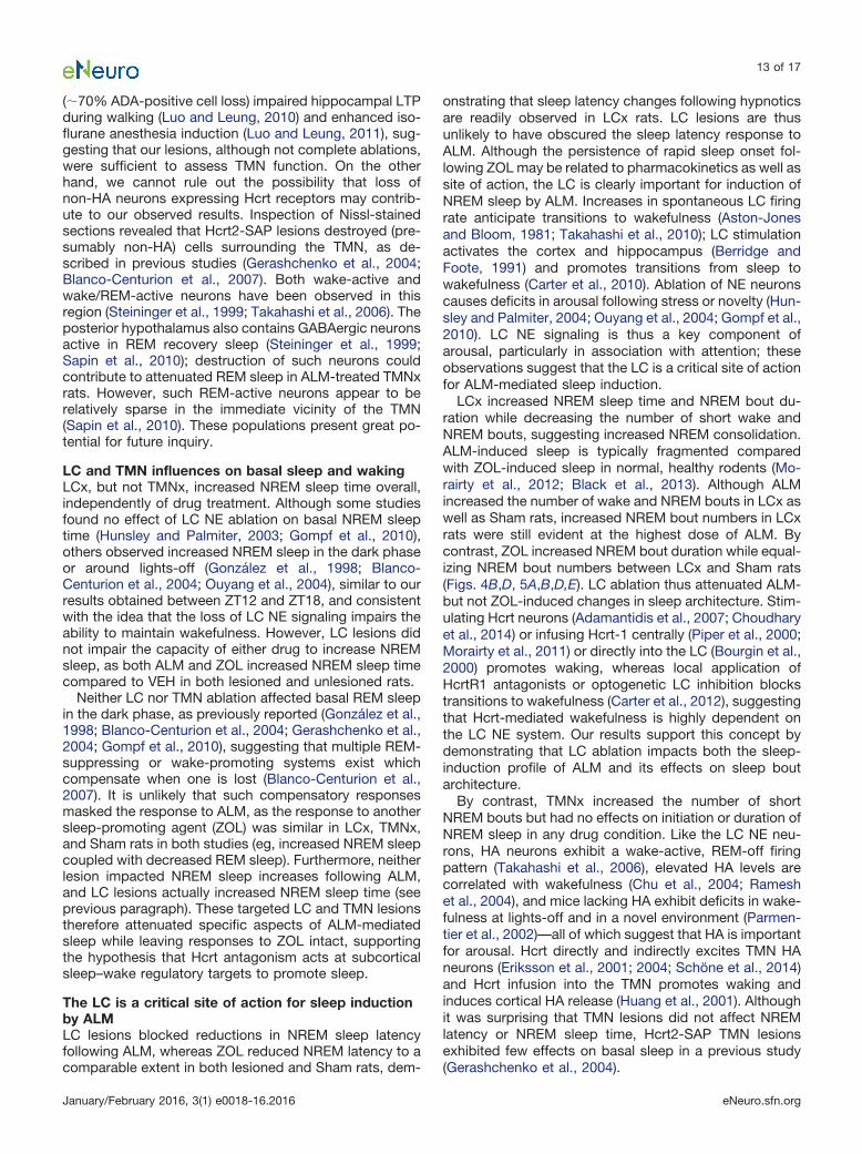
ventral, and caudal TMN subgroups highlighted. ADA-immunostaining clearly visualized the HA neurons inSham-injected rats (Fig. 6D). There were 1375 � 78 ADA-positive cells in the TMN of Sham rats (combined count ofdorsal, ventral, and caudal TMN). Hcrt2-SAP injectionsdecreased ADA-immunostaining in the TMN (Fig. 6E,G).Inspection of Nissl-stained sections revealed that Hcrt2-SAP also destroyed some cells surrounding the TMN (Fig.6H), as described in previous studies (Gerashchenkoet al., 2004; Blanco-Centurion et al., 2007).
Of 13 Hcrt2-SAP injected rats, six exhibited substantialbilateral reductions in ADA-positive cell number (rangingfrom 16% to 45% of the Sham group mean). These sixrats were used as the TMNx group. This TMNx groupexhibited 445 � 73 ADA-positive TMN cells, with individ-ual lesions ranging from 216 to 612 ADA-positive cells,significantly fewer than in the Sham group (t(11) � 8.731;p � 0.001n; Fig. 6F, individual counts from each TMNx rat
Figure 4. Total number of wake (A, B), NREM (C, D) and REM (E, F) bouts for 6 h following ALM (A, C, E) and ZOL (B, D, F) LCx andSham lesioned rats. Doses are mg/kg. �p�0.05 versus vehicle; #p�0.05 (LCx vs Sham); #p�0.06 (LCx vs Sham); �p�0.05, pairedcomparison F test (LCx vs Sham).
8 of 17
January/February 2016, 3(1) e0018-16.2016 eNeuro.sfn.org

superimposed on the group mean). The remaining ratsexhibited little to no ADA cell loss (75% of Sham groupmean), and were excluded from further analysis on thebasis of being essentially unlesioned.
TMNx attenuates REM sleep promotion by ALMAs in the LCx study described above, there was a signif-icant main effect of drug treatment on NREM sleep la-tency (F(6,66) � 11.243; p � 0.001)o such that ZOL but not
Figure 5. Number of wake (A, D, G, J), NREM (B, E, H, K), and REM (C, F, I, L) bouts as a function of bout duration in Sham (A–C,G–I) and LCx rats (D–F, J–L) following ALM (A–F) or ZOL (G–L). �p�0.05 vs vehicle; #p�0.05 (LCx vs Sham).
9 of 17
January/February 2016, 3(1) e0018-16.2016 eNeuro.sfn.org

ALM significantly shortened the latency to NREM sleep(data not shown). There was also a significant main effectof drug treatment on REM sleep latency (F(6,66) � 5.390;p � 0.001)p; whereas post hoc tests showed no signifi-cant changes compared with VEH, ALM tended to de-crease REM sleep latency, whereas ZOL tended toincrease it. Neither NREM nor REM sleep latency wasaffected by TMN lesion.
Consistent with the LCx study, both ALM and ZOLdecreased wake time (main effect of drug: F(6,66) �29.346, p � 0.001; Fig. 7A,B)q and increased NREM sleeptime (main effect of drug: F(6,66) � 27.612, p � 0.001; Fig.7C,D)r, but with no main or interaction effect of TMNlesion. By contrast, ALM (100 and 300 mg/kg) increasedREM sleep time compared with VEH in both Sham andTMNx rats (drug � lesion interaction: F(6,66) � 2.436, p �0.035; Fig. 7E,F)s. Pairwise comparisons of TMNx andSham rats in each drug treatment condition revealed thatTMNx rats had less total REM sleep time following ALM(30 and 300 mg/kg) compared to Shams, whereas therewere no differences in REM sleep time between Shamand TMNx rats following ZOL (Fig. 7E,F). REM–NREM wassignificantly increased by ALM (100 and 300 mg/kg) anddecreased by ZOL (100 mg/kg; main effect of drug: F(6,66)
� 28.419, p � 0.001; Fig. 7G,H)t. However, these drugeffects were qualified by a borderline interaction effect
(F(6,66) � 2.131; p � 0.061)t, such that TMNx decreasedREM–NREM compared to Shams following ALM (300mg/kg; pairwise comparison, p� 0.05). Thus, TMNx af-fected ALM-induced REM sleep increases in a similarmanner to that seen following LCx.
TMNx attenuates increases in REM bout numberfollowing ALMALM (all doses) increased the total number of wake andNREM bouts compared to VEH (main effect of drug: wake,F(6,66) � 48.670, p � 0.001u; NREM, F(6,66) � 46.346, p �0.001v) without an effect of TMN lesion (Fig. 8A,C). ZOLdid not affect the total number of wake or NREM bouts(Fig. 8B,D). Further analysis of bout duration histogramsshowed that TMNx preferentially increased the number ofshort (�0.5 min) NREM bouts (bout � lesion interaction:F(5,55) � 3.401; p � 0.010)w with no additional influence ofdrug treatment (data not shown).
Whereas ALM (100 and 300 mg/kg) increased REMbout number in TMNx and Sham rats, TMNx significantlyattenuated this increase at the highest dose of ALM (in-teraction: F(6,66) � 2.860, p � 0.015; Fig. 8E)x. ZOL did notaffect the total number of REM bouts in either lesiongroup (Fig. 8F). There were no additional effects of lesionor drug treatment on total REM, wake bout numbers, oron the distribution of bout numbers as a function of their
Figure 6. Characterization of Hcrt2-SAP lesions. A–C, Schematic showing location of TMN HA neurons targeted by Hcrt2-SAPinfusions. Panels adapted from Swanson (2004). D, E, HA-positive neurons in the dTMN and vTMN in a Sham-injected rat (D) and aHcrt2-SAP injected rat (E). Photomicrographs depict the TMN at approximately the same rostrocaudal level as B. F, The TMNx group(n � 6) exhibited 445 � 73 ADA-positive TMN cells, with individual lesions ranging from 216to 612 ADA-positive cells. G, H,Composite photomicrographs depicting ADA-immunostaining (G) and Nissl (H) in adjacent brain sections of a rat that was unilaterallyinjected with Hcrt2-SAP. Dotted lines in each panel indicate location of the vTMN in the injected hemisphere. 3v, Third ventricle; cpd,cerebral peduncle; f, fornix; LHA, lateral hypothalamic area; MM, medial mammillary nuclei; mt, mammillary tract; PH, posteriorhypothalamic nucleus. Scale bars: D, E, 200 �m; G, H, 500 �m. �p � 0.05 (LCx vs Sham).
10 of 17
January/February 2016, 3(1) e0018-16.2016 eNeuro.sfn.org

duration (data not shown). Thus, TMNx attenuated thepromotion of REM sleep by ALM primarily by decreasingthe number of REM episodes, while increasing the num-ber of short NREM sleep bouts independently of drugtreatment.
DiscussionThe Hcrt system promotes wakefulness in part throughexcitation of wake-active monoaminergic populations, in-cluding the noradrenergic LC and histaminergic TMN. Inthis study, neurotoxic lesions of the LC NE neurons or
Figure 7. Total wake (A, B), NREM (C, D), and REM (E, F) sleep time, and the ratio of REM to NREM sleep (G, H) for 6 h followingALM (A, C, E, G) and ZOL (B, D, F, H) in TMNx and Sham lesioned rats. Doses are mg/kg. �p � 0.05 vs vehicle; �p � 0.05, pairedcomparison F test (TMNx vs Sham).
11 of 17
January/February 2016, 3(1) e0018-16.2016 eNeuro.sfn.org
TMN HA neurons selectively attenuated ALM-mediatedincreases in REM sleep but not NREM sleep. Further-more, neither lesion altered efficacy of the GABAA recep-tor agonist ZOL. These findings support the hypothesisthat ALM promotes sleep via selective disfacilitation ofsubcortical arousal systems. In addition, these resultshighlight the important role of Hcrt input to monoaminer-gic populations in regulating REM sleep.
Lesion efficacyDBH-SAP infusion caused a near-complete loss of LCDBH-immunoreactive cells, with no signs of collateral ornonspecific damage. DBH-SAP is highly selective for LCNE neurons when delivered intracerebroventricularly orintraparenchymally (Wrenn et al., 1996; Blanco-Centurionet al., 2004). Medullary and pontine NE populations re-ceive Hcrt projections (Baldo et al., 2003) and are sug-gested to play a role in the inhibition of REM sleep (Feniket al., 2002; Rukhadze et al., 2008; Léger et al., 2009).
Although we cannot rule out the possibility of collateraldamage to these non-LC NE groups, the nearby A5 nor-adrenergic neurons appeared intact in our LCx rats fol-lowing 3�g DBH-SAP, consistent with previous workshowing that higher doses are required to lesion thesepopulations (Wrenn et al., 1996). Furthermore, LC lesionsincreased NREM sleep but not REM sleep following VEH.We therefore conclude that the observed effects on ALMand ZOL efficacy are attributable to NE cell loss in the LC,and not to damage of neighboring noradrenergic or othercell groups.
In contrast to our LC lesions, our TMN-HA lesions wereless extensive (55–84% ADA-positive cell loss in TMNxrats). However, more complete TMN lesions still leavebasal sleep–wake parameters largely intact (Gerash-chenko et al., 2004; Blanco-Centurion et al., 2007). Fur-thermore, significant deficits in motivated behavior werereported at a threshold of 45% ADA-positive cell loss(Valdés et al., 2010), whereas lesions comparable to ours
Figure 8. Total number of wake (A, B), NREM (C, D) and REM (E, F) bouts for 6 h following ALM (A, C, E) and ZOL (B, D, F) in TMNxand Sham lesioned rats. Doses are mg/kg. �p � 0.05 versus vehicle; #p � 0.05 (TMNx vs Sham).
12 of 17
January/February 2016, 3(1) e0018-16.2016 eNeuro.sfn.org
(�70% ADA-positive cell loss) impaired hippocampal LTPduring walking (Luo and Leung, 2010) and enhanced iso-flurane anesthesia induction (Luo and Leung, 2011), sug-gesting that our lesions, although not complete ablations,were sufficient to assess TMN function. On the otherhand, we cannot rule out the possibility that loss ofnon-HA neurons expressing Hcrt receptors may contrib-ute to our observed results. Inspection of Nissl-stainedsections revealed that Hcrt2-SAP lesions destroyed (pre-sumably non-HA) cells surrounding the TMN, as de-scribed in previous studies (Gerashchenko et al., 2004;Blanco-Centurion et al., 2007). Both wake-active andwake/REM-active neurons have been observed in thisregion (Steininger et al., 1999; Takahashi et al., 2006). Theposterior hypothalamus also contains GABAergic neuronsactive in REM recovery sleep (Steininger et al., 1999;Sapin et al., 2010); destruction of such neurons couldcontribute to attenuated REM sleep in ALM-treated TMNxrats. However, such REM-active neurons appear to berelatively sparse in the immediate vicinity of the TMN(Sapin et al., 2010). These populations present great po-tential for future inquiry.
LC and TMN influences on basal sleep and wakingLCx, but not TMNx, increased NREM sleep time overall,independently of drug treatment. Although some studiesfound no effect of LC NE ablation on basal NREM sleeptime (Hunsley and Palmiter, 2003; Gompf et al., 2010),others observed increased NREM sleep in the dark phaseor around lights-off (González et al., 1998; Blanco-Centurion et al., 2004; Ouyang et al., 2004), similar to ourresults obtained between ZT12 and ZT18, and consistentwith the idea that the loss of LC NE signaling impairs theability to maintain wakefulness. However, LC lesions didnot impair the capacity of either drug to increase NREMsleep, as both ALM and ZOL increased NREM sleep timecompared to VEH in both lesioned and unlesioned rats.
Neither LC nor TMN ablation affected basal REM sleepin the dark phase, as previously reported (González et al.,1998; Blanco-Centurion et al., 2004; Gerashchenko et al.,2004; Gompf et al., 2010), suggesting that multiple REM-suppressing or wake-promoting systems exist whichcompensate when one is lost (Blanco-Centurion et al.,2007). It is unlikely that such compensatory responsesmasked the response to ALM, as the response to anothersleep-promoting agent (ZOL) was similar in LCx, TMNx,and Sham rats in both studies (eg, increased NREM sleepcoupled with decreased REM sleep). Furthermore, neitherlesion impacted NREM sleep increases following ALM,and LC lesions actually increased NREM sleep time (seeprevious paragraph). These targeted LC and TMN lesionstherefore attenuated specific aspects of ALM-mediatedsleep while leaving responses to ZOL intact, supportingthe hypothesis that Hcrt antagonism acts at subcorticalsleep–wake regulatory targets to promote sleep.
The LC is a critical site of action for sleep inductionby ALMLC lesions blocked reductions in NREM sleep latencyfollowing ALM, whereas ZOL reduced NREM latency to acomparable extent in both lesioned and Sham rats, dem-
onstrating that sleep latency changes following hypnoticsare readily observed in LCx rats. LC lesions are thusunlikely to have obscured the sleep latency response toALM. Although the persistence of rapid sleep onset fol-lowing ZOL may be related to pharmacokinetics as well assite of action, the LC is clearly important for induction ofNREM sleep by ALM. Increases in spontaneous LC firingrate anticipate transitions to wakefulness (Aston-Jonesand Bloom, 1981; Takahashi et al., 2010); LC stimulationactivates the cortex and hippocampus (Berridge andFoote, 1991) and promotes transitions from sleep towakefulness (Carter et al., 2010). Ablation of NE neuronscauses deficits in arousal following stress or novelty (Hun-sley and Palmiter, 2004; Ouyang et al., 2004; Gompf et al.,2010). LC NE signaling is thus a key component ofarousal, particularly in association with attention; theseobservations suggest that the LC is a critical site of actionfor ALM-mediated sleep induction.
LCx increased NREM sleep time and NREM bout du-ration while decreasing the number of short wake andNREM bouts, suggesting increased NREM consolidation.ALM-induced sleep is typically fragmented comparedwith ZOL-induced sleep in normal, healthy rodents (Mo-rairty et al., 2012; Black et al., 2013). Although ALMincreased the number of wake and NREM bouts in LCx aswell as Sham rats, increased NREM bout numbers in LCxrats were still evident at the highest dose of ALM. Bycontrast, ZOL increased NREM bout duration while equal-izing NREM bout numbers between LCx and Sham rats(Figs. 4B,D, 5A,B,D,E). LC ablation thus attenuated ALM-but not ZOL-induced changes in sleep architecture. Stim-ulating Hcrt neurons (Adamantidis et al., 2007; Choudharyet al., 2014) or infusing Hcrt-1 centrally (Piper et al., 2000;Morairty et al., 2011) or directly into the LC (Bourgin et al.,2000) promotes waking, whereas local application ofHcrtR1 antagonists or optogenetic LC inhibition blockstransitions to wakefulness (Carter et al., 2012), suggestingthat Hcrt-mediated wakefulness is highly dependent onthe LC NE system. Our results support this concept bydemonstrating that LC ablation impacts both the sleep-induction profile of ALM and its effects on sleep boutarchitecture.
By contrast, TMNx increased the number of shortNREM bouts but had no effects on initiation or duration ofNREM sleep in any drug condition. Like the LC NE neu-rons, HA neurons exhibit a wake-active, REM-off firingpattern (Takahashi et al., 2006), elevated HA levels arecorrelated with wakefulness (Chu et al., 2004; Rameshet al., 2004), and mice lacking HA exhibit deficits in wake-fulness at lights-off and in a novel environment (Parmen-tier et al., 2002)—all of which suggest that HA is importantfor arousal. Hcrt directly and indirectly excites TMN HAneurons (Eriksson et al., 2001; 2004; Schöne et al., 2014)and Hcrt infusion into the TMN promotes waking andinduces cortical HA release (Huang et al., 2001). Althoughit was surprising that TMN lesions did not affect NREMlatency or NREM sleep time, Hcrt2-SAP TMN lesionsexhibited few effects on basal sleep in a previous study(Gerashchenko et al., 2004).
13 of 17
January/February 2016, 3(1) e0018-16.2016 eNeuro.sfn.org

Lesioning either LC or TMN attenuates ALM-inducedREM sleepBlockade of Hcrt signaling with ALM increased REM sleepin Sham rats, as previously reported in intact animals(Brisbare-Roch et al., 2007); lesioning either the LC NE orTMN HA neurons selectively attenuated the promotion ofREM sleep by ALM. LC lesions significantly decreasedboth REM–NREM and REM bout number at 100 and 300mg/kg, whereas these measures were only affected at the300 mg/kg dose in TMNx rats (Figs. 7E,G, 8E), suggestinga less robust influence of TMN HA neurons on REM sleepcompared to the LC NE neurons. Alternatively, survivingHA neurons in lesioned rats may have been sufficient tomaintain normal histaminergic regulation of REM sleep.However, the common influence of either lesion on ALM-induced REM sleep suggests a specialized role for Hcrtsignaling to both of these nuclei in regulating REM sleep.
The LC NE neurons exhibit a wake-active, “REM-off”firing profile (Takahashi et al., 2010). The LC inhibits cho-linergic brainstem “REM-on” neurons (Hobson et al.,1975; McCarley and Hobson, 1975). The LC NE neuronsare a major target of the Hcrt neurons (Peyron et al., 1998)and express HcrtR1 almost exclusively (Marcus et al., 2001).Local Hcrt-1/orexin-A infusion activates the LC and sup-presses REM sleep (Bourgin et al., 2000), whereas HcrtR1knockout (Mieda et al., 2011), systemic HcrtR1 antagonism(Smith et al., 2003; Morairty et al., 2012), or intra-LC HcrtR1blockade blocks Hcrt-mediated REM suppression (Bourginet al., 2000; Choudhary et al., 2014), and siRNA downregu-lation of HcrtR1 in the LC increases spontaneous REM sleepin the dark phase (Chen et al., 2010). HcrtR1-mediated Hcrtsignaling thus powerfully regulates the REM-suppressingrole of the LC NE neurons, although this effect may be moreclearly observed following LC-specific manipulations ratherthan systemic treatments.
TMN HA neurons, which express HcrtR2 (Marcus et al.,2001), also exhibit a wake-active, REM-off firing profile(Takahashi et al., 2006). Mice unable to synthesize HAexhibit increased REM sleep time (Parmentier et al., 2002;Anaclet et al., 2009). HA inhibits hypothalamic melanin-concentrating hormone neurons (Parks et al., 2014) thathave been implicated in REM sleep (Verret et al., 2003;Clément et al., 2012; Jego et al., 2013), and administrationof either ALM or a HcrtR2 antagonist decreases extracel-lular HA levels in the LH (Dugovic et al., 2009), consistentwith a role for Hcrt-HA signaling in REM sleep regulation.Thus, the acute blockade of excitatory Hcrt input by ALMwould “disfacilitate” REM-off activity in the LC and theTMN, resulting in the downstream disinhibition of REM-active populations such as brainstem cholinergic neuronstargeted by the LC or the hypothalamic MCH neuronstargeted by HA, respectively.
Lesioning either the LC, which expresses only HcrtR1,or the TMN, which expresses only HcrtR2 (Marcus et al.,2001), produced similar effects on ALM efficacy. Recentstudies differ regarding whether blockade of Hcrt signal-ing at one or both Hcrt receptors is critical for promotingsleep (Smith et al., 2003; Dugovic et al., 2009; Mang et al.,2012; Morairty et al., 2012; Steiner et al., 2013). However,deletion of either Hcrt receptor modulates REM sleep
response following Hcrt1 infusion (Mieda et al., 2011),while coadministration of HcrtR1 and R2 antagonists elic-ited synergistic effects distinct from those of either drugadministered alone (Dugovic et al., 2009). The sleep-promoting effects of Hcrt receptor antagonism may thusdepend on the resulting balance between HcrtR1 andHcrtR2 activity (Hoyer et al., 2013). In this light, localizedmanipulations such as lesions could also significantlyalter drug efficacy by eliminating critical points in the Hcrtsignaling pathway. Such manipulations would depend asmuch on the anatomical site of action as the receptortype(s) expressed there. For example, REM sleep is in-creased by siRNA knockdown of either HcrtR1 in the LC(Chen et al., 2010) or HcrtR2 in the lateral pontomesen-cephalic tegmentum (Chen et al., 2013), whereas lesion-ing the basal forebrain, which expresses both Hcrtreceptors (Marcus et al., 2001), attenuates ALM-inducedNREM, but not REM sleep (Vazquez-DeRose et al., 2014). Inthe present study, eliminating either HcrtR1 signaling in theLC or HcrtR2 signaling in the TMN yielded similar effects onALM-induced REM sleep, independently of the Hcrt recep-tor type expressed in each region. Indeed, the effects ofablating the LC, which is thought to respond to Hcrt exclu-sively via HcrtR1, suggest strongly that HcrtR1-mediated LCNE activity represents a critical pathway for ALM-mediatedsleep induction and REM promotion. Together, these find-ings suggest that the site of action, not just the specificreceptor or receptors targeted, is a key determinant of howHcrt receptor antagonism facilitates sleep.
ConclusionsDORAs, including ALM, promote sleep by blocking Hcrtsignaling. In this study, lesions of the wake-promoting LC NEor TMN HA populations compromised the REM sleep-promoting efficacy of ALM, whereas previous work hasshown that ALM, but not ZOL, requires an intact BF formaximum NREM-promoting efficacy (Vazquez-DeRoseet al., 2014). Together, these studies indicate that Hcrt neu-rotransmission influences distinct aspects of sleep at differ-ent locations in the sleep–wake regulatory network.Furthermore, our results, particularly our finding that LClesions attenuate ALM efficacy, suggest that site of action isat least as important a consideration for Hcrt antagonistefficacy as the receptor targeted. By selectively disfacilitat-ing these subcortical wake-promoting populations, ALM ef-fectively promotes sleep by eliciting neurochemical eventsconsistent with the transition to normal sleep (Vazquez-DeRose et al., 2014). These properties are likely to underliethe findings that ALM also promotes sleep without nega-tively impacting cognitive performance (Morairty et al., 2014)and without globally blocking the capability for arousal(Parks et al., 2016).
ReferencesAdamantidis AR, Zhang F, Aravanis AM, Deisseroth K, de Lecea L
(2007) Neural substrates of awakening probed with optogenetic con-trol of hypocretin neurons. Nature 450:420–424. CrossRef Medline
Anaclet C, Parmentier R, Ouk K, Guidon G, Buda C, Sastre JP,Akaoka H, Sergeeva OA, Yanagisawa M, Ohtsu H, Franco P, HaasHL, Lin JS (2009) Orexin/hypocretin and histamine: distinct roles in
14 of 17
January/February 2016, 3(1) e0018-16.2016 eNeuro.sfn.org

the control of wakefulness demonstrated using knock-out mousemodels. J Neurosci 29:14423–14438. CrossRef Medline
Aston-Jones G, Bloom FE (1981) Activity of norepinephrine-containinglocus coeruleus neurons in behaving rats anticipates fluctuations inthe sleep-waking cycle. J Neurosci 1:876–886. Medline
Baldo BA, Daniel RA, Berridge CW, Kelley AE (2003) Overlapping dis-tributions of orexin/hypocretin- and dopamine-beta-hydroxylase im-munoreactive fibers in rat brain regions mediating arousal, motivation,and stress. J Comp Neurol 464:220–237. CrossRef Medline
Berridge CW, Foote SL (1991) Effects of locus coeruleus activationon electroencephalographic activity in neocortex and hippocam-pus. J Neurosci 11:3135–3145. Medline
Black SW, Morairty SR, Fisher SP, Chen T-M, Warrier DR, Kilduff TS(2013) Almorexant promotes sleep and exacerbates cataplexy in amurine model of narcolepsy. Sleep 36:325–336. CrossRef Medline
Blanco-Centurion C, Gerashchenko D, Salin-Pascual RJ, ShiromaniPJ (2004) Effects of hypocretin2-saporin and antidopamine-beta-hydroxylase-saporin neurotoxic lesions of the dorsolateral pons onsleep and muscle tone. Eur J Neurosci 19:2741–2752. CrossRef
Blanco-Centurion C, Gerashchenko D, Shiromani PJ (2007) Effects ofsaporin-induced lesions of three arousal populations on daily levels ofsleep and wake. J Neurosci 27:14041–14048. CrossRef Medline
Bourgin P, Huitron-Huitrón-Résendiz S, Spier AD, Fabre V, Morte B,Criado JR, Sutcliffe JG, Henriksen SJ, de Lecea L (2000)Hypocretin-1 modulates rapid eye movement sleep through activa-tion of locus coeruleus neurons. J Neurosci 20:7760–7765. Medline
Brightwell JJ, Taylor BK (2009) Noradrenergic neurons in the locuscoeruleus contribute to neuropathic pain. Neuroscience 160:174–185. CrossRef Medline
Brisbare-Roch C, Dingemanse J, Koberstein R, Hoever P, AissaouiH, Flores S, Mueller C, Nayler O, van Gerven J, de Haas SL, HessP, Qiu C, Buchmann S, Scherz M, Weller T, Fischli W, Clozel M,Jenck F (2007) Promotion of sleep by targeting the orexin systemin rats, dogs and humans. Nat Med 13:150–155. CrossRef Medline
Carter ME, Brill J, Bonnavion P, Huguenard JR, Huerta R, de Lecea L(2012) Mechanism for Hypocretin-mediated sleep-to-wake transi-tions. Proc Natl Acad Sci U S A 109:E2635–E2644. CrossRefMedline
Carter ME, Yizhar O, Chikahisa S, Nguyen H, Adamantidis A, NishinoS, Deisseroth K, de Lecea L (2010) Tuning arousal with optoge-netic modulation of locus coeruleus neurons. Nat Neurosci 13:1526–1533. CrossRef Medline
Chemelli RM, Willie JT, Sinton CM, Elmquist JK, Scammell T, Lee C,Richardson JA, Williams SC, Xiong Y, Kisanuki Y, Fitch TE, Naka-zato M, Hammer RE, Saper CB, Yanagisawa M (1999) Narcolepsyin orexin knockout mice: molecular genetics of sleep regulation.Cell 98:437–451. Medline
Chen L, McKenna JT, Bolortuya Y, Brown RE, McCarley RW (2013)Knockdown of orexin type 2 receptor in the lateral pontomesen-cephalic tegmentum of rats increases REM sleep. Eur J Neurosci37:957–963. CrossRef Medline
Chen L, McKenna JT, Bolortuya Y, Winston S, Thakkar MM, BasheerR, Brown RE, McCarley RW (2010) Knockdown of orexin type 1receptor in rat locus coeruleus increases REM sleep during thedark period. Eur J Neurosci 32:1528–1536. Medline
Choudhary RC, Khanday MA, Mitra A, Mallick BN (2014) Perifornicalorexinergic neurons modulate REM sleep by influencing locus coer-uleus neurons in rats. Neuroscience 279:33–43. CrossRef Medline
Chu M, Huang ZL, Qu WM, Eguchi N, Yao MH, Urade Y (2004)Extracellular histamine level in the frontal cortex is positively cor-related with the amount of wakefulness in rats. Neurosci Res49:417–420. CrossRef Medline
Clément O, Sapin E, Libourel PA, Arthaud S, Brischoux F, Fort P, LuppiPH (2012) The lateral hypothalamic area controls paradoxical (REM)sleep by means of descending projections to brainstem GABAergicneurons. J Neurosci 32:16763–16774. CrossRef Medline
Dang A, Garg A, Rataboli PV (2011) Role of zolpidem in the man-agement of insomnia. CNS Neurosci Ther 17:387–397. CrossRefMedline
Dugovic C, Shelton JE, Aluisio LE, Fraser IC, Jiang X, Sutton SW,Bonaventure P, Yun S, Li X, Lord B, Dvorak CA, Carruthers NI,Lovenberg TW (2009) Blockade of orexin-1 receptors attenuatesorexin-2 receptor antagonism-induced sleep promotion in the rat.J Pharmacol Exp Therapeut 330:142–151. CrossRef Medline
Eriksson KS, Sergeeva O, Brown RE, Haas HL (2001) Orexin/hypo-cretin excites the histaminergic neurons of the tuberomammillarynucleus. J Neurosci 21:9273–9279. Medline
Eriksson KS, Sergeeva OA, Selbach O, Haas HL (2004) Orexin(hypocretin)/dynorphin neurons control GABAergic inputs to tube-romammillary neurons. Eur J Neurosci 19:1278–1284. CrossRefMedline
Estabrooke I, McCarthy M, Ko E, Chou T, Chemelli R, Yanagisawa M,Saper C, Scammell TE (2001) Fos expression in orexin neuronsvaries with behavioral state. J Neurosci 21:1656–62.
Fenik V, Marchenko V, Janssen P, Davies RO, Kubin L (2002) A5 cellsare silenced when REM sleep-like signs are elicited by pontinecarbachol. J Appl Physiol 93:1448–1456. CrossRef Medline
Gerashchenko D, Chou TC, Blanco-Centurion CA, Saper CB, Shiro-mani PJ (2004) Effects of lesions of the histaminergic tuberomam-millary nucleus on spontaneous sleep in rats. Sleep 27:1275–1281.Medline
Gerashchenko D, Kohls MD, Greco M, Waleh NS, Salin-Pascual R,Kilduff TS, Lappi DA, Shiromani PJ (2001) Hypocretin-2-saporinlesions of the lateral hypothalamus produce narcoleptic-like sleepbehavior in the rat. J Neurosci 21:7273–7283. Medline
Gompf HS, Mathai C, Fuller PM, Wood DA, Pedersen NP, Saper CB,Lu J (2010) Locus ceruleus and anterior cingulate cortex sustainwakefulness in a novel environment. J Neurosci 30:14543–14551.CrossRef Medline
González MM, Debilly G, Valatx JL (1998) Noradrenaline neurotoxinDSP-4 effects on sleep and brain temperature in the rat. NeurosciLett 248:93–96. Medline
Hagan J, Leslie RA, Patel S, Evans ML, Wattam TA, Holmes S,Benham CD, Taylor SG, Routledge C, Hemmati P, Munton RP,Ashmeade TE, Shah AS, Hatcher JP, Hatcher PD, Jones DN,Smith MI, Piper DC, Hunter AJ, Porter RA, et al. (1999) Orexin Aactivates locus coeruleus cell firing and increases arousal in therat. Proc Natl Acad Sci U S A 96:10911–6. Medline
Hobson JA, McCarley RW, Wyzinski PW (1975) Sleep cycle oscilla-tion: reciprocal discharge by two brainstem neuronal groups. Sci-ence 189:55–58. Medline
Horvath TL, Peyron C, Diano S, Ivanov A, Aston-Jones G, Kilduff TS,van den Pol AN (1999) Hypocretin (orexin) activation and synapticinnervation of the locus coeruleus noradrenergic system. J CompNeurol 415:145–159. Medline
Hoyer D, Dürst T, Fendt M, Jacobson LH, Betschart C, HintermannS, Behnke D, Cotesta S, Laue G, Ofner S, Legangneux E, Gee CE(2013) Distinct effects of IPSU and suvorexant on mouse sleeparchitecture. Front Neurosci 7:235. CrossRef Medline
Huang ZL, Qu WM, Li WD, Mochizuki T, Eguchi N, Watanabe T,Urade Y, Hayaishi O (2001) Arousal effect of orexin A depends onactivation of the histaminergic system. Proc Natl Acad Sci U S A98:9965–9970. CrossRef Medline
Hunsley MS, Palmiter RD (2003) Norepinephrine-deficient mice exhibitnormal sleep-wake states but have shorter sleep latency after mildstress and low doses of amphetamine. Sleep 26:521–526. Medline
Hunsley MS, Palmiter RD (2004) Altered sleep latency and arousalregulation in mice lacking norepinephrine. Pharmacol BiochemBehav 78:765–773. CrossRef Medline
Jego S, Glasgow SD, Herrera CG, Ekstrand M, Reed SJ, Boyce R,Friedman J, Burdakov D, Adamantidis AR (2013) Optogeneticidentification of a rapid eye movement sleep modulatory circuit inthe hypothalamus. Nat Neurosci 16:1637–1643. CrossRef Medline
Ko EM, Estabrooke IV, McCarthy M, Scammell TE (2003) Wake-related activity of tuberomammillary neurons in rats. Brain Res992:220–226. Medline
Koberstein R, Aissaoui H, Bur D, Clozel M, Fischli W, Jenck F,Mueller C, Nayler O, Sifferlen T, Treiber A, Weller T (2003) Tetra-hydroisoquinolines as orexin receptor antagonists: strategies for
15 of 17
January/February 2016, 3(1) e0018-16.2016 eNeuro.sfn.org

lead optimization by solution-phase chemistry. CHIMIA 57:270–275. CrossRef
Koberstein R, Fischli W, Clozel M, Aissaoui H, Weller T (2005)Substituted 1,2,3,4-tetrahydroisoquinoline derivatives. World pat-ent: WO 2005118548.
Lee MG, Hassani OK, Jones BE (2005) Discharge of identified orexin/hypocretin neurons across the sleep-waking cycle. J Neurosci25:6716–6720. CrossRef Medline
Léger L, Goutagny R, Sapin E, Salvert D, Fort P, Luppi P-H (2009)Noradrenergic neurons expressing Fos during waking and para-doxical sleep deprivation in the rat. J Chem Neuroanat 37:149–157. CrossRef Medline
Lin L, Faraco J, Li R, Kadotani H, Rogers W, Lin X, Qiu X, de Jong PJ,Nishino S, Mignot E (1999) The sleep disorder canine narcolepsy iscaused by a mutation in the hypocretin (orexin) receptor 2 gene.Cell 98:365–376. Medline
Luo T, Leung LS (2010) Endogenous histamine facilitates long-termpotentiation in the hippocampus during walking. J Neurosci 30:7845–7852. CrossRef Medline
Luo T, Leung LS (2011) Involvement of tuberomamillary histaminer-gic neurons in isoflurane anesthesia. Anesthesiology 115:36–43.CrossRef Medline
Mang GM, Dürst T, Bürki H, Imobersteg S, Abramowski D,Schuepbach E, Hoyer D, Fendt M, Gee CE (2012) The dual orexinreceptor antagonist almorexant induces sleep and decreasesorexin-induced locomotion by blocking orexin 2 receptors. Sleep35:1625–1635. CrossRef Medline
Marcus JN, Aschkenasi CJ, Lee CE, Chemelli RM, Saper CB, Yanag-isawa M, Elmquist JK (2001) Differential expression of orexin recep-tors 1 and 2 in the rat brain. J Comp Neurol 435:6–25. Medline
McCarley RW, Hobson JA (1975) Neuronal excitability modulationover the sleep cycle: a structural and mathematical model. Sci-ence 189:58–60. Medline
Mieda M, Hasegawa E, Kisanuki YY, Sinton CM, Yanagisawa M,Sakurai T (2011) Differential roles of orexin receptor-1 and -2 in theregulation of non-REM and REM sleep. J Neurosci 31:6518–6526.CrossRef Medline
Morairty SR, Hedley L, Flores J, Martin R, Kilduff TS (2008) Selective5HT(2A) and 5HT(6) receptor antagonists promote sleep in rats.Sleep 31:34–44. Medline
Morairty SR, Revel FG, Malherbe P, Moreau J-L, Valladao D, Wet-tstein JG, Kilduff TS, Borroni E (2012) Dual hypocretin receptorantagonism is more effective for sleep promotion than antagonismof either receptor alone. PLoS One 7:e39131. CrossRef Medline
Morairty SR, Wilk AJ, Lincoln WU, Neylan TC, Kilduff TS (2014) Thehypocretin/orexin antagonist almorexant promotes sleep withoutimpairment of performance in rats. Front Neurosci 8:3. CrossRefMedline
Morairty SR, Wisor J, Silveira K, Sinko W, Kilduff TS (2011) Thewake-promoting effects of hypocretin-1 are attenuated in old rats.Neurobiol Aging 32:1514–1527. CrossRef Medline
Ohno K, Sakurai T (2008) Orexin neuronal circuitry: role in the regu-lation of sleep and wakefulness. Front Neuroendocrinol 29:70–87.CrossRef Medline
Ouyang M, Hellman K, Abel T, Thomas SA (2004) Adrenergic signal-ing plays a critical role in the maintenance of waking and in theregulation of REM sleep. J Neurophysiol 92:2071–2082. CrossRefMedline
Parks GS, Olivas ND, Ikrar T, Sanathara NM, Wang L, Wang Z, CivelliO, Xu X (2014) Histamine inhibits the melanin-concentrating hor-mone system: implications for sleep and arousal. J Physiol (Lond)592:2183–2196. CrossRef Medline
Parks GS, Warrier DR, Dittrich L, Schwartz MD, Palmerston JB, NeylanTC, Morairty SR, Kilduff TS (2016) The dual hypocretin receptorantagonist almorexant is permissive for activation of wake-promotingsystems. Neuropsychopharmacology 41:1144-1155.
Parmentier R, Ohtsu H, Djebbara-Hannas Z, Valatx JL, Watanabe T,Lin JS (2002) Anatomical, physiological, and pharmacological
characteristics of histidine decarboxylase knock-out mice: evi-dence for the role of brain histamine in behavioral and sleep-wakecontrol. J Neurosci 22:7695–7711. Medline
Peyron C, Tighe DK, van den Pol AN, de Lecea L, Heller HC, SutcliffeJG, Kilduff TS (1998) Neurons containing hypocretin (orexin) projectto multiple neuronal systems. J Neurosci 18:9996–10015. Medline
Piper DC, Upton N, Smith MI, Hunter AJ (2000) The novel brainneuropeptide, orexin-A, modulates the sleep-wake cycle of rats.Eur J Neurosci 12:726–730. Medline
Ramesh V, Thakkar MM, Strecker RE, Basheer R, McCarley RW(2004) Wakefulness-inducing effects of histamine in the basalforebrain of freely moving rats. Behav Brain Res 152:271–278.CrossRef Medline
Rukhadze I, Fenik VB, Branconi JL, Kubin L (2008) Fos expression inpontomedullary catecholaminergic cells following rapid eye move-ment sleep-like episodes elicited by pontine carbachol in urethane-anesthetized rats. Neuroscience 152:208–222. CrossRef Medline
Sapin E, Bérod A, Léger L, Herman PA, Luppi P-H, Peyron C (2010)A very large number of GABAergic neurons are activated in thetuberal hypothalamus during paradoxical (REM) sleep hypersom-nia. PLoS One 5:e11766. CrossRef
Schöne C, Apergis-Schoute J, Sakurai T, Adamantidis A, BurdakovD (2014) Coreleased orexin and glutamate evoke nonredundantspike outputs and computations in histamine neurons. Cell Rep7:697–704. CrossRef Medline
Smith MI, Piper DC, Duxon MS, Upton N (2003) Evidence implicatinga role for orexin-1 receptor modulation of paradoxical sleep in therat. Neurosci Lett 341:256–258. Medline
Soffin EM, Evans ML, Gill CH, Harries MH, Benham CD, Davies CH(2002) SB-334867-A antagonises orexin mediated excitation in thelocus coeruleus. Neuropharmacology 42:127–133. Medline
Steiner MA, Gatfield J, Brisbare-Roch C, Dietrich H, Treiber A, JenckF, Boss C (2013) Discovery and characterization of ACT-335827,an orally available, brain penetrant orexin receptor type 1 selectiveantagonist. Chem Med Chem 8:898–903. CrossRef Medline
Steininger TL, Alam MN, Gong H, Szymusiak R, McGinty D (1999)Sleep-waking discharge of neurons in the posterior lateral hypo-thalamus of the albino rat. Brain Res 840:138–147. Medline
Swanson LW (2004) Brain maps: structure of the rat brain, 3rdedition.
Takahashi K, Kayama Y, Lin JS, Sakai K (2010) Locus coeruleusneuronal activity during the sleep-waking cycle in mice. Neurosci-ence 169:1115–1126. CrossRef Medline
Takahashi K, Lin JS, Sakai K (2006) Neuronal activity of histaminergictuberomammillary neurons during wake-sleep states in the mouse.J Neurosci 26:10292–10298. CrossRef Medline
Thannickal TC, Moore RY, Nienhuis R, Ramanathan L, Gulyani S,Aldrich M, Cornford M, Siegel JM (2000) Reduced number of hypo-cretin neurons in human narcolepsy. Neuron 27:469–474. Medline
Valdés JL, Sánchez C, Riveros ME, Blandina P, Contreras M, FaríasP, Torrealba F (2010) The histaminergic tuberomammillary nucleusis critical for motivated arousal. Eur J Neurosci 31:2073–2085.CrossRef Medline
Vazquez-DeRose J, Schwartz MD, Nguyen AT, Warrier DR, Gulati S,Mathew TK, Neylan TC, Kilduff TS (2014) Hypocretin/orexin antago-nism enhances sleep-related adenosine and GABA neurotransmis-sion in rat basal forebrain. Brain Struct Funct 221:923–940.
Verret L, Goutagny R, Fort P, Cagnon L, Salvert D, Léger L, BoissardR, Salin P, Peyron C, Luppi PH (2003) A role of melanin-concentrating hormone producing neurons in the central regula-tion of paradoxical sleep. BMC Neurosci 4:19. CrossRef
Wiley RG, Kline RH IV (2000) Neuronal lesioning with axonally trans-ported toxins. J Neurosci Methods 103:73–82. Medline
Wrenn CC, Picklo MJ, Lappi DA, Robertson D, Wiley RG (1996)Central noradrenergic lesioning using anti-DBH-saporin: anatom-ical findings. Brain Res 740:175–184. Medline
16 of 17
January/February 2016, 3(1) e0018-16.2016 eNeuro.sfn.org