Embed Size (px)
Citation preview

Usher syndrome type I G (USH1G) is caused bymutations in the gene encoding SANS, a proteinthat associates with the USH1C protein, harmonin
Dominique Weil1,{, Aziz El-Amraoui1,{, Saber Masmoudi2,{, Mirna Mustapha1,
Yoshiaki Kikkawa3, Sophie Laine1, Sedigheh Delmaghani1, Avital Adato1,4,
Sellama Nadifi5, Zeineb Ben Zina6, Christian Hamel7, Andreas Gal8, Hammadi Ayadi2,
Hiromichi Yonekawa3 and Christine Petit1,*
1Unite de Genetique des Deficits Sensoriels, CNRS URA1968, Institut Pasteur, 25 rue du Dr Roux,
75724 Paris cedex 15, France, 2Laboratoire de Genetique Moleculaire Humaine, Faculte de Medecine,
Sfax, Tunisia, 3Department of Laboratory Animal Science, The Tokyo Metropolitan Institute of
Medical Science (Rinshoken), Tokyo, Japan, 4Department of Molecular Genetics and The Crown Human
Genome Center, Weizmann Institute of Science, Rehovot, Israel, 5Laboratoire de Genetique Humaine,
Faculte de Medecine et de Pharmacie, Casablanca, Morocco, 6Service d’Ophtalmologie, C.H.U. H. Bourguiba,
Sfax, Tunisia, 7INSERM U254, Laboratoire de Neurobiologie de l’Audition, Montpellier, France and8Institut fur Humangenetik, Universitatsklinikum Hamburg-Eppendorf, Hamburg, Germany
Received December 6, 2002; Revised and Accepted January 7, 2003
Usher syndrome type I (USH1) is the most frequent cause of hereditary deaf–blindness in humans. Sevengenetic loci (USH1A-G) have been implicated in this disease to date, and four of the corresponding geneshave been identified: USH1B, C, D and F. We carried out fine mapping of USH1G (chromosome 17q24–25),restricting the location of this gene to an interval of 2.6Mb and then screened genes present within thisinterval for mutations. The genes screened included the orthologue of the Sans gene, which is defective inthe Jackson shaker deaf mutant and maps to the syntenic region in mice. In two consanguineous USH1G-affected families, we detected two different frameshift mutations in the SANS gene. Two brothers from aGerman family affected with USH1G were found to be compound heterozygotes for a frameshift and amissense mutation. These results demonstrate that SANS underlies USH1G. The SANS protein contains threeankyrin domains and a sterile alpha motif, and its C-terminal tripeptide presents a class I PDZ-binding motif.We showed, by means of co-transfection experiments, that SANS associates with harmonin, a PDZ domain-containing protein responsible for USH1C. In Jackson shaker mice the hair bundles, the mechanoreceptivestructures of inner ear sensory cells, are disorganized. Based on the known interaction between USH1B(myosin VIIa), USH1C (harmonin) and USH1D (cadherin 23) proteins and the results obtained in this study, wesuggest that a functional network formed by the USH1B, C, D and G proteins is responsible for the correctcohesion of the hair bundle.
INTRODUCTION
Usher syndrome (USH) is an autosomal recessive disease thataffects both the inner ear and the retina. It is the most frequentcause of hereditary deaf-blindness, affecting 1 child in 25 000.Three clinical subtypes have been defined (1). USH type I
(USH1), the most severe, involves severe to profoundcongenital sensorineural deafness, constant vestibular dysfunc-tion and retinitis pigmentosa with prepubertal onset. USH2differs from USH1 mainly in the deafness being less severe, theabsence of vestibular dysfunction and the onset of retinitispigmentosa after puberty. Finally, USH3 differs from USH1
*To whom correspondence should be addressed. Email: [email protected]{The authors wish it to be known that, in their opinion, the first three authors should be regarded as joint First Authors.
Human Molecular Genetics, 2003, Vol. 12, No. 5 463–471DOI: 10.1093/hmg/ddg051
Human Molecular Genetics, Vol. 12, No. 5 # Oxford University Press 2003; all rights reserved
Downloaded from https://academic.oup.com/hmg/article-abstract/12/5/463/707029by gueston 18 February 2018

and USH2 in the progressiveness of hearing loss and theoccasional presence of vestibular dysfunction. USH1 isgenetically heterogeneous. Seven loci responsible for thisdisease have been defined (USH1A-G) (2,3) and four of thecorresponding genes have been identified: USH1B, C, D and F.USH1B encodes the actin-based motor protein myosin VIIa (4).USH1C encodes a PDZ domain-containing protein, harmonin(5,6). At least 10 isoforms of harmonin have been described.They may be classified into three subclasses referred to asharmonin a, b and c, and are collectively referred to asharmonin (6). Finally, mutations in the genes encoding twocadherin-related proteins, cadherin 23 and protocadherin 15,have been shown to cause USH1D (7,8) and USH1F (9,10),respectively.
Mutations have also been reported in three murine orthologsof the USH1 genes: Myosin VIIa, which is defective in shaker-1(sh1) mutants (11); Cadherin 23, which is defective in waltzer(v) mutants (12), and Protocadherin 15, which is responsiblefor Ames waltzer (av) mutants (13). These mouse mutants arecongenitally deaf and display vestibular dysfunction. Inaddition, the sensory cells of the inner ears of these mousemutants display similar disorganization of the stereociliacomprising the hair bundle (12–14). We recently showed thatmyosin VIIA, harmonin and cadherin 23 interact in thedeveloping inner ear sensory cells, ensuring the correctdevelopment of the hair bundle (15).
We studied a consanguineous Jordanian family affected byUSH type 1 (JO-US1), and mapped the USH1G locus to aninterval of 23 cM between markers D17S1350 and D17S1830on chromosome 17q24–25 (3). In this study, we narroweddown the USH1G linkage interval, thanks to another USH1G-affected family, and identified SANS, the human orthologue ofthe gene defective in Jackson shaker ( js) mutant mice (16), asthe causative gene. We also showed, in co-transfectionexperiments, that SANS associated with harmonin, the proteinunderlying USH1C.
RESULTS AND DISCUSSION
Narrowing the limits of the USH1G interval
Eight members of a large family from Southern Tunisia (familyMB) were recognized as suffering from profound congenitaldeafness. Vestibular dysfunction was assessed by caloric testsin patients MB15, 16, 18, 20, 29 and 30 (Fig. 1). Severeretinitis pigmentosa was assessed by funduscopy in all theaffected children (see Materials and Methods). All the affectedchildren were over the age of 16 years at the time ofexamination, with the exception of MB20, who was only5 years old. The clinical features observed in this family qualifythe disease as Usher syndrome type I.
We tested this family for co-segregation of the disease withany of the seven known USH1 loci (USH1A-G), usingpolymorphic microsatellite markers (see Materials andMethods). Linkage was detected only with the markers of theUSH1G interval, D17S1807 and D17S1603. We then tried tonarrow down the USH1G interval, using markers locatedbetween D17S1350 and D17S1830, the two markers flankingthe homozygosity region in the original USH1G family (Fig. 1).
As affected individual MB20 was heterozygous for markerD17S1831, and the two affected individuals MB148 andMB152 were heterozygous for marker D17S1603, we wereable to limit the USH1G interval to a 2.6 Mb region betweenthese two markers (maximum LOD score 4.6, at y¼ 0 formarkers D17S1807 and D17S1839).
SANS mutations cause USH1G
We first considered as candidate genes two genes located withinthe USH1G interval that corresponded to two cDNAs isolatedfrom a subtracted cDNA library prepared from the vestibularsensory patches of the mouse inner ear (6). The first, the humangiant larvae gene homologue (LLGL2) (GenBank accessionnumber X87342) corresponded to a 3480 bp cDNA, extendingover 19 kb, and comprised 23 exons (the 50 end is missing fromthe Human Sequence Draft). Analysis of the sequences of theseexons in USH1G-affected families (JO-US1 and MB) resultedin the detection of only silent polymorphisms. The other gene,solute carrier family 9 (sodium/hydrogen exchanger), isoform3 regulatory factor 1 (SLC9A3R1) (GenBank accession numberXM_046932), corresponded to a 1978 bp cDNA, extendingover 21 kb and comprising 6 exons. No mutation of this genewas detected in either of the two affected families in analysingthe sequences of the coding exons.
We previously reported that the locus involved in js mousemutant, which presents deafness and vestibular dysfunction(17), mapped to a chromosomal region syntenic to the humanUSH1G region (3). Although the USH1G interval identifiedhere was much smaller, it nonetheless still matched with the jslocus. As the Sans gene was recently shown to be mutated in jsmice (16), we considered the human orthologue SANS(GenBank accession number AK091243) as a candidate generesponsible for USH1G. SANS is located between markersD17S1807 and D17S1839. The cDNA for this gene is 3559 bplong. Comparison with human genome sequences (GenBankaccession number AC068874) revealed that SANS encompasses7.2 kb and comprises three exons, two of which are coding. The1380 bp open reading frame (ORF) is predicted to encode a 460amino acid (aa) protein. The translation initiation site wasidentified at position 184 on the basis of the presence of aKozak consensus sequence (gcgccATGaacga) preceded by anin-frame stop codon, 170 bp upstream. The human and mouseORFs display 90% nucleotide sequence identity, and the twopredicted protein sequences are 96% identical. Sequenceanalysis of the encoded protein showed this protein to containthree ankyrin-like domains (18) at the N-terminal end (aa 31–63, 64–96 and 97–129), a central region (aa 130–385), and aSAM (sterile alpha motif) domain (19) (aa 384–446; Fig. 2)and a PDZ-binding motif at the C-terminal end. This proteinwas therefore named SANS for scaffold protein containingankyrin repeats and SAM domain. Ankyrin domains areinvolved in protein–protein interactions (18). SAM domains,originally identified in yeast (19), are present in several proteinsinvolved in the regulation of a number of developmentalprocesses (e.g. protein kinase receptors, cytoplasmic scaffold-ing proteins, transcription factors). These domains, which arethought to be involved in protein–protein interactions, mayundergo homo- or heterodimerization with other SAM domains(20,21).
464 Human Molecular Genetics, 2003, Vol. 12, No. 5
Downloaded from https://academic.oup.com/hmg/article-abstract/12/5/463/707029by gueston 18 February 2018

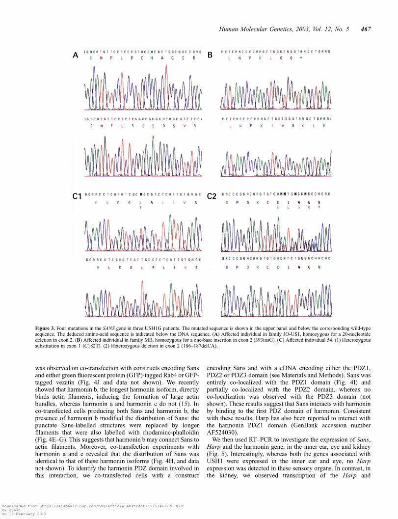
We designed primers (Table 1) to amplify and sequence thetwo coding exons and the flanking intronic regions. In the threeaffected members of family JO-US1 (3), a 20 bp deletion (829–848del) was detected in exon 2, in the homozygous state (Figs 2and 3A). The deletion was also found in a heterozygous state inboth parents of these individuals and in their three unaffectedsiblings. This frameshift deletion is predicted to lead to theproduction of a truncated protein, only 326 aa long (i.e. withoutthe SAM domain), the last 70 residues of which are notin-frame. In the eight affected members of family MB, theinsertion of a single base (393insG) was detected in exon 2, inthe homozygous state (Figs 2 and 3B). The mutation waspresent in the heterozygous state in the parents, and was absent
from the unaffected individuals MB17 and MB151. Thisframeshift mutation is predicted to lead to the production of a133 aa protein, in which only the three ankyrin repeats couldpossibly be preserved.
We then analysed 39 sporadic and familial USH1 cases fromGermany, France, Israel, Iran and Morocco. In 19 of thesecases, no mutation had been detected in the gene encodingmyosin VIIA (responsible for USH1B) or in the gene encodingharmonin (USH1C). In six other patients, no linkage had beenfound to the USH1B locus. Two brothers (patients 54 and 55)of German origin were found to be compound heterozygotesfor SANS mutations. One allele carried a C to T transition(142C>T) in exon 1, which was predicted to lead to a L48P
Figure 1. Segregation of eight polymorphic microsatellite markers from the 17q24–25 chromosomal region in family MB. Solid symbols represent affectedindividuals. Analysis of the haplotypes of individuals MB20, MB152 and MB148 defined the USH1G minimal interval as the 2.6 Mb region betweenD17S1831 and D17S1603.
Human Molecular Genetics, 2003, Vol. 12, No. 5 465
Downloaded from https://academic.oup.com/hmg/article-abstract/12/5/463/707029by gueston 18 February 2018

substitution, whereas a dinucleotide deletion in exon 2(186–187delCA) of the second allele (Figs 2 and 3C1 andC2) was predicted to lead to the production of a truncatedprotein 132 aa long, the last 70 residues of which werepredicted not to be in-frame. Each mutation was found in aheterozygous state in one parent, the mother carrying themissense mutation and the father the frameshift deletion. TheL48P aa substitution occurs in the first ankyrin domain ofthe SANS protein, at a position at which none of the 442ankyrin domain sequences analysed has a proline (leucine,valine or isoleucine is present at this position in 75% ofthe sequences; http://smart.embl-heidelberg.de) and is thereforeexpected to be deleterious. None of the detected mutations wasfound in 80 control individuals. These results identify SANS asthe gene responsible for USH1G.
We previously suggested (3) that USH1G and two geneticforms of dominant late-onset hearing loss, DFNA20 (22) andDFNA26 (23), might be allelic disorders because they haveoverlapping linkage intervals. As SANS is located outside theinterval defined for these two forms of isolated deafness, it isvery unlikely that SANS is also involved in either DFNA20 orDFNA26.
Sans interacts with harmonin
As ankyrin repeat-containing proteins have been reported to belinked to the cytoskeleton (18), we explored the possibleassociation of Sans with the cytoskeleton. A full-length SanscDNA encoding a myc-tagged Sans protein was generated. Intransfected HeLa cells, immunolabelling for Sans gave a uniformpattern of punctate staining throughout the body of the cell(Fig. 4A). Double immunolabelling with rhodamine-phalloidinor with antibodies directed against a-tubulin, cytokeratin 18 orvimentin, or a pan-cytokeratin antibody showed that Sans wasnot associated with actin microfilaments, microtubules orintermediate filaments (Fig. 4A and data not shown).
A database search for sequences similar to that of Sansrevealed that the most similar protein was Harp, which wasreported as harmonin interacting protein (GenBank accessionnumber AF524030). The Harp and Sans proteins were predictedto display 41% sequence identity and 65% sequence similarity(the ankyrin and SAM domains of the two proteins werepredicted to be 72 and 51% identical, respectively). The twoproteins differed principally in the central region, which wasmore divergent (26% identity) than the terminal regions. Thelast three aa of the human and murine Sans proteins (TEL) andof the murine Harp (TSL) protein match the class-I PDZ-interacting consensus sequence (S/T-X-F, where X is any aa andF is hydrophobic (24). We therefore carried out co-transfectionexperiments in HeLa cells to determine whether any of the threeharmonin subclasses (a, b and c) (6) interacted with Sans. Incells producing both harmonin and Sans, harmonin b wasassociated either with punctate structures (Fig. 4B–D) or withlong, curved filaments (Fig. 4E–G), throughout the cytoplasm,displaying perfect co-localization with Sans (Fig. 4D). Thisco-localization appeared to be specific because no such pattern
Figure 2. Predicted amino-acid sequence of the human SANS protein. The three predicted ankyrin domains are indicated in red and blue (aa positions 31–63,64–96 and 97–129) and the SAM domain (aa positions 384–446) in green. (A) Site of the deletion found in the family JO-US1. (B) Site of the insertion foundin family MB. (C) Site of the deletion found in patient 54, the substitution resulting from the 142C>T transition in patient 54 (L48P) is highlighted in yellow.
Table 1. Sequences of primers used for the amplification and sequencing of thetwo Sans coding exons. Primers a and m were used for amplification andsequencing, and primers 2F and 2R for sequencing only
Exon 1a 50-GGGTGAGCGTTTCAGATGTCTTG-30
Exon 1m 50-GGCAGCTCAGAGGAGTGGTGGA-30
Exon 2a 50-CTGTGACAGTGGGGAAGCTCCC-30
Exon 2m 50-CCTGAATAGGCAGATCTGTACCCCC-30
Exon 2F 50-TCTCCGAGGATGGGCGCAAG-30
Exon 2R 50-GAGGAACATGTCCCGGAGCGG-30
466 Human Molecular Genetics, 2003, Vol. 12, No. 5
Downloaded from https://academic.oup.com/hmg/article-abstract/12/5/463/707029by gueston 18 February 2018
was observed on co-transfection with constructs encoding Sansand either green fluorescent protein (GFP)-tagged Rab4 or GFP-tagged vezatin (Fig. 4J and data not shown). We recentlyshowed that harmonin b, the longest harmonin isoform, directlybinds actin filaments, inducing the formation of large actinbundles, whereas harmonin a and harmonin c do not (15). Inco-transfected cells producing both Sans and harmonin b, thepresence of harmonin b modified the distribution of Sans: thepunctate Sans-labelled structures were replaced by longerfilaments that were also labelled with rhodamine-phalloidin(Fig. 4E–G). This suggests that harmonin b may connect Sans toactin filaments. Moreover, co-transfection experiments withharmonin a and c revealed that the distribution of Sans wasidentical to that of these harmonin isoforms (Fig. 4H, and datanot shown). To identify the harmonin PDZ domain involved inthis interaction, we co-transfected cells with a construct
encoding Sans and with a cDNA encoding either the PDZ1,PDZ2 or PDZ3 domain (see Materials and Methods). Sans wasentirely co-localized with the PDZ1 domain (Fig. 4I) andpartially co-localized with the PDZ2 domain, whereas noco-localization was observed with the PDZ3 domain (notshown). These results suggest that Sans interacts with harmoninby binding to the first PDZ domain of harmonin. Consistentwith these results, Harp has also been reported to interact withthe harmonin PDZ1 domain (GenBank accession numberAF524030).
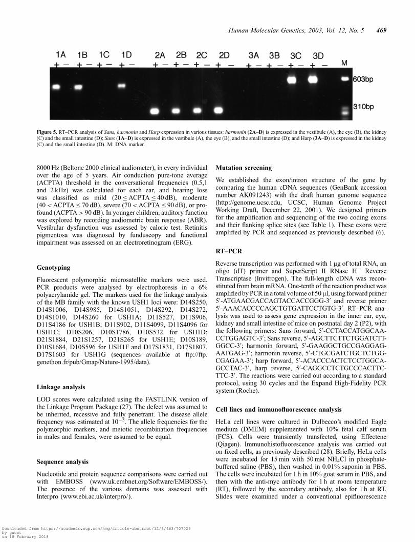
We then used RT–PCR to investigate the expression of Sans,Harp and the harmonin gene, in the inner ear, eye and kidney(Fig. 5). Interestingly, whereas both the genes associated withUSH1 were expressed in the inner ear and eye, no Harpexpression was detected in these sensory organs. In contrast, inthe kidney, we observed transcription of the Harp and
Figure 3. Four mutations in the SANS gene in three USH1G patients. The mutated sequence is shown in the upper panel and below the corresponding wild-typesequence. The deduced amino-acid sequence is indicated below the DNA sequence. (A) Affected individual in family JO-US1, homozygous for a 20-nucleotidedeletion in exon 2. (B) Affected individual in family MB, homozygous for a one-base insertion in exon 2 (393insG). (C) Affected individual 54. (1) Heterozygoussubstitution in exon 1 (C182T). (2) Heterozygous deletion in exon 2 (186–187delCA).
Human Molecular Genetics, 2003, Vol. 12, No. 5 467
Downloaded from https://academic.oup.com/hmg/article-abstract/12/5/463/707029by gueston 18 February 2018

harmonin genes but detected no Sans transcripts (Fig. 5). Thedifferential tissue expression of Sans and Harp suggests thatthe central region of Sans, which diverges from that of Harp,plays a specific role in the inner ear and the eye.
Sans, which is produced in the sensory hair cells (16), may beconnected to the actin cytoskeleton via its interaction withharmonin b, an isoform largely restricted to the inner ear (6).From our previous (15) and present studies, the followingpicture emerges: Sans interacts with harmonin, a scaffoldingprotein that itself binds to myosin VIIa and cadherin 23; theharmonin b isoform also binds actin filaments (see Fig. 6).Harmonin, myosin VIIa and cadherin 23 are detected in thegrowing hair bundles of the inner ear sensory cells (15,24).Harmonin b may anchor cadherin 23-containing interstereoci-liar links to the actin filament cores of growing stereocilia(Fig. 6) (15).
The results of this study suggest that Sans is involved in thefunctional network formed by harmonin, cadherin 23 andmyosin VIIa that is required for cohesion of the growing hair
bundle. Consistently, the js (Sans) mouse mutant displaysdisorganization of the hair bundle (26) similar to that observedin sh1 (myosin VIIa) (14) and v (cadherin 23) (12) mousemutants. The possible involvement in the network of proto-cadherin 15 (USH1F) and of the proteins underlying the otherforms of USH1 remains to be determined.
MATERIALS AND METHODS
Patients
Informed consent was obtained from adult subjects and fromthe parents of patients below the age of consent.
Clinical studies
Hearing loss was quantified by pure-tone audiometry with aerialand bone conduction at 250, 500, 1000, 2000, 4000 and
Figure 4. (A) Sans in transfected HeLa cells. Sans (green) is distributed throughout the cytoplasm. No association with actin filament-labelled structures (red) isobserved. (B–D) Sans associates with harmonin b. In HeLa cells producing harmonin b (B) and Sans (C), the two proteins show identical distributions throughoutthe cell (D). (E–G) Harmonin b connects Sans to F-actin. The presence of harmonin b clearly changes the distribution of Sans-labelled structures (green). The cellon the right produces Sans only whereas the cell on the left (asterisk) produces both Sans and harmonin b. In the cell on the right, Sans displays a punctate dis-tribution and no co-localization with actin filaments (red). In contrast, in cells producing both Sans and harmonin b (asterisk), the punctate or the long filamentousstructures labelled show the same distribution as actin (arrowheads). (H–J) Sans associates with harmonin a (H) and the GFP-tagged PDZ1 domain of harmonin (I),but not with GFP-tagged vezatin (J). In co-transfected HeLa cells, Sans immunolabelling (red) displayed a distribution identical to that of harmonin a (H) and GFP-tagged PDZ1 (I), throughout the cell cytoplasm (yellow). In contrast, no co-localization of Sans (red) with GFP-vezatin (green in J) was observed. Bars: 10mm.
468 Human Molecular Genetics, 2003, Vol. 12, No. 5
Downloaded from https://academic.oup.com/hmg/article-abstract/12/5/463/707029by gueston 18 February 2018
8000 Hz (Beltone 2000 clinical audiometer), in every individualover the age of 5 years. Air conduction pure-tone average(ACPTA) threshold in the conversational frequencies (0.5,1and 2 kHz) was calculated for each ear, and hearing losswas classified as mild (20�ACPTA� 40 dB), moderate(40<ACPTA� 70 dB), severe (70<ACPTA� 90 dB), or pro-found (ACPTA> 90 dB). In younger children, auditory functionwas explored by recording audiometric brain response (ABR).Vestibular dysfunction was assessed by caloric test. Retinitispigmentosa was diagnosed by funduscopy and functionalimpairment was assessed on an electroretinogram (ERG).
Genotyping
Fluorescent polymorphic microsatellite markers were used.PCR products were analysed by electrophoresis in a 6%polyacrylamide gel. The markers used for the linkage analysisof the MB family with the known USH1 loci were: D14S250,D14S1006, D14S985, D14S1051, D14S292, D14S272,D14S1010, D14S260 for USH1A; D11S527, D11S906,D11S4186 for USH1B; D11S902, D11S4099, D11S4096 forUSH1C; D10S206, D10S1786, D10S532 for USH1D;D21S1884, D21S1257, D21S265 for USH1E; D10S189,D10S1684, D10S596 for USH1F and D17S1831, D17S1807,D17S1603 for USH1G (sequences available at ftp://ftp.genethon.fr/pub/Gmap/Nature-1995/data).
Linkage analysis
LOD scores were calculated using the FASTLINK version ofthe Linkage Program Package (27). The defect was assumed tobe inherited, recessive and fully penetrant. The disease allelefrequency was estimated at 10�3. The allele frequencies for thepolymorphic markers, and meiotic recombination frequenciesin males and females, were assumed to be equal.
Sequence analysis
Nucleotide and protein sequence comparisons were carried outwith EMBOSS (www.uk.embnet.org/Software/EMBOSS/).The presence of the various domains was assessed withInterpro (www.ebi.ac.uk/interpro/).
Mutation screening
We established the exon/intron structure of the gene bycomparing the human cDNA sequences (GenBank accessionnumber AK091243) with the draft human genome sequence(http://genome.ucsc.edu, UCSC, Human Genome ProjectWorking Draft, December 22, 2001). We designed primersfor the amplification and sequencing of the two coding exonsand their flanking splice sites (see Table 1). These exons wereamplified by PCR and sequenced as previously described (6).
RT–PCR
Reverse transcription was performed with 1 mg of total RNA, anoligo (dT) primer and SuperScript II RNase H� ReverseTranscriptase (Invitrogen). The full-length cDNA was recon-stituted from brain mRNA. One-tenth of the reaction product wasamplified by PCR in a total volume of 50 ml, using forward primer50-ATGAACGACCAGTACCACCGGG-30 and reverse primer50-AAACACCCCAGCTGTGATTCCTGTG-30. RT–PCR ana-lysis was used to assess gene expression in the inner ear, eye,kidney and small intestine of mice on postnatal day 2 (P2), withthe following primers: Sans forward, 50-CCTACCATGGCAA-CCTGGAGTC-30; Sans reverse, 50-AGCTTCTTCTGGATCTT-GGCC-30; harmonin forward, 50-GAAGGCTGCCGAGGAG-AATGAG-30; harmonin reverse, 50-CTGCGATCTGCTCTGG-CGAGAA-30; harp forward, 50-ACACCCACTCTCCTGGCA-GCCTAC-30, harp reverse, 50-CAGGCCTCTGCCCACTTC-TTC-30. The reactions were carried out according to a standardprotocol, using 30 cycles and the Expand High-Fidelity PCRsystem (Roche).
Cell lines and immunofluorescence analysis
HeLa cell lines were cultured in Dulbecco’s modified Eaglemedium (DMEM) supplemented with 10% fetal calf serum(FCS). Cells were transiently transfected, using Effectene(Qiagen). Immunohistofluorescence analysis was carried outon fixed cells, as previously described (28). Briefly, HeLa cellswere incubated for 15 min with 50 mM NH4Cl in phosphate-buffered saline (PBS), then washed in 0.01% saponin in PBS.The cells were incubated for 1 h in 10% goat serum in PBS, andthen with the anti-myc antibody for 1 h at room temperature(RT), followed by the secondary antibody, also for 1 h at RT.Slides were examined under a conventional epifluorescence
Figure 5. RT–PCR analysis of Sans, harmonin and Harp expression in various tissues: harmonin (2A–D) is expressed in the vestibule (A), the eye (B), the kidney(C) and the small intestine (D); Sans (1A–D) is expressed in the vestibule (A), the eye (B), and the small intestine (D); and Harp (3A–D) is expressed in the kidney(C) and the small intestine (D). M: DNA marker.
Human Molecular Genetics, 2003, Vol. 12, No. 5 469
Downloaded from https://academic.oup.com/hmg/article-abstract/12/5/463/707029by gueston 18 February 2018

microscope (Leica) or a laser scanning confocal microscope,LSM-540 (Zeiss).
The following mouse monoclonal antibodies were used: anti-Myc (clone 9E10) (Santa Cruz); anti-a-tubulin; and anti-pancytokeratin (Sigma). Rhodamine-phalloidin (Sigma) stainingwas used to visualize actin filaments.
The full-length mouse cDNAs encoding harmonin isoformsa, b, and c subcloned into pcDNA (Invitrogen) have beendescribed elsewhere (15). cDNAs encoding truncated forms ofharmonin—PDZ1 (aa: 72–88), PDZ2 (189–307) and PDZ3(738–849)—were inserted into the pECFP vector (Clontech).The full-length mouse cDNA encoding Sans was inserted intothe myc-tagged pCMV vector (Stratagene).
ACKNOWLEDGEMENTS
We thank Marino Zerial (Dresden, Germany) for the gift of theGFP-Rab4 plasmid, J. Levilliers, J.-P. Hardelin, M. Leibovici,E. Verpy and S. Cure for critical reading of this manuscript,S. Nouaille for help with genotyping and S. Chardenoux fordrawing the figures. We also thank M. Leibovici and E. Verpyfor supplying us with mouse tissue RNAs. This work wassupported by grants from Fondation pour la Recherche
Medicale (ARS2000), INSERM/CNCPRST Sante Publique,Fondation Srittmatter (Retina France), A. and M. SuchertForschung contra Blindheit-Initiativ Usher syndrome and theEuropean Community (QLG2-CT-1999-00988).
REFERENCES
1. Otterstedde, C.R., Spandau, U., Blankenagel, A., Kimberling, W.J.and Reisser, C. (2001) A new clinical classification for Usher’s syndromebased on a new subtype of Usher’s syndrome type I. Laryngoscope,111, 84–86.
2. Petit, C. (2001) Usher syndrome: from genetics to pathogenesis.A. Rev. Genomics Hum. Genet., 2, 271–297.
3. Mustapha, M., Chouery, E., Torchard-Pagnez, D., Nouaille, S., Khrais, A.,Sayegh, F.N., Megarbane, A., Loiselet, J., Lathrop, M., Petit, C. et al.(2002) A novel locus for Usher syndrome type I, USH1G, maps tochromosome 17q24-25. Hum. Genet., 110, 348–350.
4. Weil, D., Blanchard, S., Kaplan, J., Guilford, P., Gibson, F., Walsh, J., Mburu, P.,Varela, A., Levilliers, J., Weston, M.D. et al. (1995) Defective myosin VIIAgene responsible for Usher syndrome type 1B. Nature, 374, 60–61.
5. Bitner-Glindzicz, M., Lindley, K.J., Rutland, P., Blaydon, D., Smith, V.V.,Milla, P.J., Hussain, K., Furth-Lavi, J., Cosgrove, K.E., Shepherd, R.M.et al. (2000) A recessive contiguous gene deletion causing infantilehyperinsulinism, enteropathy and deafness identifies the Usher type 1Cgene. Nat. Genet., 26, 56–60.
Figure 6. Schematic diagram illustrating the possible interactions of harmonin b with myosin VIIa, cadherin 23, Sans and F-actin (25). The cytodomain of cadherin23 (USH1D), which is located in the membrane of stereocilia, interacts with harmonin b (USH1C) via the PDZ2 domain. The actin-based motor protein, myosinVIIa (USH1B), also binds to harmonin b, via the PDZ1 domain. Harmonin b also binds actin filaments via its C-terminal region, and is therefore thought to connectcadherin 23 stereocilia laterally to the stereocilia microfilaments. Sans (USH1G) binds harmonin b, via the PDZ1 domain. PDZ, post synaptic density, disc large,zonula occludens domains; CC, coiled-coil domain; PST, proline, serine, threonine (PST)-rich region; Ank, ankyrin repeats; SAM, sterile alpha motif; FERM, 4.1,ezrin, radixin, moesin; MyTH4, myosin tail homology 4; SH3, src homology-3; IQ, isoleucine-glutamine motifs; EC, extracellular cadherin repeats.
470 Human Molecular Genetics, 2003, Vol. 12, No. 5
Downloaded from https://academic.oup.com/hmg/article-abstract/12/5/463/707029by gueston 18 February 2018

6. Verpy, E., Leibovici, M., Zwaenepoel, I., Liu, X.-Z., Gal, A., Salem, N.,Mansour, A., Blanchard, S., Kobayashi, I., Keats, B.J.B. et al. (2000)A defect in harmonin, a PDZ domain-containing protein expressed in theinner ear sensory hair cells, underlies Usher syndrome type 1C.Nat. Genet., 26, 51–55.
7. Bolz, H., von Brederlow, B., Ramirez, A., Bryda, E.C., Kutsche, K.,Nothwang, H.G., Seeliger, M., Salcedo Cabrera, M.d.C., Vila, M.C.et al. (2001) Mutations of CDH23, encoding a new member of thecadherin gene family, causes Usher syndrome type 1D. Nat. Genet.,27, 108–112.
8. Bork, J.M., Peters, L.M., Riazuddin, S., Bernstein, S.L.,Ahmed, Z.M., Ness, S.L., Polomeno, R., Ramesh, A., Schloss, M.,Srisailpathy, C.R.S. et al. (2001) Usher syndrome 1D and non-syndromic autosomal recessive deafness DFNB12 are caused by allelicmutations of the novel cadherin-like gene CDH23. Am. J. Hum. Genet.,68, 26–37.
9. Ahmed, Z.M., Riazuddin, S., Bernstein, S.L., Ahmed, Z., Khan, S.,Griffith, A.J., Morell, R.J., Friedman, T.B., Riazuddin, S. and Wilcox, E.R.(2001) Mutations of the protocadherin gene PCDH15 cause Ushersyndrome type 1F. Am. J. Hum. Genet., 69, 25–34.
10. Alagramam, K.N., Yuan, H., Kuehn, M.H., Murcia, C.L.,Wayne, S., Srisailpathy, R., Lowry, R.B., Knaus, R., Van Laer, L.,Bernier, F.P. et al. (2001) Mutations in the novel protocadherinPCDH15 cause Usher syndrome type 1F. Hum. Mol. Genet.,10, 1709–1718.
11. Gibson, F., Walsh, J., Mburu, P., Varela, A., Brown, K.A.,Antonio, M., Beisel, K.W., Steel, K.P. and Brown, S.D.M. (1995) Atype VII myosin encoded by the mouse deafness gene Shaker-1. Nature,374, 62–64.
12. Di Palma, F., Holme, R.H., Bryda, E.C., Belyantseva, I.A., Pellegrino, R.,Kachar, B., Steel, K.P. and Noben-Trauth, K. (2001) Mutations in Cdh23,encoding a new type of cadherin, cause stereocilia disorganization inwaltzer, the mouse model for Usher syndrome type 1D. Nat Genet.,27, 103–107.
13. Alagramam, K.N., Murcia, C.L., Kwon, H.Y., Pawlowski, K.S.,Wright, C.G. and Woychik, R.P. (2001) The mouse Ames waltzer hearing-loss mutant is caused by mutation of Pcdh15, a novel protocadherin gene.Nat. Genet., 27, 99–102.
14. Self, T., Mahony, M., Fleming, J., Walsh, J., Brown, S.D. and Steel, K.P.(1998) Shaker-1 mutations reveal roles for myosin VIIA in bothdevelopment and function of cochlear hair cells. Development,125, 557–566.
15. Boeda, B., El-Amraoui, A., Bahloul, A., Goodyear, R., Daviet, L.,Blanchard, S., Perfettini, I., Fath, K.R., Shorte, S., Reiners, J. et al. (2002)Myosin VIIa, harmonin and cadherin 23, three Usher I gene products thatcooperate to shape the sensory hair cell bundle. EMBO J., 21, 6689–6699.
16. Kikkawa, Y., Shitara, H., Wakana, S., Kohara, Y., Takada, T., Okamoto, M.,Taya, C., Kamiya, K., Yoshikawa, Y., Tokano, H., Kitamura, K., Shimizu, K.,Wakabayashi, Y., Shiroishi, T., Kominami, R. and Yonekawa, H. (2003)Mutations in a new scaffold protein Sans cause deafness in Jackson shakermice. Hum. Mol. Genet., 12, 453–461.
17. Roderick, T.H. (1972) Position of Jackson shaker. Mouse News Lett., 47, 37.18. Sedgwick, S.G. and Smerdon, S.J. (1999) The ankyrin repeat: a diversity of
interactions on a common structural framework. Trends Biochem. Sci.,24, 311–316.
19. Ponting, C.P. (1995) SAM: a novel motif in yeast sterile and Drosophilapolyhomeotic proteins. Protein Sci., 4, 1928–1930.
20. Schultz, J., Ponting, C.P., Hofmann, K. and Bork, P. (1997) SAM as aprotein interaction domain involved in developmental regulation. ProteinSci., 6, 249–253.
21. Stapleton, C., Balan, I., Pawson, T. and Sicheri, F. (1999) The crystalstructure of an Eph receptor SAM domain reveals a mechanism for modulardimerization. Nat. Struct. Biol., 6, 44–49.
22. Morell, R.J., Friderici, K.H., Wei, S., Elfenbein, J.L., Friedman, T.B. andFisher, R.A. (2000) A new locus for late-onset, progressive, hereditaryhearing loss DFNA20 maps to 17q25. Genomics, 63, 1–6.
23. Yang, T. and Smith, R. (2000) A novel locus DFNA26 maps tochromosome 17q25 in two unrelated families with progressive autosomaldominant hearing loss. Am. J. Hum. Genet., 67, 300.
24. Sheng, M. and Sala, C. (2001) PDZ domains and the organization ofsupramolecular complexes. A. Rev. Neurosci., 24, 1–29.
25. Siemens, J., Kazmierczak, P., Reynolds, A., Sticker, M., Littlewood-Evans, A.and Muller, U. (2002) The Usher syndrome proteins cadherin 23 andharmonin form a complex by means of PDZ-domain interactions.Proc. Natl Acad. Sci. USA, 99, 14946–14951.
26. Kitamura, K., Kakoi, H., Yoshikawa, Y. and Ochikubo, F. (1992)Ultrastructural findings in the inner ear of Jackson shaker mice.Acta Otolaryngol. (Stockh.), 112, 622–627.
27. Lathrop, G.M., Lalouel, J.M., Julier, C. and Ott, J. (1985) Multilocuslinkage analysis in humans: detection of linkage and estimation ofrecombination. Am. J. Hum. Genet., 37, 482–498.
28. Kussel-Andermann, P., El-Amraoui, A., Safieddine, S., Nouaille, S.,Perfettini, I., Lecuit, M., Cossart, P., Wolfrum, U. and Petit, C. (2000)Vezatin, a novel transmembrane protein, bridges myosin VIIA to thecadherin/catenins complex. EMBO J., 19, 6020–6029.
Human Molecular Genetics, 2003, Vol. 12, No. 5 471
Downloaded from https://academic.oup.com/hmg/article-abstract/12/5/463/707029by gueston 18 February 2018