Embed Size (px)
Citation preview
Urine
1Dr.Mohamed Saad Daoud
Reference Books:
Urinanalysis and body fluids (Susan King Strasinger- Marjorie Schaub De Lorenzo) Fifth edition
Fundamentals of Clinical Chemistry (Tietz) Sixth edition
2Dr.Mohamed Saad Daoud
Urine:
Sterile fluid (in the absence of a disease condition) is
secreted by the kidneys through a process called urination
and excreted through the urethra.
Urine contains a whole range of substances that the body
has no need for. It might contain chemicals that are
potentially harmful but the levels are going to be very low
when expelled by the body, and even lower when diluted by
water.3Dr.Mohamed Saad Daoud
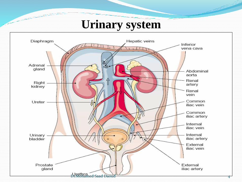
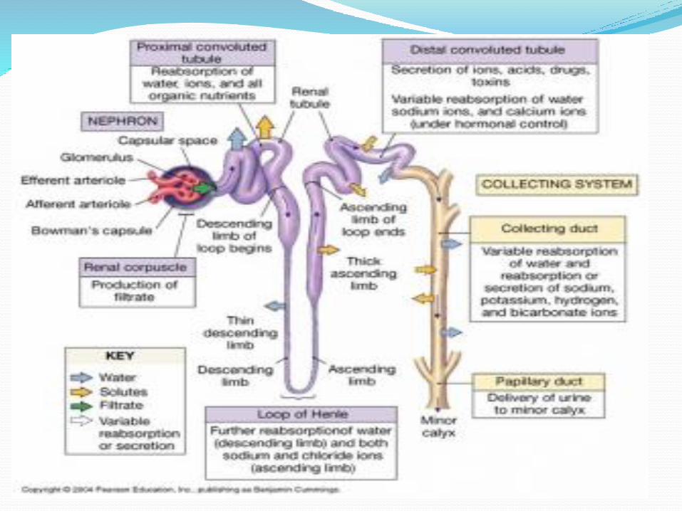
Urinary system
4Dr.Mohamed Saad Daoud
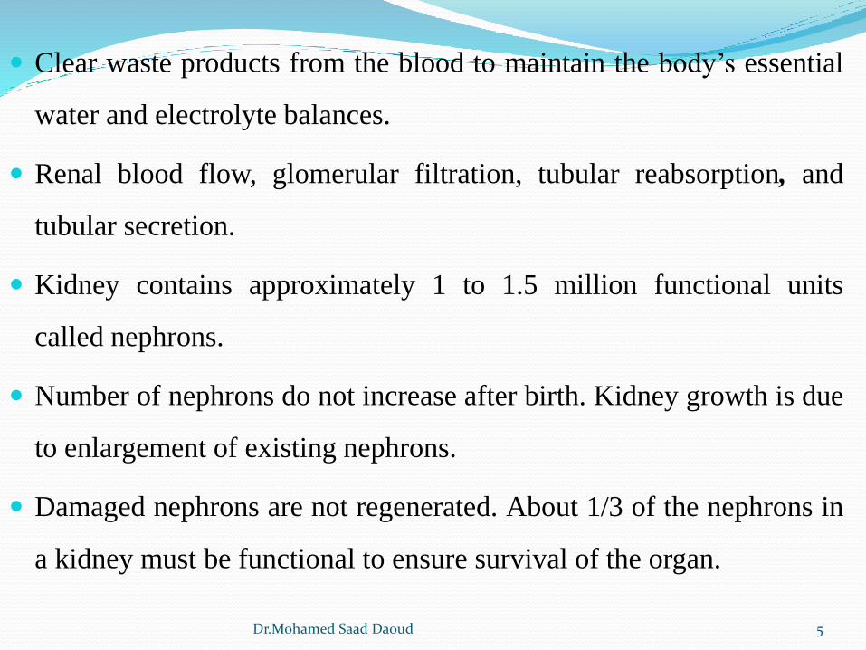
Clear waste products from the blood to maintain the body’s essential
water and electrolyte balances.
Renal blood flow, glomerular filtration, tubular reabsorption, and
tubular secretion.
Kidney contains approximately 1 to 1.5 million functional units
called nephrons.
Number of nephrons do not increase after birth. Kidney growth is due
to enlargement of existing nephrons.
Damaged nephrons are not regenerated. About 1/3 of the nephrons in
a kidney must be functional to ensure survival of the organ.
5Dr.Mohamed Saad Daoud
6Dr.Mohamed Saad Daoud
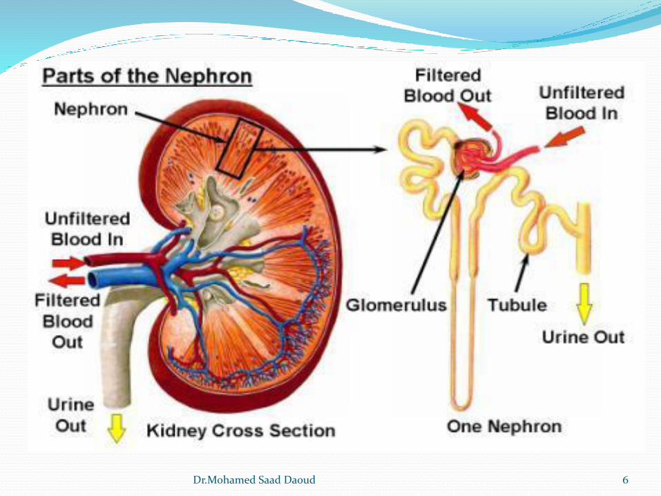
Urine formation
The kidneys continuously form urine as an ultra-filtrate of
plasma.
Reabsorption of water and filtered substances essential to
body function (170,000 mL of filtered plasma urine give 1200
mL urine per day).
Tubular secretion serves two major functions: elimination of
waste products not filtered by the glomerulus and regulation
of the acid-base balance in the body through the secretion of
hydrogen ions.
7Dr.Mohamed Saad Daoud
8Dr.Mohamed Saad Daoud
Glomerular Filtration
Glomerulus serves as a nonselective filter of plasma substances
Factors affected on filtration:
The presence of hydrostatic pressure resulting from the smaller
size of the efferent arteriole and the glomerular capillaries
enhances filtration.
Hydrostatic pressure opposite pressures from the fluid within
Bowman’s capsule and the oncotic pressure of unfiltered plasma
proteins in the glomerular capillaries.
The glomerular apparatus maintains the glomerular blood
pressure at a relatively constant rate by increasing or decreasing
the size of the afferent arteriole.
9Dr.Mohamed Saad Daoud
Action of the renin-angiotensin-aldosterone system
Dilation of the afferent arteriole and constriction of the efferent
arteriole.
Stimulation of sodium reabsorption in the proximal convoluted
tubule.
Triggers the adrenal cortex to release the sodium retaining
hormone, aldosterone, to cause reabsorption of sodium and
excretion of potassium in the distal convoluted tubule
and collecting duct.
Triggers release of antidiuretic hormone by the hypothalmus to
stimulate water reabsorption in the collecting duct.
10Dr.Mohamed Saad Daoud
Glomerular mechanisms, every minute approximately two to
three million glomeruli filter approximately 120 ml of water-
containing low-molecular weight substances.
Tubular Reabsorption
The plasma ultrafiltrate enters the proximal convoluted
tubule, the nephrons, through cellular transport
mechanisms, begin reabsorbing these essential substances
and water.
The cellular mechanisms involved in tubular reabsorption
are termed active and passive transport.
11Dr.Mohamed Saad Daoud
For active transport to occur, the substance to be
reabsorbed must combine with a carrier protein contained
in the membranes of the renal tubular cells.
Passive transport is the movement of molecules across a
membrane as a result of differences in their concentration
or electrical potential on opposite sides of the membrane.
12Dr.Mohamed Saad Daoud
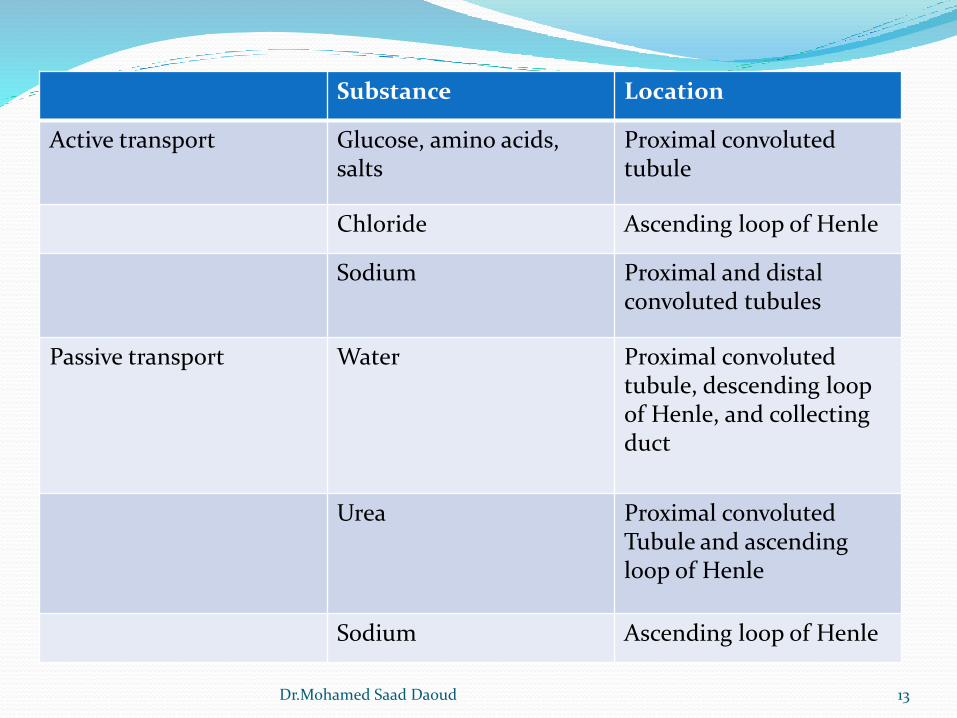
Substance Location
Active transport Glucose, amino acids, salts
Proximal convolutedtubule
Chloride Ascending loop of Henle
Sodium Proximal and distal convoluted tubules
Passive transport Water Proximal convolutedtubule, descending loop of Henle, and collectingduct
Urea Proximal convolutedTubule and ascendingloop of Henle
Sodium Ascending loop of Henle
13Dr.Mohamed Saad Daoud
Renal threshold
When the plasma concentration of a substance that is
normally completely reabsorbed reaches an abnormally
high level, the filtrate concentration exceeds the maximal
reabsorptive capacity (Tm) of the tubules, and the
substance begins appearing in the urine. The plasma
concentration at which active transport stops is termed the
renal threshold.
For glucose, the renal threshold is 160 to 180 mg/dL,
and glucose appears in the urine when the plasma
concentration reaches this level.14Dr.Mohamed Saad Daoud
Tubular Secretion
Elimination of waste products not filtered by the glomerulus
(foreign substances, such as medications).
The major site for removal of these nonfiltered substances is
the proximal convoluted tubule.
Regulation of the acid-base balance in the body through the
secretion of hydrogen ions.
15Dr.Mohamed Saad Daoud
Acid-Base Balance
The normal blood pH of 7.4, the blood must buffer and
eliminate the excess acid formed by dietary intake and
body metabolism.
The buffering capacity of the blood depends on
bicarbonate (HCO3-) ions, which are readily filtered by
the glomerulus and must be expediently returned to the
blood to maintain the proper pH.
A disruption in these secretory functions can result in
metabolic acidosis or renal tubular acidosis, the inability
to produce an acid urine.
16Dr.Mohamed Saad Daoud
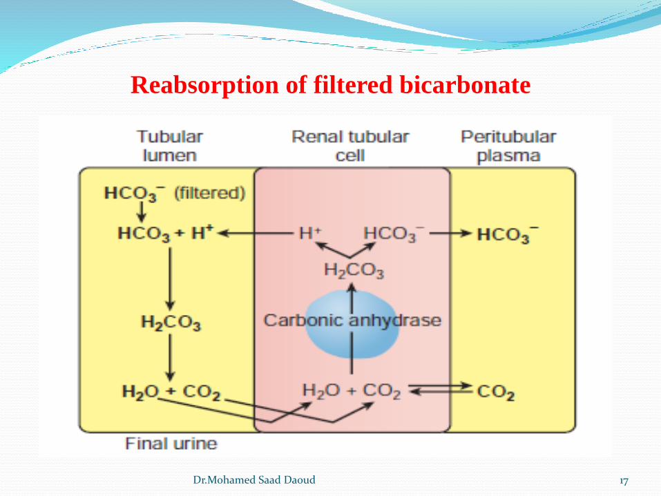
Reabsorption of filtered bicarbonate
17Dr.Mohamed Saad Daoud
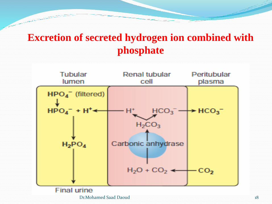
Excretion of secreted hydrogen ion combined with
phosphate
18Dr.Mohamed Saad Daoud
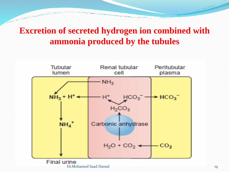
Excretion of secreted hydrogen ion combined with
ammonia produced by the tubules
19Dr.Mohamed Saad Daoud
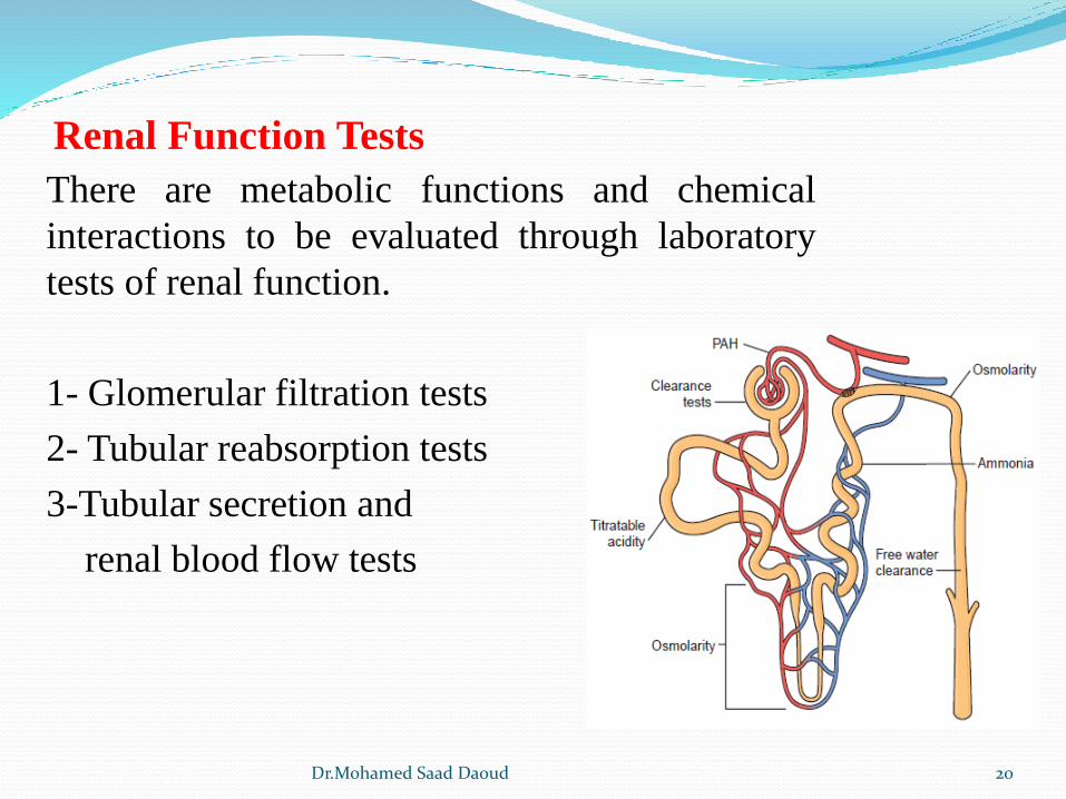
Renal Function Tests
There are metabolic functions and chemical
interactions to be evaluated through laboratory
tests of renal function.
1- Glomerular filtration tests
2- Tubular reabsorption tests
3-Tubular secretion and
renal blood flow tests
20Dr.Mohamed Saad Daoud
Glomerular Filtration Tests
The standard test used to measure the filtering capacity of
the glomeruli is the clearance test.
A clearance test measures the rate at which the kidneys are
able to remove substance from the blood.
the substance analyzed must be one that is neither
reabsorbed nor secreted by the tubules.
the stability of the substance in urine during a possible
24-hour collection period
the consistency of the plasma level the substance’s
availability to the body, and the availability of tests for
analysis of the substance.
21Dr.Mohamed Saad Daoud
Creatinine Clearance
Creatinine, a waste product of muscle metabolism that is
normally found at a relatively constant level in the blood,
provides the laboratory with an endogenous procedure for
evaluating glomerular function.
The GFR is reported in mL/min; therefore, determining the
number of milliliters of plasma from which the clearance
substance (creatinine) is completely removed during 1
minute is necessary.
22Dr.Mohamed Saad Daoud
Procedure
Urine volume in mL (V), urine creatinineconcentration in mg/dL (U), and plasma creatinineconcentration in mg/dL (P).
The urine volume is calculated by dividing the numberof milliliters in the specimen by the number ofminutes used to collect the specimen (V mL/min).
The plasma and urine concentrations are determinedby chemical testing.
The standard formula used to calculate the millilitersof plasma cleared per minute (C) is: C = UV/P
23Dr.Mohamed Saad Daoud
Normal creatinine clearance values 120 mL/min (men
107 to 139 mL/min; women, 87 to 107 mL/min). The
normal plasma creatinine is 0.5 to 1.5 mg/dL.
These normal values take into account variations in size
and muscle mass. Values are considerably lower in older
people, however, and an adjustment may also have to be
made to the calculation when dealing with body sizes that
deviate greatly from 1.73 m2 of surface, such as with
children.
24Dr.Mohamed Saad Daoud
To adjust a clearance for body size, the formula is:
C= UV/P x 1.73/A
with A being the actual body size in square meters of
surface. The actual body size may be calculated as:
log A= (0.425 x log weight)+ (0.725 x log height) -2.144
25Dr.Mohamed Saad Daoud
Clinical Significanse
The GFR is determined by the number of functioningnephrons and also by the functional capacity of thesenephrons.
Determination the extent of nephron damage in knowncases of renal disease, to monitor the effectiveness oftreatment designed to prevent further nephron damage.
Determination the feasibility of administeringmedications, which can build up to dangerous bloodlevels if the GFR is markedly reduced.
26Dr.Mohamed Saad Daoud
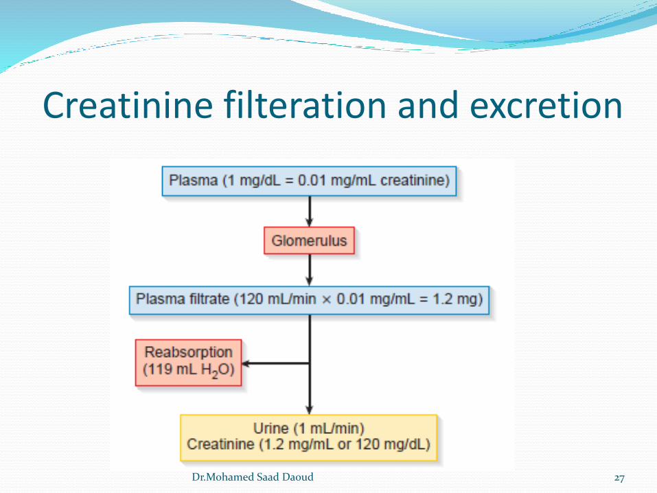
Creatinine filteration and excretion
27Dr.Mohamed Saad Daoud
Tubular Reabsorption Tests
Tests to determine the ability of the tubules to reabsorb the
essential salts and water that have been non selectively
filtered by the glomerulus are called concentration tests.
the ultrafiltrate that enters the tubules has a specific gravity
of 1010
Osmolarity
Specific gravity depends on the number of particles present
in a solution and the density of these particles.
Renal concentrating ability, monitoring the course of renal
disease, monitoring fluid and electrolyte therapy.
28Dr.Mohamed Saad Daoud
Establishing the differential diagnosis of hypernatremia and
hyponatremia, and evaluating the secretion of and renal response
to ADH. These evaluations may require determination of serum in
addition to urine osmolarity
Normal serum osmolarity values (275 -300 mOsm).
Normal values for urine osmolarity are difficult to establish,
because factors such as fluid intake and exercise can greatly
influence the urine concentration. Values can range from 50 to
1400 mOsm.
The ratio of urine to serum osmolarity should be at least 1:1; after
controlled fluid intake, it should reach 3:1
29Dr.Mohamed Saad Daoud
Tubular Secretion and Renal Blood Flow Tests
The test most commonly associated with tubular secretion
and renal blood flow is the p-aminohippuric acid (PAH)
test.
Excretion of the dye phenolsulfonphthalein (PSP) was
used to evaluate these functions.
PAH test
To measure the exact amount of blood flowing through the
kidney, it is necessary to use a substance that is
completely removed from the blood (plasma) each time it
comes in contact with functional renal tissue.
30Dr.Mohamed Saad Daoud
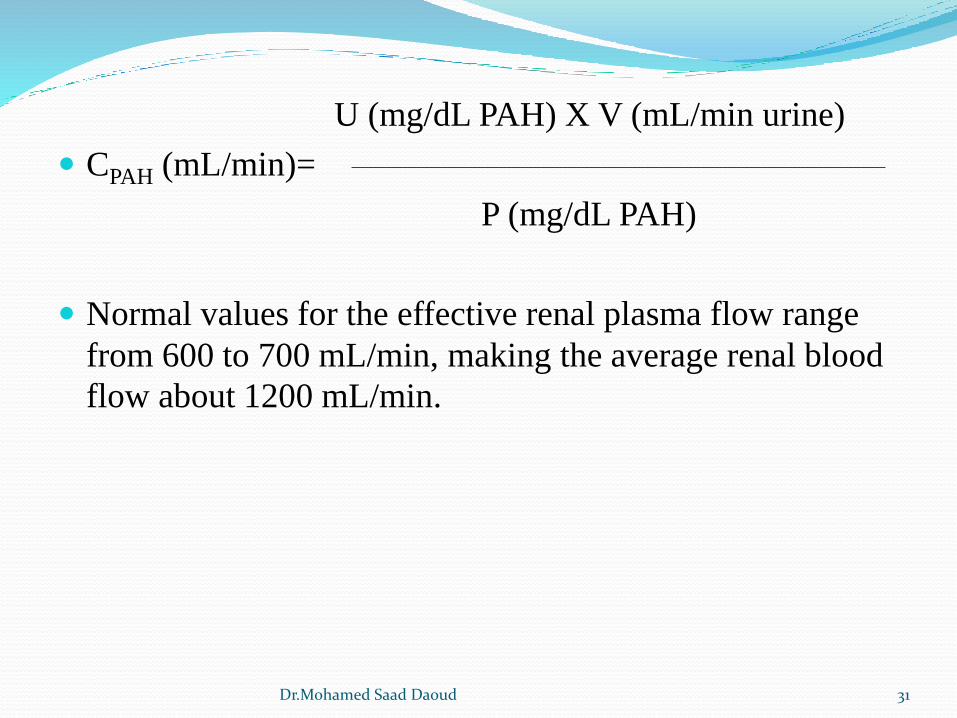
U (mg/dL PAH) X V (mL/min urine)
CPAH (mL/min)=
P (mg/dL PAH)
Normal values for the effective renal plasma flow range
from 600 to 700 mL/min, making the average renal blood
flow about 1200 mL/min.
31Dr.Mohamed Saad Daoud

Urine analysis
Purpose
General evaluation of health
Diagnosis of disease or disorders of the kidneys or urinary
tract
Diagnosis of other systemic disease that affect kidney
function
Monitoring of patients with diabetes
Screening for drug abuse
32Dr.Mohamed Saad Daoud
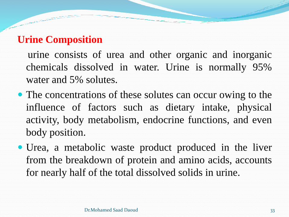
Urine Composition
urine consists of urea and other organic and inorganic
chemicals dissolved in water. Urine is normally 95%
water and 5% solutes.
The concentrations of these solutes can occur owing to the
influence of factors such as dietary intake, physical
activity, body metabolism, endocrine functions, and even
body position.
Urea, a metabolic waste product produced in the liver
from the breakdown of protein and amino acids, accounts
for nearly half of the total dissolved solids in urine.
33Dr.Mohamed Saad Daoud
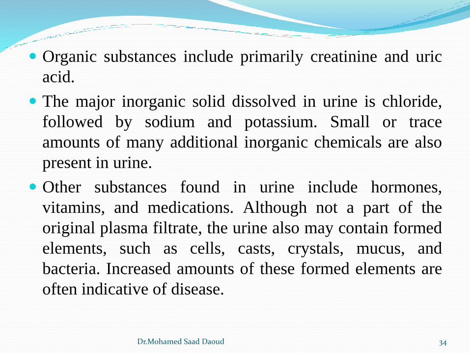
Organic substances include primarily creatinine and uric
acid.
The major inorganic solid dissolved in urine is chloride,
followed by sodium and potassium. Small or trace
amounts of many additional inorganic chemicals are also
present in urine.
Other substances found in urine include hormones,
vitamins, and medications. Although not a part of the
original plasma filtrate, the urine also may contain formed
elements, such as cells, casts, crystals, mucus, and
bacteria. Increased amounts of these formed elements are
often indicative of disease.
34Dr.Mohamed Saad Daoud
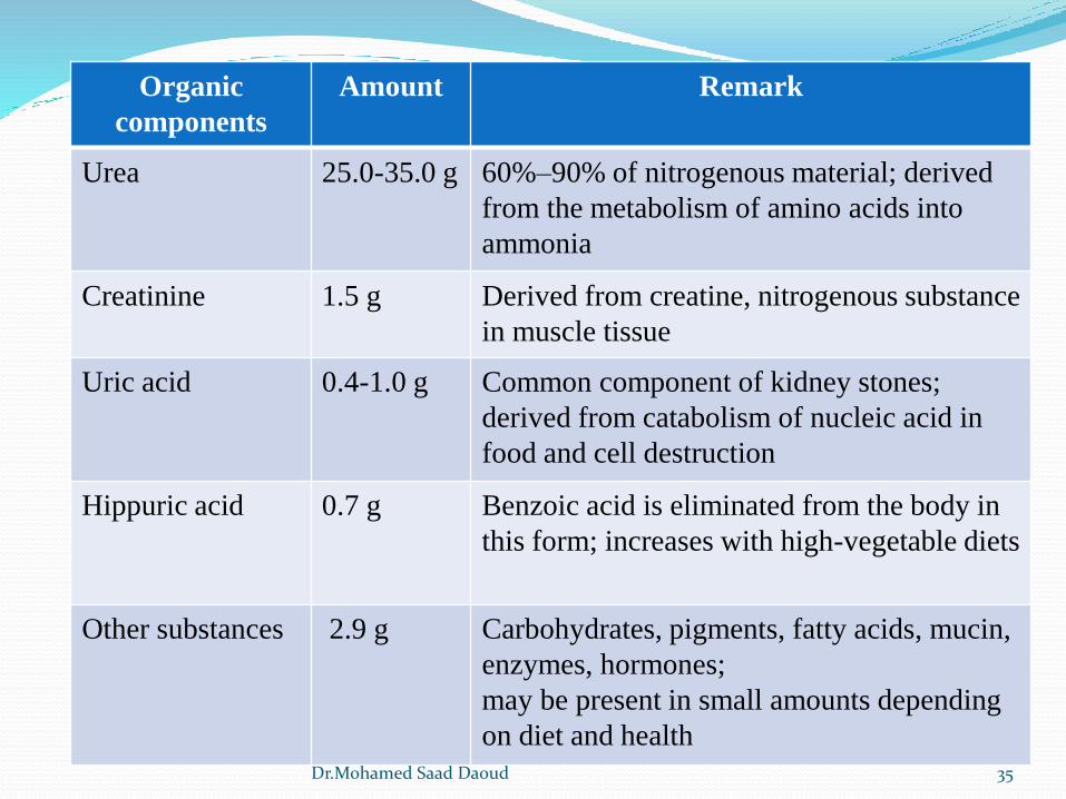
Organic
components
Amount Remark
Urea 25.0-35.0 g 60%–90% of nitrogenous material; derived
from the metabolism of amino acids into
ammonia
Creatinine 1.5 g Derived from creatine, nitrogenous substance
in muscle tissue
Uric acid 0.4-1.0 g Common component of kidney stones;
derived from catabolism of nucleic acid in
food and cell destruction
Hippuric acid 0.7 g Benzoic acid is eliminated from the body in
this form; increases with high-vegetable diets
Other substances 2.9 g Carbohydrates, pigments, fatty acids, mucin,
enzymes, hormones;
may be present in small amounts depending
on diet and health35Dr.Mohamed Saad Daoud
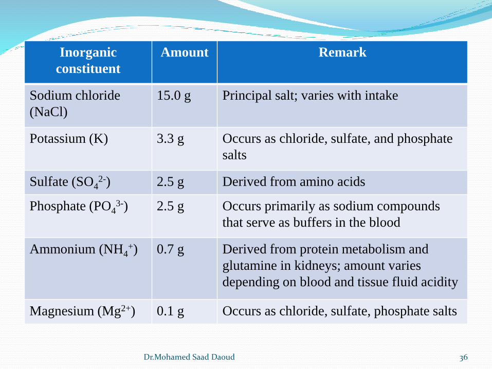
Inorganic
constituent
Amount Remark
Sodium chloride
(NaCl)
15.0 g Principal salt; varies with intake
Potassium (K) 3.3 g Occurs as chloride, sulfate, and phosphate
salts
Sulfate (SO42-) 2.5 g Derived from amino acids
Phosphate (PO43-) 2.5 g Occurs primarily as sodium compounds
that serve as buffers in the blood
Ammonium (NH4+) 0.7 g Derived from protein metabolism and
glutamine in kidneys; amount varies
depending on blood and tissue fluid acidity
Magnesium (Mg2+) 0.1 g Occurs as chloride, sulfate, phosphate salts
36Dr.Mohamed Saad Daoud
Urine Volume
Urine volume depends on the amount of water that the kidneys
excrete.
Factors that influence urine volume include fluid intake,
fluid loss from non renal sources, variations in the secretion of
antidiuretic hormone, and need to excrete increased amounts of
dissolved solids, such as glucose or salts.
Normal daily urine output is usually 1200 to 1500 ml, a
range of 600 to 2000 ml is considered normal.
37Dr.Mohamed Saad Daoud
Oliguria: A decrease in urine output
Less than 1 ml/kg/hr in infants
less than 0.5 ml/kg/hr in children
less than 400 ml/day in adults
Causes of oliguria: When the body enters a state of dehydration
as a result of excessive water loss from vomiting, diarrhea,
perspiration, or severe burns
Oliguria leading to anuria
Anuria: Cessation of urine flow, may result from any serious
damage to the kidneys or from a decrease in the flow of blood
to the kidneys.
38Dr.Mohamed Saad Daoud
Nocturia: An increase in the nocturenal excretion of urine, the
kidneys excrete two to three times more urine during the night
than during the day.
Polyuria: an increase in daily urine volume
greater than 2.5 l ml/kg/day in adults
Greater than 2.5–3 ml/kg/day in children
Diabetes mellitus and diabetes insipidus
Induced by diuretics, caffeine, or alcohol, all of which suppress
the secretion of antidiuretic hormone.
39Dr.Mohamed Saad Daoud
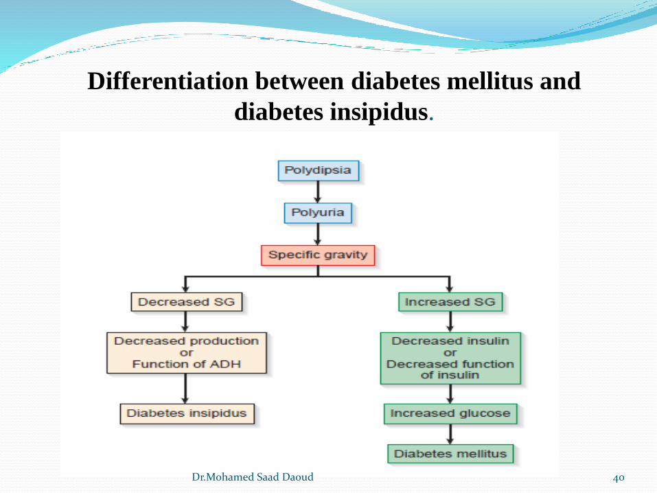
Differentiation between diabetes mellitus and
diabetes insipidus.
40Dr.Mohamed Saad Daoud
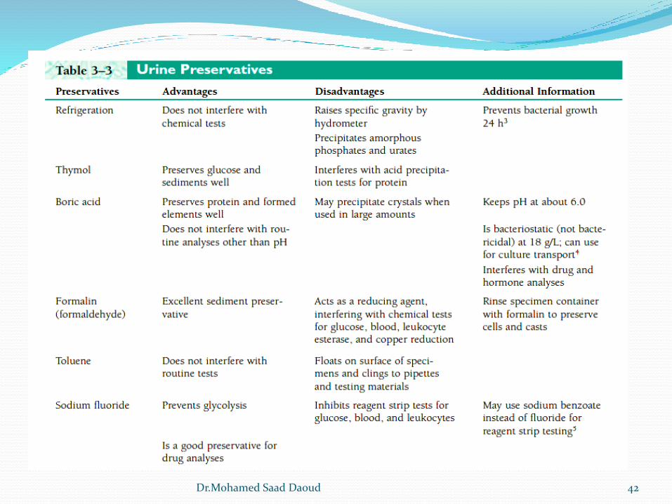
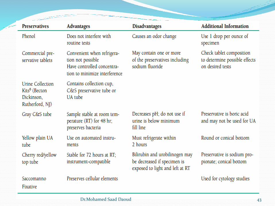
Urine Collection
Specimens must be collected in clean, dry and disposable
containers.
Individually packaged sterile containers with secure closures
should be used for microbiologic urine studies. Sterile
containers are also suggested if more than 2 hours elapse
Following collection, specimens should be delivered to the
laboratory and tested within 2 hours. A specimen that cannot be
delivered and tested within 2 hours should be refrigerated or
have an appropriate chemical preservative added.
41Dr.Mohamed Saad Daoud
42Dr.Mohamed Saad Daoud
43Dr.Mohamed Saad Daoud
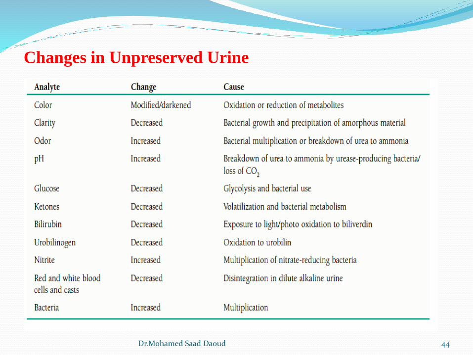
Changes in Unpreserved Urine
44Dr.Mohamed Saad Daoud
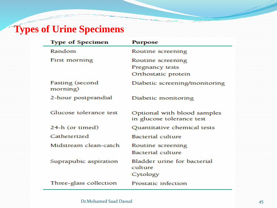
Types of Urine Specimens
45Dr.Mohamed Saad Daoud
The physical examination of urine includes the determination of the
urine color, clarity, and specific gravity
Color:
The color of urine varies from almost colorless to black. These
variations may be due to normal metabolic functions, physical
activity, ingested materials, or pathologic conditions.
Normal Urine Color
The yellow color of urine is caused by the presence of a pigment
named urochrome (product of endogenous metabolism)
Physical examination
46Dr.Mohamed Saad Daoud
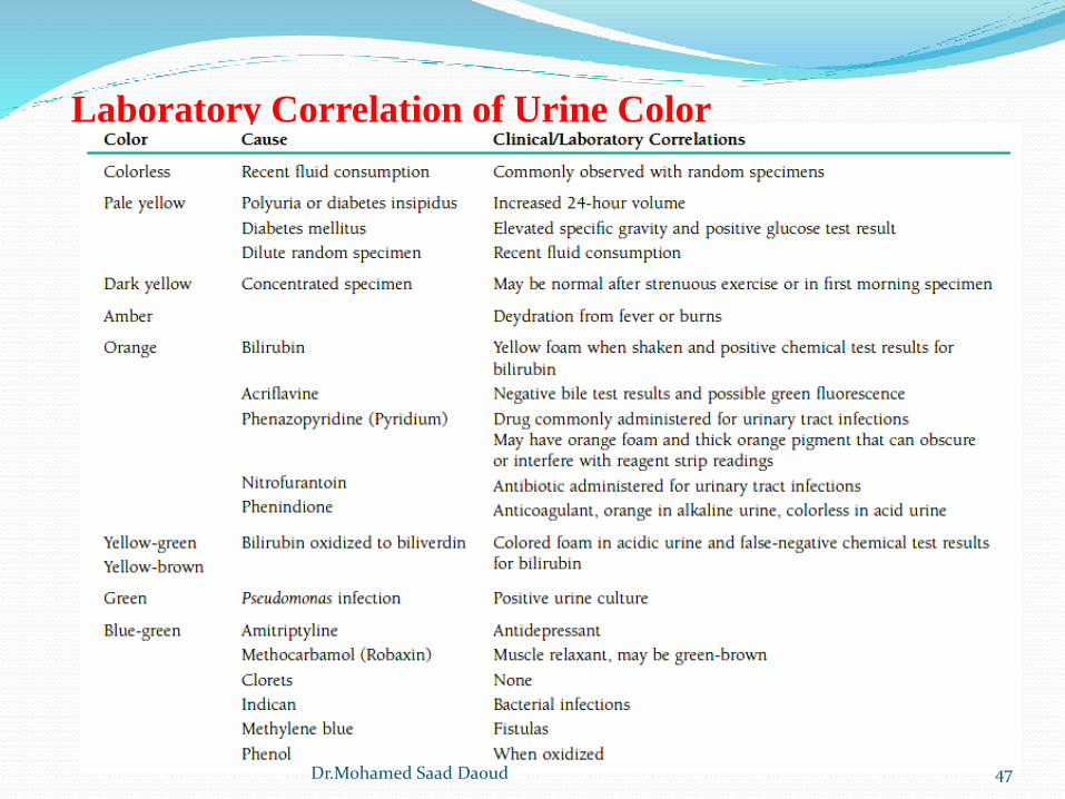
Laboratory Correlation of Urine Color
47Dr.Mohamed Saad Daoud
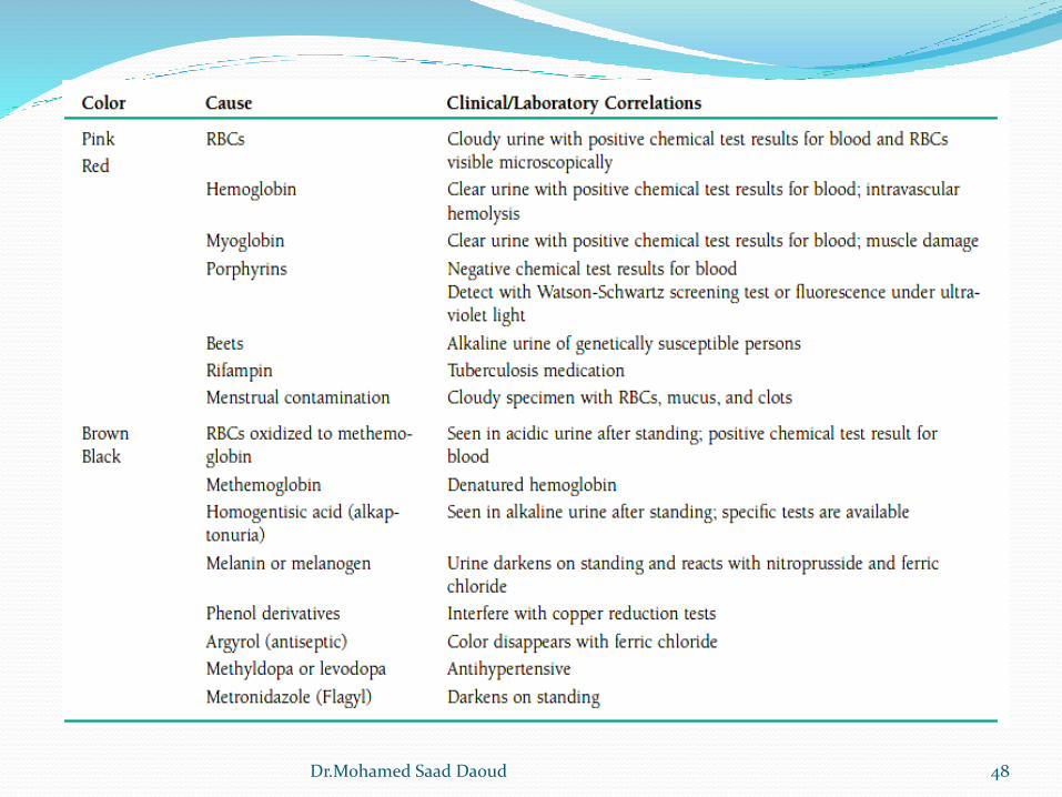
48Dr.Mohamed Saad Daoud
Clarity:
General term that refers to the transparency /turbidity of a urine
specimen.
Freshly voided normal urine is usually clear, particularly if it is a
midstream clean-catch specimen. Precipitation of amorphous
phosphates and carbonates may cause a white cloudiness.
The presence of squamous epithelial cells and mucus,
particularly in specimens from women, can result in a hazy but
normal urine.
49Dr.Mohamed Saad Daoud
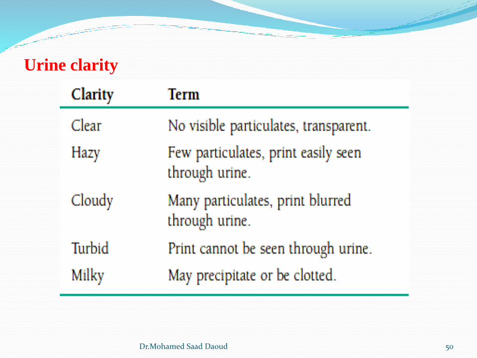
Urine clarity
50Dr.Mohamed Saad Daoud
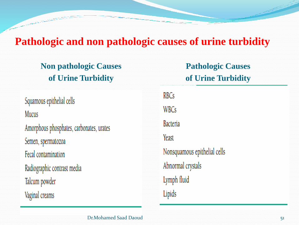
Pathologic and non pathologic causes of urine turbidity
Non pathologic Causes
of Urine Turbidity
Pathologic Causes
of Urine Turbidity
51Dr.Mohamed Saad Daoud
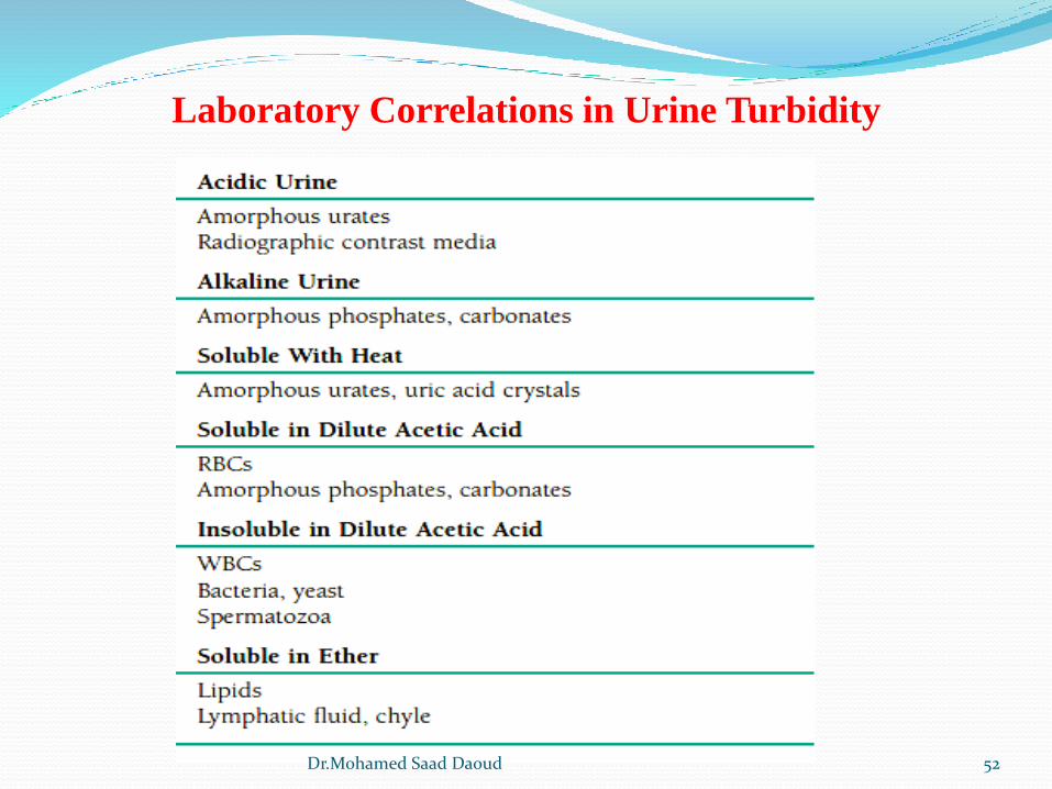
Laboratory Correlations in Urine Turbidity
52Dr.Mohamed Saad Daoud
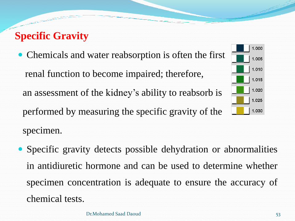
Specific Gravity
Chemicals and water reabsorption is often the first
renal function to become impaired; therefore,
an assessment of the kidney’s ability to reabsorb is
performed by measuring the specific gravity of the
specimen.
Specific gravity detects possible dehydration or abnormalities
in antidiuretic hormone and can be used to determine whether
specimen concentration is adequate to ensure the accuracy of
chemical tests.
53Dr.Mohamed Saad Daoud
54Dr.Mohamed Saad Daoud
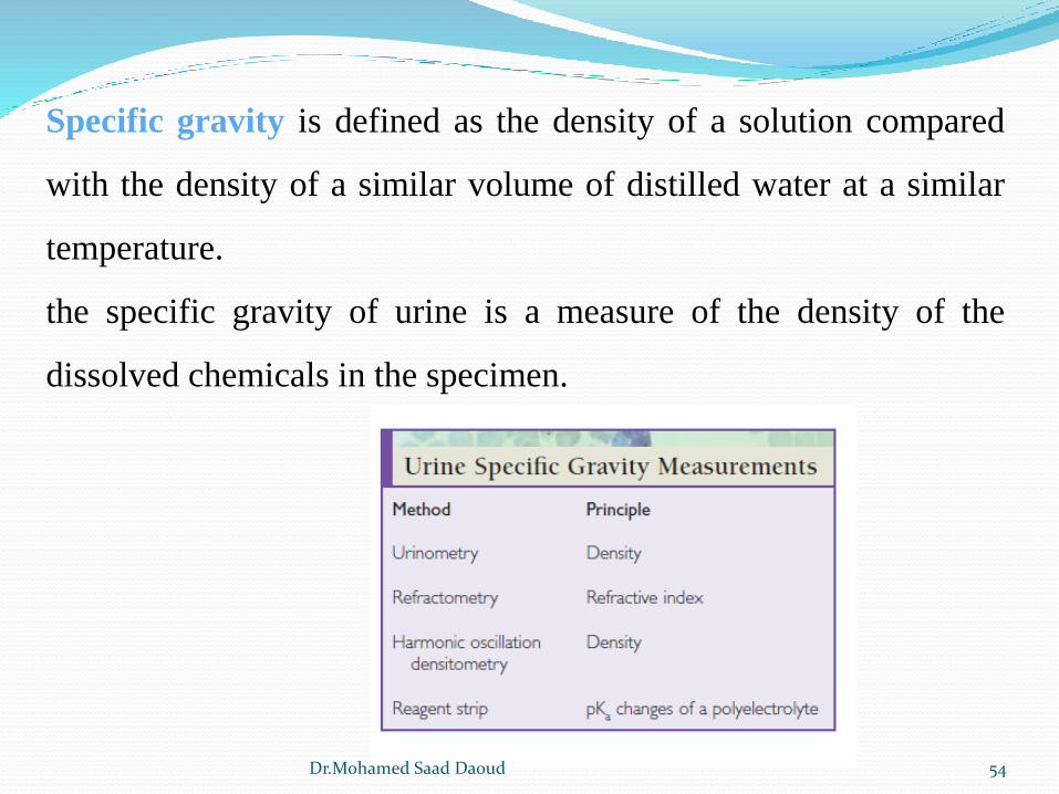
Specific gravity is defined as the density of a solution compared
with the density of a similar volume of distilled water at a similar
temperature.
the specific gravity of urine is a measure of the density of the
dissolved chemicals in the specimen.
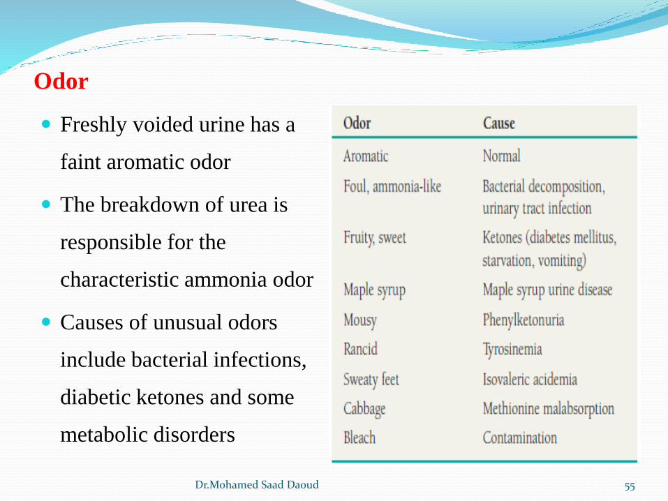
Odor
Freshly voided urine has a
faint aromatic odor
The breakdown of urea is
responsible for the
characteristic ammonia odor
Causes of unusual odors
include bacterial infections,
diabetic ketones and some
metabolic disorders
55Dr.Mohamed Saad Daoud
Chemical Examination of Urine
Reagent Strips:
Care of Reagent Strips
1. Store with desiccant in an opaque, tightly closed container.
2. Store below 30°C; do not freeze.
3. Do not expose to volatile fumes.
4. Do not use past the expiration date.
5. Do not use if chemical pads become discolored.
6. Remove strips immediately prior to use.
56Dr.Mohamed Saad Daoud
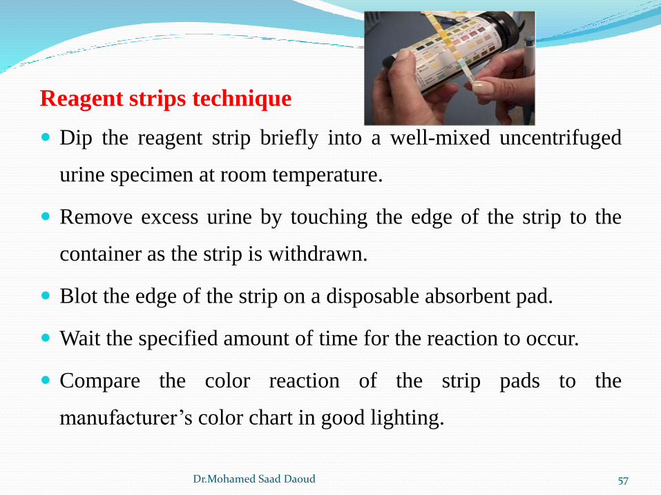
Reagent strips technique
Dip the reagent strip briefly into a well-mixed uncentrifuged
urine specimen at room temperature.
Remove excess urine by touching the edge of the strip to the
container as the strip is withdrawn.
Blot the edge of the strip on a disposable absorbent pad.
Wait the specified amount of time for the reaction to occur.
Compare the color reaction of the strip pads to the
manufacturer’s color chart in good lighting.
57Dr.Mohamed Saad Daoud
Quality Control
1.Test open bottles of reagent strips with known positive and
negative controls every 24 hr.
2. Resolve control results that are out of range by further testing.
3.Test reagents used in backup tests with positive and negative
controls.
4. Perform positive and negative controls on new reagents and
newly opened bottles of reagent strips.
5. Record all control results and reagent lot numbers.
58Dr.Mohamed Saad Daoud
59Dr.Mohamed Saad Daoud
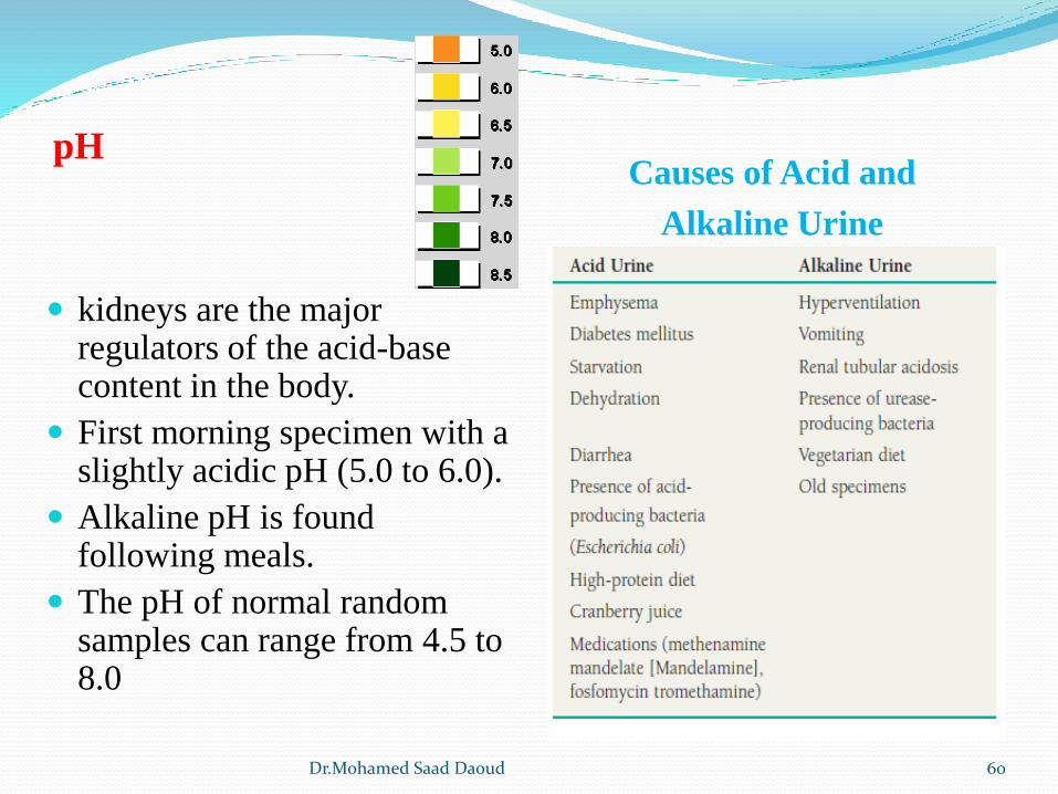
pHCauses of Acid and
Alkaline Urine
kidneys are the major regulators of the acid-base content in the body.
First morning specimen with a slightly acidic pH (5.0 to 6.0).
Alkaline pH is found following meals.
The pH of normal random samples can range from 4.5 to 8.0
60Dr.Mohamed Saad Daoud
Protein
The presence of proteinuria is often associated with early renal
disease
Normal urine contains less than 10 mg/dL or 100 mg per 24
hours is excreted (low molecular weight protein, Albumin).
Clinical proteinuria is indicated at ≥30 mg/dL (300 mg/L).
The causes of proteinuria are varied and can be grouped into
three major categories: prerenal, renal, and postrenal, based
on the origin of the protein.
61Dr.Mohamed Saad Daoud
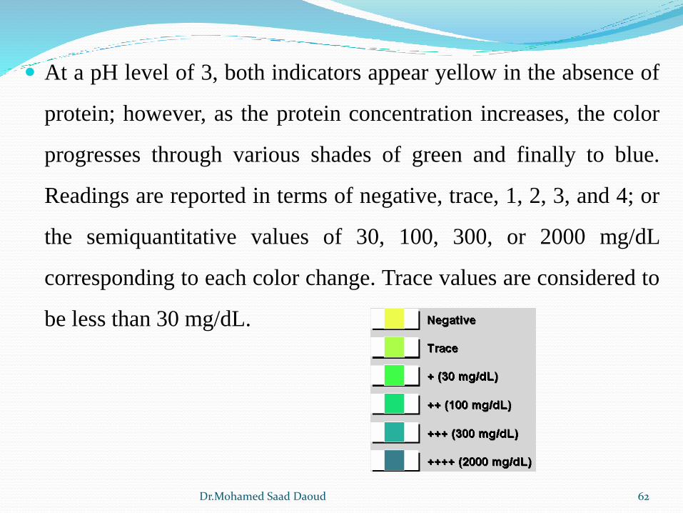
At a pH level of 3, both indicators appear yellow in the absence of
protein; however, as the protein concentration increases, the color
progresses through various shades of green and finally to blue.
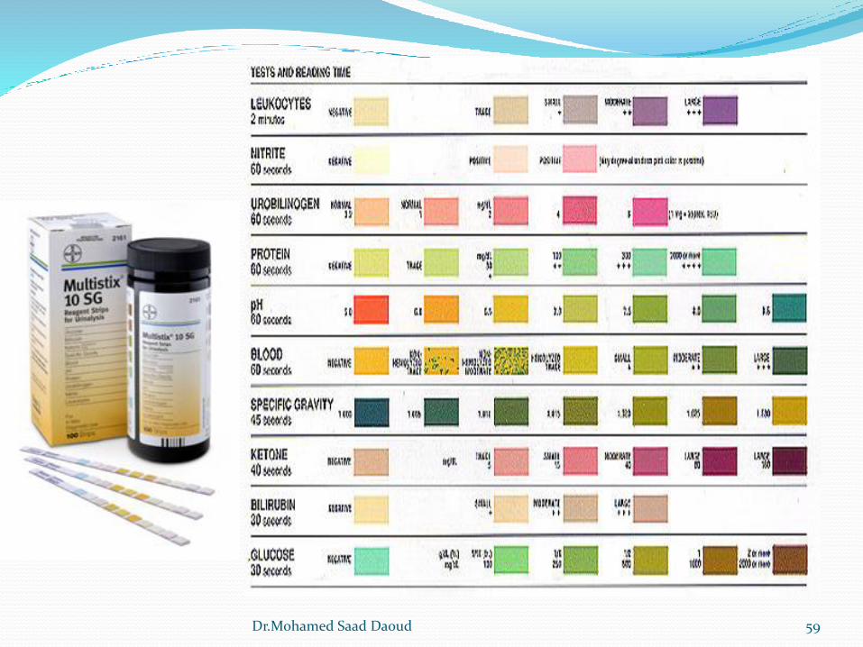
Readings are reported in terms of negative, trace, 1, 2, 3, and 4; or
the semiquantitative values of 30, 100, 300, or 2000 mg/dL
corresponding to each color change. Trace values are considered to
be less than 30 mg/dL.
62Dr.Mohamed Saad Daoud
Clinical Significance of Urine Protein:
Prerenal Proteinuria
caused by conditions affecting the plasma prior to its reaching the
kidney and, therefore, is not indicative of actual renal disease.
Increased levels of low molecular-weight plasma proteins such as
hemoglobin, myoglobin, and the acute phase reactants associated
with infection and inflammation.
Intravascular hemolysis
Muscle injury
Acute phase reactants
Multiple myeloma (Bence Jones protein)
63Dr.Mohamed Saad Daoud
Renal Proteinuria
Proteinuria associated with true renal disease may be the result of
either glomerular or tubular damage.
Glomerular disorders
the glomerular membrane is damaged, selective filtration is
impaired, and increased amounts of serum protein and
eventually red and white blood cells pass through the membrane
and are excreted in the urine
e.g., amyloid material, toxic substances, and the immune
complexes found in lupus erythematosus and streptococcal
glomerulonephritis
The amount of protein that appears in the urine ranges from
slightly above normal to 4 g/day
64Dr.Mohamed Saad Daoud
Tubular Proteinuria
Increased albumin is also present in disorders affecting tubular reabsorption because the normally filtered albumin can no longer be reabsorbed.
Causes of tubular dysfunction include exposure to toxic substances and heavy metals, severe viral infections.
Benign proteinuria is usually transient and can be produced by conditions such as strenuous exercise, high fever, dehydration, and exposure to cold.
65Dr.Mohamed Saad Daoud
Postrenal Proteinuria
Protein can be added to a urine specimen as it passes through the
structures of the lower urinary tract (ureters, bladder, urethra,
prostate, and vagina).
Bacterial and fungal infections and inflammations produce
exudates containing protein from the interstitial fluid.
The presence of blood as the result of injury or menstrual
contamination contributes protein, as does the presence of
prostatic fluid and large amounts of spermatozoa.
66Dr.Mohamed Saad Daoud
Glucose
The glucose filtered by the glomerulus is reabsorbed in the
proximal convoluted tubule; therefore, urine contains only
minute amounts of glucose. Tubular reabsorption of glucose is
by active transport in response to the body’s need to maintain
an adequate concentration of glucose.
The blood level of glucose become elevated (hyperglycemia),
as occurs in diabetes mellitus, the tubular transport of glucose
ceases, and glucose appears in the urine. The blood level at
which tubular reabsorption stops (renal threshold) for glucose
is approximately 160 to 180 mg/dL.
67Dr.Mohamed Saad Daoud
Hyperglycemia that occurs during pregnancy and disappears after
delivery is called gestational diabetes.
Hyperglycemia of nondiabetic origin is seen in a variety of disorders
and also produces glycosuria. (pancreatitis, pancreatic cancer,
acromegaly, Cushing syndrome and hyperthyroidism and The hormones
(glucagon, epinephrine, cortisol, thyroxine, and growth hormone, which
are increased in these disorders, work in opposition to insulin, thereby
producing hyperglycemia and glucosuria.
Glycosuria occurs in the absence of hyperglycemia when the
reabsorption of glucose by the renal tubules is compromised. “renal
glycosuria” is seen in end-stage renal disease.
68Dr.Mohamed Saad Daoud
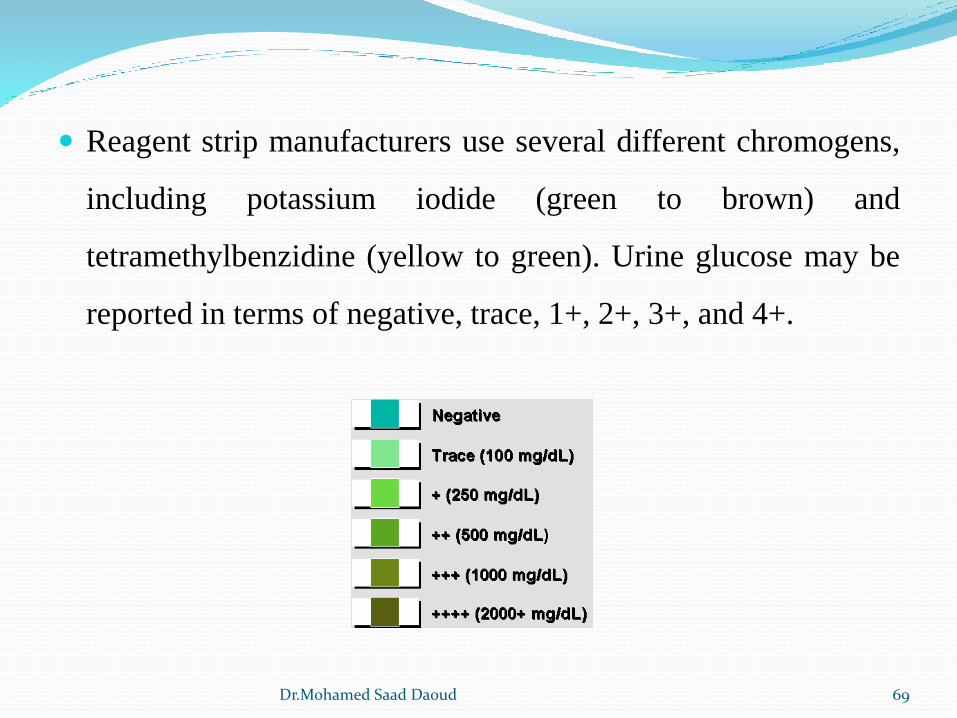
Reagent strip manufacturers use several different chromogens,
including potassium iodide (green to brown) and
tetramethylbenzidine (yellow to green). Urine glucose may be
reported in terms of negative, trace, 1+, 2+, 3+, and 4+.
69Dr.Mohamed Saad Daoud
Ketones
Including three ketone bodies: The products of fat metabolism
acetoacetic acid 20%
acetone 2%
β-hydroxybutyric acid 78%
Clinical Significance of Urine Ketones
1. Diabetic acidosis
2. Insulin dosage monitoring
3. Starvation
4. Malabsorption/pancreatic disorders
5. Strenuous exercise
6.Vomiting
7. Inborn errors of amino acid metabolism
70Dr.Mohamed Saad Daoud
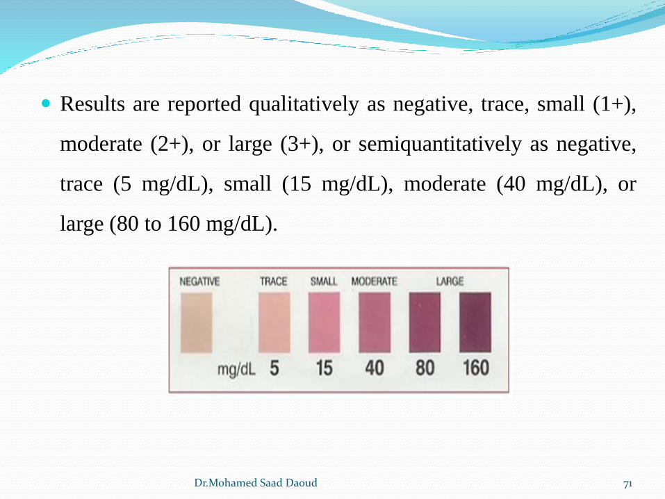
Results are reported qualitatively as negative, trace, small (1+),
moderate (2+), or large (3+), or semiquantitatively as negative,
trace (5 mg/dL), small (15 mg/dL), moderate (40 mg/dL), or
large (80 to 160 mg/dL).
71Dr.Mohamed Saad Daoud
Blood
Blood may be present in the urine either in the form of intact red
blood cells (hematuria) or as the product of red blood cell
destruction, hemoglobin (hemoglobinuria) and myoglobin
(myoglobinuria).
blood present in large quantities can be detected visually;
hematuria produces a cloudy red urine, and hemoglobinuria
appears as a clear red specimen.
Any amount of blood greater than five cells per microliter of urine
is considered clinically significant,
72Dr.Mohamed Saad Daoud
Hematuria (Renal calculi, Glomerulonephritis, Pyelonephritis,
Tumors, Trauma, Exposure to toxic, chemicals, Anticoagulants
and Strenuous exercise).
Hemoglobinuria (Transfusion reactions, Hemolytic anemias,
Severe burns, Infections/malaria, Strenuous exercise/red blood
cell trauma and Brown recluse spider bites).
Myoglobinuria (Muscular trauma/crush syndromes, Prolonged
coma, Convulsions, Muscle-wasting diseases,
Alcoholism/overdose, Drug abuse, Extensive exertion and
Cholesterol-lowering statin medications
73Dr.Mohamed Saad Daoud
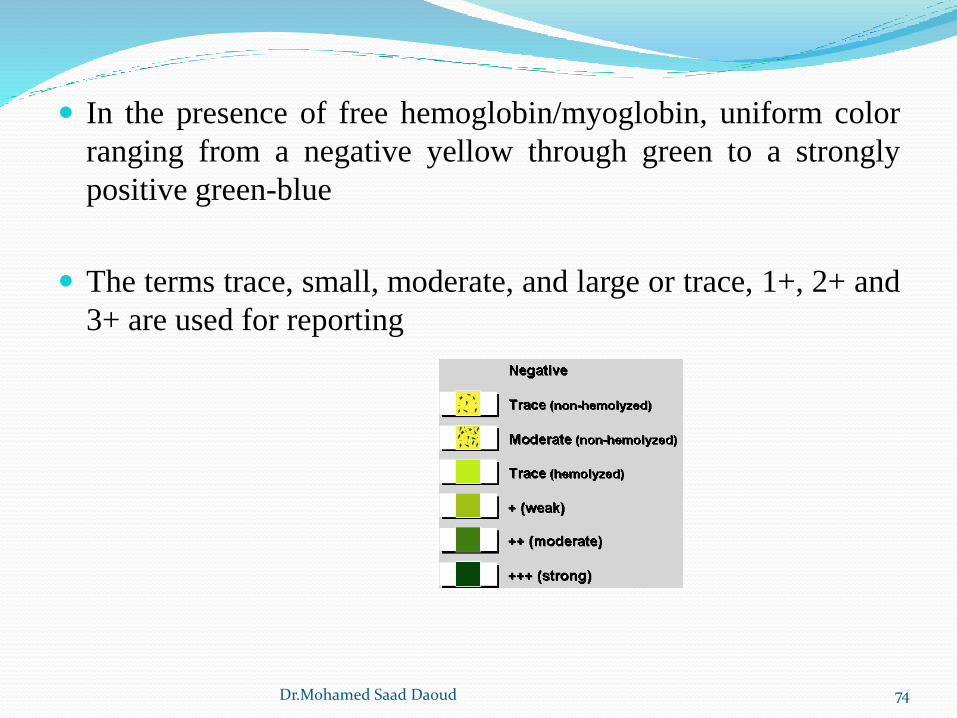
In the presence of free hemoglobin/myoglobin, uniform color
ranging from a negative yellow through green to a strongly
positive green-blue
The terms trace, small, moderate, and large or trace, 1+, 2+ and
3+ are used for reporting
74Dr.Mohamed Saad Daoud
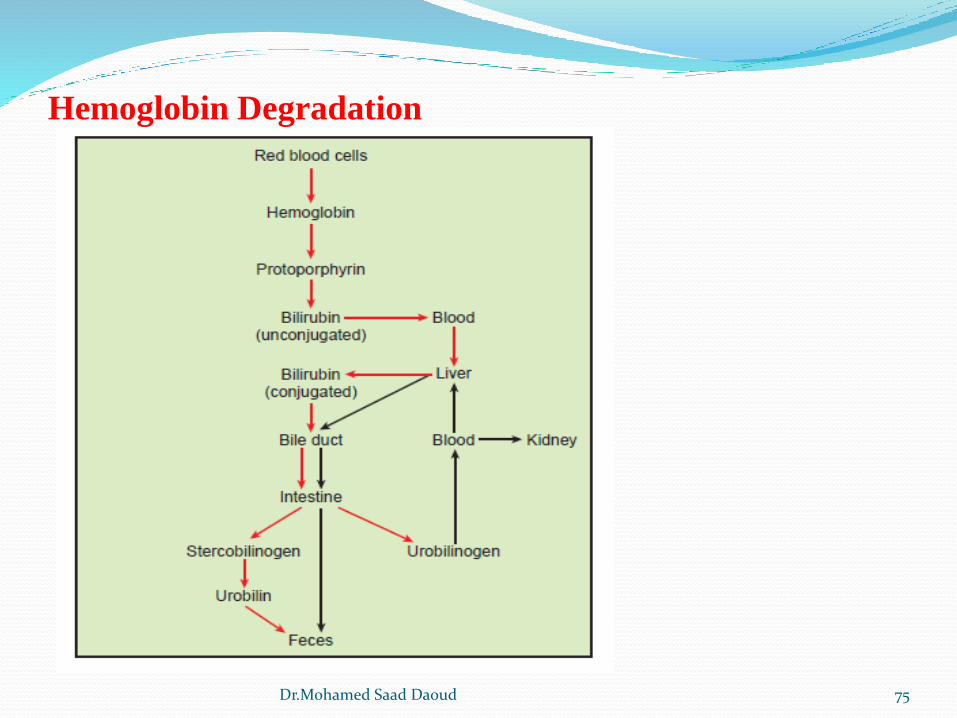
Hemoglobin Degradation
75Dr.Mohamed Saad Daoud
Bilirubin
The appearance of bilirubin in the urine can provide an early
indication of liver disease.
Highly pigmented yellow compound.
Degradation product of hemoglobin.
Conjugated bilirubin appears in the urine when the normal
degradation cycle is disrupted by obstruction of the bile duct
(e.g., gallstones or cancer) or when the integrity of the liver is
damaged, allowing leakage of conjugated bilirubin into the
circulation. Hepatitis and cirrhosis are common examples of
conditions that produce liver damage, resulting in bilirubinuria.
76Dr.Mohamed Saad Daoud
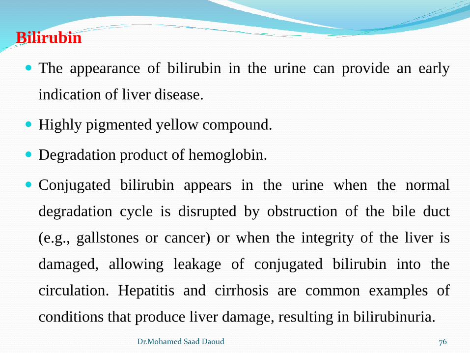
Routine testing for urinary bilirubin by reagent strip uses the
diazo reaction. Colors ranging from increasing degrees of tan or
pink to violet, respectively. Qualitative results are reported as
negative, small, moderate, or large, or as negative, 1+, 2+, or 3+.
77Dr.Mohamed Saad Daoud
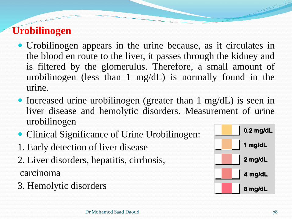
Urobilinogen
Urobilinogen appears in the urine because, as it circulates inthe blood en route to the liver, it passes through the kidney andis filtered by the glomerulus. Therefore, a small amount ofurobilinogen (less than 1 mg/dL) is normally found in theurine.
Increased urine urobilinogen (greater than 1 mg/dL) is seen inliver disease and hemolytic disorders. Measurement of urineurobilinogen
Clinical Significance of Urine Urobilinogen:
1. Early detection of liver disease
2. Liver disorders, hepatitis, cirrhosis,
carcinoma
3. Hemolytic disorders
78Dr.Mohamed Saad Daoud
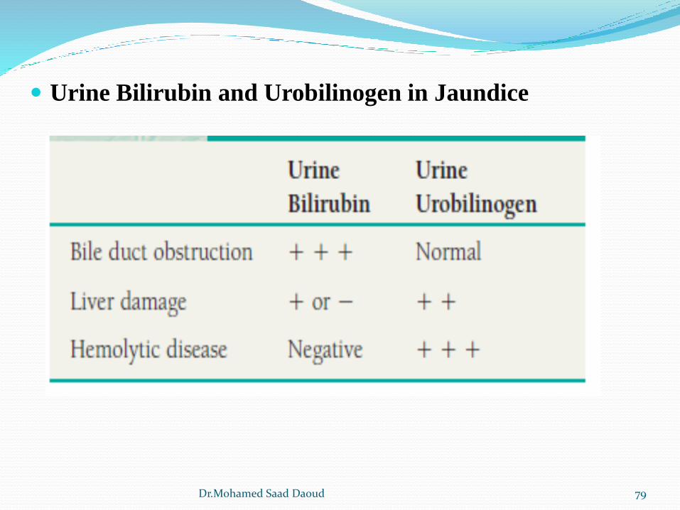
Urine Bilirubin and Urobilinogen in Jaundice
79Dr.Mohamed Saad Daoud

Nitrite
The reagent strip test for nitrite provides a rapid screening test for
the presence of urinary tract infection (UTI).
Based on the ability of certain bacteria to reduce nitrate, a normal
constituent of urine, to nitrite.
Clinical Significance of Urine Nitrite:
1. Cystitis
2. Pyelonephritis
3. Evaluation of antibiotic therapy
4. Monitoring of patients at high risk for urinary tract infection
5. Screening of urine culture specimens
80Dr.Mohamed Saad Daoud
Leukocyte Esterase
Leukocyte estrase test detects the presence of leukocytes that havebeen lysed, particularly in dilute alkaline urine, and would notappear in the microscopic examination.
Normal values for leukocytes are based on the microscopicsediment examination and vary from 0 to 2 to 0 to 5 per high powerfield.
Increased urinary leukocytes are indicators of UTI.
The LE test detects the presence of esterase in the granulocyticwhite blood cells (neutrophils, eosinophils, and basophils) andmonocytes. Neutrophils are the leukocytes most frequentlyassociated with bacterial infections
81Dr.Mohamed Saad Daoud
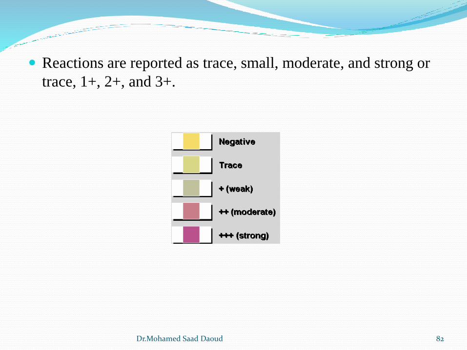
Reactions are reported as trace, small, moderate, and strong or
trace, 1+, 2+, and 3+.
82Dr.Mohamed Saad Daoud
Renal calculi
Renal calculi (kidney stones) may form in the calyces and
pelvis of the kidney, ureters, and bladder.
the calculi vary in size from barely visible to large, staghorn
calculi resembling the shape of the renal pelvis and smooth,
round bladder stones with diameters of 2 or more inches. Small
calculi may be passed in the urine, subjecting the patient to
severe pain radiating from the lower back to the legs. Larger
stones cannot be passed and may not be detected until patients
develop symptoms of urinary obstruction.
83Dr.Mohamed Saad Daoud
Conditions favoring the formation of renal calculi are similar to
those favoring formation of urinary crystals, including pH,
chemical concentration, and urinary stasis.
The finding of clumps of crystals in freshly voided urine suggests
that conditions may be right for calculus formation.
Analysis of the chemical composition of renal calculi plays an
important role in patient management.
Approximately 75% of the renal calculi are composed of calcium
oxalate or phosphate. Magnesium ammonium phosphate, uric acid,
and cystine are the other primary calculi constituents.
84Dr.Mohamed Saad Daoud
Calcium calculi are frequently associated with metabolic calcium
and phosphate disorders and occasionally diet. Magnesium
ammonium phosphate calculi are frequently accompanied by
urinary infections involving urea-splitting bacteria. The urine pH
is often higher than 7.0. Uric acid calculi may be associated with
increased intake of foods with high purine content. The urine pH
is acidic. Most cystine calculi are seen in conjunction with
hereditary disorders of cystine metabolism
85Dr.Mohamed Saad Daoud