Embed Size (px)
Citation preview
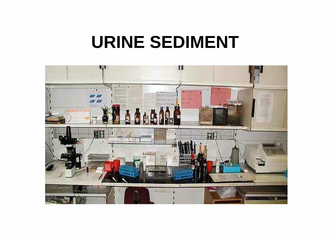
URINE SEDIMENT
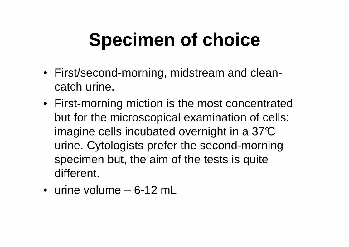
Specimen of choice
• First/second-morning, midstream and clean-catch urine.
• First-morning miction is the most concentratedbut for the microscopical examination of cells: imagine cells incubated overnight in a 37°C urine. Cytologists prefer the second-morningspecimen but, the aim of the tests is quitedifferent.
• urine volume – 6-12 mL
Sample preparation
• Centrifugation (5 minutes at 400 RCF)
• Supernatant aspiration• Resuspension
• Staining– Sternheimer
• Alcian blue - mucopolysaccharides• Pyronin B – red colour of other sediment components, maily
cell cytoplasms, matrix of waxy casts…)
– Gram-staining– Sedi-Stain
Common Crystals
A number of in vivo and in vitro factors influence the typesand numbers of urinary crystals in a given sample:
• In vivo factors: – concentration and solubility of crystallogenic substances
contained in the specimen, – urine pH, and– excretion of diagnostic and therapeutic agents.
• In vitro factors : – temperature (solubility decreases with temperature), – evaporation (increases solute concentration), and– urine pH (changes with standing and bacterial overgrowth).
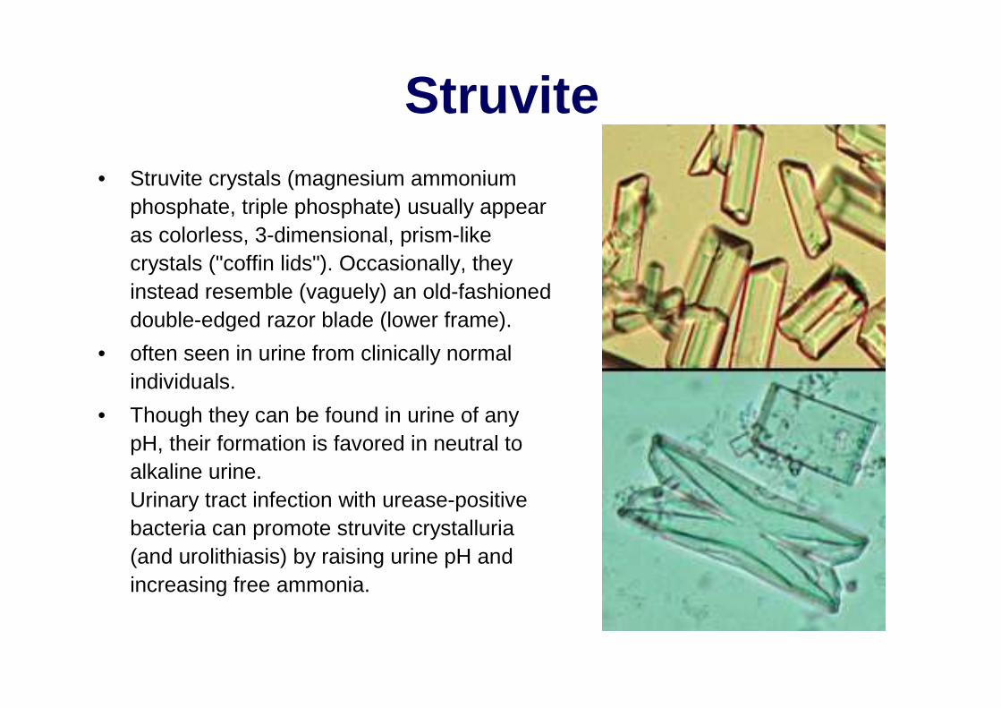
Struvite• Struvite crystals (magnesium ammonium
phosphate, triple phosphate) usually appearas colorless, 3-dimensional, prism-likecrystals ("coffin lids"). Occasionally, theyinstead resemble (vaguely) an old-fashioneddouble-edged razor blade (lower frame).
• often seen in urine from clinically normalindividuals.
• Though they can be found in urine of anypH, their formation is favored in neutral to alkaline urine. Urinary tract infection with urease-positive bacteria can promote struvite crystalluria(and urolithiasis) by raising urine pH andincreasing free ammonia.
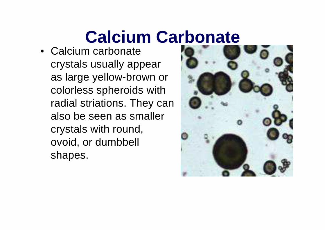
Calcium Carbonate• Calcium carbonate
crystals usually appearas large yellow-brown orcolorless spheroids withradial striations. They canalso be seen as smallercrystals with round, ovoid, or dumbbellshapes.

Calcium oxalate dihydrate
• colorless squares whosecorners are connected by intersecting lines (resemblingan envelope)
• urine of any pH. • The crystals vary in size from
quite large to very small. In some cases, large numbersof tiny oxalates may appearas amorphous unlessexamined at highmagnification. – Urolithiasis due to calcium
oxalate– Ethylene glycol intoxication
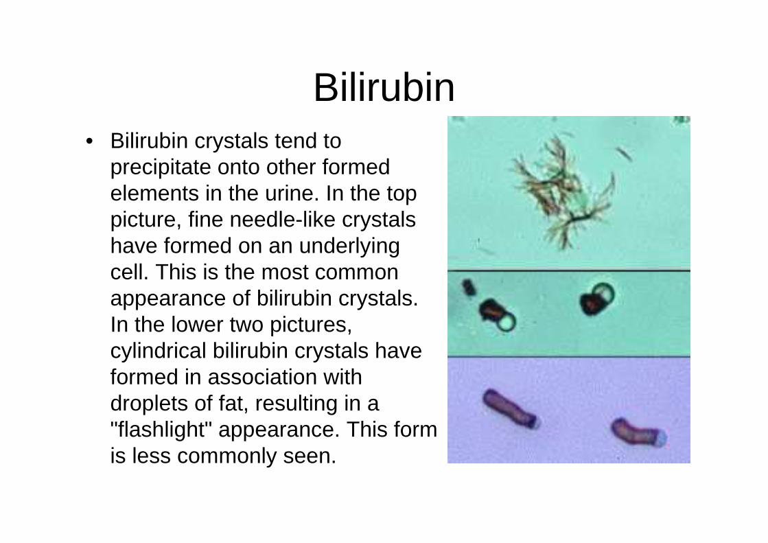
Bilirubin• Bilirubin crystals tend to
precipitate onto other formedelements in the urine. In the top picture, fine needle-like crystalshave formed on an underlyingcell. This is the most commonappearance of bilirubin crystals. In the lower two pictures, cylindrical bilirubin crystals haveformed in association withdroplets of fat, resulting in a "flashlight" appearance. This formis less commonly seen.

„Amorphous" crystals• aggregates of finely granular material without any
defining shape at the light microscopic level. • urates (Na, K, Mg, or Ca salts) tend to form in
acidic urine, and may have a yellow or yellow-brown color.
• phosphates are similar in general appearance, buttend to form in alkaline urine and lack color.
• Calcium oxalate dihydrate crystals sometimes alsocan present as "amorphous" when the individualcrystals are very small. Examination at highermagnification will reveal the typical "envelope" appearance.
• Xanthine crystals are usually in the form of"amorphous" crystals.
• Generally, no specific clinical interpretation can bemade based on the finding of amorphous crystals. Small amorphous crystals can be confused withbacterial cocci in some cases, but can bedistinguished by Gram-staining.
Less Common Crystals
• Cystine• Biurates• Drug Crystals• Tyrosine• Ca Oxalate Monohydrate
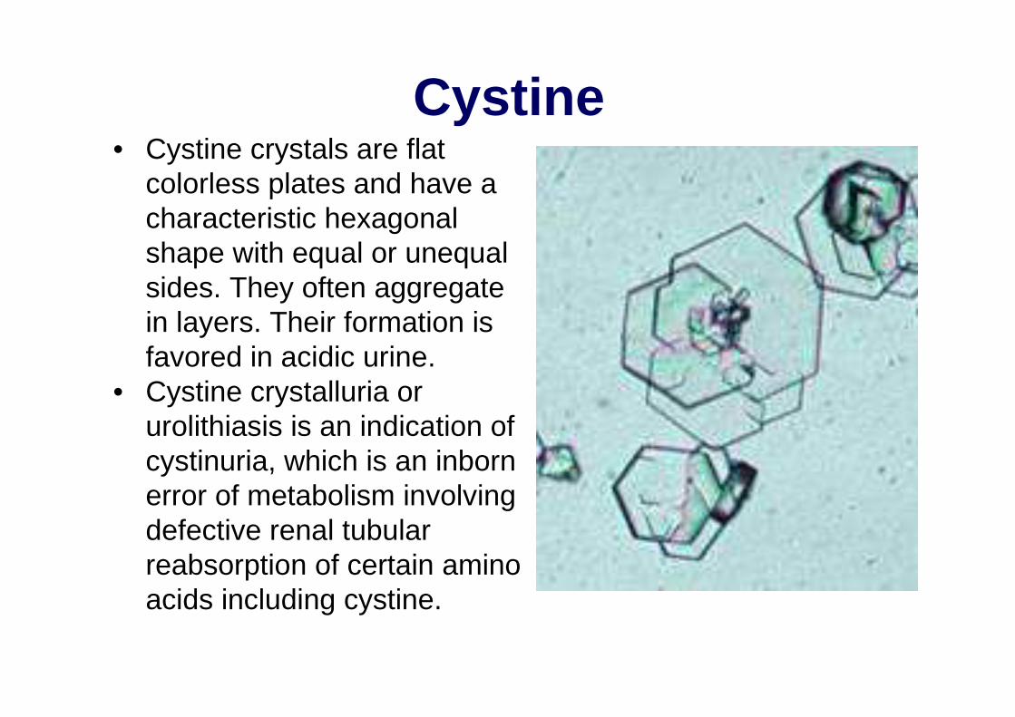
Cystine• Cystine crystals are flat
colorless plates and have a characteristic hexagonalshape with equal or unequalsides. They often aggregatein layers. Their formation isfavored in acidic urine.
• Cystine crystalluria orurolithiasis is an indication ofcystinuria, which is an inbornerror of metabolism involvingdefective renal tubularreabsorption of certain aminoacids including cystine.
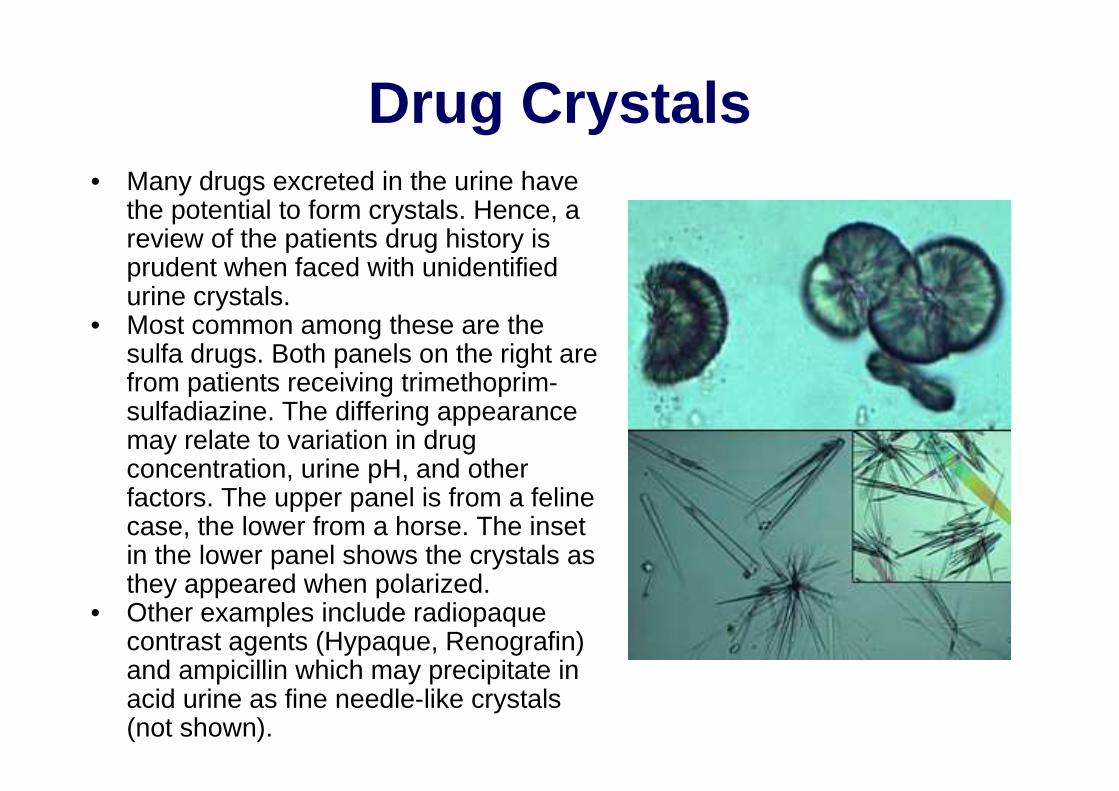
Drug Crystals• Many drugs excreted in the urine have
the potential to form crystals. Hence, a review of the patients drug history isprudent when faced with unidentifiedurine crystals.
• Most common among these are thesulfa drugs. Both panels on the right are from patients receiving trimethoprim-sulfadiazine. The differing appearancemay relate to variation in drugconcentration, urine pH, and otherfactors. The upper panel is from a felinecase, the lower from a horse. The insetin the lower panel shows the crystals as they appeared when polarized.
• Other examples include radiopaquecontrast agents (Hypaque, Renografin) and ampicillin which may precipitate in acid urine as fine needle-like crystals(not shown).

Calcium Oxalate Monohydrate• Calcium oxalate monohydrate
crystals vary in size and mayhave a spindle, oval, ordumbbell shape. Most commonly, they appear as flat, elongated, six-sided crystals("fence pickets") such as shownto the right. The arrow in thephoto indicates a "daughter" crystal forming on the face of a larger underlying crystal.
• virtually always associatedwithethylene glycol intoxication.

BiuratesAmmonium urate (orbiurate) crystals generallyappear as brown oryellow-brown sphericalbodies with irregularprotrusions ("thorn-apples"). Though possiblein urine of any pH, theirformation is favored in neutral to alkaline urine.
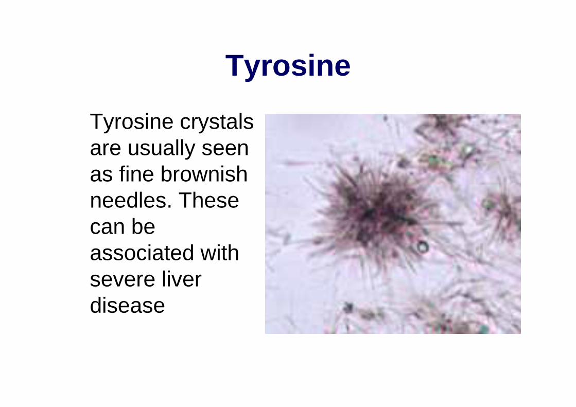
Tyrosine
Tyrosine crystalsare usually seenas fine brownishneedles. These can beassociated withsevere liverdisease
Cells in Urine Sediment• Urine is a hostile environment for cells since they
encounter abnormal osmotic pressures, pH changes, and exposure to toxic metabolites. For these reasons, post-collection delay of examination should beminimized. If delay is unavoidable, refrigeration will slowdegeneration of cells.
• For routine purposes, cells are examined as unstainedwet-mounts of sedimented urine. Under somecircumstances, air-dried smears are prepared andstained with hematologic stains.
• Red blood cells and leukocytes are quantified as cells/HPF (High Power Field - 40x objective). Other cell types are usually subjectively listed as "few, moderate, or many".
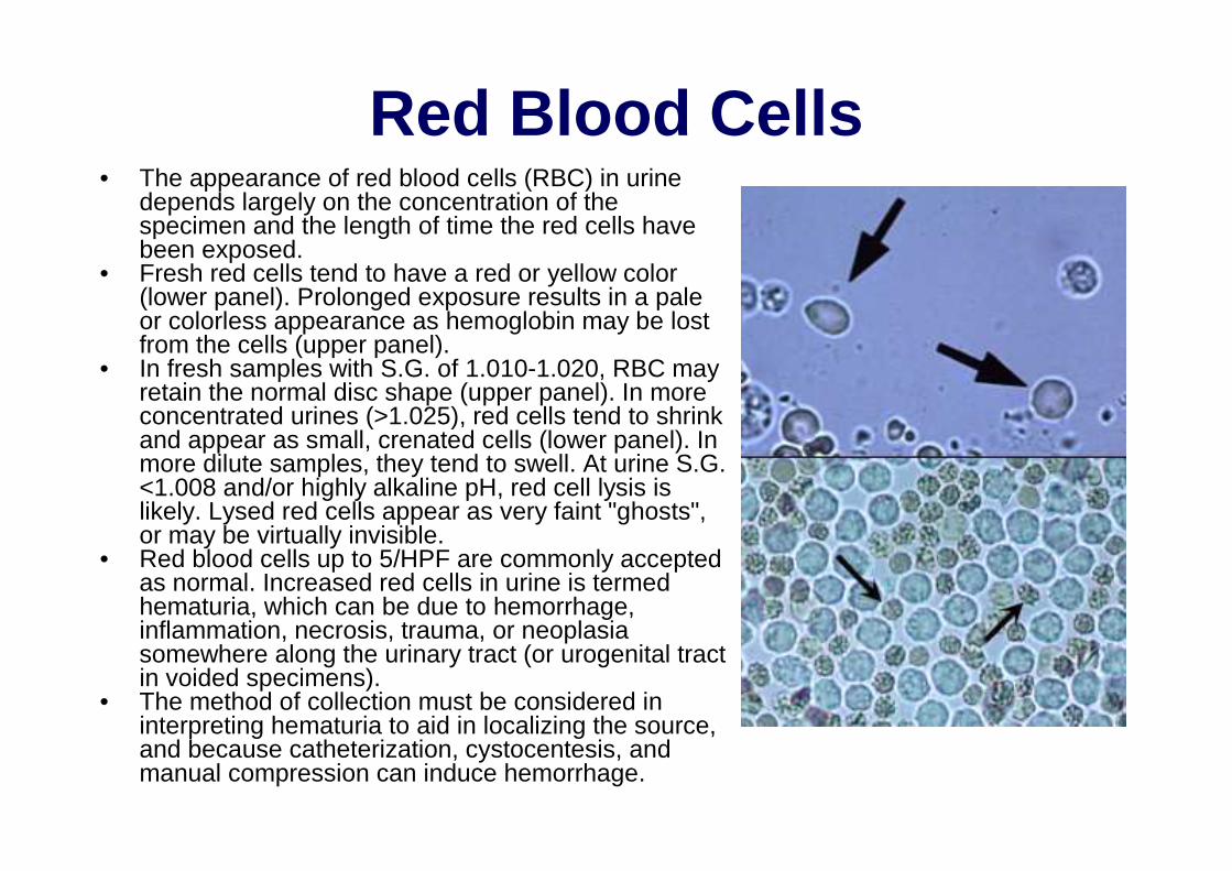
Red Blood Cells• The appearance of red blood cells (RBC) in urine
depends largely on the concentration of thespecimen and the length of time the red cells havebeen exposed.
• Fresh red cells tend to have a red or yellow color(lower panel). Prolonged exposure results in a pale or colorless appearance as hemoglobin may be lostfrom the cells (upper panel).
• In fresh samples with S.G. of 1.010-1.020, RBC mayretain the normal disc shape (upper panel). In more concentrated urines (>1.025), red cells tend to shrinkand appear as small, crenated cells (lower panel). In more dilute samples, they tend to swell. At urine S.G. <1.008 and/or highly alkaline pH, red cell lysis islikely. Lysed red cells appear as very faint "ghosts", or may be virtually invisible.
• Red blood cells up to 5/HPF are commonly acceptedas normal. Increased red cells in urine is termedhematuria, which can be due to hemorrhage, inflammation, necrosis, trauma, or neoplasiasomewhere along the urinary tract (or urogenital tractin voided specimens).
• The method of collection must be considered in interpreting hematuria to aid in localizing the source, and because catheterization, cystocentesis, andmanual compression can induce hemorrhage.
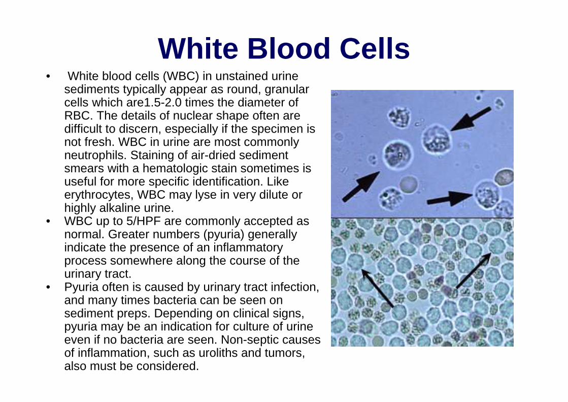
White Blood Cells• White blood cells (WBC) in unstained urine
sediments typically appear as round, granularcells which are1.5-2.0 times the diameter ofRBC. The details of nuclear shape often are difficult to discern, especially if the specimen isnot fresh. WBC in urine are most commonlyneutrophils. Staining of air-dried sediment smears with a hematologic stain sometimes isuseful for more specific identification. Likeerythrocytes, WBC may lyse in very dilute orhighly alkaline urine.
• WBC up to 5/HPF are commonly accepted as normal. Greater numbers (pyuria) generallyindicate the presence of an inflammatoryprocess somewhere along the course of theurinary tract.
• Pyuria often is caused by urinary tract infection, and many times bacteria can be seen on sediment preps. Depending on clinical signs, pyuria may be an indication for culture of urine even if no bacteria are seen. Non-septic causesof inflammation, such as uroliths and tumors, also must be considered.
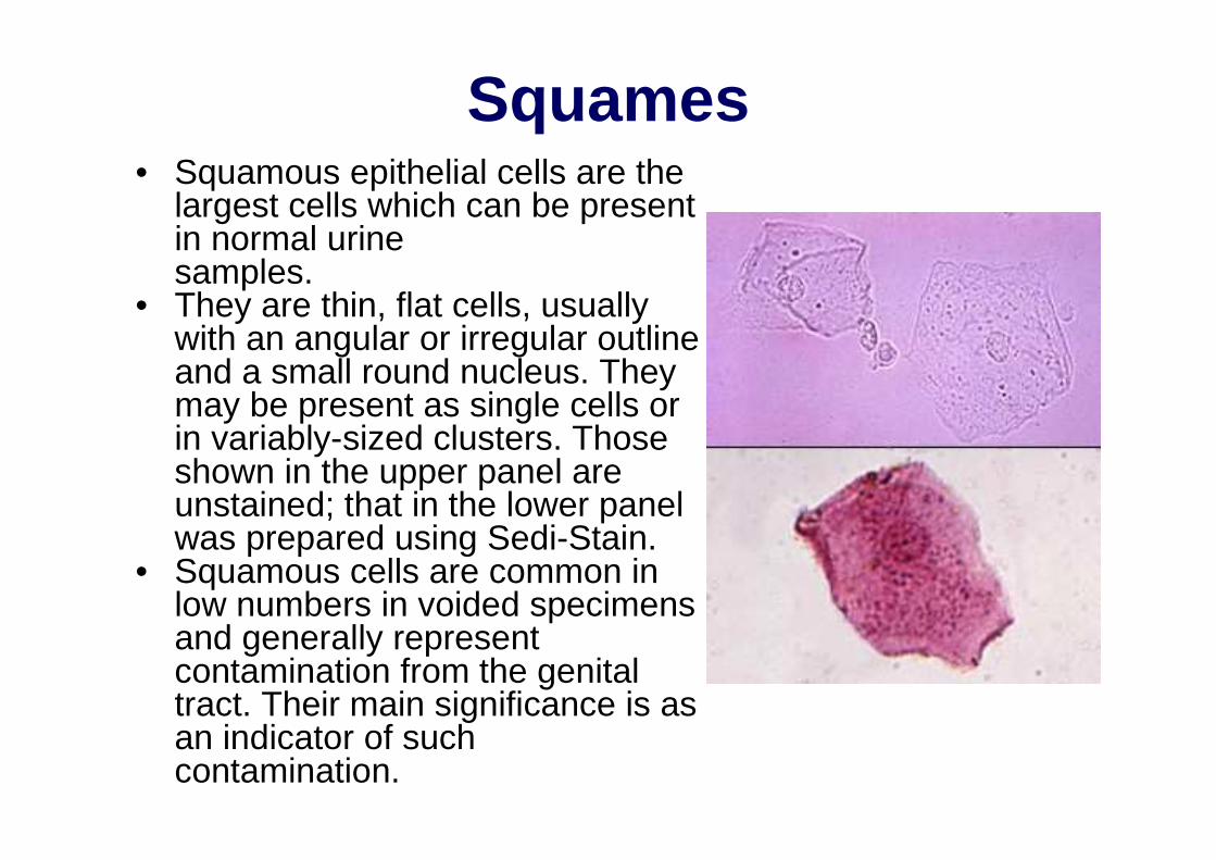
Squames• Squamous epithelial cells are the
largest cells which can be presentin normal urine samples.
• They are thin, flat cells, usuallywith an angular or irregular outlineand a small round nucleus. Theymay be present as single cells orin variably-sized clusters. Thoseshown in the upper panel are unstained; that in the lower panel was prepared using Sedi-Stain.
• Squamous cells are common in low numbers in voided specimensand generally representcontamination from the genitaltract. Their main significance is as an indicator of such contamination.
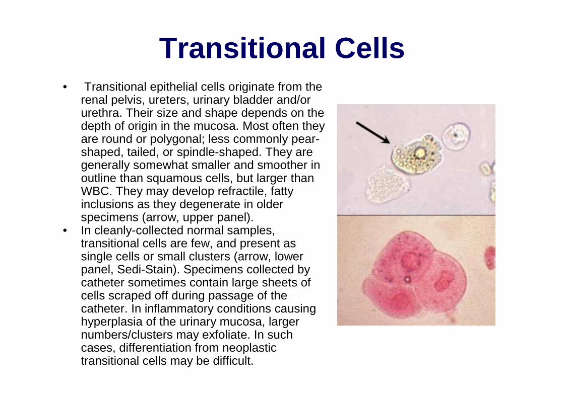
Transitional Cells• Transitional epithelial cells originate from the
renal pelvis, ureters, urinary bladder and/orurethra. Their size and shape depends on thedepth of origin in the mucosa. Most often theyare round or polygonal; less commonly pear-shaped, tailed, or spindle-shaped. They are generally somewhat smaller and smoother in outline than squamous cells, but larger thanWBC. They may develop refractile, fattyinclusions as they degenerate in olderspecimens (arrow, upper panel).
• In cleanly-collected normal samples, transitional cells are few, and present as single cells or small clusters (arrow, lowerpanel, Sedi-Stain). Specimens collected by catheter sometimes contain large sheets ofcells scraped off during passage of thecatheter. In inflammatory conditions causinghyperplasia of the urinary mucosa, largernumbers/clusters may exfoliate. In such cases, differentiation from neoplastictransitional cells may be difficult.

Neoplastic Cells• Neoplastic cells may be seen in urine sediments
of patients with tumors of the urinary tract.• Transitional cell carcinomas arising in the urinary
bladder or urethra are most likely to spontaneously exfoliate, but cannot be ruled outbased on a failure to identify malignant cells in urine. Rarely, lymphomas and renal carcinomasalso can be diagnosed from urine sediment.
• The pictures shown are from a case oftransitional cell carcinoma. Though the presence of neoplastic cells may be suspected on examination of unstained wet-mounts (upperpanel), evaluation of air-dried sediment smearsor cytocentrifuge preps stained with hematologicstains (lower panel) is necessary for confirmation. In the case shown here, thecytologic criteria of malignancy are clearlyfulfilled; in other cases a distinction fromhyperplastic cells cannot be made with certaintywithout a tissue biopsy.
Urinary Casts
Casts are cylindrical structures composed mainly ofmucoprotein (the Tamm-Horsefall mucoprotein ) whichis secreted by epithelial cells lining the loops of Henle, the distal tubules and the collecting ducts. The factorsresponsible for the precipitation of this mucoprotein are not fully understood, but may relate to the concentrationand pH of urine in these areas. Casts may form in thepresence or absence of cells in the tubular lumen. If cells(epithelial cells, WBC) are present as a cast forms, theymay adhere to, and subsequently be surrounded by, thefibrillar protein network
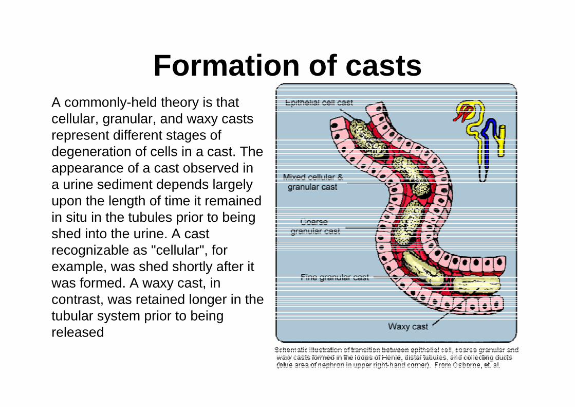
Formation of castsA commonly-held theory is thatcellular, granular, and waxy castsrepresent different stages ofdegeneration of cells in a cast. Theappearance of a cast observed in a urine sediment depends largelyupon the length of time it remainedin situ in the tubules prior to beingshed into the urine. A castrecognizable as "cellular", for example, was shed shortly after itwas formed. A waxy cast, in contrast, was retained longer in thetubular system prior to beingreleased
General Interpretation of casts-• Casts are quantified for reporting as the number seen
per low power field (10x objective) and classified as to type (e.g., waxy casts, 5-10/LPF). Casts in urine fromnormal individuals are few or none.
• An absence of casts does not rule out renal disease. Casts may be absent or very few in cases of chronic, progressive, generalized nephritis. Even in cases ofacute renal disease, casts can be few or absent in a single sample since they tend be shed intermittently. Furthermore, casts are unstable in urine and are proneto dissolution with time, especially in dilute and/oralkaline urine.
• Although the presence of numerous casts is solid evidence of generalized (usually acute) renal disease, itis not a reliable indicator of prognosis. If the underlyingcause can be removed or diminished, regeneration ofrenal tubular epithelium can occur (provided thebasement membrane remains intact).
:
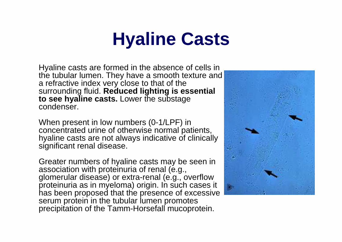
Hyaline CastsHyaline casts are formed in the absence of cells in the tubular lumen. They have a smooth texture anda refractive index very close to that of thesurrounding fluid. Reduced lighting is essentialto see hyaline casts. Lower the substagecondenser.
When present in low numbers (0-1/LPF) in concentrated urine of otherwise normal patients, hyaline casts are not always indicative of clinicallysignificant renal disease.
Greater numbers of hyaline casts may be seen in association with proteinuria of renal (e.g., glomerular disease) or extra-renal (e.g., overflowproteinuria as in myeloma) origin. In such cases ithas been proposed that the presence of excessiveserum protein in the tubular lumen promotesprecipitation of the Tamm-Horsefall mucoprotein.
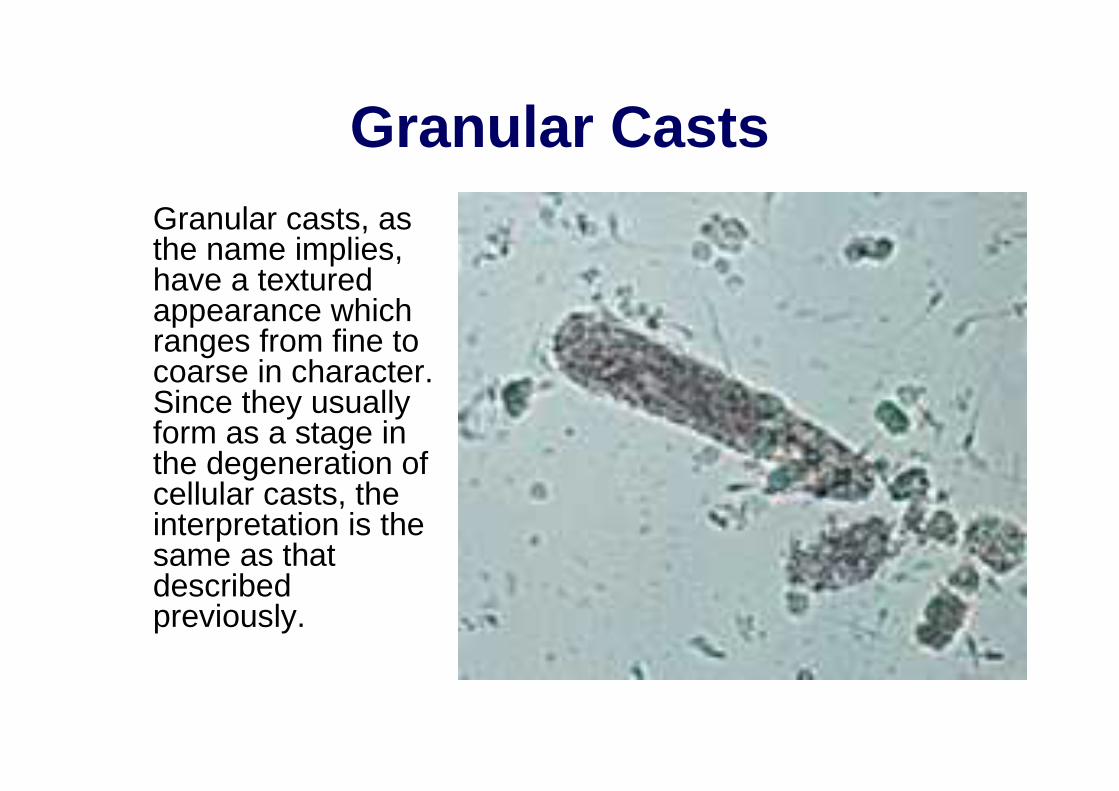
Granular CastsGranular casts, as the name implies, have a texturedappearance whichranges from fine to coarse in character. Since they usuallyform as a stage in the degeneration ofcellular casts, theinterpretation is thesame as thatdescribedpreviously.

Cellular CastsCellular casts most commonly result whendisease processes such as ischemia, infarction, or nephrotoxicity cause degeneration andnecrosis of tubular epithelial cells. The presence of these casts indicates acute tubular injury butdoes not indicate the extent or reversibility of theinjury.
A common scenario is the patient with decreasedrenal perfusion and oliguria secondary to severe dehydration. Ischemic injury results in degeneration and sloughing of the epithelial cells. The resulting casts often are prominent in urine produced following rehydration with fluid therapy. The restoration of urine flow "flushes" numerouscasts out of the tubules.
Leukocytes can also be incorporated into casts in cases of tubulo-interstitial inflammation (eg, pyelonephritis). It is rarely possible to distinguishbetween epithelial casts and leukocyte casts in routine sediment preparations, however, sincenuclear detail is obscured by the degeneratedstate of the cells.
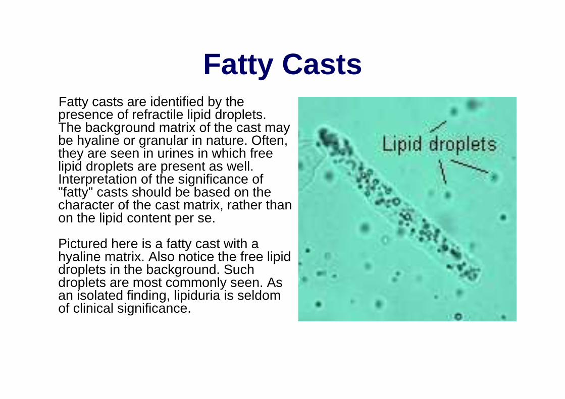
Fatty CastsFatty casts are identified by thepresence of refractile lipid droplets. The background matrix of the cast maybe hyaline or granular in nature. Often, they are seen in urines in which freelipid droplets are present as well. Interpretation of the significance of"fatty" casts should be based on thecharacter of the cast matrix, rather thanon the lipid content per se.
Pictured here is a fatty cast with a hyaline matrix. Also notice the free lipid droplets in the background. Such droplets are most commonly seen. As an isolated finding, lipiduria is seldomof clinical significance.
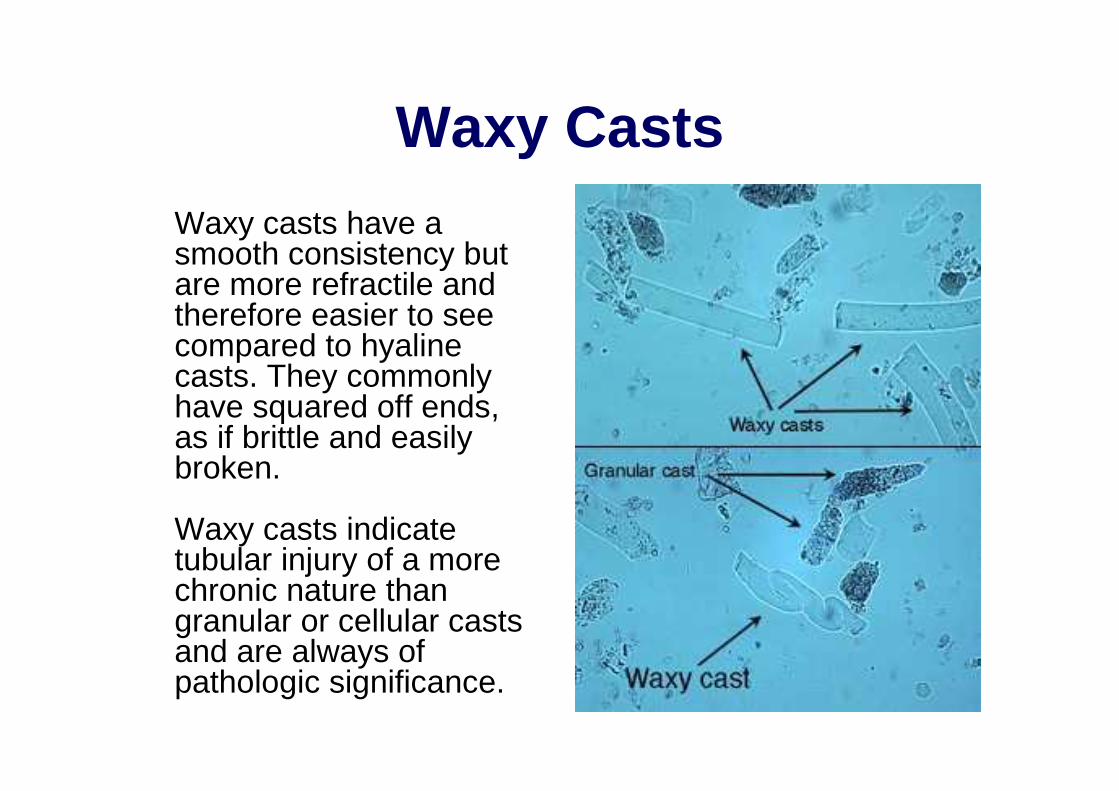
Waxy CastsWaxy casts have a smooth consistency butare more refractile andtherefore easier to seecompared to hyalinecasts. They commonlyhave squared off ends, as if brittle and easilybroken.
Waxy casts indicatetubular injury of a more chronic nature thangranular or cellular castsand are always ofpathologic significance.

Infectious Agents
• Infectious agents of various classes canbe observed in urine sediments. In most cases, their significance can be properlyassessed only in light of the clinical signs, method of collection, post-collectioninterval, and other findings in theurinalysis. – Yeasts– Bacteria
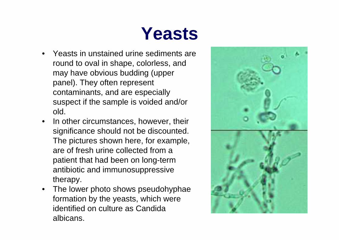
Yeasts• Yeasts in unstained urine sediments are
round to oval in shape, colorless, andmay have obvious budding (upperpanel). They often representcontaminants, and are especiallysuspect if the sample is voided and/orold.
• In other circumstances, however, theirsignificance should not be discounted. The pictures shown here, for example, are of fresh urine collected from a patient that had been on long-term antibiotic and immunosuppressivetherapy.
• The lower photo shows pseudohyphaeformation by the yeasts, which wereidentified on culture as Candidaalbicans.

Bacteria• Bacteria can be identified in unstained urine
sediments when present in sufficient numbers. Rod-shaped bacteria and chains of cocci are often readilyidentifiable. The images at right show E.coli bacillifrom a case of cystitis. However, small amorphouscrystals, cellular debris, and small fat droplets caneither mask or mimic cocci. If there is any doubt aboutthe presence of bacteria, a Gram-stained smear ofurine sediment (middle panel) should be examined.
• Urine in the bladder of normal animals is sterile. Though bacteria from the distal urethra and/or genitaltract may contaminate voided specimens, they are usually too few to see if a good mid-stream collectionwas obtained.
• Although phagocytized bacteria cannot be seen in unstained wet mounts of urine sediment, they mayfound in stained smears of sediment. The lower panel at the right shows a neutrophil containingphagocytized bacteria. Notice that the nucleus in thiscell is round; nuclei tend to become round as neutrophils age in urine.
• Bacteriuria of clinical significance, e.g., bacterialcystitis, is usually accompanied by increased numbersof white cells (pyuria). The presence of a few bacteriawithout pyuria is very rarely significant of infection.
Contaminants in Urine Sediment
• Extraneous contaminating materials of many types can make their way into urine specimens,.
• Striving for optimal collection and transport of specimens will help maximize useful results and minimize confusing findings.
• Spores and Pollens • Microbial Overgrowth • Fibers • Starch Granules

Spores and PollenMold spores and pollens come in a widevariety of shapes and sizes. Shownhere is an Alternaria spore surroundedby amorphous crystals and a few lipid droplets.
Pollen grains are generally round to oval and some have a yellow to brown tint. They are most likely to be confused for parasitic ova. A good knowledge of theactual appearance of the few trueparasite eggs that can occur in urine iseasier to achieve than specificrecognition of all types of pollen andmold spores. Again, optimizingspecimen collection and handling willreduce the chances of seeing potentiallyconfusing structures.

Overgrowth of MicrobesSpecimens mailed to laboratories withoutrefrigeration or preservatives are subject to overgrowth of microbes, whether contaminantsor pathogens. Shown here is a dense mat offungal hyphae which was seen in a sediment prep of a canine urine specimen which had been several days in transit. Since fungalinfection of the urinary tract in dogs is quiteuncommon, the odds are that this representsovergrowth of contaminants.
Bacteria, whether pathogens or contaminants, also can multiply when analysis is delayed. This often clouds the interpretation of bothsediment examination and culture results.
Refrigeration is perhaps the best all-aroundmethod for preserving a specimen. Somelaboratories also suggest specific transport media or swabs when sending a specimen for culture.
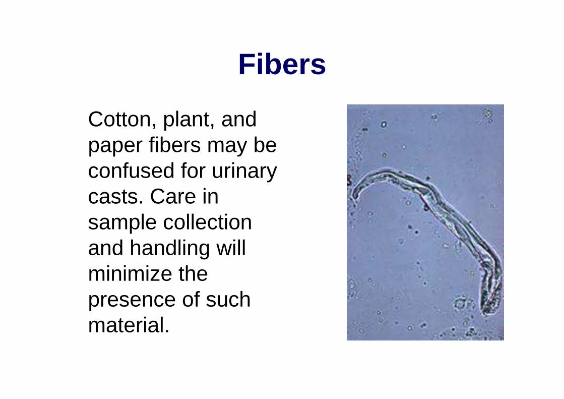
Fibers
Cotton, plant, andpaper fibers may beconfused for urinarycasts. Care in sample collectionand handling willminimize thepresence of such material.
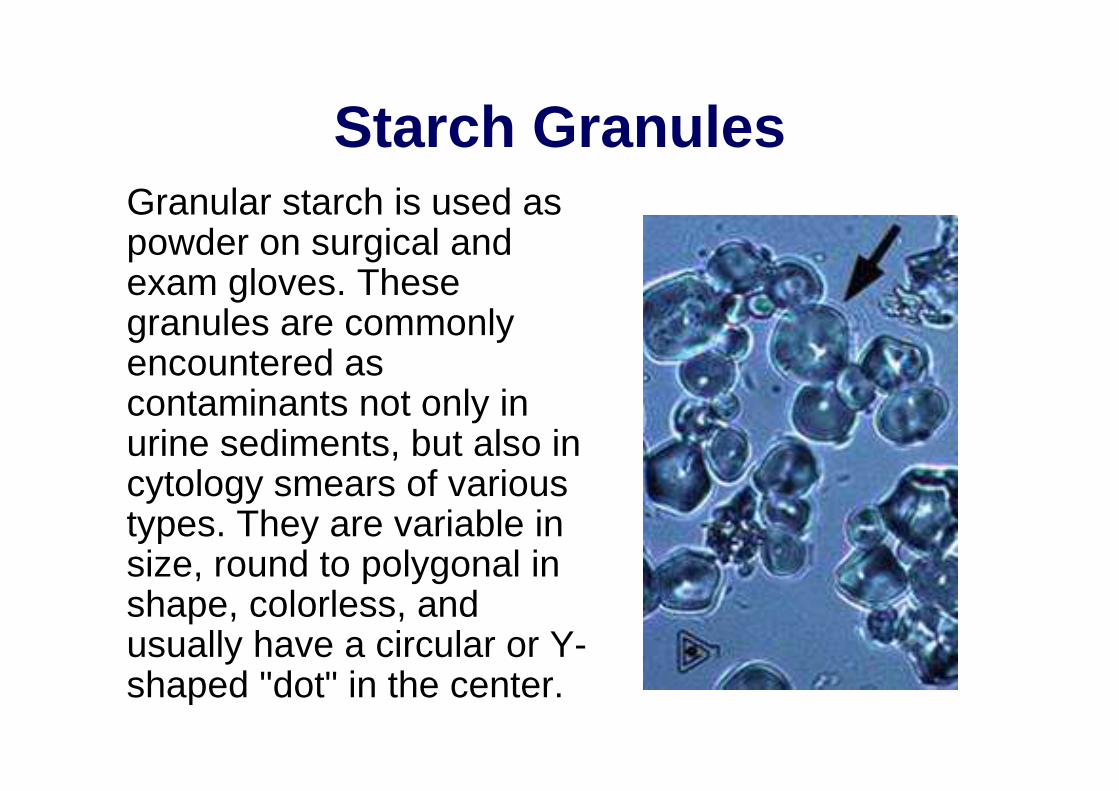
Starch GranulesGranular starch is used as powder on surgical andexam gloves. These granules are commonlyencountered as contaminants not only in urine sediments, but also in cytology smears of varioustypes. They are variable in size, round to polygonal in shape, colorless, andusually have a circular or Y-shaped "dot" in the center.
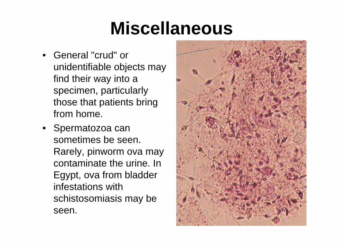
Miscellaneous• General "crud" or
unidentifiable objects mayfind their way into a specimen, particularlythose that patients bringfrom home.
• Spermatozoa cansometimes be seen. Rarely, pinworm ova maycontaminate the urine. In Egypt, ova from bladderinfestations withschistosomiasis may beseen.
![Urine analysis analysis[3359].pdfUrine sediment (Microscopic examination of urine sediment) •Should be performed by trained lab staff •Crystals –uric acid, Ca P or oxalate, Cysteine,](https://img.pdfslide.us/doc/110x75/5ec80a2cfe46c315f91a2ba4/urine-analysis-analysis3359pdf-urine-sediment-microscopic-examination-of-urine.jpg)