Embed Size (px)
Citation preview
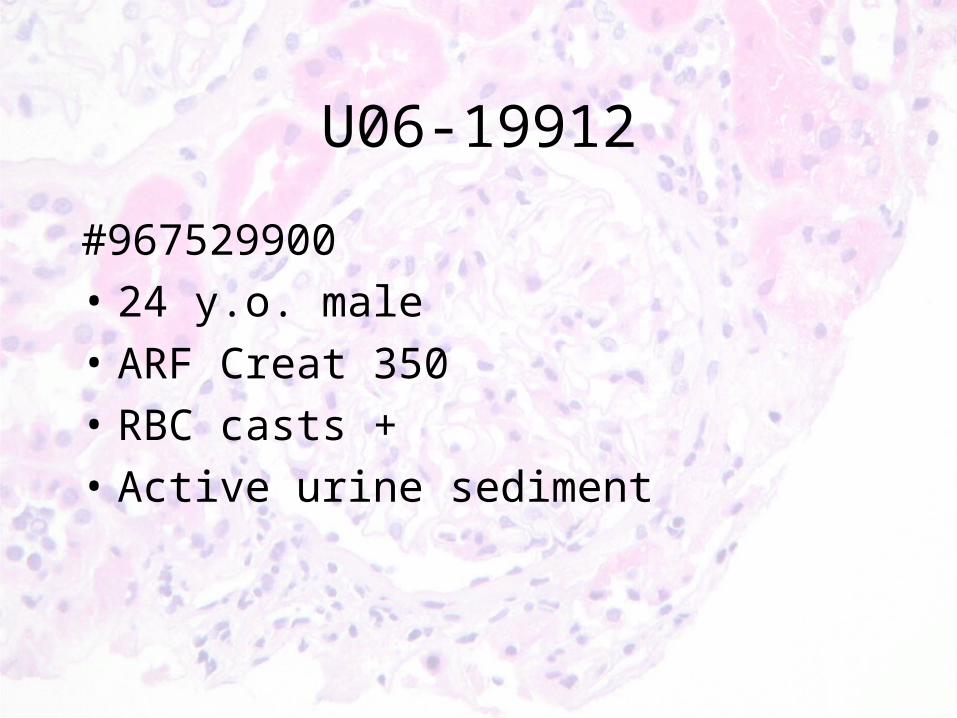
U06-19912
#967529900
• 24 y.o. male
• ARF Creat 350
• RBC casts +
• Active urine sediment
Renal Biopsy Rounds (November 3):
24 yo Asian male, previously well. ARF Crmax=355(spontaneously resolved)
HPI
• Sept-Oct: Strenuous training for Edmonton Police Service Exam x several weeks (weight lifting, aerobic exercise)
• Unwell x 1 week prior to presentation: URTI and fatigue rests
• Few days later: intermittent R sided abdominal pain (no flank pain, no radiation, no hematuria); no N/V/diarrhea
• October 18: performs EPS exam to point of exhaustion and syncope x 10 seconds (no preceding SOB/CP/palpitations, no seizure activity) ER. Cr 291
• ROS: No change in urine volume/color, no lower urinary sx. No constitutional/CTD sx. No SOB/orthopnea/edema. No cough/hemoptysis. No NSAIDS/tylenol/steroids/herbs (only whey protein supplements). No IVDU.
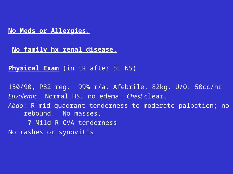
No Meds or Allergies.
No family hx renal disease.
Physical Exam (in ER after 5L NS)
150/90, P82 reg. 99% r/a. Afebrile. 82kg. U/O: 50cc/hr
Euvolemic. Normal HS, no edema. Chest clear.
Abdo: R mid-quadrant tenderness to moderate palpation; no rebound. No masses.
? Mild R CVA tenderness
No rashes or synovitis
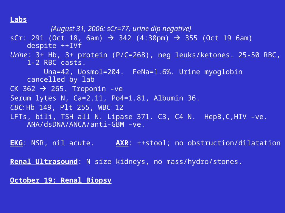
Labs [August 31, 2006: sCr=77, urine dip negative]sCr: 291 (Oct 18, 6am) 342 (4:30pm) 355 (Oct 19 6am) despite ++IVfUrine: 3+ Hb, 3+ protein (P/C=268), neg leuks/ketones. 25-50 RBC, 1-2 RBC casts. Una=42, Uosmol=204. FeNa=1.6%. Urine myoglobin cancelled by labCK 362 265. Troponin -ve Serum lytes N, Ca=2.11, Po4=1.81, Albumin 36. CBC: Hb 149, Plt 255, WBC 12LFTs, bili, TSH all N. Lipase 371. C3, C4 N. HepB,C,HIV –ve.
ANA/dsDNA/ANCA/anti-GBM –ve.
EKG: NSR, nil acute. AXR: ++stool; no obstruction/dilatation
Renal Ultrasound: N size kidneys, no mass/hydro/stones.
October 19: Renal Biopsy
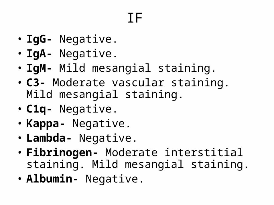
IF
• IgG- Negative.• IgA- Negative.• IgM- Mild mesangial staining. • C3- Moderate vascular staining. Mild mesangial
staining.• C1q- Negative.• Kappa- Negative.• Lambda- Negative.• Fibrinogen- Moderate interstitial staining. Mild
mesangial staining.• Albumin- Negative.
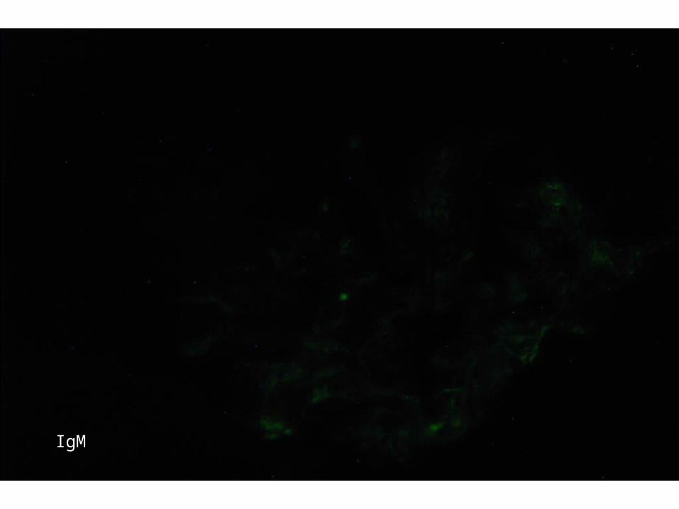
IgM
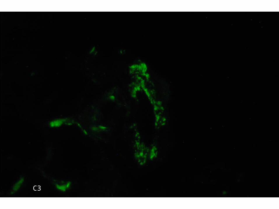
C3
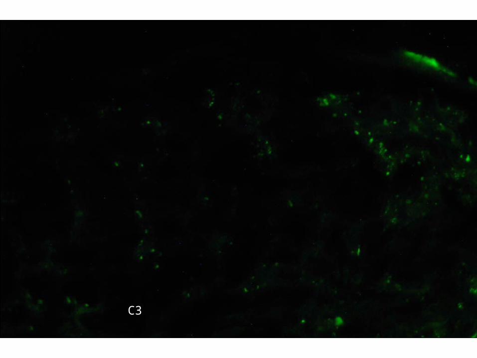
C3
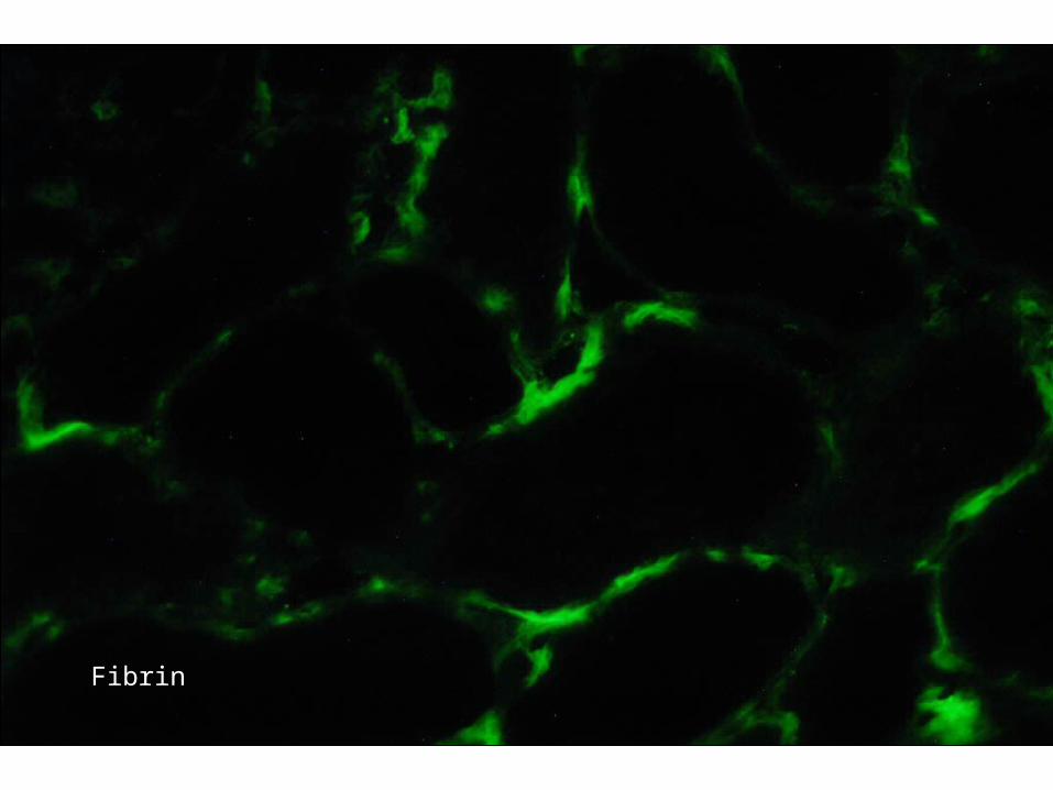
Fibrin
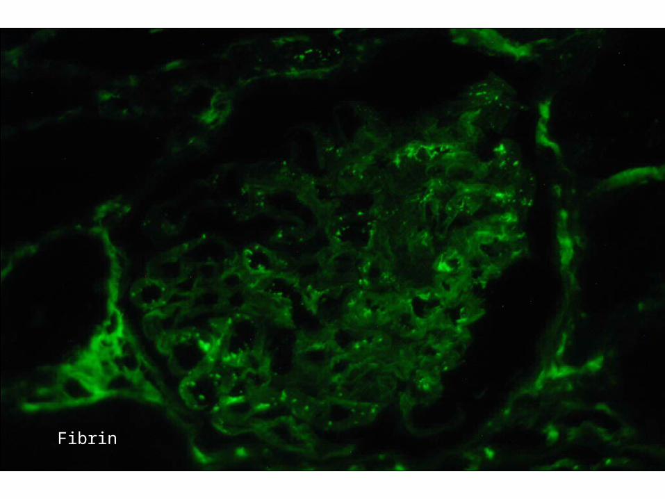
Fibrin
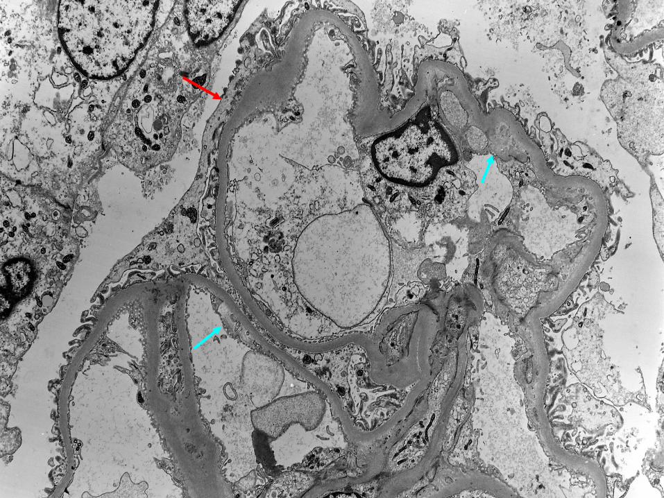
DiagnosisRenal Biopsy:
• Patchy severe congestion of glomeruli, arterioles and capillaries with foci of intense polymorph infiltrate.
• Rule out parasitic disease.
Comment
• Medullary foci of PMN inflammation and intense congestion are quite impressive and unusual
• It is likely that there has been considerable regional venous stasis brought about by the disease process (probable parasite forms in the cortex?) that has probably lead to the renal functional impairment
Comment
• Renal vein thrombosis seems ruled out by imaging studies.
• The congestion suggests that within the kidney there is quite profound venous obstruction or impairment of blood flow.
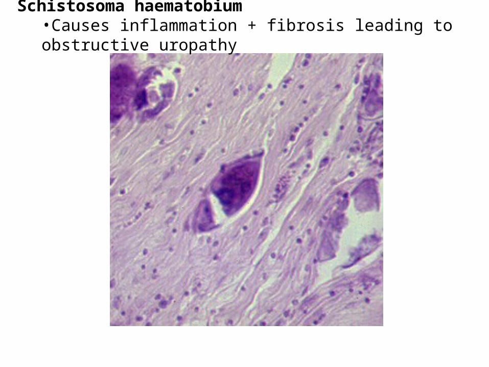
Schistosoma haematobium•Causes inflammation + fibrosis leading to obstructive uropathy
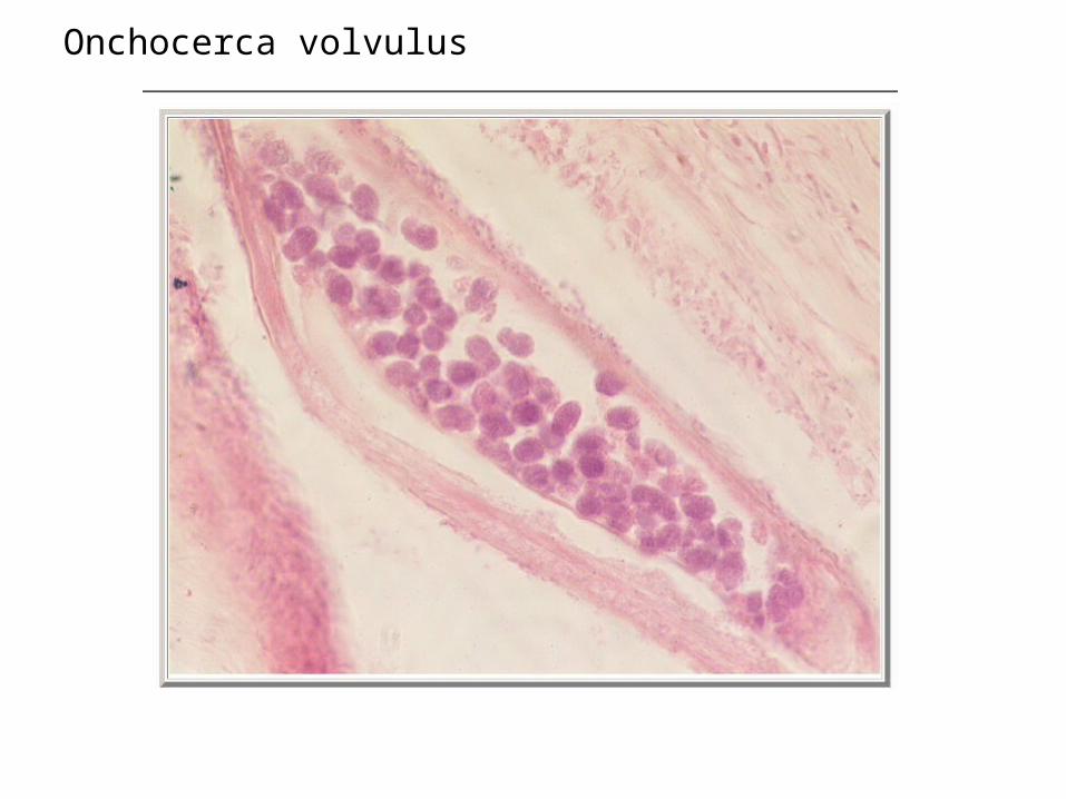
Onchocerca volvulus
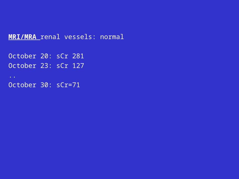
MRI/MRA renal vessels: normal
October 20: sCr 281
October 23: sCr 127
..
October 30: sCr=71