Embed Size (px)
Citation preview
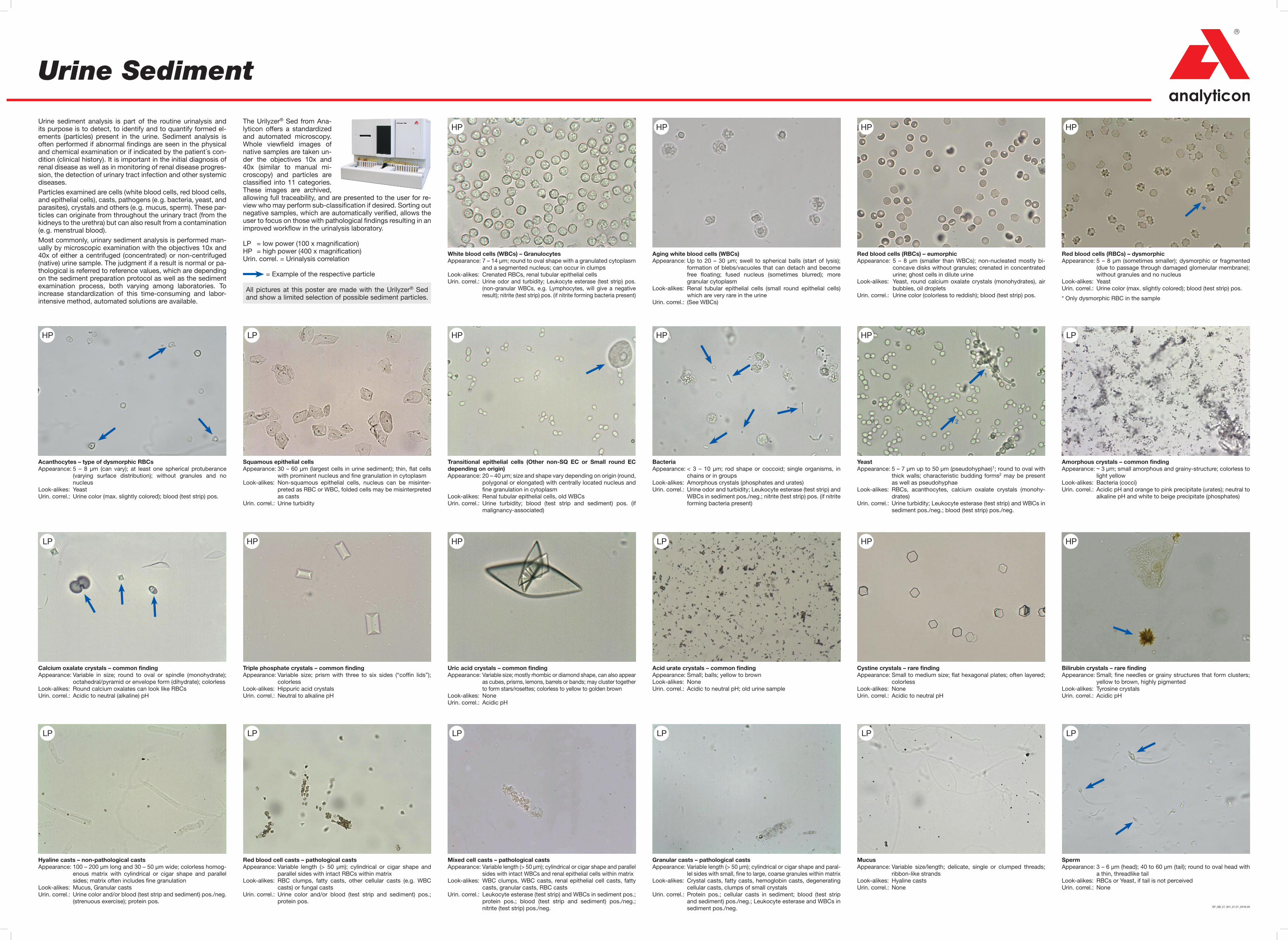
Urine Sediment
Calcium oxalate crystals – common findingAppearance: Variable in size; round to oval or spindle (monohydrate);
octahedral/pyramid or envelope form (dihydrate); colorlessLook-alikes: Round calcium oxalates can look like RBCs Urin. correl.: Acidic to neutral (alkaline) pH
Triple phosphate crystals – common findingAppearance: Variable size; prism with three to six sides (“coffin lids”);
colorlessLook-alikes: Hippuric acid crystalsUrin. correl.: Neutral to alkaline pH
Uric acid crystals – common findingAppearance: Variable size; mostly rhombic or diamond shape, can also appear
as cubes, prisms, lemons, barrels or bands; may cluster together to form stars/rosettes; colorless to yellow to golden brown
Look-alikes: NoneUrin. correl.: Acidic pH
Acid urate crystals – common findingAppearance: Small; balls; yellow to brownLook-alikes: NoneUrin. correl.: Acidic to neutral pH; old urine sample
Cystine crystals – rare findingAppearance: Small to medium size; flat hexagonal plates; often layered;
colorlessLook-alikes: NoneUrin. correl.: Acidic to neutral pH
Bilirubin crystals – rare findingAppearance: Small; fine needles or grainy structures that form clusters;
yellow to brown, highly pigmentedLook-alikes: Tyrosine crystalsUrin. correl.: Acidic pH
LP HP HP HPHPLP
Hyaline casts – non-pathological castsAppearance: 100 – 200 µm long and 30 – 50 µm wide; colorless homog-
enous matrix with cylindrical or cigar shape and parallel sides; matrix often includes fine granulation
Look-alikes: Mucus, Granular castsUrin. correl.: Urine color and/or blood (test strip and sediment) pos./neg.
(strenuous exercise); protein pos.
Red blood cell casts – pathological castsAppearance: Variable length (> 50 µm); cylindrical or cigar shape and
parallel sides with intact RBCs within matrixLook-alikes: RBC clumps, fatty casts, other cellular casts (e.g. WBC
casts) or fungal castsUrin. correl.: Urine color and/or blood (test strip and sediment) pos.;
protein pos.
Mixed cell casts – pathological castsAppearance: Variable length (> 50 µm); cylindrical or cigar shape and parallel
sides with intact WBCs and renal epithelial cells within matrixLook-alikes: WBC clumps, WBC casts, renal epithelial cell casts, fatty
casts, granular casts, RBC castsUrin. correl.: Leukocyte esterase (test strip) and WBCs in sediment pos.;
protein pos.; blood (test strip and sediment) pos./neg.; nitrite (test strip) pos./neg.
LP LP LP
MucusAppearance: Variable size/length; delicate, single or clumped threads;
ribbon-like strands Look-alikes: Hyaline castsUrin. correl.: None
LP
Granular casts – pathological castsAppearance: Variable length (> 50 µm); cylindrical or cigar shape and paral-
lel sides with small, fine to large, coarse granules within matrixLook-alikes: Crystal casts, fatty casts, hemoglobin casts, degenerating
cellular casts, clumps of small crystalsUrin. correl.: Protein pos.; cellular casts in sediment; blood (test strip
and sediment) pos./neg.; Leukocyte esterase and WBCs in sediment pos./neg.
LP
SpermAppearance: 3 – 6 µm (head); 40 to 60 µm (tail); round to oval head with
a thin, threadlike tailLook-alikes: RBCs or Yeast, if tail is not perceivedUrin. correl.: None
LP
BacteriaAppearance: < 3 – 10 µm; rod shape or coccoid; single organisms, in
chains or in groupsLook-alikes: Amorphous crystals (phosphates and urates)Urin. correl.: Urine odor and turbidity; Leukocyte esterase (test strip) and
WBCs in sediment pos./neg.; nitrite (test strip) pos. (if nitrite forming bacteria present)
YeastAppearance: 5 – 7 µm up to 50 µm (pseudohyphae)1; round to oval with
thick walls; characteristic budding forms2 may be present as well as pseudohyphae
Look-alikes: RBCs, acanthocytes, calcium oxalate crystals (monohy-drates)
Urin. correl.: Urine turbidity; Leukocyte esterase (test strip) and WBCs in sediment pos./neg.; blood (test strip) pos./neg.
Amorphous crystals – common findingAppearance: ~ 3 µm; small amorphous and grainy-structure; colorless to
light yellowLook-alikes: Bacteria (cocci)Urin. correl.: Acidic pH and orange to pink precipitate (urates); neutral to
alkaline pH and white to beige precipitate (phosphates)
Squamous epithelial cellsAppearance: 30 – 60 µm (largest cells in urine sediment); thin, flat cells
with prominent nucleus and fine granulation in cytoplasmLook-alikes: Non-squamous epithelial cells, nucleus can be misinter-
preted as RBC or WBC, folded cells may be misinterpreted as casts
Urin. correl.: Urine turbidity
LP
Transitional epithelial cells (Other non-SQ EC or Small round EC depending on origin)Appearance: 20 – 40 µm; size and shape vary depending on origin (round,
polygonal or elongated) with centrally located nucleus and fine granulation in cytoplasm
Look-alikes: Renal tubular epithelial cells, old WBCsUrin. correl.: Urine turbidity; blood (test strip and sediment) pos. (if
malignancy-associated)
HP HPHP LP
Acanthocytes – type of dysmorphic RBCsAppearance: 5 – 8 µm (can vary); at least one spherical protuberance
(varying surface distribution); without granules and no nucleus
Look-alikes: YeastUrin. correl.: Urine color (max. slightly colored); blood (test strip) pos.
HP
1
2
Aging white blood cells (WBCs)Appearance: Up to 20 – 30 µm; swell to spherical balls (start of lysis);
formation of blebs/vacuoles that can detach and become free floating; fused nucleus (sometimes blurred); more granular cytoplasm
Look-alikes: Renal tubular epithelial cells (small round epithelial cells) which are very rare in the urine
Urin. correl.: (See WBCs)
Red blood cells (RBCs) – eumorphicAppearance: 5 – 8 µm (smaller than WBCs); non-nucleated mostly bi-
concave disks without granules; crenated in concentrated urine; ghost cells in dilute urine
Look-alikes: Yeast, round calcium oxalate crystals (monohydrates), air bubbles, oil droplets
Urin. correl.: Urine color (colorless to reddish); blood (test strip) pos.
Red blood cells (RBCs) – dysmorphicAppearance: 5 – 8 µm (sometimes smaller); dysmorphic or fragmented
(due to passage through damaged glomerular membrane); without granules and no nucleus
Look-alikes: YeastUrin. correl.: Urine color (max. slightly colored); blood (test strip) pos.
* Only dysmorphic RBC in the sample
Urine sediment analysis is part of the routine urinalysis and its purpose is to detect, to identify and to quantify formed el-ements (particles) present in the urine. Sediment analysis is often performed if abnormal findings are seen in the physical and chemical examination or if indicated by the patient´s con-dition (clinical history). It is important in the initial diagnosis of renal disease as well as in monitoring of renal disease progres-sion, the detection of urinary tract infection and other systemic diseases.Particles examined are cells (white blood cells, red blood cells, and epithelial cells), casts, pathogens (e.g. bacteria, yeast, and parasites), crystals and others (e.g. mucus, sperm). These par-ticles can originate from throughout the urinary tract (from the kidneys to the urethra) but can also result from a contamination (e.g. menstrual blood).Most commonly, urinary sediment analysis is performed man-ually by microscopic examination with the objectives 10x and 40x of either a centrifuged (concentrated) or non-centrifuged (native) urine sample. The judgment if a result is normal or pa-thological is referred to reference values, which are depending on the sediment preparation protocol as well as the sediment examination process, both varying among laboratories. To increase standardization of this time-consuming and labor- intensive method, automated solutions are available.
The Urilyzer® Sed from Ana-lyticon offers a standardized and automated microscopy. Whole viewfield images of native samples are taken un-der the objectives 10x and 40x (similar to manual mi-croscopy) and particles are classified into 11 categories. These images are archived, allowing full traceability, and are presented to the user for re-view who may perform sub-classification if desired. Sorting out negative samples, which are automatically verified, allows the user to focus on those with pathological findings resulting in an improved workflow in the urinalysis laboratory.
LP = low power (100 x magnification)HP = high power (400 x magnification)Urin. correl. = Urinalysis correlation
= Example of the respective particle
White blood cells (WBCs) – GranulocytesAppearance: 7 – 14 µm; round to oval shape with a granulated cytoplasm
and a segmented nucleus; can occur in clumpsLook-alikes: Crenated RBCs, renal tubular epithelial cellsUrin. correl.: Urine odor and turbidity; Leukocyte esterase (test strip) pos.
(non-granular WBCs, e.g. Lymphocytes, will give a negative result); nitrite (test strip) pos. (if nitrite forming bacteria present)
All pictures at this poster are made with the Urilyzer® Sed and show a limited selection of possible sediment particles.
*
HP HPHPHP
SP_GB_27_001_01.01_2018-03
![Urine analysis analysis[3359].pdfUrine sediment (Microscopic examination of urine sediment) •Should be performed by trained lab staff •Crystals –uric acid, Ca P or oxalate, Cysteine,](https://img.pdfslide.us/doc/110x75/5ec80a2cfe46c315f91a2ba4/urine-analysis-analysis3359pdf-urine-sediment-microscopic-examination-of-urine.jpg)