Embed Size (px)
DESCRIPTION
mnm
Citation preview
Micturition Micturition is the process by which the
urinary bladder empties when it becomes
filled. This involves two main steps:
First, the bladder fills progressively until
the tension in its walls rises above a
threshold level; this elicits the
second step, which is a nervous reflex
called the micturition reflex that empties
the bladder or, if this fails, at least causes
a conscious desire to urinate.
Although the micturition reflex is an
autonomic spinal cord reflex, it can also
be inhibited or facilitated by centers in
the cerebral cortex or brain stem.
Physiologic Anatomy and Nervous
Connections of the Bladder
The urinary bladder is a smooth muscle
chamber composed of two main parts:
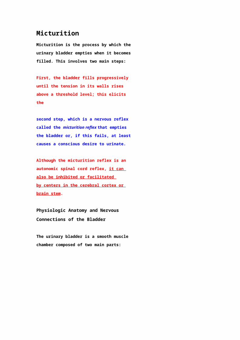
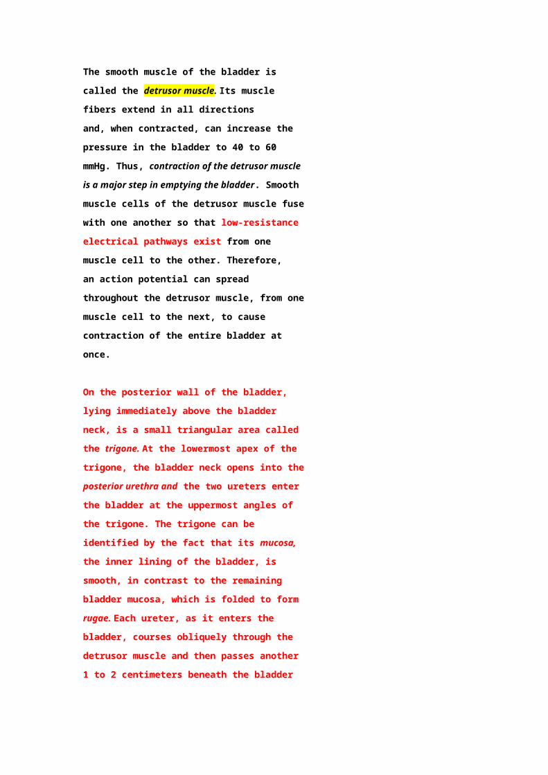
(1) the body, which is the major part of
the bladder in which urine collects and
(2) the neck, which is a funnel-
shaped extension of the body, passing
inferiorly and anteriorly into the
urogenital triangle and connecting with
the urethra. The lower part of the bladder
neck is also called the posterior urethra
because of its relation to the urethra.
The smooth muscle of the bladder is called
the detrusor muscle. Its muscle fibers
extend in all directions and, when
contracted, can increase the pressure in
the bladder to 40 to 60 mmHg. Thus,
contraction of the detrusor muscle is a
major step in emptying the
bladder. Smooth muscle cells of the
detrusor muscle fuse with one another so
that low-resistance electrical
pathways exist from one muscle cell to the
other. Therefore, an action potential can
spread throughout the detrusor muscle,
from one muscle cell to the next, to cause
contraction of the entire bladder at once.
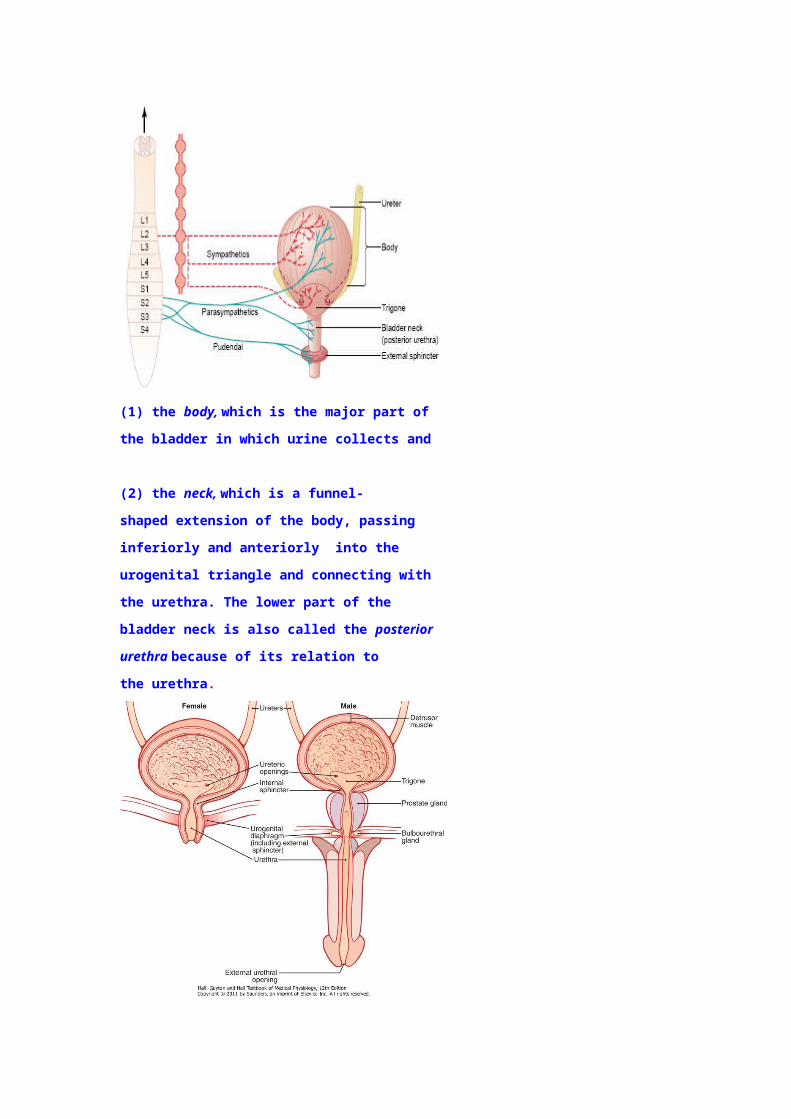
On the posterior wall of the bladder, lying
immediately above the bladder neck, is a
small triangular area called the trigone. At
the lowermost apex of the trigone, the
bladder neck opens into the posterior
urethra and the two ureters enter the
bladder at the uppermost angles of the
trigone. The trigone can be identified
by the fact that its mucosa, the inner
lining of the bladder, is smooth, in
contrast to the remaining bladder
mucosa, which is folded to form rugae.
Each ureter, as it enters the bladder,
courses obliquely through the
detrusor muscle and then passes another
1 to 2 centimeters beneath the bladder
mucosa before emptying into the bladder.
The bladder neck (posterior urethra) is 2
to 3 centimeters long and its wall is
composed of detrusor muscle interlaced
with a large amount of elastic tissue.
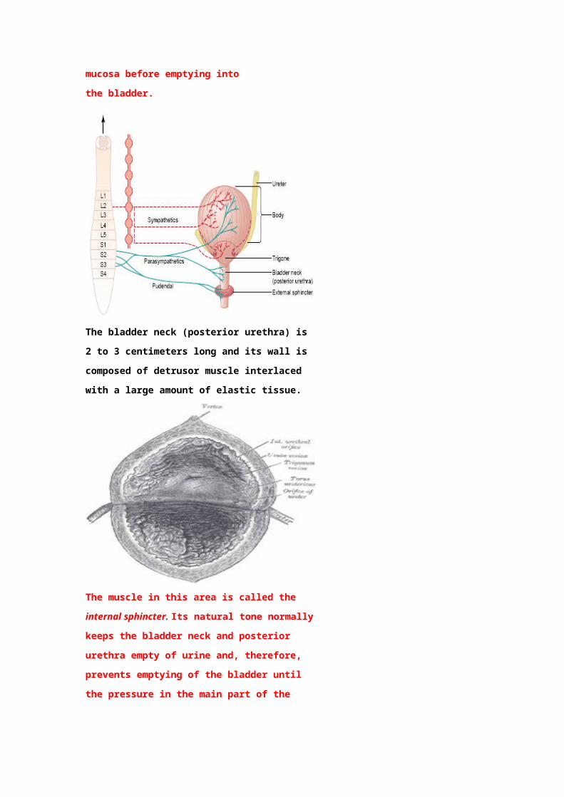
The muscle in this area is called the
internal sphincter. Its natural tone
normally keeps the bladder neck
and posterior urethra empty of urine and,
therefore, prevents emptying of the
bladder until the pressure in the main part
of the bladder rises above a critical
threshold.
Beyond the posterior urethra, the urethra
passes through the urogenital diaphragm,
which contains a layer of muscle called
the external sphincter of the bladder. This
muscle is a voluntary skeletal muscle,
in contrast to the muscle of the bladder
body and bladder neck, which is entirely
smooth muscle. The external sphincter
muscle is under voluntary control of
the nervous system and can be used to
consciously prevent urination even when
involuntary controls are attempting to
empty the bladder.
Innervation of the Bladder
The principal nerve supply of the bladder
is by way of the pelvic nerves, which
connect with the spinal cord through the
sacral plexus, mainly connecting with
cord segments S-2 and S-3.
Coursing through the pelvic nerves are
both sensory nerve fibers and motor
nerve fibers.
The sensory fibers detect the degree of
stretch in the bladder wall.
Stretch signals from the posterior
urethra are especially strong and are
mainly responsible for initiating the
reflexes that cause bladder emptying.
The motor nerves transmitted in the
pelvic nerves are parasympathetic
fibers. These terminate on ganglion cells
located in the wall of the bladder. Short
postganglionic nerves then innervate
the detrusor muscle.
In addition to the pelvic nerves, two other
types of innervation are important in
bladder function.
1- Most important are the skeletal
motor fibers transmitted through
the pudendal nerve to the external
bladder sphincter. These are
somatic nerve fibers that
innervate and control the voluntary
skeletal muscle of the sphincter.
2- Also, the bladder receives
sympathetic innervation from the
sympathetic chain through the
hypogastric nerves, connecting
mainly with the L-2 segment of
the spinal cord. These sympathetic
fibers stimulate mainly the blood
vessels and have little to do with
bladder contraction.
Some sensory nerve fibers also pass
by way of the sympathetic nerves
and may be important in
the sensation of fullness and, in
some instances, pain.
Transport of Urine from the Kidney
Through the Ureters and into the
Bladder
Urine that is expelled from the bladder
has essentially the same composition as
fluid flowing out of the collecting ducts;
there are no significant changes in
the composition of urine as it flows
through the renal calyces and ureters to
the bladder.
Urine flowing from the collecting ducts
into the renal calyces stretches the
calyces and increases their inherent
pacemaker activity, which in turn
initiates peristaltic contractions that
spread to the renal pelvis and then
downward along the length of the
ureter, thereby forcing urine from the
renal pelvis toward the bladder.
The walls of the ureters contain
smooth muscle and are innervated by both
sympathetic and parasympathetic nerves
as well as by an intramural plexus of
neurons and nerve fibers that extends
along the entire length of the ureters. As
with other visceral smooth muscles, peristaltic
contractions in the ureter are enhanced by
parasympathetic stimulation and inhibited by
sympathetic stimulation.
The normal tone of the detrusor muscle in
the bladder wall tends to compress the
ureter, thereby preventing backflow of
urine from the bladder when pressure
builds up in the bladder during micturition
or bladder compression.
Each peristaltic wave along the
ureter increases the pressure within the
ureter so that the region passing through
the bladder wall opens and allows urine to
flow into the bladder.
In some people, the distance that the
ureter courses through the bladder wall is
less than normal, so that contraction of
the bladder during micturition does
not always lead to complete occlusion of
the ureter. As a result, some of the urine
in the bladder is propelled backward into
the ureter, a condition called
vesicoureteral reflux. Such reflux can lead
to enlargement of the ureters and, if
severe, can increase the pressure in the
renal calyces and structures of the
renal medulla, causing damage to these
regions.
Pain Sensation in the Ureters
and the Uretero-renal Reflex.
The ureters are well supplied with pain
nerve fibers. When a ureter becomes
blocked (e.g., by a ureteral stone), intense
reflex constriction occurs, associated with
severe pain. Also, the pain impulses cause
a sympathetic reflex back to the kidney to
constrict the renal arterioles, thereby
decreasing urine output from the kidney.
This effect is called the ureterorenal reflex
and is important for preventing excessive
flow of fluid into the pelvis of a kidney
with a blocked ureter.
Filling of the Bladder and
Bladder Wall Tone;
the Cystometrogram
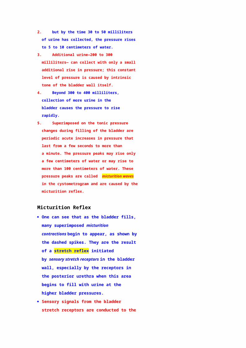
Figure 26–7 shows the approximate changes in
intravesical pressure as the bladder fills with
urine.
1. When there is no urine in the bladder, the
intravesical pressure is about 0,
2. but by the time 30 to 50 milliliters of urine
has collected, the pressure rises to 5 to 10
centimeters of water.
3. Additional urine—200 to 300 milliliters— can
collect with only a small additional rise
in pressure; this constant level of pressure is
caused by intrinsic tone of the bladder wall
itself.
4. Beyond 300 to 400 milliliters, collection of
more urine in the bladder causes the
pressure to rise rapidly.
5. Superimposed on the tonic pressure changes
during filling of the bladder are periodic
acute increases in pressure that last from a
few seconds to more than a minute. The
pressure peaks may rise only a few
centimeters of water or may rise to more
than 100 centimeters of water. These
pressure peaks are called micturition waves
in the cystometrogram and are caused by
the micturition reflex.
Micturition Reflex
One can see that as the bladder fills,
many superimposed micturition
contractions begin to appear, as shown
by the dashed spikes. They are the
result of a stretch reflex initiated
by sensory stretch receptors in the
bladder wall, especially by the receptors
in the posterior urethra when this area
begins to fill with urine at the higher
bladder pressures.
Sensory signals from the bladder stretch
receptors are conducted to the sacral
segments of the cord through the pelvic
nerves and then reflexively back again
to the bladder through the
parasympathetic nerve fibers by way of
these same nerves.
When the bladder is only partially filled ,
these micturition contractions usually
relax spontaneously after a fraction of a
minute, the detrusor muscles stop
contracting and pressure falls back to
the baseline.
As the bladder continues to fill, the
micturition reflexes become more
frequent and cause greater
contractions of the detrusor muscle.
Once a micturition reflex begins, it is
“self-regenerative.” That is, initial
contraction of the bladder activates the
stretch receptors to cause a greater
increase in sensory impulses from the
bladder and posterior urethra, which
causes a further increase in reflex
contraction of the bladder; thus, the
cycle is repeated again and again until
the bladder has reached a strong degree
of contraction.
Then, after a few seconds to more than
a minute, the self-regenerative reflex
begins to fatigue and the regenerative
cycle of the micturition reflex ceases,
permitting the bladder to relax.
Thus, the micturition reflex is a single
complete cycle of :
(1) progressive and
rapid increase of pressure,
(2) a period of
sustained pressure and
(3) return of
the pressure to the basal tone of
the bladder.
Once a micturition reflex has occurred
but has not succeeded in emptying the
bladder, the nervous elements of
this reflex usually remain in an inhibited
state for a few minutes to 1 hour or
more before another micturition reflex
occurs.
As the bladder becomes more and
more filled, micturition reflexes occur
more and more often and more and
more powerfully.
Once the micturition reflex becomes
powerful enough, it causes another
reflex, which passes through the
pudendal nerves to the external
sphincter to inhibit it.
If this inhibition is more potent in the
brain than the voluntary constrictor
signals to the external
sphincter, urination will occur.
If not enough potent, urination will not
occur until the bladder fills still further
and the micturition reflex becomes more
powerful.
Facilitation or Inhibition of Micturition by the
Brain
The micturition reflex is a completely
autonomic spinal cord reflex, but it can be
inhibited or facilitated by centers in the
brain.
These centers include:
(1) strong facilitative and inhibitory
centers in the brain stem, located
mainly in the pons and
(2) several centers located in the
cerebral cortex that are mainly
inhibitory but can become excitatory.
The micturition reflex is the basic cause of
micturition, but the higher centers
normally exert final control of micturition
as follows:
1. The higher centers keep the micturition
reflex partially inhibited, except when
micturition is desired.
2. The higher centers can prevent
micturition, even if the micturition reflex
occurs, by continual tonic contraction of
the external bladder sphincter until a
convenient time presents itself.
3. When it is time to urinate, the cortical
centers can facilitate the sacral
micturition centers to help initiate a
micturition reflex and at the same
time inhibit the external urinary
sphincter so that urination can occur.
4. Voluntary urination is usually initiated in
the following way:
First, a person voluntarily contracts
his or her abdominal muscles, which
increases the pressure in the bladder
and allows extra urine to enter
the bladder neck and posterior
urethra under pressure, thus
stretching their walls. This stimulates
the stretch receptors, which excites
the micturition reflex
and simultaneously inhibits the
external urethral sphincter.
5. Ordinarily, all the urine will be emptied,
with rarely more than 5 to 10 milliliters
left in the bladder.
Abnormalities of Micturition
Atonic Bladder Caused by Destruction
of Sensory Nerve Fibers.
Micturition reflex contraction cannot occur if
the sensory nerve fibers from the bladder to
the spinal cord are destroyed, thereby
preventing transmission of stretch signals from
the bladder. When this happens, a person loses
bladder control, despite intact efferent fibers
from the cord to the bladder and despite
intact neurogenic connections within the brain.
Instead of emptying periodically, the bladder
fills to capacity and overflows a few drops at a
time through the urethra. This is called
overflow incontinence.
A common cause of atonic bladder is
crush injury to the sacral region of the spinal
cord.
Certain diseases can also cause damage to
the dorsal root nerve fibers that enter the
spinal cord. For example, syphilis can
cause constrictive fibrosis around the dorsal
root nerve fibers, destroying them. This
condition is called tabes dorsalis and the
resulting bladder condition is called
tabetic bladder.
Automatic Bladder Caused by Spinal
Cord Damage Above the Sacral
Region.
If the spinal cord is damaged above the sacral
region but the sacral cord segments are still
intact, typical micturition reflexes can still
occur. However, they are no longer controlled
by the brain. During the first few days to
several weeks after the damage to the cord
has occurred, the micturition reflexes are
suppressed because of the state of “spinal
shock” caused by the sudden loss of
facilitative impulses from the brain stem and
cerebrum. However, if the bladder is
emptied periodically by catheterization to
prevent bladder injury caused by
overstretching of the bladder, the
excitability of the micturition reflex gradually
increases until typical micturition reflexes
return; then, periodic (but
unannounced) bladder emptying occurs.
Some patients can still control urination in this
condition by stimulating the skin (scratching or
tickling) in the genital region, which
sometimes elicits a micturition reflex.
Uninhibited Neurogenic Bladder
Caused by
Lack of Inhibitory Signals from the
Brain.
Another abnormality of micturition is the so-
called uninhibited neurogenic bladder, which
results in frequent and relatively
uncontrolled micturition. This condition derives
from partial damage in the spinal cord or the
brain stem that interrupts most of the
inhibitory signals. Therefore, facilitative
impulses passing continually down the cord
keep the sacral centers so excitable that even
a small quantity of urine elicits an
uncontrollable micturition reflex, thereby
promoting frequent urination.