Embed Size (px)
Citation preview
Upper GI Endoscopy
Upper GI endoscopy, sometimes called
EGD (esophagogastroduodenoscopy) is a
visual examination of the upper intestinal
tract using a lighted, flexible video
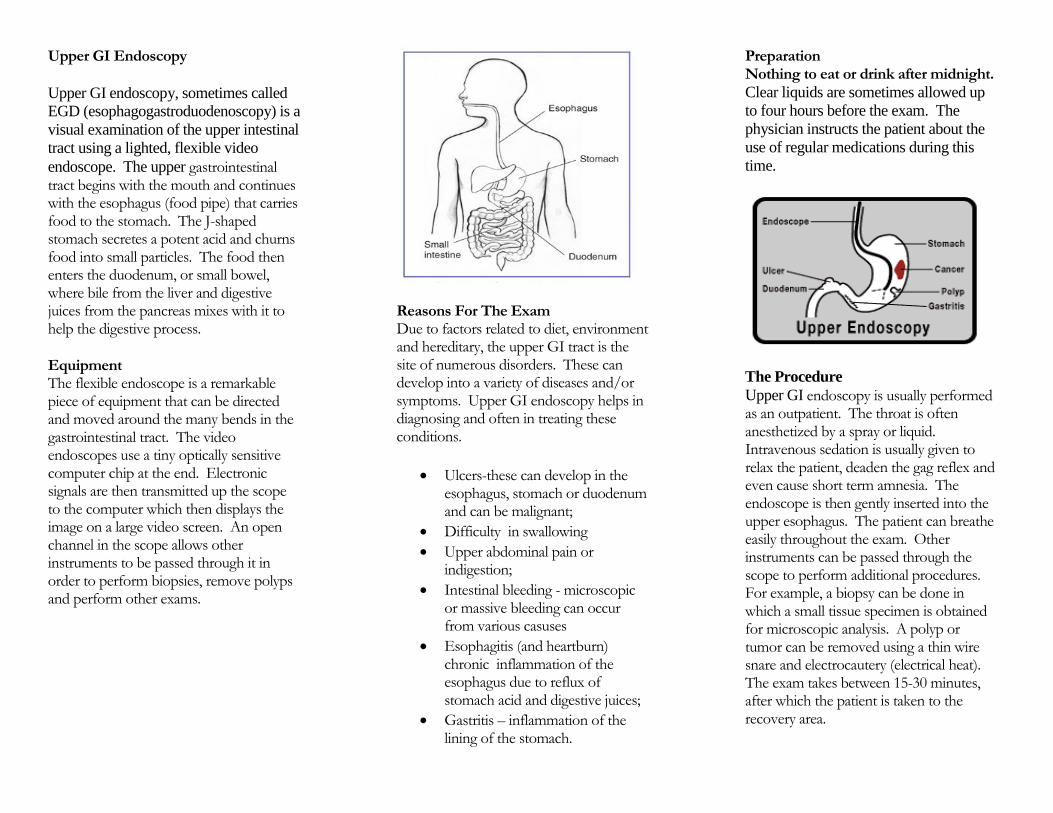
endoscope. The upper gastrointestinal tract begins with the mouth and continues with the esophagus (food pipe) that carries food to the stomach. The J-shaped stomach secretes a potent acid and churns food into small particles. The food then enters the duodenum, or small bowel, where bile from the liver and digestive juices from the pancreas mixes with it to help the digestive process.
Equipment The flexible endoscope is a remarkable piece of equipment that can be directed and moved around the many bends in the gastrointestinal tract. The video endoscopes use a tiny optically sensitive computer chip at the end. Electronic signals are then transmitted up the scope to the computer which then displays the image on a large video screen. An open channel in the scope allows other instruments to be passed through it in order to perform biopsies, remove polyps and perform other exams.
Reasons For The Exam Due to factors related to diet, environment and hereditary, the upper GI tract is the site of numerous disorders. These can develop into a variety of diseases and/or symptoms. Upper GI endoscopy helps in diagnosing and often in treating these conditions.
Ulcers-these can develop in the esophagus, stomach or duodenum and can be malignant;
Difficulty in swallowing
Upper abdominal pain or indigestion;
Intestinal bleeding - microscopic or massive bleeding can occur from various casuses
Esophagitis (and heartburn) chronic inflammation of the esophagus due to reflux of stomach acid and digestive juices;
Gastritis – inflammation of the lining of the stomach.
Preparation Nothing to eat or drink after midnight.
Clear liquids are sometimes allowed up
to four hours before the exam. The
physician instructs the patient about the
use of regular medications during this
time.
The Procedure
Upper GI endoscopy is usually performed as an outpatient. The throat is often anesthetized by a spray or liquid. Intravenous sedation is usually given to relax the patient, deaden the gag reflex and even cause short term amnesia. The endoscope is then gently inserted into the upper esophagus. The patient can breathe easily throughout the exam. Other instruments can be passed through the scope to perform additional procedures. For example, a biopsy can be done in which a small tissue specimen is obtained for microscopic analysis. A polyp or tumor can be removed using a thin wire snare and electrocautery (electrical heat). The exam takes between 15-30 minutes, after which the patient is taken to the recovery area.
Results After the exam, the physician will explain the results to the patient and family. If the effects of sedatives are prolonged, the physician may suggest an interview at a later date when the results can be fully understood. If a biopsy has been performed or a polyp removed the results of these are not available for three to seven days.
Benefits An upper GI endoscopy is performed primarily to identify and/or correct a problem in the upper gastrointestinal tract. This means the test enables a diagnosis to be made upon which specific treatment can be given. If a bleeding site is identified, treatment can be administered to stop the bleeding, or if a polyp is found, it can be removed without the need for a major operation. Other treatments can be given through the endoscope when necessary.
Alternative Testing Alternative tests to upper GI endoscopy include barium x-ray and ultrasound (sonogram) that study the organs of the upper abdomen. These exams, however, do not allow direct visualization of the esophagus, stomach and duodenum, or removal of polyps or biopsies. In addition, study of the stools, blood, and stomach juices can provide certain indirect information about a gastrointestinal condition.
Side Effects and Risks A temporary, mild sore throat sometimes
occurs after the exam. Serious risks with upper GI endoscopy however are very uncommon. One such risk is excessive bleeding especially with the removal of a polyp. In extremely rare instances a perforation, or tear in the esophagus can occur. These complications may require hospitalization and, rarely, surgery. Quite uncommonly, a diagnostic error or oversight may occur. Due to the mild sedation, the patient should not drive or operate machinery following the exam. For this reason, a driver must be available.
In Summary Upper GI endoscopy is a simple out-patient exam that is often performed with the patient lightly sedated. The procedure provides significant information upon which specific treatment can be given. In certain cases, therapy can be administered directly through the endoscope. Serious complications rarely occur from an upper GI endoscopy. The physician can answer any questions that the patient has.
Special Instructions:
Upper GI Endoscopy
310 West Ninth Street
Frederick, Maryland 21701
301-695-6800