Embed Size (px)
Citation preview

University of Birmingham
Lessons learned from two decades of anticancerdrugsLiu, Zhichao; Delavan, Brian ; Roberts, Ruth; Tong, Weida
DOI:10.1016/j.tips.2017.06.005
License:Creative Commons: Attribution-NonCommercial-NoDerivs (CC BY-NC-ND)
Document VersionPeer reviewed version
Citation for published version (Harvard):Liu, Z, Delavan, B, Roberts, R & Tong, W 2017, 'Lessons learned from two decades of anticancer drugs', Trendsin Pharmacological Sciences, vol. 38, no. 10, pp. 852-872. https://doi.org/10.1016/j.tips.2017.06.005
Link to publication on Research at Birmingham portal
Publisher Rights Statement:Checked for eligibility: 19/07/2019
This document is the Author Accepted Manuscript version of a published work which appears in its final form in Trends in PharmacologicalSciences. The final Version of Record can be found at: https://doi.org/10.1016/j.tips.2017.06.005
General rightsUnless a licence is specified above, all rights (including copyright and moral rights) in this document are retained by the authors and/or thecopyright holders. The express permission of the copyright holder must be obtained for any use of this material other than for purposespermitted by law.
•Users may freely distribute the URL that is used to identify this publication.•Users may download and/or print one copy of the publication from the University of Birmingham research portal for the purpose of privatestudy or non-commercial research.•User may use extracts from the document in line with the concept of ‘fair dealing’ under the Copyright, Designs and Patents Act 1988 (?)•Users may not further distribute the material nor use it for the purposes of commercial gain.
Where a licence is displayed above, please note the terms and conditions of the licence govern your use of this document.
When citing, please reference the published version.
Take down policyWhile the University of Birmingham exercises care and attention in making items available there are rare occasions when an item has beenuploaded in error or has been deemed to be commercially or otherwise sensitive.
If you believe that this is the case for this document, please contact [email protected] providing details and we will remove access tothe work immediately and investigate.
Download date: 05. Dec. 2021
1
1
Lessons Learned from Two Decades of Anticancer Drugs 2
3
Zhichao Liu1*, Brian Delavan1, 2, Ruth Roberts3, 4, Weida Tong1* 4
5
1 National Center for Toxicological Research, U.S. Food and Drug Administration, Jefferson, 6
Arkansas, 72079, USA 7
2 University of Arkansas at Little Rock, Little Rock, AR, 72204, USA 8
3 ApconiX, BioHub at Alderley Park, Alderley Edge, SK10 4TG, UK 9
4 University of Birmingham, Edgbaston, Birmingham, B15 2TT, UK 10
11
* To whom correspondence should be addressed: 12
[email protected] or [email protected] 13
14
Disclaimer: The views presented in this article do not necessarily reflect current or future 15
opinion or policy of the US Food and Drug Administration. Any mention of commercial products 16
is for clarification and not intended as an endorsement. 17
18
19
2
Tremendous efforts have been made to elucidate the basis of cancer biology with the aim of 20
promoting anticancer drug development. Especially in the past twenty years, anticancer drug 21
development has developed from conventional cytotoxic agents to target-based and immune-22
related therapies. Consequently, more than 200 anticancer drugs are available on the market. 23
However, anticancer drug development still suffers high attrition in the later phases of clinical 24
development and is considered to be a difficult and risky therapeutic category within the drug 25
development arena. The disappointing performance of investigational anticancer candidates 26
implies that there are some shortcomings in the translation of preclinical in vitro and in vivo 27
models to humans, and that heterogeneity in the patient population presents a significant 28
challenge. Here, we summarize both successful and failed experiences in anticancer 29
development during the past 20 years and help identify why the paradigm may be suboptimal. 30
We also offer potential strategies for improvement. 31
32
Current progress of anticancer drug development 33
Cancer, which is characterized by uncontrolled growth of cells in the body, is one of the most 34
difficult and complex diseases to treat [1-3]. Cancer patients suffer high mortality rates, which 35
range from 1.1% for prostate cancer to 92.3% for pancreatic cancer within five years after 36
cancer diagnosis. Therefore, anticancer drug research and development (R&D) is a challenging 37
and daunting activity, and the likelihood of failure is high [4]. Fewer than 5% of developed 38
anticancer compounds reach the market [5]. Furthermore, compared to other therapeutic 39
categories such as cardiovascular disease and arthritis, an anticancer drug has approximately 40
one-third to one-half greater failure rate per attempt [5, 6]. Although there are a lot of 41
difficulties and barriers in anticancer drug development, drug makers are still pursuing 42
opportunities for anticancer drug candidates due to their high cost-benefit rate [7, 8]. For 43
example, oncology is ranked in the top therapeutic class by global sales which amounted to 44
78.94 billion US dollars in 2015. 45
3
Approved anticancer drugs 46
The ultimate task for an anticancer drug is to kill the tumor cells and/or control proliferation of 47
tumor cells to prolong patient survival and improve their quality of life. However, there are 48
many different mechanisms by which this can be achieved. Based on the key biochemical 49
mechanism of anticancer action, anticancer drugs can be are categorized as: (i) nucleic acid 50
biosynthesis blocker; (ii) the structure and function of DNA interferer; (iii) transcription interferer 51
and RNA synthesis blocker;(iv) protein synthesis and function interferer; (v) hormone 52
homeostasis influencer; (vi) immune system modulators. Consequently, drug makers have 53
produced four major groups of anticancer drugs including cytotoxic drugs (alkylating agents, 54
antimetabolites, antibiotics, plant extracts, and miscellaneous cytotoxic drugs), targeted-based 55
agents (e.g. bevacizumab), hormones and hormones antagonists (e.g. tamoxifen), and 56
immunomodulators (e.g. nivolumab). From the classic “Seed and soil hypothesis” that first 57
described metastasis [9] to the first description of “immune-based cancer therapy” [10], every 58
milestone made in the cancer field has driven a wave of anticancer drug development [11] 59
(Figure 1). In the past two decades, anticancer drug development has moved on from 60
conventional non-specific cytotoxic agents which often kill proliferating normal cells as well as 61
tumor cells [12, 13]. In the place of cytotoxic agents, there is a focus on specific target-based 62
cancer therapy [14] designed to hit tumor cells only, and on immune-related modulators that 63
help the patient’s immune system to defeat tumor cells [10, 15, 16]. Furthermore, a series of 64
regulations, initiatives, and guidance have been developed to facilitate anticancer drug 65
development [17, 18]. 66
According to the USA National Cancer Institute drug repository, there are a total of 227 67
approved anticancer drugs (Supplementary Table S1) to treat about 40 different types of 68
cancers. There are multiple drug options developed for leukemia, non-Hodgkin lymphoma and 69
breast cancers. In contrast, for some cancer types such as penile or liver cancer, there is only 70
one drug treatment available. On average, available anticancer drugs are used to treat 3.44 71
cancer types. For example, nivolumab (Opdivo®) is a monoclonal antibody that works as a 72
checkpoint inhibitor by inhibiting the programmed cell death receptor 1 (PD-1), which is 73
4
overexpressed on diverse of tumor cells and is in charge of down-regulating the immune 74
system and suppressing T cell inflammatory activity. Activated PD-1 blocks T-cell activation and 75
aids the tumor in escaping immune detection. By blocking this PD-1 activation, nivolumab aids 76
the immune system in attacking the tumor cells [19, 20]. Nivolumab was initially approved as a 77
first-line anticancer drug to treat advanced melanoma in 2014. In 2015, the indication of 78
nivolumab was expanded to squamous cell lung cancer and as a second line anticancer drug to 79
treat renal cell carcinoma . In addition, nivolumab was also approved in 2016 to treat classical 80
Hodgkin lymphoma (cHL) in patients who have relapsed or progressed after post-81
transplantation brentuximab vedotin and autologous hematopoietic stem cell transplantation 82
(auto-HSCT) . In contrast, approximately 45% of rare diseases are rare oncological diseases [21], 83
some of which have no treatment options available on the market [22]. There is no obvious 84
correlation between the number of drugs to treat a particular cancer and the five-year survival 85
rate/estimated new cases for different cancer types, which may imply that developments in 86
treatment is mainly based on the understanding of cancer nature history and on our 87
accumulated knowledge on pathogenesis and etiology of cancer (Figure 2). 88
Clinical trials related cancers and other neoplasms 89
Anticancer drug development remains a major focus of clinical trials and approximately 40% of 90
studies in clinicaltrial.gov are relevant to the condition “Cancers and Other Neoplasms” [23]. 91
These clinical studies are widely sponsored by drug makers, academic researchers, and federal 92
governments. For example, the NCI has supported or sponsored a total of more than 5000 93
cancer-related clinical trials. Among 5154 cancer-related clinical trials, about 73% (3735/5114) 94
of clinical studies are aimed at developing treatment options for cancers. These cancer 95
treatment-related studies are in different clinical phases with 45% in phase I, 53% in Phase II, 96
8.6% in Phase III and only 0.6% in Phase IV. This shows that although many compounds enter 97
the early phases (I & II), relatively few make it to Phase III or beyond. 98
High failure rate of anticancer drugs 99
5
Ongoing efforts have uncovered cancer genetics, novel therapeutic targets, and clinical 100
biomarkers related to survival rate, which have led to better understanding of the molecular 101
basis of cancer. However, it seems that our ability to translate these research findings into 102
more effective clinical cancer treatments is still remarkably limited [24, 25]. Many factors are 103
responsible for a high attrition rate of anticancer drug development at each phase from 104
preclinical to post-marketing of drug development. 105
In vitro assay approaches 106
The key challenge for preclinical in vitro and in vivo tools such as cancer-cell-lines and animal 107
models is whether they can be used to make reliable “go/no go” decisions on which candidates 108
to progress into the clinical phases. Concerns have been raised as to whether cancer-cell-line 109
based assay systems can meaningfully reproduce the tumor cell behaviors in cancer patients. 110
High-throughput screening (HTS) based In vitro assays have a lot of advantages since they can 111
be used to conduct a rapid screen of anticancer drug candidates against different endpoints 112
using different cancer cells [26, 27]. In the current preclinical setting of anticancer discovery, 113
HTS in vitro assays together with combinatorial chemistry have become a standard tool to 114
readily identify agents with clinical potential. In vitro assays have been widely applied in various 115
cancer preclinical studies and diverse platforms such as NCI60 [28], LINCS project led by NIH 116
[29], and anticancer drug sensitivity studies from both the Broad Institute [30] and the 117
Wellcome Trust Sanger Institute [31]. 118
There are two major types of in vitro assay approaches for anticancer drug discovery – 119
phenotypic screening and target-based screening. Unlike the target-based approaches based on 120
engineered cloned genes either in cell-based or biochemical in vitro assays, phenotypic 121
screening assays have relatively straightforward endpoints for ameliorating the cancer 122
phenotype, which are exemplified by selectively killing cancer cells, eliminating cancer cell 123
proliferation or decreasing the cancer cell size [32]. There is some debate on which technology 124
contributes more to discovery of first-in-class drugs [33, 34]. Based on FDA approved drugs 125
statistics (1999 ~ 2008), phenotypic screening took more first-in-class drugs to the market than 126
6
target-based approaches. However, another much larger scale of studies based on from 1999 to 127
2013 drew an opposite conclusion that 78 of 113 FDA-approved first-in-class drugs are based on 128
target-based approaches. In the anticancer drug area, target-based approaches introduced 129
more anticancer drugs to the market [35], but both types of screening have their own values 130
and can lead to viable drugs [37, 41]. The target-based screening approach is hypothesis-driven, 131
in which cancer disease modeling and pathway analysis leads to a candidate protein or 132
proteins. Compounds that perturb or interfere with the candidate protein are considered as 133
lead compounds. The target-based approaches have had a lot of success, especially in kinase 134
inhibitors [36]. Between 1999 and 2013, 21 of 31 oncology new molecule entities (NMEs) 135
discovered by target-based approaches are kinase inhibitors [32]. However, the target 136
identification and validation for anticancer drug development is of great challenge. First, 137
validated anticancer drug targets are far more difficult to identify than we expected. Candidate 138
anticancer targets are initially identified from different biological based HTS efforts, which is 139
mainly hypothesis-driven. Therefore, further in-depth validation experiments are needed to 140
establish that the proposed candidate targets have desired therapeutic effects and low risk 141
[37]. There are fewer than 100 anticancer targets implicated in FDA-approved anticancer drugs, 142
which is still a small proportion compared to the 20,000 human genes that encode 143
approximately 500,000 proteins in the human genome [38, 39]. Furthermore, due to the limited 144
and incomplete knowledge of cancer-related proteins involved in specific human malignancies, 145
even drug candidates with high potency identified in the screening process may have little or no 146
value. For example, colorectal tumors harboring a KRAS mutation that activate the EGFR 147
protein signaling pathway fail to respond to EGFR inhibitors such as cetuximab (Erbitux) in 148
mutated KRAS-related colorectal patients [40]. Also, some cancer-related tumor-suppressor 149
genes such as RAS are not directly “druggable”, which creates another hurdle to apply target-150
based screen approaches [41, 42]. For example, the GTPases were identified as the key 151
enzymes to activate RAS protein. Therefore, efforts were made to inhibit GTPases to control 152
RAS activation. However, the low molar affinity between small molecules and GTPases made 153
inhibiting GTPases untenable. Furthermore, RAS protein function is highly associated with the 154
inner face of the plasma membrane, further complicating controlling RAS activation, since small 155
7
molecules could not reach the RAS protein. [43]. Some advanced cell-based assay technologies 156
including 3D in vitro assay models [44], organ-on-a-chip systems [45-47], cellular imaging [48, 157
49], and iPSC stem cells [50] may improve the performance of target-based in vitro assay 158
performance. For instance, the multicellular co-culture system mimics the tumor 159
microenvironment by migrating tumor cells to adjunct microenvironment cell types such as 160
endothelial cells and fibroblasts, thereby modeling the complex pathological features of 161
different cancer types. This strategy has been applied for drug efficacy screening for breast 162
cancer [51]. 163
Meanwhile, phenotypic based screening seems to be experiencing a resurgence in anticancer 164
drug discovery [52, 53]. Phenotypic in vitro screening is considered as a semi-empirical 165
approach that does not require knowledge of the underlying mode of action and molecular 166
mechanisms of the compounds being evaluated. Cancer phenotypes can be observed in cell 167
lines, and thus compounds that disrupt that phenotype may be viable drugs. In particular, 168
human primary cells, immortalized primary cells, and iSPCs have been widely applied to the 169
phenotypic screening assays, which has provided a lot of success in anticancer drug discovery 170
[34]. One example is carfilzomib, which is a selective proteasome inhibitor used to treat 171
multiple myeloma after the patients received prior therapies such as bortezomib and 172
lenalidomide. Proteasome inhibitors could induce apoptosis and inhibited tumor growth. 173
Carfilzomib could reversibly bind to the chymotrypsin-like (ChT-L) active sites in the 20S 174
proteasome, which potently control the cell growth and proliferation. The efficacy of 175
carfilzomib was originally discovered by using a cytotoxicity screen [54]. One difficulty of the 176
phenotypic screening approach is dosage optimation since there is no clear target for the 177
cancer types. Other challenges include optimising chemistry against an unknown target and 178
prediction of unwanted toxicities that may normally be elucidated from target distribution. 179
Besides considerations regarding the biological nature of cell-based assays for anticancer drug 180
discovery, the quality of HTS assays and how to interpret the results also play a role in better 181
harnessing the technology. One example is the inconsistency in two large drug response data 182
sets from the Cancer Cell line Encyclopedia (CCLE) [30] and the Genomics of Drug Sensitivity in 183
8
Cancer [31] based on cell-based HTS assays [55]. There are 15 common drugs characterized in 184
431 cancer cell lines between the two studies, which showed a substantial divergence in drug 185
response, although the gene expression similarity is well-established [55, 56]. There are a lot of 186
underlying reasons contributing to the divergence. The batch effect of fetal bovine serum used 187
in the different studies, the mathematical equation employed in curve-fitting of concentration-188
response curves, and even the coating on plastic wells may be influential. Another potential 189
influence on this divergence is which measure, quantitative or qualitative, should be used as 190
the assay endpoint. For example, the method employed to measure the metabolic activity by 191
assessing levels of the energy transfer molecule ATP, could influence the assay’s endpoint, 192
contributing to the observed divergence. Due to these concerns, in vitro assay results should 193
not be interpreted as a pure statistical measurement, but rather interpreted in the context of 194
the generated hypotheses that each drug was tested under. [57]. Undoubtedly, the 195
reproducibility of cell-based screening assays for anticancer drug development is of great 196
importance [58, 59]. Considering cell-based assays are plagued with the concerns of false 197
positive and false negatives [60], the statistical practices [61] and application domain of assays 198
[62] need to be standardized and defined [63]. Wassermann et. al. [64] revisited the screening 199
collection that never showed biological activity based on HTS techniques, and therefore 200
became defined as 'dark chemical matter' (DCM). It was found that some of the false negative 201
compounds based on HTS screening did show biological relevance under the quality control 202
assays such as prospective reporter-gene assay gene expression experiments. Therefore, critical 203
data quality control and wise design of experiments is a “must” to ensure reproducible and 204
reliable results generated from cell-based assays [65]. 205
Animal models 206
Animal models are widely used to verify the biological relevance of the identified target for 207
tumor response, to predict the first-in-human (FIH) dose and maximum tolerated dose (MTD), 208
to determine the potency of anticancer drug exposure target, and to detect the qualified pre-209
clinical prognosis, diagnosis and predictive biomarkers [66, 67]. The principle behind animal 210
9
models is that the physiological features of animals closely resemble humans in genetic, 211
epigenetic, and environmental factors, which is open to debate. 212
The lessons learned from animal models in anticancer drug development are mainly related to 213
how animal models could better resemble cancer pathophysiology in humans. The challenges in 214
extrapolation from animal studies to humans for anticancer drug development may be not only 215
attributed to the technical and biological transferability of the animal model itself but may also 216
involve the design, execution, and interpretation of the results from animal models [68]. Below 217
we explore lessons learned on how to improve the animal model performance such as animal 218
model application, and PK/PD model optimization (Figure 3). 219
Application domain of animal models 220
Various cancer animal models have been developed to mimic patient tumors, including human 221
cancer cell-line based xenograft models [69], patient-derived xenografts (PDXs) [70-72], 222
immune-competent models [73], and genetically engineered mice (GEM) [74]. The pros and 223
cons of different kinds of animal models for anticancer drug development have been intensively 224
discussed [66, 75-77]. The human cell-line based xenograft model was established by using the 225
mouse as an immune-deficient host for transplanted human cancer cell-line growth. The classic 226
example of human cancer cell-line based xenograft model is so called athymic ‘nude 227
mouse'[69]. The transplanted human cancer cell-line model is easily tractable, controlled and 228
experimentally convenient. However, there are also some shortcomings of this cell-line based 229
xenograft models. First, the nude animal is immune-deficient, which does not resemble the 230
immune environment of human tumors. Therefore, the human cell-line based xenograft models 231
are not applicable for immune-related anticancer drug development. Second, because cell lines 232
adapt through the clonal selection process as they grow on plastic, they do not repeat the 233
genetic diversity seen in human tumors, nor do the cell lines reflect intratumoral heterogeneity. 234
Additionally, the human cancer cell line is typically extracted from early-stage cancer patients. 235
Finally, the subcutaneous location may not foster important tissue-specific stromal infiltration, 236
which means the model is a poor fit for soft tissue sarcomas with typical tumor growth. Due to 237
10
these limitations, some reports suggest combining the different human cell line types in 238
xenograft models may improve the performance, which has been successfully for ER+ and 239
triple-negative breast cancers[78]. The human PDX model, which directly implants the human 240
tumors into a mouse, has been widely applied in both academia and industry for anticancer 241
drug development [79, 80]. The PDXs suffer similar concerns as the human cancer cell-line 242
based xenografts regarding to lack of immune features and difficulty of tumor growth in 243
subcutaneous regions. However, the PDXs could better replicate the mutational heterogeneity 244
and reflect the intricacies of tumor subpopulations [81]. For example, some mutation-related 245
cancer subtypes such as mutated ESR1 related ER+ breast cancer could be identified only in the 246
PDX model but did not show any signal in cell-based xenograft [82]. One of the big concerns of 247
animal models is how to mimic the immune-comprised systems of cancer patients in the mice. 248
The immune-competent models and genetically engineered mice (GEM) successfully reproduce 249
the immune features and tumor interaction in animals by employing different bioengineering 250
techniques [83]. The immune-competent model is established by transplanting mouse cell line 251
and tumor tissues to the immune-competent host with immune cells and fibroblast 252
incorporated. The immune-competent models provide interactive immune system features and 253
mimic tumor microenvironment, thus more closely approximating human cancers. However, 254
the limited available cell lines for immune-component models coupled with rapid and 255
uncontrolled cell growth limit its wide application[66]. The GEM model aims to manipulate the 256
mouse genome to introduce the germline mutation or conditional mutations for different 257
tumor types. Especially with the rapid development of gene editing technology, the GEM model 258
has a promising future for cancer etiology, epigenomics and personalized cancer treatment 259
[74]. One promising example of application of the GEM model for anticancer development is 260
selumetinib (clinical Phase I/II/III), which is designed for multiple cancer types including triple-261
negative breast cancer [84], non-squamous cell lung cancer [85], pancreatic cancer [86], and 262
neurofibroma [87, 88]. One of the indications is KRAS-mutant non-small cell lung cancer 263
(NSCLC). A co-clinical trial that combines preclinical and clinical models was employed to 264
observe the drug response (selumetinib and docetaxel) for NSCLC in humans and in genetically 265
engineered mice and found the selumetinib could significantly increase the efficacy of 266
11
docetaxel, a standard chemotherapy [85]. Meanwhile, there are also many cases of failure of 267
animal models in anticancer drug development (Table 1). 268
No single animal model will fit all purposes. For example, the cell-based xenografts and human 269
PDXs models are more suitable for tumor-cell-derived signal detection such as cell death and 270
proliferation but not fit for immune-related anticancer drug discovery. GEM models and 271
immune-component models may not be useful for intratumoral subclonal identification due to 272
limited types of mutations and technical hurdles for monitoring internal organs[89]. A 273
combination of animal models and cell based in vitro assays could provide more robust results. 274
Furthermore, some novel animal models such as the 3D organoids based cell-line xenografts 275
may also offer alternative means to further update and improve animal model 276
performances[90]. 277
PK/PD model optimization 278
Anticancer drugs are considered as one of the most toxic drug classes in the therapeutic 279
spectrum [12, 91, 92]. Associated adverse drug reactions cover almost every organ system and 280
are known to cause multiple organ toxicities, which could be explained by the nature of 281
anticancer drugs which are intended to kill cells together with their tendency to off-target 282
promiscuity [93]. A major difficulty is the unexpected side effects observed in the clinical phase 283
that could not be detected in animal models, and vice versa. Dose is the key factor to balance 284
the efficacy and safety profiles for anticancer drugs [94-96]. Due to the anticipated toxicities, 285
Phase I clinical trials are often conducted in cancer patients under The International Council for 286
Harmonisation of Technical Requirements for Pharmaceuticals for Human Use (ICH)S9 . 287
However, for some less toxic anticancer therapies such as targeted therapies, Phase 1 trials may 288
be conducted in volunteers under ICH M3. For these latter trials, one of the most important 289
tasks for animal models is to establish the maximum recommended starting dose (MRSD) for 290
clinical Phase I study for healthy human volunteers. The FDA has developed guidelines for the 291
industry such as “Guidance for Industry Estimating the Maximum Safe Starting Dose in Initial 292
Clinical Trials for Therapeutics in Adult Healthy Volunteers”. The conventional MRSD dose 293
12
prediction strategies are based on no observed adverse effect level (NOAEL) [97] and the 294
minimum anticipated biological effect level (MABEL) [98] approaches and have been widely 295
applied to first-in-human (FIH) dose estimation. Currently, the FIH dose is typically calculated by 296
using one tenth of the toxic dose in 10% of the animals (STD10), which is the dose that causes 297
severe toxicity in 10% of rodents [99]. Typically, at least two species are required in 298
toxicological studies: one rodent such as the rat or mouse) and one nonrodent such as the dog, 299
minipig or monkey. 300
PK/PD models play an increasingly important role in preclinical studies [100]. Since anticancer 301
drugs often have a very narrow therapeutic index (TI), a more precise PK/PD model is required 302
to estimate the FIH dose. Novel PK/PD models tends to combine diverse properties including 303
pharmacology ( potency, selectivity), preclinical safety profiles (doses and exposure related to 304
toxicity), risk assessment (target and chemical assessment) and surrogate biomarkers such as 305
those related to clinical and toxicity endpoints into the same framework to better predict the 306
FIH dose [101-104]. One recent example added a pharmacogenomics dimension to the PK/PD 307
model to define equivalent PK/PD dosing regimens for different genetically distinct tumor 308
models [105]. Such a concept has been successfully used to define the FIH dose of epidermal 309
growth factor receptor (EGFR) inhibitors such as gefitinib for different EGFR mutation carrier 310
groups [106]. 311
Preclinical models may perform well and effectively, but only when the context is well -defined 312
and data are interpreted with care. To reproduce successful cases and apply valuable 313
experience into anticancer drug preclinical practice, the comprehensive and critical re-314
evaluation of cell-based and animal models is essential. Some of the large-scale consortium 315
efforts and available public datasets make it possible to conduct meaningful retrospective 316
analyses of quality control suitability of the disease context and the utility of preclinical 317
anticancer tools [107-109]. Furthermore, some alternative approaches such as the Phase 0 318
clinical trial may be a promising complementary tool in pre-clinical anticancer models [110-319
112]. A phase 0 clinical trial is conducted prior to the conventional clinical phase I dose 320
escalation, tolerability assessment, and safety evaluation with limited human expose (usually 321
13
10-15 patients) and short period (typically with one week) and aims to optimize the PK/PD 322
features especially oral bioavailability and half-life of anticancer drugs. 323
324
Divergence between Clinical Phase II and Phase III 325
According to statistics of Clinical Development Success Rates between 2006-2015 326
(https://www.bio.org/sites/default/files/Clinical%20Development%20Success%20Rates%20200327
6-2015%20-%20BIO,%20Biomedtracker,%20Amplion%202016.pdf), anticancer drug 328
development suffers a higher failure rate (75.4%) from clinical Phase II to Phase III when 329
compared to non-oncology drugs (65.7%). The FDA recently published a report entitled “22 330
Case Studies Where Phase 2 and Phase 3 Trials Had Divergent Results” 331
(http://www.fda.gov/aboutfda/reportsmanualsforms/reports/ucm535541.htm). Among the 22 332
cases, five drugs (5/22=22.7%) are oncological agents (see Table 2). The major reason for 333
anticancer drug failure from clinical Phase II to Phase III is lack of efficacy [113, 114]. 334
Improvement of survival rate in patients is considered as the gold standard for anticancer drugs 335
in clinical trial. Clinical endpoints such as overall survival (OS), disease-free survival (DFS), 336
progress-free (PFS), time to progression (TTP) are also widely applied in cancer clinical studies. 337
One of the difficult lessons from the past few decades of anticancer drug development is that 338
positive results in Phase II do not guarantee a subsequent success in Phase III. This could be 339
because the limited patient population in Phase II trials may not accurately reflect the broader 340
patient population in Phase III trials. Furthermore, clinical endpoints in Phase II may be related 341
to controlling signs of disease over the short-term such as PFS which is easier to achieve than 342
the desired clinical endpoint for success in Phase III, which is lengthening lifespan . Thus, these 343
two endpoints may not correlate. In addition, the statistical measure in the smaller Phase II 344
population may suffer from over-fitting, in which the benefits ascribed to the drug treatment 345
are actually the result of random noise, and thus do not translate into larger populations. 346
Specifically, the statistical model may outperform within the context of Phase II but not within 347
14
the extended patient population in the clinical Phase III. Alternatively, there may be simple bias 348
which is less likely to occur in the larger patient population in Phase III. 349
Elimination of the divergence between Phase II (“therapeutic exploratory”) and Phase III 350
(“therapeutic confirmatory”) is the key to improving successful rates for anticancer drug 351
development. Patient recruitment in the late-stage clinical trials has been a great stumbling 352
block. Around 20% of cancer clinical trials were never finished due to insufficient patient 353
enrollment, which is largely attributed to uncertain benefit to the cancer patients participating 354
in the trials [115] n addition, more sensitive surrogate biomarkers are needed for use in the 355
clinical trials. Patient recruitment in a clinical trial is mainly based on the pathology and 356
morphology of diseases, which aims to collect homogeneous populations. However, patients 357
collected in Phase III are substantially genetically heterogeneous [116]. For instance, the 358
patients may carry different genetic mutations that are related to wholly different cancer 359
subtypes, and therefore the compound under evaluation may have widely varied effects on 360
these diverse tumors. With the advances in high-throughput “omics” techniques, it is possible 361
to collect more information on patients such as genetic background and epigenetic properties 362
to facilitate patient recruitment in Phase III. 363
The divergence among the population does not just exist in different clinical trial phases but 364
also manifests in the post-marketing stage. One example is bevacizumab (Avastin®). 365
Bevacizumab a vascular endothelial growth factor (VEGF) inhibitor, is the best-selling 366
anticancer drug in the world, which was approved to treat multiple cancers such as colon 367
cancer, lung cancer, and glioblastoma. In 2008, bevacizumab was approved by the FDA to treat 368
metastatic breast cancer. However, this approval was revoked by FDA due to hypertension and 369
kidney toxicity and poor progression-free survival profiles from post-marketing studies [117]. 370
Another example is ponatinib, a BCR-ABL tyrosine kinase inhibitor (TKI). In clinical Phase II 371
studies of ponatinib, there were a total of 449 patients involved. Among the 449 patients, only 372
the 128 patients carrying the T351l mutations in BCR-ABL had a positive response to the 373
treatment. However, ponatinib was given fast-track approval by the FDA on the basis of these 374
Phase II results, and as such, was approved for chronic myeloid leukemia (CML) for the general 375
15
population in December 2012. Then, some published results the following year reported 376
incidents of severe cardiovascular toxicity in patients taking ponatinib, causing the FDA to 377
suspend approval of the drug. Just seven weeks later, the FDA provided guidance to 378
reintroduce ponatinib back to the U.S. market for a more specific patient group (T351I mutation 379
carriers) with CML [118]. These two examples highlight the divergence between the clinical 380
study and general population groups and its consequence to the anticancer drug approval 381
process, which also stimulates us to rethink the current clinical design of anticancer drug trials. 382
First, the current clinical endpoints for anticancer drugs are focused on the time-to-event type, 383
which creates a lot of problems when translated from one clinical phase to another due to both 384
unclear biological meaning and statistical measures. More effective biomarkers relevant to 385
cancer pathology and drug pharmacology are urgently needed to improve the translation from 386
one clinical trial to the next. Biomarkers that more accurately reflect the efficacy and clinical 387
benefits of anticancer treatment may improve performance of the compounds in clinical trials. 388
Examples of possible biomarkers include circulating tumor DNA, concentration of antigen KI67 389
in the serum level, and circulating tumor cell (CTC) counts [117]. Secondly, the clear endpoints 390
and desired target population should be fully taken into consideration in the design of clinical 391
trials and patient recruitments. Specifically, the genetic background of recruited patients could 392
be helpful to identify patients with specific genetic mutations most likely to benefit from the 393
study drug. With the decreased cost (less than $1,000) of sequencing techniques and advanced 394
PCR assays, it is more possible to implement genetic testing as part of clinical trials. 395
Anticancer drug resistance 396
One of the chief lessons that has emerged in the past two decades of anticancer drug 397
development is that the promise of targeted therapy is tempered by the realities of drug 398
resistance. Cancer drug resistance, in which the tumor cells are either inherently unresponsive 399
to the treatment drug or develop changes that allow them to tolerate the drug, is one of the 400
biggest challenges in anticancer drug development. Some known mechanisms that promote or 401
induce anticancer drug resistance include drug transport and metabolism such as drug efflux 402
and drug activation/inactivation, drug target alterations, DNA damage repair and downstream 403
16
resistance mechanisms such as deregulation of apoptosis and autophagy [119]. These 404
mechanisms are divided into two categories: intrinsic and acquired. Intrinsic resistance means 405
the resistant is pre-existing in the tumor cells before the chemotherapy. Acquired resistance 406
occurs in the cancer development process, which can involve sub-cloning of tumor somatic 407
mutations, increased target expression level and recruitment of alternative compensatory 408
signaling pathways [120]. Moreover, molecular and genetic heterogeneity present in tumors 409
contributes substantially to drug resistance [121]. 410
Although diverse underlying mechanisms of anticancer drug resistance have been deciphered in 411
the past two decades, we still have a long road ahead before we have sufficient knowledge to 412
overcome this issue. [119]. Often tumors display multiple drug resistance (MDR), which is one 413
of the major reasons for ineffectiveness and toxicity of chemotherapeutic agents [122]. The 414
ATP-binding cassette (ABC) transporter family was identified as one of the causal factors for 415
MDR. There are a total of 49 ABC transporters. However, very few proteins such as MDR1, 416
MPR1, and BCRP have has been studied and identified in relation to MDR [123]. Initial efforts to 417
develop ABC transporter inhibitors such as MDR1 inhibitors to overcome tumor resistance have 418
yielded disappointing clinical outcomes. The first generation of MDR inhibitors had low affinity 419
for ABC transporters, and increased dosing caused unexpected side effects [124]. The second 420
and third generation MDR inhibitors had improved pharmacological profiles with higher affinity 421
to ABC transporter. However, the clinical effectiveness is still suboptimal. For instance, the 422
MDR1 inhibitor tariquidar was proposed as an adjuvant against multidrug resistance in late-423
stage breast cancer. However, the clinical trial (phase II) showed no benefit for patients’ 424
survival[125]. The possible reason may be the functional redundancy within the ABC 425
transporter family or that other contributors, beyond ABC transporters, affect tumor resistance. 426
Some preclinical cell-based HTS screening panels has been developed for ABC transporter 427
screening, which could be important reference information in monitoring potential MDR [126]. 428
Furthermore, rational drug combinations have been proposed to conquer MDR by targeting 429
multiple components of the cancer process to improve the efficacy and overcome tumor 430
resistance [127]. Research increasingly indicates that drug combinations that target multiple 431
17
pathways are more effective than targeting multiple targets within the same cancer-related 432
pathway [119]. Tumors evolve over time in terms of their epigenetics, genetics and gene 433
expression levels, which causes tumor initiation, metastasis, and drug resistance. Mutations 434
that arise in the early stage tumors could further evolve into very different mutation types, 435
which may cause the tumor to adapt and develop resistance to treatment. One example of 436
evolving mutations is provided by gefitinib, which is an epidermal growth factor receptor 437
(EGFR) inhibitor designed for non-small-cell lung cancer (NSCLC) treatment. Gefitinib is effective 438
in patients with specific activating mutations in EGFR such as L858R in exon 21 but these 439
benefits often last only for the first year of treatment. However, the evolving tumor acquires a 440
new gatekeeper mutation named EGFR-T790M to maintain the genetic information and control 441
the tumor growth, which cause 50% of patients to experience drug resistance and ultimately 442
treatment failure [128]. In some cases, researchers have designed second-generation drugs that 443
overcome the initial resistance. One example is the BCR-ABL1 oncogenic kinase inhibitors for 444
chronic myeloid leukemia (CML). The first BCR-ABL1 inhibitor was effective but patients 445
relapsed due to sub-cloning of the T315I mutation of BCR-ABL1. Drug makers developed the 446
second generation of BCR-ABL1 inhibitors such as dasatinib and bosutinib against the T351I 447
mutation in BCR-ABL1 [129]. 448
With the wide spectrum of cancer drug resistance mechanisms, it seems unlikely that the 449
dream of a “magic bullet” to cure cancer will ever be realized [119]. However, we should not 450
lose sight of significant progress being made as anticancer drugs have become more precise 451
and have prolonged and improved patients’ lives. With the assistance of modern omics 452
techniques, we are experiencing a substantial increase in our ability to identify the molecular 453
mechanisms for cancer drug resistance. Thus, the cumulative experience of cancer drug 454
resistance research, from conventional chemotherapy to target-based therapies, can serve as 455
the foundation to drive further research and to increase the number and effectiveness of 456
anticancer drugs. 457
New trends for anticancer drug development 458
18
As novel technology is increasingly applied to the challenge of cancer, new opportunities are 459
emerging to innovate in anticancer drug development. Here is a glimpse at some of the great 460
strides including precision medicine, cancer stem cells, and drug repositioning (more in Figure 461
4). 462
Precision medicine 463
Precision medicine is an approach to integrate molecular and clinical information to better 464
understand of disease by using novel genomics techniques such as next generation sequencing 465
[18, 130]. Precision medicine aims to utilize the unique genetic profiles of patients to look for 466
better treatment solution, which provides the right drug, the right dose to the right patients 467
with reduced safety concern. 468
Targeted cancer therapy as an important practice of precision medicine is considered as an 469
indispensable components of current anticancer drugs development [131]. Unlike the 470
conventional chemotherapy, targeted cancer therapy works on the specific target in cancer-471
related molecular pathways to treat cancer. Targeted cancer agents are broadly divided into 472
small molecules and monoclonal antibodies. The small molecule based targeted cancer agent is 473
able to interact with the target inside the cell by penetrating the cell membrane, and 474
monoclonal antibodies are designed to target specific antigen on the cell surface. For example, 475
trastuzumab as a monoclonal antibody is designed to treat HER2 related breast cancer, which is 476
only beneficial to the patients with HER2 protein overexpressed [132]. The precision medicine 477
provides more deep resolution of genetic feature of cancer patients, which makes the patients 478
with different genetic mutation as a group to receive the specific treatment option possible. 479
The successful examples include imatinib for patients with chronic myeloid leukemia carrying a 480
BCR-ABL mutation [133] and vemurafenib for those with melanoma or thyroid cancer who have 481
the BRAF V600E variant [134]. The implementation of precision medicine requires the 482
integration of molecular diagnosis into the anticancer drug discovery process [135]. Currently, 483
there are approximately 35% (71/203) established pharmacogenomics biomarkers for approved 484
anticancer drugs and incorporated into FDA-approved drug labeling . 485
19
The qualified biomarker or “fit-for-purpose” biomarker is the key to precision medicine practice 486
[136]. Rich resources on genetic variants and their relationship in human cancers are available 487
[137, 138]; however, understanding of how to leverage these findings into clinical practice 488
(from the relationship, correlation to translation) is still suboptimal. Furthermore, there are 489
some concerns over how many patients could actually benefit from precision medicine [139, 490
140]. One disappointing report on personalized cancer treatment based on genetic biomarkers 491
found only 30% patients had a positive response to personalized cancer treatment strategy and 492
this amounted to an average two-month improvement of progression-free survival [141]. As 493
highlighted recently, in ‘precision medicine’ the word ‘precision’ is being used in a colloquial 494
sense, to mean both ‘accurate’ and ‘precise’. Precision implies a high degree of certainty of an 495
outcome but in fact, the opposite will probably result. The new tools for tailoring treatment will 496
demand a greater tolerance of uncertainty, a greater ability to interpret ‘omics’ data and a 497
greater facility for calculating and interpreting probabilities than we have been used to as 498
physicians and patients [142, 143]. Furthermore, although the price of next generation 499
sequencing for diagnosis is continually decreased, the expanse for development personalized 500
medicine based on individual genetic characteristics is still huge. Therefore, more efforts should 501
be encouraged to standardize precision medicine practice in both clinical translation and the 502
regulatory setting [144, 145]. 503
Cancer stem cells 504
The discovery of cancer stem cells (CSCs) in the late 1990s triggered intense research efforts 505
into this specialized subpopulation of tumor cells. CSCs, also referred to as tumor-initiating cells 506
(TICs) can self-renew and drive tumorigenesis [146, 147]. CSCs play an important role in cancer 507
initiation [148, 149], maintenance [150, 151], metastasis [152] and recurrence[153-155]. 508
Therefore, a lot of efforts have been made to decipher CSC function in cancer pathogenesis, 509
and to apply these findings in anticancer drug development [156]. 510
To date, CSCs have been discovered in multiple types of solid tumors such as breast cancer 511
[157], lung cancer [158], and brain cancer [159]. Some targeting cellular surface markers 512
20
including CD133 [160], CD90 [161], CD33 [162] and PKA [163], key pathways such as Norch, 513
Hedgehog, Wnt, and NF-κB signaling pathways [164], and transporters including ATP-binding 514
cassette (ABC) transporters [165] have been detected in CSCs. Studies have sought ways to 515
specifically target CSCs. Fang et. al. [166] performed HTS screening of small molecules and 516
found LF3 (a 4-thioureido-benzenesulfonamide derivative) could effectively block the self-517
renewal of cancer stem cells and suppresses tumorigenesis. The finding was also verified by 518
using a mouse xenograft model of colon cancer. Masuda et. al. [167] found that small-molecule 519
Traf2- and Nck-interacting kinase (TNIK) inhibitor, NCB-0846 could downregulate Wnt/β-catenin 520
signaling by using Tnik−/−/Apcmin/+ mutant mice, which is essential to maintain the function of 521
CSCs. 522
Translation of CSC research findings into anticancer drug development is still in the early stages 523
[168]. The underlying mechanisms of how CSCs contribute to cancer progression are still not 524
fully uncovered, and so efforts continue to unravel the biology [169-171]. However, CSCs 525
remain a promising tool in anticancer drug development. Novel strategies such as 526
nanomedicine targeting the CSC microenvironment are also being explored [172, 173]. 527
Drug repositioning 528
Drug repositioning, an approach of finding new uses for existing drugs, has been attracting a lot 529
of attention [22, 174]. By integrating different biological, chemical and genomics data profiles, 530
drug repositioning can provide a rapid method to verify hypotheses and generate candidates 531
for clinical validation. With the successful clinical application of non-cancer drugs for cancer 532
treatment, drug repositioning becomes a powerful tool for anticancer development. 533
Considering cancer often involves multiple pathologies [175], drug repositioning for 534
combination therapy may be a promising direction [176]. 535
Various drug repositioning approaches have been developed and could be potentially applied 536
to anticancer drug development with initial evidence coming from preclinical models or 537
controlled population studies (Table 3). The classic story is thalidomide, which was first 538
marketed in 1957 in West Germany as a sedative and hypnotic. Afterward, it was also used 539
21
against nausea or alleviating morning sickness in pregnant women. However, severe adverse 540
reactions characterized by birth defects occurred and 60% of affected children died. Later, 541
researchers found thalidomide could inhibit NF-ƙB and STAT3, and it was approved by the FDA 542
for treating multiple myeloma in 2006 Another example is metformin. Metformin, as a first-line 543
drug for type II diabetes, has been demonstrated to be an alternative therapy for multiple 544
cancers with both chemopreventive and chemotherapeutic functions by single or combination 545
therapy with other drugs [177-179]. The cancer prevention and anticancer activity of metformin 546
have been demonstrated in cell-based assays[180, 181], animal models [181, 182] and 547
controlled population studies [183]. Furthermore, aspirin as a nonsteroidal anti-inflammatory 548
drug (NSAID) has been reported to reduce cancer risk with regular intake. Currently, the world-549
largest clinical Phase III trial is underway in the UK to evaluate aspirin for its potential 550
effectiveness to treat cancers such as breast, colorectal, and prostate [184]. 551
The increasing interest in drug repositioning for anticancer treatment development is mainly 552
driven by the desire to use discontinued drugs and further exploit existing drugs with known 553
PK/PD properties and safety profiles [176]. Some promising directions for anticancer 554
repositioning include treating cancer by targeting the microenvironment, triggering immune 555
systems by approved drugs [185]. Brian et. al. [185] mapped 1309 drugs onto 221 immune cell 556
types based on their transcriptomic signature and predicted ~70,000 interactions. In addition, 557
the authors experimentally validated the influence of one candidate drug (clioquinol) on 558
neutrophil migration from the bone marrow to the blood in 6- to 12-week-old female C57Bl/c 559
mice to investigate how the drug perturbs the immune systems. The proposed methodology 560
may be useful for immune-related anticancer drug candidate profiling. However, attention 561
should be paid to the complex pathological and etiological features of cancers, which are very 562
different from other common diseases. For example, cancer patients are a vulnerable 563
population and a drug that does not have safety issues in healthier patients might trigger 564
problems for them, especially if used in novel combinations [186]. Furthermore, the rationale 565
behind non-cancer drugs treating cancer is that off-target effects driven by the 566
polypharmacology of non-cancer drugs could be beneficial to the cancer patents’ survival. Since 567
22
the known PK/PD properties of non-cancer drugs are derived from data in the original 568
indication, it is not guaranteed that the PK/PD features are still the same. Accordingly, the 569
safety profiles should be also evaluated. 570
Concluding Remarks 571
By revisiting the anticancer drug development in the past two decades, we observe that a lot of 572
encouraging progress has been made to improve cancer patients’ survival and quality of life. 573
Meanwhile, there are still a lot of hurdles and unsolved difficulties in anticancer drug 574
development (see the Outstanding Questions). Furthermore, anticancer drugs tend to 575
command extremely high prices due to unmet and urgent needs of the market and patients 576
[187]. We have highlighted here some successes from the past twenty years, along with the 577
challenges posed by translational from preclinical to clinical trials, from a small population to 578
the larger population, and limited qualified biomarkers in the anticancer drug paradigm. All 579
three of these issues draw attention to the need to reevaluate our current anticancer drug 580
development tools and redefine clinical context for their implementation. With the advantage 581
of biology, genetic engineering, and emerging techniques, more and more novel concepts such 582
as precision oncology and animal models such as PDXs have been successfully applied to drive 583
innovation in the anticancer drug discovery pipeline. However, utilization to truly harness these 584
advances to facilitate and accelerate anticancer development is still suboptimal. Some 585
uncertainties still exist with novel techniques, providing a barrier to robust and reliable results. 586
It is suggested that more perspective-retrospective studies should be conducted to build the 587
standards and guidance for application of novel anticancer development tools with 588
multidisciplinary efforts from regulatory agencies, drugmakers, and academic researchers. We 589
are delighted that a lot of activities have been advocated and promoted such as Cancer 590
Moonshot [188], Patient-Reported Outcomes (PROs) [189, 190], FDA Biomarker Qualification 591
Program , and PrecisionFDA , which build the communication bridges among the patients, drug-592
makers and regulatory agencies to move this field forward. 593
23
Anticancer drug development covers a wide spectrum of multidisciplinary fields. Some points 594
not touched on and covered in depth here also hold promise in anticancer drug development. 595
For example, genetic elements such as miRNAs also provide a new avenue for looking for 596
cancer treatment options [191]. In addition, one of the gene therapies approach aims to add 597
new genes to a patient's cells to replace missing or malfunctioning genes [192, 193], which may 598
play an important role in future cancer treatment development with precise gene editing 599
technologies such as CRISPR/Cas9 gene editing now available [194, 195]. Furthermore, cancer-600
derived induced pluripotent stem cells (iPSCs) also provide a tremendous opportunity to model 601
the effects of the cancer genome back to animal models for anticancer drug discovery [50, 196, 602
197]. 603
Anticancer drug development has shifted from conventional cytotoxics agents to targeted-604
based therapy and immunotherapy in the past two decades. Whether the new concepts and 605
models truly fit within the established anticancer drug development paradigm is still an open 606
question. A rethink of the existing anticancer drug discovery pipeline could refresh our minds to 607
define pitfalls and further improve successful rates. Furthermore, cancer drug development is a 608
collaborative activity that requires drug makers, researchers, patients and regulatory agencies 609
to form a cohesive strategy to accelerate and improve drug development to improve the life 610
quality of cancer patients. 611
Resources 612
The International Council for Harmonisation of Technical Requirements for Pharmaceuticals for 613
Human Use (ICH) S9: https://www.fda.gov/downloads/Drugs/Guidances/ucm085389.pdf). 614
NCI cancer-related clinical trials: https://www.cancer.gov/about-cancer/treatment/clinical-615
trials/advanced-search 616
The International Council for Harmonisation of Technical Requirements for Pharmaceuticals for 617
Human Use (ICH) M3: 618
https://www.fda.gov/downloads/Drugs/GuidanceComplianceRegulatoryInformation/Guidances619
/UCM292340.pdf 620
24
Guidance for Industry Estimating the Maximum Safe Starting Dose in Initial Clinical Trials for 621
Therapeutics in Adult Healthy Volunteers: 622
http://www.fda.gov/downloads/drugs/guidances/ucm078932.pdf 623
Table of Pharmacogenomic Biomarkers in Drug Labeling: 624
https://www.fda.gov/Drugs/ScienceResearch/ResearchAreas/Pharmacogenetics/ucm083378.ht625
m 626
FDA Biomarker Qualification Program: 627
https://www.fda.gov/Drugs/DevelopmentApprovalProcess/DrugDevelopmentToolsQualificatio628
nProgram/BiomarkerQualificationProgram/default.htm) 629
PrecisionFDA: https://precision.fda.gov/ 630
Seed and soil hypothesis: 631
http://www.nature.com/milestones/milecancer/full/milecancer01.html 632
NCI Cancer Statistics: https://www.cancer.gov/about-cancer/understanding/statistics 633
NCI drug repository: https://www.cancer.gov/about-cancer/treatment/drugs 634
References 635
1. Begley, C.G. and Ellis, L.M. (2012) Drug development: Raise standards for preclinical cancer research. 636 Nature 483 (7391), 531-533. 637 2. Hanahan, D. and Weinberg, R.A. (2000) The Hallmarks of Cancer. Cell 100 (1), 57-70. 638 3. Hanahan, D. and Weinberg, R.A. (2011) Hallmarks of Cancer: The Next Generation. Cell 144 (5), 646-639 674. 640 4. Hait, W.N. (2010) Anticancer drug development: the grand challenges. Nat Rev Drug Discov 9 (4), 253-641 254. 642 5. Kola, I. and Landis, J. (2004) Can the pharmaceutical industry reduce attrition rates? Nat Rev Drug 643 Discov 3 (8), 711-716. 644 6. Kamb, A. (2005) What's wrong with our cancer models? Nat Rev Drug Discov 4 (2), 161-165. 645 7. Mailankody, S. and Prasad, V. (2015) Five years of cancer drug approvals: Innovation, efficacy, and 646 costs. JAMA Oncology 1 (4), 539-540. 647 8. Bach, P.B. (2014) Indication-specific pricing for cancer drugs. JAMA 312 (16), 1629-1630. 648 9. Paget, S. (1889) The distribution of secondary growths in cancer of the breast. The Lancet 133 (3421), 649 571-573. 650 10. Scott, A.M. et al. (2012) Antibody therapy of cancer. Nat Rev Cancer 12 (4), 278-287. 651 11. Dobbelstein, M. and Moll, U. (2014) Targeting tumour-supportive cellular machineries in anticancer 652 drug development. Nat Rev Drug Discov 13 (3), 179-196. 653
25
12. Ewer, M.S. and Ewer, S.M. (2015) Cardiotoxicity of anticancer treatments. Nat Rev Cardiol 12 (9), 654 547-558. 655 13. Cheung-Ong, K. et al. (2013) DNA-Damaging Agents in Cancer Chemotherapy: Serendipity and 656 Chemical Biology. Chemistry & Biology 20 (5), 648-659. 657 14. Kummar, S. et al. (2010) Utilizing targeted cancer therapeutic agents in combination: novel 658 approaches and urgent requirements. Nat Rev Drug Discov 9 (11), 843-856. 659 15. Schilsky, R.L. (2010) Personalized medicine in oncology: the future is now. Nat Rev Drug Discov 9 (5), 660 363-366. 661 16. Mackall, C.L. et al. (2014) Immune-based therapies for childhood cancer. Nat Rev Clin Oncol 11 (12), 662 693-703. 663 17. Marchetti, S. and Schellens, J.H.M. (2007) The impact of FDA and EMEA guidelines on drug 664 development in relation to Phase 0 trials. Br J Cancer 97 (5), 577-581. 665 18. Collins , F.S. and Varmus , H. (2015) A New Initiative on Precision Medicine. New England Journal of 666 Medicine 372 (9), 793-795. 667 19. Sharma, P. and Allison, J.P. (2015) The future of immune checkpoint therapy. Science 348 (6230), 56-668 61. 669 20. Pardoll, D.M. (2012) The blockade of immune checkpoints in cancer immunotherapy. Nat Rev Cancer 670 12 (4), 252-264. 671 21. Liu, Z. et al. (2016) Potential Reuse of Oncology Drugs in the Treatment of Rare Diseases. Trends in 672 Pharmacological Sciences 37 (10), 843-857. 673 22. Liu, Z. et al. (2013) In silico drug repositioning – what we need to know. Drug Discovery Today 18 (3–674 4), 110-115. 675 23. Aggarwal, S. (2010) Targeted cancer therapies. Nat Rev Drug Discov 9 (6), 427-428. 676 24. Hutchinson, L. and Kirk, R. (2011) High drug attrition rates-where are we going wrong? Nat Rev Clin 677 Oncol 8 (4), 189-190. 678 25. Adams, D.J. (2012) The Valley of Death in anticancer drug development: a reassessment. Trends in 679 Pharmacological Sciences 33 (4), 173-180. 680 26. Weinstein, J.N. (2012) Drug discovery: Cell lines battle cancer. Nature 483 (7391), 544-545. 681 27. Feng, Y. et al. (2009) Multi-parameter phenotypic profiling: using cellular effects to characterize 682 small-molecule compounds. Nat Rev Drug Discov 8 (7), 567-578. 683 28. Shoemaker, R.H. (2006) The NCI60 human tumour cell line anticancer drug screen. Nat Rev Cancer 6 684 (10), 813-823. 685 29. Yu, C. et al. (2016) High-throughput identification of genotype-specific cancer vulnerabilities in 686 mixtures of barcoded tumor cell lines. Nat Biotech 34 (4), 419-423. 687 30. Barretina, J. et al. (2012) The Cancer Cell Line Encyclopedia enables predictive modelling of 688 anticancer drug sensitivity. Nature 483 (7391), 603-307. 689 31. Garnett, M.J. et al. (2012) Systematic identification of genomic markers of drug sensitivity in cancer 690 cells. Nature 483 (7391), 570-575. 691 32. Moffat, J.G. et al. (2014) Phenotypic screening in cancer drug discovery - past, present and future. 692 Nat Rev Drug Discov 13 (8), 588-602. 693 33. Eder, J. et al. (2014) The discovery of first-in-class drugs: origins and evolution. Nat Rev Drug Discov 694 13 (8), 577-587. 695 34. Swinney, D.C. and Anthony, J. (2011) How were new medicines discovered? Nat Rev Drug Discov 10 696 (7), 507-519. 697 35. Hoelder, S. et al. (2012) Discovery of small molecule cancer drugs: Successes, challenges and 698 opportunities. Molecular Oncology 6 (2), 155-176. 699
26
36. Wu, P. et al. FDA-approved small-molecule kinase inhibitors. Trends in Pharmacological Sciences 36 700 (7), 422-439. 701 37. Benson, J.D. et al. (2006) Validating cancer drug targets. Nature 441 (7092), 451-456. 702 38. Imming, P. et al. (2006) Drugs, their targets and the nature and number of drug targets. Nat Rev 703 Drug Discov 5 (10), 821-834. 704 39. Overington, J.P. et al. (2006) How many drug targets are there? Nat Rev Drug Discov 5 (12), 993-996. 705 40. Karapetis , C.S. et al. (2008) K-ras Mutations and Benefit from Cetuximab in Advanced Colorectal 706 Cancer. New England Journal of Medicine 359 (17), 1757-1765. 707 41. Fleuren, E.D.G. et al. (2016) The kinome 'at large' in cancer. Nat Rev Cancer 16 (2), 83-98. 708 42. Hopkins, A.L. and Groom, C.R. (2002) The druggable genome. Nat Rev Drug Discov 1 (9), 727-730. 709 43. Cox, A.D. et al. (2014) Drugging the undruggable RAS: Mission Possible? Nat Rev Drug Discov 13 (11), 710 828-851. 711 44. Lovitt, C.J. et al. (2014) Advanced Cell Culture Techniques for Cancer Drug Discovery. Biology 3 (2), 712 345-367. 713 45. Horvath, P. et al. (2016) Screening out irrelevant cell-based models of disease. Nat Rev Drug Discov 714 15 (11), 751-769. 715 46. Bhatia, S.N. and Ingber, D.E. (2014) Microfluidic organs-on-chips. Nat Biotech 32 (8), 760-772. 716 47. Esch, E.W. et al. (2015) Organs-on-chips at the frontiers of drug discovery. Nat Rev Drug Discov 14 717 (4), 248-260. 718 48. Lang, P. et al. (2006) Cellular imaging in drug discovery. Nat Rev Drug Discov 5 (4), 343-356. 719 49. Conway, J.R.W. et al. (2014) Developments in preclinical cancer imaging: innovating the discovery of 720 therapeutics. Nat Rev Cancer 14 (5), 314-328. 721 50. Papapetrou, E.P. (2016) Patient-derived induced pluripotent stem cells in cancer research and 722 precision oncology. Nat Med 22 (12), 1392-1401. 723 51. Jaganathan, H. et al. (2014) Three-Dimensional In Vitro Co-Culture Model of Breast Tumor using 724 Magnetic Levitation. Scientific Reports 4, 6468. 725 52. Kang, J. et al. (2016) Improving drug discovery with high-content phenotypic screens by systematic 726 selection of reporter cell lines. Nat Biotech 34 (1), 70-77. 727 53. Wagner, B.K. (2016) The resurgence of phenotypic screening in drug discovery and development. 728 Expert Opinion on Drug Discovery 11 (2), 121-125. 729 54. Demo, S.D. et al. (2007) Antitumor Activity of PR-171, a Novel Irreversible Inhibitor of the 730 Proteasome. Cancer Research 67 (13), 6383-6391. 731 55. Haibe-Kains, B. et al. (2013) Inconsistency in large pharmacogenomic studies. Nature 504 (7480), 732 389-393. 733 56. Weinstein, J.N. and Lorenzi, P.L. (2013) Cancer: Discrepancies in drug sensitivity. Nature 504 (7480), 734 381-383. 735 57. The Cancer Cell Line Encyclopedia, C. and The Genomics of Drug Sensitivity in Cancer, C. (2015) 736 Pharmacogenomic agreement between two cancer cell line data sets. Nature 528 (7580), 84-87. 737 58. Hatzis, C. et al. (2014) Enhancing Reproducibility in Cancer Drug Screening: How Do We Move 738 Forward? Cancer Research 74 (15), 4016-4023. 739 59. Begley, C.G. and Ioannidis, J.P.A. (2015) Reproducibility in Science. Improving the Standard for Basic 740 and Preclinical Research 116 (1), 116-126. 741 60. Bibette, J. (2012) Gaining confidence in high-throughput screening. Proceedings of the National 742 Academy of Sciences 109 (3), 649-650. 743 61. Malo, N. et al. (2006) Statistical practice in high-throughput screening data analysis. Nat Biotech 24 744 (2), 167-175. 745
27
62. Lipinski, C. and Hopkins, A. (2004) Navigating chemical space for biology and medicine. Nature 432 746 (7019), 855-861. 747 63. Johnson, J.I. et al. (2001) Relationships between drug activity in NCI preclinical in vitro and in vivo 748 models and early clinical trials. Br J Cancer 84 (10), 1424-1431. 749 64. Wassermann, A.M. et al. (2015) Dark chemical matter as a promising starting point for drug lead 750 discovery. Nat Chem Biol 11 (12), 958-966. 751 65. Gillet, J.-P. et al. (2013) The Clinical Relevance of Cancer Cell Lines. JNCI: Journal of the National 752 Cancer Institute 105 (7), 452-458. 753 66. Gould, S.E. et al. (2015) Translational value of mouse models in oncology drug development. Nat 754 Med 21 (5), 431-439. 755 67. Workman, P. et al. (2010) Guidelines for the welfare and use of animals in cancer research. Br J 756 Cancer 102 (11), 1555-1577. 757 68. Hollingshead, M.G. (2008) Antitumor Efficacy Testing in Rodents. JNCI: Journal of the National 758 Cancer Institute 100 (21), 1500-1510. 759 69. Pantelouris, E.M. (1968) Absence of Thymus in a Mouse Mutant. Nature 217 (5126), 370-371. 760 70. Hidalgo, M. et al. (2014) Patient-derived xenograft models: an emerging platform for translational 761 cancer research. Cancer Discovery 4 (9), 998-1013. 762 71. Byrne, A.T. et al. (2017) Interrogating open issues in cancer precision medicine with patient-derived 763 xenografts. Nat Rev Cancer advance online publication. 764 72. Morton, C.L. and Houghton, P.J. (2007) Establishment of human tumor xenografts in 765 immunodeficient mice. Nat. Protocols 2 (2), 247-250. 766 73. Hegde, G.V. et al. (2013) Blocking NRG1 and Other Ligand-Mediated Her4 Signaling Enhances the 767 Magnitude and Duration of the Chemotherapeutic Response of Non–Small Cell Lung Cancer. Science 768 Translational Medicine 5 (171), 171ra18-171ra18. 769 74. Singh, M. et al. (2012) Genetically Engineered Mouse Models: Closing the Gap between Preclinical 770 Data and Trial Outcomes. Cancer Research 72 (11), 2695-2700. 771 75. McMillin, D.W. et al. (2013) The role of tumour-stromal interactions in modifying drug response: 772 challenges and opportunities. Nat Rev Drug Discov 12 (3), 217-228. 773 76. Doroshow, J.H. and Kummar, S. (2014) Translational research in oncology-10 years of progress and 774 future prospects. Nat Rev Clin Oncol 11 (11), 649-662. 775 77. Kung, A.L. (2006) Practices and Pitfalls of Mouse Cancer Models in Drug Discovery. In Advances in 776 Cancer Research, pp. 191-212, Academic Press. 777 78. Neve, R.M. et al. (2006) A collection of breast cancer cell lines for the study of functionally distinct 778 cancer subtypes. Cancer Cell 10 (6), 515-527. 779 79. Siolas, D. and Hannon, G.J. (2013) Patient Derived Tumor Xenografts: transforming clinical samples 780 into mouse models. Cancer research 73 (17), 5315-5319. 781 80. Tentler, J.J. et al. (2012) Patient-derived tumour xenografts as models for oncology drug 782 development. Nature reviews. Clinical oncology 9 (6), 338-350. 783 81. Hoffman, R.M. (2015) Patient-derived orthotopic xenografts: better mimic of metastasis than 784 subcutaneous xenografts. Nat Rev Cancer 15 (8), 451-452. 785 82. Li, S. et al. (2013) Endocrine-Therapy-Resistant ESR1 Variants Revealed by Genomic Characterization 786 of Breast-Cancer-Derived Xenografts. Cell Reports 4 (6), 1116-1130. 787 83. Shultz, L.D. et al. (2012) Humanized mice for immune system investigation: progress, promise and 788 challenges. Nat Rev Immunol 12 (11), 786-798. 789 84. Maiello, M.R. et al. (2015) EGFR and MEK Blockade in Triple Negative Breast Cancer Cells. Journal of 790 Cellular Biochemistry 116 (12), 2778-2785. 791
28
85. Chen, Z. et al. (2012) A murine lung cancer co-clinical trial identifies genetic modifiers of therapeutic 792 response. Nature 483 (7391), 613-617. 793 86. Bodoky, G. et al. (2012) A phase II open-label randomized study to assess the efficacy and safety of 794 selumetinib (AZD6244 [ARRY-142886]) versus capecitabine in patients with advanced or metastatic 795 pancreatic cancer who have failed first-line gemcitabine therapy. Investigational New Drugs 30 (3), 796 1216-1223. 797 87. Hutchinson, L. (2017) Targeted therapies: Selumetinib MEKing differences in NF1. Nat Rev Clin Oncol 798 advance online publication. 799 88. Dombi, E. et al. (2016) Activity of Selumetinib in Neurofibromatosis Type 1–Related Plexiform 800 Neurofibromas. New England Journal of Medicine 375 (26), 2550-2560. 801 89. Aparicio, S. et al. (2015) Examining the utility of patient-derived xenograft mouse models. Nat Rev 802 Cancer 15 (5), 311-316. 803 90. Fatehullah, A. et al. (2016) Organoids as an in vitro model of human development and disease. Nat 804 Cell Biol 18 (3), 246-254. 805 91. King, P.D. and Perry, M.C. (2001) Hepatotoxicity of Chemotherapy. The Oncologist 6 (2), 162-176. 806 92. Moslehi, J.J. (2016) Cardiovascular Toxic Effects of Targeted Cancer Therapies. New England Journal 807 of Medicine 375 (15), 1457-1467. 808 93. Tacar, O. et al. (2013) Doxorubicin: an update on anticancer molecular action, toxicity and novel drug 809 delivery systems. Journal of Pharmacy and Pharmacology 65 (2), 157-170. 810 94. Gurney, H. (1996) Dose calculation of anticancer drugs: a review of the current practice and 811 introduction of an alternative. Journal of Clinical Oncology 14 (9), 2590-2611. 812 95. Reagan-Shaw, S. et al. (2008) Dose translation from animal to human studies revisited. The FASEB 813 Journal 22 (3), 659-661. 814 96. Prasad, V. et al. (2014) Oral Anticancer Drugs: How Limited Dosing Options and Dose Reductions 815 May Affect Outcomes in Comparative Trials and Efficacy in Patients. Journal of Clinical Oncology 32 (15), 816 1620-1629. 817 97. Dorato, M.A. and Engelhardt, J.A. (2005) The no-observed-adverse-effect-level in drug safety 818 evaluations: Use, issues, and definition(s). Regulatory Toxicology and Pharmacology 42 (3), 265-274. 819 98. Muller, P.Y. et al. (2009) The minimum anticipated biological effect level (MABEL) for selection of 820 first human dose in clinical trials with monoclonal antibodies. Current Opinion in Biotechnology 20 (6), 821 722-729. 822 99. Senderowicz, A.M. (2010) Information Needed to Conduct First-in-Human Oncology Trials in the 823 United States: A View from a Former FDA Medical Reviewer. Clinical Cancer Research 16 (6), 1719-1725. 824 100. Mould, D.R. et al. (2015) Developing Exposure/Response Models for Anticancer Drug Treatment: 825 Special Considerations. CPT: Pharmacometrics & Systems Pharmacology 4 (1), e00016. 826 101. Gibbs, J.P. (2010) Prediction of Exposure–Response Relationships to Support First-in-Human Study 827 Design. The AAPS Journal 12 (4), 750-758. 828 102. Zhou, Q. and Gallo, J.M. (2011) The Pharmacokinetic/Pharmacodynamic Pipeline: Translating 829 Anticancer Drug Pharmacology to the Clinic. The AAPS Journal 13 (1), 111-120. 830 103. Tuntland, T. et al. (2014) Implementation of pharmacokinetic and pharmacodynamic strategies in 831 early research phases of drug discovery and development at Novartis Institute of Biomedical Research. 832 Frontiers in Pharmacology 5 (174). 833 104. Gallo, J.M. (2010) Pharmacokinetic/Pharmacodynamic-Driven Drug Development. The Mount Sinai 834 journal of medicine, New York 77 (4), 381-388. 835 105. Petros, W.P. and Evans, W.E. (2004) Pharmacogenomics in cancer therapy: is host genome 836 variability important? Trends in Pharmacological Sciences 25 (9), 457-464. 837
29
106. Wang, S. et al. (2008) Preclinical pharmacokinetic/pharmacodynamic models of gefitinib and the 838 design of equivalent dosing regimens in EGFR wild-type and mutant tumor models. Molecular Cancer 839 Therapeutics 7 (2), 407-417. 840 107. Ince, T.A. et al. (2015) Characterization of twenty-five ovarian tumour cell lines that phenocopy 841 primary tumours. Nature Communications 6, 7419. 842 108. Development Organization Workgroup Asn, A.T.C.C.S. (2010) Cell line misidentification: the 843 beginning of the end. Nat Rev Cancer 10 (6). 844 109. Nardone, R.M. et al. (2016) Cancer: Authenticate new xenograft models. Nature 532 (7599), 313-845 313. 846 110. Gupta, U.C. et al. (2011) Phase 0 clinical trials in oncology new drug development. Perspectives in 847 Clinical Research 2 (1), 13-22. 848 111. Kummar, S. et al. (2007) Compressing drug development timelines in oncology using phase '0' trials. 849 Nat Rev Cancer 7 (2), 131-139. 850 112. Kummar, S. et al. (2009) Phase 0 Clinical Trial of the Poly (ADP-Ribose) Polymerase Inhibitor ABT-851 888 in Patients With Advanced Malignancies. Journal of Clinical Oncology 27 (16), 2705-2711. 852 113. Jardim, D.L. et al. (2017) Factors associated with failure of oncology drugs in late-stage clinical 853 development: A systematic review. Cancer Treatment Reviews 52, 12-21. 854 114. Seruga, B. et al. (2015) Failures in Phase III: Causes and Consequences. Clinical Cancer Research 21 855 (20), 4552-4560. 856 115. Stensland, K.D. et al. (2014) Adult Cancer Clinical Trials That Fail to Complete: An Epidemic? JNCI: 857 Journal of the National Cancer Institute 106 (9), dju229-dju229. 858 116. Kent, D.M. et al. (2010) Assessing and reporting heterogeneity in treatment effects in clinical trials: 859 a proposal. Trials 11, 85-85. 860 117. Carey, K. (2012) Avastin loses breast cancer indication. Nat Biotech 30 (1), 6-6. 861 118. Sidaway, P. (2016) Haematological cancer: Ponatinib in CML [mdash] keeping PACE with multiple 862 mutations. Nat Rev Clin Oncol 13 (3), 135-135. 863 119. Holohan, C. et al. (2013) Cancer drug resistance: an evolving paradigm. Nat Rev Cancer 13 (10), 714-864 726. 865 120. Gottesman, M.M. (2002) Mechanisms of Cancer Drug Resistance. Annual Review of Medicine 53 866 (1), 615-627. 867 121. Swanton, C. (2012) Intratumor Heterogeneity: Evolution through Space and Time. Cancer Research 868 72 (19), 4875-4882. 869 122. Szakacs, G. et al. (2006) Targeting multidrug resistance in cancer. Nat Rev Drug Discov 5 (3), 219-870 234. 871 123. Gottesman, M.M. et al. (2002) Multidrug resistance in cancer: role of ATP-dependent transporters. 872 Nat Rev Cancer 2 (1), 48-58. 873 124. Wu, C.-P. et al. (2008) Reversal of ABC drug transporter-mediated multidrug resistance in cancer 874 cells: Evaluation of current strategies. Current molecular pharmacology 1 (2), 93-105. 875 125. Pusztai, L. et al. (2005) Phase II study of tariquidar, a selective P-glycoprotein inhibitor, in patients 876 with chemotherapy-resistant, advanced breast carcinoma. Cancer 104 (4), 682-691. 877 126. Szakács, G. et al. (2004) Predicting drug sensitivity and resistance: Profiling ABC transporter genes 878 in cancer cells. Cancer Cell 6 (2), 129-137. 879 127. Ozben, T. (2006) Mechanisms and strategies to overcome multiple drug resistance in cancer. FEBS 880 Letters 580 (12), 2903-2909. 881 128. Sequist, L.V. et al. (2008) First-Line Gefitinib in Patients With Advanced Non–Small-Cell Lung Cancer 882 Harboring Somatic EGFR Mutations. Journal of Clinical Oncology 26 (15), 2442-2449. 883
30
129. Hughes, T.P. et al. (2015) BCR-ABL1 mutation development during first-line treatment with 884 dasatinib or imatinib for chronic myeloid leukemia in chronic phase. Leukemia 29 (9), 1832-1838. 885 130. Jameson, J.L. and Longo, D.L. (2015) Precision Medicine — Personalized, Problematic, and 886 Promising. New England Journal of Medicine 372 (23), 2229-2234. 887 131. Friedman, A.A. et al. (2015) Precision medicine for cancer with next-generation functional 888 diagnostics. Nat Rev Cancer 15 (12), 747-756. 889 132. Gajria, D. and Chandarlapaty, S. (2011) HER2-amplified breast cancer: mechanisms of trastuzumab 890 resistance and novel targeted therapies. Expert review of anticancer therapy 11 (2), 263-275. 891 133. Druker, B.J. et al. (2006) Five-Year Follow-up of Patients Receiving Imatinib for Chronic Myeloid 892 Leukemia. New England Journal of Medicine 355 (23), 2408-2417. 893 134. Flaherty, K.T. et al. (2010) Inhibition of Mutated, Activated BRAF in Metastatic Melanoma. New 894 England Journal of Medicine 363 (9), 809-819. 895 135. Peták, I. et al. (2010) Integrating molecular diagnostics into anticancer drug discovery. Nat Rev Drug 896 Discov 9 (7), 523-535. 897 136. Yap, T.A. et al. (2010) Envisioning the future of early anticancer drug development. Nat Rev Cancer 898 10 (7), 514-523. 899 137. Landrum, M.J. et al. (2016) ClinVar: public archive of interpretations of clinically relevant variants. 900 Nucleic Acids Research 44 (D1), D862-D868. 901 138. Forbes, S.A. et al. (2017) COSMIC: somatic cancer genetics at high-resolution. Nucleic Acids 902 Research 45 (D1), D777-D783. 903 139. Prasad, V. (2016) Perspective: The precision-oncology illusion. Nature 537 (7619), S63-S63. 904 140. Meric-Bernstam, F. et al. (2015) Feasibility of Large-Scale Genomic Testing to Facilitate Enrollment 905 Onto Genomically Matched Clinical Trials. Journal of Clinical Oncology 33 (25), 2753-2762. 906 141. Schwaederle, M. et al. (2016) Association of biomarker-based treatment strategies with response 907 rates and progression-free survival in refractory malignant neoplasms: A meta-analysis. JAMA Oncology 908 2 (11), 1452-1459. 909 142. Hunter, D.J. (2016) Uncertainty in the Era of Precision Medicine. New England Journal of Medicine 910 375 (8), 711-713. 911 143. Ashley, E.A. (2016) Towards precision medicine. Nat Rev Genet 17 (9), 507-522. 912 144. Aronson, S.J. and Rehm, H.L. (2015) Building the foundation for genomics in precision medicine. 913 Nature 526 (7573), 336-342. 914 145. Altman, R.B. et al. (2016) A research roadmap for next-generation sequencing informatics. Science 915 Translational Medicine 8 (335), 335ps10-335ps10. 916 146. Reya, T. et al. (2001) Stem cells, cancer, and cancer stem cells. Nature 414 (6859), 105-111. 917 147. Bonnet, D. and Dick, J.E. (1997) Human acute myeloid leukemia is organized as a hierarchy that 918 originates from a primitive hematopoietic cell. Nat Med 3 (7), 730-737. 919 148. Kreso, A. and Dick, John E. (2014) Evolution of the Cancer Stem Cell Model. Cell Stem Cell 14 (3), 920 275-291. 921 149. Seton-Rogers, S. (2015) Cancer stem cells: Different in the details. Nat Rev Cancer 15 (10), 574-574. 922 150. Malanchi, I. et al. (2008) Cutaneous cancer stem cell maintenance is dependent on beta-catenin 923 signalling. Nature 452 (7187), 650-653. 924 151. Ito, K. and Suda, T. (2014) Metabolic requirements for the maintenance of self-renewing stem cells. 925 Nature reviews. Molecular cell biology 15 (4), 243-256. 926 152. Li, S. and Li, Q.I.N. (2014) Cancer stem cells and tumor metastasis. International Journal of Oncology 927 44 (6), 1806-1812. 928 153. Dave, B. et al. (2012) Epithelial-mesenchymal transition, cancer stem cells and treatment 929 resistance. Breast Cancer Research 14 (1), 202. 930
31
154. Flemming, A. (2015) Cancer stem cells: Targeting the root of cancer relapse. Nat Rev Drug Discov 14 931 (3), 165-165. 932 155. Merlos-Suárez, A. et al. (2011) The Intestinal Stem Cell Signature Identifies Colorectal Cancer Stem 933 Cells and Predicts Disease Relapse. Cell Stem Cell 8 (5), 511-524. 934 156. Chen, K. et al. (2013) Understanding and targeting cancer stem cells: therapeutic implications and 935 challenges. Acta Pharmacol Sin 34 (6), 732-740. 936 157. Al-Hajj, M. et al. (2003) Prospective identification of tumorigenic breast cancer cells. Proceedings of 937 the National Academy of Sciences 100 (7), 3983-3988. 938 158. Kim, C.F.B. et al. (2005) Identification of Bronchioalveolar Stem Cells in Normal Lung and Lung 939 Cancer. Cell 121 (6), 823-835. 940 159. Piccirillo, S.G.M. et al. (2006) Bone morphogenetic proteins inhibit the tumorigenic potential of 941 human brain tumour-initiating cells. Nature 444 (7120), 761-765. 942 160. Wu, Y. and Wu, P.Y. (2009) CD133 as a Marker for Cancer Stem Cells: Progresses and Concerns. 943 Stem Cells and Development 18 (8), 1127-1134. 944 161. He, J. et al. (2012) CD90 is Identified as a Candidate Marker for Cancer Stem Cells in Primary High-945 Grade Gliomas Using Tissue Microarrays. Molecular & Cellular Proteomics 11 (6). 946 162. Walter, R.B. et al. (2012) Acute myeloid leukemia stem cells and CD33-targeted immunotherapy. 947 Blood 119 (26), 6198-6208. 948 163. Pattabiraman, D.R. et al. (2016) Activation of PKA leads to mesenchymal-to-epithelial transition and 949 loss of tumor-initiating ability. Science 351 (6277). 950 164. Takebe, N. et al. (2015) Targeting Notch, Hedgehog, and Wnt pathways in cancer stem cells: clinical 951 update. Nat Rev Clin Oncol 12 (8), 445-464. 952 165. Kruger, J.A. et al. (2006) Characterization of stem cell–like cancer cells in immune-competent mice. 953 Blood 108 (12), 3906-3912. 954 166. Fang, L. et al. (2015) A small-molecule antagonist of the β-catenin/TCF4 interaction blocks the self-955 renewal of cancer stem cells and suppresses tumorigenesis. Cancer Research. 956 167. Masuda, M. et al. (2016) TNIK inhibition abrogates colorectal cancer stemness. Nature 957 Communications 7, 12586. 958 168. Alison, M.R. et al. (2011) Cancer stem cells: problems for therapy? The Journal of Pathology 223 (2), 959 148-162. 960 169. Sancho, P. et al. (2015) MYC/PGC-1α Balance Determines the Metabolic Phenotype and Plasticity of 961 Pancreatic Cancer Stem Cells. Cell Metabolism 22 (4), 590-605. 962 170. Hu, B. et al. (2016) Epigenetic Activation of WNT5A Drives Glioblastoma Stem Cell Differentiation 963 and Invasive Growth. Cell 167 (5), 1281-1295.e18. 964 171. Ho, T.-C. et al. (2016) Evolution of acute myelogenous leukemia stem cell properties following 965 treatment and progression. Blood. 966 172. Vinogradov, S. and Wei, X. (2012) Cancer stem cells and drug resistance: the potential of 967 nanomedicine. Nanomedicine (London, England) 7 (4), 597-615. 968 173. Markman, J.L. et al. (2013) Nanomedicine therapeutic approaches to overcome cancer drug 969 resistance. Advanced Drug Delivery Reviews 65 (13–14), 1866-1879. 970 174. Ashburn, T.T. and Thor, K.B. (2004) Drug repositioning: identifying and developing new uses for 971 existing drugs. Nat Rev Drug Discov 3 (8), 673-683. 972 175. The Cancer Genome Atlas Research, N. (2014) Comprehensive molecular characterization of 973 urothelial bladder carcinoma. Nature 507 (7492), 315-322. 974 176. Ding, X. (2016) Drug screening: Drug repositioning needs a rethink. Nature 535 (7612), 355-355. 975 177. Kasznicki, J. et al. (2014) Metformin in cancer prevention and therapy. Annals of Translational 976 Medicine 2 (6), 57. 977
32
178. Coyle, C. et al. (2016) Metformin as an adjuvant treatment for cancer: a systematic review and 978 meta-analysis. Annals of Oncology 27 (12), 2184-2195. 979 179. Elgogary, A. et al. (2016) Combination therapy with BPTES nanoparticles and metformin targets the 980 metabolic heterogeneity of pancreatic cancer. Proceedings of the National Academy of Sciences 113 981 (36), E5328-E5336. 982 180. Wu, L. et al. (2016) An Ancient, Unified Mechanism for Metformin Growth Inhibition in C. elegans 983 and Cancer. Cell 167 (7), 1705-1718.e13. 984 181. Benjamin, D. et al. (2016) Syrosingopine sensitizes cancer cells to killing by metformin. Science 985 Advances 2 (12). 986 182. Li, J. et al. (2015) Anti-Tumor Effects of Metformin in Animal Models of Hepatocellular Carcinoma: A 987 Systematic Review and Meta-Analysis. PLoS ONE 10 (6), e0127967. 988 183. Kautzky-Willer, A. et al. (2017) Use of statins offsets insulin-related cancer risk. Journal of Internal 989 Medicine 281 (2), 206-216. 990 184. Drew, D.A. et al. (2016) Aspirin and colorectal cancer: the promise of precision chemoprevention. 991 Nat Rev Cancer 16 (3), 173-186. 992 185. Kidd, B.A. et al. (2016) Mapping the effects of drugs on the immune system. Nat Biotech 34 (1), 47-993 54. 994 186. Gupta, S.C. et al. Cancer drug discovery by repurposing: teaching new tricks to old dogs. Trends in 995 Pharmacological Sciences 34 (9), 508-517. 996 187. Kantarjian, H. et al. (2014) High Cancer Drug Prices in the United States: Reasons and Proposed 997 Solutions. Journal of Oncology Practice 10 (4), e208-e211. 998 188. Lowy, D. et al. (2016) Cancer moonshot countdown. Nat Biotech 34 (6), 596-599. 999 189. Donovan, J.L. et al. (2016) Patient-Reported Outcomes after Monitoring, Surgery, or Radiotherapy 1000 for Prostate Cancer. New England Journal of Medicine 375 (15), 1425-1437. 1001 190. Fallowfield, L. et al. (2016) Patient-reported outcomes in metastatic castration-resistant prostate 1002 cancer. Nat Rev Clin Oncol 13 (10), 643-650. 1003 191. Rupaimoole, R. and Slack, F.J. (2017) MicroRNA therapeutics: towards a new era for the 1004 management of cancer and other diseases. Nat Rev Drug Discov advance online publication. 1005 192. Cross, D. and Burmester, J.K. (2006) Gene Therapy for Cancer Treatment: Past, Present and Future. 1006 Clinical Medicine and Research 4 (3), 218-227. 1007 193. Naldini, L. (2015) Gene therapy returns to centre stage. Nature 526 (7573), 351-360. 1008 194. Cong, L. et al. (2013) Multiplex Genome Engineering Using CRISPR/Cas Systems. Science 339 (6121), 1009 819-823. 1010 195. Jinek, M. et al. (2012) A Programmable Dual-RNA–Guided DNA Endonuclease in Adaptive Bacterial 1011 Immunity. Science 337 (6096), 816-821. 1012 196. Miyawaki, S. et al. (2016) Tumour resistance in induced pluripotent stem cells derived from naked 1013 mole-rats. Nature Communications 7, 11471. 1014 197. Trounson, A. and DeWitt, N.D. (2016) Pluripotent stem cells progressing to the clinic. Nat Rev Mol 1015 Cell Biol 17 (3), 194-200. 1016 198. Hanke, T. (2006) Lessons from TGN1412. The Lancet 368 (9547), 1569-1570. 1017 199. Schneider, C.K. et al. (2006) TGN1412[mdash]a regulator's perspective. Nat Biotech 24 (5), 493-496. 1018 200. Lee, M.J. et al. (2012) Hedgehog pathway inhibitor saridegib (IPI-926) increases lifespan in a mouse 1019 medulloblastoma model. Proceedings of the National Academy of Sciences 109 (20), 7859-7864. 1020 201. Olive, K.P. et al. (2009) Inhibition of Hedgehog Signaling Enhances Delivery of Chemotherapy in a 1021 Mouse Model of Pancreatic Cancer. Science 324 (5933), 1457-1461. 1022 202. DeFrancesco, L. (2016) Juno's wild ride. Nat Biotech 34 (8), 793-793. 1023
33
203. Darash-Yahana, M. et al. (2016) Breast cancer tumorigenicity is dependent on high expression 1024 levels of NAF-1 and the lability of its Fe-S clusters. Proceedings of the National Academy of Sciences 113 1025 (39), 10890-10895. 1026 204. Cimini, A. et al. (2017) Flavopiridol: An Old Drug With New Perspectives? Implication for 1027 Development of New Drugs. Journal of Cellular Physiology 232 (2), 312-322. 1028 205. Morran, D.C. et al. (2014) Targeting mTOR dependency in pancreatic cancer. Gut. 1029 206. Shirakawa, K. et al. (2016) Salicylate, diflunisal and their metabolites inhibit CBP/p300 and exhibit 1030 anticancer activity. eLife 5, e11156. 1031 207. Burikhanov, R. et al. (2017) Chloroquine-Inducible Par-4 Secretion Is Essential for Tumor Cell 1032 Apoptosis and Inhibition of Metastasis. Cell Reports 18 (2), 508-519. 1033 208. Jostes, S. et al. (2016) The bromodomain inhibitor JQ1 triggers growth arrest and apoptosis in 1034 testicular germ cell tumours in vitro and in vivo. Journal of Cellular and Molecular Medicine. 1035
1036
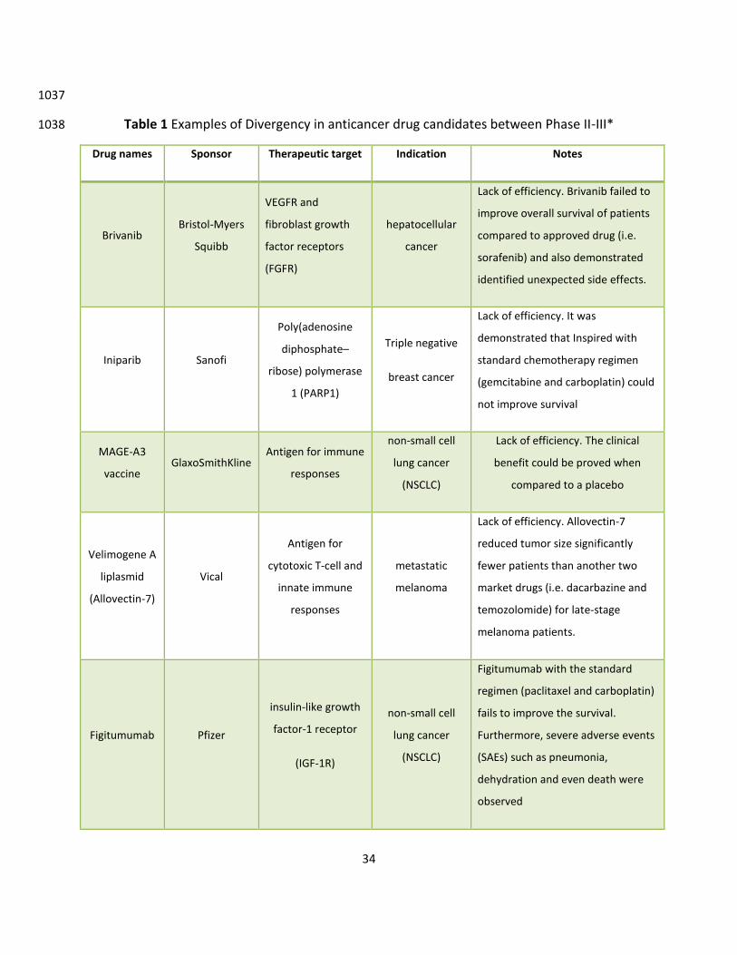
34
1037
Table 1 Examples of Divergency in anticancer drug candidates between Phase II-III* 1038
Drug names Sponsor Therapeutic target Indication Notes
Brivanib Bristol-Myers
Squibb
VEGFR and
fibroblast growth
factor receptors
(FGFR)
hepatocellular
cancer
Lack of efficiency. Brivanib failed to
improve overall survival of patients
compared to approved drug (i.e.
sorafenib) and also demonstrated
identified unexpected side effects.
Iniparib Sanofi
Poly(adenosine
diphosphate–
ribose) polymerase
1 (PARP1)
Triple negative
breast cancer
Lack of efficiency. It was
demonstrated that Inspired with
standard chemotherapy regimen
(gemcitabine and carboplatin) could
not improve survival
MAGE-A3
vaccine GlaxoSmithKline
Antigen for immune
responses
non-small cell
lung cancer
(NSCLC)
Lack of efficiency. The clinical
benefit could be proved when
compared to a placebo
Velimogene A
liplasmid
(Allovectin-7)
Vical
Antigen for
cytotoxic T-cell and
innate immune
responses
metastatic
melanoma
Lack of efficiency. Allovectin-7
reduced tumor size significantly
fewer patients than another two
market drugs (i.e. dacarbazine and
temozolomide) for late-stage
melanoma patients.
Figitumumab Pfizer
insulin-like growth
factor-1 receptor
(IGF-1R)
non-small cell
lung cancer
(NSCLC)
Figitumumab with the standard
regimen (paclitaxel and carboplatin)
fails to improve the survival.
Furthermore, severe adverse events
(SAEs) such as pneumonia,
dehydration and even death were
observed

35
* The data is from “22 Case Studies Where Phase 2 and Phase 3 Trials Had Divergent Results” and curated from 1039
https://www.fda.gov/aboutfda/reportsmanualsforms/reports/ucm535541.htm 1040
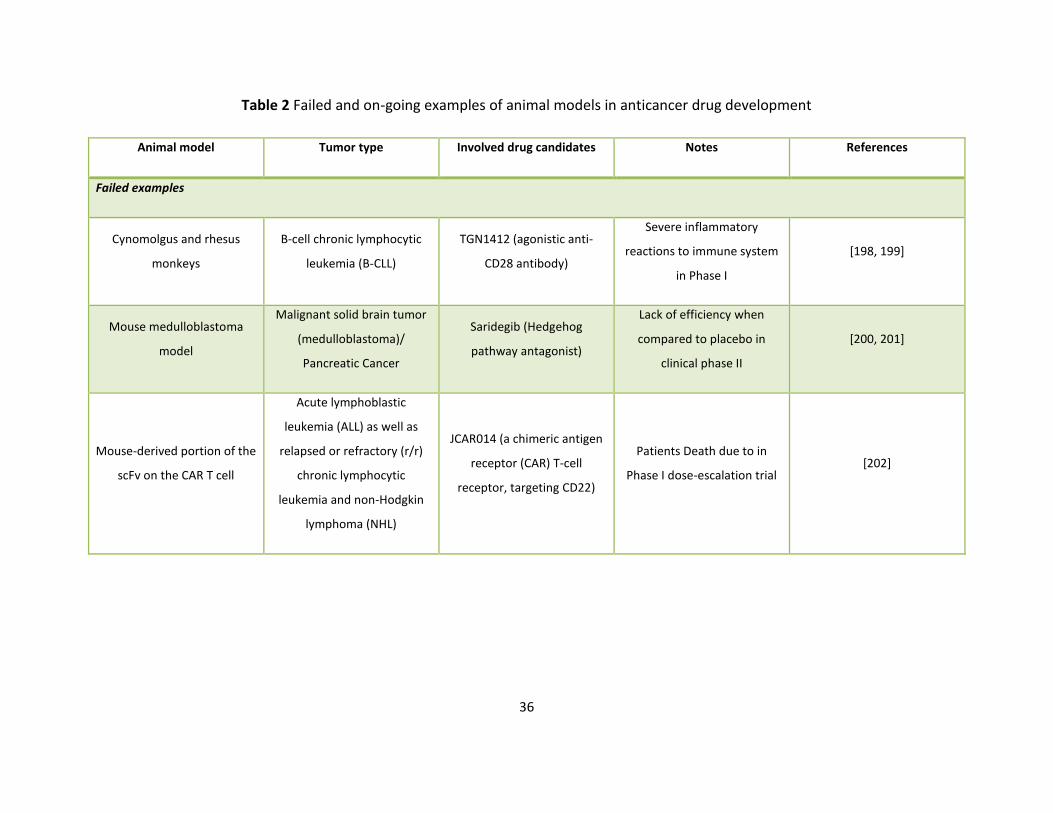
36
Table 2 Failed and on-going examples of animal models in anticancer drug development
Animal model Tumor type Involved drug candidates Notes References
Failed examples
Cynomolgus and rhesus
monkeys
B-cell chronic lymphocytic
leukemia (B-CLL)
TGN1412 (agonistic anti-
CD28 antibody)
Severe inflammatory
reactions to immune system
in Phase I
[198, 199]
Mouse medulloblastoma
model
Malignant solid brain tumor
(medulloblastoma)/
Pancreatic Cancer
Saridegib (Hedgehog
pathway antagonist)
Lack of efficiency when
compared to placebo in
clinical phase II
[200, 201]
Mouse-derived portion of the
scFv on the CAR T cell
Acute lymphoblastic
leukemia (ALL) as well as
relapsed or refractory (r/r)
chronic lymphocytic
leukemia and non-Hodgkin
lymphoma (NHL)
JCAR014 (a chimeric antigen
receptor (CAR) T-cell
receptor, targeting CD22)
Patients Death due to in
Phase I dose-escalation trial [202]
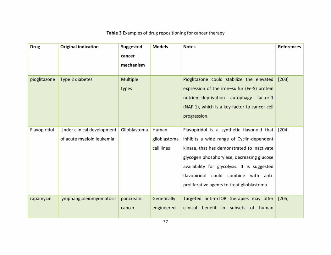
37
Table 3 Examples of drug repositioning for cancer therapy
Drug Original indication Suggested
cancer
mechanism
Models Notes References
pioglitazone Type 2 diabetes Multiple
types
Pioglitazone could stabilize the elevated
expression of the iron–sulfur (Fe-S) protein
nutrient-deprivation autophagy factor-1
(NAF-1), which is a key factor to cancer cell
progression.
[203]
Flavopiridol Under clinical development
of acute myeloid leukemia
Glioblastoma Human
glioblastoma
cell lines
Flavopiridol is a synthetic flavonoid that
inhibits a wide range of Cyclin-dependent
kinase, that has demonstrated to inactivate
glycogen phosphorylase, decreasing glucose
availability for glycolysis. It is suggested
flavopiridol could combine with anti-
proliferative agents to treat glioblastoma.
[204]
rapamycin lymphangioleiomyomatosis pancreatic
cancer
Genetically
engineered
Targeted anti-mTOR therapies may offer
clinical benefit in subsets of human
[205]
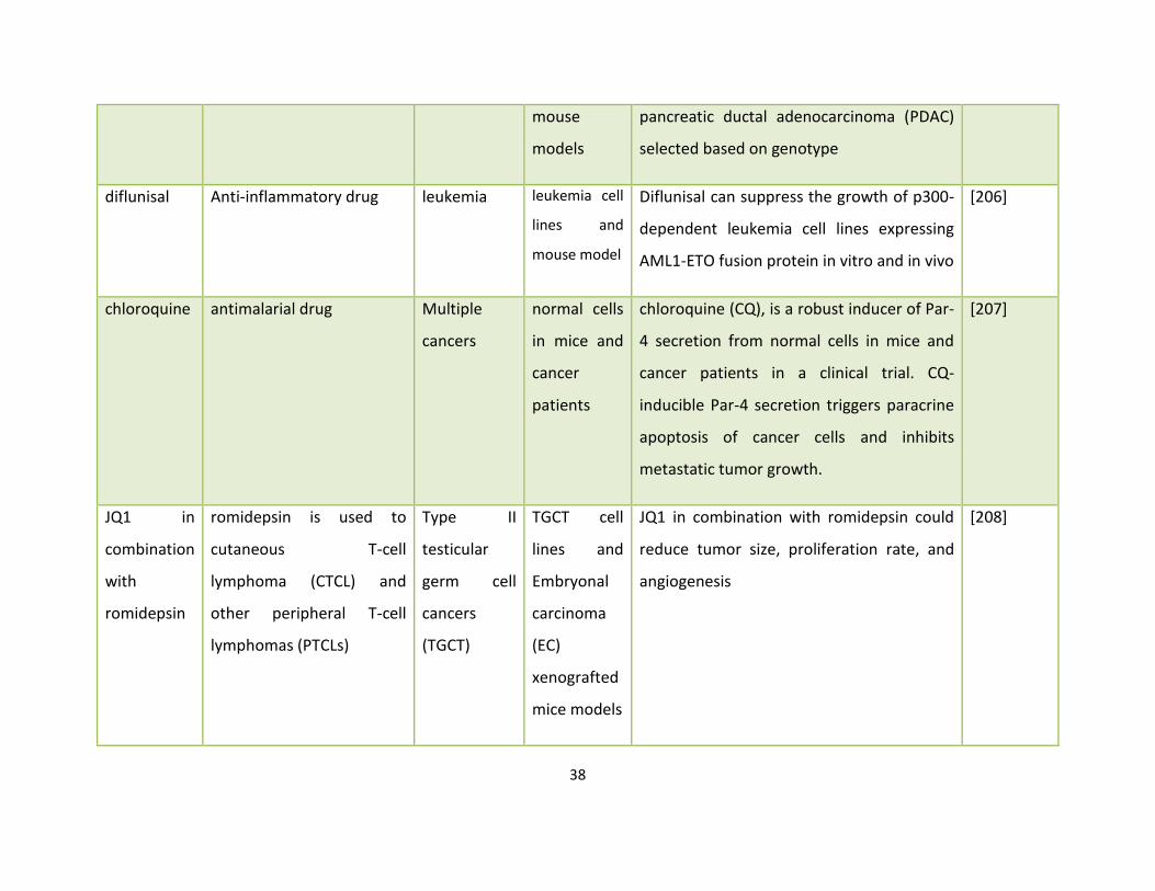
38
mouse
models
pancreatic ductal adenocarcinoma (PDAC)
selected based on genotype
diflunisal Anti-inflammatory drug leukemia leukemia cell
lines and
mouse model
Diflunisal can suppress the growth of p300-
dependent leukemia cell lines expressing
AML1-ETO fusion protein in vitro and in vivo
[206]
chloroquine antimalarial drug Multiple
cancers
normal cells
in mice and
cancer
patients
chloroquine (CQ), is a robust inducer of Par-
4 secretion from normal cells in mice and
cancer patients in a clinical trial. CQ-
inducible Par-4 secretion triggers paracrine
apoptosis of cancer cells and inhibits
metastatic tumor growth.
[207]
JQ1 in
combination
with
romidepsin
romidepsin is used to
cutaneous T-cell
lymphoma (CTCL) and
other peripheral T-cell
lymphomas (PTCLs)
Type II
testicular
germ cell
cancers
(TGCT)
TGCT cell
lines and
Embryonal
carcinoma
(EC)
xenografted
mice models
JQ1 in combination with romidepsin could
reduce tumor size, proliferation rate, and
angiogenesis
[208]
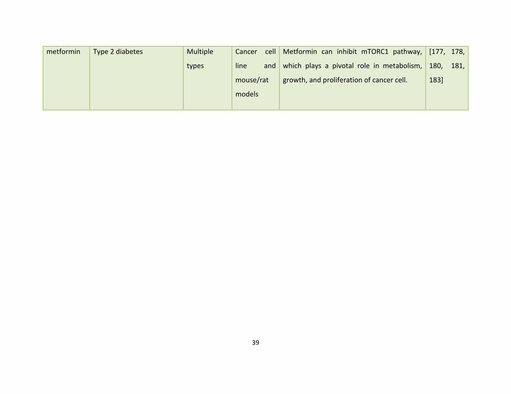
39
metformin Type 2 diabetes Multiple
types
Cancer cell
line and
mouse/rat
models
Metformin can inhibit mTORC1 pathway,
which plays a pivotal role in metabolism,
growth, and proliferation of cancer cell.
[177, 178,
180, 181,
183]
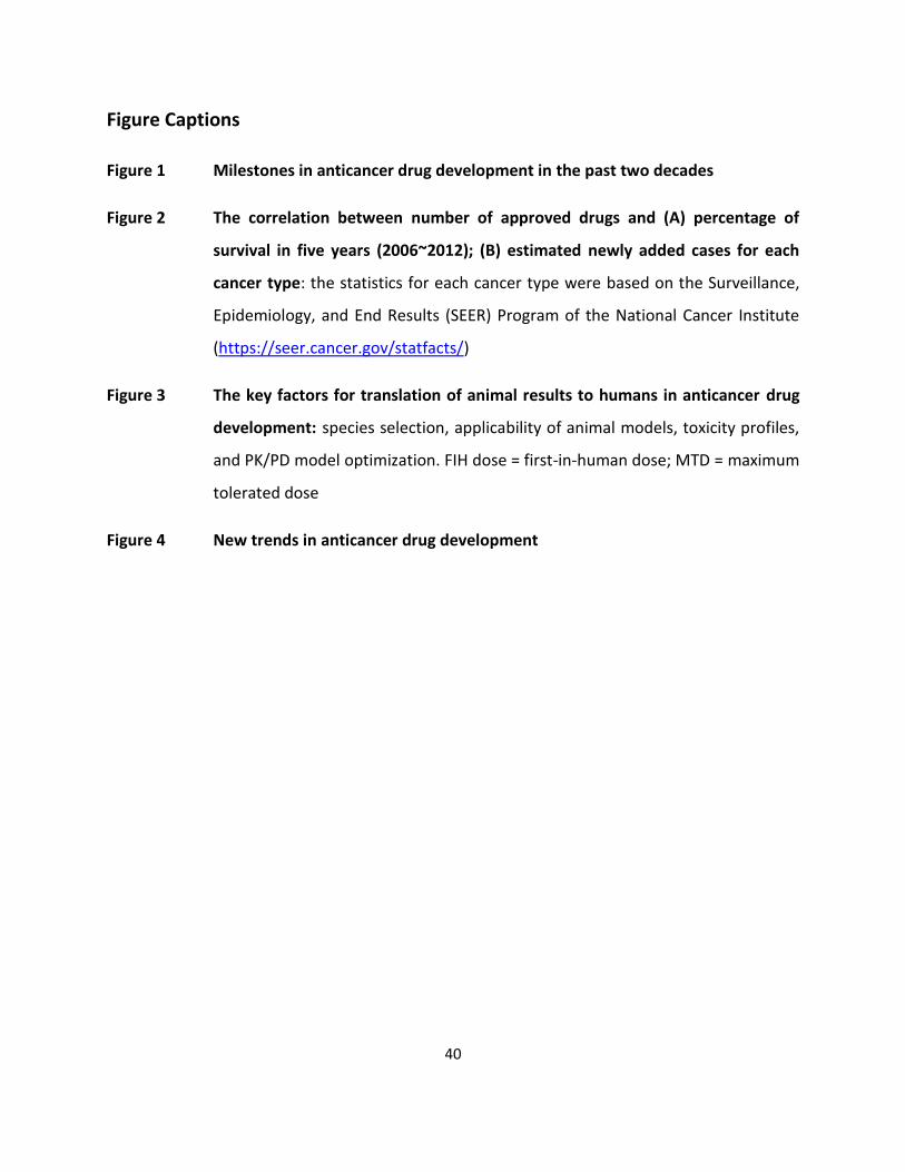
40
Figure Captions
Figure 1 Milestones in anticancer drug development in the past two decades
Figure 2 The correlation between number of approved drugs and (A) percentage of
survival in five years (2006~2012); (B) estimated newly added cases for each
cancer type: the statistics for each cancer type were based on the Surveillance,
Epidemiology, and End Results (SEER) Program of the National Cancer Institute
(https://seer.cancer.gov/statfacts/)
Figure 3 The key factors for translation of animal results to humans in anticancer drug
development: species selection, applicability of animal models, toxicity profiles,
and PK/PD model optimization. FIH dose = first-in-human dose; MTD = maximum
tolerated dose
Figure 4 New trends in anticancer drug development
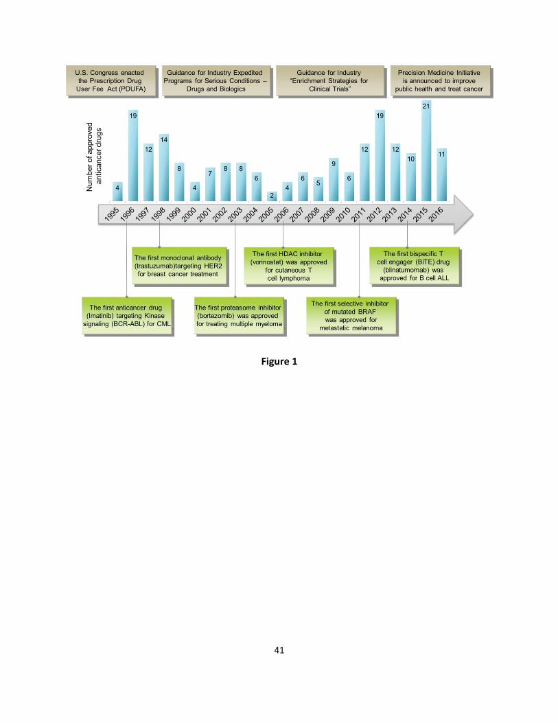
41
Figure 1
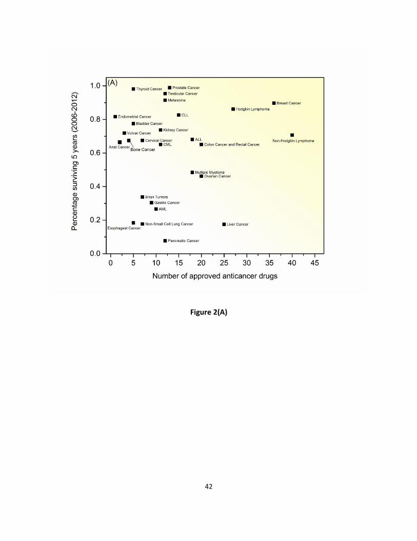
42
Figure 2(A)
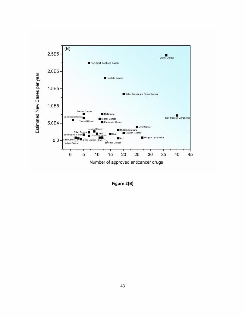
43
Figure 2(B)
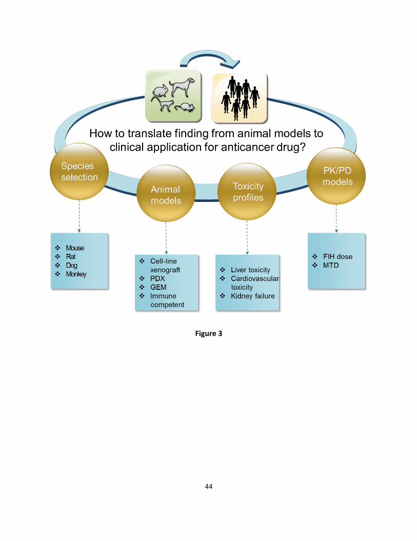
44
Figure 3
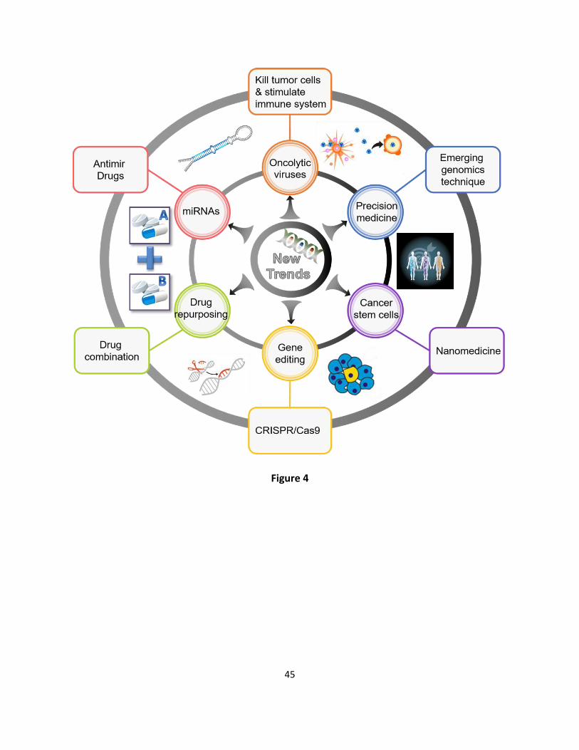
45
Figure 4