Embed Size (px)
Citation preview
UNIVERSITA’ DEGLI STUDI DI PADOVA
Sede Amministrativa: Università degli Studi di Padova
Dipartimento di Psicologia dello Sviluppo e dei Processi di Socializzazione
SCUOLA DI DOTTORATO DI RICERCA IN SCIENZE PSICOLOGICHE
INDIRIZZO: PSICOLOGIA DELLO SVILUPPO E DEI PROCESSI DI SOCIALIZZAZIONE
XXIII CICLO
HOW THE ADULT SOCIAL BRAIN BECOMES THE WAY IT IS.
THE ORIGIN AND THE DEVELOPMENTAL TIME COURSE
OF FACE PROCESSING
Direttore della Scuola: Ch.ma Prof.ssa Clara Casco
Coordinatore dell’indirizzo: Ch.ma Prof.ssa Lucia Mason
Supervisore: Ch.ma Prof.ssa Francesca Simion
Dottoranda: Elisa Di Giorgio


A mia madre, a mio padre e a mia sorella Erica,
che hanno permesso la realizzazione di questo traguardo


“William James once stated that the perceptual world
of the infant must be a “blooming, buzzing confusion”.
What our research and other research on infants has
indicated is that the infant’s world may be a bit more
blooming and a bit less buzzing than James has suspected”.
L. B. Cohen, 1972


Index
Riassunto I
Abstract V
CHAPTER 1: THE ARCHITECTURE OF THE MIND.
THE NEOCONSTRUCTIVISTIC APPROACH
1.1 The dichotomy between general and specific innate mechanisms
as determinants of cognitive development 3
1.2 The Neoconstructivism 5
1.3 The social brain 10
1.3.1 The case of face processing as an example of the
progressive specialization of the cognitive system 14
CHAPTER 2: FACE PROCESSING IN ADULTS
Introduction 19
2.1 Are faces a special class of visual stimuli for adults? 20
2.2 Face processing strategies in adults 24
2.3 A cognitive model for face processing 33
2.4 A neural model for face processing 36
Conclusion 39

CHAPTER 3: THE NATURE OF FACE REPRESENTATION
IN THE FIRST MONTHS OF LIFE
Introduction 43
3.1 The emergence of cognitive specialization for face processing 44
3.2 Models of face preference at birth 46
3.3 The nature of face representation at birth and in the first
few months of age 59
3.4 Study 1 66
Experiment 1 67
Experiment 2 71
Experiment 3 73
Conclusion from Experiments 1-3 77
Experiment 4 78
Experiment 5 83
Experiment 6 86
Conclusion from Experiments 4-6 88
Experiment 7 91
Conclusion 94
CHAPTER 4: FACE DETECTION IN COMPLEX VISUAL DISPLAYS
Introduction 97
4.1 Face detection in complex displays in adults 97
4.2 Visual search strategies in infancy 104
4.3 Face detection in complex displays in infancy 110

4.4 Study 2 114
Experiment 8 115
Experiments 9 and 10 129
4.4.1 Mixed effects model approach to analyze data 152
Experiments 11 and 12 157
Conclusion 167
CHAPTER 5: THE ORIGIN AND THE DEVELOPMENTAL TIME
COURSE OF HOLISTIC FACE PROCESSING
Introduction 171
5.1 The study of holistic face processing in adults 172
5.2 Developmental studies on face processing in infants 173
5.3 Holistic face processing in children and infants 175
5.4 Study 3 180
Experiment 13 181
Experiment 14a 187
Experiment 14b 191
Experiment 15 193
Conclusion 200
General Conclusion 203
References 207
I
Riassunto
Uno dei problemi fondamentali nello studio dello sviluppo cognitivo è comprendere come
la cognizione emerga e quali siano i cambiamenti a cui essa va incontro nel corso dello
sviluppo per raggiungere il livello maturo osservato negli adulti. Una grande sfida per tutti i
ricercatori dello sviluppo riguarda il riuscire a determinare quali sono le abilità e le
predisposizioni che il neonato possiede alla nascita, a comprendere i processi cognitivi che
mette in atto per acquisire la conoscenza del mondo che lo circonda, e a studiare se e come
tali predisposizioni si modificano in funzione dell’esperienza durante il corso dello
sviluppo. Poichè è stato dimostrato che il volto è uno stimolo speciale per gli adulti, in
quanto elaborato da aree neurali (Kanwisher, 2000) e da processi percettivi specifici e
diversi da quelli utilizzati per l’elaborazione degli oggetti (Farah, Wilson, Drain, & Tanaka,
1998), lo studio dell’origine e dello sviluppo della capacità di elaborare tale stimolo risulta
essere funzionale allo studio di un processo di specializzazione cognitiva.
In quest’ottica, la presente tesi di dottorato vuole essere un contributo allo studio
della specializzazione funzionale del sistema umano per l’elaborazione del volto nei primi
mesi di vita, con particolare riferimento alle modificazioni che il sistema subisce nella
rappresentazione ed elaborazione di tale stimolo in funzione dell’esperienza. In particolare,
i cambiamenti evolutivi a cui va incontro il sistema cognitivo per raggiungere il livello
maturo osservato negli adulti sono stati esaminati confrontando in modo diretto le
prestazioni di neonati, bambini di tre e sei mesi ed adulti attraverso l’utilizzo degli stessi
paradigmi di ricerca (ricerca visiva, composite face paradigm). L’ipotesi su cui si basa
questo lavoro è che la specializzazione cognitiva per il volto umano osservata negli adulti
non sia presente alla nascita, ma sia il prodotto di un processo di sviluppo continuo e
dinamico in cui l’esperienza esperita nell’ambiente di vita specie-specifico gioca un ruolo
fondamentale (Nelson, 2001, 2003; Johnson, 1993).
I primi due capitoli sono a carattere teorico: nel Capitolo 1 viene descritto
l’approccio Neocostruttivista, considerato il quadro teorico di riferimento entro cui si
inseriscono gli esperimenti presenti nella tesi, e viene spiegato il motivo per cui viene scelto
il volto come stimolo paradigmatico per lo studio della specializzazione cognitiva. Nel
Capitolo 2 viene invece discussa la specificità, neurale e funzionale, del sistema per
l’elaborazione del volto negli adulti. Vengono riportati inoltre due modelli teorici
fondamentali per la comprensione dell’abilità di elaborazione di tale stimolo negli adulti.
Nella seconda parte della tesi sono presentati i tre studi principali che la costituiscono la
tesi e che hanno lo scopo di studiare le origini e il corso dello sviluppo della capacità di
percepire e di riconoscere un volto umano (Capitoli 3, 4, e 5).
Nello Studio 1 (Capitolo 3), attraverso l’utilizzo della preferenza e dell’abituazione
visiva, è stata indagata la natura della rappresentazione del volto alla nascita e nei primi
mesi di vita. In linea con l’idea che la rappresentazione del volto si specializzi grazie
all’esperienza visiva con tale stimolo nei primi tre mesi di vita (Turati, Valenza, Leo, &
Simion, 2005), i risultati degli esperimenti dimostrano che alla nascita tale rappresentazione
è di natura generale, mentre a 3 mesi essa diventa più specifica per questo particolare tipo
di stimolo (Esperimenti 1, 2 e 3). Inoltre, gli Esperimenti 4, 5 e 6 dimostrano che la
rappresentazione del volto alla nascita non è specie-specifica, in linea con l’ipotesi che il
neonato entra a far parte del mondo con una rappresentazione del volto abbastanza
generale da permettergli di percepire un volto umano e un volto di scimmia come
appartenenti alla stessa “categoria volto”. E’ solo a 3 mesi, grazie all’esperienza visiva con
tale stimolo, che tale rappresentazione diventa specifica per il volto umano (Nelson, 2001;
Pascalis & Kelly, 2009).
III
Lo scopo dello Studio 2 (Capitolo 4) è stato quello di studiare se la preferenza per il
volto osservata in contesti semplici (i.e., presentazione di soli due stimoli) potesse essere
osservata anche in contesti complessi, quindi più ecologici. I movimenti oculari di bambini
di tre e sei mesi e adulti sono stati registrati attraverso un sistema di eye-tracker durante un
compito di ricerca visiva. È stato indagato se bambini di pochi mesi sono in grado di
percepire ed identificare in modo efficiente un volto umano quando inserito in contesti
complessi, ossia tra oggetti (i.e., stimoli distrattori eterogenei, Esperimenti 8, 9 e 10) e tra
volti invertiti (i.e., stimoli distrattori omogenei, Esperimenti 11 e 12). I risultati hanno
dimostrato come il volto umano è in grado di catturare e mantenere l’attenzione di adulti e
bambini di sei mesi quando è inserito fra distrattori eterogenei, mentre tale stimolo cattura
e mantiene l’attenzione dei bambini di tre mesi solo quando è inserito tra distrattori
omogenei. Tali risultati sono in linea con gli studi che hanno dimostrato che il volto cattura
l’attenzione dei bambini di pochi mesi di vita quando si trova in contesti complessi (Gliga,
Elsabbagh, Andravizou, & Johnson, 2009).
Per percepire il volto target in contesti complessi, i bambini hanno dovuto
elaborare il volto come una unità complessa, una Gestalt (Tanaka & Farah, 1993). Questo
tipo di strategia di elaborazione del volto viene definita olistica e lo scopo dello Studio 3
(Capitolo 5) è stato quello di studiarne l’origine e lo sviluppo in neonati, bambini di tre
mesi e adulti utilizzando lo stesso compito chiamato “composite face paradigm” (Young,
Hellawell, & Hay, 1987) (Esperimenti 13, 14 e 15). I risultati dimostrano che, sebbene i
primi segni della capacità di elaborare un volto come un’unità complessa si osservano in
bambini di pochi mesi di vita, tuttavia è necessaria l’esperienza visiva per raffinare tale tipo
di elaborazione del volto.
Complessivamente, i dati presentati in questa tesi sono in linea con l’idea che la
specificità del sistema cognitivo per l’elaborazione del volto umano non sia presente alla

nascita, ma sia invece il risultato di un processo di sviluppo, in cui giocano un ruolo
fondamentale sia le predisposizioni innate del neonato, sia l’esperienza visiva esperita nel
proprio ambiente di vita specie-specifico nei primi mesi di vita.
V
Abstract
One central issue in cognitive developmental science is to understand how cognition grows
and change over time to reach an adult level of specialization. Determining the abilities
with which infants come equipped into the world, their mechanisms for acquiring
knowledge, and whether and how these abilities change as a function of development and
experience is a challenging issue. Face processing is an interesting topic of research in that
respect because faces form a special class of visual objects elaborated in adults by a specific
anatomical and functional face system (e.g., Kanwisher, 2000; Farah, Wilson, Drain, &
Tanaka, 1998). Since what determines this specialization and how this specialization
emerges during development still remain unknown, the purpose of my PhD dissertation is
to study cognitive specialization during early infancy through the investigation of the
development of infants’ abilities to process faces. In particular, my hypothesis is that the
face processing specificity is not present at birth, but emerges gradually from the
interaction between general constraints and attentional biases present in the first months of
life and the critical visual input provided by the specie-specific environment (Nelson, 2001,
2003; Johnson, 1993).
With this consideration in mind, my thesis begins with two theoretical chapters:
Chapter 1 describes a neoconstructivistic approach to the emergence of cognition as the
theoretical framework and discuss the specialty of the face stimulus for humans, whereas
Chapter 2 is about the specificity of the face processing system in adults and I review two
theoretical models of face processing and the neural bases underlying this skill.
Subsequently, in the second part of the thesis I describe three studies aiming at
investigating the origin and the developmental time course of both face detection and face
recognition (Chapters 3, 4 and 5). Importantly, to examine both the emergence and the
developmental time course of the face processing abilities to become specialized, the same
experimental paradigms are employed with newborns, 3- and 6-month-old infants and
adults (e.g., composite face paradigm, a modified visual search paradigm). This allows a
direct comparison between adults’ and infants’ performance.
In Study 1 (Chapter 3), using both the visual preference and visual habituation
techniques, a first series of experiments investigates the nature of face representation in
newborns and in 3-month-old infants. According to recent evidence showing that infants’
response to faces becomes more and more tuned to the face category over the first three-
months of life (Turati, Valenza, Leo, & Simion, 2005), collected data demonstrate that 3-
month-old infants, but not newborns, are sensitive to specific perceptual cues within a face,
such as the correct position and orientation of the eyes (Experiments 1, 2 and 3).
Furthermore, results obtained from Experiments 4, 5, 6 and 7 demonstrate that early facial
representation is not human-specific, corroborating the hypothesis that newborns come
into the world with a face representation that is sufficiently general as to bias newborns’
visual attention toward multiple categories of faces (e.g., monkey faces vs. human faces),
and that this face representation, due to the visual experience that infants do in the specie-
specific environment, becomes more specific to human face during the first 3 months of
life (Nelson, 2001; Pascalis & Kelly, 2009).
Due to the social relevance of the face stimulus and due to the ability of 3-month-
old infants to form a specific representation of the human face, the aim of Study 2 (Chapter
4) is to investigate whether human face grab and maintain infants’ attention in complex
visual scenes. Specifically, using an eye-tracker system, adults’ and 6- and 3-month-old
infants’ visual search behavior is compared in a modified visual search task of a target face
among heterogeneous (e.g., various objects, Experiments 8, 9 and 10) and homogeneous

VII
distractors (e.g., inverted faces, Experiments 11, 12). Results demonstrate that a face
among heterogeneous distractors captures and maintains adults’ and 6-month-old infants’
attention and that 3-month-old infants detect a target face only when embedded among
inverted faces (e.g., homogeneous distractors), corroborating previous findings showing the
face detection advantage in infants (Gliga, Elsabbagh, Andravizou, & Johnson, 2009).
Importantly, to detect a target face among other distractors, infants have to process
a face as a Gestalt, where the whole is more than the sum of its constituent parts (Tanaka
& Farah, 1993). This kind of face processing, called “holistic”, is investigated in newborns,
3-month-old-infants, and adults through a modified version of the composite face
paradigm (Young, Hellawell, & Hay, 1987) and the recording of eye movements in Study 3
(Chapter 5). The main outcome of the present study is that the tuning toward holistic
information appears very early in life, although gradual experience-based developmental
processes will progressively refine early holistic processing abilities (Experiments 13, 14
and 15).
Overall, these data demonstrate that face specificity is not prewired, but rather
arises from general perceptual processes that, during development, become progressively
tuned to the human face, as a result of extensive experience with this stimulus category in
the first months of life.


CHAPTER 1
THE ARCHITECTURE OF THE MIND:
THE NEOCONSTRUCTIVISTIC APPROACH


3
1.1 The dichotomy between general and specific innate mechanisms as
determinants of cognitive development
Arguments over the developmental origins of human knowledge are ancient, founded in
the writings of Plato, Aristotle, Descartes, Hume and Kant. Indeed, developmental theories
have been dominated by two different views both on the origin of knowledge and on the
initial mechanisms that form the basis for cognitive development. On one view supports
the hypothesis that knowledge emerges on the basis of domain general mechanisms of
learning that are sufficient to explain how children learn about specific domain of
knowledge such as language, number, space or faces. Although Piagetian, Behaviourist and
more recently Connectionist theories fall within this view, Piaget‟s position, known as
epigenetic constructivism, differs because cognitive development is considered as the outcome
of a self organizing system that is structured and shaped by its interaction with the
environment. The mind of the newborn is essentially unstructured and knowledge-free; it is
equipped with just three domain-general processes (i.e., accomodation, assimilation and
equilibration) which, in conjunction with a few innate reflexes (i.e., sucking, looking,
grasping), are all that the child brings to the developmental process. The environment
supplies the rest. The most important thing is that the child acts on the environment,
initially just employing the few sensorimotor reflexes at its disposal, and the environment
in its turns has an important role to play in the gradual emergence of structure in the mind.
In other words, Piaget‟s constructivist theory holds that cognitive development is a
continuous process of building knowledge on previous skills (e.g., perception, memory)
and existing knowledge structures, from a foundation at birth consisting largely of reflexes
and sensory impressions.
This constructivist view of development was challenged by the nativist view,
according to which the early appearence of abilities hitherto unsuspected has supported the
notion that knowledge begins early in life and constitutes parts of humans‟ innate
endowment. Some authors maintain that human cognition is built on domain-specific
system of knowledge and that the natural selection may have favored the evolution of
mechanisms that leads to this knowledge (Kellman, 1993). From this point of view, the
neonate is seen as having domain-specific predispositions allowing it to process specific
types of inputs (Spelke, 1990; Baillargeon & Wang, 2002). More specifically, the human
mind is thought to be a collection of special purpose mechanisms, each shaped, through
adaptation to the environment during evolution, to perform a particular function. This
nativistic view asserts that humans are born either with the innate capacity to develop
information processing systems or cognitive or “cognitive modules” that allow them to
make sense of the world, or that learning is guided by innately specified and content-
specific principles that determine the entities on which subsequent learning takes place
(Gelman, 1990; Spelke, 1991). In this perspective, deeply influenced by Chomskyan
linguistics (1988) and by Fodor‟s modularity theory (1983), the infant comes into the world
prepared to process different domains of knowledge. For example, the infant comes
prepared to process faces or number. Accordingly, cognition would be specialized from the
outset in processing content-specific inputs able to mediate complex cognitive functions
(Spelke, 1991; Wynn, 1995). However, this approach seems to preclude the epigenetic
constructivism principle to development because biological forms are not considered as a
product of any dynamic interaction between the genes and the environment.
The dichotomy between general and specific innate mechanisms as determinans of
cognitive development has been overcome by the neo-constructivistic approach to
cognition that combines these two different explanations.
5
1.2 The Neoconstructivism
Karmiloff-Smith wrote: “I suggest that nativism (when redefined within a truly epigenetic
perspective of genetic expression rather than genetic unfolding), on the one hand, and
Piaget‟s constructivism, on the other, are complementary in fundamental ways, and that the
ultimate theory of human cognition will encompass aspects of both (Karmiloff-Smith,
1994, p. 693)”. Karmiloff-Smith (1994) argued that domain-specific predispositions give
development a small but significant kickstart by focusing the young infant‟s attention on
prioritary inputs. The crucial point is that the early period is followed by intricate
interaction with environmental that shape the development. That is, dichotomy between
general and specific innate mechanisms as determinants of cognitive development has been
overcome by the neo-constructivistic approach to cognition (i.e., the term was generated by
combining neo, taken from the Greek neos, meaning “new”, and constructivism, taken
from the Piaget‟s costructivist approach), that combines these two different explanations
and states that nativism and epigenetic principle are not incompatible because it can be
assumed the existence of some innate specified predispositions that would give the
epigenetic process a head start in each domain of knowledge (see Simion & Leo, 2010).
Neoconstructivism views the cognitive development as a continuous process that
emerges through the dynamic of probabilistic epigenesis and that progressively leads to an
increasing functional specialization of neural circuits (Bates & Elman, 1993; Johnson, 1997;
Karmiloff-Smith, 1992). The probabilistic epigenesis view of development (Gottlieb, 1992)
emphasizes that gene activity, instead of following a strictly pre-specified schedule (i.e.,
predetermined epigenesis), is regulated by signals from the external and internal
environment and that development is therefore subject to bidirectional interactions
between gene activity, neural activity, behavior and the environment (Gottlieb, 2007). In

this perspective, cognitive activity is seen as emerging gradually as a product of the
interaction between innate constraints and the structure of the input provided by the specie-
typical environment (de Schonen, 2002; Johnson, 1993; Nelson & Luciana, 2001). Therefore,
in contrast to the classic nativist/modular thesis (Fodor, 1983; Spelke, 1991) that considers
the infant brain as provided with build-in domain-specific representational contents, more
recent models stress the role of a number of innately specified constraints or biases in the
emergence of representations and thus in the origin of knowledge. The main hypothesis is
that specific cognitive structures observed in adults may arise from primary, general innate
constraints shaped by the nature of the experience the organism is exposed to in a given
period of time (Karmiloff-Smith, 1992).
Within this theoretical framework, innate constraints are architectural, computational
and temporal biases that shape information processing, limiting the types of input to be
selected and constraining the computations on the input. More specifically, a behavior is
seen as innate when its development is constrained concerning the neural architecture of
the brain, to the types of computation applied, or to the timing of events in the
developmental process (Elman, Bates, Johnson, Karmiloff-Smith, Parisi, & Plunkett, 1996).
Consequently, the word "constraints" does not carry on any negative connotation, but
rather, it possesses a positive connotation. Constraints are defined as biases in the
information processing due to the properties of the brain architecture or of the perceptual
systems in a given period of development. Benefits from these biases consist in selectively
focusing the cognitive system toward certain aspects of the surrounding environment or
facilitating processing of certain kinds of inputs, thus strengthening learning of some
categories of stimuli rather than others, and, consequently, tuning the system to become
specialized. In this vein, the constraints imposed by the development of the sensory

7
systems may actually facilitate subsequent perceptual development by reducing the range of
stimuli that infants has to deal with.
Importantly, there are crucial time windows (i.e., sensitive and/or critical periods,
Greenough, Black, & Wallace, 1987; Greenough & Black, 1992; Nelson, 2001, 2003),
during which experience manipulations profoundly affect cognitive development. Domain
specific cognitive activity is, therefore, strictly linked to the exposure to certain experiences.
For instance, the deprivation of early visual input due to bilateral congenital cataract in the
first few months of life impairs face processing even after years from surgery,
demonstrating that the visual experience during the first few months of life is necessary for
the normal development of expert face processing (e.g., Le Grand, Mondloch, Maurer, &
Brent, 2003).
Finally, as suggested before, experience shapes the development that occurs in a
specie-specific environment. For instance, with regards to face processing, it has been
demonstrated that infants of 6 months of age are better at discriminating monkey faces
than are 9-month-old infants and adults (Pascalis, de Hann, & Nelson 2002). These results
suggests that younger infants exhibit a more broadly tuned face-processing system that can
discriminate among exemplars within multiple categories of faces (e.g., both human and
monkey faces) and that this system becomes more specific (e.g., discrimination between
human faces only), corroborating the idea that the face processing system is shaped by the
faces encountered more often in the visual environment in the first months of life, that are
human faces (de Schonen & Mathivet, 1989; Nelson, 2001; Scott, Pascalis, & Nelson,
2007). Another example of the role of experience in a specific environment comes from
the domain of language. For example, speech perception is characterized by a loss of ability
with age, such that 4-to 6-month-olds can discriminate phonetic differences that
distinguish syllables in both their native and unfamiliar languages, whereas 10- to 12-

month-olds can only discriminate the phonetic variations used in their native language
(Cheour, Ceponiene, Lehtokoski, Luuk, Allik, Alho, et al.,1998; Kuhl, Williams, Lacerda,
Stevens, & Lindblom, 1992).
To summarize, in contrast to the classic nativist/modular thesis, the
neoconstructivism framework suggests that the specific cognitive systems are the product
of a “process of modularization”; that is to say, the modular architecture is the result of a
gradual development rather than the starting point. Consequently, brain specialization,
domain specificity and cognitive modules are considered to emerge epigenetically and
developmentally through the interaction with postnatal development, rather than being
assumed as genetically pre-specified. Evolution has pre-specified many innate biological
constraints on development that are domain general mechanisms becoming “domain
specific” with the process of development. During this process the same general
mechanisms used repeatedly to process a certain class of stimuli become specific. Some
apparent constraints contribute to the development of new structures and new modes of
functioning, which will be advantageous at later stages of development (Karmiloff-Smith,
1992) and provide starting points that channel the subsequent perceptual and cognitive
development (e.g., Turkewitz & Kenny, 1982).
The neuropsychological equivalent of the neoconstructivism is the neuro-
constructivism, which emphasizes the interrelation between brain development and cognitive
development (Karmiloff-Smith, 1992; Sirois, Spratling, Thomas, Westermann, Mareschal,
& Johnson, 2008; Quartz & Sejnowski, 1997). From a neocostructivistic point of view, the
development is a progressive increase in the complexity of representations, with the
consequence that new competences develop based on earlier and simpler ones. In the same
vein, the neuroconstructivistic approach considers this increase in representational
complexity as realized in the brain by a progressive specialization of cortical structures.
9
Specifically, neuroconstructivism offers a theoretical framework in which cognitive
development is closely linked to the development of the underlying cortical structures in
the brain. By characterizing the constraints that operate on the development of neural
structures that support mental representations, cognitive development is explained as a
trajectory emerging from the interplay of these constraints.
Brain development is viewed as an increasing restriction of the fate of component
elements, such as neurons and neural circuits. In other words, as development proceeds,
neurons and cortical circuits become increasingly specialized, dedicated to particular
functions and less capable of change. The cerebral cortex does not appear to contain
intrinsic pre-specified representations to support functions such as face recognition or
linguistic processing. Rather, the appropriate representations emerge through the
constraints of the complex cortical and subcortical networks and through the interaction
between the infant and the statistical regularities latent in the environment. Endogenous
constraints select the aspect of the environment to which orienting attention and,
interacting, with the structure of the input typical of the infant‟s environment, guide and
shape the gradual emerging of specialized processing (Werker & Vouloumanos, 2001). In
this perspective, experience appears to play a prominent role in recruiting the cortical areas
potentially suited to be activated by certain kind of stimuli. The activation of these cortical
networks leads as a consequence to a process of a progressive neural specialization
(Nelson, 2001).
To conclude, the neocostructivistic framework provides an integrated view of
development and adult processing because the adult state is viewed as merely a state along
the developmental trajectory. From this perspective, the investigation of adult processing
benefits from being analyzed through a developmental lens to reveal which constraints
have shaped development to reach the adult state. Adult cognitive processing is often
characterized as consisting in a set of qualitatively different, specialized, domain-specific
modules. The neoconstructivist perspective (and the neuroconstructivism as well) instead
focuses on how regions of functional specialization are formed given the outlined
constraints, providing explanations of adult processing that are less focused on qualitatively
different encapsulated modules. Moreover, progress in research in the neuroconstructivist
framework will be made by a better understanding of the constraints operating on neural
development, by improved methods of linking brain and behavior in developing children
(see Aslin & Fiser, 2005), and by computational modelling which has the potential to offer
explanations of the interactions between brain and cognitive development (Mareschal,
Sirois, Westermann, & Johnson, 2007; Westermann, Mareschal, Johnson, Sirois, Spratling,
& Thomas, 2007).
It is within this theoretical framework that many researchers investigate the origin
and the developmental time course of the “social brain”.
1.3 The social brain
One of the most prominent, and to an extent unique, characteristics of the human brain is
the ability to process stimuli in a social context (Adolphs, 1999; Brothers, 1996). Detecting
and discriminating humans from objects is critical for adaptive behavior. Many vertebrates
orient toward or look longer at social agents. Newly hatched chicks attend to patterns
similar to the head region of their caregivers (Morton & Johnson, 1991) and detect social
agents on the basis of the way they move (Regolin, Tommasi, & Vallortigara, 2000).
Similarly, monkeys manifest a preference for faces as compared to objects (Sugita, 2008).
These findings support the idea of the existence of hard wired mechanisms to detect social
stimuli which might be present in animals including humans (Johnson, 2007).

11
Human adults have areas of the brain specialized for processing and integrating
sensory information about the identity, behavior, and intentions of other humans,
corroborating the idea that a network of specific cortical circuits preferentially processes
social information. Indeed, a variety of cortical areas have been implicated in the „social
brain‟ including the superior temporal sulcus (STS), the fusiform „face area‟ (FFA) and
orbitofrontal cortex (Farah, Rabinowitz, Quinn, & Liu, 2000; Kanwisher, 2000; Allison,
Puce, & McCarthy, 2000).
One of the major debates in cognitive neuroscience concerns the origins of the
„social brain‟ in humans, and theoretical arguments abound about the extent to which this
is acquired through experience. The ontogeny of the social brain network is one aspect of
human postnatal functional brain development, and it is therefore useful to consider work
within this field within the context of three general perspectives on developing brain
function. Johnson (2001, 2005) reviewed three perspectives on how the neuroanatomical
development of the brain could be related to the changes in motor, perceptual, and
cognitive abilities observed during infancy and childhood: a maturational perspective, a
skill-learning viewpoint, and interactive specialization.
The maturational view assumes that, through evolutionary pressure, specific parts of
the brain and areas of the cortex have become dedicated to process social information.
Some of the specific circuits to process social stimuli would be present and functioning at
birth, in contrast others circuits would become available through maturation later during
development. The sequence of the maturational timetable would not be affected by
experience. Evidence concerning the differential neuroanatomical development of cortical
regions is used to determine an age when a particular region is likely to become functional.
Success in a new behavioral task at one specific age is then attributed to the maturation of a
newly functional brain region.
The skill-learning hypothesis maintains that social stimuli are not different from
other stimuli. Some circuits become specialized for social stimuli simply because adults
become experts in processing them. The specialization would arise not because of the
social nature of stimuli (i.e. domain specificity), but because of the expert-level
discrimination for processing complex visual patterns, independently of the category to
which the stimuli belong (i.e. process specificity). It is hypothesize that the regions active in
infants during the onset of new perceptual or behavioral abilities are the same as those
involved in skill acquisition in adults (Johnson, Grossman, & Farroni, 2008).
The interactive specialization view emphasises the importance of the initial biases
to “bootstrap” later developing systems, and the notion of partial functioning of neural
pathways which, interacting with the environment, shapes the subsequent functional and
structural development. This alternative point of view assumes that postnatal functional
brain development, at least within the cerebral cortex, involves a process of organizing
interregional interactions (Johnson, 2002), by which cortical regions go from initially
having very broadly tuned functions to having increasingly finely tuned (more specialized)
functions (Johnson, 2001). Starting from a constructivist viewpoint, this third hypothesis
maintains that the structural and functional changes in regions of the cortex co-develop as
a function of the interaction with the environment (Johnson, 2000) and that the timing of
events plays a critical role in developmental trajectories. The specialization of the cognitive
system cannot be ascribed to the pre-specification of a particular region of the cortex, but
to a particular sequence of interactions between pre- and post-natal environment and
cortical circuits, resulting in successive reorganizations of the cortical circuits themselves
(de Schonen, 2002). The specific properties of a brain region are partly determined by its
pattern of connectivity to other regions and to their pattern of activity. Cognitive
specialization is, therefore, an activity-dependent and an experience-dependent process,

13
strictly linked to the exposure to certain experiences occurring over a particular period of
time, called critical or sensitive period (Greenough & Black, 1992). The onset of new
behavioral competencies during infancy and childhood will therefore be associated with
changes in activity (adjustments in tuning functions) over several regions and not just by
the onset of activity in one or more additional region(s). According to this view, brain
regions do not develop in isolation but are heavily constrained by their connections and
interactions with other regions, a phenomenon recently termed embrainment (Mareschal,
Johnson, Sirois, Spratling, Thomas, & Westermann, 2007). The interactive specialization
approach suggests that when a new computation or skill is acquired, there is a
reorganization of interactions between different brain structures and regions. This
reorganization process could change previously existing mappings between cortical regions
and their functions. Thus, the same behavior could potentially be supported by different
neural substrates at different ages during development.
To summarize, during prenatal development, spontaneous activity in sensory
systems appears to play an important role in contributing to the differentiation of cortical
regions. In early postnatal life, infants contribute further to the specialization of their brain
by preferentially orienting and attending to certain types of stimuli, such as faces. Later,
social experience and interaction with caregivers may contribute to the specialization of
developing parts of the cerebral cortex. Much of later postnatal brain development,
therefore, can be viewed as an active process to which infant contribute. In this vein,
studying the postnatal emergence of cortical specialization for different cognitive
functions offers the possibility of new perspectives not only on the study of perceptual
and cognitive development in healthy human infants, but also for social development and
atypical developmental pathways. Indeed, developmental disorders, such as autism, can be
understood through altered constraints that push the developmental trajectory off its

normal track to reach a different endstate (Karmiloff-Smith, 1998; Thomas & Karmiloff-
Smith, 2003). Thus, atypical development can, like typical development, be characterized
as an adaptation to multiple interacting constraints, with the only exception that the
constraints are different. These atypical constraints then lead to different outcomes
through the same processes of representation construction.
In line with this interactive specialization developmental point of view, the present
thesis will examine the emergence of the specialized cognitive system devoted to
processing social stimuli and how innate mechanisms and perceptual experience contribute
to the development of the social brain. To this end, we will focus on the evidence on
infants‟ abilities since birth of processing social stimuli on the basis of the presence of a
face (i.e. face detection). Evidence will be presented to demonstrate the innate
predispositions of the human system to detect social stimuli at birth and how the pre-
wired perceptual constraints and attentional biases interact with experience to guide and
shape the emerging of a specialized system to process social stimuli.
1.3.1 The case of face processing as an example of the progressive
specialization of the cognitive system
To address the issue about how a cognitive system becomes a specialized system in
adulthood, a peculiar class of visual stimuli, namely faces, will be considered. Indeed, the
functional and neural specialization present in the adults‟ system for processing faces (e.g.,
Farah et al., 2000; Kanwisher, McDermott, & Chun, 1997; Kanwisher, 2000), renders faces
an ideal class of stimuli to investigate the time course and the factors affecting such
specialization. Indeed, interest in face recognition has played prominently in various
scientific disciplines for much of this century, and even parts of the last (Darwin, 1859).
15
Cognitive psychologists have been interested in this phenomenon because there is evidence
that faces are somehow perceived differently than other patterned objects, and thus, may
represent a „special‟ class of stimuli. In the same vein, cognitive neuroscientists are
interested in face recognition because there is evidence that this ability is subserved by
discrete neural circuits, and thus, represents a specialized brain function.
Faces play an important role in social interaction. It is commonly accepted that the
ability of fast and accurate face analysis plays a crucial role for people. Indeed, faces are the
unique source of information concerning human beings. Merely looking at somebody‟s
face enables us to determine sex, age, race and attractiveness, and what is even more
important, tentatively estimate mood, intelligence and honesty, and friendly and hostile
attitudes in its owner.
The crucial issue pertains to the specificity of face perception, that is, whether the
face is an extraordinary stimulus (Kanwisher et al., 1997; Farah et al., 1998) or if the brain
processes faces in the same manner as any other category of objects, like animals or
buildings (Diamond & Carey, 1986; Chao, Haxby, & Martin, 1999). Another issue concerns
the underlying mechanism of this extraordinary human competence. Some authors argue
that the ability to process faces as special stimuli is due to the presence of inborn
predispositions (Johnson & Morton 1991; Morton & Johnson 1991; Farah et al., 1998,
2000; Johnson, 2005, McKone, Kanwisher, & Duchaine, 2007). Other authors emphasize
the role of learning processes, claiming that we become experts in face recognition just by
experience (experience-dependent; e.g., Gauthier & Logothetis, 2000; Gauthier & Tarr,
1997).
Thus, developmental behavioral and neuropsychological work can potentially
contribute to this debate by providing evidence about the developmental trajectory of face-
processing abilities in the human brain. Since development cannot be explained in terms of

innate, building in representational contents, it becomes relevant for developmental
researchers to investigate what types of general perceptual constrains and attentional biases
are present in the first months of life and how they contribute to the specialization of the
cognitive system (Simion & Leo, 2010).
CHAPTER 2
FACE PROCESSING IN ADULTS

19
Introduction
Human faces are the most important stimuli in our visual world. The human face
represents a unique, highly salient and biologically significant visual stimulus that reveals a
great deal of cognitive and social information to a perceiver. There is no doubt that a
person can be identified by voice, body shape, gait and so on, but a face is the most
distinctive and widely used key to a person‟s identity. Indeed, a face provides many
information regarding not only identity, but also direction of attention (Langton, 2000),
intentions (Baron-Cohen, 1995) and emotions (Ekman & Friesen, 1982). Adults are experts
in processing faces and can recognize thousands of individual faces.
Although different behavioral and neuropsychological evidences showed that faces
are special (e.g., Yin, 1969; Kanwisher, 2000), there is no completely agreement yet as how
the term “special” should be defined. Several lines of neuropsychological research have
suggested that faces are special, by means that the human face is an extraordinary visual
stimulus processed by dedicated brain areas (e.g., FFA, Kanwisher, 2000; Kanwisher &
Yovel, 2006). Moreover, different behavioral evidence showed that face recognition is
different from object recognition (e.g., Farah et al., 1998). For instance, the face inversion
effect, discussed in more detail later, provided indication that face recognition is different
from other kinds of object recognition (e.g., Yin, 1969). However, some authors emphasize
the role of learning processes, claiming that face is not a special stimulus per se, but that
human adults become experts in face processing thanks to the visual experience with this
stimulus (Diamond & Carey, 1986; Gauthier & Tarr, 1997; Gauthier & Logothetis, 2000).
For instance, it has been demonstrated that activity in the fusiform face area (FFA) could
be enhanced by stimuli other than faces by increasing expertise with them (e.g., birds, Tarr

& Gauthier, 2000; Gauthier, Tarr, Anderson, Skudlarsky, & Gore, 1999). Furthermore, dog
experts showed an inversion effect comparable to the face inversion effect, demonstrating
the role of the visual experience in shaping visual processing (Diamond & Carey, 1986).
Considering this open question pertains to the specificity of neural and perceptual
processes involved in face perception and to the role of the visual experience, the focus of
the present Chapter will be to describe the specialized adult face processing system,
presenting both a cognitive (Bruce & Young, 1986) and a neural model (Haxby, Hoffmann,
& Gobbini, 2000) employed to interpret face processing in adults.
2.1 Are faces a special class of visual stimuli for adults?
A first evidence of a specialized neural system for face perception came from studies of
non-human primates, whose brains are most similar to ours. Research indicated that
regions of the superior temporal sulcus (STS) and the inferior temporal gyrus contain
neurons which exhibit activity when a monkey is shown a picture of human or monkey
face (Gross, Roche-Miranda, & Bender, 1972; Hasselmo, Ross, & Baylis, 1989; Perrett &
Mistlin, 1990; Desimone, 1991), providing the first evidence for face selective neurons.
Importantly, it has been demonstrated that neurons in the STS analyzed mainly the
changeable aspects of the face, like emotional expression (Hasselmo et al., 1989), eye gaze,
head position (Perrett, Smith, Potter, Mistlin, Head, Milner, & Jeeves, 1985), whereas
neurons in the inferior temporal gyrus seem to process the invariant features of faces
(Hasselmo et al., 1989; Perret & Mistlin, 1990). Furthermore, neurons responsive to faces
have also been observed in the amygdala (Rolls, 1984; Nahm, Perrett, Amaral, & Albright,
1991). Overall, this evidence demonstrated that the primate brain has specialized groups of
neurons that selectively respond to faces.
21
But what about humans? The main issue concerns the fact that it is not clear to what
extent the face-specific brain regions in monkeys are functionally similar to those in
humans (Gauthier & Logothetis 2000; Haxby et al., 2000, Kanwisher & Moscovitch, 2000).
With the development of the brain imaging techniques1, the brain regions involved in face
processing could be studied non-invasively in the intact human brain (Sergent, Ohta, &
MacDonald, 1992; Kanwisher et al., 1997; Haxby et al., 2000; Haxby, Hoffman, &
Gobbini, 2002). The perception of faces has been found to involve a distributed network
of brain areas, including regions in the medial temporal lobe and prefrontal cortex (Haxby
et al., 2002), as well as in the fusiform gyrus (Puce, Allison, Gore, & McCarthy, 1995).
There is a specific region that has received particular attention: the “fusiform face area” (FFA,
Kanwisher et al., 1997) in the lateral fusiform gyrus. Its activation is high (especially in the
right hemisphere, Sergent et al., 1992) when participants are looking at faces; in contrast
activation decreases remarkably when participants are looking at non-face stimuli such as
houses or landscapes (Haxby, Ungerleider, Clark, Schouten, Hoffmann, & Martin, 1999;
Ishai, Ungerleider, Martin, Schouten, & Haxby, 1999; Epstein & Kanwisher, 1998).
Considering this neural evidence, it has been suggested that the FFA is a specific cortical
area in the human brain specialized for face perception (Kanwisher, 2000).
Perhaps, the most important evidence about the existence of a dedicated face
processing system in the human brain was suggested first by the observation of prosopagnosic
patients, who had a selectively impaired ability to recognize familiar face, but a relatively
unimpaired ability to recognize other objects (e.g., de Renzi, 1986; McNeil & Warrington,
1993). Prosopagnosia is due to from damage in the ventral occipitotemporal and temporal
1 Brain imaging techniques: fMRI (functional magnetic resonance imaging) is a form of
neuroimaging that uses magnetic resonance to measure hemodynamic responses in relation to neural activity; PET (positron emission tomography) is a unique technique which allows the measurement of tissue function in vivo using compound labelled with short-lived positron emitting radionuclides. This technique has been used extensively in neuroscience for functional mapping of the brain.
cortex. An example of prosopagnosia can be found in patient L.H. (Farah, 1996), who was
very impaired in recognizing familiar faces, although his general object perception was
intact. Conversely, Moscovitch and colleagues (1997) have reported a case of patient C. K.,
who had intact impaired object processing but intact face processing abilities (e.g., he was
suffering from associative object agnosia), providing additional evidence of the double
dissociation between face and objects recognition (Moscovitch, Winicour, & Behrmann,
1997). Evidence in favor of this view comes also from a single case study (Farah et al.,
2000). The patient suffered from prosopagnosia acquired as a result from damage to
ventral temporo-occipital cortex at 1 day of age. When tested at 16 years of age, he showed
impairments with faces and a more moderate deficit with objects. The authors concluded
that “the distinction between face and object processing, and the anatomical localization of
face processing, are explicitly specified in the genome” (Farah et al., 2000). Overall, the
evidences described here seem to support the idea that faces are a special class of visual
stimuli because of the existence of dedicated brain areas involved selectively in face
processing. However, a different view is that cortical specialization for faces emerges
during a prolonged developmental process involving accumulated experience with faces
(Gauthier & Nelson, 2001; Gauthier, Tarr, Moylan, Skundlarski, Gore, & Anderson, 2000).
Recent neuroimaging studies indicated that expertise with non-face stimuli, such as cars
and birds, recruits the same ventral temporal regions of the adult brain that are selective for
face processing (Gauthier, Skudlarski, Gore, & Anderson, 2000; Gauthier et al., 1999).
Gauthier and colleagues (Gauthier & Tarr, 1997) argued that face-responsive regions are
specialized for visual experience, and not for faces per se. According to the expertise
hypothesis, they proposed that these regions respond to any objects that the subjects
perceived as distinct individuals (i.e., subordinate level of categorization), rather than as
generic exemplars of a category (i.e., categorical level). Indeed, in an fMRI study, it has
23
been found that individuals who are experts at bird and car recognition showed increased
FFA activity for objects in their domain of expertise, compared with non-experts.
Moreover, in the best car experts, car activity has been found to be equivalent or superior
to that for faces (Gauthier et al., 1999).
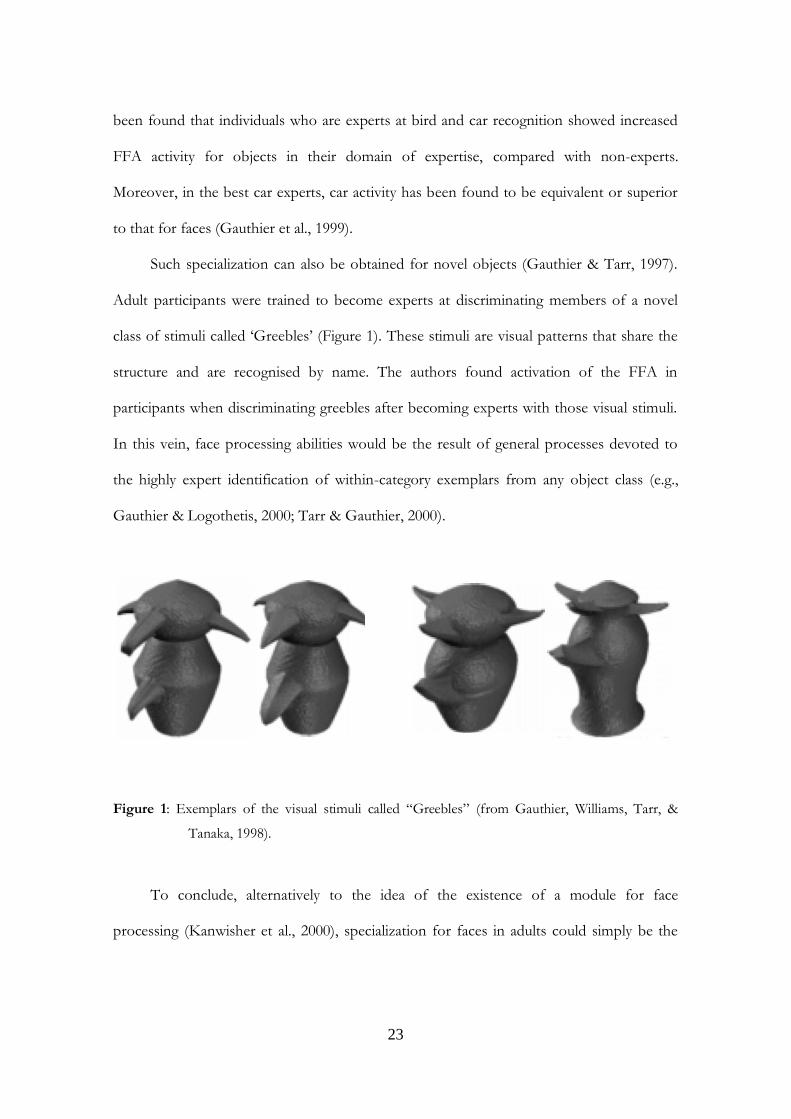
Such specialization can also be obtained for novel objects (Gauthier & Tarr, 1997).
Adult participants were trained to become experts at discriminating members of a novel
class of stimuli called „Greebles‟ (Figure 1). These stimuli are visual patterns that share the
structure and are recognised by name. The authors found activation of the FFA in
participants when discriminating greebles after becoming experts with those visual stimuli.
In this vein, face processing abilities would be the result of general processes devoted to
the highly expert identification of within-category exemplars from any object class (e.g.,
Gauthier & Logothetis, 2000; Tarr & Gauthier, 2000).
Figure 1: Exemplars of the visual stimuli called “Greebles” (from Gauthier, Williams, Tarr, &
Tanaka, 1998).
To conclude, alternatively to the idea of the existence of a module for face
processing (Kanwisher et al., 2000), specialization for faces in adults could simply be the

result of our experience with these objects (i.e., we recognize faces at a more specific level
than most objects and we acquire a lot of experience for such visual stimuli).
2.2 Face processing strategies in adults
At a functional level, behavioral studies showed that in adults face processing involves
cognitive processes different from those involved in the recognition of other objects (e.g.
Farah et al., 1998). Indeed in literature, a convincing demonstration that faces are a special
class of visual stimuli and that their processing is the result of a dedicated face-processing
system is the so called “face inversion effect”, and refers to the phenomenon by which upside-
down faces are disproportionately more difficult to recognize than other inverted objects
(Yin, 1969). The inversion effect has been considered as the hallmark of face-specific
recognition and is often used as a marker of expertise in face processing. Interestingly, this
effect is thought to interfere with perceptual face processing strategies, which are referred
as configural information.
It is generally accepted that the exceptionally proficient face recognition abilities
seen in adults are mainly derived from the rapid and efficient use of specific perceptual
processing strategies which involve the encoding of configural information that emerges
from the spatial relations among facial features, which is contrasted with the processing of
the shape of individual features (e.g., featural processing). It has been proposed that the use
of configural processing strategies for faces derives from the high degree of expertise with
this stimulus category that humans naturally develop (Diamond & Carey, 1986). The
expertise hypothesis is supported by findings from behavioral studies conducted with both
natural experts, who had become extremely skilled in the recognition of particular classes
of real-world objects, such as birds and cars (Diamond & Carey, 1986), and laboratory-
25
trained experts of artificial objects (i.e. greebles; Gauthier & Tarr, 1997). In these studies,
experts show a wide range of face-specific behavioral effects when tested within their
domain of expertise, and some authors interpret this evidence as an indication that experts
use configural processing to recognize objects of expertise in the same way as they do with
faces (e.g. Bukach, Gauthier, & Tarr, 2006)
Diamond and Carey (1986) proposed a model that subdivides configural
information into first-order and second-order relational information. First-order relational
information refers to qualitative spatial relations among facial features, that is the basic
arrangement of face features with two eyes above a nose, which is above a mouth. Second-
order relational information refers to fine spatial relations between features (for example,
the distance between the eyes). According to this model, Maurer, Le Grand, & Mondloch
(2002) suggested that configural face processing includes sensitivity to first-order relations,
holistic processing, and sensitivity to second-order relations.
Detecting face-like first-order relations, that specifies the stimulus as a face, is
facilitated by the fact that all faces share the same basic configuration. Adults have a
remarkable ability to detect faces based on first-order relations even without normal facial
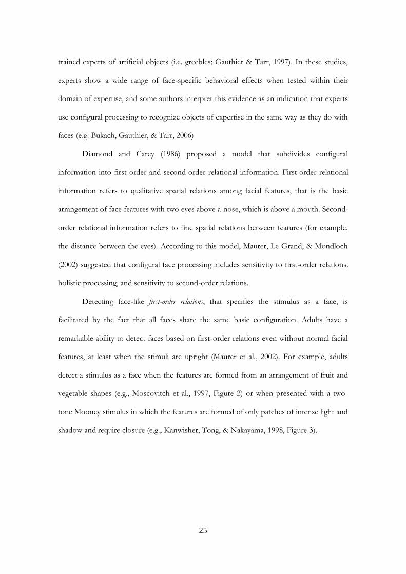
features, at least when the stimuli are upright (Maurer et al., 2002). For example, adults
detect a stimulus as a face when the features are formed from an arrangement of fruit and
vegetable shapes (e.g., Moscovitch et al., 1997, Figure 2) or when presented with a two-
tone Mooney stimulus in which the features are formed of only patches of intense light and
shadow and require closure (e.g., Kanwisher, Tong, & Nakayama, 1998, Figure 3).
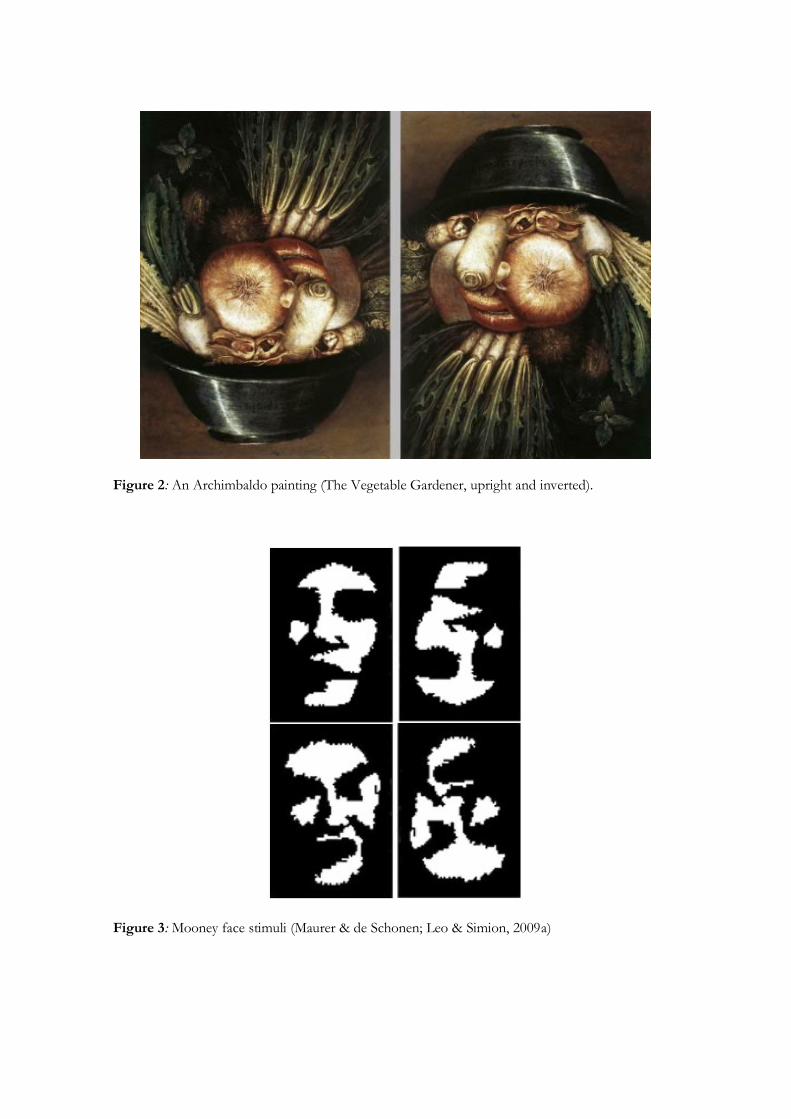
Figure 2: An Archimbaldo painting (The Vegetable Gardener, upright and inverted).
Figure 3: Mooney face stimuli (Maurer & de Schonen; Leo & Simion, 2009a)

27
To detect the first-order relations of a face, adults tend to integrate facial features
into a whole and then process the stimulus as a Gestalt, rendering the processing of
individual features less accessible. More specifically, the expression holistic processing
refers to encoding of the overall structure of the face, in which face parts and their
relations are not explicitly represented, but glued together in an undifferentiated whole
(Tanaka & Farah, 1993).
Evidence for the holistic type of processing comes from different paradigms. One
of the most influential study was designed by Tanaka and Farah (1993), who
operationalised the concept of holistic processing by developing a task in which
participants were presented with a series of faces and were tested on their recognition of
features such as eyes, mouth, or nose. The test consisted of showing the feature in isolation
and showing it within the context of the whole face, hence the name „Part-Whole
Paradigm‟. Faces could be presented in the canonical orientation or upside-down. Better
performance is tipically observed for the whole compared to the part condition (Tanaka &
Farah, 1993; Tanaka, Kay, Grinnell, Stansfield, & Szechter, 1998) (Figure 4). Indeed,
participants were more accurate in the recognition of the features presented in the whole
condition than in isolation, but only when faces are presented upright. On the contrary,
when features where presented inverted, the performance on whole condition decreased,
whereas accuracy on the part condition was not influenced by inversion manipulation.
Thus an inversion effect was only observed in the whole-face condition (Tanaka & Farah,
1993).
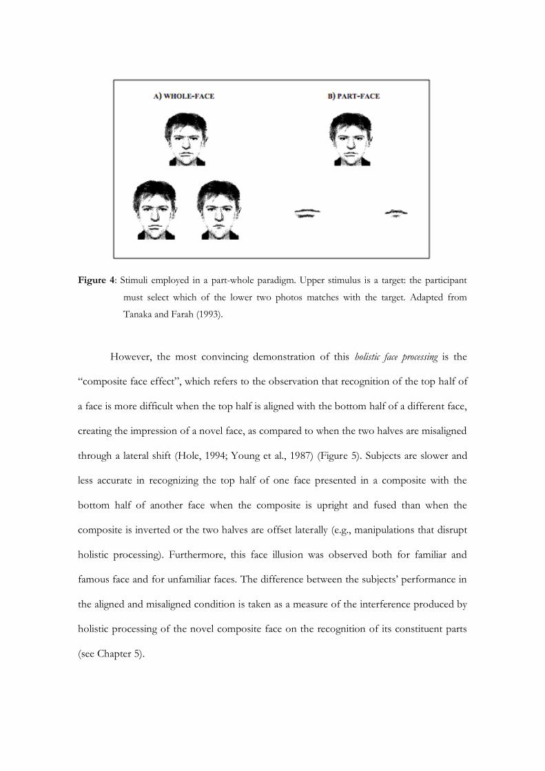
Figure 4: Stimuli employed in a part-whole paradigm. Upper stimulus is a target: the participant
must select which of the lower two photos matches with the target. Adapted from
Tanaka and Farah (1993).
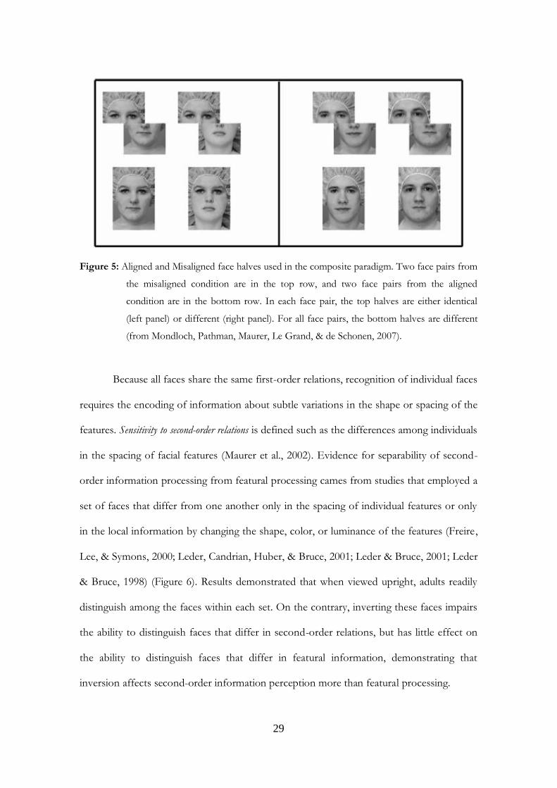
However, the most convincing demonstration of this holistic face processing is the
“composite face effect”, which refers to the observation that recognition of the top half of
a face is more difficult when the top half is aligned with the bottom half of a different face,
creating the impression of a novel face, as compared to when the two halves are misaligned
through a lateral shift (Hole, 1994; Young et al., 1987) (Figure 5). Subjects are slower and
less accurate in recognizing the top half of one face presented in a composite with the
bottom half of another face when the composite is upright and fused than when the
composite is inverted or the two halves are offset laterally (e.g., manipulations that disrupt
holistic processing). Furthermore, this face illusion was observed both for familiar and
famous face and for unfamiliar faces. The difference between the subjects‟ performance in
the aligned and misaligned condition is taken as a measure of the interference produced by
holistic processing of the novel composite face on the recognition of its constituent parts
(see Chapter 5).
29
Figure 5: Aligned and Misaligned face halves used in the composite paradigm. Two face pairs from
the misaligned condition are in the top row, and two face pairs from the aligned
condition are in the bottom row. In each face pair, the top halves are either identical
(left panel) or different (right panel). For all face pairs, the bottom halves are different
(from Mondloch, Pathman, Maurer, Le Grand, & de Schonen, 2007).
Because all faces share the same first-order relations, recognition of individual faces
requires the encoding of information about subtle variations in the shape or spacing of the
features. Sensitivity to second-order relations is defined such as the differences among individuals
in the spacing of facial features (Maurer et al., 2002). Evidence for separability of second-
order information processing from featural processing cames from studies that employed a
set of faces that differ from one another only in the spacing of individual features or only
in the local information by changing the shape, color, or luminance of the features (Freire,
Lee, & Symons, 2000; Leder, Candrian, Huber, & Bruce, 2001; Leder & Bruce, 2001; Leder
& Bruce, 1998) (Figure 6). Results demonstrated that when viewed upright, adults readily
distinguish among the faces within each set. On the contrary, inverting these faces impairs
the ability to distinguish faces that differ in second-order relations, but has little effect on
the ability to distinguish faces that differ in featural information, demonstrating that
inversion affects second-order information perception more than featural processing.
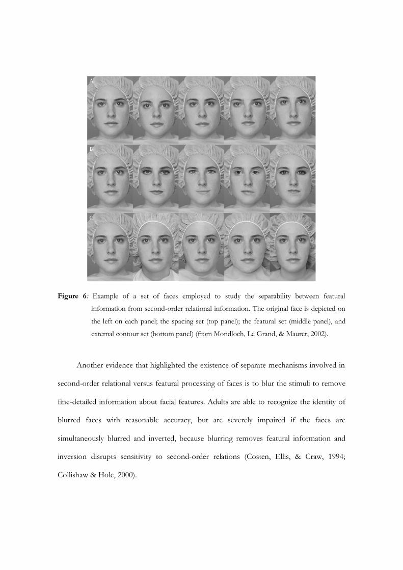
Figure 6: Example of a set of faces employed to study the separability between featural
information from second-order relational information. The original face is depicted on
the left on each panel; the spacing set (top panel); the featural set (middle panel), and
external contour set (bottom panel) (from Mondloch, Le Grand, & Maurer, 2002).
Another evidence that highlighted the existence of separate mechanisms involved in
second-order relational versus featural processing of faces is to blur the stimuli to remove
fine-detailed information about facial features. Adults are able to recognize the identity of
blurred faces with reasonable accuracy, but are severely impaired if the faces are
simultaneously blurred and inverted, because blurring removes featural information and
inversion disrupts sensitivity to second-order relations (Costen, Ellis, & Craw, 1994;
Collishaw & Hole, 2000).
31
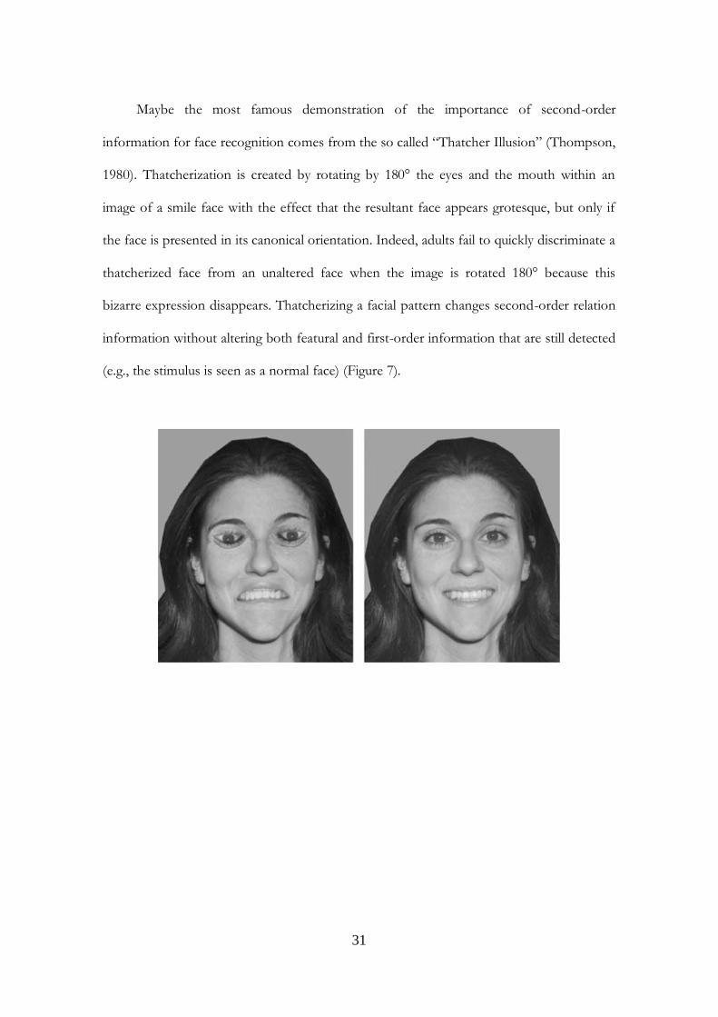
Maybe the most famous demonstration of the importance of second-order
information for face recognition comes from the so called “Thatcher Illusion” (Thompson,
1980). Thatcherization is created by rotating by 180° the eyes and the mouth within an
image of a smile face with the effect that the resultant face appears grotesque, but only if
the face is presented in its canonical orientation. Indeed, adults fail to quickly discriminate a
thatcherized face from an unaltered face when the image is rotated 180° because this
bizarre expression disappears. Thatcherizing a facial pattern changes second-order relation
information without altering both featural and first-order information that are still detected
(e.g., the stimulus is seen as a normal face) (Figure 7).
Figure 7: Examples of Thatcherized face stimuli (from Leo & Simion, 2009b).
Importantly, as suggested at the beginning of the paragraph, inversion interferes with
all three types of configural processing, since it increases difficulty in detecting the first-
order relations of a face and mitigates both the composite face effect and the part–whole
recognition effect that mark holistic processing of upright faces. Finally, it also seriously
impairs the accuracy of adults in discriminating among faces that differ only in second-
order relations (Maurer et al., 2002).
In conclusion, it has been demonstrated that there are at least three different types of
configural processing of faces: sensitivity to the first-order relations, holistic processing,
and sensitivity to second-order relations. These three types can be distinguished by
behavioral tasks and it seems logical that the three types operate in a hierarchical order:
detection of the face based on first-order relations as a necessary first step before holistic
processing and detection of second-order relations among the features. However, none of
the existing data rules out the possibility that the three types of configural processing
operate largely in parallel (Maurer et al., 2002).
33
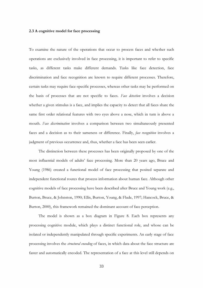
2.3 A cognitive model for face processing
To examine the nature of the operations that occur to process faces and whether such
operations are exclusively involved in face processing, it is important to refer to specific
tasks, as different tasks make different demands. Tasks like face detection, face
discrimination and face recognition are known to require different processes. Therefore,
certain tasks may require face-specific processes, whereas other tasks may be performed on
the basis of processes that are not specific to faces. Face detection involves a decision
whether a given stimulus is a face, and implies the capacity to detect that all faces share the
same first order relational features with two eyes above a nose, which in turn is above a
mouth. Face discrimination involves a comparison between two simultaneously presented
faces and a decision as to their sameness or difference. Finally, face recognition involves a
judgment of previous occurrence and, thus, whether a face has been seen earlier.
The distinction between these processes has been originally proposed by one of the
most influential models of adults‟ face processing. More than 20 years ago, Bruce and
Young (1986) created a functional model of face processing that posited separate and
independent functional routes that process information about human face. Although other
cognitive models of face processing have been described after Bruce and Young work (e.g.,
Burton, Bruce, & Johnston, 1990; Ellis, Burton, Young, & Flude, 1997; Hancock, Bruce, &
Burton, 2000), this framework remained the dominant account of face perception.
The model is shown as a box diagram in Figure 8. Each box represents any
processing cognitive module, which plays a distinct functional role, and whose can be
isolated or independently manipulated through specific experiments. An early stage of face
processing involves the structural encoding of faces, in which data about the face structure are
faster and automatically encoded. The representation of a face at this level still depends on
both the viewing condition (i.e., angle of profile, lighting) and facial configuration (i.e., eye
gaze, expression and mouth position). The face representation produced by structural
encoding is then processed by separate systems that perceive personal identity, expression
and speech related mouth movements. According to the authors, at this early stage, view-
centred descriptions provide information for the analysis of facial speech (e.g., the facial speech
code related to lips and tongue movements during speech) and for the analysis of expression
(e.g., the expression code encodes information about facial expression), whereas the
expression-independent descriptions provide information for the face recognition units
(FRUs).
Figure 8: The functional cognitive model of face processing in adults proposed by Bruce & Young
(1986) (from Bruce & Young, 1986).

35
Each face recognition unit contains stored structural descriptions, which allows views
of one known face to be discriminated from views of other faces, whether known or
unknown. The basic idea is that there is a separate face recognition unit for every familiar
face, and that a unit will become active when any view of the appropriate face is seen.
Importantly, the face recognition units respond only to face and do not respond to a
person‟s voice or name. The activation of a face recognition unit in turn leads to the
activation of the appropriate person identity code (PIN). It is the stage at which person
recognition, as opposed to face recognition, is achieved. The person identity code allows
access to semantic information about the identity of each known individual and encodes
additional information related to the seen face, such as information about the owner‟s
occupation, friends, his / her living place and so on, helping to establish the identity of the
person to whom the face belongs. Unlike FRUs, PIN may be accessed by many routes
since the same PIN will be made active not only by a person‟s face, but for example also by
a person‟s voice. It appears clear that face recognition can break down whereas person
recognition by other visual cues remains intact, since prosopagnosics become adept at
using other visual cues (such as the voice). The last stage, called the name generation, stores
information related to the name of the recognized person and can occur only by the PINs,
according to this model. As one can see from the Figure, the main system is called cognitive
system, that is responsible for the generation of the visually derived semantic codes, using
information from the analysis of expression, facial-speech, structural encoding, directed
visual processing, the face recognition units, person identification nodes and finally the
name generation. Importantly, in their model of cognitive system for face perception,
Bruce and Young (1986) proposed an organization that is hierarchical and branching, in
which all the systems processed different face information independent from each other.
In summary, this functional model proposes that face processing is a sequential
process, which involves several stages and is independent of and parallel to the other
processes designed to deal with different kinds of facial information.
2.4. A neural model for face processing
Currently, Bruce and Young‟s model is often used as a first step to more contemporary
theoretical considerations (Palermo & Rhodes 2007; Vuilleumier & Pourtois, 2007). One
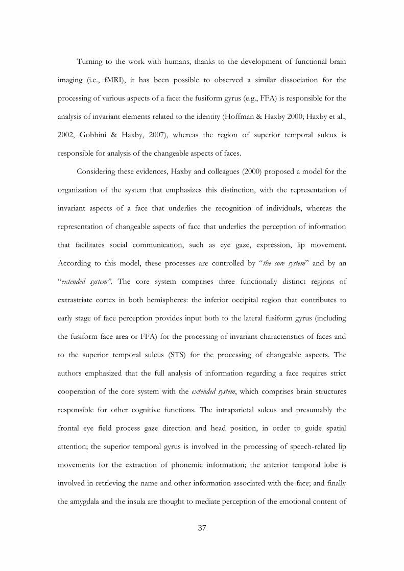
example is the model proposed by Haxby and colleagues (Haxby et al., 2000; Gobbini &
Haxby, 2007). These researchers argued that face perception is mediated by a distributed
neural system in humans that is formed by multiple, bilateral regions. They attempted to
show how cognitively distinct aspects of face perception are mediated by distinct neural
representations. The logic behind is that to recognize a face, the brain has to process
information related to the changeable face aspects such as facial expression eye gaze
direction or head position separately from features which are invariant allowing
identification of the face‟s owner (Figure 9).
As described before in this chapter, evidence of a specialized neural system for face
perception came from studies of non-human primates. Single unit recording studies in
macaques have identified neurons in the superior temporal sulcus (STS) that are involved
more in the perception of facial movement and static images of changeable aspects of face,
and neurons in the inferior temporal cortex that appears to be involved more in perceiving
facial identity (Perrett et al., 1985; Hasselmo et al., 1989; Desimone, 1991). Overall, the
findings from single-neuron recording studies in the monkey suggested a dissociation
between the roles of face-selective cells in the STS and inferior temporal cortex.
37
Turning to the work with humans, thanks to the development of functional brain
imaging (i.e., fMRI), it has been possible to observed a similar dissociation for the
processing of various aspects of a face: the fusiform gyrus (e.g., FFA) is responsible for the
analysis of invariant elements related to the identity (Hoffman & Haxby 2000; Haxby et al.,
2002, Gobbini & Haxby, 2007), whereas the region of superior temporal sulcus is
responsible for analysis of the changeable aspects of faces.
Considering these evidences, Haxby and colleagues (2000) proposed a model for the
organization of the system that emphasizes this distinction, with the representation of
invariant aspects of a face that underlies the recognition of individuals, whereas the
representation of changeable aspects of face that underlies the perception of information
that facilitates social communication, such as eye gaze, expression, lip movement.
According to this model, these processes are controlled by “the core system” and by an
“extended system”. The core system comprises three functionally distinct regions of
extrastriate cortex in both hemispheres: the inferior occipital region that contributes to
early stage of face perception provides input both to the lateral fusiform gyrus (including
the fusiform face area or FFA) for the processing of invariant characteristics of faces and
to the superior temporal sulcus (STS) for the processing of changeable aspects. The
authors emphasized that the full analysis of information regarding a face requires strict
cooperation of the core system with the extended system, which comprises brain structures
responsible for other cognitive functions. The intraparietal sulcus and presumably the
frontal eye field process gaze direction and head position, in order to guide spatial
attention; the superior temporal gyrus is involved in the processing of speech-related lip
movements for the extraction of phonemic information; the anterior temporal lobe is
involved in retrieving the name and other information associated with the face; and finally
the amygdala and the insula are thought to mediate perception of the emotional content of
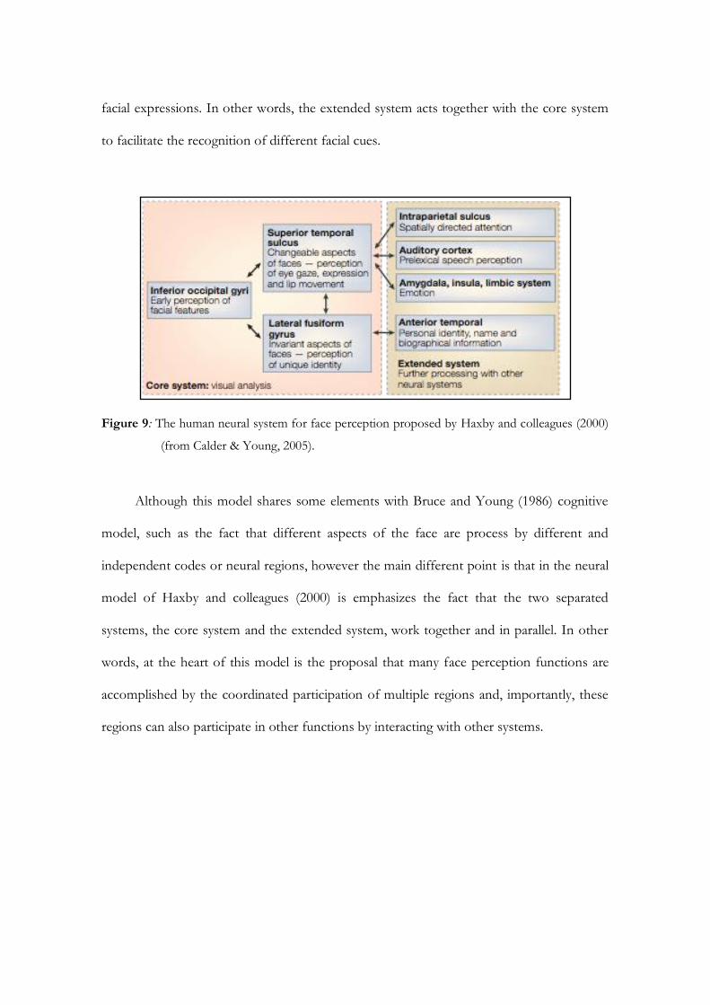
facial expressions. In other words, the extended system acts together with the core system
to facilitate the recognition of different facial cues.
Figure 9: The human neural system for face perception proposed by Haxby and colleagues (2000)
(from Calder & Young, 2005).
Although this model shares some elements with Bruce and Young (1986) cognitive
model, such as the fact that different aspects of the face are process by different and
independent codes or neural regions, however the main different point is that in the neural
model of Haxby and colleagues (2000) is emphasizes the fact that the two separated
systems, the core system and the extended system, work together and in parallel. In other
words, at the heart of this model is the proposal that many face perception functions are
accomplished by the coordinated participation of multiple regions and, importantly, these
regions can also participate in other functions by interacting with other systems.
39
Conclusion
Evidence from behavioral, brain lesion and neuroimaging studies suggests that in adult face
processing involves distinct, domain-specific perceptual processing (Maurer et al., 2002)
carried out by dedicate brain areas (e.g., Farah et al., 2000; Kanwisher, 2000). The
functional and neural specialization presented in the adult face processing system renders
faces an ideal class of stimuli to investigate the time course and the factors affecting such
specialization. Some authors claim the existence of a specialized system for face processing
already at birth (experience-independent; e.g., Farah et al., 2000), whereas others raise the
possibility that such specialization is a product of experience (experience-dependent; e.g.,
Gauthier & Logothetis, 2000). So, data from infants, and newborns especially, become
relevant for resolving this debate because they do not have the years of experience with
faces necessary to acquire expertise. The aim of the following chapters will be to support
the idea that the face specificity is not prewired, but rather arises from general perceptual
processes that, during development, become progressively tuned to the human face, as a
result of extensive experience with this stimulus category.


CHAPTER 3
THE NATURE OF FACE REPRESENTATION
IN THE FIRST MONTHS OF LIFE

43
Introduction
As described in Chapter 2, evidence from behavioral, brain lesions and neuroimaging
studies suggested that in adults face processing involves distinct, domain specific
perceptual processing (Maurer et al., 2002) carried out by dedicated brain areas (Farah et
al., 2000; Kanwisher et al., 1997; Kanwisher, 2000). However, what determines this
specialization and how this specialization emerges during development still remain largely
unknown. Recent findings support the hypothesis that the presence at birth of general
perceptual biases on visual processing seems sufficient to cause the human face to be a
frequent focus on newborns‟ visual attention, allowing, through experience, the gradual
development of a face processing system (Simion, Macchi Cassia, Turati, & Valenza, 2001;
Turati, et al., 2005; Nelson 2001, 2003). In this vein, it has been suggested that humans
might be born with some representation of the human face (Johnson & Morton, 1991;
Johnson, 2005; Valenza, Simion, Macchi Cassia, & Umiltà, 1996; Macchi Cassia, Turati, &
Simion, 2004), but a still open question concerns whether this face representation is innate
and a product of an evolutionary pressure or whether it is only a product of a rapid
learning process derived from experience with faces during the first hours of life.
Considering this literature, the aim of the present Chapter is to examine cognitive
specialization during early infancy through the investigation of the development of face
representation. In the first section of the Chapter will be described the theoretical models
interpreting face preference at birth and in the first few months of life. Evidence will be
reviewed supporting the contention that newborns‟ face preference is due to a set of non-
specific constraints that stem from the general characteristics of the human visuo-
perceptual system, rather than to a representational bias for faces. In the second section,

experiments will be reported that shown that infants‟ response to faces becomes more and
more tuned to the face category over the first three-months of life, revealing a gradual
progressive specialization of the face processing system, as the perceptual narrowing
hypothesis claims (Nelson, 2001, 2003).
3.1 The emergence of cognitive specialization for face processing
Three major approaches to the development of the face processing system have been
distinguished (de Haan & Halit, 2001).
A first view argues that the development of face specialization is an experience-
independent process (Farah et al., 2000). Due to the social relevance of face for humans,
natural selection led, through phylogenesis, to the evolution of innate face-specific devices
that would be available before any postnatal experience and explicitly specified in the
genome (Farah et al., 1998, 2000). Evidence in favor of this view comes from a single case
study that described an infant, who, at 1 day of age, sustained brain damage that resulted in
a profound impairment in face processing (Farah et al., 2000). This module has been
thought to be an experience-independent mechanism dedicated to face processing and selective
response of newborns to face stimuli is considered a the direct precursor of the adult
cortical face processing system.
A second approach characterizes the development of face processing as an
experience-dependent process, hypothesizing that extensive and prolonged experience with faces
would gradually render humans exceptionally proficient in recognizing individual exemplars
belonging to this special class of visual stimuli. From this point of view, that highlight the
role played by experience in the ontogenetic development of face processing, general
learning processes are thought to be sufficient to explain thel emergence of a cortical
45
system for the processing of faces (Diamond & Carey, 1986; Gauthier & Logothetis, 2000;
Tarr & Gauthier, 2000). Importantly, within this experience-dependent approach, the
general learning processes can occur at any time during development and might involve
any class of visual stimuli.
Recently however, many studies on the development of face processing system
share the idea that the emergence of the ability to process faces is the result of the
interaction between innately specified predispositions and the extensive experience
everyone has with faces (de Schonen 2002; Johnson, 1993; Le Grand et al., 2003; Nelson,
2001, 2003). This third approach describes the development of face processing system as
an experience-expectant process, claiming that the functional and neuroanatomical specialization
for faces processing revealed in human adults would emerge gradually from the interaction
between innate constraints and the structure of input provided by the specie-typical
environment, with innate constraints having the function of potentiating early learning
(Johnson, 1993). The cortical tissue has gained, through evolutionary pressures, the
potential to become specialized for face processing. However, this specialization would
emerge on condition that the critical type of input is provided within crucial time windows
(Nelson, 2001, 2003). According to a probabilistic epigenesis perspective (see Chapter 1),
the interactions between genes, structural brain changes, and psychological functions are
bidirectional and contribute to cognitive development (Greenough & Black, 1992). In
particular, the partial functioning of neural pathways would shape subsequent development
of neural structures and circuits that are the basis for further functional development (see
Simion, Turati, Valenza, & Leo, 2006). This process would be a progressive tuning of
certain cerebral tissues, from a large range of visual information to the specific type of
information that faces convey (Johnson, 2000; Nelson, 2001, 2003).
One way of disentangling the issue of the origin and the development of cognitive
specialization for faces might be to study the developmental time course of a phenomenon
present few hours after birth, such as the newborns‟ face preference.
3.2 Models of face preference at birth
A great number of behavioral studies established that humans are born with a
predisposition or bias to attend to faces. Indeed, despite their lack of experience, newborns
prefer both schematic and real faces over almost any other category of visual stimuli
(Goren, Sarty, & Wu, 1975; Johnson, Dziuarawiec, Ellis, & Morton, 1991; Valenza et al.,
1996; Macchi Cassia et al., 2004; Johnson, 2005). Although the existence of the face
preference few hours after birth may not provide conclusive evidence for an innate face
processing system, it seems suggestive of a specialized cognitive system operating from
very early in life. There is little dispute over the existence of a newborns‟ face preference,
however the debate concerns what mechanisms underlay this preference at birth.
Indeed, some researchers hypothesized that faces, already at birth, represent a
special class of visual stimuli because humans are born with a specific and innate
mechanism selectively tuned to the face geometry (Johnson & Morton, 1991; Johnson,
2005). It has been proposed that a subcortical visuomotor system, named Conspec, is
present at birth, which allows newborns to detect a face in the visual environment (Morton
& Johnson, 1991; Johnson & Morton, 1991). This subcortical system is thought to possess
a very simple representation of a face, that is three high-contrast blobs arranged in a
triangular formation within a bounded contour. This representation would be suitable only
for directing the newborn gaze towards face-like patterns, but is insufficient for more
elaborate face discrimination. Interestingly, the finding that newborns only display a face
47
preference when stimuli are presented in the temporal visual field (which is related to
subcortical pathways), but not when they are presented in the nasal visual field (related to
cortical pathways) is consistent with the subcortical location hypothesis (Simion, Valenza,
Umiltà, & Dalla Barba, 1998). A two-process model was hypothesized in which at birth
infants posses a subcortical orienting mechanism, Conspec, that guides the preference for
faces since its primary function is to ascertain that facial input is maximized during the first
two months of life (de Schonen & Mathivet, 1989), before a second cortical face-learning
mechanism, Conlern, comes up. That is, at approximately 2 months of age, infant
preference is controlled by another mechanism responsible for learning about faces. By
ensuring that infants have visual experience with faces, the subcortical mechanism would
favor the gradual emergence of the specialized cortical circuits that underlay face
processing in adults. In other words, Conlern functions at the cortical level as a non-
specific mechanism that facilitates learning about the characteristics of the face one
Conspec has served to orient the newborn to conspecifics faces. As suggested by the
authors, there is no linkage between the two mechanisms, rather they may interact
indirectly through the environment. That is, “Conlearn [...] is a non-specialized learning
mechanism. Effectively it learns about the characteristics of faces because the infant pays a
lot of attention to them. The role of Conspec is to direct this attention.” (p.175).
Recently, Johnson (2005) has updated his model arguing the existence of a low-
spatial frequency (LSF) face configuration detector, provided by evolutionary pressure
active throughout the life span. Indeed, recent evidence from behavioral (Tomalski, Csibra,
& Johnson, 2009), electroencephalograph and magnetoencephalograph techniques (Bailey,
Braeutigam, Jousmaki, & Switheneby, 2005) demonstrated subcortical processing in adults,
suggesting that a rapid LSF face detector cannot be dismissed. Considering this evidence, it
is likely that Conspec does not vanish at 2 months of age but is rather a rudimentary LSF
face detector that endures throughout life to perform this important function. In this
model, face detection would be supported by a “quick and dirty” subcortical route sensitive
to a raised surface with darker areas corresponding to the locations of eyes. This
subcortical pathway might be important to trigger the network of cortical regions that
makes up the adult social brain. In this vein, specialization of the face cortical circuits
might emerge due to a combination of factors including subcortical mechanisms that
function to guide infants‟ visual attention to faces during the first weeks of life, biasing the
input to the developing cortical system, increasing experience with faces, and increasing
demands to process faces as infants develop. Importantly, it has been proposed that
impairments to the subcortical route result in specific types of atypical development, such
as autism. Specifically, if the subcortical face-processing route is important for the
development of the adult social brain network, then disruption of this pathway by
congenital factors could have important negative consequences for the social brain network
as a whole (Johnson, 2005).
An alternative and complementary model hypothesizes that both Conspec and
Conlern mechanisms are present at birth and that the visual cortex also heavily contributes
from birth to develop a face recognition system (Acerra, Burnod, & de Schonen, 2002).
This computational model predicts that feedbacks between the subcortical and cortical system
are likely, arguably making them an integrated system. In line with anatomical data on
human newborn brains that showing that V1 neurons (i.e., primary visual cortex) are able
to receive and convey information to other neurons (LeRoy Conel, 1939), the authors
suggest that those neurons are involved in face preference at birth, together with other
occipital cortical areas and subcortical structures, such as the superior colliculus. In this
vein, face processing at birth is underlying by low-level (V1) processing and by subcortical
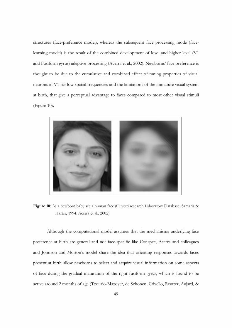
49
structures (face-preference model), whereas the subsequent face processing mode (face-
learning model) is the result of the combined development of low- and higher-level (V1
and Fusiform gyrus) adaptive processing (Acerra et al., 2002). Newborns‟ face preference is
thought to be due to the cumulative and combined effect of tuning properties of visual
neurons in V1 for low spatial frequencies and the limitations of the immature visual system
at birth, that give a perceptual advantage to faces compared to most other visual stimuli
(Figure 10).
Figure 10: As a newborn baby see a human face (Olivetti research Laboratory Database; Samaria &
Harter, 1994; Acerra et al., 2002)
Although the computational model assumes that the mechanisms underlying face
preference at birth are general and not face-specific like Conspec, Acerra and colleagues
and Johnson and Morton‟s model share the idea that orienting responses towards faces
present at birth allow newborns to select and acquire visual information on some aspects
of face during the gradual maturation of the right fusiform gyrus, which is found to be
active around 2 months of age (Tzourio-Mazoyer, de Schonen, Crivello, Reutter, Aujard, &
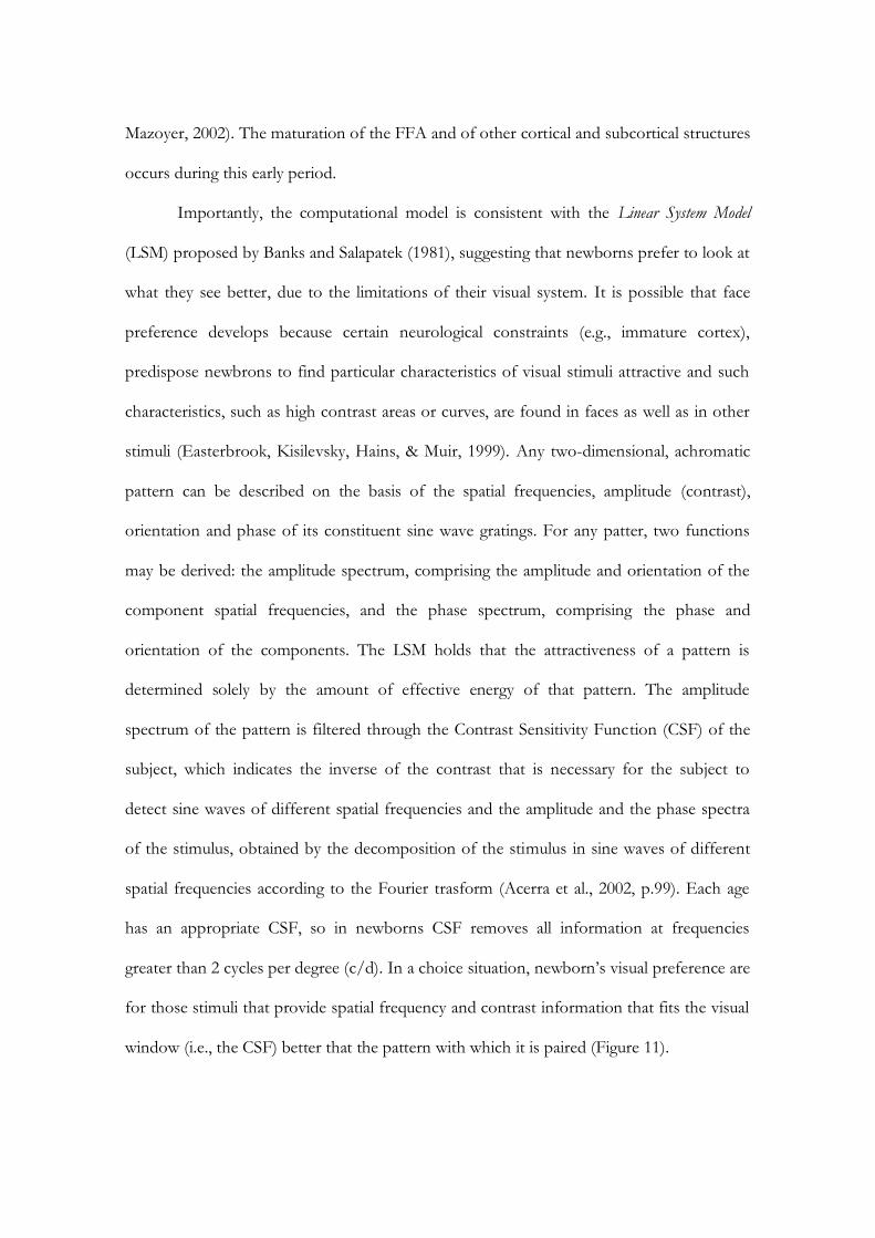
Mazoyer, 2002). The maturation of the FFA and of other cortical and subcortical structures
occurs during this early period.
Importantly, the computational model is consistent with the Linear System Model
(LSM) proposed by Banks and Salapatek (1981), suggesting that newborns prefer to look at
what they see better, due to the limitations of their visual system. It is possible that face
preference develops because certain neurological constraints (e.g., immature cortex),
predispose newbrons to find particular characteristics of visual stimuli attractive and such
characteristics, such as high contrast areas or curves, are found in faces as well as in other
stimuli (Easterbrook, Kisilevsky, Hains, & Muir, 1999). Any two-dimensional, achromatic
pattern can be described on the basis of the spatial frequencies, amplitude (contrast),
orientation and phase of its constituent sine wave gratings. For any patter, two functions
may be derived: the amplitude spectrum, comprising the amplitude and orientation of the
component spatial frequencies, and the phase spectrum, comprising the phase and
orientation of the components. The LSM holds that the attractiveness of a pattern is
determined solely by the amount of effective energy of that pattern. The amplitude
spectrum of the pattern is filtered through the Contrast Sensitivity Function (CSF) of the
subject, which indicates the inverse of the contrast that is necessary for the subject to
detect sine waves of different spatial frequencies and the amplitude and the phase spectra
of the stimulus, obtained by the decomposition of the stimulus in sine waves of different
spatial frequencies according to the Fourier trasform (Acerra et al., 2002, p.99). Each age
has an appropriate CSF, so in newborns CSF removes all information at frequencies
greater than 2 cycles per degree (c/d). In a choice situation, newborn‟s visual preference are
for those stimuli that provide spatial frequency and contrast information that fits the visual
window (i.e., the CSF) better that the pattern with which it is paired (Figure 11).
51
Figure 11: Contrast Sensitivity Functions for adults and 1-month-old infants and the CSF for 3-
day-old infants (from Banks & Ginsburg, 1985; Acerra et al., 2002).
The sensory hypothesis claims that newborn‟s preferences for visual patterns are
determined solely by their visibility. In this vein, faces are not different from other visual
stimuli. In other words, this model accounting for newborns‟ visual preferences in terms of
the match between the characteristics of the newborns visual system and the
psychophysical properties of the stimuli as described by both the low level properties (i.e.
contrasts and spatial frequencies content described by the amplitude spectrum) and higher
level variables (i.e., the structural properties of a stimulus described by the phase spectrum)
according to the Fourier‟s transforms. This sensory hypothesis suggests that the newborns‟
preference for a visual stimulus is determined by the amplitude spectra of the pattern
filtered by the CSF of the newborns and when two stimuli with identical amplitude
spectrum, the infant‟s visual preference is determined by their phase spectrum (Kleiner,
1987).
Consistent with the LSM model, an alternative account explains face preference at
birth as due to the match between perceptual and structural properties present in a face
and the constraints of the newborns‟ sensory system (Simion et al., 2001; Simion, Macchi
Cassia, Turati, & Valenza, 2003). This alternative hypothesis suggests that the presence at
birth of non-face-specific attentional biases is sufficient to produce the emergence of the
functional and neural specialization for faces observed later during development. The adult
face processing system appears thus capable of bootstrapping from minimal information,
not requiring highly specific predispositions. Considering the alternative view, face
preference at birth would be the result of the cumulative effect of a set of non-specific
constraints that stem from the general characteristics of the immature visual system and a
collection of general structural properties that attracts newborns‟ attention. This claim
derives mainly from the demonstration that, besides facedness, newborns manifest
spontaneous preference for other structural properties of visual stimuli. For example, when
horizontal gratings were paired with vertical gratings, newborns preferred the horizontal
ones (i.e., Slater, Earle, Morison, & Rose, 1985; Farroni, Valenza, Simion, & Umiltà, 2000).
Because the patterns were equated for quantity of energy (Low Spatial Frequencies), one
can assume that orientation, which is a structural property of the stimulus, was the crucial
factor in determining the preference for horizontal gratings. Therefore, facedness might be
preferred because of the addictive effect of a collection of structural and perceptual
properties. Indeed, faces are symmetrical along the vertical axis, present more patterning in
the upper compared to the lower half and have rounded rather straight edges. The
possibility exists that some, if not each of these properties, plays a role in promoting the
newborns‟ attentional response toward face-like stimuli and veridical face images.
Many studies showed that at least two non-specific structural properties not only
are preferred at birth when embedded in non-face geometric configurations (Macchi
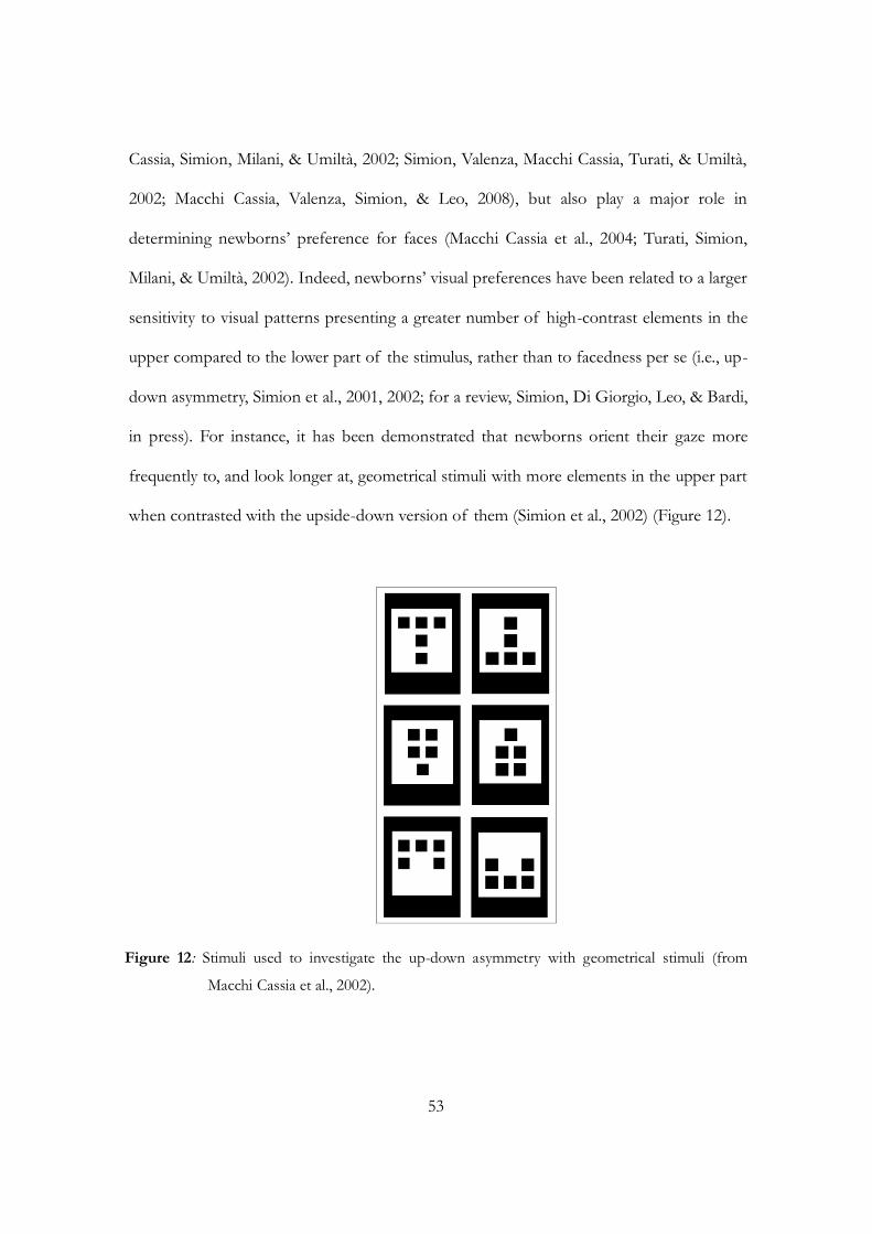
53
Cassia, Simion, Milani, & Umiltà, 2002; Simion, Valenza, Macchi Cassia, Turati, & Umiltà,
2002; Macchi Cassia, Valenza, Simion, & Leo, 2008), but also play a major role in
determining newborns‟ preference for faces (Macchi Cassia et al., 2004; Turati, Simion,
Milani, & Umiltà, 2002). Indeed, newborns‟ visual preferences have been related to a larger
sensitivity to visual patterns presenting a greater number of high-contrast elements in the
upper compared to the lower part of the stimulus, rather than to facedness per se (i.e., up-
down asymmetry, Simion et al., 2001, 2002; for a review, Simion, Di Giorgio, Leo, & Bardi,
in press). For instance, it has been demonstrated that newborns orient their gaze more
frequently to, and look longer at, geometrical stimuli with more elements in the upper part
when contrasted with the upside-down version of them (Simion et al., 2002) (Figure 12).
Figure 12: Stimuli used to investigate the up-down asymmetry with geometrical stimuli (from
Macchi Cassia et al., 2002).
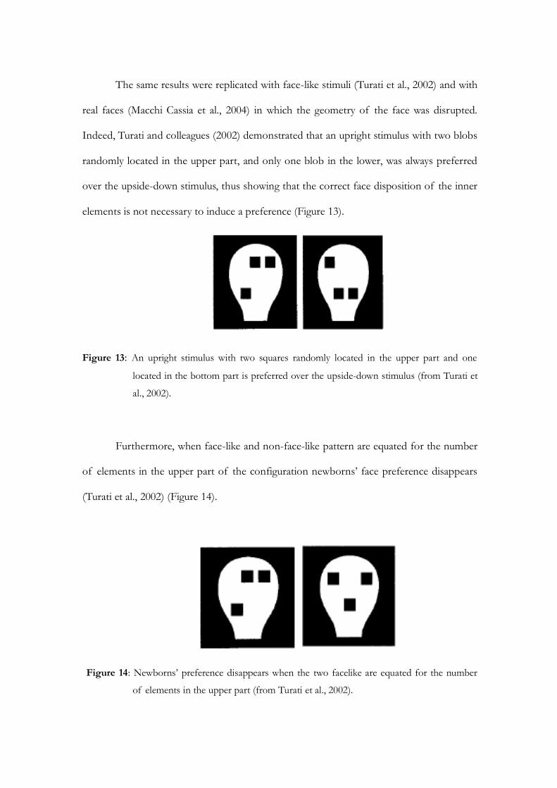
The same results were replicated with face-like stimuli (Turati et al., 2002) and with
real faces (Macchi Cassia et al., 2004) in which the geometry of the face was disrupted.
Indeed, Turati and colleagues (2002) demonstrated that an upright stimulus with two blobs
randomly located in the upper part, and only one blob in the lower, was always preferred
over the upside-down stimulus, thus showing that the correct face disposition of the inner
elements is not necessary to induce a preference (Figure 13).
Figure 13: An upright stimulus with two squares randomly located in the upper part and one
located in the bottom part is preferred over the upside-down stimulus (from Turati et
al., 2002).
Furthermore, when face-like and non-face-like pattern are equated for the number
of elements in the upper part of the configuration newborns‟ face preference disappears
(Turati et al., 2002) (Figure 14).
Figure 14: Newborns‟ preference disappears when the two facelike are equated for the number
of elements in the upper part (from Turati et al., 2002).
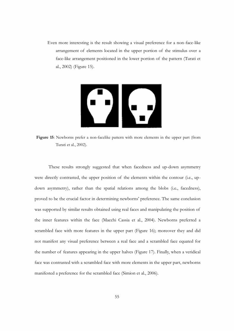
55
Even more interesting is the result showing a visual preference for a non-face-like
arrangement of elements located in the upper portion of the stimulus over a
face-like arrangement positioned in the lower portion of the pattern (Turati et
al., 2002) (Figure 15).
Figure 15: Newborns prefer a non-facelike pattern with more elements in the upper part (from
Turati et al., 2002).
These results strongly suggested that when facedness and up-down asymmetry
were directly contrasted, the upper position of the elements within the contour (i.e., up-
down asymmetry), rather than the spatial relations among the blobs (i.e., facedness),
proved to be the crucial factor in determining newborns‟ preference. The same conclusion
was supported by similar results obtained using real faces and manipulating the position of
the inner features within the face (Macchi Cassia et al., 2004). Newborns preferred a
scrambled face with more features in the upper part (Figure 16); moreover they and did
not manifest any visual preference between a real face and a scrambled face equated for
the number of features appearing in the upper halves (Figure 17). Finally, when a veridical
face was contrasted with a scrambled face with more elements in the upper part, newborns
manifested a preference for the scrambled face (Simion et al., 2006).
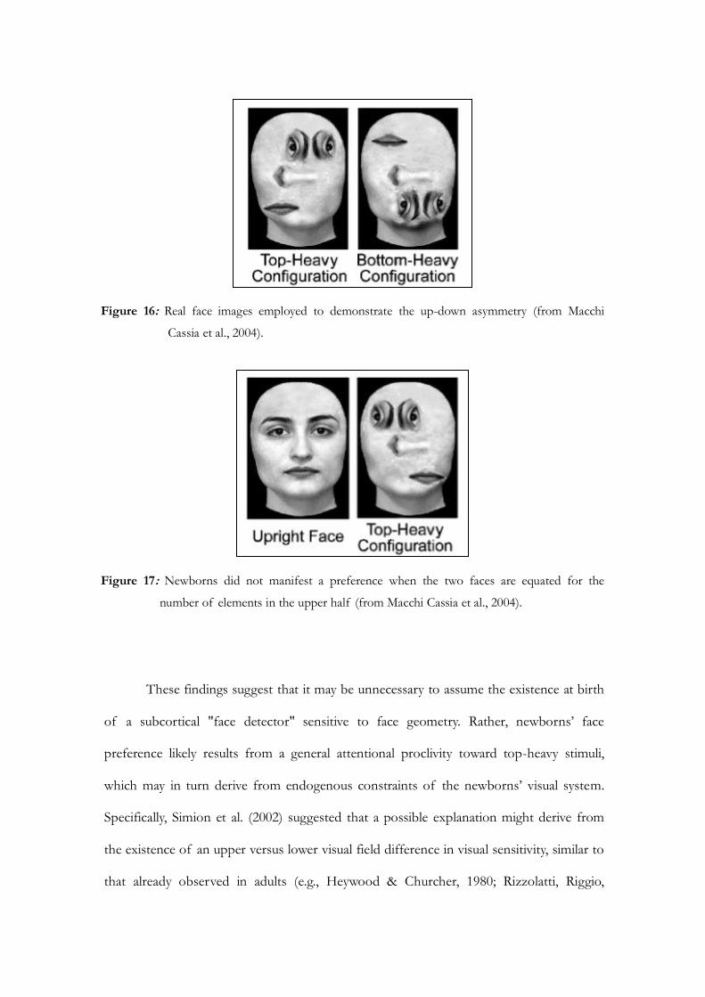
Figure 16: Real face images employed to demonstrate the up-down asymmetry (from Macchi
Cassia et al., 2004).
Figure 17: Newborns did not manifest a preference when the two faces are equated for the
number of elements in the upper half (from Macchi Cassia et al., 2004).
These findings suggest that it may be unnecessary to assume the existence at birth
of a subcortical "face detector" sensitive to face geometry. Rather, newborns‟ face
preference likely results from a general attentional proclivity toward top-heavy stimuli,
which may in turn derive from endogenous constraints of the newborns‟ visual system.
Specifically, Simion et al. (2002) suggested that a possible explanation might derive from
the existence of an upper versus lower visual field difference in visual sensitivity, similar to
that already observed in adults (e.g., Heywood & Churcher, 1980; Rizzolatti, Riggio,
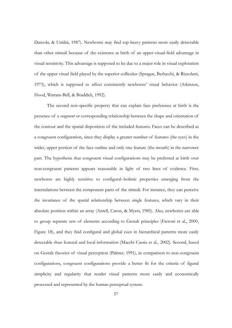
57
Dascola, & Umiltà, 1987). Newborns may find top-heavy patterns more easily detectable
than other stimuli because of the existence at birth of an upper-visual-field advantage in
visual sensitivity. This advantage is supposed to be due to a major role in visual exploration
of the upper visual field played by the superior colliculus (Sprague, Berlucchi, & Rizzolatti,
1973), which is supposed to affect consistently newborns‟ visual behavior (Atkinson,
Hood, Wattam-Bell, & Braddick, 1992).
The second non-specific property that can explain face preference at birth is the
presence of a congruent or corresponding relationship between the shape and orientation of
the contour and the spatial disposition of the included features. Faces can be described as
a congruent configuration, since they display a greater number of features (the eyes) in the
wider, upper portion of the face outline and only one feature (the mouth) in the narrower
part. The hypothesis that congruent visual configurations may be preferred at birth over
non-congruent patterns appears reasonable in light of two lines of evidence. First,
newborns are highly sensitive to configural–holistic properties emerging from the
interrelations between the component parts of the stimuli. For instance, they can perceive
the invariance of the spatial relationship between single features, which vary in their
absolute position within an array (Antell, Caron, & Myers, 1985). Also, newborns are able
to group separate sets of elements according to Gestalt principles (Farroni et al., 2000,
Figure 18), and they find configural and global cues in hierarchical patterns more easily
detectable than featural and local information (Macchi Cassia et al., 2002). Second, based
on Gestalt theories of visual perception (Palmer, 1991), in comparison to non-congruent
configurations, congruent configurations provide a better fit for the criteria of figural
simplicity and regularity that render visual patterns more easily and economically
processed and represented by the human perceptual system.
Figure 18: Stimuli employed to demonstrate newborns‟ ability to group separate sets of elements
according to Gestalt principles (from Farroni et al., 2000).
Because newborns have been shown to perceive and organize visual arrays
according to Gestalt principles, such as lightness similarity (Farroni et al., 2000) and
common motion (Valenza & Bulf, 2007), it seems reasonable to hypothesize that
newborns may be sensitive to other stimulus dimensions that contribute to figural
goodness, such as symmetry, repetition, and regularity. Each of these dimensions is
maximally present in congruent visual configurations. Evidence revealed that when
congruent and non-congruent non-face configurations were compared (using both
triangles and trapezoids), a reliable tendency to prefer the congruent pattern was observed
in newborns. A top-heavy congruent stimulus was preferred over a top-heavy non-
congruent stimulus, thus indicating that the congruency and top-heavy properties have an
additive effect on newborns‟ preferences (Macchi Cassia et al., 2008) (Figure 19).
Overall, the results described here support the idea that face preference is a
preference for some general perceptual and structural properties that faces share with
other visual stimuli.
59
Figure 19: Stimuli employed to demonstrate that a top-heavy congruent stimulus was preferred
over a top-heavy non-congruent stimulus (Macchi Cassia et al., 2008).
3.3 The nature of face representation at birth and in the first few months of age
As well as the debate concerning the mechanisms underlying face preference at birth,
another long-standing issue concerns the nature of the face representation at birth. There
is agreement about the idea that humans might be born with some representation of the
human face (Johnson & Morton, 1991; Valenza et al., 1996; Macchi Cassia et al., 2004), but
a still open question concerns whether this face representation is innate and a product of
an evolutionary pressure or whether it is only a product of a rapid learning process from
experience with faces during the first hours of life.
Several empirical findings support the nativist hypothesis, suggesting that newborn
babies may begin life with a specific representation of the human face (Slater & Kirby,
1998; Slater, von der Schulenburg, Brown, Badenoch, Butterworth, Parsons, & Samuels,
1998; Meltzoff & Moore, 1977; Slater & Quinn, 2001). From this perspective, an early
appearing representation bias that is specific to faces may be more elaborate than a

preference for three dark dots in a triangle corresponding to eyes and mouth (e.g., Config).
Indeed, newborns imitated a variety of facial gestures that they see, giving aid to the
hypothesis that the innate face representation might be very detailed from the beginning
(Meltzoff & Moore, 1977). The authors maintained that this could not be possible without
a specific and innate representation of their own face. Recently, Quinn and Slater (2003)
suggested that the face-processing system could be innately provided by evolutionary
pressure and that newborns‟ initial face representation could be formed through
proprioception in utero, since proprioceptive feedback provided by facial movements
could contribute to the formation of a face specific representation at birth. In this vein, the
face representation formed in utero would be responsible of orienting of attention toward
matching configurations (i.e., other kinds of faces). However, Pascalis and Kelly (2009)
suggested that if the exact mechanisms supporting early face preference remain unclear, it
could include both fast learning based on early interactions in the post partum period as
well as an innate system selected by evolution. Considering the limited amount of
experience newborns have with a limited number of face, it could be suggested that face
representation will be crude in the first days of life. With experience, the human specificity
of the representation will increase.
An alternative interpretation of face preference at birth is in term of prototype and
cognitive averaging process (“learning account”, Langlois & Roggman, 1990; Rubenstein,
Kalakanis, & Langlois, 1999). Learning about faces and the formation of a representation
of faces might be extremely rapid in the first days of life and according to a single study,
newborns might be able to form a face representation in less than 1 minute after birth
(Walton & Bower, 1993). The results of a preference for attractive over non-attractive faces
have been interpreted as in favour of the learning account hypothesis, since attractive faces
might match more closely the prototype that newborns have formed during their brief
61
experience in the first hours after birth (Slater & Kirby, 1998; Slater, Bremner, Johnson,
Sherwood, Hayes, & Brown, 2000; Slater, Quinn, Hayes, & Brown, 2000). However this
interpretation has been questioned by recent results showing that the preference for
attractive face is not human-specific. These findings corroborate the idea that the initial
settings of our perceptual system push infants to look at some entities because of the
presence of a family of preferred perceptual features (Quinn, Kelly, Lee, Pascalis, & Slater,
2008).
In line with this interpretation face preference at birth may be a product of a
general perceptual and information processing mechanism that subsequently becomes
“tuned” to human faces as a direct consequence of the extensive experience with this
stimulus category provided by the species-typical environment within the first days of life
(Scott et al., 2007). This “perceptual narrowing” hypothesis (Lewkowicz & Ghazanfar, 2006;
Nelson, 2001, 2003) suggests that at birth, the nature of face representation is broadly
general and that subsequently becomes finely tuned as a function of the progressive
increase in the selectivity and localization of the cortical circuits involved in face
processing, due to the facial input received (Johnson, 2000). This concept of a face
representation is well understood within the framework of the multidimensional face space
model proposed by Valentine (1991). This norm-based coding model suggests that faces
are encoded as vectors according to their deviation from a prototypical average of the face
space. As proposed by Nelson (2001), this face prototype is broadly tuned at birth and the
dimensions that this prototype encodes may differ both qualitatively and quantitatively in
infants compared with adults. Pascalis and colleagues (2005) suggested that “a good way to
think about the development or formation of a face prototype is based on the experience
or kinds of faces one meets. For example, if this prototype is thought of as a continuum of

all incoming faces, then the more a face deviates from the prototype (other-race and other-
species faces), the less this face is easily discriminated, compared with faces that are more
similar to the prototype. Importantly, the development of the face prototype is likely
influenced by a number of factors, including exposure time (number of faces seen),
dynamic and emotionally salient information provided within the face and changes in the
categorization of individuation of people. Combined, these experiences gradually lead to
the face prototype becoming more specific (Pascalis et al., 2005, p. 5298)”. The
development of face prototype is greatly influenced by the experience with certain kinds of
faces present in the environment, as well as the exposure time to those faces (Pascalis,
Scott, Kelly, Shannon, Nicholson, Coleman, & Nelson, 2005).
Early in life, infants possess a remarkable ability to discriminate among and
between a large corpus of different faces, such as faces from an unfamiliar species or an
unfamiliar race. With experience, the infant‟s face-representation system becomes more
precise and increasingly restricted to faces with which infants are most familiar. This, in
turn, results in the development of expertise, in which the ability to discriminate between
faces that one has not been exposed to (or has had less exposure to) is not as good as
discrimination between faces which one has experienced. An intriguing demonstration of
perceptual narrowing and of the importance of early visual experience has recently been
observed in animals by Sugita (2008). In this study, infant Japanese macaques were
separated from their mothers at birth and reared by human caregiver for 6-24 months.
During this period, the monkeys had no interaction with other monkeys or with humans.
Indeed, the monkeys were prevented from seeing any faces: human caregivers wore masks
whenever interacting with the monkeys. When tested using a preferential looking paradigm
during the deprivation period, all monkeys showed a preference for both monkey and
human faces over objects. But when human and monkeys faces were presented

63
simultaneously, monkeys did not exhibit a preference for either category of face. After the
deprivation period, the infant monkeys were exposed for the first time to either human
faces or monkey faces. Interestingly, when monkeys were tested a month after the
deprivation period, their preferences had altered. Indeed, they exhibit a preference for
facial features to which they were exposed. Monkeys that were exposed to human faces
showed a preference for human faces. In contrast, monkeys that were exposed to monkey
faces manifested a preference for monkey faces. These results seems to be consistent with
the hypothesis of the existence of a broadly tuned face representation at birth as well as
an apparent sensitive period during which a broad but flexible face prototype develops
into a concrete one for efficient detection of familiar face thanks to the visual experience
(see Di Giorgio, Leo, Pascalis, & Simion, submitted). Specifically, according to Nelson‟s
hypothesis, the face processing system should become specific to human faces around or
soon after three months, as a function of the faces seen in the visual environment (de
Schonen and Mathivet, 1989; Nelson, 2001). An example of how the social environment
influences the tuning of face processing during the first months of life is the so called
“other-gender effect”. It has been demonstrated that 3-month-old infants prefer to look at
female faces when paired with male faces and this preference was interpreted as a gender
bias of the face prototype toward the primer caregiver (Quinn, Yar, Kuhn, Slater, &
Pascalis, 2002). Another example is the well known „„other-race effect‟‟ (ORE), in which
adults find it easier to differentiate faces from their own ethnic group (Meissner &
Brigham, 2001). It has been demonstrated that selectivity based on ethnic facial differences
emerges at the same age, with 3-month-old infants preferring to look at faces from their
own group, as opposed to faces from other ethnic groups (Bar-Haim, Talee, Lamy, &
Hodes, 2006; Kelly, Slater, Lee, Gibson, Smith, Ge, & Pascalis, 2005). The lack of
preference for either own-or other-race faces in newborns indicates that the infant‟s face
representation may be “ethnically unspecified” at birth, but is subsequently shaped
according to the ethnicity of faces viewed within the visual environment (Kelly et al.,
2005). A final example of the importance of early experience is the “other-species effect”:
6-month-old infants are able to discriminate between both human and monkey faces, but
the ability to discriminate monkey faces has diminished by 9 months of age (Pascalis et al.,
2002).
The neuropsychological equivalent of this process of perceptual narrowing would
be an increase in the selectivity and localization of the cortical circuits involved in face
processing (Johnson, 2000). Over time, these circuits would pass from being activated by a
broader range of stimuli to responding to only certain kinds of stimuli, thus leading to a
more localized and specialized neural response. Note that this recently proposed model of
face processing development differs from that previously proposed by Johnson and
Morton (1991) in that it assumes that general, rather than specific initial input, is sufficient
to set the stage for the development of the face processing system into its adult-like,
specialized form. Indeed, some recent neuropsychological studies that measured event-
related-potential (ERP; Halit, de Haan, & Johnson, 2003) or performed positron emission
tomography (PET) scans (Tzourio-Mazoyer et al., 2002) suggested that, by 2-to 3-months
of age, there are the first signs of cortical specialization for faces.
Considering the evidence of face processing specialization within the first months
of life, several behavioral studies were carried out to verify whether the same general
biases that induce face preference at birth, such as the top-down asymmetry and the
congruency, still operate and explain face preference three months later. For instance, it
has been demonstrated that upright natural faces are still preferred and this result confirms
the presence of the "inversion effect" at three months of age (Turati et al., 2005). This

65
preference for the upright face was present not only when the face was contrasted with an
inverted face, which has fewer elements than the face in the upper part, but also when it
was contrasted with a scrambled face equated for the number of elements in the upper
half (Turati et al., 2005; Macchi Cassia, Kuefner, Westerlund, & Nelson, 2006). Crucially,
when an image of an upright face was presented together with a top-heavy scrambled face
with a greater number of elements in the upper part of the configuration, 3-month-olds
still manifest a preference for the face (Simion et al., 2006). Altogether, these results
showed that 3-month-old infants always prefer the real face, demonstrating that at this age
the up-down asymmetry in the distribution of the inner features can no longer be
considered as a crucial factor able to induce infants‟ preference for a face. Such pattern of
evidence differs from that observed at birth, where faces are no longer preferred if they
are paired with configurations equated with up-down asymmetry (Macchi Cassia et al.,
2004; Turati et al., 2002).
To summarize, from the results described here, at birth face preference seems to
depend on the existence of general biases that orient newborns‟ attention toward certain
structural properties that faces share with other complex visual stimuli (Simion et al., 2001,
2002; Turati et al., 2002). In contrast at 3 months of age the face preference appears to be
the product of more specific mechanisms that respond more selectively to those
perceptual characteristics that distinguish faces from other stimulus categories. These data
suggest the existence of different mechanisms that underlie face preference at birth and in
3-month-old infants. This is line with an experience-expectant perspective (Nelson, 2003)
that highlight the importance of both general constraints of the human visuo-perceptual
system and exposure to certain experiences shortly after birth to drive the system to
become functionally specialized to process faces in the first months of life.
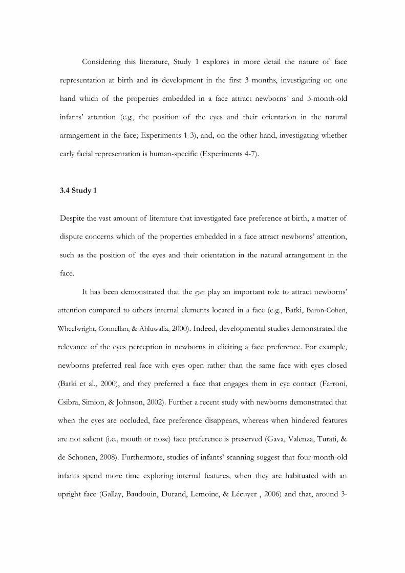
Considering this literature, Study 1 explores in more detail the nature of face
representation at birth and its development in the first 3 months, investigating on one
hand which of the properties embedded in a face attract newborns‟ and 3-month-old
infants‟ attention (e.g., the position of the eyes and their orientation in the natural
arrangement in the face; Experiments 1-3), and, on the other hand, investigating whether
early facial representation is human-specific (Experiments 4-7).
3.4 Study 1
Despite the vast amount of literature that investigated face preference at birth, a matter of
dispute concerns which of the properties embedded in a face attract newborns‟ attention,
such as the position of the eyes and their orientation in the natural arrangement in the
face.
It has been demonstrated that the eyes play an important role to attract newborns‟
attention compared to others internal elements located in a face (e.g., Batki, Baron-Cohen,
Wheelwright, Connellan, & Ahluwalia, 2000). Indeed, developmental studies demonstrated the
relevance of the eyes perception in newborns in eliciting a face preference. For example,
newborns preferred real face with eyes open rather than the same face with eyes closed
(Batki et al., 2000), and they preferred a face that engages them in eye contact (Farroni,
Csibra, Simion, & Johnson, 2002). Further a recent study with newborns demonstrated that
when the eyes are occluded, face preference disappears, whereas when hindered features
are not salient (i.e., mouth or nose) face preference is preserved (Gava, Valenza, Turati, &
de Schonen, 2008). Furthermore, studies of infants‟ scanning suggest that four-month-old
infants spend more time exploring internal features, when they are habituated with an
upright face (Gallay, Baudouin, Durand, Lemoine, & Lécuyer , 2006) and that, around 3-

67
months of age, among all of the facial features, the eyes appear to be of particular
importance in face perception (Hainline, 1978) independently from their location in the
face (Maurer, 1985).
With this in mind, Experiments 1, 2 and 3 were aimed at investigating which of
perceptual cues (i.e., position and orientation of eyes within a face) make face so attractive
at birth and at 3 months of age. Two variables have been manipulated: the position
(Experiment 1) and the orientation of the eyes (Experiments 2 and 3) within a face.
Experiment 1
The aim of experiment 1 was to test whether at birth the position of the eyes within a face
is relevant to induce a face preference.
Method
Participants
Participants were 12 (4 females) healthy, full term newborns (mean age 43.5 hours),
recruited at the maternity ward of the Pediatric Clinic of the University of Padova. All of
them were middle-class infants and Caucasian. Four additional newborns were tested, but
were not included in the final sample because one showed a strong position preference
(i.e., he looked to one direction more than 80% of the time) and three did not complete
testing due to fussiness. All of them met the screening criteria of normal delivery, a birth
weight between 2445 and 4280 g, and an Apgar score between 9 and 10 at 5 minutes.
Newborns were tested only if they were awake and in an alert state (Prechtl & O‟Brien,
1982), after parents gave their informed consent. The ethic committee of the Hospital of
Padova, where all the testing was conducted, granted permission.
Stimuli
Three pairs of stimuli were presented, each composed of an upright canonical face and the
same inverted face with the eyes correctly oriented but located in the lower part (Figure
20). Black-and-white photographs of women‟s faces were used. Photographs were
modified with Adobe Photoshop 7.1. More specifically, hair was removed from each
photograph and, on the upside-down face, only the inner portion of the face was rotated
180° whereas the orientation of the eyes was keep constant, although positioned in the
lower part. The stimuli were about 18 cm high X 11.5 cm wide (about 34° X 26° of visual
angle).
Figure 20: Stimuli employed in Experiment 1 to investigate the role of the position of the eyes
within a face to induce newborns‟ preference.
Apparatus
The newborn baby sat on the experimenter‟s lap, 30 cm from a 30-inch computer screen
(2560 x 1600). Infants‟ eyes were aligned with a red flickering LED at the center of the
screen, which was used to attract the infant‟s gaze at the start of each trial and to check that
the infant‟s sight was level with the horizontal midline of the screen during the testing
69
session. Both stimuli were projected bilaterally on a black screen at a distance of
approximately 8 cm (15°) from a central fixation point. A video camera, mounted above
the central screen, recorded the infant‟s eyes movements. To prevent interference from
irrelevant distracters, plain white curtains were drawn on both sides of the chair where the
experimenter sat with the baby in her lap.
Procedure
The experiment was carried out using a preferential looking procedure, previously used
with newborns (Simion et al., 2002). Every trial began with the center flashing light. When
the infant fixated the light, one of the experimenters, who watched the infant‟s eyes by
means of a video monitor system, started the sequence of the trial by pressing a key on the
computer keyboard. This automatically turned off the central LED and activated the slide
projector, which presented the stimuli on the screen. The stimuli remained on as long as
the infant fixated on one of them (infant control procedure). When the infant shifted the
gaze from the display for more than 10 s, the observer turned off the stimuli and the center
light automatically turned on. All the infants were submitted to two trials in which the two
stimuli were shown bilaterally, one on the left and one on the right of the central LED.
Left/Right position of the stimuli was counterbalanced between the trials. The session
ended when the baby did not look at either stimulus for more than 10 s. Videotapes of
infant‟s eye movements throughout the trial were subsequently analyzed frame by frame by
two coders (an experimenter and a student); both coders were unaware of the kind of
stimulus presented. The coders recorded, separately for each stimulus and each position,
the total fixation time, that is, the sum of all fixations, and the number of orienting
responses. The mean estimated reliability between on-line and off-line coding was r (10) =

.87, p <. 001, N = 12 (Pearson correlation) for either dependent variable (total fixation
time and number of orientations).
Results and Discussion
To determine whether newborns showed a spontaneous visual preference toward one of
the stimuli presented, two separate t-test for dependent samples were performed to
compare total fixation time and the number of orienting response. Newborns looked
longer at the canonical face (M = 51443 ms, SD = 16332) compared to the same inverted
face whit the eyes correctly oriented but located in the lower part (M = 34922 ms, SD =
14866), t (11) = 2.7, p < .02. Effect size analyses revealed that newborns‟ preference for
upright canonical face corresponded to a medium-large effect size in Cohen‟s standard
(Cohen, 1988) (Cohen‟s d = .70). Additional analyses were conducted on preferences
scores (percentages) for the natural face. Each infant‟s looking time at canonical face was
divided by the total looking time on both test stimuli and converted into a percentage
score. Therefore, only scores significantly above 50% indicated a preference for the upright
canonical face compared to the face with the eyes located in the lower part. Preference
score for the canonical face were significantly above the chance level of 50 per cent (M =
59.7%, SD = 18.1, one sample t (11) = 2.9, p < .02 (Cohen‟s d = .72). As regard to the
number of orientations, this dependent variable did not reach the significant level. Results
demonstrated that newborn infants preferred the canonical upright face, where all the inner
elements are correctly positioned and oriented. However, this results could be due to the
newborns‟ preference toward configurations with more elements in the upper part (i.e.,
top-down asymmetry). To verify this issue, Experiment 2 presented newborns with two
faces with the same number of inner elements in the upper part (i.e., the eyes) and that
differed only for the orientation of the eyes.
71
Experiment 2
Method
Participants
The final sample was formed by 12 (7 females) healthy, full term newborns (mean age 46.3
hours), recruited at the maternity ward of the Pediatric Clinic of the University of Padova.
All of them were middle-class infants and Caucasian. Two additional newborns were
tested, but were not included in the final sample because they showed a strong position
preference (i.e., he looked to one direction more than 80% of the time). All of them met
the screening criteria of normal delivery, a birth weight between 2650 and 3780 g, and an
Apgar score between 8 and 10 at 5 minutes. Newborns were tested only if they were awake
and in an alert state, after parents gave their informed consent.
Stimuli
Three pairs of stimuli were presented, each composed of an upright canonical face and the
same face with the eyes correctly located in the upper part of the face but misoriented
(Figure 21). Black-and-white photographs of women‟s faces were used. Photographs were
modified with Adobe Photoshop 7.1.
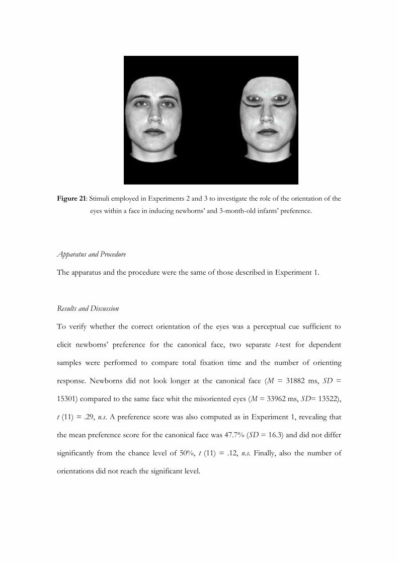
Figure 21: Stimuli employed in Experiments 2 and 3 to investigate the role of the orientation of the
eyes within a face in inducing newborns‟ and 3-month-old infants‟ preference.
Apparatus and Procedure
The apparatus and the procedure were the same of those described in Experiment 1.
Results and Discussion
To verify whether the correct orientation of the eyes was a perceptual cue sufficient to
elicit newborns‟ preference for the canonical face, two separate t-test for dependent
samples were performed to compare total fixation time and the number of orienting
response. Newborns did not look longer at the canonical face (M = 31882 ms, SD =
15301) compared to the same face whit the misoriented eyes (M = 33962 ms, SD= 13522),
t (11) = .29, n.s. A preference score was also computed as in Experiment 1, revealing that
the mean preference score for the canonical face was 47.7% (SD = 16.3) and did not differ
significantly from the chance level of 50%, t (11) = .12, n.s. Finally, also the number of
orientations did not reach the significant level.

73
Thus, the present result indicated that newborns were not sensitive to the
orientation of the eyes within a face, corroborating the idea that face representation at birth
is broad and not sensitive to specific cues within a face. However, in line with the literature
that demonstrated the effect of the visual experience for the formation of a specific face
representation in the first few months of life, it seemed relevant to investigate whether 3-
month-old infants were sensitive to the correct orientation of the eyes within a face
(Experiment 3).
Experiment 3
Method
Participants
Participants were 12 3-month-old healthy and full-term infants (7 females, mean age = 98
days). They were middle-class infants and all of them were Caucasian. Five additional
infants were tested but not included in the final sample because they showed a strong
position preference (i.e., they looked to one direction more than 90% of the time). Infants
were tested only if awake and in an alert state after parents gave their informed consent.
Stimuli
The same stimuli used in Experiment 2 were employed in Experiment 3.
Apparatus
The experiment was run using an apparatus that allows the automatic recording of eye
movements direction because of an infrared camera (i.e., an eye-tracker system). Infants
were placed in an infant seat at a distance of about 60 cm from the computer screen where
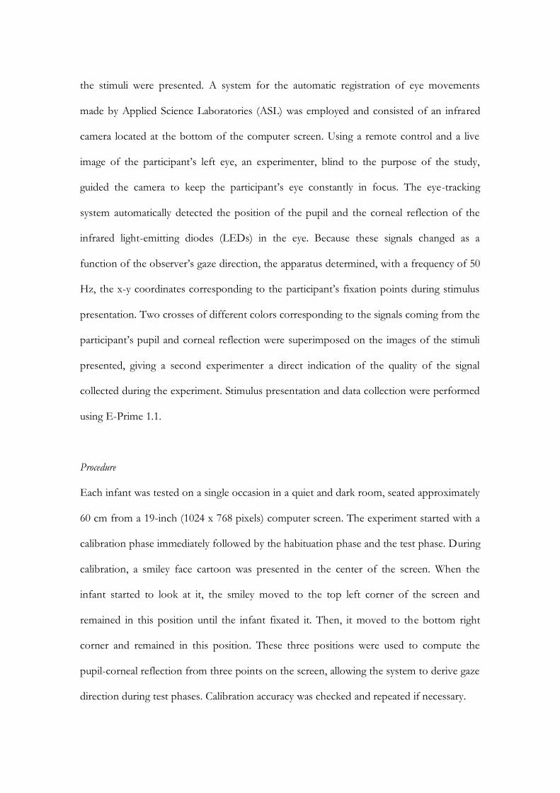
the stimuli were presented. A system for the automatic registration of eye movements
made by Applied Science Laboratories (ASL) was employed and consisted of an infrared
camera located at the bottom of the computer screen. Using a remote control and a live
image of the participant‟s left eye, an experimenter, blind to the purpose of the study,
guided the camera to keep the participant‟s eye constantly in focus. The eye-tracking
system automatically detected the position of the pupil and the corneal reflection of the
infrared light-emitting diodes (LEDs) in the eye. Because these signals changed as a
function of the observer‟s gaze direction, the apparatus determined, with a frequency of 50
Hz, the x-y coordinates corresponding to the participant‟s fixation points during stimulus
presentation. Two crosses of different colors corresponding to the signals coming from the
participant‟s pupil and corneal reflection were superimposed on the images of the stimuli
presented, giving a second experimenter a direct indication of the quality of the signal
collected during the experiment. Stimulus presentation and data collection were performed
using E-Prime 1.1.
Procedure
Each infant was tested on a single occasion in a quiet and dark room, seated approximately
60 cm from a 19-inch (1024 x 768 pixels) computer screen. The experiment started with a
calibration phase immediately followed by the habituation phase and the test phase. During
calibration, a smiley face cartoon was presented in the center of the screen. When the
infant started to look at it, the smiley moved to the top left corner of the screen and
remained in this position until the infant fixated it. Then, it moved to the bottom right
corner and remained in this position. These three positions were used to compute the
pupil-corneal reflection from three points on the screen, allowing the system to derive gaze
direction during test phases. Calibration accuracy was checked and repeated if necessary.
75
A visual preference procedure adapted to 3-month-old infants was used. Applying the ASL
algorithm, a fixation was defined as a period of a least 100 ms during which the fixation
point did not change by more than 1 degree of visual angle. The experimental session
began with the presentation, in the middle of the screen, of a central fixation point
particularly attractive for the infant (i.e., a colored moving smiley). This central fixation
point was used to attract infants‟ attention toward the computer screen where the stimuli
were shown, and to check that the infants‟ gaze was aligned with the horizontal midline of
the screen during the entire experimental session. As soon as the infant looked at the
central fixation point, the first pair of stimuli automatically appeared on the computer
screen and the central fixation was removed. Each stimulus pair was presented for 8 s.
Afterwards, the central fixation point reappeared in the middle of the screen and the trial
loop started again. Each of the five pairs of stimuli was presented four times, in a
pseudorandom sequence, for a total of 20 trials. The left vs. right position of the stimuli
within each pair was counterbalanced. A software program processed the raw data coming
from the eye-tracker system, calculating infants‟ total fixation time toward the upright
canonical face and the same face with the eyes misoriented.
Results and Discussion
Two separate series of statistical analyses were performed. The first series of analyses were
concerned with the overall looking time on the stimuli. In this way, the eye-tracker
potentials were used to avoid human coders, and data obtained paralleled those classically
registered in a visual preference paradigm. So results obtained with 3-month-old infants in
Experiment 3 were comparable with those obtained with newborns in Experiment 2. It is
important to note that, at present the eye-tracker system is inappropriate for use with

newborns because of the poor differentiation of the pupil at birth, that renders almost
impossible for the infrared camera to detect the position of the pupil and the corneal
reflection of the infrared light-emitting diodes in the newborns‟ eye. A second series of
analyses were run to examine which perceptual cues within the canonical face and the face
with the misoriented eyes attracted infants‟ gaze, an opportunity allowed by the eye-tracker.
Overall total looking time
To test whether 3-month-old infants exhibited a visual preference toward one of
the stimuli presented, t-test for dependent samples were performed to compare total
fixation time. Statistical analyses revealed that 3 months infants looked longer the canonical
face (M = 19512 ms, SD = 11082) compared to the face with the misoriented eyes (M =
15600 ms, SD = 8688), t (11) = 3.06, p < .02 (d = 1.1). Moreover, the mean preference
score for the canonical face was above the chance level (M = 55.8%, SD = 4.8), t (11) =
4.27, p < .02 (d = 1.2). These findings clearly demonstrated that the 3-month-olds‟ gaze
seems to be attracted by the interrelation between the arrangement of the inner features
and by the position and orientation of the single facial features (i.e., eyes correctly oriented
and located in the upper part of a face).
Distribution of looking time
To understand which perceptual cues of each stimulus attracted infants‟ gaze, a
series of statistical analyses were performed on percentage of looking toward four selected
areas (i.e., areas of interest, AOI) corresponding to the upper and the lower parts of each
stimulus within the pair. One-sample t tests were applied to verify whether such
percentages of looking differed from a chance level of 25%. The following distribution of
looking was observed. The 3-month-old infants looked at the upper half of the canonical
face for 14243.2 ms, corresponding to 41% of total fixation time (M = 40.8%, SD = 13.9),
77
t (11) = 3.7, p < .05 (d = .39), and at the lower part for 5266.8 ms of the face
corresponding to 15% (M = 15.3%, SD = 11.3), t (11) = 2.9, p < .05 (d = .63). The upper
half of the face with misoriented eyes was fixed for 11587 ms corresponding to 33% of
total fixation time (M = 33.1%, SD = 11), t (11) = 2.5, p < .05 (d = .25), and the lower part
for 4014.3 ms corresponding to 11% of total fixation time (M = 10.7%, SD = 8.3), t (11) =
5.7, p < .01 (d = 1.3). Taken together, these findings indicated that, contrary to newborns‟
performances, 3-month-old infants showed a preference for the canonical human face,
showing that at this age infants are sensitive to the face-specific perceptual characteristics.
Conclusion from Experiments 1-3
The main aim of those experiments was to explore which perceptual cues, such as the
location and the orientation of the eyes within a face, are relevant in inducing a face
preference in newborns and whether these perceptual cues were able to elicit the same
preference in 3-month-old infants. Taken together, the present results corroborated other
findings (Turati et al., 2005; Macchi Cassia et al., 2004) showing that, although newborns
preferred the canonical face when contrasted when the same upside-down face
(Experiment 1), their face preference is determined by the activity of general perceptual
constraints rather than a content-determined bias toward the face geometry, since
newborns did not respond selectively (i.e., they did not look longer to) to a specific
perceptual characteristic of human face, that is the correct orientation of the eyes
(Experiment 2). Contrary to the data obtained with newborns, results from Experiment 3
suggested that face preference in 3-month-old infants is the result of perceptual cues
specific to faces. Indeed, a major role in inducing face preference at 3 months seems to be
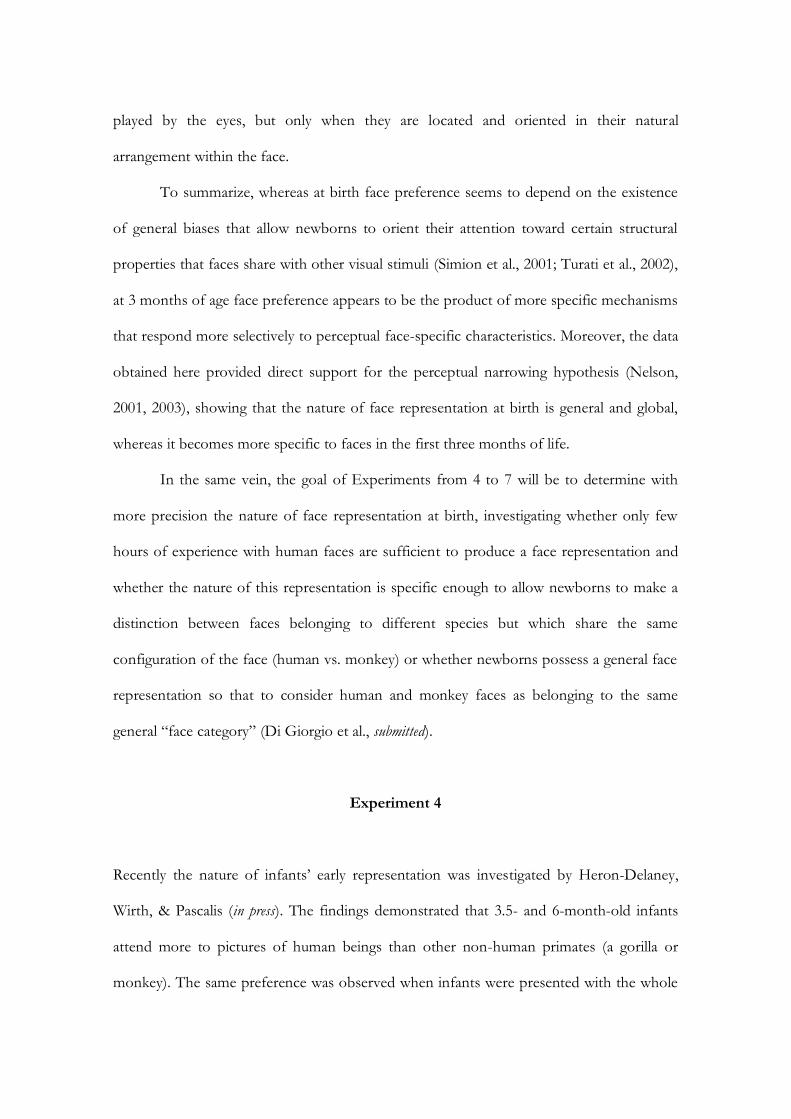
played by the eyes, but only when they are located and oriented in their natural
arrangement within the face.
To summarize, whereas at birth face preference seems to depend on the existence
of general biases that allow newborns to orient their attention toward certain structural
properties that faces share with other visual stimuli (Simion et al., 2001; Turati et al., 2002),
at 3 months of age face preference appears to be the product of more specific mechanisms
that respond more selectively to perceptual face-specific characteristics. Moreover, the data
obtained here provided direct support for the perceptual narrowing hypothesis (Nelson,
2001, 2003), showing that the nature of face representation at birth is general and global,
whereas it becomes more specific to faces in the first three months of life.
In the same vein, the goal of Experiments from 4 to 7 will be to determine with
more precision the nature of face representation at birth, investigating whether only few
hours of experience with human faces are sufficient to produce a face representation and
whether the nature of this representation is specific enough to allow newborns to make a
distinction between faces belonging to different species but which share the same
configuration of the face (human vs. monkey) or whether newborns possess a general face
representation so that to consider human and monkey faces as belonging to the same
general “face category” (Di Giorgio et al., submitted).
Experiment 4
Recently the nature of infants‟ early representation was investigated by Heron-Delaney,
Wirth, & Pascalis (in press). The findings demonstrated that 3.5- and 6-month-old infants
attend more to pictures of human beings than other non-human primates (a gorilla or
monkey). The same preference was observed when infants were presented with the whole
79
body or the face only. In contrast, a preference for human faces was also observed in
newborns only in the condition when the body was not present. The authors interpreted
the results as a demonstration that newborns have learned something about human faces
during the first few days of life. However, since the stimuli used in that study were not
equated as for low level perceptual properties, the face preference might be due to the
difference in the visibility of the two stimuli such as a difference in contrast. Moreover, in
those pictures the non human primate faces stand out as their fur was dramatically
different from the human‟s skin so that the perceptual cue that newborns might have used
is the fur and not the difference in the global configuration of the two faces. The aim of
Experiment 4 was to investigate whether newborns show a spontaneous preference
between a human and a monkey face. Importantly, in order to prevent that differences in
perceptual characteristics of the stimuli, such as the presence of the fur or the different
external contour that may affect newborns‟ discrimination and preference performance,
both the human and monkey faces were equated for low-level variables (i.e., low spatial
frequencies and high contrast areas). Importantly, the only perceptual characteristic that
differentiate the two stimuli is the presence, only in the human face, of the correct contrast
polarity within the eyes (i.e., the black pupil surrounded by the white sclera, Kobayashi &
Kohshima, 1997).
Method
Participants
Eighteen normal, healthy, full-term Caucasian newborns were selected from the Pediatric
Clinic of the University of Padova. Six babies were removed from the study for the
following reasoning: three babies changed their state during the testing (the newborn
become too tired or started to cry) and three babies had a strong position bias (looking

more than 80% of the time in one direction). So the final sample consisted of 12 newborns
(6 males). All infants met the screening criteria of normal delivery, a birth weight between
2570 and 3980 g, and a 5-min Apgar score above 8. Their ages at the time of testing ranged
from 24 to 72 hr. Newborns were tested only if they were awake and in an alert state and
after parents gave their informed consent.
Stimuli
Grey scale digitized full-frontal images of two human and two monkey faces were prepared
using Adobe Photoshop 7.0 (Figure 22). Two pairs of stimuli comprising a human and a
monkey face were presented. Monkey faces were manipulated taking off the hair on the
cheeks, to equate monkey and human faces for all the low-level variables (i.e., low-spatial
frequency, contrast, luminance). The stimuli were about 18.5 cm high X 13.5 cm wide
(about 34° X 26° of visual angle).
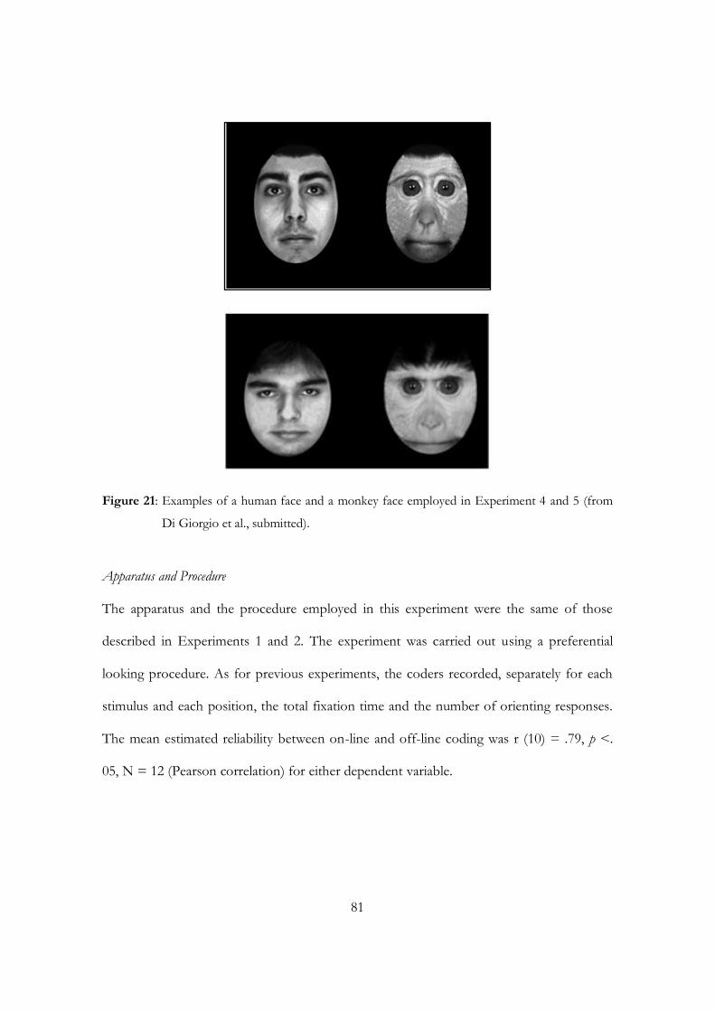
81
Figure 21: Examples of a human face and a monkey face employed in Experiment 4 and 5 (from
Di Giorgio et al., submitted).
Apparatus and Procedure
The apparatus and the procedure employed in this experiment were the same of those
described in Experiments 1 and 2. The experiment was carried out using a preferential
looking procedure. As for previous experiments, the coders recorded, separately for each
stimulus and each position, the total fixation time and the number of orienting responses.
The mean estimated reliability between on-line and off-line coding was r (10) = .79, p <.
05, N = 12 (Pearson correlation) for either dependent variable.

Results and discussion
To determine whether newborns showed a spontaneous visual preference for one of the
two stimuli presented, two separate t-test for dependent samples were performed to
compare total fixation time and the number of orienting response. Newborns did not
manifest a visual preference response. Indeed, they did not look longer at the human face
(M = 33194 ms, SD = 13958) rather than to the monkey face (M = 32529 ms, SD =
15119), t (11) = .09, n.s. As in the previous experiments additional analyses on preferences
scores (percentages) for the human face were conducted. Preference was not above the
chance level of 50 per cent (M = 50%, SD = 18.1, one sample t (11) = .12, n.s., two-tailed).
Furthermore, newborns did not orient more frequently to the human face (M = 13.2, SD =
5.5) than to the monkey face (M= 14, SD= 5.3), t (11) = .38, n.s. Finally, examination of the
data for individual infants revealed that only 5 out of 12 newborns looked longer at the
human face (binomial test, n.s.) and that 7 out of 12 newborns oriented more frequently to
the human face (binomial test, n.s.). Finally, the correlation between the age of newborns
(hours) and the preference score was not significant (r = .06, n.s.).
Heron-Delaney et al. (in press) have found a preference for human faces over
monkey faces during the first week of life, in an experimental condition where hair/fur was
present. In contrast, present result demonstrated that newborns did not show any
spontaneous visual preference when the two faces were equated for low-level variables,
even if the eyes of the two faces differ because of the contrast between sclera and iris. A
possible interpretation for the lack of any visual preference might be that the face
representation during the first week of life is not precise enough to be labelled human
specific but rather includes other primates‟ faces. However, one possible reason why
newborns did not manifest any visual preference could be that, due to their visual
limitations, they were not able to discriminate between the two stimuli. Experiment 5 was

83
aimed to verify this hypothesis if newborns were able to discriminate between the stimuli
presented.
Experiment 5
Experiment 5 tested whether newborn babies were capable of discriminating, after
exposure, a human face from a monkey face, equated for all level-variables. Considering the
hypothesis of the existence of a general face representation at birth (Nelson, 2001), it was
predict that newborns should be able to process both faces and therefore to discriminate
between them.
Method
Participants
The final sample consisted of 14 healthy and full-term newborn Caucasian babies (5 males)
from the Pediatric Clinic of the University of Padova. Three newborns who changed their
state during the testing were excluded from the final sample. All infants met the screening
criteria of normal delivery, a birth weight between 3100 and 3980 g, and a 5-min Apgar
score between 8 and 10. Their ages at the time of testing ranged from 24 to 72 hr. All were
tested only if they were awake and in an alert state. Informed consent was obtained from
their parents.
Stimuli
The same grey scale full-frontal images of the human and monkey faces used in previous
experiment were employed in Experiment 5.
Apparatus
The apparatus was the same of that described in Experiments 1, 2 and 4.
Procedure
The experiment was carried out using an infant-control habituation procedure (Horowitz,
Paden, Bhana & Self, 1972). The infant was judged to have habituated when, from the
fourth fixation on, the sum of any three consecutive fixations was 50% or less than the
total of the first three fixations (Slater et al., 1985). Half of the newborns were habituated
to the human face, the other half of the sample was habituated to the monkey face).
During the habituation phase, the same stimulus was presented side by side. The stimuli
were projected bilaterally and remained on the screen until the habituation criterion was
reached. Bilateral rather than central presentation was chosen for two reasons. First, when
newborns look at a centrally presented stimulus, it is difficult for an observer to decide if
they are actually looking at the stimulus or if they simply do not move their eyes from the
central position. Second, at birth photoreceptors in the central fovea are very immature,
thus resulting in a poor vision in the central area of visual field (Abramov, Gordon,
Hendrickson, Hainline, Dobson, & LaBossiere, 1982; Atkinson & Braddick, 1989).
The habituation phase was followed by a preference test in which a preference could be
expressed between the familiar face and a novel one. The two test stimuli were shown in
both left and right positions, the positions being reversed from the first to the second
presentation. During the preference test phase, the experimenter recorded the duration of
infant‟s fixations on each stimulus by pressing two different push buttons depending on
whether the infant looked at the right or the left position. Presentation lasted until each
stimulus had been fixated at least once and a total of 20 s of looking had been accumulated.
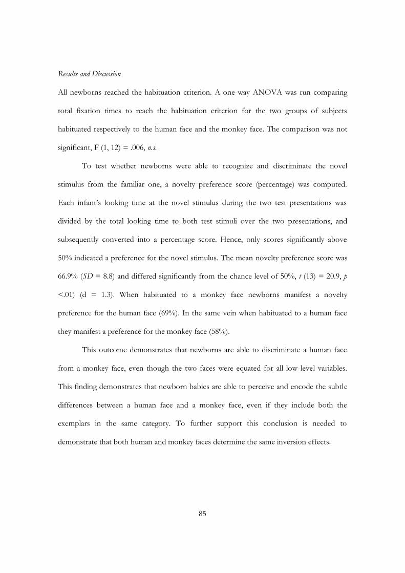
85
Results and Discussion
All newborns reached the habituation criterion. A one-way ANOVA was run comparing
total fixation times to reach the habituation criterion for the two groups of subjects
habituated respectively to the human face and the monkey face. The comparison was not
significant, F (1, 12) = .006, n.s.
To test whether newborns were able to recognize and discriminate the novel
stimulus from the familiar one, a novelty preference score (percentage) was computed.
Each infant‟s looking time at the novel stimulus during the two test presentations was
divided by the total looking time to both test stimuli over the two presentations, and
subsequently converted into a percentage score. Hence, only scores significantly above
50% indicated a preference for the novel stimulus. The mean novelty preference score was
66.9% (SD = 8.8) and differed significantly from the chance level of 50%, t (13) = 20.9, p
<.01) (d = 1.3). When habituated to a monkey face newborns manifest a novelty
preference for the human face (69%). In the same vein when habituated to a human face
they manifest a preference for the monkey face (58%).
This outcome demonstrates that newborns are able to discriminate a human face
from a monkey face, even though the two faces were equated for all low-level variables.
This finding demonstrates that newborn babies are able to perceive and encode the subtle
differences between a human face and a monkey face, even if they include both the
exemplars in the same category. To further support this conclusion is needed to
demonstrate that both human and monkey faces determine the same inversion effects.

Experiment 6
The purpose of Experiment 6 was to investigate whether an empirical phenomenon
thought to indicate face-specific processing, the inversion effect (Yin, 1969; Slater et al.,
2000; Turati et al., 2006), might also extended to processing of monkey faces at birth. If
newborns perceive monkey faces as belonging to the “face category”, as well as human
faces, they should prefer the upright monkey face rather than the inverted one.
Method
Participants
The participants were 12 full-term healthy Caucasian newborns (8 males) recruited at the
maternity ward of the Pediatric Clinic of the University of Padova. A further newborn was
excluded from the final sample because he changes his state during the testing was. All
infants met the screening criteria of normal delivery, a birth weight between 2620 and 3700
g, and a 5-min Apgar score between 8 and 10. Their ages at the time of testing ranged from
24 to 72 hr. The criteria for selection of the babies were identical to those used in the
previous experiments.
Stimuli
The same grey scale full-frontal images of the monkey faces used in Experiment 4 and 5
were employed (Figure 22). An upright monkey face was contrasted to the same inverted
monkey face.
87
Figure 22: Upright and inverted monkey faces employed in Experiment 6 (from Di Giorgio et al.,
submitted).
Apparatus and Procedure
A visual preference was employed and the apparatus was the same of the previous
experiments.
Results and Discussion
To verify newborns preference toward the stimuli, t-test for dependent samples were
performed, one for each dependent variable. Newborns looked longer toward the upright
monkey face (M = 38650 ms, SD = 15980) when compared to the same inverted monkey
face (M = 25518 ms, SD = 7259), t (11) = 2.5, p <. 05 (d = .70). Preference scores for the
upright monkey face were above the chance level of 50 per cent (M = 59%, SD = 12), t
(11) = 2.5, p <.05 (Cohen‟s d = .70). Examination of the data for individual infants found
that 8 out of 12 newborns looked longer at the upright monkey face (binomial test, n.s.).
Furthermore, t test performed on the number of orientation revealed that newborns did
not orient more frequently to the upright monkey face (M = 12, SD = 2.7) rather than to
the inverted monkey face (M = 10, SD = 2.1), t (11) = 1.8, n.s. Examination of the data for
individual infants found that 8 out of 12 newborns oriented more to the upright monkey
face (binomial test, n.s.). Finally, as in Experiment 2, the correlation between the age of
newborns (hours) and the preference score was not significant (r = .02, n.s.).
The result of Experiment 6 shows that, as well as for human face, the inversion
manipulation affects newborns‟ visual preference of monkey faces. This outcome
demonstrates that newborns perceive monkey faces and human faces as belonging to the
same “face category”. Alternative interpretations of the preference for the human-monkey
upright face can refer to either the presence of a greater number of elements in the upper
part or to the presence of first-order configural information (Simion et al., 2002). Indeed,
recent results with Mooney-face patterns, in which the face is formed only from white lit
surfaces and black unlit shadows, equated for the number of elements in the upper part
support the role of first order configural information since newborns prefer to look at
holistic patterns that are closer to upright faces (Leo & Simion, 2009).
Conclusions from Experiments from 4-6
There is prominent evidence in support of the proposal that at birth the face
representation is broad and unspecified and that the face processing system is shaped by
the faces seen in the visual environment in the first months of life (de Schonen &
Mathivet, 1989; Nelson, 2001; Sugita, 2008). Newborns did not manifest a spontaneous
visual preference for faces from own- or other-ethnic groups (Kelly et al., 2005) and they
did not respond differentially to the gender of faces presented (Quinn, Uttley, Lee, Gibson,
Smith, Slater, & Pascalis, 2008), however no study had yet investigate whether face
representation is specie-specific in newborns. The experiments presented here were aimed
to disentangle this question by contrasting human face and monkey face equated for low
level perceptual properties. Results from Experiment 4 demonstrates that newborns did
89
not manifest any visual preference for a human face over a face that is human like
(primate) corroborating the idea that face representation is not human-specific at birth.
This results corroborate previous findings obtained with animal studies (Sugita, 2008) that
reveals clear evidence of a basic, coarsely tuned face-recognition system in primate present
at birth. The lack of any preference for one of the two faces cannot be substantiate by a
lack of discrimination between them because results of Experiment 5 showed that
newborns are able to discriminate between a human face and a monkey face.
Importantly, the only perceptual characteristic that differentiate the two stimuli is the
presence, only in the human face, of the correct contrast polarity within the eyes (i.e., the
black pupil surrounded by the white sclera). The human species seem to be the only
primate species with a white sclera and a dark iris (Kobayashi & Kohshima, 1997).
Although the presence of this perceptual characteristic might explain newborns‟
discrimination in Experiment 5, it fails to explain the null preference for the same stimuli in
Experiment 4. A possibility to explain why newborns discriminated the two faces but failed
to prefer a human face over a monkey face comes from Experiment 6, which showed that
at birth the inversion effect affects the perception of monkey faces, just as affects the
perception of human faces (Macchi Cassia et al., 2004), suggesting the idea that at birth
both human and monkey faces are processed in the same way and are members of the
same category. It is possible that at birth the face category includes not only the basic
category of human faces, which in turn includes the subordinate categories of Caucasian
and Asian faces (Kelly et al., 2005), but includes also faces of non-human primates.
Notwithstanding, one could interpret the preference for the upright monkey face
compared to the upside-down one with the formation of the prototype in the few hours
after birth, since faces are encountered in the upright orientation much more frequently
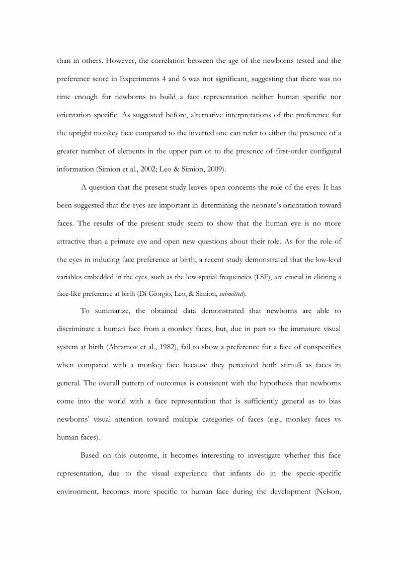
than in others. However, the correlation between the age of the newborns tested and the
preference score in Experiments 4 and 6 was not significant, suggesting that there was no
time enough for newborns to build a face representation neither human specific nor
orientation specific. As suggested before, alternative interpretations of the preference for
the upright monkey face compared to the inverted one can refer to either the presence of a
greater number of elements in the upper part or to the presence of first-order configural
information (Simion et al., 2002; Leo & Simion, 2009).
A question that the present study leaves open concerns the role of the eyes. It has
been suggested that the eyes are important in determining the neonate‟s orientation toward
faces. The results of the present study seem to show that the human eye is no more
attractive than a primate eye and open new questions about their role. As for the role of
the eyes in inducing face preference at birth, a recent study demonstrated that the low-level
variables embedded in the eyes, such as the low-spatial frequencies (LSF), are crucial in eliciting a
face-like preference at birth (Di Giorgio, Leo, & Simion, submitted).
To summarize, the obtained data demonstrated that newborns are able to
discriminate a human face from a monkey faces, but, due in part to the immature visual
system at birth (Abramov et al., 1982), fail to show a preference for a face of conspecifics
when compared with a monkey face because they perceived both stimuli as faces in
general. The overall pattern of outcomes is consistent with the hypothesis that newborns
come into the world with a face representation that is sufficiently general as to bias
newborns‟ visual attention toward multiple categories of faces (e.g., monkey faces vs
human faces).
Based on this outcome, it becomes interesting to investigate whether this face
representation, due to the visual experience that infants do in the specie-specific
environment, becomes more specific to human face during the development (Nelson,

91
2001; Pascalis et al., 2002; Pascalis & Kelly, 2009), as suggested by studies demonstrating
that 3 months of experience are sufficient to induce a gender (i.e., female vs male, Quinn et
al., 2002) and an own-race preference (Kelly et al., 2005). So, the aim of Experiment 7 was
to verify whether 3-month-old infants show a visual preference for a human face over a
monkey face.
Experiment 7
Method
Participants
Participants were 12 3-month-old healthy and full-term infants (6 females, mean age = 97.6
days). They were middle-class infants and all of them were Caucasian. Five additional
infants were tested but not included in the final sample because they showed a strong
position preference (i.e., they looked to one direction more than 90% of the time). Infants
were tested only if awake and in an alert state after parents gave their informed consent.
Stimuli
Stimuli employed in Experiment 4 were used here.
Apparatus and Procedure
The experiment was run using the eye-tracker system during and infants were tested in a
visual preference paradigm. The procedure was the same of that used in Experiment 3.
Results and Discussion
As in Experiment 3, the results are presented in two sections: the overall looking time and
the distribution of looking time.

Overall looking time
To test whether 3-month-old infants exhibited a visual preference toward one of
the stimuli presented, t test for dependent samples were performed to compare total
fixation time. Statistical analyses revealed that 3 months infants looked longer at the human
face (M = 36737 ms, SD = 15895) compared to the monkey face (M = 20363 ms, SD =
8238), t (11) = 2.7, p < .05 (d = 1.3). Moreover, the mean preference score for the human
face was above the chance level (M = 62.5%, SD = 18.4), t (11) = 2.3, p < .05 (d = .67). As
for the number of orientations, infants looked more frequently the human face (M = 108.5,
SD = 41.9) than to the monkey face (M = 66.7, SD = 38.7), t (11)= 2.9, p < .05 (d = 1.1).
These findings clearly demonstrated that the 3-month-olds‟ attention seems to be attracted
more by the human face than by the monkey face.
Distribution of looking time
To understand which perceptual cues of each stimulus attracted infants‟ gaze, a
series of statistical analyses were performed on percentage of looking toward four selected
areas (i.e., areas of interest, AOI) corresponding to the upper and the lower parts of each
stimulus within the pair. One-sample t tests were applied to verify whether such
percentages of looking differed from a chance level of 25%. The following distribution of
looking was observed. The 3-month-old infants looked at the upper half of the human face
for 25110 ms corresponding to 43% of total fixation time (M = 42.6%, SD = 24.6), t (11)
= 2.5 p < .001 (d = .73) and at the lower part for 11626 ms corresponding to 20% of total
fixation time (M = 19.8%, SD = 22.9), t (11) =.78, n.s. The upper half of the monkey face
was fixed for 15479 ms corresponding to 29% of total fixation time (M = 29%, SD =
20.3), t (11) = .69, n.s., and the lower part of the monkey face for 4884 ms corresponding
to 8% of total fixation time (M = 8.3%, SD = 8.9), t (11) = 6.4, p < .001 (d = 1.1) (Figure
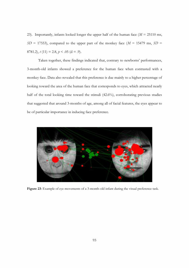
93
23). Importantly, infants looked longer the upper half of the human face (M = 25110 ms,
SD = 17553), compared to the upper part of the monkey face (M = 15479 ms, SD =
8781.2), t (11) = 2.8, p < .05 (d = .9).
Taken together, these findings indicated that, contrary to newborns‟ performances,
3-month-old infants showed a preference for the human face when contrasted with a
monkey face. Data also revealed that this preference is due mainly to a higher percentage of
looking toward the area of the human face that corresponds to eyes, which attracted nearly
half of the total looking time toward the stimuli (42.6%), corroborating previous studies
that suggested that around 3-months of age, among all of facial features, the eyes appear to
be of particular importance in inducing face preference.
Figure 23: Example of eye movements of a 3-month-old infant during the visual preference task.
Conclusion
The goal of the Study 1 was to provide direct evidence in favor of the perceptual narrowing
hypothesis (Nelson, 2001, 2003) concerning the presence of a broad face representation at
birth and its developmental time course in the first months of life. Taken together, the data
obtained corroborated previous findings (Turati et al., 2005; Kelly et al., 2005; Quinn et al.,
2008, 2003) showing that at birth face representation was broad and that at three months
of age face representation becomes more specific to human face category. Evidence
described herein suggests that face specificity is not prewired, but rather arises from general
perceptual processes that, during development, become progressively tuned to the human
face, as a result of extensive experience with this stimulus category.
Overall, the data presented in this chapter are in line with an experience-expectant
perspective, that emphasizes the relevance of both general constraints of the human visuo-
perceptual system and exposure to certain experiences shortly after birth to drive the
system to become functionally specialized to process faces in the first month of life.
Importantly, the perceptual narrowing approach highlight the role of the visual experience
as a determinant factor for the specialization of the face processing system. However, at
present, little is known about the specific experiences that lead to expert face processing or
how early perceptual experience contributes to the specialization of the neural structures
underlying face processing. These issues should be the focus of future research, employing
both behavioral and neuropsychological tasks (e.g., ERP and NIRS2).
2 Event related potentials (ERPs): a set of voltage changes contained within a period of electroencephalogram (EEG) that are time-locked to an event, for example, presentation of an object. This is a noninvasive technique with excellent temporal resolution. Near infrared spectroscopy (NIRS): a neuroimaging technique that uses infrared resonance to measure changes in blood and tissue oxygenation in a noninvasive way.

CHAPTER 4
FACE DETECTION IN
COMPLEX VISUAL DISPLAYS

97
Introduction
As described in Chapter 3, the majority of developmental studies that investigated face
detection and recognition employed habituation-and novelty- and familiarity-preference
paradigms, in which the total fixation time towards a stimulus is the main measure. In these
studies, however, only a single stimulus is presented, so attentional selection is not required
as there is no competition with other stimuli. Consequently, although these studies have
considerably expanded our knowledge of infants’ perceptual processing, they have shed
little light on the development of mechanisms responsible for selecting a target amidst
competing stimuli.
Indeed, human visual world is rich of many stimuli that represent possible input for
visual and cognitive processing and for guiding behavior. Because our visual system has a
limited capacity, selection must occur to prioritize important stimuli for their subsequent
processing, while ignoring less important ones. Despite the large amount of evidence on
infants’ face detection and recognition (de Haan, 2001), only a few studies have
investigated whether infants are capable to detect a face within complex visual scenes.
Due to the social relevance of the face stimuli early in life and due to the emergence
of a specific face representation at 3 months (Chapter 3), the aim of Study 2 will be to
investigate whether infants can detect a target face in a more ecological situations, that is
when a face is embedded among both heterogeneous and homogeneous objects.
4.1 Face detection in complex displays in adults
For adaptive reasons and because of their ubiquity, faces are probably the most biologically
and socially significant visual stimuli for humans. For this reason, a great debate in adult
literature concerns whether faces, due to their social and biological relevance for humans,
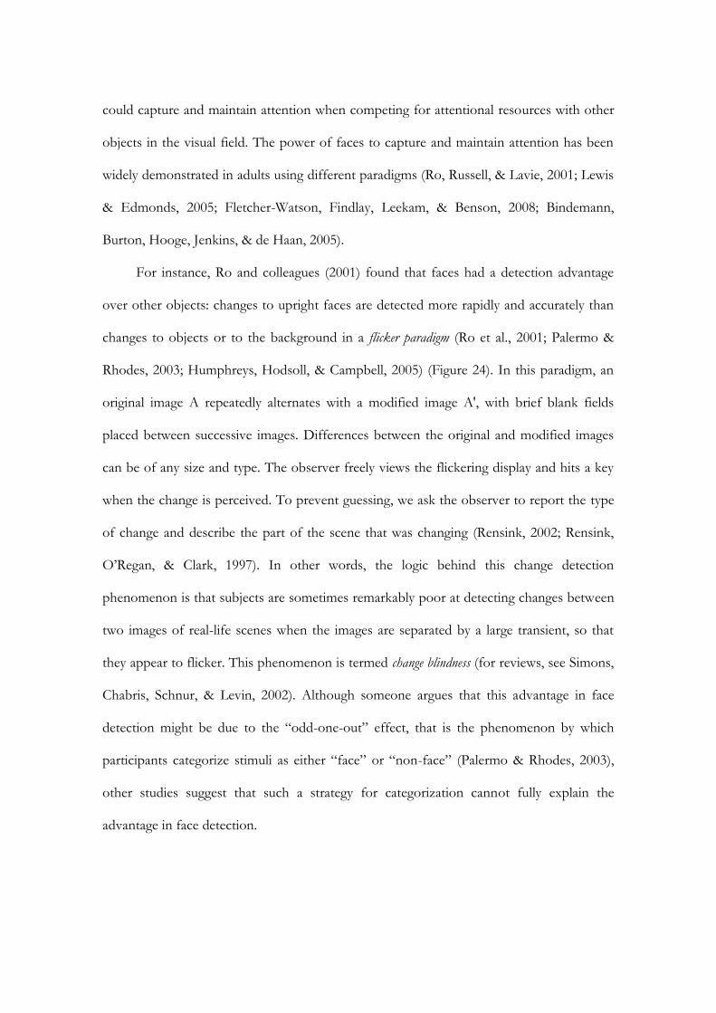
could capture and maintain attention when competing for attentional resources with other
objects in the visual field. The power of faces to capture and maintain attention has been
widely demonstrated in adults using different paradigms (Ro, Russell, & Lavie, 2001; Lewis
& Edmonds, 2005; Fletcher-Watson, Findlay, Leekam, & Benson, 2008; Bindemann,
Burton, Hooge, Jenkins, & de Haan, 2005).
For instance, Ro and colleagues (2001) found that faces had a detection advantage
over other objects: changes to upright faces are detected more rapidly and accurately than
changes to objects or to the background in a flicker paradigm (Ro et al., 2001; Palermo &
Rhodes, 2003; Humphreys, Hodsoll, & Campbell, 2005) (Figure 24). In this paradigm, an
original image A repeatedly alternates with a modified image A', with brief blank fields
placed between successive images. Differences between the original and modified images
can be of any size and type. The observer freely views the flickering display and hits a key
when the change is perceived. To prevent guessing, we ask the observer to report the type
of change and describe the part of the scene that was changing (Rensink, 2002; Rensink,
O’Regan, & Clark, 1997). In other words, the logic behind this change detection
phenomenon is that subjects are sometimes remarkably poor at detecting changes between
two images of real-life scenes when the images are separated by a large transient, so that
they appear to flicker. This phenomenon is termed change blindness (for reviews, see Simons,
Chabris, Schnur, & Levin, 2002). Although someone argues that this advantage in face
detection might be due to the “odd-one-out” effect, that is the phenomenon by which
participants categorize stimuli as either “face” or “non-face” (Palermo & Rhodes, 2003),
other studies suggest that such a strategy for categorization cannot fully explain the
advantage in face detection.
99
Figure 24: Example of the events for a trial in Ro et al.’ study (2001). Following an inter-trial
interval of fixation for 2,000 ms, the first frame of object items appeared for 533 ms.
A transient (blank white screen) was then presented for 83 ms, followed by the
presentation of the second frame of objects for another 533 ms. Another transient
was then presented, and this sequence repeated until the subject responded, or 20 s
had elapsed. Six objects, one from each of six different categories (faces, food,
clothes, musical instruments, appliances, and plants), were presented in each display,
and the center of each object was placed 5° from fixation. Across trials, six instances
from each category were used (from Ro et al., 2001).
An example of this comes from the study of Humphreys and colleagues (Humphreys et al.,
2005), in which participants were presented with images featuring four females and found
that changes in faces were detected faster than changes in bodies, which in turn were
detected faster than changes in the background. This study demonstrated that the face
detection advantage in the change blindness paradigm is based on the attentional bias
toward the face, independent of the rapid categorization of the face as opposed to other
object categories.
Moreover, evidences showed that attention may be preferentially directed to face in
natural scenes (Lewis & Edmonds, 2005; Fletcher-Watson et al., 2008) and that faces not
only grab attention but also have an advantage in retaining adults’ attention over other
stimulus categories (i.e., go/no-go classification task, Bindemann et al., 2005) and, finally,
that faces gave rise o inhibition of return alongside a concurrent non-face objects.
As well as the other paradigms, the visual search task has been increasingly employed
to study face perception in complex arrays (Hansen & Hansen, 1988; Lewis & Edmonds,
2005; Langton, Law, Burton, & Schweinberger, 2008; Suzuki & Cavanagh, 1995; Calvo &
Nummenmaa, 2008; Tong & Nakayama, 1999). In a typical visual-search task, participants
are asked to detect a target stimulus among distractors (Treisman & Gelade, 1980). The
time to find the target is measured as a function of the number of distractors present in the
array. The logic behind these studies is the following: if the target stimulus draws attention
automatically, then the number of distractors in the array will have a minimal or null effect
on search time (Treisman & Gelade, 1980). The search is defined “efficient” (e.g., parallel
search) when the number of distractors does not influence the search time, whereas the
search is “inefficient” when the number of distractors present in the array affects the
search time for the target (e.g., serial search). Several studies, using a visual search
paradigm, failed to demonstrate that a schematic face or a more realistic face popped out
among scrambled and inverted faces as distractors.
Nothdurft (1993), using an upright drawing of a face with hair as target and the same
drawings but inverted as distractors, failed to find pop out. However, when the facial
features were removed from the drawings and only the hair remained, the face target
popped out. The pop out phenomenon in this case was interpreted as due to a perceptual
101
characteristic of the stimulus employed and not due to the significance of the face as a
visual stimulus.
Brown, Huey, and Findlay (1997) used black-and-white photographs with the hair
removed and set in ovoid templates in a special visual search paradigm, in which the target
and distractors are presented in the periphery, around a fixation point. The subjects were
asked to move their eyes to the target as quick as possible. Targets were upright or inverted
faces with distractors being in the opposite orientation. This study again failed to establish
pop out for faces. Importantly, the authors found a practice effect specific for upright, but
not for inverted faces. Subjects trained on upright face targets improved markedly in
latency and accuracy for upright faces only, while those trained on inverted faces improved
only slightly for both upright and inverted faces equally. The authors conclude that upright
faces have a special status in tasks that require configural learning.
Kuehn and Jolicoeur (1994) investigated whether perceptual features of the stimuli
presented such as orientation, quality and similarity affect visual search for faces. Results
demonstrated that faces do not pop out when embedded among distractors containing
facial features, but that search for a face became easier and faster when the distractors
looked less like faces. Indeed, when the distractor did not contain any facial features, but
was a globe in the shape of a face, the upright face did pop out.
Other researchers have used schematic or real faces to investigate whether some
kinds of emotional expression could be detect faster than others (Purcell & Stewart, 1986;
Ohman, Lundqvist, Esteves, 2001; Calvo & Nummenmaa, 2008; Figure 25). For example,
Hansen and Hansen (1988) demonstrated that a face with an angry expression pops out
from an array of happy faces. However, Purcell, Stewart, and Skov (1996) argued that this
pop out effect was the result of an artifact of extraneous dark areas in the angry faces,
which the subjects became aware of and used to detect the target. In sum, none of the
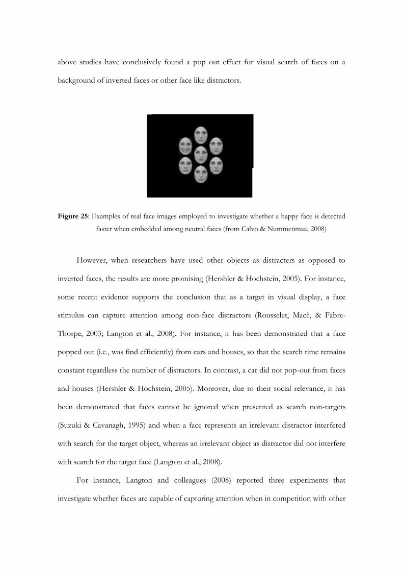
above studies have conclusively found a pop out effect for visual search of faces on a
background of inverted faces or other face like distractors.
Figure 25: Examples of real face images employed to investigate whether a happy face is detected
faster when embedded among neutral faces (from Calvo & Nummenmaa, 2008)
However, when researchers have used other objects as distracters as opposed to
inverted faces, the results are more promising (Hershler & Hochstein, 2005). For instance,
some recent evidence supports the conclusion that as a target in visual display, a face
stimulus can capture attention among non-face distractors (Rousselet, Macè, & Fabre-
Thorpe, 2003; Langton et al., 2008). For instance, it has been demonstrated that a face
popped out (i.e., was find efficiently) from cars and houses, so that the search time remains
constant regardless the number of distractors. In contrast, a car did not pop-out from faces
and houses (Hershler & Hochstein, 2005). Moreover, due to their social relevance, it has
been demonstrated that faces cannot be ignored when presented as search non-targets
(Suzuki & Cavanagh, 1995) and when a face represents an irrelevant distractor interfered
with search for the target object, whereas an irrelevant object as distractor did not interfere
with search for the target face (Langton et al., 2008).
For instance, Langton and colleagues (2008) reported three experiments that
investigate whether faces are capable of capturing attention when in competition with other
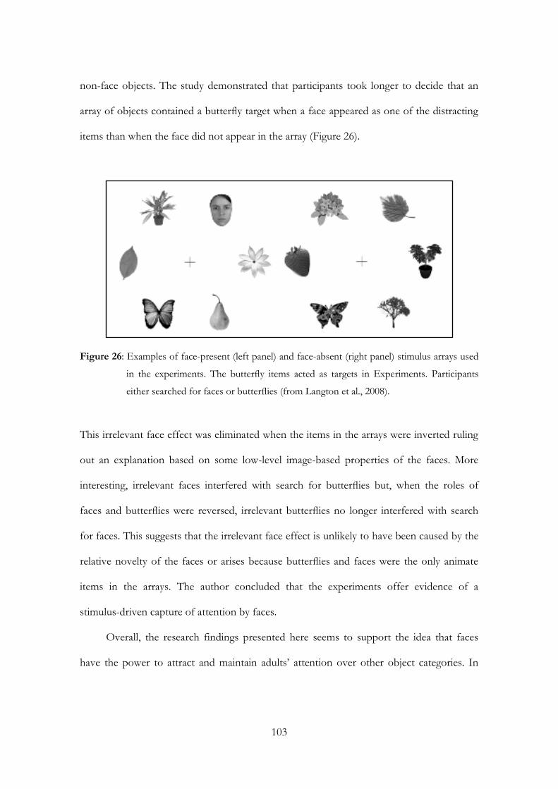
103
non-face objects. The study demonstrated that participants took longer to decide that an
array of objects contained a butterfly target when a face appeared as one of the distracting
items than when the face did not appear in the array (Figure 26).
Figure 26: Examples of face-present (left panel) and face-absent (right panel) stimulus arrays used
in the experiments. The butterfly items acted as targets in Experiments. Participants
either searched for faces or butterflies (from Langton et al., 2008).
This irrelevant face effect was eliminated when the items in the arrays were inverted ruling
out an explanation based on some low-level image-based properties of the faces. More
interesting, irrelevant faces interfered with search for butterflies but, when the roles of
faces and butterflies were reversed, irrelevant butterflies no longer interfered with search
for faces. This suggests that the irrelevant face effect is unlikely to have been caused by the
relative novelty of the faces or arises because butterflies and faces were the only animate
items in the arrays. The author concluded that the experiments offer evidence of a
stimulus-driven capture of attention by faces.
Overall, the research findings presented here seems to support the idea that faces
have the power to attract and maintain adults’ attention over other object categories. In
other words, these studies demonstrate the special perceptual processing of the human
face.
From a developmental point of view, infants’ face detection have been extensively
investigated in displays containing one or two faces, leaving open the question as whether
the same face preference obtained in infants of few months would be obtained also when a
face is embedded among competing visual stimuli. There is no doubt that we are a social
species that depends highly on face-mediate social interaction between conspecifics and
thus we should benefit from mechanisms which allow us to orient and to maintain
attention toward faces from the first months of life (Gliga & Csibra, 2007; Johnson, 2005).
4.2 Visual search strategies in infancy
Developmental studies investigated both infants’ ability to detect and recognize faces and
infants’ capacity to efficiently detect a discrepant target object in complex visual display. As
regard to face processing, it has been demonstrated that from birth, human newborns are
biased to pay attention towards faces over other visual stimuli in the environment. For
instance, it has been widely demonstrated that they not only attend to and prefer faces over
other visual stimuli (Johnson et al., 1991; Valenza et al., 1996; Macchi Cassia et al., 2004),
but they recognize the face of their mother (Bushnell, Sai, & Mullin, 1989; but see Pascalis
& de Schonen, 1994) and they recognize a face that changes in viewpoint (Turati, Bulf, &
Simion, 2008).
As regard to the ability to efficiently detect a target stimulus in complex visual
displays, studies that used the mobile conjugate reinforcement procedure (Adler,
Gerhardstein, & Rovee-Collier, 1998; Adler, Inslicht, Rovee-Collier, & Gerhardstein, 1998;
Rovee-Collier, Bhatt, & Chazin, 1996; Rovee-Collier, Hankins, & Bhatt, 1992), the classical
105
visual habituation or preference paradigms (Colombo, Ryther, Frick, & Gifford, 1995), or
more recent eye-tracker technique (Adler & Orprecio, 2006; Bulf, Valenza, & Simion,
2009), provided evidence that young infants are able to exhibit “pop-out”, where their
attention is captured by a stimulus (or patch of stimuli) that is surrounded by dissimilar
stimuli.
For instance, the visual habituation procedure was used to demonstrate that some
perceptual features, such as line-crossing and orientation, are efficiently detected in
complex displays by 3- and 4-month-old infants, confirming that young infants are
sensitive to some of the same fundamental features that are believed to direct adult
attention (Quinn & Bhatt, 1998). In the line crossing condition, 3- and 4-month-old infants
were habituated with homogenous arrays of either Ls, or +s. They were then tested with
two concurrently presented test stimulus arrays, one of which contained a single novel
character amid 24 familiar distractors, and the other contained a single familiar character
amid 24 novel distractors (Figure 27). The authors hypothesized that if infants exhibit
perceptual pop-out, the individual discrepant element (e.g., L or +s) should be detected
amid the homogeneous distractors, holding the infants’ attention. Then infants would
perceive the array with the novel discrepant element as the novel one, even though 24 out
of the 25 elements in the array are familiar. The same result was obtained in the orientation
condition. These findings provided evidence of pop out based on line crossing and
orientation information in 3- and 4-month-old infants.
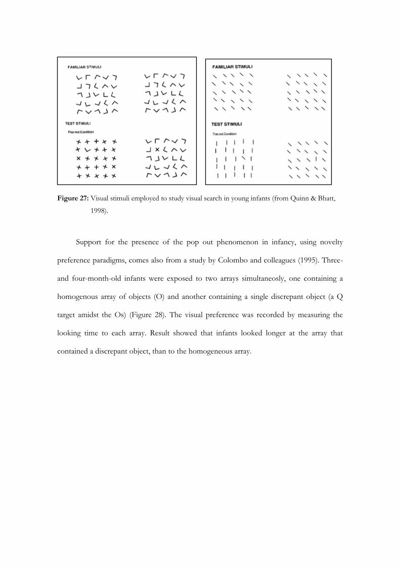
Figure 27: Visual stimuli employed to study visual search in young infants (from Quinn & Bhatt,
1998).
Support for the presence of the pop out phenomenon in infancy, using novelty
preference paradigms, comes also from a study by Colombo and colleagues (1995). Three-
and four-month-old infants were exposed to two arrays simultaneosly, one containing a
homogenous array of objects (O) and another containing a single discrepant object (a Q
target amidst the Os) (Figure 28). The visual preference was recorded by measuring the
looking time to each array. Result showed that infants looked longer at the array that
contained a discrepant object, than to the homogeneous array.
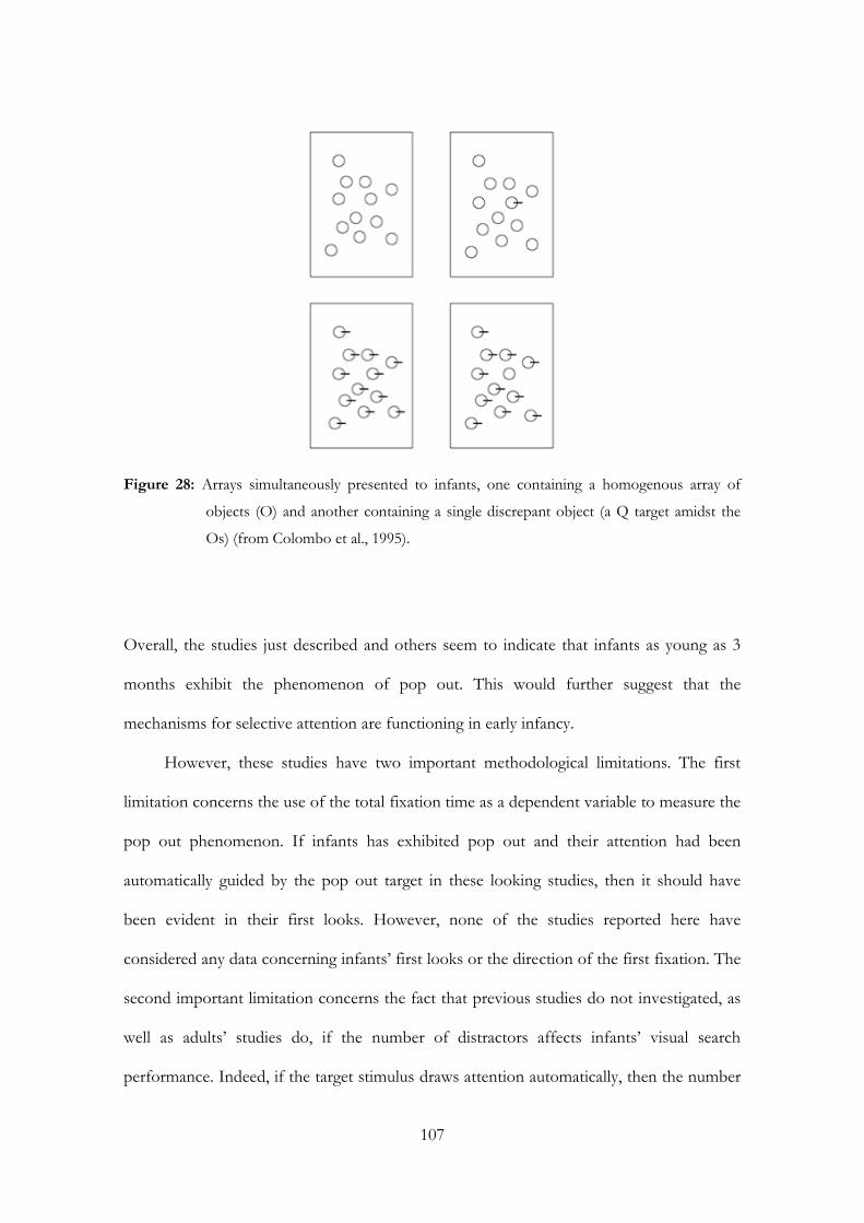
107
Figure 28: Arrays simultaneously presented to infants, one containing a homogenous array of
objects (O) and another containing a single discrepant object (a Q target amidst the
Os) (from Colombo et al., 1995).
Overall, the studies just described and others seem to indicate that infants as young as 3
months exhibit the phenomenon of pop out. This would further suggest that the
mechanisms for selective attention are functioning in early infancy.
However, these studies have two important methodological limitations. The first
limitation concerns the use of the total fixation time as a dependent variable to measure the
pop out phenomenon. If infants has exhibited pop out and their attention had been
automatically guided by the pop out target in these looking studies, then it should have
been evident in their first looks. However, none of the studies reported here have
considered any data concerning infants’ first looks or the direction of the first fixation. The
second important limitation concerns the fact that previous studies do not investigated, as
well as adults’ studies do, if the number of distractors affects infants’ visual search
performance. Indeed, if the target stimulus draws attention automatically, then the number

of distractors stimuli in the array will have a minimal or null effect on search time
(Treisman & Gelade, 1980).
To overcome these limitations, two recent studies employed an eye-tracker system
to measure infants’ and adults’ saccade latencies to visual arrays that contained a visual
geometrical stimulus as target among a variable number of distractor elements (Adler &
Orprecio, 2006; Bulf et al., 2009).
Many studies have indicated a linkage between adults’ eye movement and
attentional processing in visual search tasks. For example, findings from these studies
demonstrated that the number of distinct eye movements are positively correlated with
search times (Zelinsky, 1996), or that the latency and accuracy of the initial saccade to a
visual search target is a function of the spatial certainty of the target (Findlay, 1997) and the
number of distractors in the visual array (McSorley & Findlay, 2003; Motter & Belky,
1998). These findings indicate that an assessment of eye movements can be used as an
accurate measure of visual search, pop out and mechanisms of attentional processing.
Adler & Orprecio (2006) measured 3-month-olds’ and adults’ saccade latencies to
visual arrays that contained either a+ among Ls (target-present condition) or all L (target-
absent) with set sizes of 1, 3, 5 and 8 items (Figure 29).
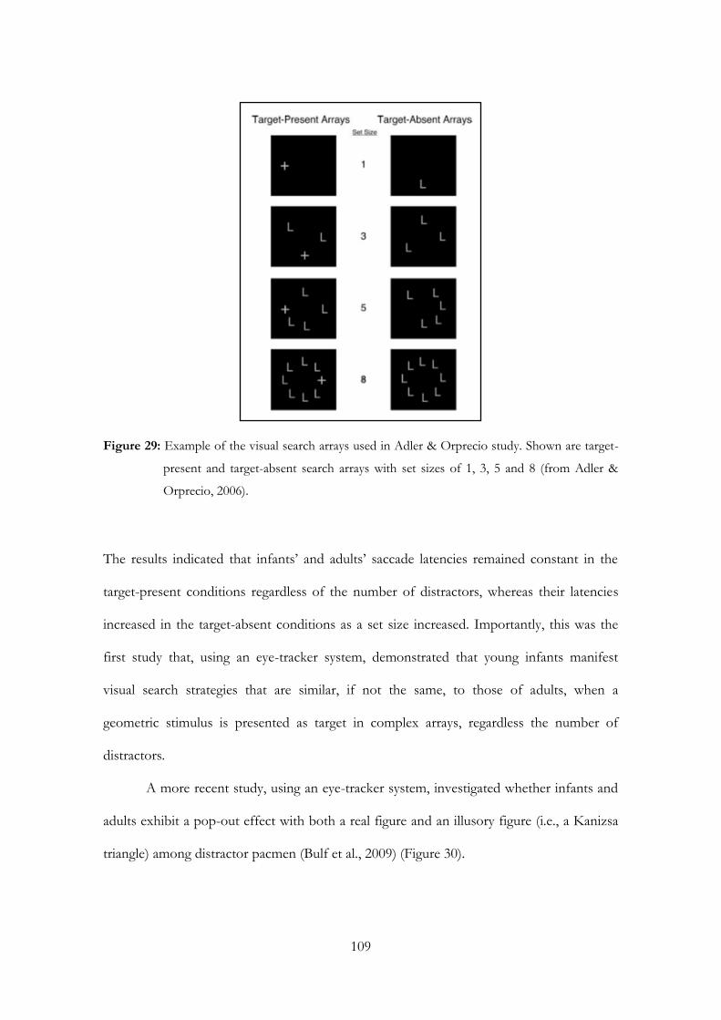
109
Figure 29: Example of the visual search arrays used in Adler & Orprecio study. Shown are target-
present and target-absent search arrays with set sizes of 1, 3, 5 and 8 (from Adler &
Orprecio, 2006).
The results indicated that infants’ and adults’ saccade latencies remained constant in the
target-present conditions regardless of the number of distractors, whereas their latencies
increased in the target-absent conditions as a set size increased. Importantly, this was the
first study that, using an eye-tracker system, demonstrated that young infants manifest
visual search strategies that are similar, if not the same, to those of adults, when a
geometric stimulus is presented as target in complex arrays, regardless the number of
distractors.
A more recent study, using an eye-tracker system, investigated whether infants and
adults exhibit a pop-out effect with both a real figure and an illusory figure (i.e., a Kanizsa
triangle) among distractor pacmen (Bulf et al., 2009) (Figure 30).
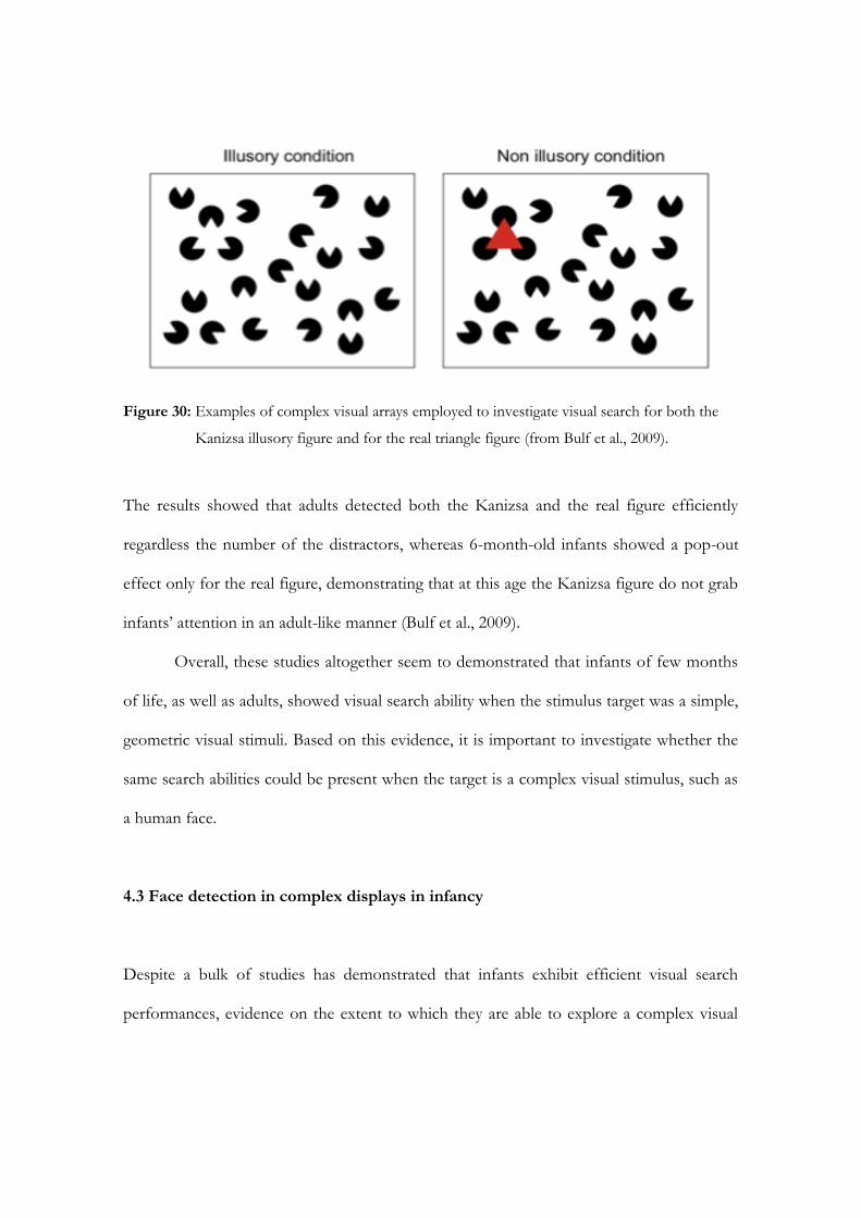
Figure 30: Examples of complex visual arrays employed to investigate visual search for both the
Kanizsa illusory figure and for the real triangle figure (from Bulf et al., 2009).
The results showed that adults detected both the Kanizsa and the real figure efficiently
regardless the number of the distractors, whereas 6-month-old infants showed a pop-out
effect only for the real figure, demonstrating that at this age the Kanizsa figure do not grab
infants’ attention in an adult-like manner (Bulf et al., 2009).
Overall, these studies altogether seem to demonstrated that infants of few months
of life, as well as adults, showed visual search ability when the stimulus target was a simple,
geometric visual stimuli. Based on this evidence, it is important to investigate whether the
same search abilities could be present when the target is a complex visual stimulus, such as
a human face.
4.3 Face detection in complex displays in infancy
Despite a bulk of studies has demonstrated that infants exhibit efficient visual search
performances, evidence on the extent to which they are able to explore a complex visual
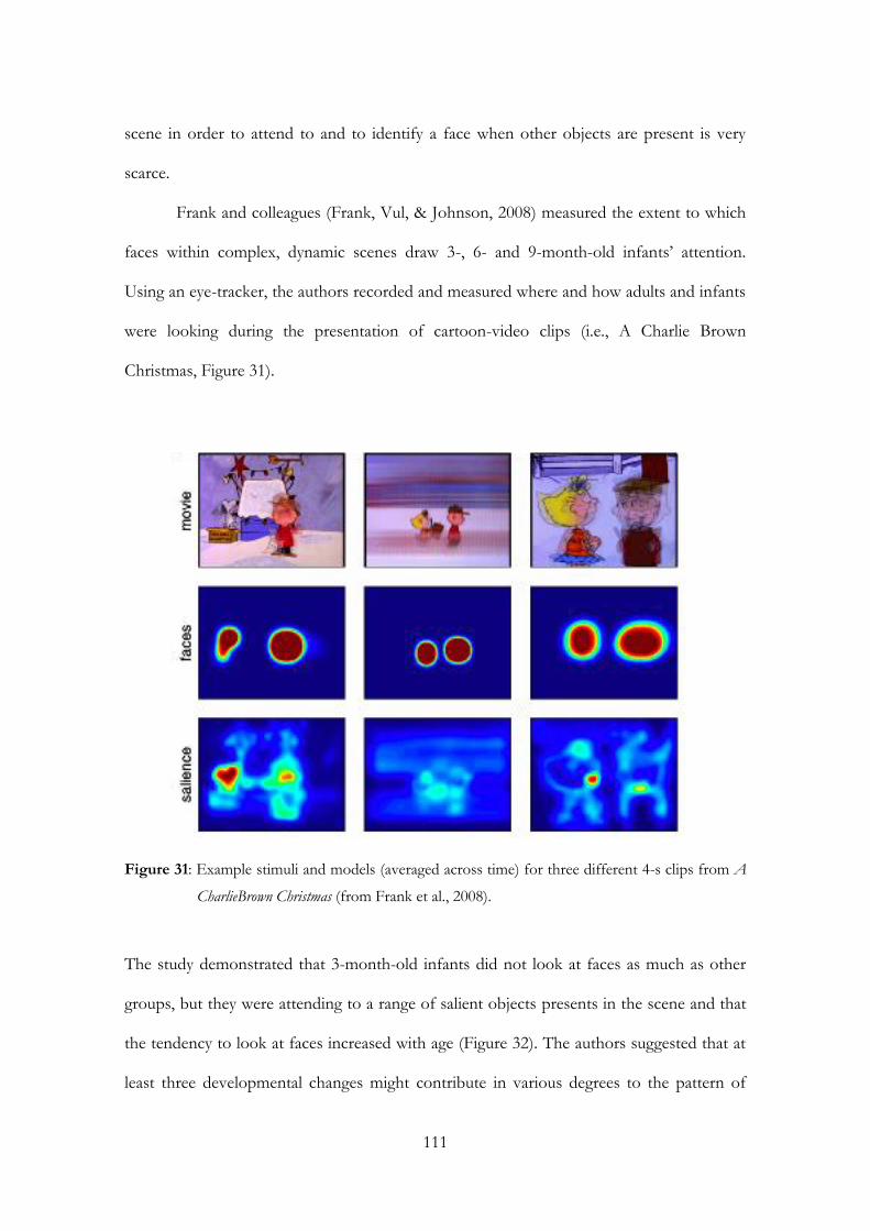
111
scene in order to attend to and to identify a face when other objects are present is very
scarce.
Frank and colleagues (Frank, Vul, & Johnson, 2008) measured the extent to which
faces within complex, dynamic scenes draw 3-, 6- and 9-month-old infants’ attention.
Using an eye-tracker, the authors recorded and measured where and how adults and infants
were looking during the presentation of cartoon-video clips (i.e., A Charlie Brown
Christmas, Figure 31).
Figure 31: Example stimuli and models (averaged across time) for three different 4-s clips from A
CharlieBrown Christmas (from Frank et al., 2008).
The study demonstrated that 3-month-old infants did not look at faces as much as other
groups, but they were attending to a range of salient objects presents in the scene and that
the tendency to look at faces increased with age (Figure 32). The authors suggested that at
least three developmental changes might contribute in various degrees to the pattern of
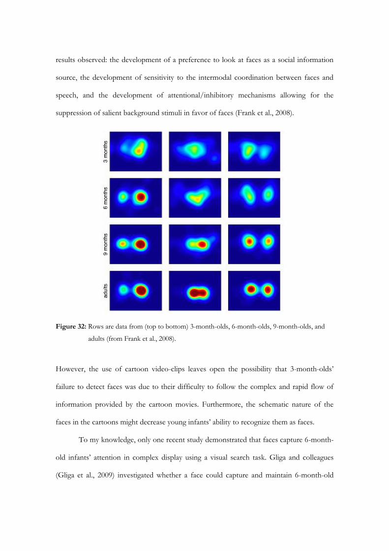
results observed: the development of a preference to look at faces as a social information
source, the development of sensitivity to the intermodal coordination between faces and
speech, and the development of attentional/inhibitory mechanisms allowing for the
suppression of salient background stimuli in favor of faces (Frank et al., 2008).
Figure 32: Rows are data from (top to bottom) 3-month-olds, 6-month-olds, 9-month-olds, and
adults (from Frank et al., 2008).
However, the use of cartoon video-clips leaves open the possibility that 3-month-olds’
failure to detect faces was due to their difficulty to follow the complex and rapid flow of
information provided by the cartoon movies. Furthermore, the schematic nature of the
faces in the cartoons might decrease young infants’ ability to recognize them as faces.
To my knowledge, only one recent study demonstrated that faces capture 6-month-
old infants’ attention in complex display using a visual search task. Gliga and colleagues
(Gliga et al., 2009) investigated whether a face could capture and maintain 6-month-old
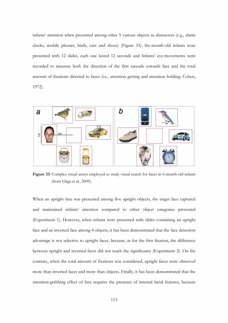
113
infants’ attention when presented among other 5 various objects as distractors (e.g., alarm
clocks, mobile phones, birds, cars and shoes) (Figure 33). Six-month-old infants were
presented with 12 slides, each one lasted 12 seconds and Infants’ eye-movements were
recorded to measure both the direction of the first saccade towards face and the total
amount of fixations directed to faces (i.e., attention getting and attention holding; Cohen,
1972).
Figure 33: Complex visual arrays employed to study visual search for faces in 6-month-old infants
(from Gliga et al., 2009).
When an upright face was presented among five upright objects, the target face captured
and maintained infants’ attention compared to other object categories presented
(Experiment 1). However, when infants were presented with slides containing an upright
face and an inverted face among 4 objects, it has been demonstrated that the face detection
advantage is not selective to upright faces, because, as for the first fixation, the difference
between upright and inverted faces did not reach the significance (Experiment 2). On the
contrary, when the total amount of fixations was considered, upright faces were observed
more than inverted faces and more than objects. Finally, it has been demonstrated that the
attention-grabbing effect of face requires the presence of internal facial features, because

faces whose internal features has been phase-scrambled (i.e., to create a noise stimulus) did
not attract nor maintain infants’ attention (Experiment 3).
To my knowledge this study is the first that investigated attention capture by faces
within complex visual arrays in infants of few months of life using a paradigm usually
employed with adults (i.e., visual search). However, this study has some limitations: i.) the
number of objects-distractors within the displays was keep constant and the authors did
not examine whether and how the number of distractors affects infant’ search performance
for faces and ii.) the authors did not compare six-month-old infants’ search performance
neither with adults nor with younger infants.
Therefore, the main aim of Study 3 will be to overcome these limitations testing 3-
and 6-month-old infants as well as adults with the same visual search paradigm, in which
the number of distractor objects will be manipulated.
4.4 Study 2
At present both the origin and the developmental time course of the ability to detect a face
target among other objects as distractors and whether the mechanisms underlying 6-
month-old infants’ ability to search efficiently a face in complex visual arrays are the same
or different from those of newborns and adults are still largely unknown. From a
developmental point of view, it appears relevant to investigate whether the ability to
efficiently detect a face in a complex array is still present in 6 months infants when the
number of distractors is variable and whether the same face detection advantage is present
before 6 months of age thanks to the importance of the human face early in life.
To this purpose, the current study investigated the origins and the time course of
visual search for a target face in complex displays in adults (Experiment 8) and in 3- and 6-
115
month-old infants (Experiments 9 and 10) through the use of a modified version of the
visual search paradigm and the recording of eye-movements. Importantly, in the classic
visual search task participants are asked to search for a specific target stimulus that is
presented among a different number of distractors stimuli. In this study the classic visual
search paradigm used with adults was modified in order to be adapted to infants, who are
unable to comprehend written or verbal instructions and to provide manual response and
all participants were tested under free viewing conditions. Each participant was presented
with complex displays consisting of a target face stimulus among a variable number of
objects as distractors. It has been used an eye-tracker system to measure the direction of
the first fixation and the number of fixations toward a target face, both considered as
measure of an early attentional orienting mechanism (e.g., attention-getting mechanism,
Cohen, 1972). In addition, the total amount of fixations directed to face, was recorded
because considered an index of maintenance of attention (e.g., attention-holding
mechanism, Cohen, 1972).
Experiment 8 tests the validity of our modified visual search paradigm with adults,
whereas Experiments 9 and 10 are aimed at investigating whether a target face presented
among objects capture and maintain 3- and 6-month-old infants’ attention. Since the
stimuli, the apparatus and the procedure are the same for adults and infants, I describe the
method one time and then I describe the results.
Experiment 8
Method
Participants
Fourteen undergraduate students were selected from the Department of Psychology at the
University of Padova to participate in the experiment. Two participants were excluded
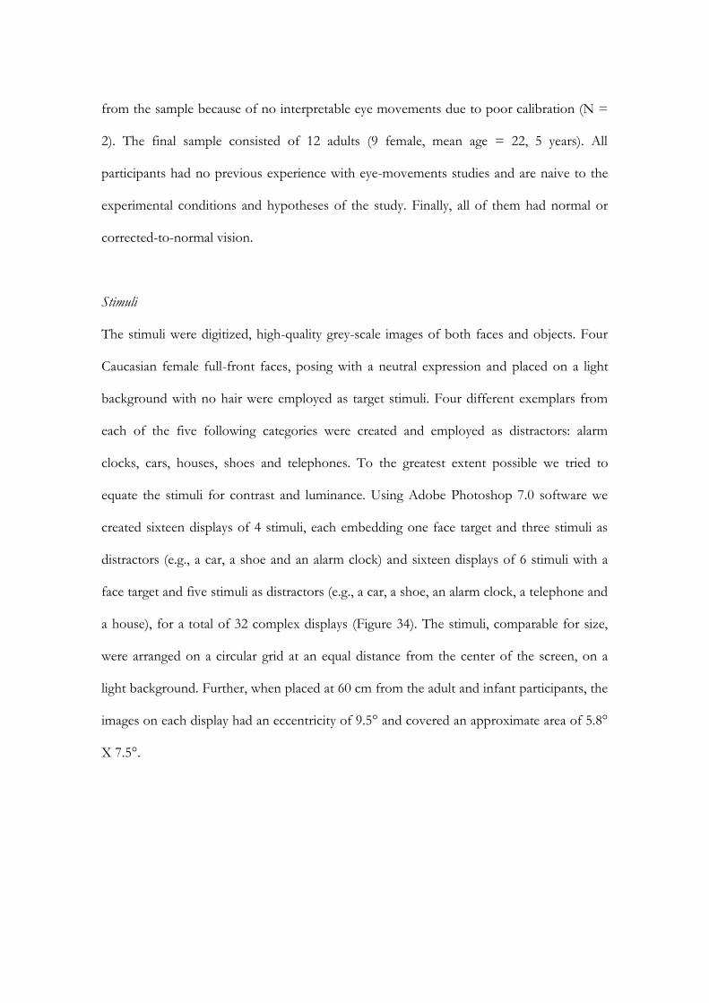
from the sample because of no interpretable eye movements due to poor calibration (N =
2). The final sample consisted of 12 adults (9 female, mean age = 22, 5 years). All
participants had no previous experience with eye-movements studies and are naive to the
experimental conditions and hypotheses of the study. Finally, all of them had normal or
corrected-to-normal vision.
Stimuli
The stimuli were digitized, high-quality grey-scale images of both faces and objects. Four
Caucasian female full-front faces, posing with a neutral expression and placed on a light
background with no hair were employed as target stimuli. Four different exemplars from
each of the five following categories were created and employed as distractors: alarm
clocks, cars, houses, shoes and telephones. To the greatest extent possible we tried to
equate the stimuli for contrast and luminance. Using Adobe Photoshop 7.0 software we
created sixteen displays of 4 stimuli, each embedding one face target and three stimuli as
distractors (e.g., a car, a shoe and an alarm clock) and sixteen displays of 6 stimuli with a
face target and five stimuli as distractors (e.g., a car, a shoe, an alarm clock, a telephone and
a house), for a total of 32 complex displays (Figure 34). The stimuli, comparable for size,
were arranged on a circular grid at an equal distance from the center of the screen, on a
light background. Further, when placed at 60 cm from the adult and infant participants, the
images on each display had an eccentricity of 9.5° and covered an approximate area of 5.8°
X 7.5°.
117
Apparatus
Adults were seated in a seat at a distance of about 60 cm from the computer screen where
the stimuli were presented. A system for the automatic registration of eye movements
made by Applied Science Laboratories (ASL) was employed (see Chapter 3).
Procedure
Each participant was tested on a single occasion in a quiet and dark room, seated
approximately 60 cm from a 19-inch (1024 x 768 pixels) computer screen. Importantly, no
instructions were given to adults, except that participants had to watch the images
appearing on the computer screen moving their eyes freely. The experiment started with a
calibration phase immediately followed by the test phase. After the calibration phase, each
participant was presented with 32 displays randomly selected. Sixteen displays comprised of
4 items (e.g., a target face among three different objects) and the other sixteen displays
comprised 6 items (e.g., a target face among 5 different objects). Across all displays, for
each set size, the participant was randomly presented with the target face occurring once in
each of the four or six possible positions. Each display presentation lasted 5 seconds and in
between the displays a fixation point was presented in the center of the screen, ensuring
that adult participant’s gaze was directed to the center before the next display was
presented. Rectangular areas of interest (AOIs) were defined manually around each image
in the displays, and for each participant we calculated the direction of the first fixation
directed to a certain category AOI and the number of fixations and the total fixation times
within an AOI. Only adult participants that looked at 32 out of 32 displays were included
in the analysis, whereas only infants that looked at least 16 out of 32 displays were included
in the analysis.
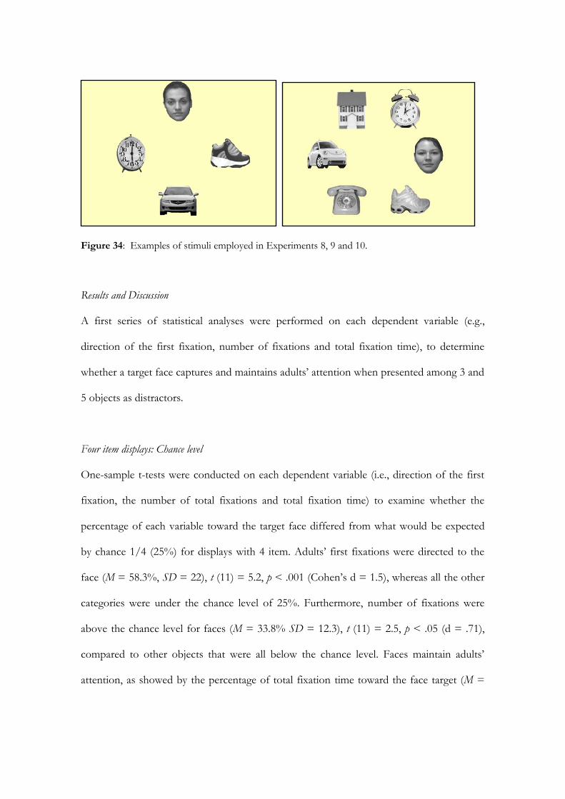
Figure 34: Examples of stimuli employed in Experiments 8, 9 and 10.
Results and Discussion
A first series of statistical analyses were performed on each dependent variable (e.g.,
direction of the first fixation, number of fixations and total fixation time), to determine
whether a target face captures and maintains adults’ attention when presented among 3 and
5 objects as distractors.
Four item displays: Chance level
One-sample t-tests were conducted on each dependent variable (i.e., direction of the first
fixation, the number of total fixations and total fixation time) to examine whether the
percentage of each variable toward the target face differed from what would be expected
by chance 1/4 (25%) for displays with 4 item. Adults’ first fixations were directed to the
face (M = 58.3%, SD = 22), t (11) = 5.2, p < .001 (Cohen’s d = 1.5), whereas all the other
categories were under the chance level of 25%. Furthermore, number of fixations were
above the chance level for faces (M = 33.8% SD = 12.3), t (11) = 2.5, p < .05 (d = .71),
compared to other objects that were all below the chance level. Faces maintain adults’
attention, as showed by the percentage of total fixation time toward the face target (M =
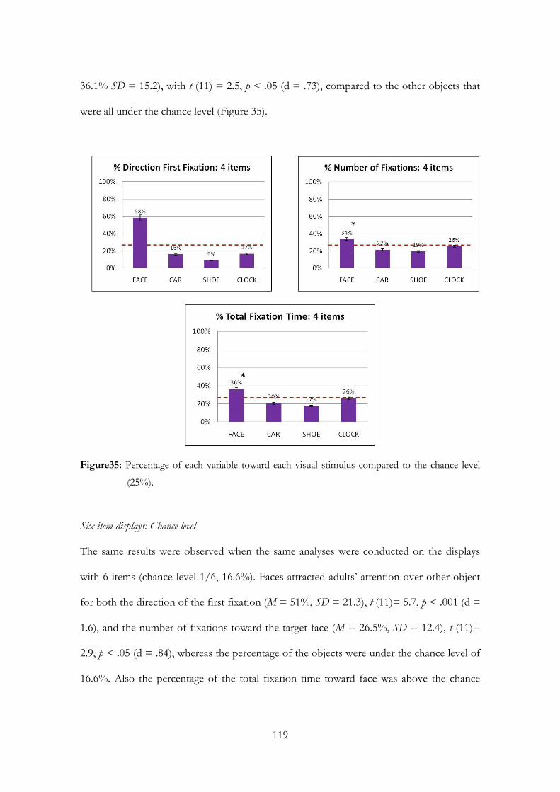
119
36.1% SD = 15.2), with t (11) = 2.5, p < .05 (d = .73), compared to the other objects that
were all under the chance level (Figure 35).
Figure35: Percentage of each variable toward each visual stimulus compared to the chance level
(25%).
Six item displays: Chance level
The same results were observed when the same analyses were conducted on the displays
with 6 items (chance level 1/6, 16.6%). Faces attracted adults’ attention over other object
for both the direction of the first fixation (M = 51%, SD = 21.3), t (11)= 5.7, p < .001 (d =
1.6), and the number of fixations toward the target face (M = 26.5%, SD = 12.4), t (11)=
2.9, p < .05 (d = .84), whereas the percentage of the objects were under the chance level of
16.6%. Also the percentage of the total fixation time toward face was above the chance

level (M = 28.2%, SD = 17.3), t (11)= 2.4, p < .05 (d = .71) compared to the other objects
presented (Figure 36).
Figure 36: Percentage of each variable toward each visual stimulus compared to the chance level
(16.6%).

121
To determine whether a target face had a detection advantage compared to other objects,
one-way repeated measure analyses of variance (ANOVAs) were performed on each
dependent variable and for both kind of displays (i.e., with 4 and 6 items). These analyses
were the same that Ro et al., (2001) and Palermo & Rhodes (2003) performed in their
studies.
Four item displays: face vs. each object
A main effect of Stimulus (i.e., face) was observed for the direction of the first fixation, F
(3, 33) = 23, p < .001, 2 = .68, for the number of fixations, F (3, 33) = 6.7, p < .001, 2 =
.38, and also for the total fixation time F (3, 33) = 6.9, p < .05, 2 = .36. Planned t-tests for
dependent sample showed that the target face (M = 9.3, SD = 3.5) received more first
fixations compared to cars (M = 2.6, SD = 1.7), t (11) = 4.8, p < .001 (d = .72), to shoes
(M = 1.4 SD = 1.5), t (11)= 5.7, p < .001(d = .6), and to alarm clocks (M = 2.7 SD = 1.7), t
(11) = 4.8, p < .01 (d = .71). T-tests for the number of fixations showed that face (M =
59.6 SD = 21.3) received more fixations compared to cars (M = 38, SD = 9.8), t (11) = 2.6,
p < .05 (d = 1.3), and compared to shoes (M = 34.2, SD = 9.5), t (11)= 3.1, p < .05 (d =
1.1), whereas there was no difference between faces and alarm clocks (M = 45.1 SD = 11),
t (11)= 1.8, n.s. Moreover, as regard to the total fixation time, t-tests revealed that the target
face (M = 20201.1 ms, SD = 9987.9) was observed more than cars (M = 11178 ms, SD =
3134.9), t (11)= 2.5, p < . 05 (d = 1.4), and more than shoes (M = 9576.7 ms, SD =
2801.3), t (11)= 2.9, p <. 05 (d = 1.2), but not more than alarm clocks (M = 14260.9 ms SD
= 3542.5), t (11)= 1.6, n.s. (Figure 37).

Figure 37: Four item displays: comparison between face target and each object as regard to each
dependent variable.
Six item displays: face vs. each object
Similar results were obtained when the same analyses were performed for 6 items display,
since a main effect of the Stimulus was obtained for the direction of the first fixation, F (5,
55) = 20.8, p < .001, 2 = .65, for the number of fixations, F (5, 55) = 7.1 p < .05, 2= .39,
and also for the total fixation time F (5, 55) = 4.8, p < .01, 2= .30. Planned t-tests for
dependent sample showed that the target face received more first fixations (M = 8.2 SD =
3.4) compared to all the other objects presents (t tests, p <. 01). Moreover, faces were
observed more frequently (M = 53.3 SD = 25.3) than the other objects (t tests, p < .05).
Finally, as regard to the total fixation time, faces (M = 15510.9 ms, SD = 9957.5) were
observed longer than cars (M = 7575 ms, SD = 1992.2), t (11)= 2.4, p <. 05 (d = 1.4) and

123
more than shoes (M = 7025.7 ms, SD = 1967), t (11)= 2.6, p < .05 (d = 1.3). However, the
difference between face and houses (M = 7799.8 ms SD = 2904.4), alarm clocks (M =
8893.7 ms, SD = 2667.3), and telephones (M = 7963.9 ms, SD = 2825.7) did not reach the
significance (t tests, n.s.) (Figure 38).
Figure 38: Six item displays: comparison between face target and each object as regard to each
dependent variable.
A series of t-tests for dependent sample were performed to compare Face category to
Object category for each dependent variable measured for each condition (i.e., 4 vs. 6
display items) This is the type of analysis used by Ro et al. (2001) and Palermo & Rhodes
(2003) to demonstrate a face detection advantage.
Four item displays: face category vs. object category
As for the direction of the first fixation, comparing the Face category (M = 9.3, SD = 3.5)
to the pooled first fixations for Object category (M = 2.2, SD= 1.2) revealed a Face
category advantage with t (11)= 5.2, p < .001 (d = .7). In the same vein, Face category
received more frequent fixations (M = 59.6, SD = 21.2) compared to Object category (M =
39.1, SD = 8.4), t (11)= 2.6, p < .05 (d = 1.4). Faces were also looked longer (M = 20201.1
ms, SD = 9987.9) than objects (M = 11671.9 ms, SD = 2798.4), t (11)= 2.3, p < .05 (d =
1.5).

125
Figure 39: Four item displays: comparison between face category vs. objects category as regard to
each dependent variable.
Six item displays: face category vs. object category
As for 4 item displays, Face category captured adults’ attention more than the other
objects, as we can see from both the direction of first fixations (Face category M = 8.2, SD
= 3.4 vs. Object category M =1.6, SD = .7, t (11)= 5.6, p < .001, d = .6) and the number of
fixations (Face category M = 53.3, SD = 25.3 vs. Object category M = 29.5, SD = 5.8, t
(11) = 2.8, p < .05, d = .9). Finally, faces maintained adults’ attention more than objects as
showed by the significant difference in total fixation time between Face category (M =
15510.9 ms, SD = 9957.5) and Object category (M = 7851.6 ms, SD = 2000.3), t (11) =
2.2, p < .05 (d = 1.5).

Figure 40: Six item displays: comparison between face category vs. objects category as regard to
each dependent variable.
Comparison between four and six item displays: face category vs. object category
Finally, to examine whether adults’ performance differed significantly between the four and
the six item displays condition a two-way repeated measures ANOVA’s were performed on
each dependent variable, with Stimulus category (Face category vs. Object category) and
Item (4 vs. 6) as within-subjects factors.
Results of the ANOVA on the direction of the first fixation showed a main effect of
the Stimulus category F (1, 11) = 31.3, p < .001, 2 = .74 and a main effect of the Item F
(1, 11) = 21.4 p < .01, 2 = .66, whereas the interaction Stimulus category X Item did not
reach the statistical significance, F (1, 11) = .57, n.s., demonstrating that a target face (M =
8.8, SD = .9) captured adults’ attention over other objects (M = 1.9, SD = .3) regardless of
127
the number of distractors presented, even though the number of first fixation toward face
is higher in the display containing four items (M = 5.8, SD = .3) than the displays
containing six items (M = 4.9, SD = .4) (Figure 41).
Figure 41: Adults’ first fixations are oriented toward face compared to other object, regardless of
the number of distractors.
Similar results were obtained for the other attention-getting index, that is the number
of fixation, since the ANOVA showed a main effect of the Stimulus category, F (1, 11) =
7.5, p < .05, 2 = .68 and a main effect of the Item F (1, 11) = 23.5 p < .01, 2 = .68, with
no Stimulus category X Item interaction, F (1, 11) = .91, n.s. Although Face category was
observed more frequently in four item displays (M = 49.3, SD = 2.4) than in six item
displays (M = 41.4, SD = 3.1), adult participants looked more frequently faces (M = 56.5,
SD = 6.6) than objects (M = 34.3, SD = 2), and this regardless of the number of objects
present (Figure 42). Finally, adults looked longer Face category (M = 17856 ms, SD =
2806.3) rather than Object category (M = 9761.8 ms, SD = 677.5), as revealed by the main
effect of Stimulus category F (1, 11) = 5.5, p < .05, 2 = .33. As for the other dependent
variables measured, faces were looked longer in four item condition (M = 15936.5 ms, SD
= 1084.5) than in six item condition (M = 11681.3 ms, SD = 1182.6), F (1,11) = 47.2, p <
.001, 2 = .81, although the Stimulus category X Item was not significant F (1, 11) = .37,
n.s. (Figure 43).
Figure 42: Adults’ number of fixations is oriented toward face compared to other object, regardless
of the number of distractors.
Figure 43: Adults look longer at target face compared to other object, regardless of the number of
distractors.
Results obtained from this experiment demonstrated that a face among various and
heterogeneous objects grab and maintain adults’ attention, regardless of the number of
distractors presents in the complex displays. These results seem to be in line with other
studies in literature that showed how faces, when competing for attention with other non-
129
face stimuli have an attention advantage (Ro et al., 2001; Langton et al., 2008). Importantly,
this study differed from the others in literature because demonstrated that the measure of
the eye-movements only is sufficient to investigate search performance for a complex
stimulus like a human face, corroborating the hypothesis of a linkage between attention
and eye movements (Zelinsky, 1996). Moreover, the data obtained here showed that the
modified visual search employed could be a good paradigm to study visual search strategies
in adults. With this in mind, the same procedure and the same paradigm were employed
with 6- and 3-month-old infants to test whether a face could grab and hold attention in
infants just as in adults.
Experiments 9 and 10
Method
Participants
Participants were twelve 6-month-old (7 females, mean age = 98.7 days) and nineteen 3-
month-old (10 females, mean age = 99.2 days) healthy and full-term infants. They were
middle-class infants and all of them were Caucasian. Eleven additional infants were
excluded from the final sample for the following reasons: two six-month-old infants
showed a strong position preference (i.e., they looked to one direction more than 90% of
the time) and nine infants of three months of age did not complete testing due to fussiness.
Infants were tested only if awake and in an alert state after parents gave their informed
consent.
The Stimuli, the Apparatus and the Procedure were the same that were employed in the previous
experiment, with only the exception that infants sat in an infant car seat. Furthermore, to

be involved in the statistical analysis, infants have to look at least to half of the trials
presented, that is 16 out to 32.
Results and Discussion
The same statistical analyses performed with adults were run with 6- and 3-month-old
infants.
Four item displays: Chance level
One-sample t-tests were conducted on each dependent variable (e.g., direction of the first
fixation, number of fixations and total fixation time) to examine whether the percentage of
each variable towards the target face differed from what would be expected by chance 1/4
(e.g., 25%) for displays with four item.
Six-month-old infants. Infants’ first fixations directed to the face (M = 21.4%, SD =
11.8) were not above the chance level of 25%, t (11) = 1.1, n.s. On the contrary, number of
fixations were above the chance level for faces (M = 36.8% SD = 10.4), t (11) = 3.9, p <
.05 (d = 1.1), compared to other objects that were all below the chance level. Furthermore,
faces maintain infants’ attention, as showed by the percentage of total fixation time toward
the face target (M = 38.9% SD = 11.1), with t (11) = 4.3, p < .01 (d = 1.2), compared to
the other objects that were all under the chance level (Figure 44).
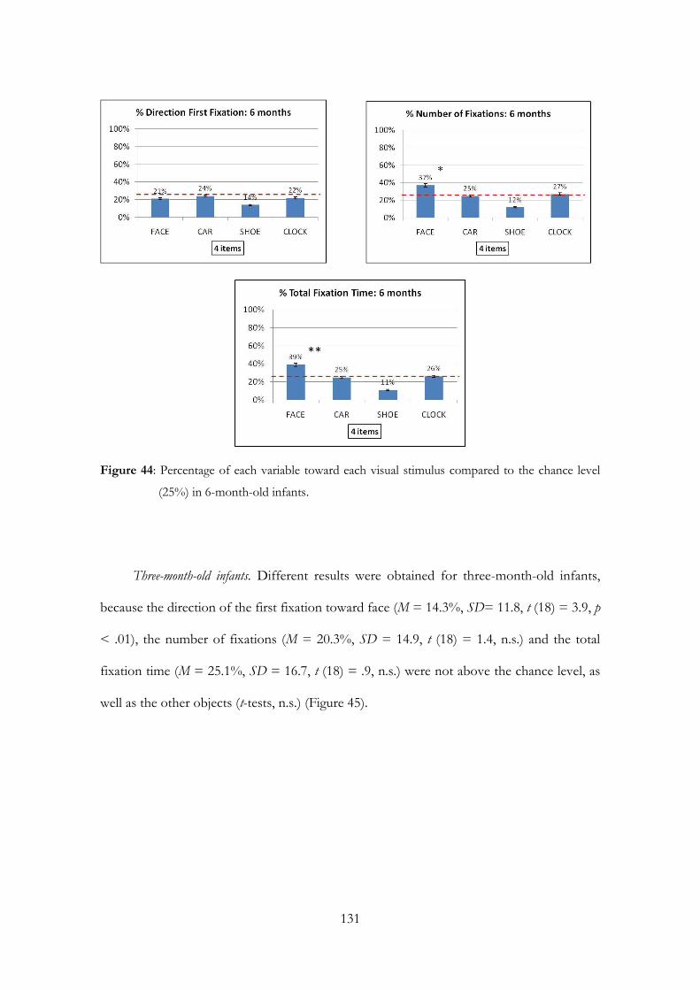
131
Figure 44: Percentage of each variable toward each visual stimulus compared to the chance level
(25%) in 6-month-old infants.
Three-month-old infants. Different results were obtained for three-month-old infants,
because the direction of the first fixation toward face (M = 14.3%, SD= 11.8, t (18) = 3.9, p
< .01), the number of fixations (M = 20.3%, SD = 14.9, t (18) = 1.4, n.s.) and the total
fixation time (M = 25.1%, SD = 16.7, t (18) = .9, n.s.) were not above the chance level, as
well as the other objects (t-tests, n.s.) (Figure 45).
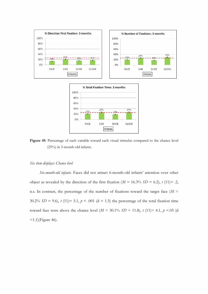
Figure 45: Percentage of each variable toward each visual stimulus compared to the chance level
(25%) in 3-month-old infants.
Six item displays: Chance level
Six-month-old infants. Faces did not attract 6-month-old infants’ attention over other
object as revealed by the direction of the first fixation (M = 16.3% SD = 6.2), t (11)= .2,
n.s. In contrast, the percentage of the number of fixations toward the target face (M =
30.2% SD = 9.6), t (11)= 5.1, p < .001 (d = 1.5) the percentage of the total fixation time
toward face were above the chance level (M = 30.1% SD = 11.8), t (11)= 4.1, p <.05 (d
=1.1)(Figure 46).
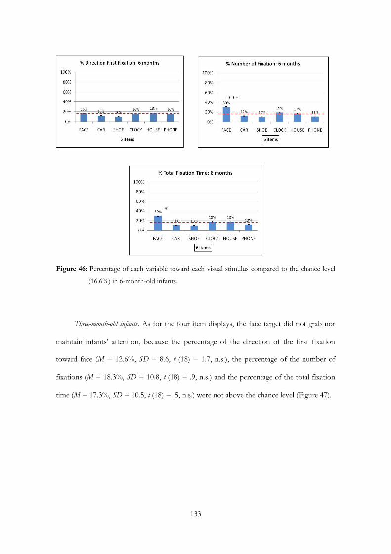
133
Figure 46: Percentage of each variable toward each visual stimulus compared to the chance level
(16.6%) in 6-month-old infants.
Three-month-old infants. As for the four item displays, the face target did not grab nor
maintain infants’ attention, because the percentage of the direction of the first fixation
toward face (M = 12.6%, SD = 8.6, t (18) = 1.7, n.s.), the percentage of the number of
fixations (M = 18.3%, SD = 10.8, t (18) = .9, n.s.) and the percentage of the total fixation
time (M = 17.3%, SD = 10.5, t (18) = .5, n.s.) were not above the chance level (Figure 47).

Figure 47: Percentage of each variable toward each visual stimulus compared to the chance level
(16.6%) in 3-month-old infants.
Four item displays: face vs. each object
Six-month-old infants. One-way repeated measure analyses of variance (ANOVAs) on
each dependent variable, revealed no main effect of the face target stimulus as for the
direction of the first fixation, F (3, 33) = 2.5, n.s., showing that 6-month-old infants did not
orient their attention first to the target face (Figure 48).
Figure 48: Four item displays: comparison between face target and each object as for the direction
of the first fixation.

135
On the contrary, a main effect of face target stimulus was obtained both for the number of
fixations, F (3, 33) = 11.8, p < .001, 2 = .52, and also for the total fixation time F (3, 33)
= 10.9, p < .001, 2 = .50. Planned t-tests for dependent sample showed that the target
face (M = 38.3, SD = 22.3) was observed more frequently than cars (M = 22.9, SD =12.8),
t (11) = 2.8, p < .05 (d = 1.2), shoes (M = 11.8, SD = 8.8), t (11)= 4.8, p < .01(d = .7), and
alarm clocks (M = 29.3, SD = 18.9), t (11) = 2.7, p < .05 (d = 1.2) (Figure 49).
Figure 49: Four item displays: comparison between face target and each object as for the number
of fixations.
T-tests for the total fixation time revealed that face (M = 9802.4 ms, SD = 5996.2) was
looked longer than cars (M = 5728.3 ms, SD = 3860.8), t (11) = 3.1, p < .05 (d = 1.1),
shoes (M = 2832.5 ms SD = 1461.1), t (11)= 4.5, p < 01 (d =.8) and alarm clocks (M =
7223.9 ms, SD = 5274.2), t (11)= 2.3, p < .05 (d = 1.4) (Figure 50).
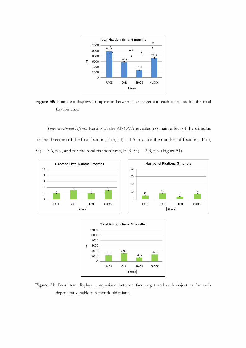
Figure 50: Four item displays: comparison between face target and each object as for the total
fixation time.
Three-month-old infants. Results of the ANOVA revealed no main effect of the stimulus
for the direction of the first fixation, F (3, 54) = 1.5, n.s., for the number of fixations, F (3,
54) = 3.6, n.s., and for the total fixation time, F (3, 54) = 2.3, n.s. (Figure 51).
Figure 51: Four item displays: comparison between face target and each object as for each
dependent variable in 3-month-old infants.

137
Six item displays: face vs. each object
Six-month-old infants. There was no main effect of the face target stimulus for the
direction of the first fixation, F (5, 55) = 1.2, n.s. (Figure 52), whereas a main effect of face
stimulus was observed for the number of fixations, F (5, 55) = 8.9 p < .001, 2 = .44, and
also for the total fixation time F (5, 55) = 5.8, p < .001, 2 = .35.
Figure 52: Six item displays: comparison between face target and each object as for the direction of
the first fixation.
Planned t-tests for dependent sample showed that the target face (M = 35.4, SD = 20.5)
was observed more frequently than all the other objects (t tests, p < .05)(Figure 53).

Figure 53: Six item displays: comparison between face target and each object as for the number of
fixations.
Finally, as regard to the total fixation time, faces (M = 9267.1 ms, SD = 7883.4) were
observed longer than cars (M = 2870.1 ms, SD = 2193.6), t (11)= 2.9, p <. 05 (d = 1.1),
more than shoes (M = 2318.5 ms, SD = 1483.5), t (11)= 3.1, p < .05 (d = 1.1) and more
than telephones (M = 3053.8 ms, SD = 2042.8), t (11) = 3.1, p < .05 (d = 1.1). However,
the difference between the target face and alarm clocks (M = 5427.1 ms, SD = 5203.2) and
houses (M = 4849.6 ms SD = 3533.5) did not reach the significance (t tests, n.s.) (Figure
54).
Figure 54: Six item displays: comparison between face target and each object as for the total
fixation time.
Three-month-old infants. No main effects were obtained for any of the dependent
variables, as revealed by the ANOVA for the direction of the first fixation F (5, 90) = .14,
n.s., the number of fixations F (5, 90) = .40, n.s., and for the total fixation time F (5, 90) =
.38, n.s (Figure 55).

139
Figure 55: Six item displays: comparison between face target and each object as for each
dependent variable in 3-month-old infants.
Four item displays: face category vs. object category
As for adults, a series of t-tests collapsing the data into Face and Objects categories for
each condition (i.e., 4 vs. 6 display items) were performed.
Six-month-old infants. As for the direction of the first fixation, comparing the Face
category (M = 4, SD = 1.6) to the pooled first fixations for Object category (M = 3.2, SD=
1.1) revealed that Face category did not reach any advantage, t (11)= 1.1, n.s (Figure 56).
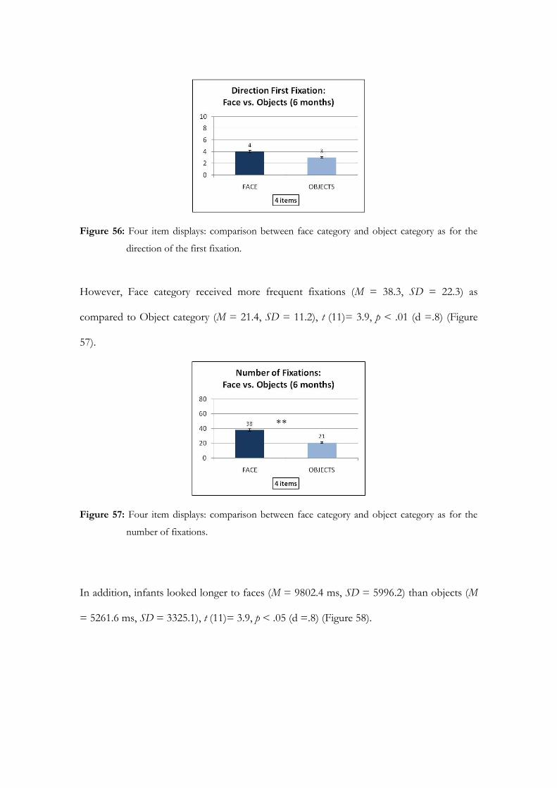
Figure 56: Four item displays: comparison between face category and object category as for the
direction of the first fixation.
However, Face category received more frequent fixations (M = 38.3, SD = 22.3) as
compared to Object category (M = 21.4, SD = 11.2), t (11)= 3.9, p < .01 (d =.8) (Figure
57).
Figure 57: Four item displays: comparison between face category and object category as for the
number of fixations.
In addition, infants looked longer to faces (M = 9802.4 ms, SD = 5996.2) than objects (M
= 5261.6 ms, SD = 3325.1), t (11)= 3.9, p < .05 (d =.8) (Figure 58).
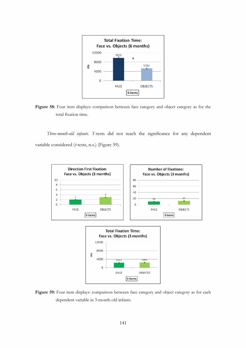
141
Figure 58: Four item displays: comparison between face category and object category as for the
total fixation time.
Three-month-old infants. T-tests did not reach the significance for any dependent
variable considered (t-tests, n.s.) (Figure 59).
Figure 59: Four item displays: comparison between face category and object category as for each
dependent variable in 3-month-old infants.

Six item displays: face category vs. object category
Six-month-old infants. As for 4 item displays, Face category did not captured 6-month-
old infants’ attention more than the other objects, at least for the direction of the first
fixation (Face category M = 2.6, SD = 1 vs. Object category M =2.5, SD = 1.2, t (11)= .2,
n.s.) (Figure 60).
Figure 60: Six item displays: comparison between face category and object category as for the
direction of the first fixation.
On the contrary the significance was observed at the level of the number of fixations (Face
category M = 35.4, SD = 20.5 vs. Object category M = 15.5, SD = 7.8, t (11) = 3.9, p < .05,
d =.8) (Figure 61).
Figure 61: Six item displays: comparison between face category and object category as for the
Number of fixations.

143
Finally, faces maintained infants’ attention more than objects as showed by the significant
difference in total fixation time between Face category (M = 9267.1 ms, SD = 7883.4) and
Object category (M = 3703.8 ms, SD = 2137.3), t (11) = 2.8, p < .05 (d = .81) (Figure 62).
Figure 62: Six item displays: comparison between face category and object category as for the total
fixation time.
Three-month-old infants. T-tests did not reach the significance for any dependent
variable considered (t-tests, n.s.) (Figure 63).
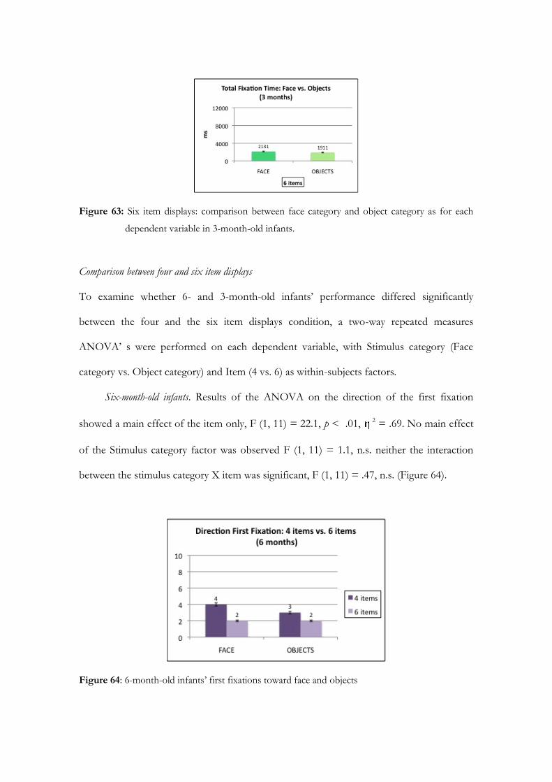
Figure 63: Six item displays: comparison between face category and object category as for each
dependent variable in 3-month-old infants.
Comparison between four and six item displays
To examine whether 6- and 3-month-old infants’ performance differed significantly
between the four and the six item displays condition, a two-way repeated measures
ANOVA’ s were performed on each dependent variable, with Stimulus category (Face
category vs. Object category) and Item (4 vs. 6) as within-subjects factors.
Six-month-old infants. Results of the ANOVA on the direction of the first fixation
showed a main effect of the item only, F (1, 11) = 22.1, p < .01, 2 = .69. No main effect
of the Stimulus category factor was observed F (1, 11) = 1.1, n.s. neither the interaction
between the stimulus category X item was significant, F (1, 11) = .47, n.s. (Figure 64).
Figure 64: 6-month-old infants’ first fixations toward face and objects
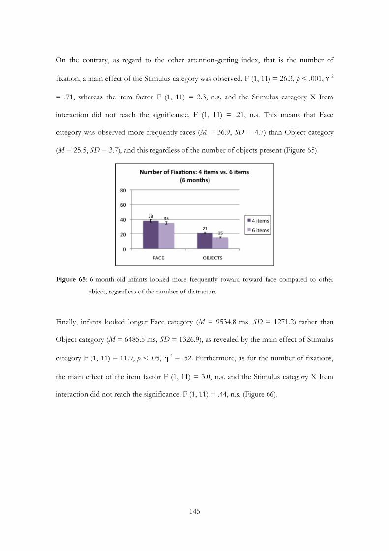
145
On the contrary, as regard to the other attention-getting index, that is the number of
fixation, a main effect of the Stimulus category was observed, F (1, 11) = 26.3, p < .001, 2
= .71, whereas the item factor F (1, 11) = 3.3, n.s. and the Stimulus category X Item
interaction did not reach the significance, F (1, 11) = .21, n.s. This means that Face
category was observed more frequently faces (M = 36.9, SD = 4.7) than Object category
(M = 25.5, SD = 3.7), and this regardless of the number of objects present (Figure 65).
Figure 65: 6-month-old infants looked more frequently toward toward face compared to other
object, regardless of the number of distractors
Finally, infants looked longer Face category (M = 9534.8 ms, SD = 1271.2) rather than
Object category (M = 6485.5 ms, SD = 1326.9), as revealed by the main effect of Stimulus
category F (1, 11) = 11.9, p < .05, 2 = .52. Furthermore, as for the number of fixations,
the main effect of the item factor F (1, 11) = 3.0, n.s. and the Stimulus category X Item
interaction did not reach the significance, F (1, 11) = .44, n.s. (Figure 66).
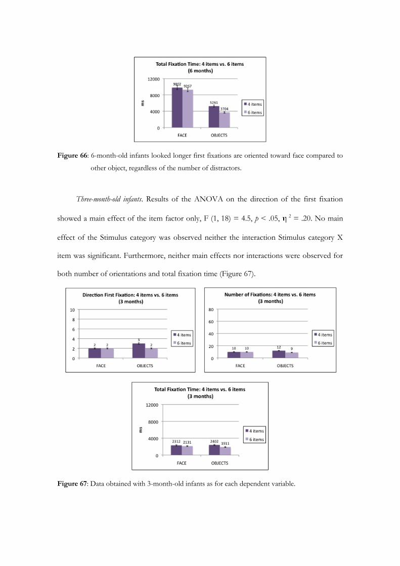
Figure 66: 6-month-old infants looked longer first fixations are oriented toward face compared to
other object, regardless of the number of distractors.
Three-month-old infants. Results of the ANOVA on the direction of the first fixation
showed a main effect of the item factor only, F (1, 18) = 4.5, p < .05, 2 = .20. No main
effect of the Stimulus category was observed neither the interaction Stimulus category X
item was significant. Furthermore, neither main effects nor interactions were observed for
both number of orientations and total fixation time (Figure 67).
Figure 67: Data obtained with 3-month-old infants as for each dependent variable.
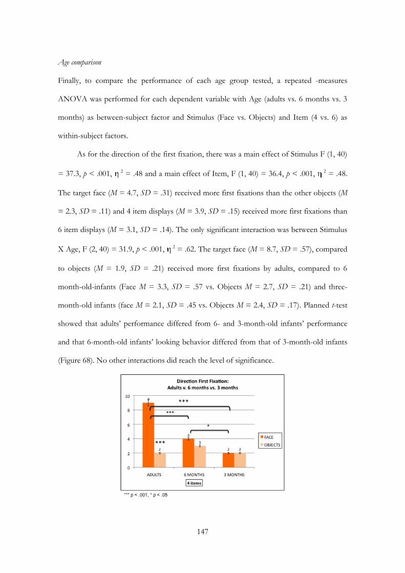
147
Age comparison
Finally, to compare the performance of each age group tested, a repeated -measures
ANOVA was performed for each dependent variable with Age (adults vs. 6 months vs. 3
months) as between-subject factor and Stimulus (Face vs. Objects) and Item (4 vs. 6) as
within-subject factors.
As for the direction of the first fixation, there was a main effect of Stimulus F (1, 40)
= 37.3, p < .001, 2 = .48 and a main effect of Item, F (1, 40) = 36.4, p < .001, 2 = .48.
The target face (M = 4.7, SD = .31) received more first fixations than the other objects (M
= 2.3, SD = .11) and 4 item displays (M = 3.9, SD = .15) received more first fixations than
6 item displays (M = 3.1, SD = .14). The only significant interaction was between Stimulus
X Age, F (2, 40) = 31.9, p < .001, 2 = .62. The target face (M = 8.7, SD = .57), compared
to objects (M = 1.9, SD = .21) received more first fixations by adults, compared to 6
month-old-infants (Face M = 3.3, SD = .57 vs. Objects M = 2.7, SD = .21) and three-
month-old infants (face M = 2.1, SD = .45 vs. Objects M = 2.4, SD = .17). Planned t-test
showed that adults’ performance differed from 6- and 3-month-old infants’ performance
and that 6-month-old infants’ looking behavior differed from that of 3-month-old infants
(Figure 68). No other interactions did reach the level of significance.
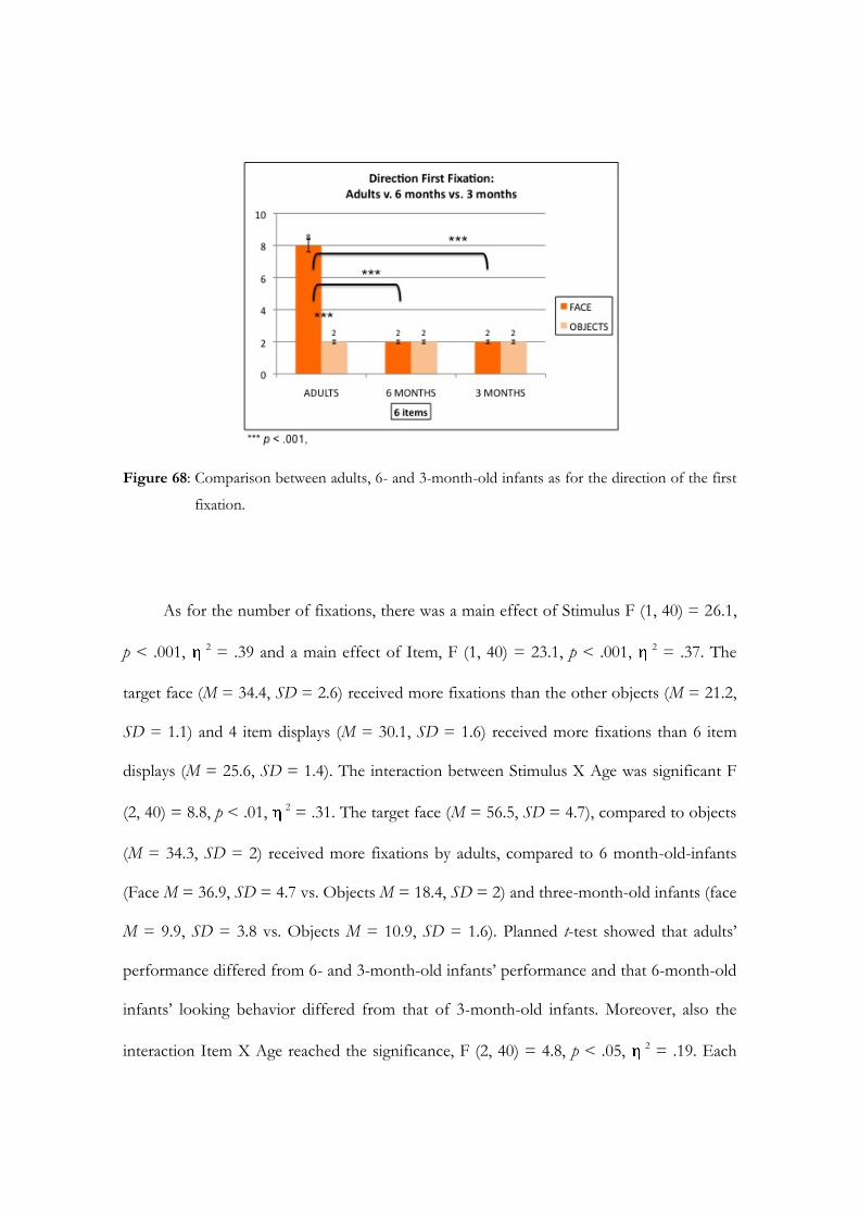
Figure 68: Comparison between adults, 6- and 3-month-old infants as for the direction of the first
fixation.
As for the number of fixations, there was a main effect of Stimulus F (1, 40) = 26.1,
p < .001, 2 = .39 and a main effect of Item, F (1, 40) = 23.1, p < .001, 2 = .37. The
target face (M = 34.4, SD = 2.6) received more fixations than the other objects (M = 21.2,
SD = 1.1) and 4 item displays (M = 30.1, SD = 1.6) received more fixations than 6 item
displays (M = 25.6, SD = 1.4). The interaction between Stimulus X Age was significant F
(2, 40) = 8.8, p < .01, 2 = .31. The target face (M = 56.5, SD = 4.7), compared to objects
(M = 34.3, SD = 2) received more fixations by adults, compared to 6 month-old-infants
(Face M = 36.9, SD = 4.7 vs. Objects M = 18.4, SD = 2) and three-month-old infants (face
M = 9.9, SD = 3.8 vs. Objects M = 10.9, SD = 1.6). Planned t-test showed that adults’
performance differed from 6- and 3-month-old infants’ performance and that 6-month-old
infants’ looking behavior differed from that of 3-month-old infants. Moreover, also the
interaction Item X Age reached the significance, F (2, 40) = 4.8, p < .05, 2 = .19. Each
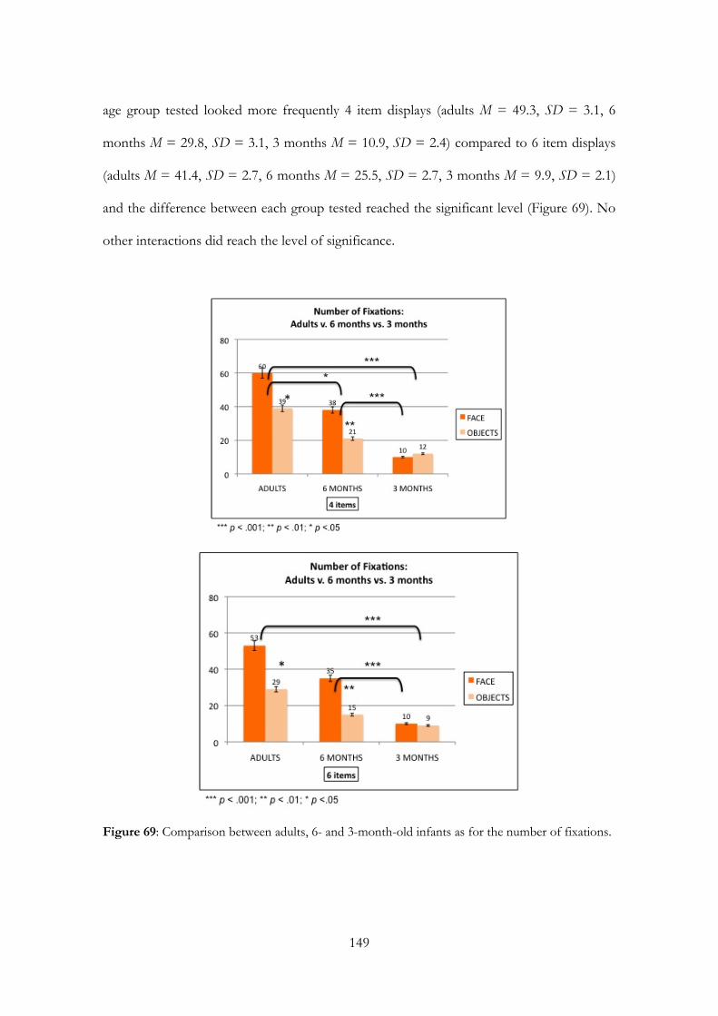
149
age group tested looked more frequently 4 item displays (adults M = 49.3, SD = 3.1, 6
months M = 29.8, SD = 3.1, 3 months M = 10.9, SD = 2.4) compared to 6 item displays
(adults M = 41.4, SD = 2.7, 6 months M = 25.5, SD = 2.7, 3 months M = 9.9, SD = 2.1)
and the difference between each group tested reached the significant level (Figure 69). No
other interactions did reach the level of significance.
Figure 69: Comparison between adults, 6- and 3-month-old infants as for the number of fixations.

Finally, for the total fixation time, there was a main effect of Stimulus F (1, 40) =
16.9, p < .001, 2 = .29 and a main effect of Item, F (1, 40) = 49.9, p < .01, 2 = .21. The
target face (M = 9870.7 ms, SD = 986.9) was looked longer than the other objects (M =
5467 ms, SD = 327.2) and 4 item displays (M = 8608.4 ms, SD = 515.5) were looked
longer than 6 item displays (M = 6729.3 ms, SD = 526.5). The interaction between
Stimulus X Age was significant F (2, 40) = 5.4, p < .001, 2 = .56. The target face (M =
17856 ms, SD = 1825), compared to objects (M = 9761.8 ms, SD = 6059) was looked
longer by adults, but not by 6 month-old-infants (Face M = 9534.8 ms, SD = 1825 vs.
Objects M = 4482.7 ms, SD = 605) and three-month-old infants (face M = 2221.2 ms, SD
= 1450.4 vs. Objects M = 2156.7 ms, SD = 480.8). Planned t-test showed that adults’
performance differed from 6- and 3-month-old infants’ performance and that 6-month-old
infants’ looking behavior differed from that of 3-month-old infants. Moreover, also the
interaction Item X Age reached the significance, F (2, 40) = 20.4, p < .001, 2 = .51. Each
age group tested looked longer 4 item displays (adults M = 15936.5 ms, SD = 953.3, 6
months M = 7531.9 ms, SD = 953.3, 3 months M = 2356.8 ms, SD = 757.6) compared to
6 item displays (adults M = 11681.3 ms, SD = 973.5, 6 months M =6485.5 ms, SD = 973.6,
3 months M = 2021.1 ms, SD = 773.8) and the difference between each group tested
reached the significance (planned t-test). No other interactions did reach the level of
significance (Figure 70).
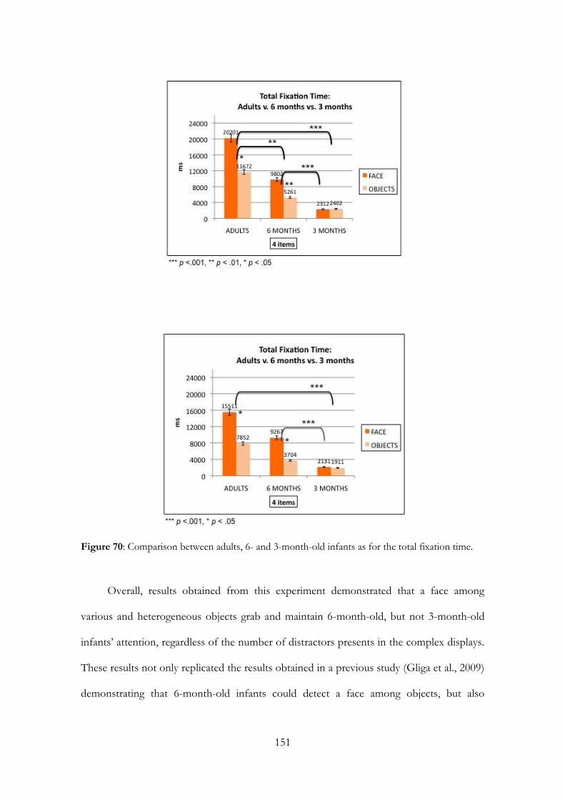
151
Figure 70: Comparison between adults, 6- and 3-month-old infants as for the total fixation time.
Overall, results obtained from this experiment demonstrated that a face among
various and heterogeneous objects grab and maintain 6-month-old, but not 3-month-old
infants’ attention, regardless of the number of distractors presents in the complex displays.
These results not only replicated the results obtained in a previous study (Gliga et al., 2009)
demonstrating that 6-month-old infants could detect a face among objects, but also

extended this previous study in manipulating the number of distractors among the visual
scenes and in testing different age groups with the same paradigm.
However, before to conclude with a general discussion in regard to these results, I’ll
present in the following section a relatively recent approach to analyze data.
4.4.1 Mixed effects model approach to analyze data
We introduce a relatively recent approach to analyze data, called mixed effects model
approach, based on maximum likelihood methods that are now in common use in many
areas of science, medicine, and engineering and specifically in eye tracking studies (e.g.,
Faraway, 2006; Goldstein, 2005; Malcom, Lanyon, Fugard, & Barton, 2008). Since the
number of trials was not the same for each participant, Generalized Mixed-Effects Models
were used to test the effect of age and number of items (e.g., fixed effects, controlled by
experimenter) and the two-way interaction between item (four vs. six) and age (adults vs. 6
months vs. 3 months) on the proportion of first fixation and the proportion of total
fixation time and number of orientation towards face, considering participants and display
as random variables (Baayen, Davidson, & Bates, 2008; Pinheiro & Bates, 2000).
Essentially four are the main advantages for using these models for our data set: i.)
to overcome the problem that the number of trials was not the same for each participant,
conducting the analyses on the number of observation (N = 1167) rather than on the
number of participants (N = 43), ii.) to provide more statistical power eliminating the need
to average across trials, iii.) to insert and to compare within the same model infants’ and
adults’ performance in the modified visual search task and, iv.) compared to the classic
ANOVA calculation, to allow the researcher to simultaneously consider all factors that
potentially contribute to the understanding of the structure of the data, such as random

153
effects related to subjects or display presented. The data analysis was conducted using lme4
package (Bates, 2007) in R (www.r-project.org).
Results for each dependent variable are presented in the following section.
Proportion of the Direction of the First Fixation: Logistic Mixed Effects Model
We used a logistic mixed effects model because the dependent variable is a dichotomic
variable, where the proportion of the direction of the first fixation towards the face target
has value 1, whereas the proportion of the direction of the first fixation towards another
object in the visual display has value 0.
The model containing all the fixed effects and the random effect of the participants was the
best-fitting model for all participants. The best-fitting model was selected using the BIC
criteria (Raftery, 1995), i.e. the model with the smallest BIC is considered the most
appropriate model for reproducing the observed data. Odds ratios are reported as a
measure of effect size. Results showed that adults made significantly more first saccades
towards faces compared to 6- (z = -5.3, p <. 001, or = .22) and 3-month-old infants (z = -
6.9, p < .001, or = .16), regardless of the number of items as distractors (e.g., the interaction
between Age X Item was not significant) (Figure 71).
In other words, from this result it appears that a face among objects do not capture 6- and
3-month old infants’ attention in an efficient way like in adults.
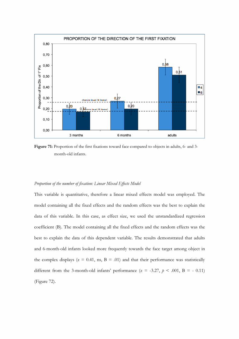
Figure 71: Proportion of the first fixations toward face compared to objects in adults, 6- and 3-
month-old infants.
Proportion of the number of fixation: Linear Mixed Effects Model
This variable is quantitative, therefore a linear mixed effects model was employed. The
model containing all the fixed effects and the random effects was the best to explain the
data of this variable. In this case, as effect size, we used the unstandardized regression
coefficient (B). The model containing all the fixed effects and the random effects was the
best to explain the data of this dependent variable. The results demonstrated that adults
and 6-month-old infants looked more frequently towards the face target among object in
the complex displays (z = 0.41, ns, B = .01) and that their performance was statistically
different from the 3-month-old infants’ performance (z = -3.27, p < .001, B = - 0.11)
(Figure 72).

155
Figure 72: Proportion of the number of fixations toward face compared to objects in adults, 6-
and 3- month-old infants.
Proportion of the total fixation time: Linear Mixed Effects Model
This variable is a quantitative variable, therefore a linear mixed effects model was employed
and unstandardized regression coefficient (B) was used as effect size measure.
The model containing all the fixed effects and the random effects was the best to explain
the data of this variable. As regard of the proportion of total fixation time towards the
target face, 6-month-old infants’ and adults’ performances are similar (z = .08, B = .00),
and differed from 3-month-old infants’ performance (z = -3.4, p <. 001, B = - 0.13). In
other words, face maintains adults’ and 6-month-old infants’ attention, but not 3-month-
old infants’ attention, once it has been detected among objects. Furthermore, the fixed
effect of Item is significant (z = -2.20, p < .05, B = - 0.1), but not the interaction between
Age and Item, demonstrating that the displays formed by 4 Items were looked longer than
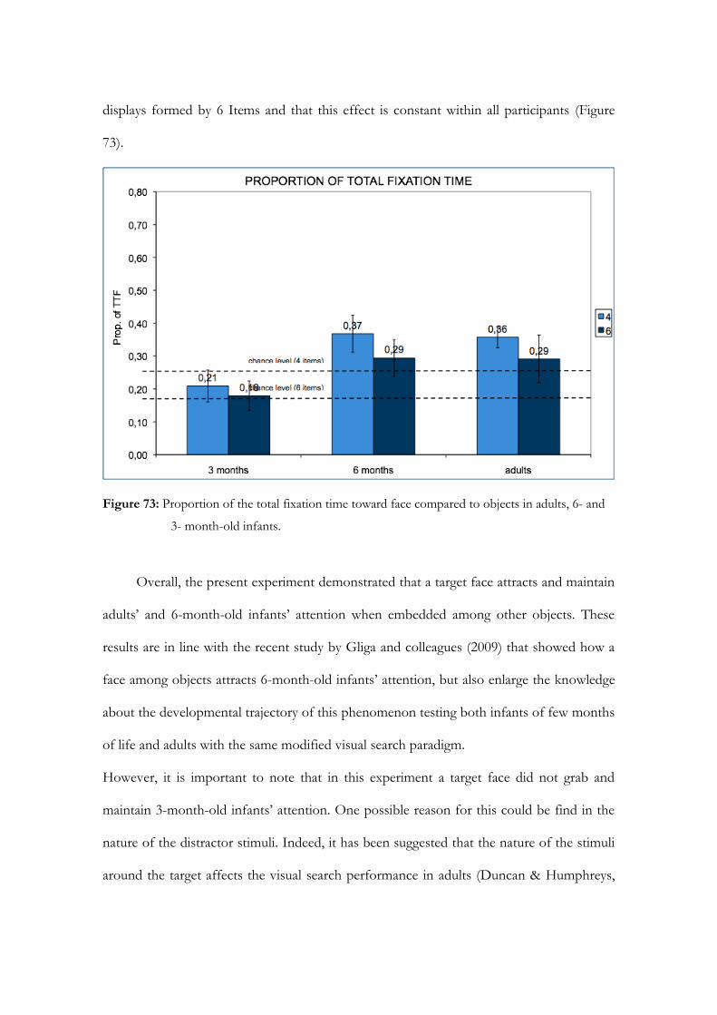
displays formed by 6 Items and that this effect is constant within all participants (Figure
73).
Figure 73: Proportion of the total fixation time toward face compared to objects in adults, 6- and
3- month-old infants.
Overall, the present experiment demonstrated that a target face attracts and maintain
adults’ and 6-month-old infants’ attention when embedded among other objects. These
results are in line with the recent study by Gliga and colleagues (2009) that showed how a
face among objects attracts 6-month-old infants’ attention, but also enlarge the knowledge
about the developmental trajectory of this phenomenon testing both infants of few months
of life and adults with the same modified visual search paradigm.
However, it is important to note that in this experiment a target face did not grab and
maintain 3-month-old infants’ attention. One possible reason for this could be find in the
nature of the distractor stimuli. Indeed, it has been suggested that the nature of the stimuli
around the target affects the visual search performance in adults (Duncan & Humphreys,
157
1989). That is, more the distractors are similar to each other and different from the target
stimulus, more efficient is the detection of the target, whereas more the distractors are
dissimilar from each other, more difficult will be the detection of the stimulus target.
Therefore, it is possible that 3-month-old infants perceived the visual displays of the
previous experiment as complex and that they did not find a target face among the other
heterogeneous and complex visual stimuli presented as distractors.
To investigate whether the nature of the distractor stimuli could affect 3-month-old
infants’ performance in the modified visual search paradigm, an experiment in which the
complex visual displays were formed by an upright target face among inverted faces as
distractors was conducted. It was predicted that, since the distractors are similar to each
other and share with the upright target face all the low-level perceptual characteristics (i.e.,
structure, luminance, contrast, complexity), 3-month-old infants’ should be able to detect
efficiently the upright target face, regardless of the number of distractors.
Experiment 11 and 12
Method
Participants
Participants were twenty-five undergraduate students selected from the Department of
Psychology at the University of Padova. Four participants were excluded from the sample
because of the presence of not interpretable eye movements due to poor calibration (N =
4). The final sample consisted of 21 adults (14 female, mean age = 23,7 years). All
participants had no previous experience with eye-movements studies and are naive to the
experimental conditions and hypotheses of the study. Finally, all of them had normal or
corrected-to-normal vision. Furthermore, fourteen 3-month-old (5 females, mean age =
97.4 days) healthy and full-term infants were tested. They were middle-class infants and all
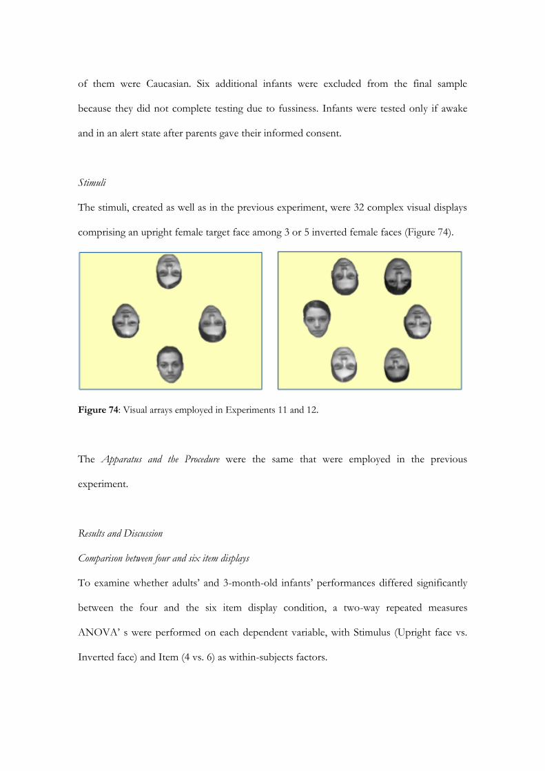
of them were Caucasian. Six additional infants were excluded from the final sample
because they did not complete testing due to fussiness. Infants were tested only if awake
and in an alert state after parents gave their informed consent.
Stimuli
The stimuli, created as well as in the previous experiment, were 32 complex visual displays
comprising an upright female target face among 3 or 5 inverted female faces (Figure 74).
Figure 74: Visual arrays employed in Experiments 11 and 12.
The Apparatus and the Procedure were the same that were employed in the previous
experiment.
Results and Discussion
Comparison between four and six item displays
To examine whether adults’ and 3-month-old infants’ performances differed significantly
between the four and the six item display condition, a two-way repeated measures
ANOVA’ s were performed on each dependent variable, with Stimulus (Upright face vs.
Inverted face) and Item (4 vs. 6) as within-subjects factors.

159
Adults. Results of the ANOVA on the direction of the first fixation showed a main
effect of the Stimulus, F (1, 20) = 20.1, p < .001, 2 = .50 and a main effect of Item, F (1,
20) = 61.3, p < .001, 2 = .75. Importantly, there is a significant interaction between
Stimulus and Item, F (1, 20) = 4.5, p < .05, 2 = .18. This data indicated that upright face
grabs adults’ attention (M = 5.9, SD = .36) more than inverted faces (M = 2.9, SD = .12).
Furthermore, 4 item displays received more first fixations (M = 4.6, SD = .12) than 6 item
displays (M = 3.2, SD = .18). As regard to the interaction Stimulus X Item, the data
demonstrated that the number of distractors affected the adults’ performance. Specifically,
upright target face received more first fixations (M = 5.9, SD = .36) than inverted faces (M
= 3.3, SD = .17) in the 4 item display compared to the number of first fixation that the
target face (M = 3.8, SD = .47) received in the six item displays compared to the inverted
faces (M = 2.5, SD = .13) (Figure 75).
Figure 75: Adults’ first fixations are oriented toward face than other objects, but the number of
distractors affects the performance.
As regard to the number of fixation, the ANOVA showed a main effect of the
Stimulus, F (1, 20) = 12.3, p < .01, 2 = .38 and the main effect of the item factor F (1, 20)
= 15.7, p < .01, 2 = .44. However, the interaction between them did not reach the
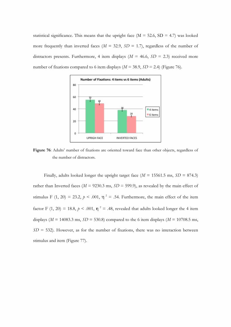
statistical significance. This means that the upright face (M = 52.6, SD = 4.7) was looked
more frequently than inverted faces (M = 32.9, SD = 1.7), regardless of the number of
distractors presents. Furthermore, 4 item displays (M = 46.6, SD = 2.3) received more
number of fixations compared to 6 item displays (M = 38.9, SD = 2.4) (Figure 76).
Figure 76: Adults’ number of fixations are oriented toward face than other objects, regardless of
the number of distractors.
Finally, adults looked longer the upright target face (M = 15561.5 ms, SD = 874.3)
rather than Inverted faces (M = 9230.3 ms, SD = 599.9), as revealed by the main effect of
stimulus F (1, 20) = 23.2, p < .001, 2 = .54. Furthermore, the main effect of the item
factor F (1, 20) = 18.8, p < .001, 2 = .48, revealed that adults looked longer the 4 item
displays (M = 14083.3 ms, SD = 530.8) compared to the 6 item displays (M = 10708.5 ms,
SD = 532). However, as for the number of fixations, there was no interaction between
stimulus and item (Figure 77).

161
Figure 77: Adults looked longer toward a target face than other objects, regardless of the number
of distractors.
Three-month-old infants. Results of the ANOVA on the direction of the first fixation
showed a main effect of the item factor only, F (1, 13) = 14.1, p < .05, 2 = .52. No main
effect of the stimulus factor was observed neither the stimulus X item interaction was
significant (Figure 78).
As for the number of fixation, that is the other attention-getting index, results
showed a main effect of both Stimulus, F (1, 13) = 8.3, p < .05, 2 = .39 and Item factor, F
(1, 13) = 7.4, p < .05, 2 = .36, but their interaction was not significant. This means that 3-
month-old infants looked more frequently the upright face (M = 18.2, SD = 3.6) compared
to inverted faces (M = 12.5, SD = 1.9), regardless of the number of distractors presented.
Moreover, 4 item displays received more number of fixations (M = 18.1, SD = 3.6)
compared to 6 item displays (M = 12.6, SD = 2) (Figure 79).
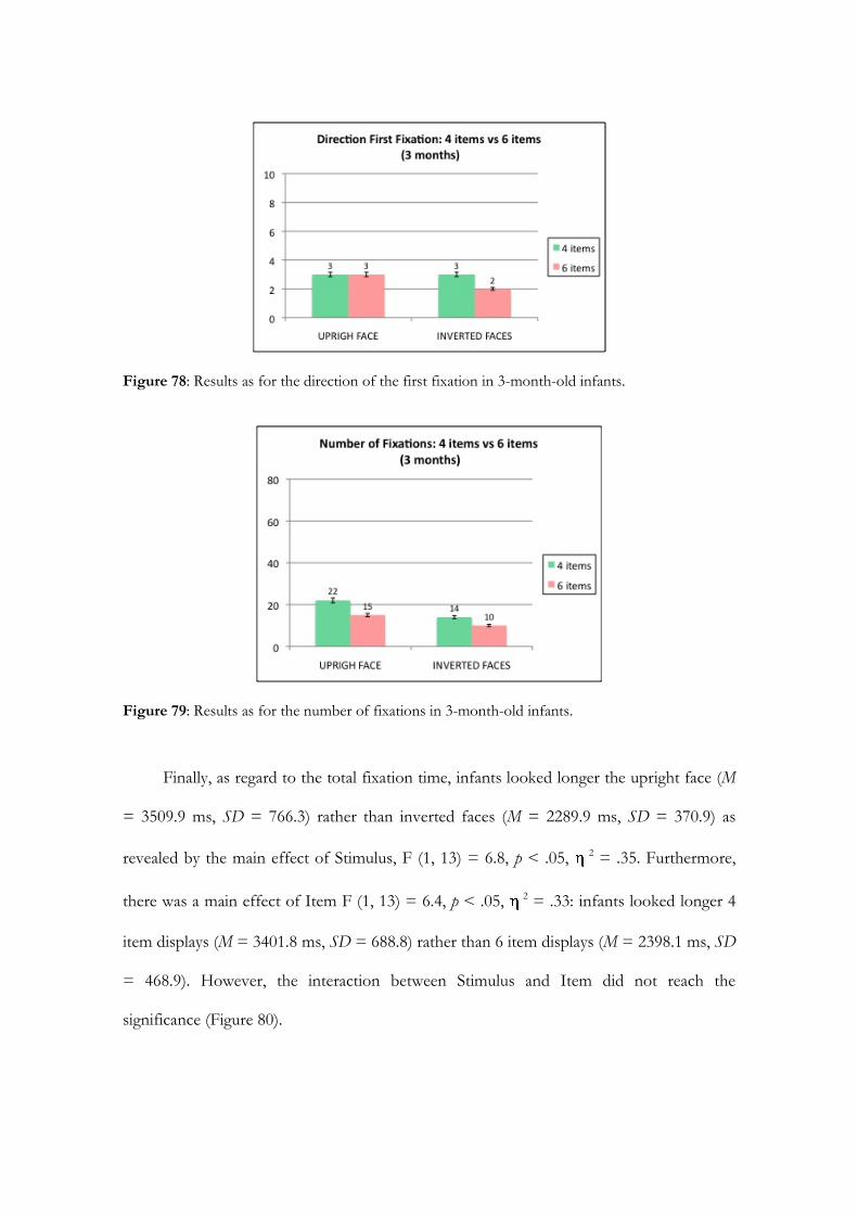
Figure 78: Results as for the direction of the first fixation in 3-month-old infants.
Figure 79: Results as for the number of fixations in 3-month-old infants.
Finally, as regard to the total fixation time, infants looked longer the upright face (M
= 3509.9 ms, SD = 766.3) rather than inverted faces (M = 2289.9 ms, SD = 370.9) as
revealed by the main effect of Stimulus, F (1, 13) = 6.8, p < .05, 2 = .35. Furthermore,
there was a main effect of Item F (1, 13) = 6.4, p < .05, 2 = .33: infants looked longer 4
item displays (M = 3401.8 ms, SD = 688.8) rather than 6 item displays (M = 2398.1 ms, SD
= 468.9). However, the interaction between Stimulus and Item did not reach the
significance (Figure 80).

163
Figure 80: Results as for the total fixation time in 3-month-old infants.
Age Comparison
To compare the performance of each age group tested, repeated-measures ANOVA was
performed for each dependent variable with Age (adults vs. 3 months) as between-subject
factor and Stimulus (Upright Face vs. Inverted Faces) and Item (4 vs. 6) as within-subject
factors.
As for the direction of the first fixation, there was a main effect of Stimulus F (1, 33)
= 16.9, p < .001, 2 = .34 and a main effect of Item, F (1, 33) = 61.9, p < .001, 2 = .65.
The target face (M = 3.9, SD = .26) received more first fixations than inverted faces (M =
2.7, SD = .09) and 4 item displays (M = 3.9, SD = .13) received more first fixations than 6
item displays (M = 2.8, SD = .13). The interaction between Stimulus X Age was significant,
F (1, 33) = 4.8, p < .05, 2 = .13. The upright target face received more first fixations
(adults M = 4.9, SD = .32, 3 months M = 3, SD = .39) compared to other inverted faces
(adults M = 2.9, SD = .12, 3 months M = 2.4, SD = .15). Planned t-test showed that adults’
performance differed from 3-month-old infants’ performance. Furthermore, Item X Age
interaction was significant, F (1, 33) = 6.3, p < .05, 2 = .16. The 4 item displays received
more first fixations (adults M = 4.6, SD = .17, 3 months, M = 3.1, SD = .21) rather than 6
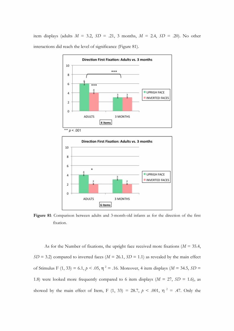
item displays (adults M = 3.2, SD = .21, 3 months, M = 2.4, SD = .20). No other
interactions did reach the level of significance (Figure 81).
Figure 81: Comparison between adults and 3-month-old infants as for the direction of the first
fixation.
As for the Number of fixations, the upright face received more fixations (M = 35.4,
SD = 3.2) compared to inverted faces (M = 26.1, SD = 1.1) as revealed by the main effect
of Stimulus F (1, 33) = 6.1, p < .05, 2 = .16. Moreover, 4 item displays (M = 34.5, SD =
1.8) were looked more frequently compared to 6 item displays (M = 27, SD = 1.6), as
showed by the main effect of Item, F (1, 33) = 28.7, p < .001, 2 = .47. Only the
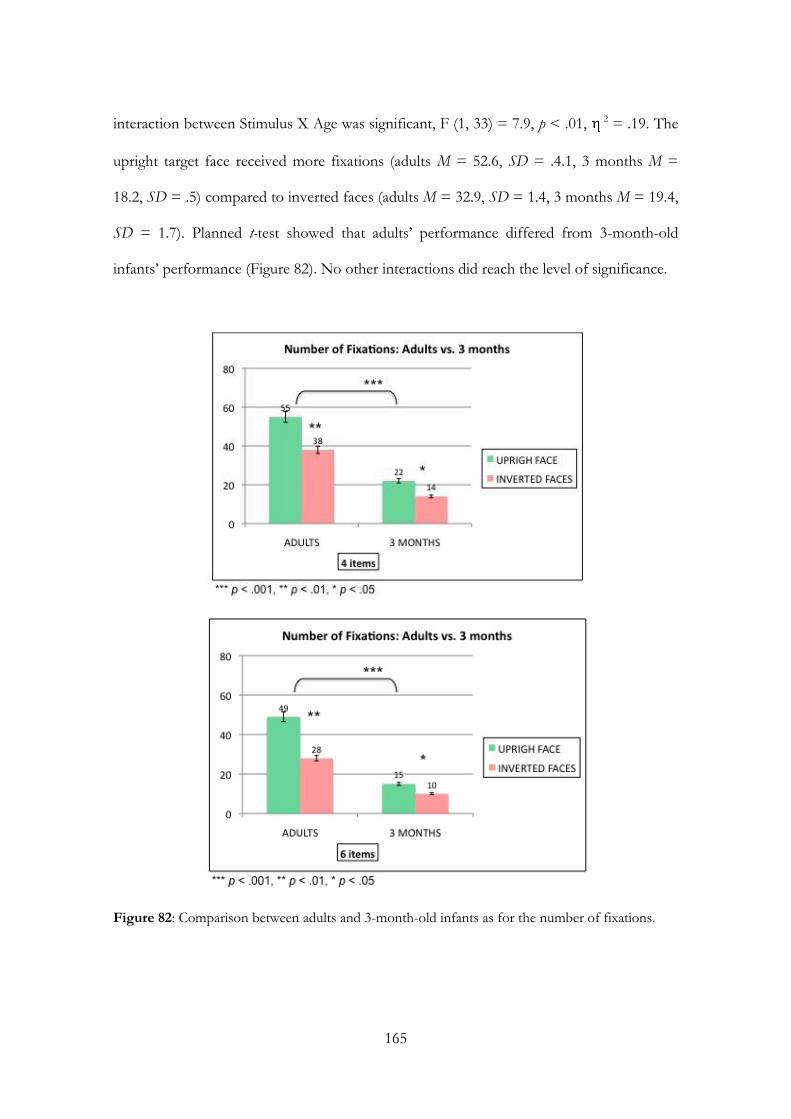
165
interaction between Stimulus X Age was significant, F (1, 33) = 7.9, p < .01, 2 = .19. The
upright target face received more fixations (adults M = 52.6, SD = .4.1, 3 months M =
18.2, SD = .5) compared to inverted faces (adults M = 32.9, SD = 1.4, 3 months M = 19.4,
SD = 1.7). Planned t-test showed that adults’ performance differed from 3-month-old
infants’ performance (Figure 82). No other interactions did reach the level of significance.
Figure 82: Comparison between adults and 3-month-old infants as for the number of fixations.
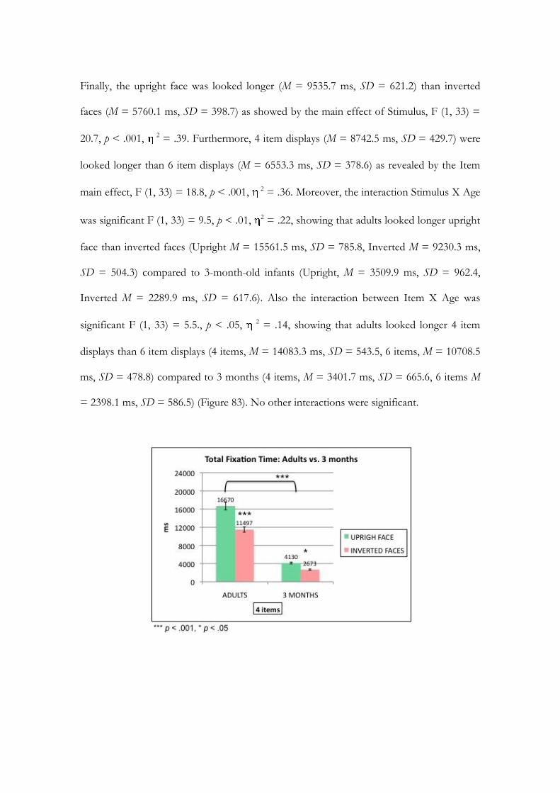
Finally, the upright face was looked longer (M = 9535.7 ms, SD = 621.2) than inverted
faces (M = 5760.1 ms, SD = 398.7) as showed by the main effect of Stimulus, F (1, 33) =
20.7, p < .001, 2 = .39. Furthermore, 4 item displays (M = 8742.5 ms, SD = 429.7) were
looked longer than 6 item displays (M = 6553.3 ms, SD = 378.6) as revealed by the Item
main effect, F (1, 33) = 18.8, p < .001, 2 = .36. Moreover, the interaction Stimulus X Age
was significant F (1, 33) = 9.5, p < .01, 2 = .22, showing that adults looked longer upright
face than inverted faces (Upright M = 15561.5 ms, SD = 785.8, Inverted M = 9230.3 ms,
SD = 504.3) compared to 3-month-old infants (Upright, M = 3509.9 ms, SD = 962.4,
Inverted M = 2289.9 ms, SD = 617.6). Also the interaction between Item X Age was
significant F (1, 33) = 5.5., p < .05, 2 = .14, showing that adults looked longer 4 item
displays than 6 item displays (4 items, M = 14083.3 ms, SD = 543.5, 6 items, M = 10708.5
ms, SD = 478.8) compared to 3 months (4 items, M = 3401.7 ms, SD = 665.6, 6 items M
= 2398.1 ms, SD = 586.5) (Figure 83). No other interactions were significant.
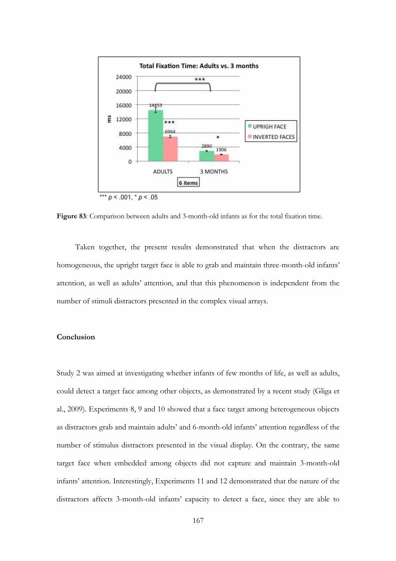
167
Figure 83: Comparison between adults and 3-month-old infants as for the total fixation time.
Taken together, the present results demonstrated that when the distractors are
homogeneous, the upright target face is able to grab and maintain three-month-old infants’
attention, as well as adults’ attention, and that this phenomenon is independent from the
number of stimuli distractors presented in the complex visual arrays.
Conclusion
Study 2 was aimed at investigating whether infants of few months of life, as well as adults,
could detect a target face among other objects, as demonstrated by a recent study (Gliga et
al., 2009). Experiments 8, 9 and 10 showed that a face target among heterogeneous objects
as distractors grab and maintain adults’ and 6-month-old infants’ attention regardless of the
number of stimulus distractors presented in the visual display. On the contrary, the same
target face when embedded among objects did not capture and maintain 3-month-old
infants’ attention. Interestingly, Experiments 11 and 12 demonstrated that the nature of the
distractors affects 3-month-old infants’ capacity to detect a face, since they are able to
detect an upright target face only when it is among homogeneous distractors, that are
inverted faces that share with the target face the complexity, high contrast areas and
luminance, but not the social relevance.
To my knowledge, the current study is the first that, using a modified visual search
with adults and infants of few months of life and recording the eye-movements of
participants, demonstrated a developmental trajectory of the attention capture by faces
within complex displays. Six-month-old infants’ performances are similar to adults’
performance, and differed from the 3-month-old infants’ performance. A possible
interpretation of these data could be that it is harder for young infants to detect a complex
visual stimulus, like a human face, among other complex and novel visual stimuli as
distractors. Indeed, one perceptual cue that guides infants’ visual preference in the first
months of life is the complexity of the visual stimuli (Easterbrook et al., 1999). Indeed,
Experiments 12 demonstrated that it is easier for young infants to detect a target stimulus
that differs from the distractor only for one perceptual characteristic, in this case the
orientation (e.g., upright face vs. inverted faces).
To conclude, the modified visual search paradigm employed in this study proved to
be a reliable measure of the capacity of face stimuli to capture and maintain infants’ and
adults’ attention. Future studies should explore whether the attention-getting and the
attention-holding mechanisms become more selective with the age to specific kind of faces
(i.e., monkey faces vs human faces, caucasic faces vs asiatic faces, and so on).

CHAPTER 5
THE ORIGIN AND THE DEVELOPMENTAL TIME
COURSE OF HOLISTIC FACE PROCESSING


171
Introduction
Face detection is the first step in the processing of a face, since detection allow infant to
select and orient attention toward salient stimuli to efficiently process them. In previous
chapters of my thesis, I described how newborns and infants detect a face in both simple
arrays (i.e., when only two stimuli are presented simultaneously) and complex visual arrays
(i.e., when faces are embedded among other complex visual stimuli). There is no doubt that
infants, to identify a target face among other visual stimuli (Chapter 4), had to perceive the
face as a whole, employing a holistic face processing strategy. Faces, unlike many other
objects, are processed holistically, and this means that they are encoded as one inseparable
unit, rather than as a group of individual features or parts (Tanaka & Farah, 1993). The
concept was probably first introduced by Francis Galton, who noticed that facial features
were not perceived and analyzed separately; that is, the face stimulus was processed as a
whole unit or as a Gestalt (Galton, 1883). Although rudimentary ability to perceive faces
holistically is present at birth (Leo & Simion, 2009), the developmental time course of this
face processing is still unclear.
The aim of the present chapter is to investigate the origin and the development of
holistic face processing in newborns and infants of few months of life by employing a
classical behavioral task, called the composite face paradigm, that was used to tap this
specific face processing in adults and older children (Young et al., 1987; Mondloch et al.,
2007). The use of the same task, throughout development, from early infancy to adulthood,
allows researchers to highlight both differences and similarities in the cognitive processes
underlying face processing, and to highlight the changes of the system during lifespan to
raise the adult face specialized level. Based on this, the current chapter is focused on

holistic processing, investigating through the testing of the composite face illusion
newborns’ and 3-month-old infants’ sensitivity to holistic face information.
5.1. The study of holistic face processing in adults
As described in Chapter 2, evidence of holistic processing in adults can be observed in
part-whole paradigms, in which subjects perform better to match or recognize individual
facial features presented within the context of a whole face than features that are presented
separately (Tanaka & Farah, 1993). Evidence of part versus whole effects is observed in
adults and in typically developing children of four years of age (Pellicano & Rhodes, 2003).
Holistic face processing can be also tap with the composite face task by examining the
effects of spatial misalignment on the ability to attend selectively to specific portions of
faces. In the typical composite face paradigm, composite faces are created by combining
the top half of one famous or familiar face with the bottom half of another face, and
subjects are then asked to identify only the person depicted in the top or the bottom half
of the face (Young, et al., 1987). When the two face halves are presented in alignment with
one another, they join to form a new face configuration and the stimulus is encoded
holistically. In this case, recognition of the individual parts of the face is difficult because
the new configuration interferes with the recognition of the individual features within each
half. By contrast, a new overall facial configuration does not result when the two halves of
the composite face are spatially misaligned with one another. In this case, there is no
interference due to holistic encoding of the stimulus and subjects recognize the source
images more easily (Young et al.,1987). Discrimination of unfamiliar faces is similarly
disrupted by holistic processing in composite tasks (Hole, 1994; Hole, George, &
Dunsmore, 1999; Le Grand et al., 2003). In these tasks, subjects are presented with two
composite faces and are asked to make same or different judgments based on only one half

173
of the faces. When the two halves of the composite face are aligned, the face is processed
holistically. However, when the two halves of the composite face are spatially misaligned,
holistic processing is disrupted, allowing better selective attention to the attended half of
the face, and consequently, better performance in the misaligned relative to the intact
condition. In sum, holistic processing is characterized by the encoding of the face as single
stimulus, with difficulty attending selectively to individual features or parts of the face.
Selective attention to features can be improved by misaligning sections of the face because
the misalignment disrupts holistic processing.
It is important to note that in the composite task, spatial misalignment of the face
halves disrupts the first-order configuration of the stimulus (two eyes centered directly
above a mouth). This configural disruption interferes with the binding of the two halves
into a gestalt and as a result, the face is not encoded holistically and the unattended half
exerts less influence on perception of the attended half. By contrast, when faces are intact
and perceived holistically, information in the unattended half strongly affects judgments of
the attended half. This characterization agrees with recent proposals that holistic
processing is a subtype of configural processing that is dependent upon the first-order
configuration of the face (de Heering, Houthuys, & Rossion, 2007; Maurer et al., 2002).
5.2 Developmental studies on face processing in infants
The functional face processing specialization seen in adults is generally considered as due
to the extensive visual experience with faces during the life time. Indeed, it has been widely
accepted that although experience-based developmental processes progressively refine early
configural face recognition abilities, available evidence demonstrates that configural
processing emerges during the first months of life rather than undergoing a long
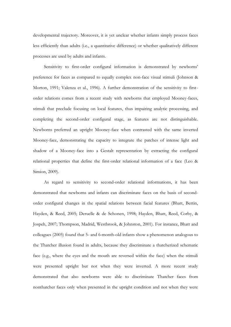
developmental trajectory. Moreover, it is yet unclear whether infants simply process faces
less efficiently than adults (i.e., a quantitative difference) or whether qualitatively different
processes are used by adults and infants.
Sensitivity to first-order configural information is demonstrated by newborns’
preference for faces as compared to equally complex non-face visual stimuli (Johnson &
Morton, 1991; Valenza et al., 1996). A further demonstration of the sensitivity to first-
order relations comes from a recent study with newborns that employed Mooney-faces,
stimuli that preclude focusing on local features, thus impairing analytic processing, and
completing the second-order configural stage, as features are not distinguishable.
Newborns preferred an upright Mooney-face when contrasted with the same inverted
Mooney-face, demonstrating the capacity to integrate the patches of intense light and
shadow of a Mooney-face into a Gestalt representation by extracting the configural
relational properties that define the first-order relational information of a face (Leo &
Simion, 2009).
As regard to sensitivity to second-order relational informations, it has been
demonstrated that newborns and infants can discriminate faces on the basis of second-
order configural changes in the spatial relations between facial features (Bhatt, Bertin,
Hayden, & Reed, 2005; Deruelle & de Schonen, 1998; Hayden, Bhatt, Reed, Corby, &
Jospeh, 2007; Thompson, Madrid, Westbrook, & Johnston, 2001). For instance, Bhatt and
colleagues (2005) found that 5- and 6-month-old infants show a phenomenon analogous to
the Thatcher illusion found in adults, because they discriminate a thatcherized schematic
face (e.g., where the eyes and the mouth are reversed within the face) when the stimuli
were presented upright but not when they were inverted. A more recent study
demonstrated that also newborns were able to discriminate Thatcher faces from
nonthatcher faces only when presented in the upright condition and not when they were

175
presented upside-down (Leo & Simion, 2009). Moreover, it has been demonstrated that
newborns, as well as 4-month-old infants (Turati, Sangrigoli, Ruel, & de Schonen, 2004),
were able to recognize upright faces either when faces were fully visible (i.e., when both
inner and outer features are present) or when only the inner and outer features were
present. In contrast, newborns failed to recognize an inverted face when only the inner
features were presented. The presence of recognition for the upright face but not for the
inverted one in the inner condition have been interpreted as due to sensitivity to second-
order relational information just for the upright face, since inversion affected all kind of
configural processing (Turati, Macchi Cassia, Simion, & Leo, 2006). Overall, these studies
seem to indicate that the ability to process second-order relational information is available
at birth and that inversion affects the processing of the configural information.
5.3 Holistic face processing in children and infants
Compared to many studies that investigated first- and second-order relation information
processing in children and infants, the processing of holistic face information has received
far less attention. As described in previous paragraphs, compelling examples of holistic
face processing come from two behavioral paradigms widely used to evaluate the existence
of holistic face processing in adults and in children: the whole-part paradigm (Tanaka &
Farah, 1993) and the composite face paradigm (Young et al., 1987; Hole et al., 1994).
As regard to the whole-part paradigm, only one study provided evidence that 4-
year-olds manifest a part-whole effect, indicating that they recognize faces holistically.
Four- to six-year-old children and adults were administered a whole-part face recognition
task. Children below the age of 6 recognized parts from upright faces better when tested in
the whole-face condition rather than in isolation condition and this effect was presented

only when faces were presented upright but not when they where presented upside-down
(Pellicano & Rhodes, 2003). Interestingly, the different age groups showed similar patterns
of performance indicating that young preschoolers, like older children and adults, are able
to recognize faces holistically.
As regard to the composite face paradigm, that is considered the most convincing
demonstration of holistic face processing, it has been used only in few studies with school
and preschool aged children. Specifically, evidence for a composite face effect for both
familiar (i.e., faces of classmates) and unfamiliar faces was found in 6- and 10-year-old
children, with the magnitude of the effect remaining constant across the two age groups
and into the adulthood (Carey & Diamond, 1994; Mondloch et al., 2007). At present, only
two studies have investigated holistic face processing in preschool-aged children using this
paradigm. The first study tested adults and 4- to 6-year-olds with the same composite
paradigm and demonstrated that holistic face processing is already mature at 4 years of age,
that is, performance was more accurate in the misaligned condition than in the aligned
condition. The second study compared the development of holistic processing for faces
and non-face visual objects by testing for the composite effect for faces and frontal images
of cars in 3- to 5-year-old children and adults (Macchi Cassia, Picozzi, Kuefner, Bricolo, &
Turati, 2009) (Figure 84).
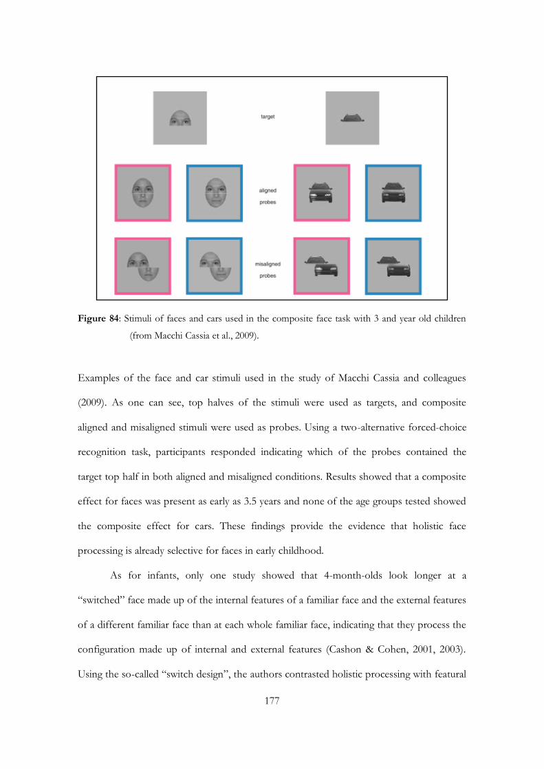
177
Figure 84: Stimuli of faces and cars used in the composite face task with 3 and year old children
(from Macchi Cassia et al., 2009).
Examples of the face and car stimuli used in the study of Macchi Cassia and colleagues
(2009). As one can see, top halves of the stimuli were used as targets, and composite
aligned and misaligned stimuli were used as probes. Using a two-alternative forced-choice
recognition task, participants responded indicating which of the probes contained the
target top half in both aligned and misaligned conditions. Results showed that a composite
effect for faces was present as early as 3.5 years and none of the age groups tested showed
the composite effect for cars. These findings provide the evidence that holistic face
processing is already selective for faces in early childhood.
As for infants, only one study showed that 4-month-olds look longer at a
“switched” face made up of the internal features of a familiar face and the external features
of a different familiar face than at each whole familiar face, indicating that they process the
configuration made up of internal and external features (Cashon & Cohen, 2001, 2003).
Using the so-called “switch design”, the authors contrasted holistic processing with featural
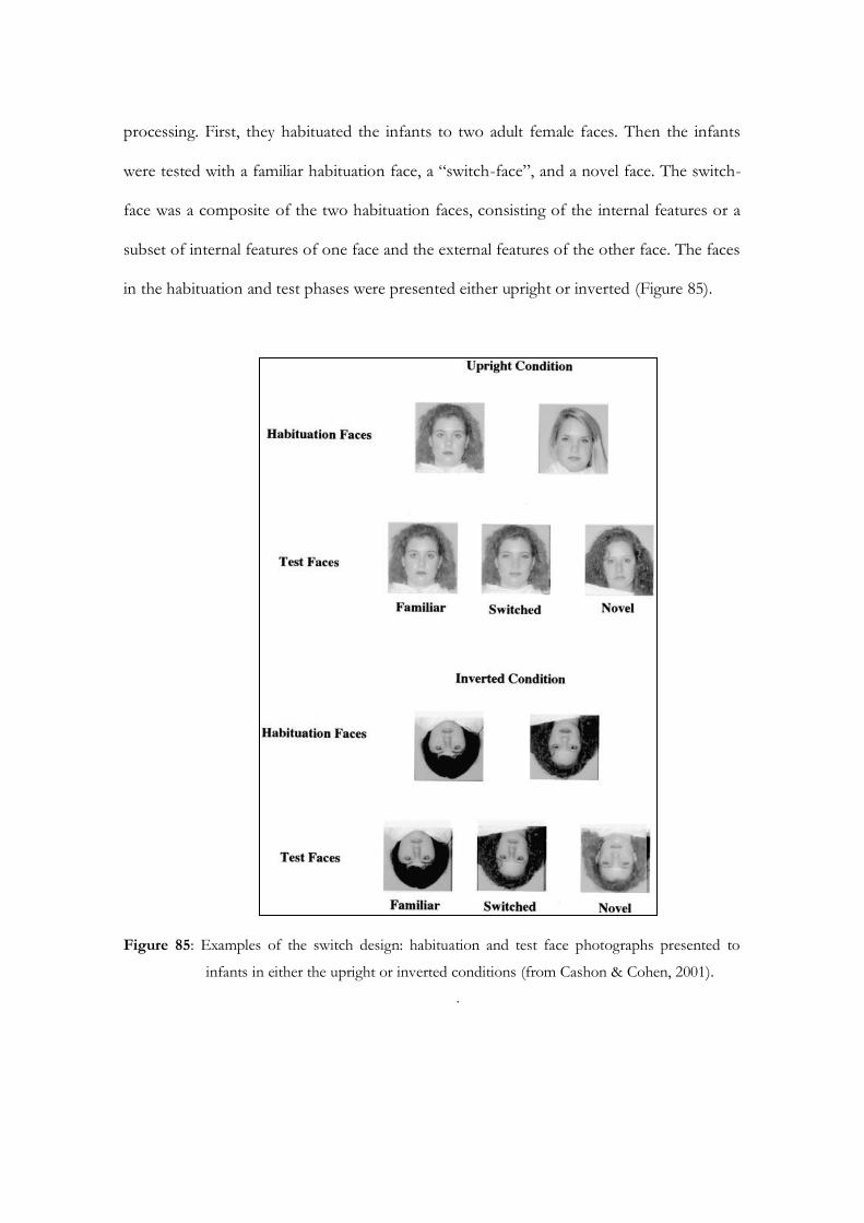
processing. First, they habituated the infants to two adult female faces. Then the infants
were tested with a familiar habituation face, a “switch-face”, and a novel face. The switch-
face was a composite of the two habituation faces, consisting of the internal features or a
subset of internal features of one face and the external features of the other face. The faces
in the habituation and test phases were presented either upright or inverted (Figure 85).
Figure 85: Examples of the switch design: habituation and test face photographs presented to
infants in either the upright or inverted conditions (from Cashon & Cohen, 2001).
.
179
To assess whether infants used featural or holistic processing, it was critical to
determine whether they looked longer at the switch-face compared to the familiar test face.
The authors reasoned that infants would look longer at the switch-face only if they were
processing more than the independent features and were integrating at least some of the
internal and external features. On the other hand, if they were processing the facial sections
independently the infants would not respond to the switch-face. The results showed that
holistic processing followed an N-shaped developmental pattern for upright faces and an
inverted U-shaped pattern for inverted faces in the age range of 3 to 7 months of age. In
more detail, this means that the youngest age group, i.e. 3-month-olds, processed upright
and inverted internal and external sections of faces independently of each other. The 4-
month-olds, in contrast, processed both upright and inverted sections holistically, while the
6-month-olds again processed both upright and inverted external and internal sections
independently of each other. Finally, the 7-month-old infants showed an adult-like pattern
of response, since they processed the internal and external facial features holistically when
faces were upright, whereas when faces were inverted they process internal and external
elements featurally. Neverthless, such studies focused exclusively on the correlations
between outer and inner face features. Yet, the inner part of the face per se contains
sufficient cues to support 4-month-old infants’ face recognition. For example, newborns,
as suggested before, use information about internal facial features in making preferences
based on attractiveness (Slater et al., 2000). Furthermore, thirty-five- to 40-day-old infants
are able to discriminate their mother from a stranger even when the whole line between
face and hair is masked by a scarf, thus relying mostly on the inner portion of the face
(Bartrip, Morton, & de Schonen, 2001). Following familiarization to unfamiliar faces,
similar results have been obtained at 1, 3, and 6 months of age (de Haan, Johnson, Maurer,
& Perrett, 2001; Pascalis, de Haan, Nelson, & de Schonen, 1998). Nevertheless, holistic
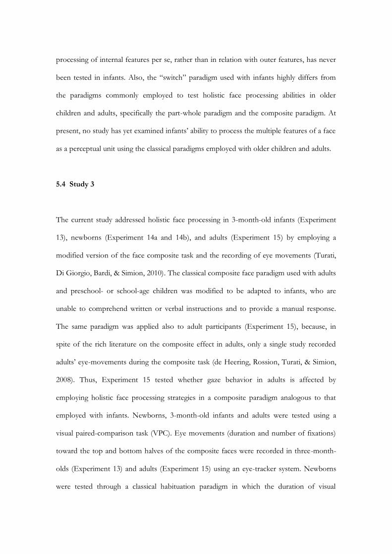
processing of internal features per se, rather than in relation with outer features, has never
been tested in infants. Also, the “switch” paradigm used with infants highly differs from
the paradigms commonly employed to test holistic face processing abilities in older
children and adults, specifically the part-whole paradigm and the composite paradigm. At
present, no study has yet examined infants’ ability to process the multiple features of a face
as a perceptual unit using the classical paradigms employed with older children and adults.
5.4 Study 3
The current study addressed holistic face processing in 3-month-old infants (Experiment
13), newborns (Experiment 14a and 14b), and adults (Experiment 15) by employing a
modified version of the face composite task and the recording of eye movements (Turati,
Di Giorgio, Bardi, & Simion, 2010). The classical composite face paradigm used with adults
and preschool- or school-age children was modified to be adapted to infants, who are
unable to comprehend written or verbal instructions and to provide a manual response.
The same paradigm was applied also to adult participants (Experiment 15), because, in
spite of the rich literature on the composite effect in adults, only a single study recorded
adults’ eye-movements during the composite task (de Heering, Rossion, Turati, & Simion,
2008). Thus, Experiment 15 tested whether gaze behavior in adults is affected by
employing holistic face processing strategies in a composite paradigm analogous to that
employed with infants. Newborns, 3-month-old infants and adults were tested using a
visual paired-comparison task (VPC). Eye movements (duration and number of fixations)
toward the top and bottom halves of the composite faces were recorded in three-month-
olds (Experiment 13) and adults (Experiment 15) using an eye-tracker system. Newborns
were tested through a classical habituation paradigm in which the duration of visual

181
fixations is coded by human expert observers. During the habituation/familiarization
phase, all participants were presented with a top half of a face. The test phase involved the
presentation of a pair of composite face stimuli. In one face stimulus of the pair, the top
half of the face was identical to the one shown during familiarization, and the top of the
other face was new. Half of the participants was tested in the Aligned condition, in which
the left/right margins of the top and bottom face parts were properly aligned, and the
other half was tested in the Misaligned condition, in which the top and bottom face parts
were offset horizontally.
Experiment 13
Method
Participants
Participants were 20 3-month-old healthy and full-term infants (13 males, mean age = 99
days). They were middle-class infants and all of them except one were Caucasian. Seven
additional infants were tested but not included in the final sample because they showed a
strong position preference (i.e., they looked to one direction more than 90% of the time).
Infants were tested only if awake and in an alert state after parents gave their informed
consent.
Stimuli
The original stimuli were digitized, high-quality grey-scale images of 12 Caucasian female
full-front faces, posing with a neutral expression and placed on a black background with no
hair. All faces were unfamiliar to participants. Face images were split into two parts at the
level of the middle of the nose, generating a top and a bottom segment. Composite faces

were created by joining the top segment of one face to the bottom segment of a different
face image (Young et al., 1987). By that matching, 6 pairs of faces were created. Within
each pair, the bottom face half was the same. In the Aligned condition, the top and the
bottom face segments were properly horizontally aligned, whereas in the Misaligned
condition the top half of each face was shifted horizontally to the left, so that the middle of
the nose in the bottom segment was positioned next to the extreme right side of the top
segment. In both conditions, the two halves of each composite face were separated by a
gap of 3 mm. When projected onto the screen, the top part presented in the habituation
phase was 8 x 12 cm (about 8° x 11° of visual angle). Aligned faces were 15.5 x 12 cm
(about 15° x 11° of visual angle). Misaligned faces were 15.5 x 17.5 cm (about 15° x 17° of
visual angle). The distance between the centre of the screen and the vertical median line of
each stimulus was about 13.5 cm (about 13° of visual angle).
Apparatus
As in the previous experiments described in my thesis, a system for the automatic
registration of eye movements made by Applied Science Laboratories (ASL) was employed.
Procedure
Infants were tested with an infant control habituation paradigm. During the habituation
phase, the top half portion of a face was presented at the centre of the screen. The infant
was judged to have habituated when, from the fourth fixation on, the sum of any three
consecutive fixations was 50 percent or less than the total of the first three. In the test
phase, each infant was given two paired presentations of the test stimuli, in which the left-
right stimulus position was reversed. In one face of the pair the top half was identical to
that shown during habituation, in the other face the top half was new. Both top halves
were matched with the same bottom half of a third new face. Each presentation lasted
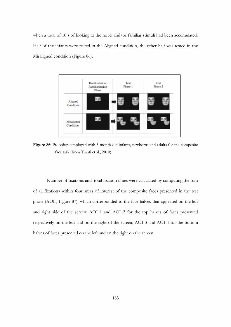
183
when a total of 10 s of looking at the novel and/or familiar stimuli had been accumulated.
Half of the infants were tested in the Aligned condition, the other half was tested in the
Misaligned condition (Figure 86).
Figure 86: Procedure employed with 3-month-old infants, newborns and adults for the composite
face task (from Turati et al., 2010).
Number of fixations and total fixation times were calculated by computing the sum
of all fixations within four areas of interest of the composite faces presented in the test
phase (AOIs, Figure 87), which corresponded to the face halves that appeared on the left
and right side of the screen: AOI 1 and AOI 2 for the top halves of faces presented
respectively on the left and on the right of the screen; AOI 3 and AOI 4 for the bottom
halves of faces presented on the left and on the right on the screen.
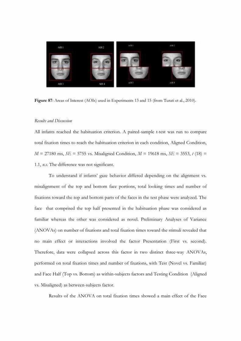
Figure 87: Areas of Interest (AOIs) used in Experiments 13 and 15 (from Turati et al., 2010).
Results and Discussion
All infants reached the habituation criterion. A paired-sample t-test was run to compare
total fixation times to reach the habituation criterion in each condition, Aligned Condition,
M = 27180 ms, SE = 5755 vs. Misaligned Condition, M = 19618 ms, SE = 3553, t (18) =
1.1, n.s. The difference was not significant.
To understand if infants’ gaze behavior differed depending on the alignment vs.
misalignment of the top and bottom face portions, total looking times and number of
fixations toward the top and bottom parts of the faces in the test phase were analyzed. The
face that comprised the top half presented in the habituation phase was considered as
familiar whereas the other was considered as novel. Preliminary Analyses of Variance
(ANOVAs) on number of fixations and total fixation times toward the stimuli revealed that
no main effect or interactions involved the factor Presentation (First vs. second).
Therefore, data were collapsed across this factor in two distinct three-way ANOVAs,
performed on total fixation times and number of fixations, with Test (Novel vs. Familiar)
and Face Half (Top vs. Bottom) as within-subjects factors and Testing Condition (Aligned
vs. Misaligned) as between-subjects factor.
Results of the ANOVA on total fixation times showed a main effect of the Face
185
Half factor, F (1, 18) = 82.5, p < .001, 2 = .82. Infants looked longer at the top face part
(M = 3382 ms, SE = 206) than at the bottom face part (M = 720 ms, SE = 159).
Furthermore, the main effect of Test indicated that the familiar face (M = 2322 ms, SE =
196) was fixated longer than the novel face (M = 1780 ms, SE = 110), F (1, 18) = 5.7, p <
.05, 2 = .24. This Test main effect could be explained in relation to the Test X Testing
Condition significant interaction, F (1, 18) = 4.6, p < .05, 2 = .20, which qualifies the main
effect (Figure 88).
Figure 88: Total fixation time at the novel and the familiar stimuli as a function of test condition
(Aligned vs Misaligned Condition) in Experiment 13.
Indeed, planned t-tests showed that, in the Misaligned test condition, infants looked longer
at the familiar (M = 5326 ms, SE = 645) than at the novel (M = 3273 ms, SE = 366)
composite stimulus, t(9) = 2.7, p < .05. On the contrary, in the Aligned test condition,
looking time toward the novel (M = 3847 ms, SE = 244) and the familiar (M = 3963 ms,
SE = 446) faces did not differ, t(9) = 0.24, n.s.
Results of the ANOVA on number of fixations showed a main effect of Face Half,

F (1, 18) = 88.6, p < .001, 2 = .83, and a significant Face Half X Testing Condition
interaction, F (1, 18) = 4.6, p < .05, 2 = .20, showing that the number of fixations infants
oriented to the top face part (M = 40, SE = 2) was greater than the number of fixations
toward the bottom face part (M = 11 ms, SE = 1), particularly in the Misaligned (M = 44,
SE = 3 vs. M = 9, SE = 3) as compared to the Aligned test condition (M = 35, SE = 1 vs.
M = 13, SE = 2).
According to previous evidence (Gallay et al., 2006; Turati et al., 2005), these
findings showed that, in both the aligned and the misaligned conditions, infants’ visual
exploration was focused on the top portion of the face, that comprised the eye region.
More interestingly, results revealed a discrepancy in infants’ looking times according to the
alignment or misalignment of the faces. Three-month-olds looked longer at the face with a
familiar top half when the top and the bottom face halves were misaligned, thus revealing
that they discriminated the two stimuli. Conversely, in the Aligned condition infants looked
equally long to the familiar and novel stimuli, indicating that they did not differentiate
between the two. This suggests that the bottom part of the composite faces affected
infants’ performance in both the Aligned and the Misaligned conditions. In the Aligned
condition, the novel bottom-face halves impaired infants’ discrimination of the two
composite faces. In the Misaligned condition, the addition of the bottom face half
determined a persistent and extensive re-exploration of the familiar top-face half. Indeed,
previous studies that tested infants’ ability to recognize a learned face over strong
modifications -such as partial occlusion, or rotation- obtained a familiarity preference
(Gava et al., 2008; Walton, Amstrong, & Bower, 1997; Sirois & Mareschal, 2002, 2004). In
the current experiment, infants in the Misaligned condition recognized the familiarity of the
top portion of the configuration, but also detected that a novel never-seen bottom face
part was added to the familiar part. This induced infants’ extensive re-exploration of the

187
whole partially familiar stimulus.
The alignment vs. misalignment of faces also affected the trajectories of infants’ eye
movements, as infants’ tendency to fixate the top- vs. the bottom-face halves was more
evident in the Misaligned condition, that is when the top part of the faces are disjoined
from the bottom parts. Thus, overall, evidence obtained employing an eye-movement
recording VPC procedure supports the existence of the composite effect in 3-month-old
infants.
Due to the presence from birth of the ability to process faces holistically, relaying
on holistic face processing (Leo & Simion, 2009), it appears interesting to investigate
whether holistic face processing could be observed employing the composite face paradigm
in newborns.
Experiment 14a
Method
Participants
Participants were 28 (14 females) healthy, full term newborns (mean age 45 hr), recruited at
the maternity ward of the Pediatric Clinic of the University of Padova. All of them were
middle-class infants and Caucasian. Nine additional newborns were tested, but were not
included in the final sample because one showed a strong position preference (i.e., he
looked to one direction more than 80% of the time) and 8 did not complete testing due to
fussiness. All of them met the screening criteria of normal delivery, a birth weight between
2460 and 4100 g, and an Apgar score between 8 and 10 at 5 minutes. Newborns were
tested only if they were awake and in an alert state, after parents gave their informed
consent.
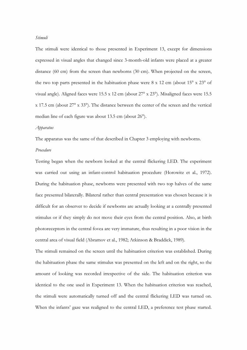
Stimuli
The stimuli were identical to those presented in Experiment 13, except for dimensions
expressed in visual angles that changed since 3-month-old infants were placed at a greater
distance (60 cm) from the screen than newborns (30 cm). When projected on the screen,
the two top parts presented in the habituation phase were 8 x 12 cm (about 15° x 23° of
visual angle). Aligned faces were 15.5 x 12 cm (about 27° x 23°). Misaligned faces were 15.5
x 17.5 cm (about 27° x 33°). The distance between the center of the screen and the vertical
median line of each figure was about 13.5 cm (about 26°).
Apparatus
The apparatus was the same of that described in Chapter 3 employing with newborns.
Procedure
Testing began when the newborn looked at the central flickering LED. The experiment
was carried out using an infant-control habituation procedure (Horowitz et al., 1972).
During the habituation phase, newborns were presented with two top halves of the same
face presented bilaterally. Bilateral rather than central presentation was chosen because it is
difficult for an observer to decide if newborns are actually looking at a centrally presented
stimulus or if they simply do not move their eyes from the central position. Also, at birth
photoreceptors in the central fovea are very immature, thus resulting in a poor vision in the
central area of visual field (Abramov et al., 1982; Atkinson & Braddick, 1989).
The stimuli remained on the screen until the habituation criterion was established. During
the habituation phase the same stimulus was presented on the left and on the right, so the
amount of looking was recorded irrespective of the side. The habituation criterion was
identical to the one used in Experiment 13. When the habituation criterion was reached,
the stimuli were automatically turned off and the central flickering LED was turned on.
When the infants’ gaze was realigned to the central LED, a preference test phase started.

189
Each infant was given two paired presentations of the stimuli. During each presentation,
newborns were simultaneously presented with a pair of composite faces. In one face of the
pair the top half was identical to that shown during habituation, in the other face the top
half was new. Both top halves were matched with the same bottom half of a third new
face. The two-paired stimuli were always shown in both left and right positions, the
position being reversed from presentation 1 to presentation 2. During the preference test
phase, the experimenter recorded the duration of infant’s fixations and the number of
fixations on each stimulus by pressing two different push buttons depending on whether
the infant looked at the right or the left position. Each presentation lasted until each
stimulus had been fixated at least once and a total of 20 s of looking had been accumulated.
The test presentations were kept longer in Experiment 14a (20 s of fixation time) than in
Experiment 13 (10 s of fixation time), to give newborns more time to process the stimuli
and eventually manifest a novelty preference. Half of the infants was tested in the Aligned
condition, the other half was tested in the Misaligned condition.
Results and Discussion
All newborns reached the habituation criterion. A paired-sample t-test was run to compare
total fixation times to reach the habituation criterion in each condition (Aligned Condition,
M = 65210 ms, SE = 10456 vs. Misaligned Condition, M = 74886 ms, SE = 11220). The
comparison did not reach statistical significance, t(27) = 1.2, n.s.
Preliminary ANOVAs on total fixation times and number of fixations toward the
stimuli revealed that no main effect or interactions involved the factor Presentation (First
vs. Second). Therefore, data were subsequently collapsed in two ANOVAs with Testing
Condition (Aligned vs Misaligned) as between-subjects factor and Test (Novel vs. Familiar)
as within-subjects factors, one for each dependent variable. The face stimulus that
comprised the top half presented in the habituation phase was considered as familiar.
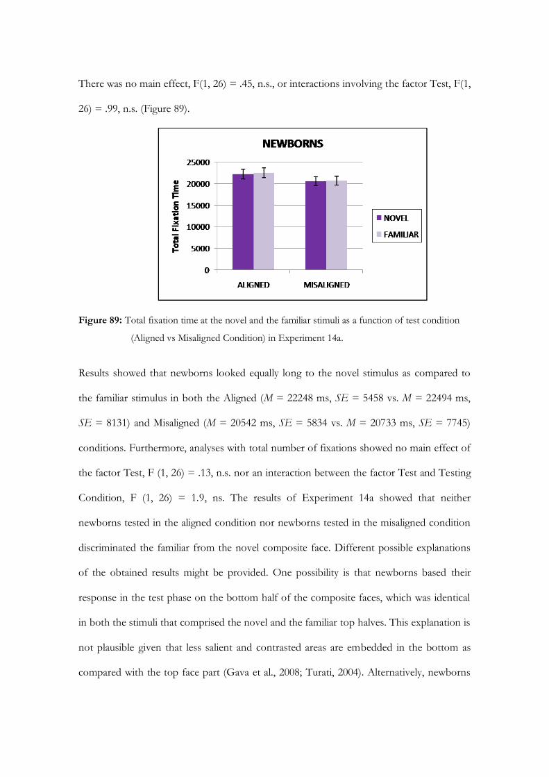
There was no main effect, F(1, 26) = .45, n.s., or interactions involving the factor Test, F(1,
26) = .99, n.s. (Figure 89).
Figure 89: Total fixation time at the novel and the familiar stimuli as a function of test condition
(Aligned vs Misaligned Condition) in Experiment 14a.
Results showed that newborns looked equally long to the novel stimulus as compared to
the familiar stimulus in both the Aligned (M = 22248 ms, SE = 5458 vs. M = 22494 ms,
SE = 8131) and Misaligned (M = 20542 ms, SE = 5834 vs. M = 20733 ms, SE = 7745)
conditions. Furthermore, analyses with total number of fixations showed no main effect of
the factor Test, F (1, 26) = .13, n.s. nor an interaction between the factor Test and Testing
Condition, F (1, 26) = 1.9, ns. The results of Experiment 14a showed that neither
newborns tested in the aligned condition nor newborns tested in the misaligned condition
discriminated the familiar from the novel composite face. Different possible explanations
of the obtained results might be provided. One possibility is that newborns based their
response in the test phase on the bottom half of the composite faces, which was identical
in both the stimuli that comprised the novel and the familiar top halves. This explanation is
not plausible given that less salient and contrasted areas are embedded in the bottom as
compared with the top face part (Gava et al., 2008; Turati, 2004). Alternatively, newborns

191
may have reacted to the significant perceptual change that occurred between the
habituation phase (in which only one top half of a face was presented) and the test phase
(in which two faces were presented, each comprising a novel bottom half) and showed a
persistent and comparable exploration of both the “familiar” and the “novel” composite
face. Finally, newborns might be unable to recognize a portion of a face -i.e., only the top
part rather than the full face-, given that first-order face information is absent and second-
order face information is poorly represented, the spatial relations between eyes and nose
and eyes and mouth being disrupted. The aim of Experiment 14b was to explore this issue
by presenting newborns with the top-face part (i.e., without the bottom-part of the face)
not only in the habituation phase, but also in the test phase.
Experiment 14b
Method
Participants
The final sample comprised 13 (8 females) healthy, full term newborns (mean age 43 hr),
recruited at the maternity ward of the Pediatric Clinic of the University of Padova. All of
them were middle-class infants and Caucasian. Four additional newborns were tested, but
were not included in the final sample because they did not complete testing due to
fussiness. All of them met the screening criteria of normal delivery, a birth weight between
2620 and 3850 g, and an Apgar score between 9 and 10 at 5 minutes. Newborns were
tested only if they were awake and in an alert state, after parents gave their informed
consent.
Stimuli
The stimuli were identical to those presented in Experiment 13 and 14a, except for the fact
that only the top halves of faces were presented both in the habituation and in test phases.
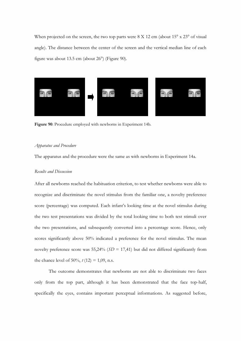
When projected on the screen, the two top parts were 8 X 12 cm (about 15° x 23° of visual
angle). The distance between the center of the screen and the vertical median line of each
figure was about 13.5 cm (about 26°) (Figure 90).
Figure 90: Procedure employed with newborns in Experiment 14b.
Apparatus and Procedure
The apparatus and the procedure were the same as with newborns in Experiment 14a.
Results and Discussion
After all newborns reached the habituation criterion, to test whether newborns were able to
recognize and discriminate the novel stimulus from the familiar one, a novelty preference
score (percentage) was computed. Each infant’s looking time at the novel stimulus during
the two test presentations was divided by the total looking time to both test stimuli over
the two presentations, and subsequently converted into a percentage score. Hence, only
scores significantly above 50% indicated a preference for the novel stimulus. The mean
novelty preference score was 55,24% (SD = 17,41) but did not differed significantly from
the chance level of 50%, t (12) = 1,09, n.s.
The outcome demonstrates that newborns are not able to discriminate two faces
only from the top part, although it has been demonstrated that the face top-half,
specifically the eyes, contains important perceptual informations. As suggested before,
193
newborns are not able to recognize a portion of a face -i.e., only the top part rather than
the full face-, given that first-order face information is absent and second-order face
information is poorly represented, the spatial relations between eyes and nose and eyes and
mouth being disrupted.
Experiment 15
To ground the results obtained in Experiments 13 and 14 in an adult model, Experiment
15 tested a group of adults in a similar VPC task. In adults, the active control of eye
fixations plays an important functional role in a wide variety of cognitive and perceptual
tasks (Henderson, 2006). Adults’ eye fixations during face perception and recognition are
predominantly directed to internal facial features, particularly the eyes, then nose and
mouth (Althoff & Cohen, 1999; Henderson, Falk, Minut, Dyer, & Mahadevan, 2001;
Stacey, Walker, & Underwood, 2005). Recently, a functional relationship between eye
movements and face perception has been demonstrated, in that a deficit in recognition
emerged when participants were required to keep their eyes fixated in one central position
(i.e., restricted viewing condition) rather than to move their eyes freely (i.e., free viewing
condition) (Henderson, Williams, & Falk, 2005). Finally, Williams and Henderson (2007)
found that the same features of a face were fixated regardless of whether the face was right
side up or upside down and interpreted their findings as indicating that the face inversion
effect is not the result of a different pattern of eye movements.
Through the recording of adults’ eye movement during a composite task,
Experiment 15 was aimed to test gaze behavior during holistic face processing. Despite
numerous studies on adults’ composite face illusion, only a single study recorded eye-
movements while adults performed a delayed matching task of top halves of faces aligned
or misaligned with bottom face halves (de Heering, Rossion, Turati, & Simion, 2008).
Results showed that participants fixated longer the top parts of aligned than misaligned
faces, suggesting the presence of a difference in eye-movements strategies between the
aligned and misaligned condition. However, manual reaction time data were strictly linked
to eye-recording data, as suggested by the high correlations between participants’ manual
response time and ocular fixation time in aligned and misaligned conditions. When the
effect of participants’ manual response time was controlled, considering the variable as a
covariate, the difference in mean fixation time on the top parts of aligned and misaligned
faces disappeared, suggesting that longer fixation time on the top parts of aligned versus
misaligned faces was due to the time devoted to respond manually at aligned vs. misaligned
faces, aligned faces being processed more slowly.
Considering this result, in the current experiment adults’ eye movements were
recorded in a free-viewing task that does not require a manual response. In parallel with the
experimental paradigm used with three-month-old and newborn infants in Experiments 13
and 14 and with studies that used the VPC task with adults (Richmond, Sowerby,
Colombo, & Hayne, 2004), participants were familiarized with the top half of a face in a
single fixed 15-sec presentation. In the subsequent two fixed 10-s presentation test phases a
pair of composite faces was shown in which one of the face top halves was identical to the
top half of the face presented during familiarization and the other top half was new. Half
of the participants was tested in the aligned condition, and the other half was tested in the
misaligned condition, so that alignment of the composite stimuli was varied between
subject as in Experiments 13 and 14. No instructions were given, except that participants
had to watch the images appearing on the computer screen moving their eyes freely.
195
Method
Participants
Twenty-two undergraduate middle-class, Caucasian students (17 females, M = 22.8 years)
from the Department of Psychology at the University of Padua (Italy) participated in the
study. Seven additional participants were tested, but excluded from the final sample due to
failure to reach criteria established for data analyses (their fixation time toward stimuli was
less than 70% of the total presentation time during both first and second test phases). All
of them had normal or corrected-to-normal vision.
Stimuli
The stimuli were identical to those used in Experiments 13 and 14a.
Apparatus and procedure
The apparatus and the procedure were the same as with 3-month-old infants in the
previous experiment, with one exception. Following the calibration phase, instead of the
habituation phase, participants were familiarized with a top half of a face projected in the
center of the screen for a fixed presentation period of 15 s. Half of the participants was
tested in the Aligned condition, while the other half in the Misaligned condition.
Participants were given no instructions except that they had to watch the images appearing
on the screen.
Results and Discussion
To compare total fixation times in the Aligned and Misaligned conditions during the
familiarization phase, a paired-sample t-test was run (Aligned Condition, M = 11773 ms,
SE = 762 vs. Misaligned Condition, M = 12709 ms, SE = 434), t(20) = 1.06, n.s. As
expected, the difference was not significant.
As with infants in Experiment 13, the aims of the statistical analyses performed on
the data were carried out to understand i.) Whether adults’ ability to discriminate the
familiar from the novel composite face varied as a function of the alignment or
misalignment of the face halves, and ii.) Whether the pattern of adults’ fixation durations to
the top and bottom halves of the composite faces varied between the two alignment
conditions.
The face stimulus that comprised the top half presented in the habituation phase
was considered familiar while the other was considered novel. Two distinct mixed-model
ANOVAs were performed on total fixation times and number of fixations with
Presentation (First vs. Second), Test (Novel vs. Familiar), and Face Half (Top vs. Bottom)
as within-subjects factors and Testing Condition (Aligned vs. Misaligned) as between-
subject factor.
Results of the ANOVA on total fixation times showed that adults looked longer at
the top face halves (M = 2869 ms, SE = 121) than at the bottom face halves (M = 1062
ms, SE = 107) as showed by the main effect of Face Half, F (1, 20) = 65.3, p < .001, 2 =
.76. More importantly, there was a significant interaction between the factors Test and
Testing Condition, F (1, 20) = 6.5, p < .05, 2 = .24 (Figure 91).
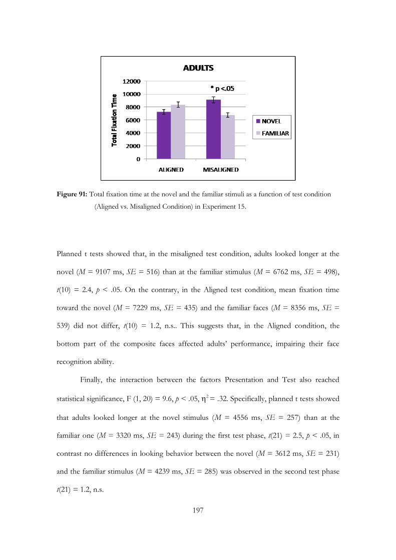
197
Figure 91: Total fixation time at the novel and the familiar stimuli as a function of test condition
(Aligned vs. Misaligned Condition) in Experiment 15.
Planned t tests showed that, in the misaligned test condition, adults looked longer at the
novel (M = 9107 ms, SE = 516) than at the familiar stimulus (M = 6762 ms, SE = 498),
t(10) = 2.4, p < .05. On the contrary, in the Aligned test condition, mean fixation time
toward the novel (M = 7229 ms, SE = 435) and the familiar faces (M = 8356 ms, SE =
539) did not differ, t(10) = 1.2, n.s.. This suggests that, in the Aligned condition, the
bottom part of the composite faces affected adults’ performance, impairing their face
recognition ability.
Finally, the interaction between the factors Presentation and Test also reached
statistical significance, F (1, 20) = 9.6, p < .05, 2 = .32. Specifically, planned t tests showed
that adults looked longer at the novel stimulus (M = 4556 ms, SE = 257) than at the
familiar one (M = 3320 ms, SE = 243) during the first test phase, t(21) = 2.5, p < .05, in
contrast no differences in looking behavior between the novel (M = 3612 ms, SE = 231)
and the familiar stimulus (M = 4239 ms, SE = 285) was observed in the second test phase
t(21) = 1.2, n.s.

The ANOVA on number of fixations revealed no main effect or interactions
involving the factor Presentation (First vs. second). Therefore, data were collapsed across
this factor in a subsequent three-way ANOVA with Test (Novel vs. Familiar) and Face
Half (Top vs. Bottom) as within-subjects factors and Testing Condition (Aligned vs.
Misaligned) as between-subjects factor.
Results indicated a main effect of Face Half, F (1, 20) = 90.3, p < .001, 2 = .82. The
number of fixations adults made to the top part of the face (M = 36, SE = 2) was greater
than the number of fixations toward the bottom part of the face (M = 13, SE = 1).
Furthermore, the significant interaction between Face Half and Testing Condition, F (1,
20) = 5.4, p < .05, 2 = .21, showed that the difference between fixations to the top part of
the face and the bottom part of the face was greater in the Aligned test condition (M = 39,
SE = 2 vs. M = 10, SE = 1) than in the Misaligned test condition (M = 34, SE = 2 vs. M =
17, SE = 3). Furthermore, there was a significant interaction between the factors Test and
Testing Condition, F (1, 20) = 6.2, p < .05, 2 = .24. Planned t tests showed that, in the
Misaligned test condition, the number of fixations toward the novel stimulus (M = 29, SE
= 2) was greater than the number of fixations toward the familiar stimulus (M = 22, SE =
2), t(10) = 2.9, p < .05. On the contrary, in the Aligned test condition, the number of
fixations toward the novel (M = 24, SE = 2) and the familiar faces (M = 25, SE = 1) did
not differ, t(10) = .61, n.s. Evidence from the present experiment appears in line with
previous findings showing that adults’ fixations toward faces are directed predominantly
toward the eye regions (e.g., Althoff & Cohen, 1999; Henderson et al. 2001, 2005).
Importantly, adults looked longer and directed a greater number of fixations toward the
face with a novel top half in the Misaligned condition. Conversely, in the Aligned condition
adults looked equally long and oriented their gaze approximately the same number of times
toward the novel and familiar stimuli. This suggests that, in the Aligned condition, the

199
bottom part of the composite faces affected adults’ performance, demonstrating the
presence of the composite face effect. Aligned vs. Misaligned stimulus conditions elicited
differential looking times and numbers of fixations for the novel and the familiar
composite face, providing evidence that eye movements may be affected by the encoding
of holistic face information.
Overall, the present work is consistent with the idea that eye movements during
face recognition are functional to face processing strategies (Henderson et al., 2005), but is
partially in contrast with the findings reported by de Heering et al. (2008) on the composite
effect and by Williams and Henderson (2007) on the inversion effect, according to which
eye movement patterns are not able to reflect holistic processing strategies. The
discrepancies between this study and previous work may be explained by at least three
major methodological differences. First, in the present study participants were not required
to provide a manual response. This may have avoided high correlations between
participants’ manual response time and ocular fixation time that could produce distortions
in the results related to eye movements, since longer manual reaction times are associated
with longer fixations. Second, with respect to the study by de Heering et al. (2008),
participants were not explicitly required to look at the top part of a face in order to
recognize it, but were allowed to freely move their eyes. According with de Heering et al.
(2008), it has been found that the top-halves of the composite faces in the test phase were
fixated longer than the bottom-halves, irrespective of whether the face-halves were aligned
or misaligned. However, differently from de Heering et al. (2008), in the current study the
Aligned and Misaligned conditions affected the amount of visual exploration of the novel
and the familiar composite faces and number of fixations directed toward the top and the
bottom composite face part. Third, differently from the study by Williams and Henderson
(2007), the use of the composite paradigm allowed us to split faces into only two critical
areas of interest (top face part and bottom face part) rather than in multiple areas related to
each face feature (left eye, right eye, nose, mouth, chin, forehead, etc...). This might have
produced a more adequate measure of holistic, rather than feature-based, face processing
strategies.
Conclusion
The main outcome of the present study is that for the first time a clear demonstration was
provided that young infants are capable of processing a face holistically, as an
undifferentiated unit in which face parts and their relations are glued together (Experiment
13). The present work extends previous findings that reported a measure of the composite
effect at 3 years of age (Macchi Cassia et al., 2009) and of the part-whole effect at 4 years of
age (Pellicano & Rhodes, 2003). Also, the present results extend previous findings obtained
by Cohen and colleagues, demonstrating that infants are not only capable of detecting
correlations between outer and inner face features (Cashon & Cohen, 2001, 2003), but also
between the sole inner features of a face.
Importantly, this study employed a version of the composite paradigm suitable for
infants that permits to compare directly the holistic face processing performance in infants,
older children and adults. Indeed, this paradigm was shown to be a potential measure of
the composite face effect also in adults (Experiment 15), demonstrating that eye
movements (duration and number of fixations) may be sensitive to holistic face processing
during the entire life span. The use of the same task throughout development, from early
infancy to adulthood, allows researchers to highlight both differences and similarities in the
visual behavior across age groups. First, results clearly show that the facial features that
attract both adults’ and infants’ ocular fixations are almost the same, in that at both ages a
clear tendency to explore the top rather than the bottom face part was found. Second,

201
although both age groups visually recognized the familiar stimulus in the misaligned but
not in the aligned condition, infants preferred looking at the face with the familiar top part
while adults tended to look longer at the face with the novel top part. Thus, adults did not
need to re-explore the stimulus that comprises the familiar top of the face and directly
focused attention on the face stimulus with a novel top. Overall, evidence from the current
study reveals that the tuning toward configural information appears very early in life,
although gradual experienced-based developmental processes will progressively refine early
configural abilities.


203
General Conclusion One central issue in cognitive developmental science is to understand how cognition grows
and change over time to reach an adult level of specialization. Determining the abilities
with which infants come equipped into the world, their mechanisms for acquiring
knowledge, and whether and how these abilities change as a function of development and
experience is a challenging issue. Face processing is an interesting topic of research in that
respect because faces form a special class of visual objects elaborated in adults by a specific
anatomical and functional face system (e.g., Kanwisher, 2000; Farah et al., 1998). Since
what determines this specialization and how this specialization emerges during
development still remain unknown, the purpose of my PhD dissertation was to study
cognitive specialization during early infancy through the investigation of the development
of infants’ abilities to process faces. In particular, my hypothesis was that the face
processing specificity is not present at birth, but emerges gradually from the interaction
between general constraints and attentional biases present in the first months of life and
the critical visual input provided by the specie-specific environment (Nelson, 2001, 2003;
Johnson, 1993).
In Study 1 (Chapter 3), using both the visual preference and visual habituation
techniques, a first series of experiments investigated the nature of face representation in
newborns and in 3-month-old infants. According to recent evidence showing that infants’
response to faces becomes more and more tuned to the face category over the first three-
months of life (Turati et al., 2005), collected data demonstrated that 3-month-old infants,
but not newborns, are sensitive to specific perceptual cues within a face, such as the correct
position and orientation of the eyes (Experiments 1, 2 and 3). Furthermore, results
obtained from Experiments 4, 5, 6 and 7 demonstrated that early facial representation is

not human-specific, corroborating the hypothesis that newborns come into the world with
a face representation that is sufficiently general as to bias newborns’ visual attention toward
multiple categories of faces (e.g., monkey faces vs. human faces), and that this face
representation, due to the visual experience that infants do in the specie-specific
environment, becomes more specific to human face during the first 3 months of life
(Nelson, 2001; Pascalis & Kelly, 2009).
Due to the social relevance of the face stimulus and due to the ability of 3-month-
old infants to form a specific representation of the human face, the aim of Study 2 (Chapter
4) was to investigate whether human face capture and maintain infants’ attention in
complex visual scenes. Specifically, using an eye-tracker system, adults’ and 6- and 3-
month-old infants’ visual search behavior was compared in a modified visual search task of
a target face among heterogeneous (e.g., various objects, Experiments 8, 9 and 10) and
homogeneous distractors (e.g., inverted faces, Experiments 11, 12). Collected data
demonstrated that a face among heterogeneous distractors captures and maintains adults’
and 6-month-old infants’ attention and that 3-month-old infants detect a target face only
when embedded among inverted faces (e.g., homogeneous distractors), corroborating
previous findings showing the face detection advantage in infants (Gliga et al., 2009).
Importantly, to detect a target face among other distractors, infants have to process
a face as a Gestalt, where the whole is more than the sum of its constituent parts (Tanaka
& Farah, 1993). This kind of face processing, called “holistic”, was investigated in
newborns, 3-month-old-infants, and adults through a modified version of the composite
face paradigm (Young et al., 1987) and the recording of eye movements in Study 3 (Chapter
5). The main outcome of the study was that the tuning toward holistic information appears
very early in life, although gradual experience-based developmental processes will
progressively refine early holistic processing abilities (Experiments 13, 14 and 15).
205
Taken together, the data obtained herein suggests that face specificity is not
prewired, but rather arises from general perceptual processes that, during development,
become progressively tuned to the human face, as a result of extensive experience with this
stimulus category. Overall, the data presented in this thesis are in line with an experience-
expectant perspective, that emphasizes the relevance of both general constraints of the
human visuo-perceptual system and exposure to certain experiences shortly after birth to
drive the system to become functionally specialized to process faces in the first month of
life. The three studies presented here highlighted the role of the visual experience as a
determinant factor for the specialization of the face processing system.
However, at present, little is known about the specific experiences that lead to
expert face processing or how early perceptual experience contributes to the specialization
of the neural structures underlying face processing (e.g., specifying exactly what kinds of
experiences are necessary, when these experiences need to occur, and lastly, for how long
they need to occur). This research will specifically require future research that
simultaneously combine the study of structural and functional changes in the brain during
development, employing both behavioral and neuropsychological tasks (e.g., NIRS and
ERP).
Finally, as suggested in the first chapter, within a neoconstructivistic theoretical
framework, developmental disorders can be understood through altered constraints that
push the developmental trajectory off its normal track to reach a different endstate
(Karmiloff-Smith, 1998; Thomas & Karmiloff-Smith, 2002). Thus, atypical development
can, like typical development, be characterized as an adaptation to multiple interacting
constraints, only that the constraints are different. These atypical constraints then lead to
different outcomes through the same processes of representation construction
(Westermann et al., 2007). In this vein, understanding the constraints that shape typical

development trajectory is an important first step to investigate which and how different
constraints shape atypical development.
207
References
Abramov, I., Gordon, J., Hendrickson, A., Hainline, L., Dobson, V., & LaBossiere, E.
(1982). The retina of the newborn human infant. Science, 217, 265-267.
Acerra, F., Burnod, Y., & de Schonen S. (2002). Modelling aspects of face processing in
early infancy. Developmental Science, 5, 98-117.
Adler, S. A., Gerhardstein, P., & Rovee-Collier, C. (1998). Levels of processing effects in
infant memory? Child Development, 69, 280-294.
Adler, S. A., Inslicht, S., Rovee-Collier, C., & Gerhardstein, P. C. (1998). Perceptual
asymmetry and memory retrieval in 3-month-old infants. Infant Behavior &
Development, 21, 253-272.
Adler, S. A., & Orprecio, J. (2006). The eyes have it: visual pop-out in infants and adults.
Developmental Science, 9, 189-206.
Adolphs, R. (1999). Social cognition and the human brain. Trends in Cognitive Science, 3, 469-
479.
Allison, T., Puce, A., & McCarthy, G. (2000). Social perception from visual cues: role of
the STS region. Trends in Cognitive Neuroscience, 4, 267-278.
Althoff, R. R., & Cohen, N. J. (1999). Eye-movement-based memory effect: A reprocessing
effect in face perception. Journal of Experimental Psychology: learning, Memory &
Cognition, 25, 997-1010.
Antell, S., Caron, A. J., & Myers, R. S. (1985). Perception of relational invariants by
newborns. Developmental Psychology, 21, 942-948.
Aslin, R. N., & Fiser, J. (2005). Methodological challenges for understanding cognitive
development in infants. Trends in Cognitive Sciences, 9, 92-98.
Atkinson, J., & Braddick, O. (1989). Development of basic visual functions. In A. Slater
and G. Bremner (Eds.). Infant Development. Erlbaum, UK.
Atkinson, J., Hood, B., Wattam-Bell, J., & Braddick, O. (1992). Changes in infants‟ ability to
switch visual attention in the first three months of life. Perception, 21, 643–653.
Baayen, R. H., Davidson, D. J., & Bates, D. M. (2008). Mixed-effcts modeling with crossed
random effects for subjects and items. Journal of Memory and Language, 59, 390-412.
Baillargeon, R., & Wang, S. (2002). Event categorization in infancy. Trends in Cognitive
Sciences, 6, 85-93.
Banks, M. S., & Ginsburg, A. P. (1985). Infant visual preferences: a review and new
theoretical treatment. Advances in Child Development and Behavior, 19, 207-245.
Banks, M. S., & Salapatek, P. (1981). Infant pattern vision: a new approach based on the
contrast sensitivity function. Journal of Experimental Child Psychology, 31, 1-45.
Bar-Haim, Y., Talee, Z., Lamy, D., & Hodes, R. M. (2006). Nature and nurture in own-race
face processing. Psychological Science, 17, 159-163.
Baron-Cohen, S. (1995). Mind blindness: an essay on autism and theory of mind.
Cambridge, MA: MIT Press.
Bartrip, J., Morton, J., & de Schonen, S. (2001). Responses to mother's face in 3-week to 5-
month-old infants. British Journal of Developmental Psychology, 19, 219-232.
Bates, D. M. (2007). Linear mixed model implementation in lme4. Manuscript, University
of Wisconsin -Madison, January, 2007.
Bates, E. A., & Elman, J. L., (1993). Connectionism and the study of change. In M.
Johnson (Ed.), Brain development and cognition: A reader (pp. 420-440). Oxford:
Blackwell Publishers.
209
Batki, A., Baron-Cohen, S., Wheelwright, S., Connellan, J., & Ahluwalia, J. (2000). Is there
an innate module? Evidence from human neonates. Infant Behavior & Development,
23, 223–229.
Bailey, A. J., Braeutigam, S., Jousmaki, V., & Swithenby, S. S. (2005). Abnormal activation
of face processing systems at early and intermediate latency in individuals with
autism spectrum disorder: a magnetoencephalographic study. European Journal of
Neuroscience, 21, 2575–2585.
Bertin, E., & Bhatt, R. S. (2004). The Thatcher illusion and face processing in infancy.
Developmental Science, 7, 431-436.
Bhatt, R. S. Bertin, E., Hayden, A., & Reed, A. (2005). Face processing in infancy:
developmental changes in the use of different kinds of relational information. Child
Development, 76, 169-181.
Bindemann, M., Burton, M., Hooge, I. T. C., Jenkins, R., & de Haan, E. H. F. (2005). Faces
retain attention. Psychonomic Bulletin & Review, 12, 1048-1053.
Brothers, L. (1996). Brain mechanisms of social cognition. Journal of Psychopharmacology, 10,
2-8.
Brown, V. Huey, D., & Findlay, J. M. (1997) Face detection in peripheral vision: do faces
pop out? Perception, 26, 1555-1570.
Bruce, V., & Young, A. (1986). Understanding face recognition. British Journal of Psychology,
77, 305-327.
Bulf, H., Valenza, E., & Simion, F. (2009). Searching for an illusory figure: A comparison
between infants and adults. Perception, 38, 1313-1327.
Bukach, C. M., Gauthier, I., & Tarr, M. J. (2006). Beyond faces and modularity: the power
of an expertise framework. Trends in Cognitive Sciences, 10, 159-166.
Burton, A. M., Bruce, V., & Johnston, R. A., (1990). Understanding face recognition with
an interactive activation model. British Journal of Psychology, 81, 361-380.
Bushnell, I.W.R., Sai, F., & Mullin, J.T. (1989). Neonatal recognition of the mother's face.
British Journal of Developmental Psychology, 7, 3-15.
Calder, A. J., & Young, A. W. (2005). Understanding the recognition of facial identity and
facial expression. Nature Reviews Neuroscience, 6, 641-651.
Calvo, M. G. & Nummenmaa, L. (2008). Detection of emotional faces: salient physical
features guide effective visual search. Journal of Experimental Psychology: General, 137,
471-494.
Carey, S., & Diamond, R. (1994). Are faces perceived as configurations more by adults than
by children? Visual Cognition, 1, 253-274.
Cashon, C. H., & Cohen, L. B. (2001). Do 7-month-old infants process independent
features or facial configurations? Infant and Child Development, 10, 83-92.
Cashon, C. H. & Cohen, L. (2003). The construction, deconstruction, and reconstruction
of infant face perception. In O. Pascalis & A. Slater (Eds.), The development of face
processing in infancy and early childhood: Current perspectives (pp. 13–25). New York: Nova
Science Publishers.
Chao, L. L., Haxby, J. V., & Martin, A. (1999). Attribute-based substrates in temporal
cortex for perceiving and knowing about objects. Nature Neuroscience, 2, 913-919.
Cheour, M., Ceponiene, R., Lehtokoski, A., Luuk, A., Allik, J., Alho, K., et al. (1998).
Development of language-specific phoneme representations in the infant brain.
Nature neuroscience, 1, 351-353.
Chomsky, N. (1988). Language and the problems of knowledge: The Managua lectures. Cambridge,
MA: MIT Press.

211
Cohen, L. B. (1972). Attention-getting and attention-holding processes of infant visual
preferences. Child Development, 43, 869-879.
Cohen, J. (1988). Statistical power analysis for the behavioral sciences (2nd Ed.). Hillsdale,
NJ: Erlbaum.
Collishaw, S. M., & Hole, G. J. (2000). Featural and configurational processes in the
recognition of faces of different familiarity. Perception, 29, 893-909.
Colombo, J., Ryther, J. S., Frick, J. E., & Gifford, J. G. (1995). Visual pop-out in infants:
evidence for preattentive search in 3- and 4-month-olds. Psychonomic Bulletin &
Review, 2, 266-268.
Costen, N. P., Ellis, H. D., & Craw, I. (1994). Masking of faces by facial and non-facial
stimuli. Visual Cognition, 1, 227-251.
Darwin, C. (1859). On the origin of the species by means of a natural selection, or,
preservation of favoured races in the struggle for life. London: Murray.
de Haan, M. (2001). The neuropsychology of face processing during infancy and
childhood. In C. A. Nelson & M. Luciana, (Eds). Handbook of Developmental Cognitive
Neuroscience (pp. 381-398). Cambridge, MA: MIT Press.
de Haan, M., & Halit, H.(2001). Neural bases and development of face recognition during
infancy. In A. Kalverboer, & A. Gramsbergen, (Eds.). Handbook of Brain and
Behaviour in Human Development (pp. 921–938). Kluwer Academic, Dordrecht.
de Haan, M., Johnson, M. H., Maurer, D., & Perrett, D. (2001). Recognition of individual
faces and average face prototypes by 1- and 3-month-old infants. Cognitive
Development, 1, 659-678.
de Heering, A., Houthuys, S., & Rossion, B. (2007). Holistic face processing is mature at 4
years of age: evidence from the composite face effect. Journal of Experimental Child
Psychology, 96, 57-70.
de Heering, A., Rossion, B., Turati, C., & Simion, F. (2008). Holistic face processing can be
independent of gaze behaviour: Evidence from the composite face illusion. Journal
of Neuropsychology, 2, 183-195.
De Renzi, E. (1986). Slowly progressive visual agnosia or apraxia without dementia. Cortex,
22, 171-180.
Deruelle, C., & de Schonen, S. (1998). Do the right and left hemispheres attend to the
same visuospatial information within a face in infancy? Developmental Neuropsychology,
14, 535-554.
de Schonen, S. (2002). Epigenesis of the cognitive brain: a task for the 21st Century. In L.
Backman and C. von Hofsten (Eds.), Psychology at the turn of the millenium
Hove, UK: Psychology Press, pp. 55–88.
de Schonen, S., & Mathivet, E. (1989). First come first served. A scenario about
development of hemispheric specialization in face recognition during infancy.
European Bulletin of Cognitive Psychology, 9, 3–44.
Desimone, R. (1991). Face-selective cells in the temporal cortex of monkeys. Journal of
Cognitive Neuroscience, 3, 1-8.
Diamond, R., & Carey, S. (1986). Why faces are and are not special: an effect of expertise.
Journal of Experimental Psychology: General, 115, 107-117.
Di Giorgio, E., Leo, I., Pascalis, O., & Simion, F. (submitted). Is early facial representation
human-specific? Developmental Psychology.
213
Di Giorgio, E., Leo, I., & Simion, F. (submitted). The role of the low-level variables in
newborns‟ eyes perception. Visual Cognition.
Duncan, J., & Humphreys, G. W. (1989). Visual search and stimulus similarity. Psychological
Review, 96, 433-458.
Ekman, P., & Friesen, W. V. (1982). Felt, false, and miserable smiles. Journal of Nonverbal
Behavior, 6, 238-252.
Ellis, A. W., Burton, A. M., Young, A., & Flude, B. M. (1997). Repetition priming between
parts and wholes: tests of a computational model of familiar faced recognition.
British Journal of Psychology, 88, 579-608.
Elman, J. L., Bates, E. A., Johnson, M. H., Karmiloff-Smith, A., Parisi, D., & Plunkett, K.
(1996). Rethinking innateness. A connectionist perspective on development.
Cambridge, MA: The MIT Press.
Epstein, R., & Kanwisher, N. (1998). A cortical representation of the local environment.
Nature, 392, 598-601.
Easterbrook, M. A., Kisilevsky, B. S., Hains, S. M. J., &Muir, D. W. (1999). Faceness or
complexity: evidence from newborn visual tracking of facelike stimuli. Infant
Behavior & Development, 22, 17-35.
Farah, M. J. (1996). Is face recognition special? Evidence from neuropsychology.
Behavioural Brain Research, 76, 181-189.
Farah, M. J., Rabinowitz, C., Quinn, G. E., & Liu, G. T. (2000). Early commitment of
neural substrates for face recognition. Cognitive Neuropsychology, 17, 117-123.
Farah, M. J., Wilson, K. D., Drain, M., & Tanaka, J. N. (1998). Psychological Review, 105, 482-
498.
Faraway, J. J. (2006). Extending the Linear Model with R: generalized linear, mixed effects
and nonparametric regression models. CRC Press.
Farroni, T., Csibra, G., Simion, F., & Johnson, M. H. (2002). Eye contact detection in
humans from birth. Proceedings of the National Academy of Sciences of the United States of
America, 99, 9602-9605.
Farroni, T. Valenza, E., Simion, F., & Umiltà, C. (2000). Configural processing at birth:
evidence for perceptual organization. Perception, 29, 355-372.
Findlay, J. M. (1997). Saccade target selection in visual search. Vision Research, 37, 617-631.
Fletcher-Watson, S., Findlay, J. M., Leekam, S. R., & Benson, V. (2008). Rapid detection of
person information in a naturalistic scene. Perception, 37, 571-583.
Fodor, J. (1983). The modularity of mind. An essay on faculty psychology. Cambridge, MA: MIT
Press.
Frank, M. C., Vul, E., & Johnson, S. P. (2009). Development of infants‟ attention to face
during the first year. Cognition, 210, 160-170.
Freire, A., Lee, K., & Symons, L. A. (2000). The face-inversion effect as a deficit in the
encoding of configural information: direct evidence. Perception, 29, 159-170.
Galton, F. (1883). Inquiries into human faculty and its development. London: Macmillan.
Gauthier, I., & Logothetis, N. K. (2000). Is face recognition unique after all? Cognitive
Neuropsychology, 17, 125-142.
Gauthier, I., & Nelson, C. A. (2001). The development of face expertise. Current Opinion in
Neurobiology, 11, 219-224.
215
Gauthier, I., Skudlarski, P., Gore, J. C., & Anderson, A. W. (2000). Expertise for cars and
birds recruits brain areas involved in face recognition. Nature Neuroscience, 3, 191-
197.
Gauthier, I., & Tarr, M. J. (1997). Becoming a greeble expert: exploring mechanisms for
face recognition. Vision Research, 37, 1673-1682.
Gauthier, I., & Tarr, M. J., & Anderson, A. W., Skudlarski, P., & Gore, J. C. (1999).
Activation of the middle fusiform “face area” increases with experience in
recognizing novel objects. Nature Neuroscience, 2, 568-573.
Gauthier, I., Tarr, M. J., Moylan, J., Skudlarski, P., Gore, J. C., & Anderson, A. W. (2000).
The fusiform face area is part of a network that processes faces at the individual
level. Journal of Cognitive Neuroscience, 12, 495-504.
Gauthier, I., Williams, P., Tarr, M. J., & Tanaka, J. (1998). Training greeble‟ experts: a
framework for studying expert object recognition processes. Vision Research, 38,
2401-2428.
Gava, L., Valenza, E., Turati, C., & de Schonen, S. (2008). Effect of partial occlusion on
newborns' face preference and recognition. Developmental Science, 11, 563-574.
Gallay, M., Baudouin, J. Y., Durand, K., Lemoine, C., & Lécuyer, R. (2006). Qualitative
differences in the exploration of upright and upside-down faces in four-mounth-old
infants: An eye-movement study. Child Development, 77, 984-996.
Gelman, R. (1990). First principles organize attention to and learning about relevant data:
number and animate-inanimate distinction as examples. Cognitive Science, 1, 79-106.
Gliga, T. & Csibra, G. (2007). Seeing the face through the eyes: a developmental
perspective on face expertise. In C. von Hofsten & K. Rosander (Eds.), Progress in
Brain Research, 164 (pp. 323-339). Elsevier.
Gliga, T., Elsabbagh, M., Andravizou, A., & Johnson, M. H. (2009). Face attract infants‟
attention in complex displays. Infancy, 14, 550-562.
Gobbini, M. I., & Haxby, J. V. (2007). Neural systems for recognition of familiar faces.
Neuropsychologia, 45, 32-41.
Goldstein, H. (2005). Multilevel models. In P. Armitage, & T. Colton (Eds.) Encyclopedia
of Biostatistics. Wiley.
Goren, C. C., Sarty, M., & Wu, P. Y. K. (1975). Visual following and pattern discrimination
of face-like stimuli by newborn infants. Pediatrics, 56, 544–549.
Gottlieb, G. (1992). Individual development and evolution: The genesis of novel behavior. Oxford,
Oxford University Press.
Gottlieb, G. (2007). Probabilistic epigenesis. Developmental Science, 10, 1-11.
Greenough, W. T., & Black, J. E. (1992). Induction of brain structure by experience:
Substrates for cognitive development. In M. Gunnar and C. A. Nelson (Eds.),
Behavioral developmental neuroscience. Vol. 24. Minnesota symposia on child psychology (pp. 35-
52). Hillsdale, NJ: Erlbaum.
Greenough, W. T., Black, J. E., & Wallace, C. S. (1987). Experience and brain development.
Child Development, 58, 539-559.
Gross, C. G., Roche-Miranda, C. E., & Bender, D. B. (1972). Visual properties of neurons
in infero-temporal cortex of the macaque. Journal of Neuropsychology, 35, 96-111.
Hainline, L., (1978). Developmental changes in visual scanning of face and nonface
patterns by infants. Journal of Experimental Child Psychology, 25, 90-115.
217
Halit, H., de Haan, M., & Johnson, M.H. (2003). Cortical specialization for face processing:
Face-sensitive event-related potential components in 3- and 12-month-old infants.
Neuroimagine, 19, 1180-1193.
Hancock, P. J. B., Bruce, V., & Burton, A. M. (2000). Recognition of unfamiliar faces. Trends
in Cognitive Sciences, 4, 330-337.
Hansen, C. H., & Hansen, R. D. (1988). Finding a face in the crowd: an angry superiority
effect. Journal of Personality and Social Psychology, 54, 917-924.
Hasselmo, M. E., Rolls, E. T., & Baylis, G. C. (1989). The role of expression and identity in
the face-selective responses of neurons in the temporal visual cortex of the
monkey. Behavioural Brain Research, 32, 203-218.
Haxby, J. V., Hoffmann, E. A., & Gobbini, M. I. (2000). The distributed human neural
system for face perception. Trends in Cognitive Sciences, 4, 223-233.
Haxby, J. V., Hoffmann, E. A., & Gobbini, M. I. (2002). Human neural system for face
recognition and social communication. Biological Psychiatry, 51, 59-67.
Haxby, J. V., Ungerleider, L. G., Clark, V. P., Schouten, J. L., Hoffmann, E. A., & Martin,
A. (1999). The effect of face inversion on activity in human neural systems for face
and object perception. Neuron, 22, 189-199.
Hayden, A., Bhatt, R. S., Reed, A., Corby, C. R., Joseph, J. E. (2007). The development of
expert face processing: are infants sensitive to normal differences in second-order
relational information? Journal of Experimental Child Psychology, 7, 85–98.
Henderson, J. M. (2006). Eye movements. In C.Senior, T. Russell, & M. Gazzaniga (Eds.),
Methods in mind (pp. 171-191). Cambridge, MA: MIT Press.
Henderson, J. M., Falk, R. J., Minut, S., Dyer, F. C., & Mahadevan, S. (2001). Gaze control
for face learning and recognition by humans and machines. In T. Shipley & P.
Kellman (Eds.), From fragments to objects: segmentation processes in vision (pp. 463-482)
Amsterdam: Elsevier.
Henderson, J. H., Williams C. C., & Falk, R. (2005). Eye movements are functional during
face learning. Memory & Cognition, 33, 98-106.
Heron-Delaney, M., Wirth, S., & Pascalis, O. (in press). Infants‟ knowledge of their own
species.
Herschler, O. & Hochstein, S. (2005). At first sight: a high-level pop-out effect for faces.
Vision Research, 45, 1707-1724.
Heywood, S., & Churcher, J. (1980). Structure of the visual array and saccadic latency:
implications for oculomotor control. Quarterly Journal of Experimental Psychology, 32,
335-341.
Hoffmann, E. A., & Haxby, J. V. (2000). Distinct representations of eye gaze and identity
in the distributed human neural system for face perception. Nature Neuroscience, 3,
80-84.
Hole, G. (1994). Configurational factors in the perception of unfamiliar faces. Perception, 23,
65-74.
Hole, G. J., George, P. A., & Dunsmore, V. (1999). Evidence for holistic processing of
faces viewed as photographic negatives. Perception, 28, 341-359.
Horowitz, F. D., Paden, L., Bhama, K., & Self, P. (1972). An infant-control procedure for
studying infant visual fixation. Developmental Psychology, 7, 90.
219
Humphreys, G. W., Hodsoll, J., & Campbell, C. (2005). Attending but not seeing: the other
race effect in face and person perception studied through change blindness. Visual
Cognition, 12, 249-262.
Ishai, A., Ungerleider, L. G., Martin, A., Schouten, J. L., & Haxby, J. V. (1999). Distributed
representation of objects in the human ventral visual pathway. Proceedings of the
National Academy of Sciences of the United States of America, 96, 9379-9384.
Johnson, M. H. (1993). Constraints on cortical plasticity. In M. J. Johnson (Eds.). Brain
Development and Cognition. A reader (pp. 703-721). Cambridge, MA: Blackwell.
Johnson, M. H. (1997). Developmental cognitive neuroscience. Blackwell Publisher.
Johnson, M. H. (2000). Functional brain development in infants: Elements of an
interactive specialization framework. Child Development, 71, 75 – 81.
Johnson, M. H. (2001). Functional brain development in humans. Nature Reviews
Neuroscience, 2, 475-483.
Johnson, M. H. (2002). Neural mechanisms of cognitive development in infancy. In: J.L.
McClelland & R. M. Thompson (Eds.), International Encyclopedia of the Social and
Behavioral Sciences (pp. 2103-2108). Elsevier Science Ltd.
Johnson, M. H. (2005). Subcortical face processing. Nature Reviews Neuroscience, 6, 766–773.
Johnson, M. H. (2006). Biological Motion: A perceptual life detector? Current Biology, 16,
R376-R377.
Johnson, M. H. (2007). The social brain in infancy: A developmental cognitive
neuroscience approach. In D. Coch, K.W, Fischer, & G. Dawson (Eds.), Human
Behavior, Learning and the Developing Brain: Typical Development.(pp. 115-137). New
York: Guilford Press.
Johnson, M. H., Dziurawiec, S., Ellis, H., & Morton, J. (1991). Newborns‟ preferential
tracking of face-like stimuli and its subsequent decline. Cognition, 40, 1-19.
Johnson, M.H., Grossman, T., & Farroni, T. (2008). The social cognitive neuroscience of
infancy: Illuminating the early development of social brain functions. Advances in
Child Development and Behavior, 36, 331-372.
Johnson, M. H., & Morton, J. (1991). Biology and cognitive development: the case of face
recognition. Oxford, UK: Blackwell.
Kanwisher, N. (2000). Domain specificity in face perception. Nature Neuroscience, 3, 759-
763.
Kanwisher, N., McDermott, J., & Chun, M. M. (1997). The fusiform face area: a module in
extrastriate cortex specialized for face perception. The Journal of Neuroscience, 17,
4302-4311.
Kanwisher, N., & Moscovitch, M. (2000). The cognitive neuroscience of face processing:
an introduction. Cognitive Neuropsychology, 17, 1-11.
Kanwisher, N., Tong, F., & Nakayama, K. (1998). The effect of face inversion on the
human fusiform face area. Cognition, 68, B1-B11.
Kanwisher, N., & Yovel, G. (2006). The fusiform face area: a cortical region specialized for
the perception of faces. Philosophical Transactions of the Royal Society B: Biological Sciences,
361, 2109-2128.
Karmiloff-Smith, A. (1992). Beyond modularity: a developmental perspective on cognitive
science. Cambridge, MA: MIT Press.
Karmiloff-Smith, A. (1994). Precis of beyond modularity: a developmental perspective on
cognitive science. Behavioral and Brain Sciences, 17, 693-707.
221
Karmiloff-Smith, A. (1998). Developmental itself is the key to understanding
developmental disorders. Trends in Cognitive Sciences, 2, 389-398.
Kellman, P. J. (1993). Kinematic foundations of infant visual perception. In C. Granrud,
Visual perception and cognition in infancy (pp. 121-173). Hillsdale, NJ: Erlbaum.
Kelly, D. J., Slater, A. M., Lee, K., Gibson, A., Smith, M., Ge, L., & Pascalis, O. (2005).
Three-month-olds, but not newborns, prefer own-race faces. Developmental Science, 8,
F31-F36.
Kleiner, K. A. (1987). Amplitude and phase spectra as indices of infants‟ pattern
preferences. Infant behavior and development, 10, 49-59.
Kobayashi, H., & Kohshima, S. (1997). Unique morphology of the human eye. Nature, 387,
767-768.
Kuehn, S. M., & Jolicoeur, P. (1994). Impact of quality of the image, orientation, and
similarity of the stimuli on visual search for faces. Perception, 23, 95-122.
Kuhl, P. K., Williams, K. A., Lacerda, F., Stevens, K. N., & Lindblom, B. (1992). Linguistic
experience alters phonetic perception in infants by 6 months of age. Science, 255,
606-608.
Langlois, J. H., & Roggman, L. A. (1990). Attractive faces are only average. Psychological
Science, 1, 115-121.
Langton, S. R. H. (2000). The mutual influence of gaze and head orientation in the analyses
of social attention direction. The Quarterly Journal of Experimental Psychology A, 3, 825-
845.
Langton, S. R. H., Law, A. S., Burton, M., & Schweinberger, S. R. (2008). Attention capture
by face. Cognition, 107, 330-342.
Leder, H., & Bruce, V. (1998). Local and relational aspects of face distinctiveness. Quarterly
Journal of Experimental Psychology, 51A, 449-473.
Leder, H., & Bruce, V. (2001). When inverted faces are recognised: the role of configural
information in face recognition. Quarterly Journal of Experimental Psychology: Human
Perception and Performance, 22, 904-915.
Leder, H., Candrian, G., Huber, O., & Bruce, V. (2001). Configural features in the context of
upright and inverted faces. Perception, 30, 73-83.
Le Grand, R., Mondloch, C. J., Maurer, D., & Brent, H. P. (2003). Expert face processing
requires visual input to the right hemisphere during infancy. Nature neuroscience, 6,
1108-1112.
Leo, I., & Simion, F. (2009a). Newborns' Mooney face perception. Infancy, 14, 641-653.
Leo, I., & Simion, F. (2009b). Face processing at birth: a Thatcher illusion study.
Developmental Science, 12, 492-498.
LeRoy Conel., J. (1939). The postnatal development of the human cerebral cortex: vol. 1.
Harvard University Press.
Lewkowicz, D. J., & Ghazanfar, A. A. (2006). The decline of cross-species intersensory
perception in human infants. Proceedings of The National Academy of Sciences of the
United States of America, 103, 6771-6774.
Lewis, M. B., & Edmonds, A. J. (2005). Searching for faces in scrambled scenes. Visual
Cognition, 12, 1309-1336.
Macchi Cassia, V., Kuefner, D., Westerlund, A., & Nelson, C. A. (2006). A behavioral and
ERP investigation of 3-month-olds‟ face preferences. Neuropsychologia, 44, 2113-
2125.
223
Macchi Cassia, V., Picozzi, M., Kuefner, D., Bricolo, & Turati, C. (2009). Holistic
processing for faces and cars in preschool-aged children and adults: evidence from
the composite effect. Developmental Science, 12, 236-248.
Macchi Cassia, V., Simion, F., Milani, I., & Umilta`, C. A. (2002). Dominance of global
visual properties at birth. Journal of Experimental Psychology: General, 4, 398-411.
Macchi Cassia, V., Turati, C., & Simion, F. (2004). Can a non-specific bias toward top-heavy
patterns explain newborns‟ face preference? Psychological Science, 15, 379-383.
Macchi Cassia, V., Valenza, E., Simion, F, Leo, I. (2008). Congruency as a nonspecific
perceptual property contributing to newborns‟ face preference. Child Development,
79, 807-820.
Malcom, G. L., Lanyon, L. J., Fugard, A. J. B., & Barton, J. J. S. (2008). Scan patterns
during the processing of facial expression versus identity: an exploration of task-
driven and stimulus-driven effects. Journal of Vision, 8, 1-9.
Mareschal., D., Johnson, M. H., Sirois, S., Spartling, M. W., Thomas, M. S. C., &
Westermann, G. (2007). Neuroconstructivism: How the brain constructs cognition. Oxford:
Oxford University Press.
Mareschal., D., Sirois, S., Westermann, G., & Johnson, M. H. (2007). Neuroconstructivism vol
II: Perspectives and prospects. Oxford: Oxford University Press.
Maurer, D. (1985). Infants‟ perception of facedness. In T. N. Field & N. Fox (Eds.), Social
perception in infants (pp. 73-100). Norwood, NJ: Ablex.
Maurer, D., Le Grand, R., & Mondloch, C. J. (2002). The many faces of configural
processing. Trends in Cognitive Sciences, 6, 255-260.
McKone, E., Kanwisher, N., & Duchaine, B. C. (2007). Can generic expertise explain
special processing for faces? Trends in Cognitive Sciences, 11, 8-15.
McNeil, J. E., & Warrington, E. K. (1993). Prosopagnosia: a face-specific disorder. Quarterly
Journal of Experimental Psychology A, 46, 1-10.
McSorley E. & Findlay J. M. (2003). Saccade target selection in visual search: accuracy
improves when more distractors are present. Journal of Vision, 3, 877-892.
Meissner, C. A., & Brigham, J.C. (2001). Thirty years of investigating the own-race bias
memory for faces: a meta-analytic review. Psychology, Public Policy, and Law, 7, 3–35.
Meltzoff, A. N., & Moore, M. K. (1977). Imitation of facial and manual gestures by human
neonates. Science, 198, 75–78.
Mondloch, C. J., Le Grand, R., & Maurer, D. (2002). Configural face processing develops
more slowly than featural face processing. Perception, 31, 553-566.
Mondloch, C. J., Pathman, T., Maurer, D., Le Grand, R., & de Schonen, S. (2007). The
composite effect in six-year-old children: evidence for adult-like holistic face
processing. Visual Cognition, 15, 564-577.
Morton, J., & Johnson, M. H. (1991). CONSPEC and CONLERN: A two-process theory
of infant face recognition. Psychological Review, 98, 164-181.
Moscovitch, M., Winocur, G., & Behrmann, M. (1997). What is special in face recognition?
Nineteen experiments on a person with visual object agnosia and dyslexia but
normal face recognition. Journal of Cognitive Neuroscience, 9, 555-604.
Motter, B. C., & Belky, E. J. (1998). The guidance of eye movements during active visual
search. Vision Research, 38, 1805-1818.
Nahm, F. D., Perret, A., Amaral, D. G., & Albright, T. D. (1997). How do monkeys look at
faces. Journal of Cognitive Neuroscience, 9, 611-623.
Nelson, C. A. (2001). The development and neural bases of face recognition. Infant and
Child Development, 10, 3-18.
225
Nelson, C. A. (2003). The developmental of face recognition reflects an experience-
expectant and experience-dependent process. In O. Pascalis and A. Slater, (Eds.)
The development of face processing in infancy and early childhood: Current Perspectives (pp. 79-
88). Nova Science Publishers, New York.
Nelson, C. A., & Luciana, M. (Eds.) (2001). Handbook of developmental cognitive neuroscience.
Cambridge, MA: MIT Press.
Nothdurft, H. C. (1993). Faces and facila expressions do not pop out. Perception, 22, 1287-
1298.
Ohman, A., Lundqvist, D., & Esteves, F. (2001). The face in the crowd revisited: a threat
advantage with schematic stimuli. Journal of Personality and Social Psychology, 80, 381-
396.
Palermo, R., & Rhodes, G. (2003). Change detection in the flicker paradigm: Do faces have
an advantage? Visual Cognition, 10, 683-713.
Palermo, R., & Rhodes, G. (2007). Are you always on my mind? A review of how face
perception and attention interact. Neuropsychologia, 45, 75-92.
Palmer, S. E. (1991). Goodness, gestalt, groups, and Garner: Local symmetry subgroups as
a theory of figural goodness. In G. R. Lockhead and J. R. Pomerantz (Eds.), The
perception of structure: Essays in honor of Wendell R. Garner (pp. 23-29). American
Psychological Association, Washington, DC.
Pascalis, O., de Haan, M., & Nelson, C. A. (2002). Is face processing specie-specific during
the first year of life? Science, 296, 1321-1323.
Pascalis, O., de Haan, M., Nelson, C. A., & de Schonen, S. (1998). Long-term recognition
memory for faces assessed by visual paired comparison in 3- and 6-month-old
infants. Journal of Experimental Psychology: Learning, Memory, and Cognition, 24, 249-260.
Pascalis, O., & de Schonen, S. (1994). Recognition memory in 3-4-day-old human infants.
NeuroReport, 5, 1721-1724.
Pascalis, O., & Kelly, D. J. (2009). The origins of face processing in humans. Perspectives on
Psychological Science, 4, 200-209.
Pascalis, O., Scott, L. S., Kelly, D. J., Shannon, R. W., Nicholson, E., Coleman, M., &
Nelson, C. A. (2005). Plasticity of face processing in infancy. Proceedings of the
National Academy of Sciences of the United States of America,12, 5297–5300.
Pellicano E., & Rhodes, R. (2003). Holistic processing of faces in preschool children and
adults. Psychological Science, 14, 618-622.
Perret, D. I., & Mistlin, A. J. (1990). Perception of facial attributes. In W. C. Stebbins and
M. A. Berkley (Eds.), Comparative perception: vol. II. Complex signal (pp.187-215). New
York: John Wiley.
Perrett, D. I., Smith, P. A., Potter, D. D., Mistlin, A. J., Head, A. S., Milner, A. D., &
Jeeves, M. A. (1985). Visual cells in the temporal cortex sensitive to face view and
gaze direction. Proceedings of the Royal Society of London - Series B: Biological Sciences, 223,
293-317.
Pinheiro, J. C., & Bates, D. M. (2000). Mixed-effects models in S and S-PLUS. New York:
Springer.
Prechtl, H., & O‟Brien, M. J. (1982). Behavioral states of the full term newborn: The
emergence of a concept. In P. Stratton (Eds.), Psychobiology of the human newborn. New
York: Wiley.
Puce, A., Allison, T., Gore, J. C., & McCarthy, G. (1995). Face-sensitive regions in human
extrastriate cortex studied by functional MRI. Journal of Neuropsychology, 74, 1192-
1199.
227
Purcell, D. G., & Stewart, A. L. (1986). The face-detection effect. Bulletin of the Psychonomic
Society, 24, 118-120.
Purcell, D. G., Stewart, A. L., & Stov, R. B. (1996). It takes a confounded face to pop out
of a crowd. Perception, 25, 1091-1108.
Quartz, S. R., & Sejnowski, T. J. (1997). The neural basis of cognitive development: a
constructivist manifesto. Behavioral and Brain Sciences, 20, 537-596.
Quinn, P. C., & Bhatt, R. S. (1998). Visual pop-out in young infants: convergent evidence
and an extension. Infant Behavior & Development, 21, 273-288.
Quinn, P. C., Kelly, D. J., Lee, K., Pascalis, O., & Slater, A. (2008). Preference for attractive
faces in human infants extends beyond conspecifics. Developmental Science, 11, 76-83.
Quinn, P. C., & Slater, A. (2003). Face perception at birth and beyond. In O. Pascalis & A.
Slater (Eds.), The development of face processing in infancy and early childhood. Current
perspectives (pp. 3-12). Huntington, NY: Nova Science.
Quinn, P. C., Uttley, L., Lee, K., Gibson, A., Smith, M., Slater, A., & Pascalis, O. (2008).
Infant preference for female faces occurs for same- but not other-race faces. Journal
of Neuropsychology, 2, 15-26.
Quinn, P. C., Yahr, J., Kuhn, A., Slater, A. M., & Pascalis, O. (2002). Representation of the
gender of human faces by infants: a preference for female. Perception, 31, 1109-1121.
Raftery, A. E. (1995). Bayesian model selection in social research. Social Methodology, 25, 111-
163.
Rensink, R. A. (2002). Change detection. Annual Review of Psychology, 53, 245–277.
Rensink, R., O'Regan, K., Clark J. J. (1997)To see or not to see: The need for attention to
perceive changes in scenes. Psychological Science, 8, 368-373
Regolin, L., Tommasi, L., & Vallortigara, G. (2000). Visual perception of biological motion
in newly hatched chicks as revealed by an imprinting procedure. Animal Cognition, 3,
53-60.
Richmond, J., Sowerby, P., Colombo, M., & Hayne, H. (2004). The effect of familiarization
time, retention interval, and context change on adults‟ performance in the visual
paired-comparison task. Developmental Psychobiology, 44, 146-155.
Rizzolatti, G., Riggio, L., Dascola, I., & Umiltà, C. A. (1987). Reorienting attention across
the horizontal and vertical meridians: evidence in favor of a premotor theory of
attention. Neuropsychologia, 25, 31-40.
Ro, T., Russell, C., & Lavie, N. (2001). Changing face: a detection advantage in the flicker
paradigm. Psychological Science, 12, 94-99.
Rolls, E. T. (1984). Neurons in the cortex of the temporal lobe and in the amygdala of the
monkey selective for faces. Human Neurobiology, 3, 209-222.
Rousselet, G. A., Mace, M. J., & Fabre-Thorpe, M. (2003). Is it an animal? Is it a human
face? Fast processing in upright and inverted natural scenes. Journal of Vision, 3,
440-455.
Rovee-Collier, C., Bhatt, R. S., & Chazin, S. (1996). Set size, novelty, and visual pop-out in
infancy. Journal of Experimental Psychology: Human Perception and Performance, 22, 1178-
1187.
Rovee-Collier, C., Hankins, E., & Bhatt, R. S., & (1992). Textons, visual pop-out effects,
and object recognition in infancy. Journal of Experimental Psychology: General, 121,
435-445.
Rubenstein, A. J., Kalakanis, L., & Langlois, J. H. (1999). Infant preferences for attractive
faces: a cognitive explanation. Developmental Psychology, 35, 848-855.
229
Samaria, F., & Harter, A. (1994). Parameterization of a stochastic model for human face
identification. Paper presented at the 2nd IEEE Workshop on Applications of
Computer Vision, Sarasota (Florida), December.
Scott, L. S., Pascalis, O., & Nelson, C. A. (2007). A domain-general theory of perceptual
development. Current Direction in Psychological Science, 16, 197-201.
Sergent, J., Ohta, S., & MacDonald, B. (1992). Functional neuroanatomy of face and object
processing, a positron emission tomography study. Brain, 115, 15-36.
Simion, F., Di Giorgio, E., Leo, I., & Bardi, L. (in press). The processing of social stimuli in
infancy: from faces to biological motion perception. Chapter to appear in: Gene
Expression to neurobiology and Behavior: Human Brain Development and Developmental
Disorders. Progress in Brain Research, 189. Elsevier (Eds.).
Simion F., & Leo, I. (2010). A neoconstructivistic approach to the emergence of a face
processing system. In S. P. Johnson (Eds.). Neoconstructivism: the new science of cognitive
development (pp. 314- 332). Oxford: Oxford University Press.
Simion, F., Macchi Cassia, V., Turati, C., & Valenza, E. (2001). The origins of face
perception: specific vs non-specific mechanisms. Infant and Child Development, 10, 59-
65.
Simion, F., Macchi Cassia, V., Turati, C. & Valenza, E. (2003). Non-specific perceptual
biases at the origins of face processing. In A. Slater & O. Pascalis (Eds.),The
development of face processing in infancy and early childhood (pp. 13-25). New York: Nova
Science.
Simion, F., Turati, C., Valenza, E., & Leo, I. (2006). The emergence of cognitive
specialization in infancy: The case of face preference. In M. Johnson and M.
Munakata (Eds.), Attention and Performance XXI, Processes of change in brain and cognitive
development (pp. 189-208).Oxford University Press.
Simion, F., Valenza, E., Macchi Cassia, V., Turati, C., & Umiltà, C. A. (2002). Newborns‟
preference for up-down asymmetrical configurations. Developmental Science, 5, 427-
434.
Simion, F., Valenza, E., Umiltà, C., & Dalla Barba, B. (1998). Preferential orienting to faces
in newborns: A temporal-nasal asymmetry. Journal of Experimental Psychology: Human
Perception and Performance, 24, 1399-1405.
Simons, D. J., Chabris, C. F., Schnur, T. T., & Levin, D. T. (2002). Evidence for preserved
representations in change blindness. Consciousness and Cognition, 11, 78–97.
Sirois, S., & Mareschal, D. (2002). Models of habituation in infancy. Trends in Cognitive
Sciences, 6, 293-298.
Sirois, S., & Mareschal, D. (2004). An interacting systems model of infant habituation.
Journal of Cognitive Neuroscience, 16, 1352-1362.
Sirois, S., Spratling, M., Thomas, M. S. C., Westermann, G., Mareschal, D., & Johnson, M.
H. (2008). Precis of neuroconstructivism: how the brain construct cognition.
Behavioral and Brain Sciences, 31, 321-356.
Slater, A., Bremner, G., Johnson, S. P., Sherwood, P., Hayes, R., & Brown, E.(2000).
Newborn infants' preference for attractive faces: The role of internal and external
facial features. Infancy, 1, 265-274.
231
Slater, A., Earle, D. C., Morison, V., & Rose, D. (1985). Pattern preferences at birth and
their interaction with habituation-induced novelty preferences. Journal of
Experimental Child Psychology, 39, 37-54.
Slater, A., & Kirby, R. (1998). Innate and learned perceptual abilities in the newborn infant.
Experimental Brain Research, 123, 90-94.
Slater, A., & Quinn, P. C. (2001). Face recognition in the newborn infant. Infant and Child
Development, 10, 21-24.
Slater, A., Quinn, P. C., Hayes, R. A. & Brown, E. (2000). The role of facial orientation in
newborn infants‟ preference for attractive faces. Developmental Science, 3, 181-185.
Slater, A., von der Schulenburg, C., Brown, E., Badenoch, M., Butterworth, G., Parsons, S.,
& Samuels, C. (1998). Newborn infants prefer attractive faces. Infant Behavior and
Development, 21, 345-354.
Spelke, E. S. (1990). Principles of object perception. Cognitive Science, 14, 29-56.
Spelke, E. S. (1991). Physical knowledge in infancy: Reflections on Piaget‟s theory. In S.
Carey & R. Gelman (Eds.).The epigenesis of mind: Essays on biology and cognition (pp.
133-169). Hilsdale, NJ: Erlbaum.
Sprague, J. M., Berlucchi, G., & Rizzolatti, G. (1973). The role of the superior colliculus
and pretectum in vision and visually guided behavior. In R. Jung (Ed.), Handbook of
sensory physiology (Vol. 7/3, part B pp. 27–101), Springer-Verlag, Germany,
Stacey, P. C.,Walker, S., & Underwood, J. D. M. (2005). Face processing and familiarity:
Evidence from eye-movement data. British Journal of Psychology, 96, 407-422.
Sugita, Y. (2008). Face perception in monkeys reared with no exposure to faces. Proceedings
of the National Academy of Sciences of the United States of America, 105, 394-398.
Suzuki, S. & Cavanagh, P. (1995). Facial organization blocks access to low-level features: an
object inferiority effect. Journal of Experimental Psychology: Human Perception and
Performance, 21, 901-913.
Tanaka, J. W., & Farah, M. J. (1993). Parts and wholes in face recognition. Quarterly Journal
of Experimental Psychology, 46A, 225-245.
Tanaka, J. W., Kay, J. B., Grinnel, E., Stansfield, B., & Szechter, L. (1998). Face recognition
in young children: When the whole is greater than the sum of its parts. Visual
Cognition, 5, 479-496.
Tarr, M. J., & Gauthier, I. (2000). FFA: a flexible fusiform area for subordinate-level visual
processing automatized by expertise. Nature Neuroscience, 3, 764-769.
Thomas, M. S. C., & Karmiloff-Smith, A. (2003). Connectionist models of development,
developmental disorders and individual differences. In R. J. Sternberg, J. Lautrey, &
T. Lubart (Eds.). Models of intelligence: International perspectives (pp. 133-150).
Washington, DC: American Psychological Association.
Thompson, L. A. (1980). Margaret Thatcher: a new illusion. Perception, 9, 483-484.
Thompson, L. A., Madrid, V., Westbrook, S., & Johnston, V. (2001). Infants attend to
second-order relational properties of faces. Psychonomic Bulletin and Review, 8, 769-
777.
Tomalski, P., Csibra, G., & Johnson, M. H. (2009). Rapid orienting toward face-like stimuli
with gaze-relevant contrast information. Perception, 38, 569-578.
Tong, F., & Nakayama, K. (1999). Robust representations for faces: Evidence from visual
search. Journal of Experimental Psychology: Human Perception and Performance, 25, 1016-
1035.
233
Treisman, A., & Gelade, G. (1980). A feature integration theory of attention. Cognitive
Psychology, 12, 97-136.
Turati, C. (2004). Why faces are not special to newborns: an alternative account of the face
preference. Current Directions in Psychological Science, 13, 5-8.
Turati, C., Bulf, H., & Simion, F. (2008). Newborns‟ face recognition over changes in
viewpoint. Cognition, 106, 1300-1321.
Turati, C., Di Giorgio, E., Bardi, L., & Simion, F. (2010). Holistic face processing in
newborns, 3-month-old infants and adults: Evidence from the composite face
effect. Child Development, 81, 1894-1905.
Turati, C., Macchi Cassia, V., Simion, F. & Leo, I. (2006). Newborns' face recognition: role
of inner and outer facial features. Child Development, 77, 297-311.
Turati, C., Sangrigoli, S., Ruel, J., & de Schonen, S. (2004). Evidence of the face inversion
effect in 4-month-old infants. Infancy, 6, 275-297.
Turati, C., Simion, F., & Milani, I., & Umiltà, C. (2002). Newborns' preference for faces:
What is crucial? Developmental Psychology, 38, 875-882.
Turati, C., Valenza, E., Leo, I., & Simion, F. (2005). Three-month-olds‟ visual preference
for faces and its underlying mechanisms. Journal of Experimental Child Psychology, 90,
255-273.
Turkewitz, G., & Kenny, P. A. (1982). Limitations on input as a basis for neural
organization and perceptual development: a preliminary theoretical statement.
Developmental Psychobiology, 15, 357-368.
Tzourio-Mazoyer, N., de Schonen, S., Crivello, F., Reutter, B., Aujard, Y., & Mazoyer, B.
(2002). Neural correlates of woman face processing by 2-month-old infants.
Neuroimagine, 15, 454-461.
Valentine, T. (1991). A unified account of the effects of distinctiveness, inversion, and race
in face recognition. Quarterly Journal of Experimental Psychology, 43, 161-204.
Valenza, E., & Bulf, H. (2007). The role of kinetic information in newborns‟ perception of
illusory contours. Developmental Science, 10, 492-501.
Valenza, E., Simion, F., Macchi Cassia, V. & Umiltà, C. (1996). Face preference at birth.
Journal of Experimental Psychology: Human Perception and Performance, 22, 892-903.
Vuilleumier, P., & Pourtois, G. (2007). Distributed and interactive brain mechanisms during
emotion face perception: Evidence from functional neuroimaging. Neuropsychologia,
45, 174-194.
Walton, G. E., Amstrong, E. S., & Bower, T. G. R. (1997). Faces as forms in the world of
the newborns. Infant Behavior and Development, 20, 537-543.
Walton, G. E., & Bower, T. G. R. (1993). Newborns form „„prototypes‟‟ in less than 1
minute. Psychological Science, 4, 203-205.
Werker, J. F., & Vouloumanos, A. (2001). Speech and language processing in infancy: A
neurocognitive approach. In C. A. Nelson & M. Luciana (Eds.), Handbook of
Developmental Cognitive Neuroscience (pp. 269-280). Cambridge, MA: MIT Press.
Westermann, G., Mareschal, D., Johnson, M. H., Sirois, S., Spratling, W., & Thomas, M. S.
C. (2007). Neuroconstructivism. Developmental Science, 10, 75-83.
Williams, C., & Henderson, J. M. (2007). The face inversion effect is not a consequence of
aberrant eye movements. Memory & Cognition, 35, 1977-1985.
Wynn, K. (1995). Origins of numerical knowledge. Mathematical Cognition, 1, 35-60.

235
Yin, R. K. (1969). Looking at upside-down faces. Journal of Experimental Psychology, 81, 141-
145.
Young, A. W., Hellawell, D., & Hay, D. (1987). Configural information in face perception.
Perception, 16, 747-759.
Zelinsky, G. J. (1996). Using eye saccades to assess the selectivity of search movements.
Vision Research, 36, 2177-2187.