Embed Size (px)
Citation preview

UNIVERSITÀ DEGLI STUDI DI PADOVA
DIPARTIMENTO DI BIOLOGIA
SCUOLA DI DOTTORATO IN BIOSCIENZE
INDIRIZZO DI GENETICA E BIOLOGIA MOLECOLARE DELLO SVILUPPO
CICLO XXIII
PATHOGENETIC ROLE OF MFN2 GENE: GENETIC ANALYSIS IN
PATIENTS WITH CHARCOT-MARIE-TOOTH NEUROPATHY AND
DISEASE MODELING IN ZEBRAFISH
Direttore della Scuola: Ch.mo Prof. Giuseppe Zanotti
Coordinatore d’indirizzo: Ch.mo Prof. Paolo Bonaldo
Supervisore: Ch.ma Prof.ssa Maria Luisa Mostacciuolo
Co-supervisore: Dott. Andrea Vettori
Dottoranda: Dott.ssa Giorgia Bergamin
31 Gennaio 2011


Table of contents
Riassunto ....................................................................................................................................... 7
Abstract ....................................................................................................................................... 11
Chapter 1: Introduction ................................................................................................................ 13
1.1: Charcot-Marie-Tooth diseases .................................................................................................................. 13
1.2: Axonal Charcot-Marie-Tooth diseases ...................................................................................................... 14
1.2.1: Classification of axonal CMTs ...................................................................................................................................... 14
1.2.2: Biological processes leading to axonopathies ............................................................................................................ 15
Axonal transport and endosomal trafficking ........................................................................................................................................ 16
Protein misfolding ................................................................................................................................................................................. 17
RNA synthesis and processing .............................................................................................................................................................. 18
1.3: Charcot-Marie-Tooth type 2A ................................................................................................................... 19
1.3.1: Molecular and clinical presentation ........................................................................................................................... 19
1.3.2: MFN2 gene and protein structure .............................................................................................................................. 20
1.3.3: MFN2 and mitochondria membranes fusion ............................................................................................................. 20
1.3.4: Mitofusin 1 and 2 have not completely redundant functions ................................................................................... 22
1.3.5: Mitofusin 2 roles beyond fusion ................................................................................................................................. 25
Mitochondria movement ...................................................................................................................................................................... 25
ER-mitochondria tethering ................................................................................................................................................................... 25
Mitofusin 2 as a signalling protein ........................................................................................................................................................ 26
Project aims ................................................................................................................................. 27
Chapter 2: Analysis of MFN2 mutations in CMT2A patients ............................................................ 29
2.1: Background ............................................................................................................................................... 29
2.1.1: MFN2 mutation distribution in CMT2A patients and inheritance patterns ............................................................. 29
2.1.2: Phenotype-genotype correlation hypothesis ............................................................................................................. 30
2.1.3: Mechanism of action MFN2 mutations ...................................................................................................................... 31
2.1.4: Animal models of CMT2A ............................................................................................................................................ 32
2.2: Materials and methods ............................................................................................................................. 35
2.2.1: Patients analysed ......................................................................................................................................................... 35
2.2.2: Blood DNA extraction .................................................................................................................................................. 35
2.2.3: PCR amplification of MFN2 gene exons ...................................................................................................................... 35
2.2.4: Direct PCR sequencing ................................................................................................................................................. 38
2.2.5: MLPA analysis of MFN2 ............................................................................................................................................... 38
2.2.6: RFLP analysis of control DNA for nonpolimorphic changes found in MFN2 gene .................................................... 39

4
2.3: Results ...................................................................................................................................................... 41
2.3.1: Screening of MFN2 gene in patients affected by CMT2A .......................................................................................... 41
Patient 5 ................................................................................................................................................................................................ 43
Patient 7 ................................................................................................................................................................................................ 44
Patient 10 .............................................................................................................................................................................................. 45
Patient 21 .............................................................................................................................................................................................. 46
Patient 24 .............................................................................................................................................................................................. 46
2.3.2: MLPA analysis .............................................................................................................................................................. 47
Chapter 3: Effects of down-regulation of MFN2 and analysis of R94Q mutation effect in zebrafish .. 49
3.1: Background ............................................................................................................................................... 49
3.1.1: Zebrafish as a model organism ................................................................................................................................... 49
3.1.2: Genetic analysis using zebrafish ................................................................................................................................. 51
Forward genetic .................................................................................................................................................................................... 51
Reverse genetics ................................................................................................................................................................................... 52
Transgenic approaches ......................................................................................................................................................................... 55
3.1.3: Zebrafish in the field of neuromuscular disorders ..................................................................................................... 56
3.2: Materials and methods............................................................................................................................. 59
3.2.1: Zebrafish keeping and maintenance .......................................................................................................................... 59
3.2.2: Bioinformatical analysis on zebrafish genome ........................................................................................................... 59
3.2.3: RNA extraction from zebrafish embryos and tissues ................................................................................................. 60
3.2.4: cDNA synthesis ............................................................................................................................................................ 60
3.2.5: Semiquantitative PCR for evaluation of zebrafish Mfn2 expression ......................................................................... 61
3.2.6: In situ hybridisation ..................................................................................................................................................... 61
Cloning of Mfn2 probe .......................................................................................................................................................................... 61
Synthesis of Mfn2 antisense and sense probes ................................................................................................................................... 63
Hybridisation reaction........................................................................................................................................................................... 64
3.2.7: Design of Morpholino oligonucleotide against Mfn2 and PCR verification of morpholino effect ........................... 65
3.2.8: Production of MFN2 mRNA ......................................................................................................................................... 66
Human MFN2 cloning and mutagenesis ............................................................................................................................................... 66
In vitro transcription of MFN2 mRNA ................................................................................................................................................... 68
3.2.9: Microinjection of zebrafish embryos .......................................................................................................................... 69
3.2.10: Histological analysis on injected embryos ................................................................................................................ 69
Acetylated tubulin immunohistochemistry .......................................................................................................................................... 70
Phalloidin staining ................................................................................................................................................................................. 70
Synaptic vesicles and acetylcholine receptor staining.......................................................................................................................... 71
3.2.11: Behavioural analysis on injected embryos ............................................................................................................... 71
3.2.12: Statistical analysis ...................................................................................................................................................... 72
3.3: Results ...................................................................................................................................................... 73
3.3.1: Identification of zebrafish mfn2 and analysis of its expression pattern ................................................................... 73
Identification of mitofusin 2 gene in zebrafish and conservation analysis .......................................................................................... 73
Expression of mfn2 in zebrafish: semiquantitative RT-PCR analysis .................................................................................................... 75
Expression of mfn2 in zebrafish: in situ hybridisation .......................................................................................................................... 76

3.3.2: Analysis of mfn2 down-regulation effects in the first stages of zebrafish development ......................................... 78
Splice-modifying morpholino design and evaluation of its effect on mfn2 splicing ............................................................................ 78
Effects of mfn2 knock-down on early zebrafish development ............................................................................................................ 79
Evaluation of motorneurons alterations in mfn2 morphants .............................................................................................................. 80
Analysis of neuromuscular junctions in mfn2 morphant larvae .......................................................................................................... 81
Effects of mfn2 knock-down on somitic muscles ................................................................................................................................ 82
3.3.3: MO-mfn2 rescue with human and R94Q mutated MFN2 transcripts ....................................................................... 83
3.3.4: Analysis of the gain of function effects of wild-type and mutated MFN2 in zebrafish ............................................ 85
Chapter 4: Discussion.................................................................................................................... 89
Bibliography ............................................................................................................................... 101


7
Riassunto
Le Charcot-Marie-Tooth (CMT) sono le più comuni patologie ereditarie del sistema nervoso
periferico. Il sottotipo CMT2A, uno dei più frequenti, è causato da mutazioni nel gene MFN2,
codificante mitofusina 2, una proteina GTPasica, per struttura simile alla dinamina, localizzata
a livello della membrana esterna dei mitocondri. MFN2 risulta implicata in diversi processi
cellulari, quali la fusione della membrana mitocondriale esterna dei mitocondri, il trasporto di
questi lungo gli assoni e il tethering tra mitocondri e reticolo endoplasmatico. Ad oggi sono
state identificate più di 80 mutazioni associate alla CMT2A e a varianti complicate di CMT2.
Tali mutazioni sono nella maggior parte dei casi mutazioni puntiformi a trasmissione
autosomica dominante, ma recentemente sono state identificate anche alcune mutazioni a
trasmissione semi-dominante o recessiva. Con lo scopo di identificare nuove mutazioni in
mitofusina 2 e individuare una possibile correlazione genotipo-fenotipo, nella prima parte di
questo progetto si è proceduto a condurre un’analisi genetica del gene in 25 pazienti con
diagnosi di CMT assonale. Tale analisi è stata condotta primariamente mediante
sequenziamento diretto degli esoni di cui il gene MFN2 è composto. Successivamente, nei
soggetti risultati negativi all’analisi per sequenziamento, si è proceduto ad eseguire un’analisi
mediante la tecnica della multiplex ligation-dependent probe amplification (MLPA), la quale
consente di identificare eventuali riarrangiamenti genici, non identificabili mediante
sequenziamento. L’analisi genetica dei pazienti ha permesso di identificare la mutazione
causativa in cinque probandi. In quattro di questi sono state trovate mutazioni in eterozigosi,
mentre un paziente affetto da una grave forma di CMT assonale è risultato essere un
eterozigote composto per due mutazioni in MFN2 (p.[K38del]+[T362M]). Delle mutazioni così
rilevate, quattro (E329del, A738V, R94P, K38del) non risultavano prima descritte. La
frequenza delle mutazioni di MFN2 nel campione indagato (20%) è in accordo con i dati di
letteratura. L’analisi di MFN2 mediante MLPA non ha invece permesso di rilevare alcun
riarrangiamento genico.
Attualmente lo studio dei meccanismi tramite cui mutazioni in MFN2 inducono
neurodegenerazione è limitato dal fatto che solo pochi modelli di CMT2A sono stati sviluppati
con successo in topo. Considerando che lo zebrafish si è recentemente distinto come un buon
modello per lo studio di molte malattie neurodegenerative, si è deciso di investigare la funzione
di mitofusina 2 e il suo ruolo nella patogenesi della CMT2A in tale organismo. Mitofusina 2 è

Riassunto
8
stata quindi silenziata durante lo sviluppo di zebrafish usando un oligo-morfolino antisenso, il
quale viene direttamente iniettato in uova fecondate. L’analisi del fenotipo dei morfanti ha
permesso di evidenziare come, in assenza di mfn2, gli embrioni presentino evidenti alterazioni
fenotipiche ed in particolare risultino avere gravi problemi di movimento, in accordo col
fenotipo osservato nei pazienti. Ulteriori analisi hanno evidenziato che il sistema
neuromuscolare delle larve è gravemente compromesso, con motoneuroni morfologicamente
alterati e una riduzione del numero di giunzioni neuromuscolari correttamente formate. Inoltre,
come nei pazienti, le fibre muscolari risultano ipotrofiche e con diametro ridotto,
probabilmente a seguito della denervazione dei muscoli dei somiti. Tali risultati, validati da
esperimenti di rescue con il trascritto umano di MFN2, confermano il ruolo fondamentale di
mitofusina 2 nello sviluppo dei motoneuroni e suggeriscono che lo zebrafish può essere uno
strumento utile per lo studio in vivo degli effetti delle mutazioni trovate nei pazienti con
CMT2.
Per tale motivo si è proceduto a mettere a punto un metodo che consentisse di valutare l’effetto
delle mutazioni in MFN2 utilizzando lo zebrafish come modello. Ci si è concentrati in primo
luogo sull’analisi della mutazione R94Q, la più frequentemente identificata nei pazienti e per la
quale sono disponibili svariate informazioni riguardanti il suo meccanismo molecolare,
ottenute sia da indagini in vitro che in vivo. Mediante esperimenti di iniezione dell’mRNA
umano mutato, sia in presenza che in assenza del morfolino contro mfn2, si è potuto dimostrare
che nei primi stadi dello sviluppo di zebrafish, R94Q è, almeno in parte, loss of function. Tale
risultato è in accordo con alcuni dati ottenuti da studi in vitro che dimostrano che tale allele
mutato di MFN2 non è in grado di ripristinare la forma del network mitocondriale in cellule
derivate da topi knock-out per Mfn2. Inoltre una linea di topi knock-in per R94Q non mostra
alcun segno patologico quando la mutazione è in eterozigosi, mentre è letale, ma tardivamente
rispetto al knock-out, in omozigosi. Tuttavia un solo meccanismo di loss of function non è in
grado di spiegare il fatto che una linea transgenica di topi in cui R94Q è sovraespressa nel
sistema nervoso mostri un fenotipo compatibile con la CMT2A a partire dai 5 mesi di vita. I
dati così ottenuti, confrontati con quelli di letteratura, suggeriscono che R94Q, pur avendo
funzionalità ridotta, potrebbe avere anche un effetto di gain of function, il quale però è in grado
di manifestarsi solo dopo lo sviluppo embrionale. Va segnalato che il metodo messo a punto,
permette di evidenziare alterazioni solo nei primi stadi dello sviluppo e per tale motivo sarà
necessario sviluppare delle linee transgeniche stabili di zebrafish per confermare il
meccanismo d’azione di R94Q. Questo potrà permettere di ampliare l’indagine anche
valutando gli effetti delle mutazioni in MFN2 rilevate con la presente indagine.

Riassunto
9
Nel complesso i dati ottenuti con questo lavoro hanno contribuito a chiarire la possibile
correlazione genotipo-fenotipo in pazienti affetti da CMT2A. Inoltre è stato possibile
identificare zebrafish come un nuovo strumento per l’analisi del meccanismo d’azione di
mutazioni in MFN2 associate a CMT2. Dato che zebrafish è un organismo modello che si
presta meglio di altri a esperimenti di drug screening, la validazione di un sistema modello per
la CMT2A in zebrafish potrebbe in futuro contribuire all’identificazione di nuovi farmaci
efficaci per il trattamento di tale patologia.


11
Abstract
Charcot-Marie-Tooth diseases (CMTs) are the most common hereditary pathologies of the
peripheral nervous system. The dominant subtype CMT2A, one of the most frequent, is caused
by mutations in MFN2 gene, coding for mitofusin 2, a dynamin-like GTPase located in the
outer membrane of mitochondria. MFN2 is involved in several cellular processes, as the fusion
of mitochondrial membranes, the transport of mitochondria along axons and the tethering of
mitochondria with endoplasmic reticulum. To date more than 80 mutations associated with
CMT2A and complicated variants of CMT2 have been described. They are mainly point
mutations with dominant transmission, but recently some semi-dominant and recessive
mutations have been reported. With the aim to identify new causing mutations and possibly
identify a genotype-phenotype correlation, in the first part of this project it was performed a
genetic analysis of MFN2 in 25 patients with diagnosis of axonal CMT. This analysis was
conducted primarily by direct sequencing of all exons of MFN2 gene. Subsequently, in subjects
where no mutations were detected, it was carried out a multiplex ligation-dependent probe
amplification (MLPA), a relatively new PCR-based technique that enables the identification of
complex gene rearrangements. The genetic analysis of patients with CMT2A allowed the
identification of the disease causing mutation in five probands. In four patients the mutation
was found in heterozygosis, while one patient affected by an uncommonly severe axonal CMT
resulted a compound heterozygote for two MFN2 mutations (p.[K38del]+[T362M]). In all, four
mutations (E329del, A738V, R94P, K38del) never reported in literature were found. The
analysis of MFN2 by MLPA did not revealed large gene rearrangements in patients without
point mutations in MFN2, thus meaning that probably other genes are responsible for the
disease in such patients and confirming the genetic heterogeneity of CMT neuropathies.
The understanding of the mechanisms by which the mutated forms of MFN2 lead to
neurodegeneration is limited by the fact that few mouse models for CMT2A were successfully
developed. Since zebrafish turned useful to study many neurological and neuromuscular
disorders, we decided to use it to investigate the function of MFN2 and its role in CMT2A
disease. Using the morpholino technique, mitofusin 2 was knocked-down in developing
embryos, which resulted severely motor-impaired in accordance with the loss of limbs motility
observed in CMT2A patients. Investigations performed on the neuromuscular system of
morphants, demonstrated that larvae exhibit misshapened motorneurons and a reduction of

Abstract
12
neuromuscular junctions. As in human pathology, possibly because their incorrect innervation,
muscular fibres appear hypotrophic and are reduced in diameter. These results, validated by the
rescue of morphants with human MFN2 mRNA, confirm the essential role of mitofusin 2 in
motorneurons development and suggest that the zebrafish could be a very useful tool to study
in living embryos the effects of mutations identified in CMT2A patients.
Therefore, with the aim to study the mutations found in our patients, we developed a method to
evaluate the effect of human mutations in MFN2 using zebrafish embryos as a model. For this
reason we first concentrated on the analysis of R94Q, which is a well studied mutation found
frequently in CMT2A patients with severe phenotype. By injecting the mutated human MFN2
mRNA alone and together with the morpholino against zebrafish mfn2, we demonstrated that,
in the first stages of zebrafish development, R94Q is, at least in part, loss of function. This
agrees with some studies performed in cells that demonstrated that this mutant allele of MFN2
cannot restore the shape of mitochondria in knock-out cells. Moreover, a previously reported
R94Q knock-in mouse displayed no phenotype in heterozygous state, while in homozygosity is
lethal, but survives longer than the complete knock-out, thus confirming the hypothesis that
R94Q mutated mitofusin 2 has a reduced activity. However a pure loss of function mechanism
was recently excluded in mouse by the finding that a transgenic model over-expressing this
mutation develops movement defects and alterations in mitochondria distributions resembling
CMT2A disease by the age of 5 months. Our data, together with that found in literature,
suggest that R94Q, even if it has a reduced functionality, may have also a gain of function
activity that is disclosed phenotypically later during development. Since our method allows the
analysis of MFN2 mutations effect only during the first stages of development, we will need a
stable transgenic zebrafish model to confirm the mechanism of action of R94Q and analyse the
effect of MFN2 mutations found in the patients we investigated.
Altogether the results obtained with this work contributed to clarify the possible genotype-
phenotype relations involved in CMT2A disease. Moreover it was proposed zebrafish as a new
tool to dissect the molecular mechanism by which mitofusin 2 mutations lead to CMT2. Being
easily amenable to drug screening, the zebrafish model could be not only a useful complement
to the studies performed in mouse, but it may also contribute to identification of effective
pharmacological compounds for the treatment of Charcot-Marie-Tooth disease.

13
Chapter 1: Introduction
1.1: Charcot-Marie-Tooth diseases
Charcot-Marie-Tooth diseases (CMTs), also known as hereditary motor and sensory
neuropathies (HMSN), first described in 1886 (3, 4), are the most common inherited
neuromuscular disorders, with an estimated average prevalence of 1:2000 (5, 6). Clinically
they are characterized by hyposteny and hypotrophy of distal limb muscles, skeletal
deformities (pes cavus is a very frequent hallmark of the disease), and decrease or absence of
deep tendon reflexes. Sensory alterations (loss of sensation to touch, pain and vibration) are
generally also present, but can be subtle and become evident late in the disease course or can
be apparent only at electrophysiological examination (7). Muscle atrophy and weakness,
particularly of lower limbs, are usually predominant, but severe impairment and loss of
autonomy are infrequent in CMT.
To date more than 20 genes associated to several subtypes of CMT disease have been
identified (8). The classification is mainly based on electroneurography examination (ENG)
and in particular on the evaluation of motor nerve conduction velocity (MNCV), usually
measured at the median or peroneal nerves. CMTs can be therefore subdivided into two main
groups: demyelinating forms (CMT1 or HMSN I), in which there is a severe reduction of
MNCV (<38 m/s), and axonal types (CMT2 or HMSN II), in which MNCV is conserved or
slightly reduced (≥38 m/s) but there is a decrease of compound motor action potential
amplitudes (CMAP) (9). Further subdivisions within those two types are based on the
inheritance pattern and on the genetic data.
Frequently, the different subtypes of CMT are clinically indistinguishable and the precise CMT
subtype assignment requires a complex approach, which comprehends necessarily the analysis
of genetic and molecular data. Nevertheless age at onset and disease severity are highly
variable, also in the case of patients with mutations in the same gene and among people of the
same kindred. Some individuals may show minimal signs of the pathology and frequently are
unconscious of being affected, while others may be significantly disabled. Marked differences
in disease severity were reported in identical twins affected by CMT1A (10), and a very high

Chapter 1: Introduction
14
frequency of non-penetrant subjects (up to 25%) was described for CMT2A (11). The reasons
for such clinical variability are unknown and the search for modifier factors is ongoing.
CMT1 accounts for more than two-thirds of all CMTs, and the subtypes CMT1A, caused by
duplication of PMP-22 gene, and CMT1B, associated to MPZ mutations, are recognized as the
major causes of the disease in the Western and in the Eastern countries respectively (12).
CMT2 represents almost one-third of cases and the most frequent subtype is CMT2A, which
comprises the 10-33% of all axonal cases and is caused by mutations in the MFN2 gene.
1.2: Axonal Charcot-Marie-Tooth diseases
1.2.1: Classification of axonal CMTs
Axonal CMTs are characterised by the constant degeneration and regeneration of distal nerves
fibres, which can be evidenced by sural nerve biopsy exam, and are easily distinguished from
the demyelinating forms through electroneurography. The majority of CMT2 cases have an
autosomal dominant inheritance pattern. Some recessive CMT2 subtypes have been described,
but are restricted only to a few families. Table 1.1 shows the current nomenclature used for the
classification of axonal CMTs.
Although CMT2A is by far the most frequent axonal form, other less common CMT2 subtypes
with unusual properties have been described. Some examples are CMT2B, characterised by
ulcerative-mutilating phenomena, CMT2C, showing vocal cord paralysis and atrophy of
diaphragm, and CMT2D, in which the involvement of hand muscles is predominant. All these
characteristics have to be considered and can be of great help in the diagnosis process.

Chapter 1: Introduction
15
Gene/Locus Phenotype
AD CMT2
CMT2A MFN2 axonal, severe, optic atrophy
CMT2B RAB7 axonal, predominant sensory involvement
CMT2C TRPV4 axonal, vocal cord and respiratory involvement
CMT2D GARS axonal, predominant hand wasting
CMT2E NEFL axonal, demielinating, early onset severe
CMT2F HSPB1 axonal, classic CMT
CMT2G 12q12-q13.3 axonal, classic CMT
CMT2I MPZ demielinating, late onset axonal
CMT2J MPZ demielinating, axonal with Adie's pupil
CMT2L HSPB8 axonal, classic CMT
AR CMT2
AR CMT2A LMNA axonal, proximal muscles involvement
AR CMT2B MED25 axonal, classic CMT
CMT2H GDAP1 axonal, intermediate, demielinating, vocal cord paralysis
CMT2K GDAP1 axonal, intermediate, demielinating, vocal cord paralysis
Table 1.1: classification of CMT diseases. Modified from Patzko and Shy (2011).(8)
1.2.2: Biological processes leading to axonopathies
The majority of the genes implicated in CMT1 are principally involved in the formation and
maintenance of peripheral myelin sheets and as a consequence are exclusively expressed in
Schwann cells. Some examples are peripheral myelin protein 22 (PMP22), myelin protein zero
(MPZ) and connexin 32 (Cx32), causing CMT1A, CMT1B and CMT1X respectively. On the
other hand, axonal CMTs are frequently caused by genes involved in many different cellular
processes, with a broad expression pattern not necessarily specific of neuronal cells. This has
been long explained by the fact that neuronal tissue has a high degree of specialisation but a
limited regenerative capacity, and both these characteristics can make neurons more sensitive
to the alteration of mechanisms in common with other cell types. Another aspect that can also
explain why CMTs manifest as a peripheral neuropathy, is the unique anatomy of secondary
motorneurons, with a cellular body that, in humans, can be far from the periphery as much as
one meter. This makes motorneurons particularly sensitive to the alteration of some cellular
processes, as endosomal trafficking, axonal transport and mitochondrial dynamics.
This functional differentiation of axonal and demyelinating CMTs has to be considered only a
general rule, which is not exempts from exceptions. The historical classification between
CMT1 and CMT2 is recently becoming less clear, since families previously classified as

Chapter 1: Introduction
16
CMT1 or CMT2 based on clinical and electrophysiological data have been found to be mutated
in the same gene and sometimes to carry also the same mutation. Some examples of this have
been described for the genes MPZ, Cx32, GDAP1, NEFL and DNM2, which can cause both
axonal and demyelinating forms of CMT, thus introducing additional complexity to the
classification of these pathologies. This provides serious challenges for the efficient clinical
application of molecular diagnosis in CMT patients and made necessary the definition of a new
group of CMT, named intermediate CMT, when motor conduction velocities are found to be
both in the CMT1 and CMT2 ranges.
Besides the evident difficulties to join clinical and molecular classifications, the identification
of main cellular pathways involved in CMT diseases is of extreme importance for the
development of an effective cure, that is still lacking. In the next paragraphs I will summarize
the most frequently cellular pathways involved in axonal CMTs (Fig. 1.1).
Figure 1.1: Structure of a peripheral nerve axon illustrating the main pathways and genes involved in axonal CMTs (modified from Zuchner
and Vance, 2006).(13)
Axonal transport and endosomal trafficking
As a consequence of their unique shape, the transport of vesicles, mitochondria and other
organelles along axons is of vital importance in the maintenance of motorneuron functionality

Chapter 1: Introduction
17
making longest neurons more susceptible to alterations in such functions. Because of the
extreme length of motor axons, motorneurons are dependent on cytoskeleton for an effective
intracellular transport. Neurofilaments, the most abundant intermediate filaments in neurons,
are formed by the polymerisation of three types of subunits: high, medium and light. NEFL, the
neurofilament-light chain coding gene, was the first to be associated to an axonal CMT (14). It
seems important both for the anterograde and the retrograde transport, in particular of
mitochondria (15). Microtubules are also relevant for axonal transport. Anterograde and
retrograde movements are mediated by the action of kinesins and by dynein-dynactin
complexes respectively. A kinesin protein (KIF1B) was initially associated to CMT2A, but
only one family with mutation in this gene has been so far described. Mutations in the MFN2
gene were subsequently related to the majority of CMT2A patients (16). Interestingly
mutations in DCTN1, coding for the motor subunit dynactin 1, are associated to distal
hereditary motor neuropathy type VII (dHMNVII) (17). dHMNs are strictly associated to
CMT, and can be distinguished only because sensory alterations are never present in dHMNs.
Dynamin 2 (DNM2) is a member of the dynamin superfamily, large GTPases that are central
players in the clathrin mediated endocytosis. DNM2 causes a dominant form of CMT with
axonal and demyelinating features (DI-CMTB) (18). This protein is ubiquitously expressed and
plays a role in receptor-mediated endocytosis, membrane trafficking from late endosomes and
Golgi complex and actin assembly (19, 20). In vitro experiments demonstrated that mutant
DNM2 cannot locate correctly to membranes and induces disorganisation of the microtubules
network (18). The RAS-associated GTP-binding proteins of the RAB family are also known to
be involved in the regulation of vesicular transport, and in particular RAB7 has been associated
to CMT2B, a form with predominant sensory deficits (21).
Protein misfolding
The growing number of neurodegenerative disorders linked to alteration in protein folding
demonstrates the importance of protein degradation for the correct neuronal homeostasis.
Neurons express molecular chaperones, able to recognize misfolded proteins and prevent their
aggregation, and co-chaperones, that sort denatured proteins between renaturation and
proteasomal degradation. Small heat shock proteins are a family of conserved proteins which
shares the presence of an α-crystallin domain at the C-terminus. To date ten different sHSPs
(HSPB1–10) have been identified in man (22). HSPB1 and HSPB8 are found mutated
respectively in CMT2F and CMT2L, two axonal CMTs with classical phenotypic
manifestations. Interestingly, mutations in these genes were identified also in two subtypes of

Chapter 1: Introduction
18
distal hereditary motor neuropathy, and it is now well accepted that CMT2F is allelic to
dHMNIIB and CMT2L to dHMNIIA.
Human small heat-shock proteins can form highly dynamic high-molecular-mass oligomers
and are involved in many different processes. HSPB1 acts as an ATP-independent chaperone
(23), but is also implicated in the maintenance of cytoskeleton structure, cell migration,
metabolism and cell survival (24). HSPB8 is part of a protein complex which stimulates the
degradation of protein substrates by macroautophagy, and in particular is directly involved in
the recognition of misfolded proteins (25). The direct interaction between HSPB1 and HSPB8
suggests they could share a common disease mechanism (26).
RNA synthesis and processing
The glycyl-tRNA synthetase gene (GARS) is mutated in CMT2D (27). This gene is
ubiquitously expressed and plays a leading role in RNA synthesis by covalently linking glycine
with its corresponding tRNAs. GARS was the first tRNA synthetase gene that was found
associated to a human disease, but to date 4 of the 36 other tRNA synthetase genes have been
linked to other neurological diseases, thus indicating a central role of these genes in the
nervous system development and maintenance (27-30). Notably YARS, one of these disease-
causing tRNA synthetases, has been associated to an intermediate form of CMT (DI-CMTC)
(29). Why the ubiquitously expressed tRNA synthetase genes are implicated in neurological
disorders however is not yet clear. There is the evidence that local protein translation can
occurs in axons and peripheral motor axon can be translationally active (31). As a consequence
one hypothesis is that mutated tRNA synthetases cannot be transported correctly trough axons,
thus causing the inhibition of peripheral protein expression.
Mitochondria dynamics alterations
Neurons are extremely sensitive to alterations in mitochondrial function, and this is mainly due
to their high metabolic rate, which needs a large amount of energy to sustain ionic transmission
and axonal transport (32). Recently a critical dependence on mitochondria dynamic was also
demonstrated. Indeed mitochondria are highly dynamic organelles, and undergo continuous
events of fusion and fission, fundamental to intermix lipids and mitochondrial DNA molecules
in order to maintain a fully functional mitochondria population in cells (33). This is also
demonstrated by the fact that in some of the major neurodegenerative diseases, including

Chapter 1: Introduction
19
Parkinson’s, Alzheimer’s and Huntington’s disease, the disruption of mitochondrial dynamics
was recognised as a key feature of the pathology (33).
Charcot-Marie-Tooth diseases were also associated to mutations in mitochondria dynamics
regulators. Above all, axonal CMTs can be caused by mutations in two genes, MFN2 and
GDAP1, directly involved in mitochondria trafficking and morphology. Here the involvement
of GDAP1 in CMT disease will be briefly discussed, while MFN2 functions and alterations in
human diseases will be deepened in the next paragraphs.
GDAP1 is an ubiquitous protein, which is mainly expressed in the nervous system, both in
motorneurons and Schwann cells (34). Bioinformatical analysis originally classified it to a
subfamily of glutathione-S-transferases (GSTs), proteins associated to the detoxification of
reactive oxigen species, GST-activity was never detected by in in vitro assays (35). Instead
GDAP1 seems involved in mitochondria dynamics and it was demonstrated that this protein,
located in the outer mitochondria membrane, is a regulator of mitochondrial fission (36).
Interestingly, GDAP1 mutations are linked to a very broad spectrum of CMT phenotypes, with
autosomal dominant or recessive inheritance and axonal, demyelinating or intermediate clinical
features. In a recent review attempting to find a genotype-phenotype correlation, it was shown
that recessive forms are generally more severe and with earlier onset, particularly in the
presence of truncating mutations (37). However the explanation of such different phenotypic
manifestations and wide intra- and inter-familial variability is still lacking.
1.3: Charcot-Marie-Tooth type 2A
1.3.1: Molecular and clinical presentation
Charcot-Marie-Tooth type 2A (CMT2A) is the most frequent form of axonal CMT. The locus
associated to CMT2A was first reported in 1993 (38), and was initially linked to mutations in
KIF1B in a Japanase kindred (39). However in any of the other CMT2 families described so far
other mutations were never reported in this gene. In 2004 was finally assessed that, in the
previous described kindreds with linkage in the CMT2A locus, MFN2 gene mutations were
responsible for the disease (16). Patients with mutations in MFN2 usually have a typical axonal
CMT, even if usually more severe than CMT1 (the 28% of patients with mutations in MFN2
are wheelchair dependent) (40). Nevertheless the severity of the disease can be extremely
variable, and a bimodal distribution was observed: some patients have an early onset and

Chapter 1: Introduction
20
severe phenotype, while other present a late onset and a milder course (40, 41). A genotype-
phenotype correlation explaining this distribution was never been found and this is also
complicated by the fact that phenotypic variations frequently occur also in the same kindred
(11).
CMT2A neuropathy in some cases can be clinically distinguished from other subtypes by the
presence of complicating additional features. Optic atrophy has been described in 10-20% of
affected individuals, particularly in the early-onset group. Indeed mutations in MFN2 were
associated also to the CMT variant HMSN-VI (42), but there is now general agree that CMT2A
and HMSN-VI represent a unique disease (8). Another common symptom found in CMT2A
patients is sensoryneural hearing loss, present in the 59% of patients (11) and found also in
other forms of demyelinating CMT. Central nervous system lesions can also be present in
CMT2A patients, particularly in those with early onset and severe phenotype (43, 44). The
involvement of primary motorneurons and pyramidal signs was also described in some patients
with the CMT subtype HMSN-V and mutations in MFN2 (45).
1.3.2: MFN2 gene and protein structure
MFN2 is located in chromosome 1, in a genomic region extending for about 33 kilobases. In its
longer form, the mRNA is built from 19 exons, of which 15 are protein-coding. A shorter
MFN2 transcript have been reported, lacking the second exon, which code for a portion of the
5’ UTR. Both MFN2 mRNAs code for a protein of 757 aminoacid residues that is located in
the outer mitochondria membrane and in the endoplasmic reticulum. MFN1, a paralogue of
MFN2, is located in chromosome 3 and codes for a protein of 741 aa which share with MFN2 a
percentage of similarity of 77%.
1.3.3: MFN2 and mitochondria membranes fusion
Events of fusion and, in reverse, fission, are fundamental for the maintenance of mitochondria
morphology. These events are mediated by a series of proteins, highly conserved from yeast to
mammals, that are subdivided in pro-fusion and pro-fission. MFN1 and MFN2 are both
involved in the fusion of outer mitochondria membranes (2). As their orthologue in
invertebrates and yeast, named fzo, mitofusins are inserted in the outer mitochondrial
membrane and share a conserved structure. They are formed by a large GTPasic domain at the
N-terminal and two heptad repeats domains (HR1 and HR2) forming α-elices and involved in

Chapter 1: Introduction
21
colied-coil interactions. HR1 and HR2 are separated by a bipartite transmembrane domain,
which allows the insertion of mitofusins in the outer mitochondrial membrane, with both N and
C-terminal exposed in the cytoplasm (46, 47). (Fig. 1.2).
Figure 1.2: Fzo homologues are found in a wide range of eukaryotes. They share a conserved protein structure, composed by a GTPasic
domain, 2-4 heptad repeat domains (HR) and a transmembrane region (TM). Some differences in the number and positions of HR regions are
present, but their functional significance is not clear. Modified from Mozdy and Shaw, 2003. (48)
The mechanism of outer mitochondria fusion mediated by MFN1/2 involve the interaction of
these proteins on opposite mitochondrial membranes. Mitofusins can form homo-dimers or
hetero-dimers through the interaction of their HR2 regions. These interactions tether adjacent
mitochondria prior to fusion. It was hypothesized that the hydrolysis of GTP, mediated by the
GTPasic domain of the two proteins, could provide biomechanical energy for outer membrane
fusion (49).
Since mitochondria are double membrane-bound organelles, fusion is a particularly complex
process and fusion of inner mitochondria membranes must follow that of the outer membranes.
OPA1, is another highly conserved dynamin related protein that, contrary to mitofusins, is
inserted in the inner membrane of mitochondria, with the C-terminal exposed in the
intermembrane space. OPA1 acts during the phases of mitochondria inner membrane tethering
and fusion, in a similar way to that described for mitofusins and, being able to interact with
MFN1, is also involved in coordinating the fusion of outer and inner membranes (50, 51).
Human OPA1 is a large gene that originates several so called “long isoforms” by alternative
splicing. These isoforms can be further modified by proteolytic cleavage, originating “short
isoforms” that lack the TM domain and are able to diffuse in the intermembrane space.
Fission depends on the activity of dynamin-related proteins as well. In mammals two proteins
are mainly involved in this process: dynamin-related protein 1 (DRP1), and FIS1. DRP1 is a
Chapter 1: Introduction
22
cytosolic GTPase, while FIS1 is inserted in the outer mitochondrial membrane. Through its
tetratricopeptide repeat motifs (TPR), FIS1 interacts directly with DRP1 and targets it into
large foci at the sites of mitochondria fission (52, 53). The understanding of the mechanisms of
mitochondria fission were better clarified in yeast, where other proteins, beyond the
orthologues of FIS1 (Fis1) and DRP1 (Dnm1), have been identified to contribute to
mitochondria fission (54). Probably, as for mitochondria fusion, the mechanisms of fission in
mammals are similar to that described in yeast and other proteins, as the already mentioned
GDAP1, may be involved in the process.
The great dependance of neurons on mitochondria fusion and fission is demonstrated by the
fact that mutations in several genes involved in these processes can lead to disorders of the
nervous system. As previosly said, MFN2 and GDAP1 are both associated to CMT disease.
Alterations of inner mitochondria membrane fusion can also induce neuropathy. In fact OPA1
mutations cause the most frequent cause of autosomic dominant optic atrophy (55) and
paraplegin, a protease that cleaves OPA1 (56), is associated to a recessive form of hereditary
spastic paraplegia that is frequently complicated by the presence of cerebral and optic atrophy
(57). Moreover, a mutation in DRP1 was identified in a newborn with a lethal syndrome
characterised by microcephaly, absence of tendon reflexes and pail optic disks (58).
1.3.4: Mitofusin 1 and 2 have not completely redundant functions
Even though the mitofusin/fuzzy onions (Fzo) protein family is present from yeast to humans,
the appearance of two mitofusins orthologues with ubiquitous expression has been described
only in the vertebrate evolutive line. Indeed Drosophila melanogaster genome codes for two
mitofusin like proteins: fzo and dmfn. However the expression pattern, more similar to that of
vertebrates mitofusins, and the higher similarity, make dmfn the direct homologue of
mitofusins rather than Drosophila fzo (59).
In all vertebrates analysed, mitofusin 1 and mitofusin 2 share a very high sequence similarity,
but their function is far from be totally overlapping. First of all, although they are considered
both ubiquitary proteins, their expression pattern is quite different. In rat the two proteins are
expressed at a comparable level in liver, kidney, and adrenal glands, but show differences in
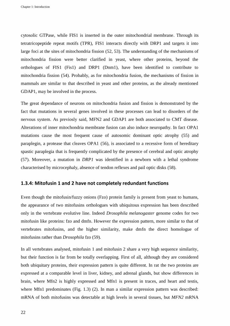
brain, where Mfn2 is highly expressed and Mfn1 is present in traces, and heart and testis,
where Mfn1 predominates (Fig. 1.3) (2). In man a similar expression pattern was described:
mRNA of both mitofusins was detectable at high levels in several tissues, but MFN2 mRNA
Chapter 1: Introduction
23
resulted specifically enriched in skeletal muscle and heart and that of MFN1 in pancreas and
liver (47).
Another difference between MFN1 and
MFN2 concerns the GTPase functional
domain. An active domain is essential
for the function of both proteins, but in
vitro studies demonstrated that MFN1
has a high GTPase activity but a low
GTP affinity, while the contrary is true
for MFN2. This implies that homotypic
interactions of MFN1 are 100-fold
higher than that of MFN2. It was
speculated that MFN1 and MFN2 could be involved in different steps of mitochondria fusion:
MFN1 should mainly induce the first step of mitochondria tethering, while MFN2 may be
involved in the fusion process (60). An involvement of MFN1 in the coordination of outer and
inner mitochondria membranes was also reported, as in vitro experiments demonstrated that
MFN1 but not MFN2 is necessary for OPA1 function. (51).
The importance of mitofusins in development is demonstrated by the fact that in mouse the
complete knock-out (KO) for either Mfn1 or Mfn2 results in embryonic death (61). Although
both KO-mice die in midgestation, some distinctive phenotypic features in embryos and
embryonic fibroblast derived cell lines (MEF) can be identified, thus leaning towards a
differentiation of mitofusins function.
Heterozygous KO-mice are fertile and show any alteration, but in homozygosity the absence of
mitofusins results lethal in both KO-lines, although at different stages. In Mfn1-KO live
embryos can be recovered up to embryonic day (e) 10,5, but are completely resorbed by e12,5.
Mfn2-KO show earlier lethality, since full viable embryos can be see only until e9,5, and are
resorbed by e11,5. Interestingly KO-embryos show different developmental alterations. In
Mfn2-KO defects of placenta are ascribed as the major cause of perinatal death. In fact at e9,5
Mfn2-KO embryos are only slightly smaller and show no obvious malformations, while the
giant cells of trophoblast, the polyploid cells that give rise to the placenta, are reduced in
quantity due to a diminished number of endoreplication cycles. This was not observed in
Mfn1-KO embryos, that at e10,5 are significantly delayed in development and malformed, but
Figure 1.3: Tissue expression of rat Mfn proteins. The total proteins
prepared from indicated tissues of adult rat were subjected to
immunoblotting using anti-rat Mfn2 (top) or Mfn1 (middle). An
immunoblot against the mitochondrial protein Tom40 was used as a
loading control (lower stripe) (2).

Chapter 1: Introduction
24
without morphological defects in trophoblasts. To assess the involvement of placenta defects in
determining the unviable phenotype of mitofusin-KO mice, some conditional KO lines, where
Mfn1 or Mfn2 are not depleted only in the trophoblasts, were developed. Surprisingly the
conditional Mfn1-KO resulted vital and fertile, and survived through adulthood with no evident
defects, thus demonstrating that indeed also Mfn1-KO lethality is due to a placental defect. On
the contrary, conditional Mfn2-KO dies by postnatal day 17 and shows severe defects in
movement and balance. These alterations were ascribed to defects in the development of
cerebellum, particularly of Purkinje cells, which appear to have less branched and shorter
dendrites and nuclear clustering of mitochondria. Since Purkinje cells have a high expression
of Mfn2 but a very low presence of Mfn1, and given that their alterations in the conditional
Mfn2-KO can be rescued with the same strength both by Mfn1 and Mfn2, it was inferred that
the loss of mitofusin function in general, rather than the specific loss of mitofusin 2, was
responsible for the phenotype (62). We can also argue that tissues where Mfn1 expression is
lower, as neuronal tissues, are more sensible to the decrease of Mfn2 activity.
As for the effects on mitochondria morphology, some differences can be evidenced in KO-
derived MEF cells (61). The loss of either Mfn1 or Mfn2 causes mitochondria fragmentation,
but at a higher degree in Mfn1-KO cells. Very importantly, similarly to what happens in
Purkinje cells, the mitochondrial fragmented phenotype of Mfn2-KO MEFs can be completely
restored by the over-expression of both Mfn1 and Mfn2. Intriguingly, although both proteins
are expressed in wt MEFs, in Mfn1-KO cells the tubular shape of mitochondria is completely
rescued by Mfn1, but of only the 25% by the over-expression of Mfn2, thus implying that
mitofusin 1 has a higher fusion capacity and that mitofusin 2 can have some distinct functions
(61). However the importance of the integration of both Mfn1 and Mfn2 for the tubulated
mitochondria morphology maintenance and particularly the significance of the formation of
functional etero-dimers of Mfn1 and Mfn2 was also demonstrated. Indeed some Mfn2 CMT2A
causing mutations lack mitochondrial fusion activity when expressed in MEFs lacking both
Mfn1 and Mfn2, but show substantial fusion activity in cells expressing only Mfn1. This
cooperation occurs in trans by the formation of hetero-dimers of Mfn1 and mutated Mfn2 (63),
and a similar complementation was found also in yeast, where a fzo1 mutated in the GTPase
domain could cooperate with one mutated in a HR region to promote mitochondrial fusion
(64). This suggests that each subunit of the mitofusins oligomer need not be fully functional to
provide function to the complex and further stresses the importance of MFN1 expression for

Chapter 1: Introduction
25
the manifestation of mitochondria alterations in cells with a lowered activity of MFN2, both
due to MFN2 mutations or ablation.
1.3.5: Mitofusin 2 roles beyond fusion
As speculated by the mentioned complementation studies performed on knock-out MEF cells,
in recent years it was demonstrated that MFN2 has a role not only in mitochondria membrane
fusion, but also in mitochondria movement along microtubules, endoplasmic reticulum-
mitochondria thetering and signaling. Here the state of art about these novel MFN2 functions
and their possible relation CMT2A pathogenesis will be briefly treated.
Mitochondria movement
In a first work, Baloh and collegues reported that in cultured dorsal root ganglion neurons
(DRG) the expression of disease-associated MFN2 mutant proteins induced alterations in
mitochondria distribution, which resulted clustered in the soma or at the neuronal terminals.
They also proved that the altered distribution of mitochondria is consecutive to a defect in
mitochondria transport, which is however not dependent on a decreased ATP production (65).
Interestingly a defect of mitochondria distribution in axons was reported also CMT2A patients.
Electron microscopy of sural nerve biopsies shows the presence of distal clustering of
fragmented mitochondria and a corresponding decrease of mitochondria along axons (40, 66,
67). In 2010 a direct interaction of MFN2 with the Miro/Milton motor protein complex was
demonstrated (68). Miro is an outer mitochondrial membrane protein involved, together with
the adaptor protein Milton, in the attachment of mitochondria to the motor proteins kinesin and
dynein (69). MFN2 is able to interact with both Miro and Milton in a specific manner not
dependent on fusion. As a consequence the absence of MFN2, or the expression of disease-
related allele, specifically disrupts mitochondria movement in axons (68).
ER-mitochondria tethering
MFN2 is also involved in the endoplasmic reticulum (ER) and mitochondria juxtaposition.
Indeed MFN2 is also expressed in the ER membranes, and is particularly enriched in the sites
of contact with mitochondria. In MEF cells, ablation of Mfn2 disrupts ER morphology and
detaches mitochondria from ER-membranes, thus demonstrating the importance of Mfn2
omodimers and Mfn1-Mfn2 heterodimers in the ER-mitochondria tethering (70). Contacts
between ER and mitochondria are physiologically important in many cellular processes, since

Chapter 1: Introduction
26
they allow the regulation of Ca++
level in cells.
In Mfn2 deficient MEFs, mitochondrial Ca++
uptake is indeed reduced, proving that a close
apposition of the two organelles is crucial
during Ca++
signalling. This may have
important implications in many other cellular
processes in which Ca++
signalling is involved,
first of all the induction of apoptosis.
Intriguingly Ca++
is also important for the
regulation of kinesin-mediated mitochondrial
motility, as an increase of Ca++
induces a
conformational change in Miro, which
becomes able to interact directly with kinesin
and induce arrest of mitochondria movement
(71).
Mitofusin 2 as a signalling protein
MFN2 acts not only as a tethering molecule, but has also signalling functions. Indeed MFN2,
but not MFN1, has a p21Ras binding site at the extreme N-terminal, before the GTP-binding
domain (72). MFN2 over-expression is able to induce cell cycle arrest by inhibition of the Ras-
MAPK-ERK pathway. This was also demonstrated in vitro by the fact that hyperproliferating
vascular smooth muscle cells (VSMCs), derived from spontaneously hypertensive rat arteries,
show a down-regulation of MFN2 expression. In these cells the artificial over-expression of
MFN2, and thus the inhibition of Ras pathway, is able to induce cell cycle arrest. This effect of
MFN2 is however independent on its function in mitochondrial fusion (72) and ER-
mitochondria tethering (73), but suggests a possible contribution of MFN2 in proliferative
cardiovascular disorders and potentially other proliferative diseases as cancer. Intriguingly a
mutation in MFN2 in a kindred with recurrent strokes occurence was identified (74) and two
patients with MFN2 mutation causing a neuropathy with pyramidal signs showed also
vasomotror troubles (75).
Figure 1.4: A fraction of MFN2 is present in the ER membrane
and assembles with MFN1 or MFN2 at the mitochondrial
surface to tethes ER to mitochondria. ER-mitochondria
juxtapositionis required for efficient Ca++ uptake into
mitochondria and may have important implications for cell
signaling and mitochondrial movement (1).

27
Project aims
Mutations in mitofusin 2 gene (MFN2) account for the most common variant of axonal
Charcot-Marie-Tooth disease (CMT2A), an inherited disorder of the peripheral nervous
system. This pathology is characterised by phenotypic variability and reduced penetrance and,
even if more than 80 MFN2 mutations have been described, a clear genotype-phenotype
correlation was not identified. Mitofusin 2 is localised in the outer mitochondria membranes
and endoplasmic reticulum. It is a multifunctional protein, and its main functions are the
tethering of mitochondria prior to fusion, the ER-mitochondria juxtaposition and the axonal
transport of mitochondria. Both in vitro and in vivo studies have been performed in order to
clarify the mechanism of action of MFN2 mutations. In vivo studies resulted particularly
complicated in mouse, and future investigations would certainly benefit of new models of the
disease. Very interesting indications may come from the zebrafish (Danio rerio), an animal
model that recently proved to be an excellent tool for the study of neurological disorders. In
order to contribute to the identification of a genotype-phenotype correlation and evaluate new
models of the disease, with this project I propose to:
a) Perform a genetic screening of MFN2 gene in a cohort of patients affected by axonal
CMT. This analysis will be conducted by direct sequencing, and MLPA (multiplex
ligation-dependent probe amplification), in order to evaluate both point-mutations and
MFN2 gene rearrangements.
b) Evaluate zebrafish as a model for CMT2A. First, to study the role of mfn2 gene in the
development of zebrafish neuromuscular system, I will perform knock-down
experiments of this gene using a morpholino-oligo. Then I will evaluate the ability of
human MFN2 transcript to complement for the loss of zebrafish mfn2. This will allow
to assess if human and zebrafish proteins are functionally conserved. In order to
elucidate the mechanism of action of MFN2 mutations, I will also study the CMT2A
disease causing mutation R94Q using zebrafish as a model. In particular, by performing
rescue experiments with the mutated transcript, I will evaluate if this allele has a loss of
function activity. On the other hand, over-expression experiments will be performed to
evaluate the gain of function activity of the R94Q allele.


29
Chapter 2: Analysis of MFN2 mutations in CMT2A patients
2.1: Background
2.1.1: MFN2 mutation distribution in CMT2A patients and inheritance
patterns
To date more than 80 disease causing
mutations have been described in MFN2.
As illustrated in figure 2.1, most of them
cluster in the GTPasic domain and in the
region linking this domain with the HR1
region. However some mutations in or
around the HR regions, even much rarer,
have also been found in CMT2A patients.
Most mutations are missense and with a
dominant inheritance. Phenotypically a
surprisingly high percentage of non-
penetrance associated to an elevated
variability in the phenotypic expression,
also among people of the same kindred,
was reported. A total of 4 nonsense and 2
frameshift mutations have also been
described. Interestingly 3 of these
mutations were found to have a recessive
inheritance pattern and were reported in
patients with compound heterozygous
MFN2 mutations. Indeed 8 cases of
recessive inheritance of MFN2 mutations
were reported so far (40, 66, 75-77).
Figure 2.1: Schematic representation of the human MFN2 protein.
Mutations found in CMT2A are indicated with their corresponding
frequency. Abbreviation: HR; heptads repeated region, TM;
transmembrane domain. In red are mutations found only in homozygous
or heterozygous state, in blue that found both recessively and dominantly
inherited. In bold are indicated intronic mutations on splicing-sites.

Chapter 2: analysis of MFN2 mutations in CMT2A patients
30
Some positions of the protein appear to be more frequently mutated and mutations in these
sites were reported several times in independent works. Above all the arginine at position 94
appears particularly prone to mutations, being the substitutions R94Q and R94W described 11
times each. Since R94Q and R94W were reported several times in patients with a de novo
mutation, it is reasonable that 94 position represents indeed a mutational hot spot (78).
2.1.2: Phenotype-genotype correlation hypothesis
Although the severity of the phenotype induced by MFN2 mutations and the presence of
complicating additional features can be extremely variable among patients (see paragraph
1.3.1), on the basis of age at onset and severity of the disease, two main clinical groups can be
identified. In the first group are included patients with an early onset, generally before 10
years, and a severe phenotype, frequently complicated by other symptoms, especially optic
atrophy. The second group comprise patients with a later onset, after 10 years, and a milder
course of the disease (40). Interestingly pyramidal signs were reported preferentially in patients
with mild or moderate forms of CMT2. This is possibly due to a masking effect of pyramidal
signs when an important peripheral motor neuropathy is present (75). Even if this clinical
subclassification is, with rare exceptions, always observed, it does not reflect an evident
genotypic-phenotypic correlation. For example it is reported that mutations located in the
GTPasic domain or in the HR regions are able to induce similar phenotypes. The only
exceptions are the mutation R418X (40) and P456L. The nonsense mutation R418X is located
in HR1 and it potentially causes the expression of a truncated form of MFN2 lacking the
transmembrane domain (TM), which therefore would not insert in the outer mitochondrial
membrane. In vitro studies demonstrated that a mutant form of MFN2 deprived of the TM
domain is able to act as a dominant negative allele (79). Interestingly the patient bearing the
R418X shows an extremely severe CMT2 phenotype, characterised by onset in infancy,
wasting of distal and proximal muscles and vocal cord paresis. However no functional studies
have been performed on this mutation that can support the hypothesis that it acts with a
dominant-negative mechanism. The other mutation, P456L, positioned between HR1 and TM,
was found in a patient with early onset cerebral stroke. The patient showed any clinical or
electrophysiological sign of neuropathy. The father, from who she inherited the MFN2
mutation, showed white matter lesions probably due to unawared stroke events, and similarly
to his daughter, has no signs of motorneuron alterations at ENG (74).

Chapter 2: analysis of MFN2 mutations in CMT2A patients
31
The only observation supporting the existence of some genotypic-phenotypic correlation in
CMT2A is the fact that individuals with homozygous or compound heterozygous MFN2
mutations have the tendency to manifest a more severe phenotype. Indeed, even if CMT2A was
originally reported as an autosomal dominant disease, in the last years is growing the evidence
that MFN2 mutations are recessively inherited in a significant number of families. To date a
total of six kindred with recessive inheritance have been reported (40, 66, 75, 76). The
proband, usually a son of two unaffected parents, tends to have an extremely early onset and a
severe phenotype. The parents are typically unaffected, although sometimes can have some
subclinical signs. Some mutations, are found only in homozygous or heterozygous state, thus
meaning they may have a real recessive inheritance. Other mutations, as for example T362M,
were found also to have dominant inheritance. Nevertheless, when present in heterozygosity,
T362M cause a mild phenotype, but when associated to other missense mutations (A164V) it
causes an extremely early onset and severe disease.
Altogether these results demonstrate the difficulties in determining a genotype-phenotype
correlation and stress the importance of deeper studies of the molecular mechanisms through
which different MFN2 mutations can induce CMT2A disease.
2.1.3: Mechanism of action MFN2 mutations
Several in vitro studies were conducted with the aim to identify the mechanism of action
through which mutations in MFN2 gene leads to CMT2A disease.
In the only extensive functional study published so far, Detmer and Chan analysed 9 MFN2
diseases causing mutations using KO-MEF cells and they first hypothesized that different
molecular mechanisms could be responsible for the effect of different MFN2 mutations (63).
This hypothesis is based on the observation that in the absence of both mitofusins, a moderate
over-expression of some mutations is able to recover the fragmented mitochondrial tubules
with the same strength of wt MFN2, while other mutations result non-functional. Intriguingly
all the non-functional alleles are in position conserved in MFN1 and MFN2, thus suggesting a
possible explanation for their different ability to recover mitochondria fragmentation in double
KO-MEFs. They also observed that in cells expressing only the non-functional alleles, the
over-expression of MFN1 but not MFN2 is able to recover the phenotype. In an elegant series
of complementation studies, they demonstrated that MFN1 is able to cooperate with mutant
MFN2 to promote mitochondria fusion through the formation of functional hetero-oligomers in

Chapter 2: analysis of MFN2 mutations in CMT2A patients
32
trans, in a way that is independent on the presence of a fully functional GTPase domain. This
effect, observed also in yeast fzo1(64), underlies the importance of MFN1 for the phenotypic
manifestation of MFN2 mutations and supports the hypothesis that tissues expressing less
MFN1 are more prone to suffer mitochondrial dynamics alterations in the presence of mutated
MFN2.
The work of Detmer and Chan is somewhat limited by the fact that they screened cells
expressing mutant MFN2 only for mitochondria alterations, while recently an involvement of
MFN2 in axonal transport of mitochondria and ER-mitochondria tethering was verified.
In the works where Baloh and Misko demonstrate a role of MFN2 in mitochondrial axonal
transport (65, 68), an analysis of the effects of some disease related MFN2 mutations on
mitochondria transport was also performed. They showed that in wt-DRG neurons, the
expression of mutated MFN2 induces proximal and distal clustering of mitochondria, similarly
to what observed in patients. Interestingly the mutations R94Q an L76P, that Detmer described
as fusion “uncompetent” and “competent” respectively, have a similar effect on the alteration
of mitochondria movement (65). Misko further demonstrated that MFN2 mutations do not
disrupt the interaction of MFN2 with Miro and Milton proteins, and propose that the disruption
of mitochondrial transport could be caused by an altered regulation of the kinesin transport
apparatus (68).
Informations about the capacity of mutations in MFN2 to alter ER-mitochondria tethering are
available only for the R94Q mutation. De Brito and Scorrano demonstrated that, contrary to wt
MFN2, R94Q cannot complement the ER morphological alterations of Mfn2-KO cells, nor
recover ER-mitochondria tethering (70).
All these results are promising and we could speculate that possibly MFN2 mutations act by
inducing ER-mitochondria detachment. This alters Ca++
exchange, which is necessary for the
correct activity of the motor proteins involved in mitochondria movement. However many
questions about the mechanism of action of MFN2 mutations remain to be elucidated. In
particular it is not explained why mutations can have different phenotypic manifestations and
why some are active only in heterozygous or homozygous state. Some important clues may
come from the generation and study of animal models.
2.1.4: Animal models of CMT2A

Chapter 2: analysis of MFN2 mutations in CMT2A patients
33
Attempts to model CMT2A disease were made uniquely in mouse. As discussed in chapter
1.3.4, the complete knock-out of either MFN1 or MFN2 is lethal mainly due to placental
defects (61). Intriguingly the conditional MFN1-KO, in which mitofusin is expressed only in
the placenta, is vital and phenotypically normal, while that for MFN2 shows motor
abnormalities, which were however ascribed to alterations in cerebellum, rather than in the
peripheral nerves (62).
Some mouse models expressing MFN2 disease related mutations were also developed. A first
attempt to model MFN2 mutations was the generation of a mouse knock-in line for the
common mutation R94Q (63, 80). Surprisingly heterozygous mice are vital and healthy,
without any sign of neuropathy, while homozygous animals have severe movement defects and
die by the third week after birth, hence much later than the complete Mfn2-KO. In addition
alterations of mitochondria morphology in MEF cells derived from homozygous animals were
not detected, and this was explained by the fact that in these cells Mfn1 is able to interact with
Mfn2R94Q
to complement the phenotype (63).
The absence of evident alterations in heterozygous mice expressing R94Q could also be
justified by the differences between human and murine motorneurons: in humans with CMT
only longer neurons are affected, whereas in mouse, characterised by shortest motorneurons
than man, the effect of MFN2 mutations could probably not disclose phenotypically. For this
reason the authors derived also a mouse transgenic line overexpressing the mutation T105M
(80). In this model the transgene (Mfn2T105M
) is driven by the HB9 promoter in order to
overexpress the mutant protein only in the peripheral nervous system. HB9-Mfn2T105M
mice
show several phenotypic alterations that recapitulate some of the motor defects observed in
CMT2A patients (abnormal gait, foot deformities, reduced number of motor axons, calf
muscles atrophy). These defects are present only in homozygous animals, while heterozygous
have only a slightly shortened tail, but retain a normal motor capacity. Alterations in the
distribution of mitochondria along axons were also evident and aggregated mitochondria
uneven distributed along axons could be noticed in homozygous animals. All these defects are
congenital and indeed the HB9 promoter is expressed embryonically coincident with motor
neuron formation and during the whole fetal life, but decreases dramatically by three weeks of
age. The absence of a control transgenic line, overexpressing the wild type construct, and the
fact that in cells the over-expression of MFN2 induces apoptosis (81), arose the suspect that the
dose dependent phenotype of this transgenic mouse could be a side effect of the over-
expression of the protein, rather than of the mutation itself.

Chapter 2: analysis of MFN2 mutations in CMT2A patients
34
Recently Cartoni and colleagues developed an alternative transgenic mouse model for the
mutation R94Q. Also in this transgenic line the mutation is expressed specifically in the
nervous system, but it is driven by the neuron specific enolase promoter (NSE). Compared with
Hb9, NSE is active in the whole neural tissue starting embryonic day 13, but continues to
function also after birth (82). The NSE-Mfn2R94Q
line shows several motor and phenotypic
alterations reminding CMT2A disease. Interestingly, as the previously described model, the
expression of the phenotype is dose dependent and was detected only in the homozygous
animal. However a control transgenic line overexpressing the wild-type construct shows any
phenotypic alteration. Thus it is reasonable to hypothesize that the phenotypic alterations found
in NSE-MfnR94Q
are a direct consequence of the expression of the mutation. An interesting
feature, which was not observed in the previous models, is that the phenotype is evident
starting 5 months after birth, thus mimicking further the degenerative feature of CMT2A
disease. As described in the HB9-Mfn2T105M
line, a strong phenotypic variation among siblings
of the same genotype can also be observed, probably due to slightly different levels of
transgene expression. Finally the observation that in motorneurons with diameter smaller than
3,5 μm there is a distal accumulation of mitochondria, as observed in CMT2A patients,
suggests that indeed defects in the retrograde movement of mitochondria could be the cause of
the pathology.
Altogether these mouse models contributed to the understanding of the mechanisms by which
mutations in MFN2 leads to CMT2A. However many questions are still unresolved, and only
the study of the genotype-phenotype correlations of MFN2 mutations and the development of
new animal models could finally lead to a complete comprehension of CMT2A disease.

Chapter 2: analysis of MFN2 mutations in CMT2A patients
35
2.2: Materials and methods
2.2.1: Patients analysed
In the present study a cohort of 25 patients diagnosed with axonal Charcot-Marie-Tooth
disease where analysed for mutations in MFN2 gene. 13 patients were diagnosed in Padua
Hospital (Clinica Neurologica) or were directed to our laboratory by the UILDM association
branch of Padua. The remaining came from other hospitals of northern Italy.
Elettroneurography was conducted on all patients, except patient 9 who was too young to
perform the exam. A positive familiar history can be traced for 17 probands. All patients
included in the study gave informed consent to carry out the genetic test.
2.2.2: Blood DNA extraction
Genomic DNA was isolated from blood samples using a modified protocol from (83). Briefly
whole blood samples were diluted 1:4 with N-N solution (NaCl 0,9%, Nonidet 0,1%) to lyse
erythrocytic membranes. Lymphocytes are recovered by a centrifugation at 6000 rpm for 30
min performed at 4°C. Cells are then resuspended in 4 ml of TEN solution (TrisHCl 10mM,
EDTA 2 mM pH8, NaCl 400 mM) and 600 μl of SDS 10% and lysed 3 hrs at 80°C. The
salting-out of proteins and cellular debris is performed by adding 1 ml of a NaCl saturated
solution. The supernatant containing the genomic DNA is recovered by centrifugation at 6000
rpm for 10 min. To purify the DNA, 1 volume of chloroform is added and, after a
centrifugation at 6000 rpm for 10 minutes, the supernatant aqueous phase containing the DNA
is recovered and precipitated by adding 1 volume of isopropanol. DNA is recovered by
centrifugation and washed 2 times with cold ethanol 70% solution, then resuspended in 300-
500 μl of sterile TE solution (Tris HCl 10 mM pH 8, EDTA 1mM). The concentration and
purity of DNA is then measured with NanoDrop 2000 (Thermo Scientific) and the integrity is
evaluated by gel electrophoresis in 1% agarose gel. DNA is then stored at 4°C.
2.2.3: PCR amplification of MFN2 gene exons
The amplification of the coding regions of MFN2 gene was performed by PCR using primers
pairs designed on the intronic flanking regions of each exon. Since MFN2 has 2 transcript
variants, for reference we referred to isoform 1 (NM_014874) that has a longer 5’UTR.

Chapter 2: analysis of MFN2 mutations in CMT2A patients
36
Exon Primers Fragment
length DMSO Ta Program Taq
1 F: ccatgatgcagtgggagtc
R: gcttgactgcatcccagac 426 - 60 N A
2 F: ttgtttggcttgaaacctga
R: ctaagcaaccagcgaaatcc 423 - 60 N A
3 F: tgacctgagatccccatattc
R: atggcagttcccttgtcttc 483 - 60 N A
4 F: gccaccatagaggatctgga
R: cctggaacgttctgtgacct 377 - 60 N A
5 F: actggcaacattgcactgaa
R: tctcccattcacctccacag 397 - 60 N A
6 F: gtggaagaagaaagggtagc R: gttgagcccatctgaaaggt
384 - 60 N A
7+8 F: caggtctgttctcagcagga
R: cactagatccaggggtgcag 489 - 60 TD A
9 F: catgcccagcctcttatgac
R: gcctctctgctcttctcagc 372 - 60 N A
10+11 F: ctgagaaggggatgctgaga
R: tgggagcccagagactca 579 - 60 TD A
12 F: tacctgggaaggggaagg R: acagggccacaactttcttc
400 - 60 N A
13+14 F: aggctgctggtttgagagg
R: cagaacctgaaggtatcgagt 668 - 60 N A
15 F: atccctggcagtagctggta R: ggaggcagggtacagactga
379 - 60 N GO
16 F: agccactctgtgtccctgtt
R: cagtggactgtggagtgtgg 371 - 61 N FS
17 F: gaaacatgaaggctccttgg
R: acgatgcttaggacccagtg 425 - 60 N GO
18 F: gctgctggcagggatataga
R: ccagcccatggtacacatct 399 8% v/v 60 N A
19 F: ggatgatgtaagggtgtgtgtc
R: tgcttcattctcttggcagt 300 - 60 N A
Table 2.1: Primers used to amplify human MFN2 gene, dimensions of amplification product and Taq and PCR conditions used. In bold are
indicated the primers used for sequencing. Ta= annealing temperature; N=standard PCR program; TD=Touchdown PCR program;
A=AmpliTaq Gold; GO=GO Taq; FS=FastStart Taq.
Primers were designed with Primer3 (v. 0.4.0) using default settings. Conditions such
annealing temperature (Ta), time of denaturation, annealing and extension phases, number of
cycles, adding of DMSO and selection of Taq polymerase to be used were adjusted
empirically. In some cases, to avoid the amplification of aspecific products, a touchdown

Chapter 2: analysis of MFN2 mutations in CMT2A patients
37
amplification program was used (84). Primer pairs and amplification conditions are described
in table 2.1 while in table 2.2 are described the PCR programs used. All the PCR
amplifications were conducted using AmpliTaq Gold (Applied Biosystem), GO Taq (Promega)
or FastStart Taq (Roche) with the reaction mixes listed in table 2.3. PCR products were tested
on a 2% agarose gel before sequencing.
A) Standard PCR program
B) Touchdown PCR program
temperature time
temperature time
95 °C 2-10 min
95 °C 2-10
min 95 °C 45"
95 °C 45"
Ta 45" x 33 (Ta+10)-1 °C every cycle 45" x 10
72 °C 45"
72 °C 45"
72 °C 10 min
95 °C 30"
15 °C 5 min
Ta 30" x 26
72 °C 45"
72 °C 10 min
15 °C 5 min
Table 2.2: details of PCR programs used .A) standard PCR program; B) touch-down PCR program.
A) AmpliTaq Gold
B) GO Taq
C) FastStart Taq
DNA (50ng/µl) 1 µl
DNA (50ng/µl) 1 µl
DNA (50ng/µl) 2 µl
Buffer 10X 1,5 µl
Buffer 5X (w MgCl2 7,5 mM)
3 µl
Buffer 5X (w MgCl2 20 mM)
1,5 µl
MgCl2 (25mM) 1,5 µl
dNTPs (1mM) 1,5 µl
dNTPs (1mM) 2,5 µl
dNTPs (1mM) 3 µl
primer F+R (10 pmol/µl)
0,5+0,5 µl
primer F+R (10 pmol/µl)
0,5+0,5 µl
primer F+R (10 pmol/µl)
0,5+0,5 µl
MilliQ H2O 8,42 µl
MilliQ H2O 7,9 µl
MilliQ H2O 6,9 µl
GOTaq (5U/µl) 0,08 µl
FastTaq (5U/µl) 0,1 µl
AmpliTaq gold
(5U/µl) 0,1 µl
Table 2.3: composition of PCR mixes used. A)Mix fof AmpliTaq Gold; B) Mix for GO Taq polymerase; C) mix for FastStart Taq.
Chapter 2: analysis of MFN2 mutations in CMT2A patients
38
2.2.4: Direct PCR sequencing
To eliminate the unincorporated primers and dNTPs which could interfere with the sequencing
reaction, 1 μl of ExoSAP-IT® solution (USB), containing exonuclease I and shrimp alkaline
phosphatase, is added to 2,5 μl of freshly made PCR and incubated at 37 °C for 15 min and
then at 80 °C for 15 min to inactivate the enzymes. At the end of the purification reaction, the
sequencing primer (10 pmol) is added to the PCR and the sample is dried at 60 °C and sent to
BMR genomics (www.bmr-genomics.it) to perform the sequencing reaction. Sanger
sequencing was carried out using Applera BigDye version 3.1 and the automated sequencers
ABI 3730XL and ABI 3100. The electropherograms were analysed with Chromas Lite version
2.01 and Seqman Pro 7.1.0 and aligned to the RefSeq of human MFN2 isoform 1
(NM_014874). All nucleotidic differences were compared to dbSNP database
(http://www.ncbi.nlm.nih.gov/projects/SNP). Differences in the coding sequence of MFN2 that
were not reported as polymorphisms were analysed in at least 100 healthy subjects using
Restriction Fragment Length Polymorphism (RFLP), when possible, or direct sequencing.
2.2.5: MLPA analysis of MFN2
Multiplex Ligation-dependent Probe Amplification (MLPA) is a relatively new method used to
identify alterations in DNA copy number of up to 40 sequences in a single reaction (85). This
method uses specific probe pairs that hybridize to genomic DNA in correspondence of every
single exon of the gene to be analyzed. The probe pair consists of a short left probe (L) with a
hybridization sequence specific for the target gene at the 3’ end and a common 19 nucleotides
sequence that is recognized by the reverse PCR primer, at the 5’ end (Fig. 2.2). The long right
probe (R), conversely, has the hybridization sequence at the 5’ and the forward primer
recognition sequence at the 3’ end. In the R probe, between the hybridization sequence and the
primer recognition sequence is included a stuffer sequence of different length for each probe
pair. The probes are designed to hybridize adjacently to the target DNA and after ligation is
possible to perform a semiquantitative multiplex PCR to amplify the derived fragments taking
advantage of the common primer recognition sequences. Since the reverse primer used is
fluorescently labelled and the stuffer sequence has different lengths for every probe pair, after
PCR is possible to separate by capillary electrophoresis the fragments to obtain an
electropherogram in which at every length correspond a probe pair that hybridize to a known
exon and with a peak area that is proportional to the amount of that exon sequence in the

Chapter 2: analysis of MFN2 mutations in CMT2A patients
39
genomic DNA of the patient analyzed. Heterozygous deletions will appear as a reduction of
35-50% of the corresponding relative peak area. However the presence of point mutations in
the vicinity of the ligation site will also cause the reduction of peak area. For this reason is
always necessary confirm the absence of nucleotide substitutions by sequencing.
Figure 2.2: step-by-step representation of Multiplex Ligation-dependent Probe Amplification (MLPA) procedure.
To analyse if rearrangements of MFN2 gene could be responsible of the disease in CMT2A
patients that resulted negative by sequencing, MLPA analysis was performed with the MLPA
kit P143 by MRC-Holland that allows the simultaneous detection of copy number variations in
MFN2 and MPZ, that is associated to CMT type 1B. The kit contains the probes for all the 19
exons of MFN2 and for the 6 coding exons of MPZ and 10 control probes located in other
human genes on different chromosomes. The reaction was performed according the
manufacturer and the statistical analysis on electropherogram peak areas was conducted with
the Coffalyser V6 software (MRC-Holland).
2.2.6: RFLP analysis of control DNA for nonpolimorphic changes found in MFN2
gene
Restriction Fragment Length Polymorphism (RFLP) was used to analyse the DNA of control
subjects for the presence of MFN2 non-polymorphic variations found in patients. DNA was

Chapter 2: analysis of MFN2 mutations in CMT2A patients
40
amplified with the primer pairs corresponding to the exon bearing the substitution, and
restriction enzymes with the recognition site over the mutation (tab. 2.4) were used to digest
the DNA. 5μl of PCR product were digested with 1-2 U of enzyme in the appropriate
restriction buffer. The reaction was performed at the optimal reaction temperature of the
enzyme for 2 hours. Then digested product were tested in a 2% agarose gel. At least 100
individuals were analysed for each mutation.
Restricion
enzyme mutation
Wild-type
fragments
Mutated
fragments
Reaction
temperature
XmnI c.987_989delAAG 147/431 578 37 °C
HpaII c.2213C>T 118/182 300 37 °C
BsaJI c.281G>C 377 261/116 60 °C
Table 2.4: restriction enzymes used in the RFLP analysis of DNA from healthy control subjects. The fragment lenghts obtained from the wild-
type and mutated allele are shown.
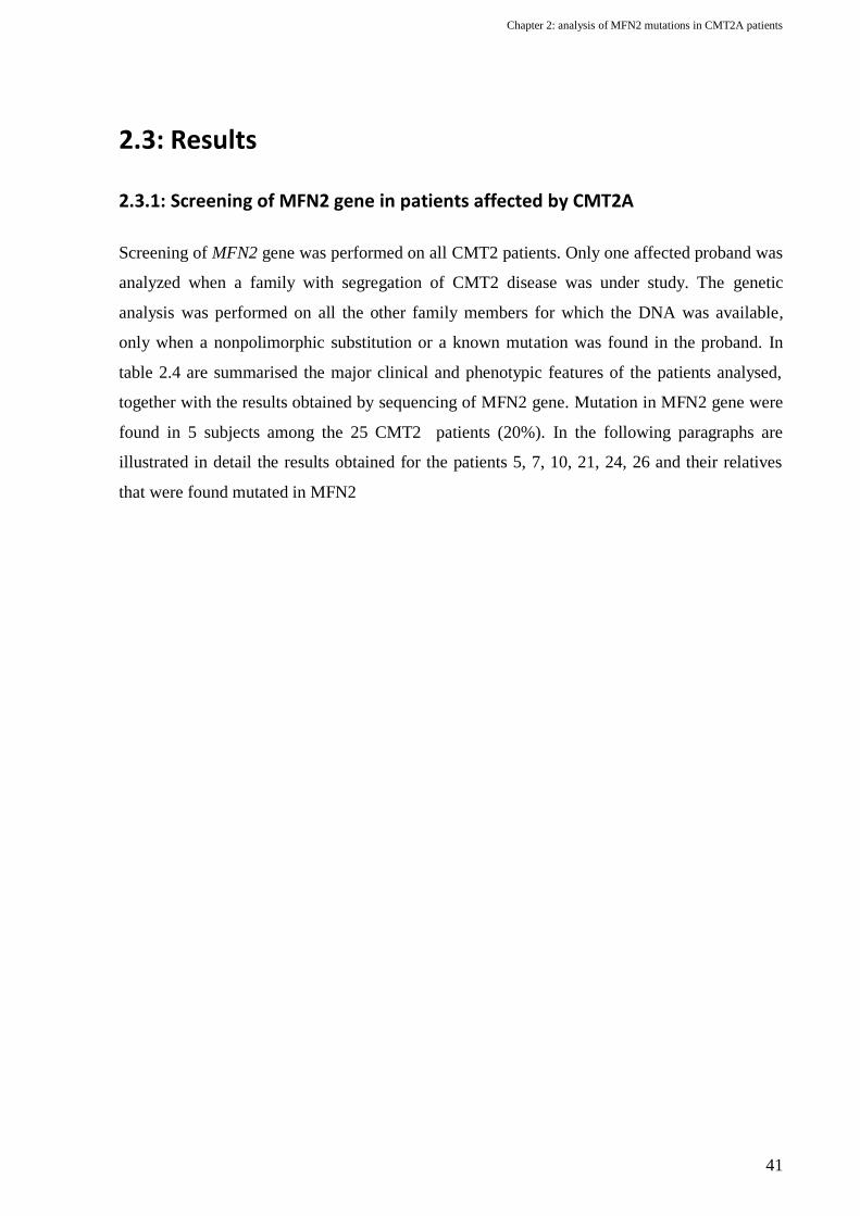
Chapter 2: analysis of MFN2 mutations in CMT2A patients
41
2.3: Results
2.3.1: Screening of MFN2 gene in patients affected by CMT2A
Screening of MFN2 gene was performed on all CMT2 patients. Only one affected proband was
analyzed when a family with segregation of CMT2 disease was under study. The genetic
analysis was performed on all the other family members for which the DNA was available,
only when a nonpolimorphic substitution or a known mutation was found in the proband. In
table 2.4 are summarised the major clinical and phenotypic features of the patients analysed,
together with the results obtained by sequencing of MFN2 gene. Mutation in MFN2 gene were
found in 5 subjects among the 25 CMT2 patients (20%). In the following paragraphs are
illustrated in detail the results obtained for the patients 5, 7, 10, 21, 24, 26 and their relatives
that were found mutated in MFN2

Chapter 2: analysis of MFN2 mutations in CMT2A patients
42
Patient
ID
Se
x
Age
onset Inheritance
UL
involvement
MNCV
(m/s)
Other
symptoms
Mfn2
Mutation,
status
Previous
reported
1 M <3 y AD - 40 - -
2 M 31 y AD - 43 - -
3 F <29 AD - 40 - -
4 F
? AD - 42 -
-
5
F <6 y S + >38 OA
p.E329del,
HZ no
6 F <31 y AD - 36,8 - -
7
M 9 y AD - >38 -
p.A738V,
HZ no
8 F <45 y AD - 42 m/s - -
9 F <7 y na - NA HL, WMA -
10
M 16 y AD + >38 -
p.R94P,
HZ no
11 M
<48 y
S - 4 - -
12 F 7 y AD + 35 - -
13 M <11 y AD - 44 - -
14 F <27 y AD + >38 - -
15 F <9 y S - 41 - -
16 F 46 y S + 45 - -
17 F 50 y AD - 40 RP -
18 M 10 y S + 47 - -
19 M <18 y S - 45 - -
20 F <45 y AD - 40 - -
21
F <35 y na + 38 m/s -
p.H165R,
HZ
(40, 41,
86)
22 M <1 y S + >38 D -
23 F <35 y AR - 40 - -
24
M 3 y AR + NE
OA, RF,
FMA, D,
HL
p.[K38del]
+[T362M],
CHZ no, (41)
25 M 5 y AD - 55 HL, OA, E -
Table 2.5: Clinical findings of CMT2 patients analysed. Abbreviations: AD: autosomal dominant; AR: autosomal recessive; S: sporadic; na:
not available; ne: not evocable; HL: Hearing Loss; D: dysphonia; OA: optic atrophy; RP: Retinitis Pigmentosa; RF: Respiratory failure; E:
epilepsy; FMA: facial muscle atrophy; WMA: white matter alterations; HZ: heterozygous; CHZ: compound heterozygous.

Chapter 2: analysis of MFN2 mutations in CMT2A patients
43
Patient 5
The patient showed difficulty in walking and muscular asteny from 1 year of age. She was
operated to lengthen the Achilles tendon at the age of 6 and 18 years. Over the years she
suffered of a gradual hypotrophy of lower limb distal muscles and from the age of 34 she
started to show weakness of right hand muscles. EMG evidenced a severe axonal sensorymotor
neuropathy of lower limbs, with a consistent peripheral denervation. Bilateral optic atrophy
was diagnosed at the age of 38 and caused a severe loss of visual acuity. Her parents are
referred healthy and nonconsanguineous.
Figure 2.3: A) electropherogram obtained by sequencing of exon 10 of MFN2 in patient 5; B) alignment of MFN2 proteins of various
organisms illustrating the conservation at aminoacid residue 329.
The sequencing of MFN2 revealed a heterozygous in frame
deletion of 3 nucleotides in exon 10 (c.987_989delAAG)
which result in the aminoacidic deletion p.E329del in a
conserved position of the protein between GTPasic domain
and coiled coil region HR1(Fig. 2.3).
Since this variant was never been reported in literature, the
DNA from healthy control subjects was analysed. Given that
the deletion causes the loss of a sequence recognition site for the restriction enzyme XmnI,
RFLP technique was used (Fig. 2.4). None of the 200 chromosomes analysed resulted bearing
the mutation and it is reasonable to conclude that the p.E329del deletion is the disease causing
mutation in patient 5.
Figure 2.4: RFLP analysis for the identification of c.987_989delAAG MFN2
mutation using XmnI restriction enzyme.
P5; patient 5, C, not mutated control; ND,
not digested PCR product.

Chapter 2: analysis of MFN2 mutations in CMT2A patients
44
Patient 7
The proband (III-3) showed walking difficulty from the age of 9 and was operated to correct
pes cavus at the age of 15. The EMG show the reduction of compound motor action potential
amplitudes (CMAP) in both sensory and motor nerves and is compatible with a sensorymotor
neuropaty of axonal nature. Also the sister (III-2) and the father (II-2) showed difficulty in
walking, as well as other relatives related through their father (I-1, II-3 and II-4).
Figure 2.5: A) family pedigree of patient 7. B) Electropherogram obtained by sequencing of exon 19 of MFN2 in patient 7 and in the indicated
other members of the family; C) Alignment of MFN2 proteins of various organisms illustrating the conservation at aminoacid residue 738.
In the proband and in his affected sister (III-2) was identified a
heterozygous single nucleotide substitution in exon 19 of MFN2
(c.2213C>T), leading to the missense mutation p.A738V (Fig. 2.5).
The c.2213C>T substitution was not found in the unaffected sister
(III-1) and in the mother (II-1) of the proband. The DNA from the
other members of the family was not available.
Also this mutation was never proviously reported in literature and
the analysis of the DNA of unaffected subjects by RFLP using HpaII
restriction enzyme revealed that none of the 200 chromosomes
Figure 2.6: RFLP analysis for the identification of c.2213C>T MFN2
mutation using HpaII restriction
enzyme. P7; patient 7, C, not
mutated control; ND, not digested
PCR product.

Chapter 2: analysis of MFN2 mutations in CMT2A patients
45
analysed bears the substitution (Fig. 2.6). For this reason we can conclude that A738V is the
mutation causing CMT2A in this family.
Patient 10
The proband (III-2) showed difficulty in walking from the age of 16 years. The disease
progressively worsened into a severe flaccid tetraparesis associated to distal hypotrophy and
mild alterations of sense in the lower limbs. EMG evidenced a marked motor and sensory
polineuropathy. Subject III-1 is also affected from the age of 15 years and has the same
symptoms observed in the proband. The mother (II-2), is mildly affected, shows only pes
cavus, while the aunt (II-3) has the same clinical picture of the proband, with a milder
phenotype.
Figure 2.7: A) family pedigree of patient 10. B) Electropherogram obtained by sequencing of exon 4 of MFN2 in patient 10; C) Alignment of
MFN2 proteins of various organisms illustrating the conservation at aminoacid residue 94.
Only the DNA of the proband was available for this family.
The analysis revealed the presence of the heterozygous
substitution c.281G>C in exon 4 leading to the missense
mutation p.R94P (Fig 2.7).
This undescribed mutation was not present in 300 healthy
subject chromosomes analysed with RFLP using the restriction
enzyme BsaJI (Fig 2.8). For this reason and considering that
codon 94 is considered a mutational hot spot of MFN2, we can
conclude that p.R94P is the causing mutation in this patient.
Figure 2.8: RFLP analysis for the
identification of c.281G>C MFN2
mutation using BsaJI restriction
enzyme. P10; patient 10, C, not mutated
control; ND, not digested PCR product.

Chapter 2: analysis of MFN2 mutations in CMT2A patients
46
Patient 21
The patient underwent neurological investigations because affected by carpal tunnel syndrome.
Through EMG study she was diagnosed a mild peripheral neuropathy involving both upper and
lower limbs with a slightly reduced nerve conduction velocity.
Figure 2.9: A) Electropherogram obtained by sequencing of exon 6 of MFN2 in patient 21; C) Alignment of MFN2 proteins of various
organisms illustrating the conservation at aminoacid residue 165.
The sequencing revealed the mutation c.494A>G in heterozygosity in exon 6 that leads to the
aminoacidic substitution p.H165R (Fig. 2.9). This mutation was previously reported in 4
unrelated kindreds with autosomal dominant inheritance (40, 41, 86).
Patient 24
The patient experienced frequent fallings and hyposteny of lower limb muscles from the age of
3 years and became wheelchair dependent from the age of 12. The disease was slowly
progressive and later involved also the upper limbs and facial muscles. Also the respiratory
muscles were involved and she uses a ventilator during the night from the age of 29 years. In
the same period she was also diagnosed a bilateral optic atrophy. She suffers of sensorineural
hearing loss from the age of 36. The EMG evidenced a nearly complete denervation of both
upper and lower distal muscles. Two brothers (II-1 and II-4) of the proband show a similar
clinical picture, while the parents (I-1 and I-2) and a sister (II-3) are referred healthy.

Chapter 2: analysis of MFN2 mutations in CMT2A patients
47
Figure 2.10: A) family pedigree of patient 24. Electropherograms obtained by sequencing of exon 3 (B) and exon 11 (C) of MFN2 in the
indicated subjects of the family of patient 10; Alignment of MFN2 proteins of various organisms illustrating the conservation at aminoacid
residues 38 (D) and 362 (E).
The sequencing revealed the presence of two MFN2 mutations in trans in the proband (II-2)
and in the other affected members of the generation II (Fig. 2.10). One is a previously reported
missense mutation (c.1085C>T) leading to the aminoacidic substitution p.T362M (41), that
was inherited from the mother. The other is an unreported 3 nucleotides in frame deletion
(c.113_115delAGA) leading to the mutation p.Lys38del, that was probably inherited from the
father, of which the DNA was not available. This mutation was not found in 400 healthy
subjects analysed by sequencing.
2.3.2: MLPA analysis
Direct sequencing is a very sensible method for the identification of single nucleotide changes
and small insertions and deletions. However it is inadequate for the detection of large genomic
rearrangements involving entire exons or genes. Recently is growing the idea that relatively
small rearrangements involving few exons can be an important cause of disease. A first case of
a patient with CMT2A bearing deletion involving exons 7 and 8 of MFN2 was just described

Chapter 2: analysis of MFN2 mutations in CMT2A patients
48
(77). For this reason we further analysed the DNA of subjects that resulted not mutated by
sequencing of MFN2 gene, using MLPA technique. We also performed the MLPA analysis on
5 healthy subjects as a control. A statistical analysis of the resulting electropherograms was
performed with the program Coffalyser. A heterozygous deletion should give a reduction of the
peak area corresponding to the probe annealing to that exon of about the 35-50% compared to
that of a control subject, and a similar gain should be noticeable in the case of a duplication. A
loss of a peak, instead, constitutes a homozygous deletion. None of the analysed patients
showed any deletion or insertion in MFN2
.

49
Chapter 3: Effects of down-regulation of MFN2 and analysis of R94Q mutation effect in zebrafish
3.1: Background
3.1.1: Zebrafish as a model organism
In the last 15 years the tropical freshwater fish Danio rerio, commonly known as zebrafish, has
been stated as the leading vertebrate model for developmental studies. As early as the 1930s,
this little teleost have been appreciated by developmental scientists for the ease of
maintenance, the exceptional spawning ability, the short generation time and the rapid external
development, which, unlike other vertebrate models, allow the observation and the
manipulation of the very first developmental stages. More recently the evolution of genetic
techniques, as cloning, mutagenesis and transgenesis, previously amenable only in other more
common animal models as Drosophila and mouse, prompted the use of this fish in other fields
of biology, and in particular for the study of human diseases.
Zebrafish is easily maintained in specialized facilities in water kept within a 25-28,5 °C
temperature range. Spawning can be induced easily, by regulating feeding and the light-dark
cycle. It is an extremely prolific animal and each female can generate clutches of 100-200
eggs, which are externally fertilised by the male and can be easily recovered at the bottom of a
spawning chamber. Embryos are transparent and development can be followed in vivo and in
real time. Organogenesis is particularly rapid: at 24 hours post fertilisation (hpf) most organ
primordials have formed and by 48-72 hpf larvae hatches and are able to move and react to
stimuli (Fig. 3.1).

Chapter 3: effects of downregulation of MFN2 and analysis of R94Q mutation effect in zebrafish
50
Figure 3.1: representation of developmental stages of zebrafish embryos from 1 cell stage to 2 days post fertilization. Modified from Kimmel
et al (1995).(87)
Despite the 400 million of years of evolution, it was found that many human genes have an
orthologue in zebrafish. Bioinformatical analysis on the zebrafish genome, whose sequencing
project, sponsored by the Sanger institute, started in 2001 and is now in completion, showed
also that extensive chromosomal regions retain a well conserved sinteny (88). Interestingly, it
was shown that zebrafish genome contains more genes than that of humans, and this is a result
of a duplication that probably occurred 100-400 million years ago (89, 90). As expected by the
genetic similarity, man and zebrafish organs are remarkably similar at morphological,
physiological and molecular level.
All this characteristics made zebrafish an invaluable animal model for the study of several
human disease, from cancer (91) to Alzheimer (92), and this was also recognized by the NIH
through the establishment of the Trans-NIH Zebrafish Coordinating Committee in 1997. As
research is ongoing, scientists can rely on increasingly genetic and genomic informations,
which are collected in the Zebrafish Model Organism Database (ZFIN) available on the web
(http://zfin.org). This provides an integrated representation of mutants, genes, genetic markers,
mapping panels, publications and community resources.
Being little and particularly permeable to any small molecule dissolved in the water, zebrafish
is also well-suited for use in chemical screens and in the last years this stimulated the
beginning of several drug screening programs that can count on a growing number of chemical
libraries (93). Hence zebrafish potentials in the field of human diseases go far beyond the

Chapter 3: effects of downregulation of MFN2 and analysis of R94Q mutation effect in zebrafish
51
modelling of pathology and potentially it could reach also a fundamental role in
pharmaceutical drug discovery.
3.1.2: Genetic analysis using zebrafish
Similarly to the widely used invertebrate models Drosophila and Caenorhabditis, zebrafish is
particularly suitable to forward genetics approaches. However in the last years, with the advent
of genomic era and the sequencing of zebrafish genome, now in completion, many reverse
genetics strategies have also been developed, that can make this little teleost a fundamental
complement of studies in mammal models. Here will be discussed briefly the many genetic
technologies that have been developed over the years, with particular attention on the more
recent reverse genetic approaches.
Forward genetic
Forward genetic analysis relies on the availability of mutant libraries and efficient and rapid
mutagenesis screening methods. Although exceptionally successful in invertebrates, this
approach has been historically of limited use in mammals model systems.
The zebrafish, unlike most vertebrate organisms, lends itself remarkably well to forward
genetic approaches. The ease of keeping many fishes in a small space, the large brood size, the
diploid genome, and the reasonably short life cycle, render large-scale genetic screens practical
and economical. Forward genetics is driven by phenotypes: mutagens, as ethylnitrosourea
(ENU), are employed to produce random changes in the DNA and the phenotypic
consequences are assayed. Zebrafish is resistant to ENU toxicity and the phenotypic
consequences of mutations can be easily screened under the microscope. Two large scale
mutagenesis screening were performed, which focused on early development and led to the
identification of 1800 mutants. They represented a great achievement and deserved the
definition of an “accomplishment of historic proportions” in Science (94) and a special issue of
Development (95). Random mutagenesis using retroviral methods has also been successfully
carried out in zebrafish (96, 97). This method, although lower in efficiency than ENU
mutagenesis, has the advantage to tag each insertion event, thus facilitating the mapping of the
mutation. These forward-genetic screening recovered also numerous mutants in genes
orthologues to that involved in human diseases. In some instances, the phenotype of the mutant
was so similar to that of man that could be identified by a candidate gene approach. One
example is the zebrafish mutant sapje, which display several muscular defects that phenocopies

Chapter 3: effects of downregulation of MFN2 and analysis of R94Q mutation effect in zebrafish
52
the alterations found in patients with Duchenne muscular dystrophy and it was found to be due
to a nonsense mutation in the orthologue of human dystrophin gene (98).
Reverse genetics
Reverse genetic is driven by genes and studies the phenotypic consequences of the disruption
or over-expression of the gene of interest. The major limitation is the fact that it relies on the
knowledge of the genomic location of genes and the possibility to alter their expression in a
site-directed way. With the advent of genomic era and the sequencing of zebrafish genome,
informations about zebrafish genes are now easily recovered. Further, besides the absence of a
gene targeting technique by homologous recombination similar to that of mouse, many ways to
specifically knock-down or induce the expression of genes have been developed in this model
organism.
Zebrafish embryos develop externally and during the first four cellular divisions the
blastomeres are open to the yolk. This allows the delivery of nucleic acid molecules directly in
the fertilised egg through microinjection. In this way transient interference or over-expression
of genes is easily achievable and, being the first developmental stages of zebrafish fast, the
effects can be rapidly screened. To induce the down-regulation of the gene of interest, the most
widely used technique is the microinjection of antisense morpholino phosphorodiamidate
oligos (morpholinos or MOs), antisense nucleic acid analogs in which a morpholine moiety
replaces the pentose ring (Fig. 3.2). Due to their small size and altered chemistry, MOs are
more stable and do not
trigger the immune response
of the organism.
MOs, that typically are long
25 bases, can be designed
on translational start sites
(TSS), in order to block
translation, or on splicing
sites, to cause the mis-
splicing of mRNA and
consequently the induction
of mRNA decay (NMD). In both cases the effect will be a dose dependent reduction of the
translation of the gene of interest and the generation of a functional knock-down embryo,
DNA Morpholino
Figure 3.2: Comparison of the chemical structure of DNA (A) and morpholino (B)
molecules.

Chapter 3: effects of downregulation of MFN2 and analysis of R94Q mutation effect in zebrafish
53
commonly known as “morphant”. Since MOs are resistant to nucleases, their activity will be
decreased over time only by the effect of dilution resulting from cell division. Depending also
on the expression levels of the targeted gene, a consistent knock-down can be achieved up to 7
dpf. The choice between translation blocking and splice-modifying MOs is dependent on the
purposes of the experiment and on the and needs of the investigator. Translation blocking MOs
binds to TSS and sterically block the attachment of ribosomes. They have the advantage to be
active on both maternal and zygotic mRNAs, but, since they do not cause the degradation of
mRNAs, immunologic methods, particularly Western blot, have to be used to check their
efficacy. This is sometimes hampered by the fact that antibodies that cross-react in zebrafish
are rarely available. Splice modifying MOs, on the contrary, act by sterically interfering with
the spliceosome. Unfortunately the effect of a splice MO is not predictable basing only on its
sequence, and MOs on donor or acceptor splice sites can both trigger intron retention, exon
skipping or activation of cryptic splice sites. Nonetheless the effect will be in most cases the
introduction of a premature stop codon in the mRNA with the consequent induction of NMD.
This is particularly advantageous since allows the monitoring of MO efficacy simply by RT-
PCR. Contrary to translation-blocking, splice-modifying MOs are not active on maternal
transcripts, and thus the knock-down effect will be evident only from the midblastula
transition, when maternal mRNA is degraded and the zygote start to express its own genes (99-
101).
Despite general high specificity, morpholinos can sometimes show non-specific effects, due to
the presence of contaminants or, more frequently, to the off-target of other genes. (102). For
this reason morpholino experiments need several controls to be sure that the phenotype
obtained is really consequent to the knock-down of the gene of interest. The most frequent
control used is the injection of a “mismatch” MO, which typically has 4 mismatches in the
sequence, or a random-sequence MO. With the injection of these control-MOs it is possible to
check only for non-specific morpholino effects and not for the more frequent off-targeting. A
more reliable way to control the off-targeting, although more expensive, is the use of two or
more independent MOs to demonstrate that they are able to induce equivalent knock-down
phenotypes. Arguably, the most convincing morpholino control of all is the phenotypic rescue,
as this approach directly addresses the issue of specificity. For some genes, the knock-down
phenotype can be efficiently reversed by restoring the gene product of interest via co-injection
of the corresponding mRNA. While this is particularly easy when using splice-modifying
MOs, some modifications of the mRNA have to be introduced when working with TSS-

Chapter 3: effects of downregulation of MFN2 and analysis of R94Q mutation effect in zebrafish
54
binding MOs, in order to avoid the binding of the MO to the injected mRNA. This can be
achieved by removing the 5’ untranslated region of the mRNA, by introducing silent mutations
into the annealing site of the MO or using mRNA from other organisms. Although such rescue
experiments are compelling, they are sometimes not easily achievable. This is particularly true
for genes with highly localized expression patterns or in cases where outcome is sensitive to
precise protein levels. The result is that frequently an amelioration rather than a complete
rescue of the morphants’ phenotype is obtained (103).
Similarly to knock-down approaches, also the over-expression of genes is readily feasible in
zebrafish by microinjection. As morpholinos, plasmid DNA or 5’-capped mRNA are injected
in 1-4 cell stage embryos. When using plasmid DNA is possible to regulate the expression of
the transcript by using different promoter to drive the expression of the gene. However the
expression will start only after the mid-blastula transition and, since plasmids distribute
unequally between cells due to their bigness, there will cause invariably a mosaic expression.
By contrast, mRNA distributes generally better and leads to a far more homogenous
distribution of the protein, with translation initiating shortly after the time of injection.
However, as in practice the mRNA often becomes asymmetrically localized, it is common to
trace the distribution of the gene product within the embryo. This can be achieved by co-
injecting an easily detectable tracer such as GFP or LacZ or by tagging the molecule of
interest, for example with a series of Myc-epitopes, to allow direct antibody detection of the
ectopic protein. This last method however can interfere with normal protein function and much
care has to be taken to avoid non-consistent results.
Altogether these methods have proven to be invaluable in dissecting early developmental
events. However MOs are inactivated by dilution and mRNA are also gradually degraded upon
injection, and this limits their use only to the study of processes occurring in the first
developmental stages.
In the last years several methods have been developed to address this limitation and to balance
for the absence of a site-directed mutagenesis methods in zebrafish. The isolation of mutant
alleles of orthologues of human gene can be achieved by TILLING (induced local lesions in
genomes). This strategy rely on traditional ENU mutagenised libraries, that are then screened
for the presence of mutation in the desired genes by high-throughput screening (104). This
method is nonetheless expensive and labour intensive, and for this reason TILLING
programmes are limited to a few centres around the world. Anyway mutant lines are usually

Chapter 3: effects of downregulation of MFN2 and analysis of R94Q mutation effect in zebrafish
55
made freely available to the scientific community and some project, as the Zebrafish Mutation
Resource of the Sanger institute (http://www.sanger.ac.uk/Projects/D_rerio/mutres/), accept
request of other laboratories for the search of mutations in specific genes.
A new reverse genetic approach that is currently object of intense investigations is known as
“genome editing” and is based on the use of zinc finger nucleases (ZFNs). Engineered ZFNs
are able to introduce a double-strand break (DSB) in a specific genomic target sequence, that is
then repaired by the cell through non-homologous end joining (NHEJ) or homology-directed
repair (HDr). Since NHEJ rapidly ligates the two broken ends with the occasional gain or loss
of genetic information, it is possible with this method to introduce small insertion or deletion
and create null alleles of the gene of interest. While the insertion of mutations mediated by
NHEJ repair is accidental, by exploiting HDr it is possible to select the mutation to introduce.
This can be achieved by providing, in conjunction to the ZFN, a donor DNA molecule
containing the desired mutation, which can be both a substitution and a little rearrangement.
This approach, developed in tissue culture systems and applied with some success in whole
organisms, including Caenorhabditis and Drosophila (105), has now been shown to be highly
efficacious in zebrafish. Although the methods for selecting the appropriate zinc fingers require
optimization, this opens up the possibility to generate mutant alleles of any gene of interest in
zebrafish, thus opening new important perspectives in the study of human diseases using this
animal model.
Transgenic approaches
Transgenesis can be exploited for a variety of tasks (analysis of regulatory elements, gene
over-expression, tracing of cellular lineages) and, in contrast to other gene over-expression
techniques, can be used to gain spatial and/or temporal gene expression control, removing the
embryo-to-embryo variability associated with the injection of molecules into fertilized eggs.
Many advances has been made in the last years, and now several techniques, based on the use
of enzymatic approaches, transposons, and retroviruses, have been implemented to increase the
efficiency of transgenes insertion into the zebrafish genome. At present probably the most
common methods are that based on the use of transposons, particularly the Tol2, a transposable
element originally isolated from the genome of the medaka fish. The natural Tol2 element is
autonomous and codes for its own transposase. If the transposase is provided in trans, only
short sequences of respectively 200 and 150 base pairs from the left and right of Tol2 are
essential for its insertion in the genome. In this way almost any DNA molecule up to 10 Kb can

Chapter 3: effects of downregulation of MFN2 and analysis of R94Q mutation effect in zebrafish
56
be inserted between the two terminal sequences and injected into fertilized eggs together with
the transposase mRNA to induce its integration (106). Tol2-mediated transgenesis allows a
frequency of insertion of up the 50%, and usually with a single insertion. The efficiency of
integration, joined to the prolific nature and rapid development of zebrafish, has led in the last
years to the development of several transgenic lines. Particularly useful are those expressing
fluorescent proteins under control of tissue-specific promoters, that turned useful for the in vivo
study of gene expression patterns during development (107).
3.1.3: Zebrafish in the field of neuromuscular disorders
Zebrafish proved to be a useful tool to dissect a wide range of human pathologies, but probably
one of the most interesting and promising application is the study of neurological disorders.
Facilitated by the small dimensions and transparency, neuronal patterning, pathfinding and
connectivity of neurons in the in the CNS have all been deciphered and were found to correlate
with that of humans (108, 109). Further, by use of in situ hybridisation, immunohistochemistry
and, more recently, transgenic lines, it is possible to identify spinal motor axons an monitor
them during development (110). Then, although the physiology and development of neurons is
similar to that of human, during the first days of development, zebrafish has a relatively simple
neuromuscular system. This facilitates the microscopic observation of axonal outgrowth in
zebrafish models of neurological diseases.
Spinal motorneurons develop in two distinct “waves”. During gastrulation (9-10 hpf) “primary
motorneurons” start to differentiate. They have large cell bodies (around 10 μm in diameter)
and can be uniquely identified by the positions of their cell body within the spinal cord
hemisegment. Initially only 3 motorneurons per hemisegment are present: Caudal Primary
(CaP), Middle Primary (MiP) and Rostral Primary (RoP). Growth cones of all primary
motorneurons leave the spinal cord approximately at 18-24 hpf and extend ventrally on the
medial surface of the ventral myotome, between the myotome and the notochord. This is
defined as “common pathway”, since all primary motorneurons extends axons along this
region. The distal end of the common pathway is delineated by a group of specialised cells, the
“muscle pioneers”, which define the nascent horizontal myoseptum, which separate dorsal
from ventral myotomes. This is an intermediate target for primary motorneurons and it
represents a choice point, because only after having reached this area, primary motorneurons
start to differentiate their pathway in order to innervate non-overlapping territories of

Chapter 3: effects of downregulation of MFN2 and analysis of R94Q mutation effect in zebrafish
57
myotomes. CaPs continue to extend their axons along the ventral myotomes, MiPs invert the
growth direction and move dorsally between the spinal cord and the dorsal myotome, while
RoPs extend laterally within the future horizontal myoseptum (Fig. 3.3).
After primary motorneurons, secondary motorneurons differentiate. They have smaller cell
bodies (7 μm of diameter) and appear at 14-15 hpf, but start to extend their axons at 26 hpf.
They are more numerous and approximately 30 secondary motorneurons are present per
hemisegment. However their axons extend along that of primary, in order to form the dorsal
and ventral motor nerves.
Figure 3.3: Neuromuscular synaptogenesis in wild-type zebrafish. (A) Primary motor neuron axons CaP (pink), MiP (blue), and RoP (green)
exit the spinal cord through the ventral root and extend along the common pathway to the choice point (asterisk) located at the horizontal
myoseptum. At the choice point, axons pause and then proceed to innervate a cell-appropriate territory. At later time points, MiP retracts its
ventral axon, the dorsal MiP and ventral CaP axons reach the edge of the myotome and turn laterally, and RoP elaborates an axonal arbor
laterally near the horizontal myoseptum. Beginning ca. 5 h after primary motor axons, secondary motor axons (dark blue) exit the spinal cord
and extend into the periphery. (B) Cross-section at 24 hpf illustrating the medial location of the common pathway, choice point, the initial
medial projections of MiP and CaP, and the lateral turn of these axons at the edge of the myotome (SC, spinal cord; NC, notochord). (C) The
mature pattern of muscle fiber innervation consists of several neuromuscular synapses distributed along the length of each fiber. Muscle fibers
are multiple innervated by one primary motor neuron (pink) and several secondary motor neurons (dark blue). From Panzer et al (2005).(110)
Some examples of successful application of zebrafish models on neuromuscular disorders are
the results recently obtained in the study of spinal muscular atrophy (SMA) and hereditary
spastic paraplegia. Similarly to CMTs, SMA affects secondary motorneurons, but the
consequent denervation induces both distal and proximal atrophy and leads to paralysis and
death. It is recessively inherited and due to mutations causing the aploinsufficiency of SMN
gene. Morphants and zebrafish lines bearing nonsense mutations in the SMN orthologue show
several alterations of axonal outgrowth in the peripheral nervous system, thus phenocoping the
human disease. A fine analysis of the neuromuscular system revealed that the loss of Smn in
zebrafish induce a specific decrease in the synaptic vesicle protein SV2, thus demonstrating
that Smn has a role in the maintenance of presynaptic integrity (111). Mutations in spastin gene

Chapter 3: effects of downregulation of MFN2 and analysis of R94Q mutation effect in zebrafish
58
(SPG4) are one of the more common cause of hereditary spastic paraplegia, a neuromuscular
disorder in which the degeneration of primary motor axons in the corticospinal tract induce
progressive lower limb spasticity. The knock-down of SPG4 orthologue in zebrafish do not
alter the differentiation of motorneurons, but inhibit their correct. It was demonstrated that this
defects was due to the alteration of microtubules stability in axons, thus providing a new light
into the comprehension of the molecular mechanism leading to hereditary spastic paraplegia
(112, 113).

Chapter 3: effects of downregulation of MFN2 and analysis of R94Q mutation effect in zebrafish
59
3.2: Materials and methods
3.2.1: Zebrafish keeping and maintenance
In this work zebrafish from the GT strain (114) were used. Zebrafish are maintained and raised
in 5 l tanks with freshwater at 28 °C with a 12 hr light/12 hr dark cycle. Embryos are obtained
by natural mating according the indications of the zebrafish book (115) and raised at 28 °C in
Petri dishes containing fish water. Embryo older than 24 hpf that had to be analysed with in
situ hybridisation or at fluorescence microscope were treated with an inhibitor of pigment
formation from 24 hpf to the time of analysis by adding the tyrosinase inhibitor 1-phenyl-2-
thiourea (PTU) to fish water.
Fish Water 50 X:
5 g/l instant ocean
7,9 g/l CaSO4
1 g/l NaHCO3
Fish Water 1 X:
Dilute 50x solution in ddH2O and add 0.2 mg/l of methylene blue
PTU 100 X
0.3% 1-phenyl-2-thiourea in 10 X PBS.
Dilute 10 X in fish water w/o methylene blue and store at 4 °C protected from light. Dilute at working
concentration in fish water 1 X w methylene blue before use.
3.2.2: Bioinformatical analysis on zebrafish genome
To identify the zebrafish MFN2 orthologue, a research in GenBank sequence database using
protein BLAST program (116) was performed. As query the sequence of the human MFN2
protein (NP_055689) was used. To better clarify the identity of the zebrafish sequences
retrieved with BLAST and evaluate the evolutionary relationship between mitofusins, a
multialignment with the aminoacidic sequences of mitofusin 1 and 2 of other organisms was
carried out using ClustalW (117). A phylogenetic tree was generated with the maximum
likelihood method using the program Phylogeny (118) and the identity of the zebrafish
mitofusins was finally confirmed by sinteny analysis made comparing the up to date
assemblies of human and zebrafish genomes using UCSC Genome Browser (119).
BLAST
http://blast.ncbi.nlm.nih.gov/Blast.cgi
ClustalW
http://www.ebi.ac.uk/Tools/msa/clustalw2/

Chapter 3: effects of downregulation of MFN2 and analysis of R94Q mutation effect in zebrafish
60
Phylogeny
http://www.phylogeny.fr
UCSC Genome Browser
http://genome.ucsc.edu/
3.2.3: RNA extraction from zebrafish embryos and tissues
RNA extraction from whole embryos and tissues was performed with TRIzol® reagent
(Invitrogen). 1 ml of TRIzol® was added every 50-100 mg of tissue in 1,5 ml tubes. For
zebrafish embryo RNA extraction, at least 20 embryos were used. Tissues/embryos were
homogenised with a sterile pestle for 1.5 ml microfuge tubes and incubated at RT for 5 min. To
separate RNA from DNA and proteins, 0,2 ml of chloroform every 1 ml of TRIzol® was added
and samples were energically mixed with a vortex. Phase separation was performed by
centrifugation at 12000 x g for 15 min at 4 °C and the superior aqueous phase containing the
RNA was transferred into a new tube. RNA was precipitated adding 0,5 ml of isopropanol for
every ml of TRIzol® used. Samples were incubated at -20 °C for 15 minutes and RNA was
pelleted by centrifugation at 12000 x g for 15 min at 4 °C. Pellet was then washed once with
75% ethanol in DEPC water, air-dried and resuspended in 50-100 μl of DEPC water. RNA was
then quantified with NanoDrop 2000 (Thermo Scientific).
3.2.4: cDNA synthesis
Total RNA retrotranscription was performed with ImProm-II™
Reverse Trascriptase (Promega)
using random primers according the manufacturer instructions. Briefly a mix A containing
RNA and random primers was prepared, denatured at 70 °C for 5 min and immediately chilled
on ice. Then a mix B containing the transcriptase (RT), the nucleotides and the appropriate
buffer was added and retrotranscription was conducted at 42 °C for 1 h. In table 3.1 the
reaction mixes and temperature reaction conditions are shown.
Chapter 3: effects of downregulation of MFN2 and analysis of R94Q mutation effect in zebrafish
61
MIX A MIX B Reaction program
RNA 500 ng nuclease free H2O 5,6 µl 25 °C 5 min
Random primers (500 ng/μl) 1 µl Buffer 5X 4 µl 42 °C 1 h
nuclease free H2O to 5 µl MgCl (25 mM) 2,4 µl 70 °C 15 min
dNTPs (10 mM) 1 µl 15 °C 5 min
RNasin (40 U/μl) 1 µl
ImProm-II™ RT 1 µl
Table 3.1: reaction mixes used for RNA retrotranscription and reaction program used.
3.2.5: Semiquantitative PCR for evaluation of zebrafish Mfn2 expression
A rough estimation of zebrafish mfn2 expression level was obtained performing a
semiquantitative PCR using AmpliTaq Gold® DNA Polymerase (Applied Biosystem).
Zebrafish mfn2 cDNA was amplified with a forward primer in exon 2 and a reverse primer in
exon 5. Amplification of a fragment of the housekeeping gene β-actin 1 was used for
normalisation. In table x.y are shown the primers and the PCR cycling conditions used.
Gene Primers
cDNA
fragment
length
Ta Number of
cycles
mfn2 F: GCGCCTACATCAAAGAAAGC
R: CAGCATCCAAATCCTCATCC 387 60 26
β-actin 1 F: TGTTTTCCCCTCCATTGTTGG
R: TTCTCCTTGATGTCACGGAC 558 55 22
Table 3.2: PCR primers used for semiquantitative RT analysis of zebrafish mfn2 and β-actin mRNA.
3.2.6: In situ hybridisation
Cloning of Mfn2 probe
To synthesize the probe used for in situ hybridisation, a fragment of 554 bp of zebrafish Mfn2
cDNA was cloned into the plasmid pCRII-TOPO (Invitrogen). Total zebrafish RNA was
obtained from 5 dpf embryos (see paragraph 3.2.3) and retrotranscribed using ImProm-II™
reverse transcriptase (Invitrogen) (see paragraph 3.2.4). A region spanning exons 2-6 of
zebrafish Mfn2 cDNA was PCR amplified using AmpliTaq Gold (Applied Biosystem) and the

Chapter 3: effects of downregulation of MFN2 and analysis of R94Q mutation effect in zebrafish
62
primers Forward: GCGCCTACATCAAAGAAAGC and Reverse:
GACTCTGAATTGGCCACCAG with an annealing temperature of 58 °C using a normal PCR
cycling program (see paragraph 2.2.3). The obtained fragment was cloned into pCRII-TOPO
vector (Invitrogen), which contain SP6 and T7 promoters at the opposite ends of the cloning
site and allow blue-white colony screening. Top10 chemically competent E. Coli (Invitrogen)
were transformed and plated on LB-Agar plates containing kanamycin spread with 40 mg/ml
X-Gal, to permit the blue-white colony screening. Plasmid DNA was prepared from white
colonies grown in kanamycin added LB-broth using PureYield Plasmid Miniprep (Promega),
and the direction of insert was determined by sequencing with M13 forward
(TTGTAAAACGACGGCCAGT) and M13 reverse (CAGGAAACAGCTATGACC) primers.
The selected construct (pCRII-zMFN2-probe) contained the Mfn2 fragment cloned in direct
sense, so that the antisense probe can be transcribed with T7 and the sense probe with SP6
RNA polymerase (Fig. 3.4).
Figure 3.4: plasmid map of pCR-II-zMFN2-probe used for the synthesis of sense and antisense probes employed in whole mount in situ
hybridisation.
LB-Broth
10 g/l bacto tryptone
5 g/l bacto yeast extract
10 g/l NaCl
To pH 7.0 with NaOH
Autoclave immediately and store at 4 °C
Add the antibiotics before use
Kanamycin 20 μg/μl
Ampicillin 50 μg/μl
LB-Agar plates
15 g/l agar in LB-broth.
Autoclave, use immediately or store at 4 °C until use.

Chapter 3: effects of downregulation of MFN2 and analysis of R94Q mutation effect in zebrafish
63
For selective plates add the antibiotic immediately before preparing the plates
Kanamycin 20 μg/μl
Ampicillin 50 μg/μl
Store the plates at 4 °C protected from light.
X-Gal stock solution
X-Gal 40 mg/ml in DMF (N, N Dimethylformamide)
Mix until completely dissolved. Store at -20 ºC protected from light.
Synthesis of Mfn2 antisense and sense probes
The synthesis of Mfn2 digoxigenated antisense and sense probes to be used for in situ
hybridisation was performed with T7 and SP6 RNA polymerases respectively (Ambion). 5 μg
of pCRII-zMFN2-probe plasmid DNA were linearized using HindIII to transcribe the antisense
probe with T7 and with NotI to transcribed the sense probe with SP6. Restrictions were
performed using 20 units of enzyme in a 50 μl reaction volume and incubating at 37 °C for 3 h.
1 μl of restricted plasmid was loaded into a 1% agarose gel together with the undigested
plasmid to check the completion of the digestion. The linearized plasmid was then purified and
concentrated with the kit DNA Clean Up (Zymoresearch) using an elution volume of 6 μl and
quantified with NanoDrop 2000. To synthesize the DIG-probes, a reaction containing 1 μg of
linearized plasmid, the DIG-NTPs and the RNA polymerase (SP6 or T7) together with
RNAsin to inhibit the RNAses, was setted up (see tab 3.3). The reaction was incubated 2 h at
37 °C.
DIG-RNA probes synthesis reaction
Linearised plasmid 1 µg
T7/SP6 Buffer 10X 2 µl
DIG-NTP 10X 2 µl
RNAsin (35 U/μl) 1 µl
T7/SP6 polymerase (10 U/μl) 2 µl
DEPC H2O to 20 μl
Table 3.3: mix reaction used for the synthesis of DIG-RNA probes to be used for in situ hybridisation.
When the reaction was completed, the template plasmid DNA was removed by adding 1μl of
TURBO DNase (Ambion) and incubating at 37 °C for 15 minutes. RNA probes were then
purified with MEGAclear columns by Ambion, gel quantified and stored at -80 °C.

Chapter 3: effects of downregulation of MFN2 and analysis of R94Q mutation effect in zebrafish
64
Hybridisation reaction
In situ hybridisation was performed on whole embryos to analyse the expression of Mfn2
during zebrafish development (87). The hybridisation was performed in parallel with the
antisense and sense probe as control. Embryos are obtained by natural mating and staged
according (120). Embryos above 24 h of development are manually deprived of their chorion
and fixed in PFA 4% ON at 4 °C or 2 h at room temperature and stored at -20 °C in absolute
methanol. Embryos are then rehydrated in solution at decreasing concentration of methanol in
PBS.
Permeabilisation: it is necessary only for embryo with more than 1 day of development. It is
performed with a proteinase K solution at 10 µg/ml according this scheme:
1 h for embryos older than 3 days
30 minutes for 2 days embryos
15 minutes for 1 day embryos
Embryos are then postfixed in 4% paraformaldehyde solution (PFA) for at least 20 minutes.
Prehybridisation: the samples are prehybridised with Hybridisation Mix solution (HM) for 2-5
h at 65 °C
Hybridisation: HM solution is substituted with a HM solution added with the RNA dig-labelled
probe (0,75 ng/μl). Incubation was performed ON at 65°C.
Washes: were performed at hybridisation temperature according this scheme:
HM 75%, 2X SSC 25%, 15 min
HM 50%, 2X SSC 50%, 15 min
HM 35%, 2X SSC 75%, 15 min
2X SSC, 15 min
Final wash was performed at RT with 0,2 X SSC (2x 30 min)
Preincubation: samples are transferred in 100% PBT by a series of incubations at RT in
solutions at decreasing concentration of SSC in PBT:
75% 0,2 X SSC 25% in 1X PBT, 10 min
50% 0,2 X SSC 50% in 1X PBT, 10 min

Chapter 3: effects of downregulation of MFN2 and analysis of R94Q mutation effect in zebrafish
65
25% 0,2 X SSC 75% in 1X PBT, 10 min
1 X PBT, 10 min
Embryos are then incubated for 2 h at RT in a block solution containing 2% sheep serum,
200mg/ml BSA in PBT.
Anti-Dig antibody incubation: Samples are incubated ON at 4 °C in 300-400 μl of anti-
digoxigenin antibody in PBT (1:5000, Roche).
Washes: antibody solution is removed and samples are washed in PBT solution (6 times for 15
min) and then in staining buffer (3 times for 5 min)
Staining: embryos are incubated in staining solution at RT in the dark and monitored under the
microscope the staining is sufficiently intense. To block the reaction embryos are washed in
stop solution. Samples are then post-fixed in 4% PFA in PBS.
Hybridisation Mix (HM)
Formamide 60%; 5X SSC; 0.1% Tween-20; Heparin 50 μg/ml; tRNA 500 μg/ml; to pH 6 with citric acid. tRNA
and heparin are added only to the prehybridisation and hybridisation HM solution and not in HM washing
solution.
1X SSC
150 mM NaCl; 15 mM citric acid in DEPC H2O.
PBT
1% Tween-20 in 1X PBS
Staining buffer
100 mM Tris-HCl pH 9.5; 50 mM MgCl2; 100 mM NaCl; 0.1% Tween-20
Staining solution
225 mg/ml NBT; 175 mg/ml BCIP in staining buffer.
(NBT: for 50 mg/ml stock solution: 50 mg Nitro Blue Tetrazolium in 0,7 ml Dimethyl-formamide anhydre, to 1
ml with water. BCIP: for 50 mg/ml stock solution: 50 mg 5-Bromo 4-Chloro 3-Indolyl Phospate, to 1 ml with
anydrous dimethyl-formamide)
Stop solution
EDTA 1 mM in PBS 1X pH 5.5
3.2.7: Design of Morpholino oligonucleotide against Mfn2 and PCR verification
of morpholino effect
Basing on the Mfn2 sequence obtained with the bioinformatical analysis, a morpholino
oligonucleotide against the acceptor splice site of intron 2 of zebrafish Mfn2 was designed
(MO-mfn2: GTTTCTGTTAAAGGGAAAACGGGCA). As control a oligo morpholino

Chapter 3: effects of downregulation of MFN2 and analysis of R94Q mutation effect in zebrafish
66
without any annealing sequence on zebrafish trascriptome was also synthesized (MO-Ctrl:
GTTAATACCAGGATTAGATTGATTG). Both morpholinos were produced by Gene Tools,
resuspended in DNAse/RNAse free water at a final concentration of 1 mM and stored at 4°C.
To verify the effect on Mfn2 splicing and expression, a semiquantitative PCR on cDNA
obtained from MO-mfn2 and MO-Ctrl injected embryos was performed using the same primers
and PCR conditions described in paragraph 3.2.5.
3.2.8: Production of MFN2 mRNA
Human MFN2 cloning and mutagenesis
Human MFN2 cDNA fused to SV40 poly(A) signaling sequence was PCR amplified with
iProof High-Fidelity DNA polymerase (Biorad) from pCI-HA-hMFN2 (gently provided by
Scorrano L.) using the primers Forward: CATATGTCCCTGCTCTTCTCTCG and Reverse:
CCTAGGAAACGTCTATCAGGGC in order to eliminate the HA tag. According
manufacturer instructions PCR product was cloned into pCR-bluntII-TOPO vector
(Invitrogen), which contain T7 promoter sequencing at the 3’ end of the cloning site. Since the
integration of the insert disrupts the cytotoxic protein ccdB gene, only positive colonies can
survive (Fig. 3.5).
Figure 3.5: plasmid map of pCR-blunt-II-hMFN2 used for the synthesis of human capped mRNA to be used in zebrafish injection experiments.

Chapter 3: effects of downregulation of MFN2 and analysis of R94Q mutation effect in zebrafish
67
After preparation of plasmid DNA, the direction of insert was determined by sequencing with
M13 forward (TTGTAAAACGACGGCCAGT) and M13 reverse
(CAGGAAACAGCTATGACC) primers and with primers internal to the human MFN2 cDNA
sequence.
Mutagenesis was performed on pCR-bluntII-hMFN2 to introduce the common mutation R94Q
(c.281G>A) found in CMT2A patients. A modified protocol from (121) was used. Briefly a
mutagenesis reaction was performed with iProof High-Fidelity DNA polymerase (Biorad)
using a single primer. The mutagenesis primer bears the desired nucleotide substitution in the
middle with at least 10 flanking residues and was designed to have a high Tm (>72 °C). In
table 3.4 are listed the mutagenesis reaction conditions.
A Primer name Sequence Tm (NN)
Mut-hMFN2-
R94Q-F
GGTGCTGGCTCaGAGGCACATGA 73,4
Table 3.4: primers (A), mutagenesis mix reaction (B) and cycling program (C) used for to introduce the c.281G>A mutation in plasmid pCR-
blunt-II-hMFN2. Melting temperature (Tm) was calculated with the nearest neighbor method.
At the end of the mutagenesis reaction, the plasmid template DNA, that is methylated, is
digested for 1 h at 37 °C with 1 μl of DpnI restriction enzyme, which cleaves only methylated
dsDNA. 2 μl of digested product are used to transform TOP-10 chemically competent cells
(Invitrogen) according the manufacturer instructions and plated in kanamycin LB-Agar plates.
The insertion of the nucleotide substitution was confirmed by sequencing with primers internal
to the insert.
B Mutagenesis reaction C Cycling program
Plasmid
DNA (100
ng/µl)
1 µl
98 °C 1 min
Buffer (5X) 5 µl 98 °C 10 sec
dNTPs
(1mM)
5 µl 68 °C 30 sec
Mutagenesis
primer (10
pmol/μl)
1,25 µl 72 °C 2 min
H2O 13 µl 72 °C 10 min
Taq iProof
(2 U/μl)
0,25 µl 15 °C 5 min
X 30

Chapter 3: effects of downregulation of MFN2 and analysis of R94Q mutation effect in zebrafish
68
In vitro transcription of MFN2 mRNA
To synthesize wild-type and mutated forms of human MFN2 RNA, 5 μg of the plasmids pCR-
bluntII-hMFN2 and the mutagenised pCR-bluntII-hMFN2R94Q
and pCR-bluntII-hMFN2T105M
were linearised with HindIII using 20 units of enzyme in a 50 μl reaction volume and
incubating 3 h at 37 °C. The linearised plasmid was loaded on a 1% agarose gel, to verify if the
digestion was completed, and purified with DNA Clean Up kit (Zymoresearch). The
transcription reaction was conducted with T7 RNA polymerase with MEGAscript® T7 kit
(Ambion). The reaction, containing the template DNA, the NTP/CAP and the T7 polymerase
(see Tab. 3.5) was incubated at 37 °C for 2 h.
Capped mRNA transcription reaction
Linearised plasmid 1 µg
T7 Buffer 10X 2 µl
NTP/CAP 2X 10 µl
T7 enzyme mix 2 µl
DEPC H2O to 20 μl
Table 3.5: reaction mix used for the synthesis of human MFN2 capped mRNA
The template DNA was digested adding 1 μl of TURBO DNase (Ambion) and incubating 15
min at 37 °C. The transcripts were then purified with MEGAclear columns (Ambion) and gel-
quantified. To verify the correct length and the integrity of the transcripts, 100 ng of RNA were
loaded in a formaldehyde agarose gel together with a RNA ladder. The gel was post-stained
incubating it for 30 min in agitation in staining solution and visualized under an UV
transilluminator.
Formaldehyde 1% agarose gel
Agarose 1% w/v
MOPS 1X
Formaldehyde 0,6 M
In DEPC water
Running buffer 1X
MOPS 1X
Formaldehyde 0,2 M
MOPS 10X
MOPS 0,4 M
NaOAc (3H2O) 0,1 M
EDTA 10 mM
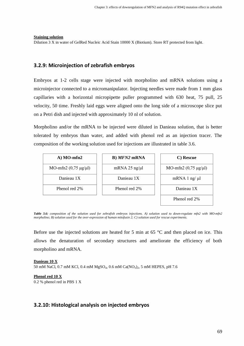
Chapter 3: effects of downregulation of MFN2 and analysis of R94Q mutation effect in zebrafish
69
Staining solution
Dilution 3 X in water of GelRed Nucleic Acid Stain 10000 X (Biotium). Store RT protected from light.
3.2.9: Microinjection of zebrafish embryos
Embryos at 1-2 cells stage were injected with morpholino and mRNA solutions using a
microinjector connected to a micromanipulator. Injecting needles were made from 1 mm glass
capillaries with a horizontal micropipette puller programmed with 630 heat, 75 pull, 25
velocity, 50 time. Freshly laid eggs were aligned onto the long side of a microscope slice put
on a Petri dish and injected with approximately 10 nl of solution.
Morpholino and/or the mRNA to be injected were diluted in Danieau solution, that is better
tolerated by embryos than water, and added with phenol red as an injection tracer. The
composition of the working solution used for injections are illustrated in table 3.6.
A) MO-mfn2 B) MFN2 mRNA C) Rescue
MO-mfn2 (0,75 μg/μl) mRNA 25 ng/μl MO-mfn2 (0,75 μg/μl)
Danieau 1X Danieau 1X mRNA 1 ng/ μl
Phenol red 2% Phenol red 2% Danieau 1X
Phenol red 2%
Table 3.6: composition of the solution used for zebrafish embryos injections. A) solution used to down-regulate mfn2 with MO-mfn2
morpholino; B) solution used for the over-expression of human mitofusin 2; C) solution used for rescue experiments.
Before use the injected solutions are heated for 5 min at 65 °C and then placed on ice. This
allows the denaturation of secondary structures and ameliorate the efficiency of both
morpholino and mRNA.
Danieau 10 X
50 mM NaCl, 0.7 mM KCl, 0.4 mM MgSO4, 0.6 mM Ca(NO3)2, 5 mM HEPES, pH 7.6
Phenol red 10 X
0.2 % phenol red in PBS 1 X
3.2.10: Histological analysis on injected embryos

Chapter 3: effects of downregulation of MFN2 and analysis of R94Q mutation effect in zebrafish
70
Acetylated tubulin immunohistochemistry
An immunohistochemistry against the pan-neural marker acetylated tubulin was performed to
analyse the motorneurons of zebrafish embryos. 48 hpf embryos, previously treated with PTU
to avoid pigments formation, were manually dechorionated and fixed in 4% PFA in PBS for 2
h at RT or at 4 °C ON. They were permeabilized with absolute methanol ON at -20 °C and
then incubated in cold acetone for 7 min at -20 °C. They were rehydrated in solutions at
decreasing concentration of methanol in PBS and washed at RT in agitation with PBTX (3
times for 10 min and 3 times for 30 min).
Embryos were then incubated with the primary mouse anti-acetylated tubulin antibody (Sigma)
diluted 1:1000 in PBTX. Incubation was performed for 48 h at 4 °C.
At the end of the incubation, embryos were washed again with PBTX (3 times for 10 min and 3
times for 30 min) and incubated ON at 4 °C with the secondary antibody diluted 1:100 in
PBTX. Depending on the need, an anti-mouse tetramethylrhodamine or Alexa Fluor® 488
conjugated (both from Invitrogen), were used. Samples were then washed again with PBTX (3
times for 10 min and 3 times for 30 min), embedded in 1,5% low-melting agarose gel and
mounted on microscope slides and analyzed with a Leica DMR microscope equipped with a
Leica DC500 digital camera. Images were processed with ImageJ and CorelDRAW X5
softwares.
PBTX
1% Triton X-100 in PBS.
Phalloidin staining
In order to examine the muscular system of treated and untreated zebrafish larvae, we
performed a staining with fluorescent phalloidin that binds to F-actin, which is particularly
abundant in muscle fibres. Whole 48 hpf embryos were fixed in PFA 4% and permeabilized in
PBTX 2% overnight at 4 °C. Unspecific binding sites were blocked incubating the samples in
blocking solution for 10 minutes. The incubation with tetramethylrhodamine B isothiocyanate-
conjugated phalloidin (Sigma) diluted 1:100 in PBTX was performed 3 h at RT. Embryos were
then washed 3 times for 10 min and 3 times for 30 min in PBTX, embedded in low melting
1,5% agarose and visualised with a Nikon Eclipse E600 microscope equipped with a Biorad
radiance LE 2000 laser scanning system. Z-stack images were generated by scanning segments

Chapter 3: effects of downregulation of MFN2 and analysis of R94Q mutation effect in zebrafish
71
located above the yolk sack extension, centred on the 12th
somite. ImageJ was used to measure
muscle fibres diameter.
PBTX 2%
2% Triton X-100 in PBS
Blocking solution
1% BSA (Bovine Serum Albumin)
1% DMSO (Dimethyl Sulfoxide)
In 1X PBS
Synaptic vesicles and acetylcholine receptor staining
To analyse the correct development of neuromuscular junctions (NMJ) in zebrafish embryos, a
staining of acetylcholine receptors (AChR) and synaptic vesicles was performed. To mark
synaptic vesicles we used a mouse sv2 primary antibody (DHSB, Iowa) together with an anti-
mouse Alexa Fluor® 488 conjugated secondary antibody. The acetylcholine receptors were
visualised with α-bungarotoxin (BTX) Alexa Fluor® 555 conjugated (Invitrogen). Embryos
are fixed in PFA and permeabilized ON at 4 °C with PBTX 2%, then they are incubated ON at
4°C with the sv2 primary antibody diluted 1:50 in PBTX. Larvae are then washed 3 times for
10 min and 3 times for 30 min in PBTX to remove the primer antibody, and then incubated in
PBTX ON at 4 °C with the green anti-mouse secondary antibody (1:100) and the red BTX
(1:200). Embryos are then washed again and mounted on microscope slides embedded with
low melting agarose. Visualization was performed with a confocal microscope and Z-stack
images were analyzed with ImageJ software.
3.2.11: Behavioural analysis on injected embryos
To evaluate the motor capability of MO-Ctrl, MO-mfn2 and rescued embryos, a simple
behavioural assay was used (122). Embryos were allowed to develop to 2 dpf and manually
dechorionated at least 3 hours before the experiment. To assess their motor ability, they were
gently touched with a mounted needle at the dorsal tip of the tail for at least three times. Fishes
were scored as “normally responding” if they can swim for a distance of at least 3 times their
length, as “impaired” if they vibrated, turned around or swim for a short distance, and as
“unresponding” if they did not react after three stimulations.

Chapter 3: effects of downregulation of MFN2 and analysis of R94Q mutation effect in zebrafish
72
3.2.12: Statistical analysis
Statistical analysis were performed using GraphPad Prism version 5.00 with a confidence
interval of 95%. Chi square test was used to analyze the data obtained from the analysis of
mortality, phenotype and movement of zebrafish injected larvae, while t-test and ANOVA
were used to analyse the data obtained from the measurement of muscle fibres.

Chapter 3: effects of downregulation of MFN2 and analysis of R94Q mutation effect in zebrafish
73
3.3: Results
3.3.1: Identification of zebrafish mfn2 and analysis of its expression pattern
Identification of mitofusin 2 gene in zebrafish and conservation analysis
To identify zebrafish mfn2, a similarity search was performed in GenBank using the program
BLAST and the human MFN2 protein sequence (NP_055689) as query. Three entries with a
high similarity were found. The one with the highest score (NP_001121726.1) is the zebrafish
mfn2 RefSeq (also named in zebrafish LOC567448), located in chromosome 8, while the
others are that of mfn1 (NP_956941.1), in chromosome 11, and a hypothetical protein
annotated as mitofusin 1-like (XP_002660753). The identity of the two zebrafish mitofusins
was confirmed by reconstructing the maximum likelihood phylogenetic tree, which was
calculated using the mitofusins sequences from other organisms. As can be seen in figure 3.6,
this analysis confirms the evolution of vertebrates’ mitofusins from an ancestral gene, which
appear to be more similar to the ubiquitous dmfn of Drosophila than to the fzo gene found in
the same organism.
Figure 3.6: The unrooted tree was generated using the web-based pipeline provided by Phylogeny.fr. The horizontal bar at the bottom of the
tree represents a distance of 0,6 substitutions per site. The RefSeq of the sequences used for the analysis are listed in the table on the right.
Genomic regions surrounding mitofusin 2 gene in man and zebrafish, analysed with UCSC
Genome Browser and the most recent genome assemblies available, revealed that the synteny
with genes near the 5’ end of mitofusin 2 is retained (Fig. 3.7 A). In addition the two mitofusin
2 genes of man and zebrafish have maintained the same exons organisation in man and
zebrafish and codes for proteins of the same length (see Fig. 3.7 B).

Chapter 3: effects of downregulation of MFN2 and analysis of R94Q mutation effect in zebrafish
74
Figure 3.7: A) Genomic regions surrounding mitofusin 2 genes in man and zebrafish. Mitofusins are in red and the surrounding genes are
coloured in function of their orthology. Synteny conservation is evident at the 5’ terminus. It is not possible to analyse the 3’end for the
presence of 2 gaps in the zebrafish genome. B) Comparison of mitofusin 2 genes, mRNAs and proteins in man and zebrafish. Note that the
different cDNA length is due to the incomplete sequencing of the UTR regions in zebrafish mfn2.
Subsequent to the genomic and genetic analysis, a comparison between human, mouse and
zebrafish mitofusin 2 proteins was performed, in order to evaluate the conservation degree at
the aminoacidic level (Fig. 3.8). The multi-alignment demonstrates that zebrafish mfn2 shares
the same percentage of similarity (92%) and identity (83%) with human and mouse proteins.
The higher conservation is observed in the functional domains and in particular in the
transmembrane region.
Figure 3.8: Multiple sequence alignment of human, mouse and zebrafish mitofusin 2 polypeptides. In colour are evidenced the domains of the
proteins: GTPase, GTPasic domain; HR1, coiled coil region 1; TM, transmembrane domain; HR2, coiled coil region 2.

Chapter 3: effects of downregulation of MFN2 and analysis of R94Q mutation effect in zebrafish
75
Expression of mfn2 in zebrafish: semiquantitative RT-PCR analysis
A semiquantitative RT-PCR analysis of mfn2 mRNA was performed on whole embryos at
different developmental stages and in different adult zebrafish tissues.
RNA was extracted from embryos between from 2 cells to 5 dpf. cDNA was normalized for the
housekeeping β-actin gene. As shown in figure 3.9, zebrafish mfn2 was found expressed in all
stages analysed. Since the early embryo is transcriptionally silent until blastula, the presence of
mfn2 transcript at 2 and 64 cells indicates that mfn2 has a maternal expression, and that mfn2
mRNA is present in mature form in the unfertilized eggs.
Figure 3.9: semiquantitative RT-PCR analysis of Mfn2 transcript in zebrafish at different stages of development from 2 cells to 5 days post
fertilisation (top panel). Total cDNA was normalised against β-actin gene (lower panel).
It is known that in humans MFN2 is expressed ubiquitously, but is enriched in some tissues
(47). To verify if the pattern of expression of zebrafish Mfn2 resembles the same pattern
observed in man, semiquantitative RT-PCR was performed on RNA extracted from different
adult zebrafish tissues and organs. Mfn2 resulted ubiquitously expressed, but is particularly
enriched in the heart and skeletal muscles, and, at a lower level, in the brain and eyes (Fig.
3.10). A very low amount of mRNA was found in swim bladder, liver, ovary and gut.
Figure 3.10: semiquantitative RT-PCR analysis of Mfn2 transcript in different adult zebrafish tissues (top panel). Total cDNA was normalised
against β-actin gene (lower panel).

Chapter 3: effects of downregulation of MFN2 and analysis of R94Q mutation effect in zebrafish
76
Expression of mfn2 in zebrafish: in situ hybridisation
To investigate more deeply the expression pattern of zebrafish mfn2, we performed a whole
mount in situ hybridisation (WISH) on zebrafish embryos and larvae at different
developmental stages. The antisense probe used was synthesized from the plasmid pCRII-
zMFN2-probe and is complementary to a region of mfn2 cDNA comprised between exon 2 and
6. To assess the specificity of the hybridization, a sense probe was used in a parallel control
experiment at all stages and no staining was detected in any embryo.
With this analysis it was possible to locate precisely the sites of mfn2 expression in embryos
from 2 cells to 48 hours post fertilisation. As can be seen in figure 3.11, mfn2 mRNA is
maternally expressed and ubiquitous until the completion of somitogenesis (Fig 3.11 A-C), but
by 24 hpf (Fig 3.11 D-F) it starts to be upregulated in the anterior part of the embryo, mainly in
the midbrain, in the growing ganglion cell layer of the retina, in the ventral mesenchyme and
along the midline of each miotome. Between 48 and 96 hpf, mfn2 mRNA localize mainly in
cortical brain neurons but remarkable signals are also retained in the otic vesicle, heart and gut
(Fig 3.11G-I).

Chapter 3: effects of downregulation of MFN2 and analysis of R94Q mutation effect in zebrafish
77
Figure 3.11: Mitofusin 2 (mfn2) localization by in situ hybridization during zebrafish embryonic development. Maternal mfn2 mRNAs
are already detectable at the two cell (A) and dome stage (B). Ubiquitous expression is seen from 50% epiboly (C) and throughout
somitogenesis (not shown). (D–F) At 24 h postfertilization (hpf) mfn2 transcripts are mostly localized in the cephalic region of the
embryo with faint gapped signal in the hindbrain (hb) and intense hybridization staining in the midbrain (mb) and retina (r), especially
in the region (see arrow in E) in more close contact to the developing crystalline lens (le). (F) The presence of mfn2 is evident also in
ventral mesenchyme (arrow in F) and along the midline of each miotome (m). At 48 hpf (G) the presence of mfn2 persists in the head
and in the eye but can be also detected in the heart (h) and in the otic vesicles (ov). (H and I) At 4 days post-fertilization the expression
pattern is mainly restricted to the encephalic region (H), in cranial neurons (cn) and otic vesicles (ov). (I) High levels of mfn2
transcripts are also detectable in the gut (g). Animal pole to the top in A–C; anterior to the left in D–I. Dorsal view in D, E, and H.
Ventral view in I. Lateral view in F and G. Scale Bar is 200 μm except for E, F, and I (50 μm).

Chapter 3: effects of downregulation of MFN2 and analysis of R94Q mutation effect in zebrafish
78
3.3.2: Analysis of mfn2 down-regulation effects in the first stages of zebrafish
development
Splice-modifying morpholino design and evaluation of its effect on mfn2 splicing
Since it is known that in mouse the complete knock-out of Mfn2 is embryonically lethal and
given that fertilised oocytes undergo rapid mitochondria movement during the first
developmental stages (123), we decided to use a splice-modifying MO against mfn2 (MO-
mfn2) to avoid the possibility of a complete lethality of the morphant embryos. Given that in
zebrafish mfn2 exon 1 and exon 2 are 5’UTR-coding, MO was designed in the most upstream
position of the protein-coding mRNA, which is the acceptor splice site of intron 2. To induce
the down-regulation of mfn2, zebrafish embryos at 2-4 cell stage were injected with MO-mfn2.
In order to control for unspecific effects induced by the injection of MO, each experiment was
performed in parallel with a control morpholino (MO-Ctrl) with no complementary RNA
targets. To verify the effect of MO-mfn2, total RNA was extracted from 48 hpf embryos
injected with increasing doses of the morpholino and semiquantitative RT-PCR was performed.
This analysis revealed that MO-mfn2 acts by inducing the skipping of exon 3, thus introducing
a premature stop codon in exon 4 (Fig 3.12). The strength of this effect appears to be dose
dependent up to 7,5 ng. At this dose, which was used for all the subsequent experiments, the
reduction of the wild-type transcript was of about the 70%.
Figure 3.12: A) site of annealing and effect of MO-mfn2 on zebrafish mfn2 mRNA. Arrows indicate the annealing sites of the primers used in
the semiquantitative RT-PCR. B) Result of the semiquantitative RT-PCR on embryos injected with MO-Ctrl and different doses of MO-mfn2.
MO-mfn2 injected embryos have an additional lower band compared to that of controls. Sequencing confirmed that the extra-band is an
incorrectly spliced transcript lacking exon 3. This induces a frameshift in the sequence and the introduction of a premature stop codon in exon
4.

Chapter 3: effects of downregulation of MFN2 and analysis of R94Q mutation effect in zebrafish
79
Effects of mfn2 knock-down on early zebrafish development
The first evident effect of mfn2 down-regulation was a reduced vitality of MO-mfn2 injected
embryos (64%) as compared to controls (90%) (Fig 3.13 D). The surviving larvae, although
slightly delayed in growth, do not show evident morphological alterations until 48 hours post
fertilisation (hpf). At this stage the 99% of MO-mfn2 injected larvae (Fig 3.13 E) show a
characteristic phenotype, and morphants were classified according the severity of the
alterations into 3 groups (Fig 3.13 A-C):
Normal: with no evident morphological alterations
Mild: showing curved tail, irregular somites, facial prognathism, underdeveloped eyes
and brain ventricles oedema
Severe: presenting shortened body axis, little head with encephalic necrosis, ventral
expansion of the yolk and trunk oedema.
Mfn2 morphants appeared also severely impaired in their motor ability, as is in the case of
CMT2A patients. To quantify this feature, MO-injected embryos were allowed to develop to 2
days post-fertilization and motility defects were scored with a behavioural assay. While the
96% of control larvae can react to the stimulus by swimming, the majority of morphants didn’t
react (31%) or reacted in a weaker way (52%) (Fig 3.13 F).
Figure 3.13: effects of mfn2 knock-down on zebrafish embryos at 48 hpf. A) At 48 hpf MO-Ctrl injected embryos show no apparent phenotypic
abnormalities, while at the same stage MO-mfn2 treated larvae display a mild (B) or a severe (C) phenotype. Histograms showing the vitality
(D), the phenotype distribution (E) and motility test reaction (F) in control embryos (MO-Ctrl), and in larvae injected with an anti-mfn2
morpholino (MO-mfn2). All the differences found between controls and morphants were tested with Chi-square test and resulted statistically
significant (p < 0.0001).

Chapter 3: effects of downregulation of MFN2 and analysis of R94Q mutation effect in zebrafish
80
Evaluation of motorneurons alterations in mfn2 morphants
Considering that mfn2 morphants showed evident motor abnormalities, we decided to
determine whether the reduced levels of mitofusin could cause growth defects in
motorneurons. Embryos were microinjected with either MO-mfn2 or MO-Ctrl and analysed by
immunohistochemistry against the pan-neural marker acetylated-tubulin. Abnormal length and
structure of ventral extending nerves innervating the somitic muscles, which are formed by the
fasciculation of caudal primary motor neurons (CaPs) and secondary motorneurons, were
analysed in 48 hpf larvae. Motor neurons were scored as “altered” when axons branched above
the horizontal myoseptum or when they appeared truncated or missing. The presence of these
defects was evaluated in trunk motorneurons above the yolk extension in both sides of each
larva. From this analysis were excluded the embryos with a severe phenotype because of the
complete disorganization of their nervous system.
When morphologically normal or mildly altered embryos were analysed, a significant
difference in motorneurons organization between MO-mfn2 and MO-Ctrl treated embryos was
observed. In 74% of MO-Ctrl injected larvae, microtubule staining demonstrated that motor
neurons spread normal axonal projections along the centre of each somite. In contrast, in the
trunk of mfn2 morphant embryos, axons were less intensely stained, the ventral projecting
nerves were abnormally developed and in 67% of MO-mfn2 treated larvae at least one
branched, truncated or missing nerve was identified (Fig. 3.14).
Figure 3.14: Immunohistochemistry of 48 hpf embryos with anti-acetylated tubulin antibody. A) Lateral view of an MO-Ctrl embryo. MO-
mfn2 injected embryos displayed several types of motor neuron alterations (arrows): premature branching (B), truncation (C) and failed
extension (D). Scale bar: 50 μm. E) Trunk motor neurons at the level of the yolk extension were evaluated and embryos were considered
altered if at least one misshaped motor neuron was present. No statistically significant difference between control (MO-Ctrl) and rescued
embryos (MO-mfn2+hMFN2-wt) was observed. Significance was determined by Fisher’s exact test. ***p < 0.0001.

Chapter 3: effects of downregulation of MFN2 and analysis of R94Q mutation effect in zebrafish
81
Analysis of neuromuscular junctions in mfn2 morphant larvae
The formation of neuromuscular junction (NMJ) is a key event in the development of
neuromuscular system. It was recently demonstrated that the aggregation of acetylcholine
receptor clusters (AChR) occurs in muscle fibres before the contact of neurons. After the
elongation of neurons, only the AChR clusters that are contacted by the growth cone can form
functional NMJs, while the others rapidly disappear. In zebrafish AChRs start to pre-pattern at
16 hpf, in advance of the extension of CaP motorneurons, and are stabilised by 20 hpf, when
the elongation of primary motorneurons is almost completed (124). Since the incorrect
formation NMJs can cause movement defects, we performed an analysis of NMJ formation in
mfn2 morphant larvae. The analysis was performed on 48 hpf larvae, since at this stage the
NMJs should be completely developed. To analyse the innervation of muscles, axon terminals
were stained in green by immunohistochemistry with sv2 antibody, which binds to synaptic
vesicles, while acetylcholine receptors were marked with a red fluorescent α-bungarotoxin
(BTX). Embryos were then mounted laterally and visualised with a confocal microscope (Fig.
3.15). This analysis revealed that in mfn2 knock-down larvae the arborisation of intact CaPs
motorneurons is highly diminished (Fig. 3.15 A and A’). Acetylcholine receptors are greatly
reduced (Fig. 3.15 B and B’) and the colocalisation with the signal of sv2, indicating the
formation of a functional neuromuscular junction, is present only in a small area of the mid-
region of some somites, (see arrows in Fig. 3.15 B’). Notably the colocalisation of sv2 and
AChRs appear reduced also in fully elongated nerves, in which there is a complete depletion of
AChR in the distal regions (see fig. 3.15 A’’-B’’).

Chapter 3: effects of downregulation of MFN2 and analysis of R94Q mutation effect in zebrafish
82
Figure 3.15: Effect of mfn2 depletion on neuromuscular synapse formation on 48 hpf larvae. Zebrafish embryos were injected with MO-Ctrl
(A–C) and MO-mfn2 (A’-C’), and stained for synaptic terminals, with sv2 antibody (green), and Acetylcholine receptors (AChR, red), with
bungarotoxin (BTX) at 48 hpf. A) In 48 hpf control larvae caudal primary motor neurons (CaP) completely extended in the mid of each
myotome and started to form many arborizations to contact the myofibres. A’) In MO-mfn2 injected larvae, CaP motor neurons appeared
disorganized and with less arborizations. BTX staining of controls embryos (B) showed the presence of AChR in the mid of each somite, in
regions where the CaP’s arborizations contact the muscular fibres (see merge panel in C). On the contrary, in mfn2-MO-injected larvae (B’),
BTX staining was severely reduced, with a high presence of AChR cluster only in the mid of each somite (arrows in B’) and along the
myosepta. AChR are totally absent from the distal regions of fully elongated motorenurons (see A’’-C’’, which are the enlargement of the grey
box in A’). Scale bar 50 μm.
Effects of mfn2 knock-down on somitic muscles
Considering the defects observed in the neuromuscular junctions formation in mfn2 morphants,
and given that CMT2A patients suffer of a degeneration of muscular fibres that is secondary to
denervation, an analysis of muscular fibres diameter was performed on MO-mfn2 injected
larvae at 48 hpf. Muscle fibres were stained with fluorescent phalloidin, which binds to actin
filaments, mounted laterally and visualized at confocal microscopy. An evident disorganization
of the muscle myosepta structure, that appears U-shaped rather than V-shaped, was observed in
48 hpf MO-mfn2 injected larvae (Fig. 3.16 A-A’). The analysis of muscle fibres diameter
revealed that, consistently with the pathology, mfn2 depletion determine also a reduction in the
size of muscle fibres (2.4 ± 0.04 μm vs. 4.2 ± 0.1 μm) which in addition appear irregularly
aligned and joined less tightly (Fig. 3.16 B).

Chapter 3: effects of downregulation of MFN2 and analysis of R94Q mutation effect in zebrafish
83
Figure 3.16: Knock-down of mfn2 in zebrafish induces somitic and muscular alterations. MO-mfn2 and MO-Ctrl injected embryo were fixed
at 48 h hpf and stained with phalloidin-tetramethylrhodamine that binds to F actin in muscle cells. Confocal microscopy confirmed the
alterations of the shape of somites which compared with controls (A), appeared thinner and U-shaped in morphants (A’). Further analysis of
the muscular fibres revealed that in MO-mfn2 treated embryos the width of the single myofibres is significantly smaller than in controls (B).
Images in A and A’ were centred in the 12th somite and resulted from the merge of 2 images taken at 2 μm of distance. Scale bar 50 μm.
Significance was determined by t test with Welch correction. ***p < 0.0001.
3.3.3: MO-mfn2 rescue with human and R94Q mutated MFN2 transcripts
The effect of morpholinos can be sometimes due to their off-target effect on other genes. For
this reason when working with MOs it is important to include some experiments in order to
control for such events. The use of a control-MO, with no target in the zebrafish transcriptome,
is common to evaluate a toxic effect of the morpholino molecule. To specifically control the
off-target other methods have to be used. The conclusive evidence of the specificity in a knock-
down experiment using morpholinos is the phenotypic rescue with the mRNA of the knocked-
down gene.
Since human and zebrafish mitofusins share a high sequence similarity, we decided to
performed rescue experiments using the human MFN2 transcript. In this way we will be able
also to determine if the human mitofusin 2 can complement the function of the endogenous
zebrafish mfn2, and thus assess if the two proteins share the same functions. In parallel to the
rescue experiments with the wild-type human MFN2 mRNA, we performed also the rescue
with a MFN2 transcript bearing the mutation R94Q in the GTPasic domain. This mutation is
one of the most frequent found in CMT2A patients. It is generally associated to an early onset
and severe phenotype and has a high penetrance. However the mechanism of action (gain or
loss of function) of this, as of other mutations, is not clear. Rescue experiments with the R94Q
mutated transcripts will help to assess if this mutation in vivo can act via a loss of function
mechanism.

Chapter 3: effects of downregulation of MFN2 and analysis of R94Q mutation effect in zebrafish
84
Vitality, morphological phenotype, movement and percentage of embryos with motorneurons
alterations were analysed in morphants rescued with human wild-type (hMFN2-wt) or mutated
(hMFN2-R94Q) MFN2 mRNA.
The coinjection of MO-mfn2 with hMFN2-wt significantly increased the vitality of larvae (p-
value=0,0033) since the percentage of embryos dying at 48 hpf passes from the 36% of
morphants to the 20% in hMFN2-wt rescue. On the other hand, hMFN2-R94Q was not able to
increase the vitality, and causes a percentage of lethality not significantly different to that
observed in morphants (Fig 3.17 A).
Although the injection of hMFN2-wt cannot completely rescue the phenotype of MO-mfn2
morphants, a statistically significant difference in the distribution of phenotypes between
morphants and hMFN2-wt injected embryos was observed (p-value<0,0001). hMFN2-wt
causes an amelioration of the phenotype, reducing the percentage of embryos with a mild and
severe phenotype and inducing an increase of normal embryos which passed from a percentage
of 1,4% in morphants to the 23,2% in hMFN2-wt rescue. Rescue with hMFN2-R94Q can also
ameliorate the phenotype of morphants, but at a lower degree than hMFN2-wt. hMFN2-R94Q
causes an increase of normal embryos to only the 8,3% and do not significantly modify the
percentage of embryos with a severe phenotype (Fig. 3.17 B).
Movement alterations were evident in the 82,8% of morphants. Rescue performed with
hMFN2-wt reduce this percentage to 67,5%. Wild-type transcript acts by increasing the
percentage of normally responding embryos, which passes from the 17,2% of morphant to the
32,5% (p-value=0,0016). Surprisingly hMFN2-R94Q could also ameliorate the motor abilities
of morphants. This effect seems to be as strong as that of hMFN2-wt, since no statistically
significant differences can be noticed in the distributions of motility-phenotypes between
hMFN2-wt and R94Q rescued larvae (Fig. 3.17 C).
The most evident effect of hMFN2-wt transcript was noticed at the level of motorneurons. The
coinjection of hMFN2-wt with MO-mfn2 was able to completely rescue the percentage of
embryos with altered motorneurons found in morphants (67,4%) to a percentage (35,3%) not
significantly different to that of MO-Ctrl injected larvae (26%). Interestingly hMFN2-R94Q
did not rescue these defects, leading to a percentage of embryos with motorneurons alterations
of 58%, not significantly different from that of morphants (p-value=0,1999).

Chapter 3: effects of downregulation of MFN2 and analysis of R94Q mutation effect in zebrafish
85
Figure 3.17: analysis of vitality (A), phenotype (B), movement (C) and motorneuron alterations (D) in morphants rescued with human wild-
type (hMFN2-wt) or mutated (hMFN2-R94Q) mRNA. The results obtained for controls (MO-Ctrl) and morphants (MO-mfn2) are shown as
comparison. A) Analysis of vitality of embryos at 48 hpf. While hMFN2-wt is able to completely rescue the vitality, hMFN2-R94Q do not
ameliorate significantly this characteristic giving a distribution not significantly different from that of morphants. B) hMFN2-wt transcript
significantly ameliorate the phenotypic alterations found in morphants. Also hMFN2-R94Q display an effect, causing a distribution
significantly different to that of morphants, but it result less effective than hMFN2-wt. C) Movement defects found in morphants are partially
rescued by the coinjection of hMFN2-wt with MO-mfn2. Rescue performed with hMFN2-R94Q resulted as efficient as with the wild-type
transcript. E) Motorneuron alterations are completely rescued by hMFN2-wt since no statistical differences between rescue hMFN2-wt and
MO-Ctrl were found with chi-square test. On the contrary, hMFN2-R94Q do not rescue the motorneuron alterations found in morphants,
being the distribution of rescue hMFN2-R94Q not significantly different compared to that of morphants.
(***p < 0.0001; **p < 0.001; *p < 0.01).
3.3.4: Analysis of the gain of function effects of wild-type and mutated MFN2
in zebrafish
Through the analysis of the effects of hMFN2-R94Q rescue we were able to evaluate if the
R94Q mutation retains some functionality in the absence of mfn2, and thus the possibility that
in patients it acts via a loss of function mechanism. To assess if it has also a gain of function
effect, we injected high doses of the mutated mRNA in fertilized zebrafish eggs, in order to
overexpress the protein during their development. We titrated the dose of mRNA and we
decided to use a dose of 250 pg of mRNA per embryo, which was the higest tolerated dose of

Chapter 3: effects of downregulation of MFN2 and analysis of R94Q mutation effect in zebrafish
86
the wild-type transcript. As a control, since mRNA is rapidly degraded, a semiquantitative RT-
PCR using primers specific for the human MFN2 mRNA was performed, in order to verify the
retention of the mRNA after 48 hpf, the time of the analysis (Fig. 3.18). As a control, for each
experiment a batch of embryos was injected in parallel with water or with the same amount of
the wild-type transcript.
Development of larvae injected with water, hMFN2-wt or R94Q
was monitored over 2 days post fertilisation. Phenotypically,
larvae overexpressing wt or R94Q mitofusins were
morphologically not distinguishable from water-injected larvae
and showed the same survival rate. In addition, in contrast with
morphant embryos and CMT2A patients, hMFN2-R94Q
overexpressing larvae showed at 48 hpf the same movement
ability observed in hMFN2-wt and water injected control larvae.
Even though morphologically MFN2 R94Q overexpressing
embryos display any alteration, it is still possible that subtle
motorneuronal and muscular alterations are present but are unable to induce a gross phenotypic
defect in the first developmental stages. For this reason we analysed the structure of ventral
motor nerves by immunohistochemistry with an anti-acetylated-tubulin antibody. As can be
seen in figure 3.19, any difference in the development of motorneurons was evidenced.
Figure 3.19: analysis of motorneuron alterations in water, and 250 pg hMFN2-wt and hMFN2-R94Q injected larvae at 48 hpf. No alterations
were evidenced in the differently injected embryos.
To further analyse the effects of R94Q over-expression in the neuromuscular system, we
performed the analysis of neuromuscular junction by staining motorneurons in green with sv2
3.18: Semiquantitative RT-PCR analysis
performed to evaluate the presence of the
human transcript at 48 hpf in embryos
injected with 250 pg of hMFN2. cDNA
obtained from injected embryos was
amplified using primers specific for the
human sequence. β-actin amplification
was used as a control.

Chapter 3: effects of downregulation of MFN2 and analysis of R94Q mutation effect in zebrafish
87
antibody and AChR in red with a fluorescent BTX. Once again no overt differences were
demonstrated between larvae injected with water, hMFN2-wt or hMFN2-R94Q mRNA (Fig.
3.19).
Figure 3.20: Analisys of the neuromuscular junction of water and 250 pg hMFN2-wt and hMFN2-R94Q injected larvae at 48 hpf. In green are
synaptic terminals stained with sv2 antibody, in red the acetylcholine receptors marked with a fluorescent bungarotoxin. No differences
between water (A-C), 250 pg hMFN2-wt (A’-C’) and hMFN2-R94Q (A’’-C’’) injected larvae were evidenced.
Finally an analysis of somitic muscle fibres was performed after staining with fluorescent
phalloidin. Somitic muscles appear normally developed both in hMFN2-R94Q and wt injected
larvae and the analysis of muscular fibres thickness revealed no significant reduction of their
diameter (Fig. 3.20).

Chapter 3: effects of downregulation of MFN2 and analysis of R94Q mutation effect in zebrafish
88
Figure 3.21: A-C) somitic muscles of 48 hpf injected larvae stained with fluorescent falloidin and visualised at confocal microscope.
Differences in the structure of muscles was not evidenced in water (A), 250 pg hMFN2-wt (B) or hMFN2-R94Q (C) injected larvae. The
analysis of muscle fibres diameter was performed in differently treated embryos, but no statistical differences were found with t-test.

89
Chapter 4: Discussion
Charcot-Marie-Tooth neuropathies (CMT) are a group of genetically heterogeneous diseases of
the peripheral nervous system. They present with progressive distal muscles weakness,
followed, during the disease course, by muscle atrophy. Alterations of sensory nerves are also
present and differentiate CMTs from other related hereditary neuropathies. With a median
prevalence of 1:2500, they are the most common hereditary pathology of the peripheral
nervous system and have been found world-wide in all races and ethnic groups. These diseases
can be extremely disabling, since patients slowly lose their use of lower and, frequently, upper
limbs and represent a serious medical problem for which an effective medical treatment is still
lacking. On the basis of electrophysiological criteria, CMTs are subdivided into demyelinating
(CMT1) and axonal (CMT2) forms. Several genes and loci have been associated to many
different demyelinating and axonal subtypes of CMT. In some cases different mutations in the
same gene are able to cause both CMT1 and CMT2, thus showing the great genetic and clinical
heterogeneity of these pathologies. Among axonal forms, CMT2A was described as the most
common subtype, being responsible for the 16-33% of all axonal cases. CMT2A is associated
to mutations in mitofusin 2 gene (MFN2), which codes for a dynamin-like GTPase found in the
outer mitochondria and on the membranes of endoplasmic reticulum (ER). Together with its
orthologue MFN1, it mediates the events leading to fusion of outer mitochondria membranes.
Both MFN1 and MFN2 are orthologues of the fzo proteins found in invertebrates and yeast and
share with them a conserved domain structure. Mammals’ mitofusin 1 and 2 have a GTPase
domain at the N-terminal, a coiled coil region (HR1) a transmembrane domain and a second
coiled coil region (HR2) at the C-terminus. Mitofusins on opposite mitochondria are able to
interact via their HR2 regions to form homo or hetero-oligomers which tether mitochondria
outer membranes prior to fusion. A functional GTPase domain is necessary to permit this
function and most likely provides the energy for the subsequent fusion event. More than 80
CMT2 disease causing mutations have been described in MFN2. They are most frequently
missense mutations with dominant inheritance and are located preferentially in the GTPase
domain or in the region between this and the HR1 region. However, recently some rare cases of
recessively inherited MFN2 mutations were reported. Mitofusin 2 mutations can frequently
lead to a CMT phenotype complicated by other symptoms. Patients with optic atrophy,
sensoryneural hearing loss or alterations in the central nervous system have been reported. The

Chapter 4: discussion
90
severity of the phenotype is usually related to the age of onset, and patients with earlier
manifestations of the pathology are frequently more severely affected. This clinical subdivision
between early-onset severe and late-onset mild patients does not transpose to a clear genotype
relation. Indeed the way mutations in MFN2 lead to CMT2A is almost unexplained. The
comprehension of the molecular mechanisms underlying this neuropathy is complicated by the
fact that recently the involvement of MFN2 in cellular processes other than mitochondria
fusion has been described. MFN2 and MFN1 have not completely redundant function, and even
if they are both ubiquitous proteins, they have some differences in tissue specific expression.
Contrary to MFN2, MFN1 is involved also in the fusion of inner mitochondria membranes,
being its presence required for the action of OPA1, the inner mitochondria membrane pro-
fusion protein. On the other hand MFN2 seems also involved in the transport of mitochondria
along microtubules. Being the highly active synaptic terminals far from the soma as much as
one meter, motorneurons are greatly dependant on axonal transport of organelles and vesicles.
The role of MFN2 in axonal transport of mitochondria was not well elucidated, but it was
demonstrated that MFN2 directly interacts with the Miro/Milton protein complex, which
allows the movement of mitochondria along microtubules in a Ca++
dependant manner. The
presence of alterations in mitochondria distribution along axons of patients seems to favour this
as the main pathological mechanism of CMT2A disease. MFN2 is found in a little extent also
on the membrane of endoplasmic reticulum (ER). There, by forming homo-dimers with other
MFN2 molecules or hetero-dimers with MFN1, it mediates the tethering of ER to
mitochondria. This is a core event allowing the efficient transport of Ca++
between these two
organelles. In the absence of MFN2 or when mutated forms are expressed, mitochondria detach
from ER causing an overload of Ca++
in the cytosol upon stimulation. This may have important
implications on Ca++
signalling and induction of apoptosis, but also on the Ca++
mediated
mitochondria movement. MFN2 has also a role in cell signalling, which is triggered by its Ras
binding domain in the N-terminal. Besides all these new evidences, the relation of these
multiple functions of MFN2 and their role in neurodegeneration are still unexplained and
particularly the mechanism of action of MFN2 mutations leading to CMT2A is not well
established. In vitro analysis demonstrated that mutations can both disrupt or retain the
capacity of the protein to induce mitochondria fusion, thus meaning that both gain and loss of
function of MFN2 could cause the disease. Interestingly the fusion-incompetent mutations, as
R94Q and T105M, are able in the presence of wild-type MFN1 but not MFN2, to induce the
recovering of mitochondria tubules. This led to the hypothesis that cells with lower MFN1
expression, as neurons, are more susceptible to alterations in MFN2 function. Some mutations

Chapter 4: Discussion
91
were reported to alter ER-mitochondria tethering and mitochondria transport, but those studies
were performed mainly on the R94Q mutation and other mutant forms of the protein have not
been intensely studied. With the aim to identify the genotype-phenotype correlation and clarify
the mechanism of action of MFN2 gene mutations, in this thesis I proceeded in two different
research lines. On one hand I performed a screening for MFN2 mutations in a cohort of 25
patients with diagnosis of CMT2 to identify new causative mutations and to get further insight
into the genotype-phenotype correlation. On the other hand, considering the lack of a useful
animal model for CMT2A, I tried to model this disease in zebrafish (Danio rerio) by knock-
down experiments and gain of function analysis of mfn2.
Patients enrolled in this study were diagnosed with axonal CMT on the basis of clinical
symptoms and electroneurography, which was performed on all adult subjects. Fourteen (56%)
displayed autosomal dominant and two (8%) autosomal recessive inheritance, while the others
were sporadic cases. They all presented with the usual CMT clinical symptoms while upper
limb involvement was reported in nine (36%) subjects. Additional complicating symptoms,
commonly found in CMT2A patients, were present in 6 (24%) subjects. Age at onset was
variable and comprised between 1 and 50 years of age.
Sequencing of all MFN2 exons allowed the identification of 6 causative mutations in 5
probands, since a patients was found to be a compound heterozygote. Of these mutations, 4
were never reported and were not found in control subjects. The MFN2 mutation frequency
found in our CMT2 patients (20%) is in perfect agreement with that reported in literature (16-
33%). All mutations located preferentially in the N-terminal region of the protein, involved
conserved aminoacidic residues. Four were missense, but two novel single aminoacid deletions
were also identified.
Three mutations involved residues that were previously known to be affected, and indeed two
of these were just described in the literature. The novel mutation R94P, identified in patient 10,
involves the arginine 94, which is the most frequently mutated aminoacid found in MFN2.
Substitutions of this residue with the aminoacids glutamine and tryptophan were reported a
total of 22 times independently, in both familial and sporadic cases of CMT2A. It was already
known that this site represents a mutational hot spot, and the finding of a different substitution
with proline at this position confirms this hypothesis. Although CMT2A is a phenotypically
heterogeneous disease, which can present also with different severity in patients of the same
kindred, mutations in arginine 94 usually cause severe disease. CMT2A linked to R94Q, for
example, generally has an onset between 2 and 7 years of age and is frequently complicated by

Chapter 4: discussion
92
additional symptoms, particularly optic atrophy. Patient 10 displays a later onset of the disease,
with the first walking difficulties appearing at 16 years. However his clinical symptoms appear
uncommonly severe for an axonal CMT, being affected by flaccid tetraparesis. From the
pedigree, is possible to speculate that the mutation was dominantly inherited through his
mother (Fig. 2.7). Being so, the mutation R94P in this kindred shows phenotypic
heterogeneity, since the affected mother has a much milder phenotype.
The mutation H165R was found in patient 21. This substitution was already reported five times
in literature and several other mutations affecting histidine 165 were described (see Fig. 2.1).
Patients with H165R mutation display a mild phenotype, generally involving only lower limbs,
and a relatively late onset (6-50 years, mean 20 years). These data are in accordance with the
clinical features of patient 21, who shows an extremely mild disease with late onset.
A third mutation, affecting a previously described residue, is the substitution of a threonine
with a methionine at position 362 identified in patient 24. Interestingly, this patient was found
to be a compound heterozygote, bearing also the previously undescribed mutation K38del.
T362M was reported only in another patient affected by CMT2A with two in trans MFN2
mutations (76). Also in that case, the parent bearing the T362M mutation was unaffected, thus
implying a recessive inheritance. Patient 24 is not the first compound heterozygote described,
and recently other subjects with mutations in both MFN2 alleles have been reported. In all
cases the parents of the proband, bearing only one mutation, result unaffected, although some
subclinical manifestations, as pes cavus, has been described (76, 77). People with compound
heterozygous or homozygous mutations have always early onset and severe phenotype, and
this is in accordance with the clinical manifestations found in patient 24. Indeed this subject
shows a disease of uncommon severity for a CMT2A.. He suffers of all the complicating
symptoms described independently in other patients (optic atrophy, deafness, upper limb
involvement, respiratory failure) with the addition of facial muscle weakness, never reported in
CMT2A. Interestingly, T362M was described also in a CMT2 family where this mutation
shows dominant inheritance and a very mild phenotype. This implies that T362M is a semi-
dominant MFN2 allele. Only another mutation (R707W) was described to cause both dominant
and recessive pattern of inheritance, while the other recessive MFN2 mutations were found in
affected patients exclusively in homozygosis or compound heterozygosis. This is fascinating,
since indicate that possibly these two groups of mutations act via different molecular
mechanisms. Specifically, the fact that mutations found in homozygosis or compound-
heterozygosis cannot induce a phenotype when alone, may indicate they act through a loss of
function mechanism. Interestingly in mouse the complete knock-out of mfn2 is lethal. The fact

Chapter 4: Discussion
93
that these putative loss of function mutations, when present in both alleles, do not cause a lethal
phenotype, probably means they retain a certain level of functionality. It was observed that in
yeast, fzo alleles with loss of function mutations in different domains of the protein are able to
complement each other (64). In this view, it is interesting to note that, of all patients with
recessive MFN2 mutations, compound heterozygotes, in 5 cases on a total of 6, bear mutations
located in two different domains of the protein.
Patient 5 was found to carry an in frame deletion of 3 nucleotides leading to the deletion of
glutamic acid residue at position 329 (E329del). This mutation, located in a conserved position
between the GTPasic domain and HR1, was never reported before and was not found in control
subjects. This mutation is associated to an early onset severe CMT2A phenotype complicated
by optic atrophy. Besides the single aminoacid deletion K38del found in patient 10, only
another in frame deletion (L379_M381del) was described in literature (40). Unfortunately in
the paper by Verhoeven and colleagues, a detailed phenotypic report associated to
L379_M381del is not given, so it is impossible to make a confrontation with the findings of
patient 5.
In one kindred with a dominant axonal CMT I found the missense mutation A738V. This
mutation is located in the C-terminal of the protein, after the HR2 domain. Mutations outside
the GTPase region or the GTPase-HR1 linking region are rare and only 6 of such mutations
have been reported so far (16, 40, 75, 125).
Among the 25 patients I investigated, 20 resulted not to be mutated. To evaluate the possibility
that MFN2 rearrangements were responsible for the pathology in these subjects, I performed a
MLPA (Multiplex Ligation-dependent Probe Amplification) analysis. This was considered
necessary since in recent years there is growing evidence that large gene rearrangements,
involving entire exons, may be causative of several human genetic diseases previously
associated only to point mutations. Some examples are the duplication of PMP22 gene that
cause CMT1A (126) and its deletion, which is associated to hereditary neuropathy with
liability to pressure palsies (HNPP) (127). Among hereditary spastic paraparesis, the subtype
SPG4 is caused in the 40% of cases by exons deletions in spastin gene (128). Moreover, in a
recent paper (77) a deletion comprising exons 7 and 8 of MFN2 was found associated to
CMT2A disease, thus proving that, even though probably rare, deletions of whole exons can be
also a cause of the pathology. MLPA analysis revealed that none of the non-mutated patients
bear a gene rearrangement in MFN2, thus meaning that probably MFN2 rearrangements are a
rare cause of CMT2A.

Chapter 4: discussion
94
The MFN2 screening in CMT2 patients allowed the identification of new disease causing
mutations. Although the phenotype of the disease was confirmed to be variable, some
suggestions of a genotype-phenotype correlation and some possible molecular explanations
were given. A whole comprehension of molecular mechanisms of MFN2 mutations can
however only be reached through in vitro and in vivo studies. While in vitro studies have
proved to be useful to understand some aspects of wild-type and mutant MFN2, in vivo studies
have been slowed down for a long time by the lack of a good model of the disease. Knock-out
mice for either Mfn1 or Mfn2 are full viable and fertile in heterozygous state, but in
homozygosity they die due to placenta defects. Mfn2 conditional knock-out mice display
perinatal lethality as well, but are also motor impaired (62). However, this is due to the
degeneration Purkinje cells in the cerebellum, rather than alterations in motorneurons. To date
three mouse lines expressing only two of the MFN2 disease related mutations (R94Q and
T105M) were developed and just one has a phenotype resembling CMT2A disease (82).
Since the study of numerous different MFN2 mutations, both in man and animal models, is
crucial to understand in detail the pathomechanisms of CMT2A, and considering that that the
strategies used so far appear to be not exhaustive, in the second part of my project I decided to
move on a different animal model. Zebrafish (Danio rerio) offers several advantages that make
it a useful complement to the mouse models of disease. With its short reproductive cycle and
the large number of progeny, contrary to mammal models, it is amenable to several forward
genetic analysis. However, as in mouse, experiments of reverse genetics, particularly knock-
down and over-expression of genes, are also possible and in some cases even more easily
performed. Furthermore, being a vertebrate, anatomy and physiology are more similar to that
of mammals rather than other widely used invertebrate models as Drosophila and
Caenorhabditis. Due to its transparency, zebrafish neuromuscular system has been
characterized in detail and it was shown that its development and functioning is exceedingly
similar to that of higher vertebrates. For this reasons zebrafish appears especially suitable for
the study of neurodegenerative and neuromuscular disorders, and indeed some important
successes in this field have just been obtained. To analyse mitofusin 2 in zebrafish, I first
performed a bioinformatical analysis to retrieve zebrafish mfn2 and evaluate its conservation
degree with the mitofusins of other organisms. mfn2 was found in zebrafish as a single copy
and maps on chromosome 8. This is an important finding, since several genes are duplicated in
zebrafish and the analysis of a gene present in two copies can be more difficult due to the fact
that the two paralogues may both retain the same redundant functions or diverge completely.

Chapter 4: Discussion
95
As expected, zebrafish genome codes also for mfn1, and this is a significant feature as well,
since is known that mitofusin 1 is able to interact and modify the phenotypic expression of
mitofusin 2 mutations. In this view studying mitofusin 2 functions and mutations in a model
with both mitofusins is of great importance, since the results obtained will be more easily and
directly translated to human beings. The conservation analysis made by comparing the protein
sequence of zebrafish mfn2 with that of other organisms, demonstrates the high conservation of
this protein, a feature that is compatible with a very important function of this gene. Mitofusins
2 appears more highly conserved than mitofusins 1, as displayed by the lengths of branches in
the phylogenetic tree reported in figure 3.6. Not only the aminoacidic sequence, but also the
intron-exon gene organisation and the genomic location are retained. A comparison between
human, mouse and zebrafish mitofusin 2 proteins reveals that zebrafish share a high percentage
of similarity (92%) with both mammals and that this similarity is particularly high in the active
domains of the protein. The transmembrane region show a 99% of identity, thus indicating it
has a fundamental role, probably for the correct localisation of the protein. Intriguingly
mutations in this domain were never found in patients with CMT2A. To find out if the pattern
of expression of mfn2 in zebrafish resembles that of man and other mammals, I performed
semiquantitative RT-PCR on embryos at different developmental stages and on several adult
tissues. This analysis revealed that, as in other animals, mfn2 is ubiquitously expressed and it
has also maternal expression. This is common for genes that are important to sustain the rapid
and energy demanding early cell divisions (129), a property that is also expected for mfn2.
While mfn2 expression shows minimal differences on embryos up to 5 days post fertilisation
(dpf), the analysis of adult tissues revealed that in zebrafish mfn2 is highly expressed in heart,
muscles, brain and eyes. This pattern of expression resembles that described in humans, in
which MFN2 is ubiquitous but enriched particularly in heart and skeletal muscles. To analyse
more deeply the expression pattern of mfn2 mRNA also in larval stages, I performed whole
mount in situ hybridisation on embryos and larvae from 1 cell to 2 dpf. This analysis confirmed
the maternal expression of the gene. Moreover it was found that its expression remains
ubiquitous in the very first stages of development, while from the stage of 18 somites it starts
to localise specifically in some regions. Interestingly by 48 hpf the mfn2 mRNA has gained
almost the same tissue specificity found in adults, being expressed mainly in somitic muscles,
heart, brain and eyes. To have an indication of the role of mfn2 during zebrafish development,
this gene has been knocked-down by injecting a specific antisense morpholino in one-four cell
stage embryos. Morpholinos (MOs) are the most widely used anti-sense knock-down tools
available in zebrafish. They have the same bases of DNA, but their neutrally charged

Chapter 4: discussion
96
phosphorodiamidate backbone results in molecules with a higher binding affinity for RNA than
siRNA. MOs, typically long 25 bases, act by steric hindrance and can be both constructed on
the translational start site or on the splice-site of a RNA species. In the first case, MOs will be
able to inhibit both maternal and zygotic transcripts, while in the second case they will only act
on zygotic mRNAs. To perform the knock-down of mfn2, a splice-morpholino (MO-mfn2) was
used essentially for 2 reasons. First the silencing effect of a splice-morpholino is more easily
monitored than translation-MO and requires the execution of a simple RT-PCR analysis, rather
than a western-blot. Second, since in mouse the complete knock-out is lethal and considering
that in zebrafish mfn2 is probably important during the first cell divisions, by using a splice-
morpholino total embryonic lethality observed in knock-out mice was avoided. MO-mfn2 was
designed over the acceptor splice site of intron 2. Being exon 1 and exon 2 5’UTR coding, this
was the most upstream protein coding region targetable. In this way it should be avoided the
possibility of the production of shorter peptides with potential gain of function effect. By
titrating different doses of MO, I found that the injection of 7,5 ng of MO-mfn2 per embryo
gave the best result in term of vitality and penetrance of the phenotype. Indeed it was
immediately clear that mfn2 morphants have a dramatically reduced vitality that resulted dose-
dependent. Moreover a semiquantitative RT-PCR analysis, performed on RNA extracted from
embryos treated with different doses of MO, showed that doses greater than 7,5 ng do not
cause a higher silencing effect. MO-mfn2 acts by inducing the skipping of exon 3, an event
which introduces a premature stop codon in exon 4. This altered transcript is supposed to code
for a protein lacking GTPase and for this reason results probably inactive. In addition, the
triggering of nonsense mediated decay (NMD) of this miss-spliced transcript can be
hypothesized on the basis of the semiquantitative RT-PCR analysis. Using this morpholino-
injection protocol, that allowed to reduce mfn2 expression of about the 70%. evident
phenotypic and behavioural abnormalities in the majority of mfn2 zebrafish morphants were
induced. These alterations were mainly evident in 48 hpf embryos. A small group of larvae
presented with heavy structural abnormalities, while a more frequent one show a distinctive
phenotype, defined by the presence of facial prognathism, underdeveloped eyes, brain
ventricles enlargement and curly-tail. Notably these alterations occurred in the tissue where
mfn2 mRNA is expressed at highest level in the 48 hpf larvae, as demonstrated by WISH.
Moreover, the morphological phenotype I obtained was remarkably similar to that observed in
two morphants developed to study hereditary spastic paraplegia, where MO knock-down
experiments were performed silencing SPG8 and SLC33A1 genes (130, 131). Morphants are
also severely impaired in motor capability. At 48 hpf one third is unable to respond by

Chapter 4: Discussion
97
swimming to a soft touch stimulus at the tail, a reaction that is commonly observed in larvae at
this stage of development. Since at 2 dpf the neuromuscular system of zebrafish is quite
completely formed, the absence of reaction or the diminished response, evident in quite a half
of larvae, could indicate the presence of severe alterations in the neuromuscular system of
morphants. An analysis of neurons structure, performed by immunohistochemistry against the
pan-neural marker acetylated-tubulin, revealed that MO-mfn2 injected larvae have evident
alterations in the morphology of primary motor neurons innervating ventral somites. Larvae
showed differently altered nerves, and both miss-branched, truncated and absent motorneurons
were noticed. Moreover the acetylated-tubulin staining performed in morphants revealed a less
strong signal than controls, with motorneurons frequently hardly visible due to their thinness.
This could imply that motorneurons are abnormally depleted of stable microtubules, a feature
that was noticed in other zebrafish morphants for genes associated to neuromuscular diseases,
as atlastin (132) and spastin (112). To further characterize the neuronal alterations in
morphants, I performed an analysis of neuromuscular junctions (NMJs) formation by staining
pre-synaptic terminals with sv2 antibody and post-synaptic acetylcholine receptors (AChR)
clusters with fluorescent α-bungarotoxin (BTX). It was demonstrated that during
neuromuscular development, AChRs pre-pattern in muscle fibres before their innervation and
this is independent from the expression of growth factors released by pre-synaptic axons. Later
in development, only innervated AChR clusters stabilise. In zebrafish, AChRs pre-patterning is
evident from 16 hpf, but by 20 hpf, with the completion of CaP motorneurons elongation,
NMJs are formed and AChRs that are not innervated are rapidly reabsorbed (124). I analysed
NMJ structure in morphant at 48 hpf, a stage in which evident morphological and movement
abnormalities are just visible, but in which the innervation of muscles and the stabilisation of
AChR clusters should be completed. This analysis revealed that mfn2 morphants have a highly
reduced axonal arborisation and an altered distribution of AChRs clusters, which are reduced in
number and size and do not co localize properly with the axon terminals, especially in the
distal part of nerves. This allows to hypothesize that the innervations of the lateral myotomes
should be slightly reduced. Since CMT2A is characterised by muscle atrophy, that is secondary
to the denervation, I decided to investigate if zebrafish mfn2 morphants display also alterations
in somitic muscles. By staining F-actin with a fluorescent phalloidin toxin, I was able to
evaluate the morphology of somites and measure the thickness of muscle fibres with a confocal
microscope. An evident disorganization of the muscle structure was observed in 48 hpf MO-
mfn2 injected larvae. While controls have a V-shaped myoseptum and well-organized
myofibres, mfn2 morphants exhibited U-shaped somites. Moreover, a significant reduction of

Chapter 4: discussion
98
muscle fibres diameter was demonstrated. Since mfn2 has a high expression in muscles, it is
not clear if these alterations are a consequence of the depletion of mfn2 in muscles or a direct
effect of their denervation. This last possibility is attractive, since it would mimic what happens
in CMT2A patients. However future studies have to be performed to confirm this hypothesis.
Besides the morpholino technique is a simple and rapid method to obtain the knock-down of a
gene in zebrafish, it is not exempted to problems. The main inconvenience is their knock-down
effect on other genes, an event known as off-target. To control for such a possibility, the most
robust method is to perform a phenotypic rescue by coinjecting the MO with a mRNA coding
for the targeted gene. I performed rescue experiment using the mRNA coding for human
MFN2. This was done not only to control for off-target, but also to have a functional indication
of the conservation of mitofusin 2 in humans and zebrafish. Human MFN2 mRNA significantly
increased the survival rate of rescued embryos, which reached almost the same level observed
in controls, suggesting that human MFN2 can efficiently compensate the depletion of
endogenous zebrafish mRNA in early stages of development. Phenotypically, the number of
embryos with a normal morphology was significantly increased and morphants with mild and
severe phenotype reduced. Human MFN2 could also ameliorate the motor impairment found in
morphants, although with a slightly less efficiency. Anyway, the most important result was
obtained at the level of motorneurons, where MFN2 succeeded in completely restore the
distinctive alterations of mfn2 morphants. The fact that the mRNA rescue could not completely
restore the phenotype of mfn2 morphants for some aspects can be easily explained. Indeed
complete rescue can be difficult to obtain for genes with a strict time-dependant regulation of
their expression pattern or for non-ubiquitous genes. In fact in these cases the mRNA injected
to rescue the MO will be expressed also in times and/or tissues where they are normally absent,
thus potentially leading to other gain of function phenotypes different from that obtained with
the MO. Moreover, mRNA distribute not as equally as MO among cells, and this can lead to a
some degree of mosaic expression of the protein. I have demonstrated that in zebrafish, mfn2
has a some degree of tissue-specificity. Moreover the mRNA I injected was almost 3 Kb long,
so I can expect it has some difficulty in equally distributing between cells. For all these
reasons, the amelioration of the phenotype I obtained is to be considered a success. Altogether
these results demonstrate that human MFN2 is able to complement for the absence of zebrafish
endogenous protein, and provide the evidence not only that the alterations found in morphant
are specific, but also that man and zebrafish mitofusin 2 genes share the same functions and
that mitofusin 2 plays an important role in the development of the neuromuscular system. This

Chapter 4: Discussion
99
may also indicate that zebrafish could be a useful tool to study in living embryos the effects of
mutations identified in CMT2A patients.
For this reason, in the last part of my project, I exploited this possibility, performing mfn2
morphant rescue experiment and gain of function analysis of the CMT2A causing mutation
R94Q. The choice to study this mutation was not accidental. This residue is the most frequently
mutated in CMT2A patients and R94Q is usually associated to early onset and severe disease.
Moreover, this mutation has been extensively studied in both in vitro and in vivo models, and it
was suggested that it disrupts the capacity of mfn2 to induce the fusion of mitochondria and
their tethering to ER (63, 70). Furthermore it was also shown that mitofusin R94Q the
capability to promote mitochondria transport along axons (65, 68). However, as for the other
MFN2 mutations, the mechanism of action of R94Q, and particularly its gain or loss of
function activity, is not clear. In mouse the R94Q knock-in line is completely healthy in the
heterozygous state. Homozygous mutants are lethal but in later developmental stages than the
complete knock-out, thus implying that probably R94Q retains some degree of functionality. A
transgenic line overexpressing R94Q in the nervous system was recently developed by Cartoni
and colleagues (82). The homozygous transgenics are motor impaired and show some features,
as muscular atrophy and alterations in mitochondria distribution along axons, which
phenocopies at some degree the human disease. Contrarily to the transgenic for T105M
mutation, that of Cartoni display these alterations late, from 5 months of age.
To study R94Q mutation in zebrafish, I first performed some rescue experiments of the mfn2
morphant using the mutated human MFN2 mRNA. This was performed to evaluate if R94Q act
via a loss of function mechanism. This mode of action seems to be favoured by in vitro studies
performed so far, which are in contrast with the findings on the mouse transgenic model.
Altogether the rescue analysis performed in zebrafish indicates that the R94Q allele has a
diminished ability than the wild-type in inducing the amelioration of morphant phenotype. This
diminished capacity is not equally distributed between the several phenotypic alterations I
analysed. R94Q in fact is completely unable to recover the lethality and the motorneuron
alterations of morphants, but is able to recover, the phenotypic alterations, although with less
efficacy than the wild-type allele. Surprisingly I found that the ability of R94Q in the
amelioration of movement alterations is of the same extent of that obtained with the non
mutated mRNA. It is to note that movement alterations are not as efficiently recovered as other
features by the wild-type mRNA. Thus slight differences in the effect of wild-type and R94Q
mRNA in rescue the morphant phenotype could be hardly noticeable. To evaluate the

Chapter 4: discussion
100
possibility that R94Q act via a gain of function mechanism, which seems favoured by the
results obtained in mouse, I induced the over-expression of the mutated R94Q allele by
injecting high doses of mRNA in 1-4 cell stage embryos. These experiments were performed
using a dose 25 times higher than that used for rescue experiments. In this way I expected to
obtain an over-expression of the protein sufficient to disclose a gain of function effect of
R94Q. Titration experiments, performed using the wild-type transcript, demonstrated that this
dose is tolerated by zebrafish embryos, thus any phenotypic alteration I would found in
zebrafish injected with R94Q mRNA will be specifically caused by the mutated protein. Over-
expression experiments did not reveal any gain of function effect of R94Q in the first stages of
zebrafish development. Larvae at 48 hpf have the same vitality and morphology of wild-type
MFN2 transcript or water control injected embryos. Moreover, they did not display any
alteration neither in the neuromuscular system, as demonstrated by immunohistochemistry and
not in NMJ and muscular fibres. These results demonstrate that, as in mouse, in the first stages
of development the over-expression of R94Q do not induce phenotypic alterations.
Unfortunately it is not possible to evaluate the effects of the over-expression of R94Q in
zebrafish using mRNA injection only, since transcripts are rapidly degraded. This analysis is
however possible using a transgenic line overexpressing R94Q along all the zebrafish life, and
several technologies that would permit to generate this line are just available.
By finding new MFN2 mutations, with this thesis work I contributed to clarify the possible
genotype-phenotype relations involved in CMT2A disease. Moreover, I demonstrated that in
zebrafish, mfn2 if required for the correct development of the neuromuscular system. Last,
since in zebrafish loss of mfn2 can be complemented by the expression of the human
orthologue, I gave also evidences that this animal model can be a useful tool to dissect the
molecular and cellular pathways leading to the disease, though probably it would be necessary
to develop transgenic lines to model MFN2 disease mutations in this animal. The validation of
a zebrafish model for CMT2A could have in future some important applicative outcomes, since
zebrafish is easily amenable to high throughput drug screens experiments, which could lead to
the identification of active molecules to be used for the treatment of CMT2A (Wheeler &
Brändli, 2009).

101
Bibliography
1. Merkwirth C & Langer T (2008) Mitofusin 2 builds a bridge between ER and
mitochondria. (Translated from eng) Cell 135(7):1165-1167 (in eng).
2. Eura Y, Ishihara N, Yokota S, & Mihara K (2003) Two mitofusin proteins, mammalian
homologues of FZO, with distinct functions are both required for mitochondrial fusion.
(Translated from eng) J Biochem 134(3):333-344 (in eng).
3. Charcot JM & Marie P (1886) Sur une forme particuliére d’atrophie musculaire
progressive souvent familial debutant par les pieds et les jambes et atteignant plus tard
les mains. Revue médicale 6:97-138.
4. Tooth HH (1886) The peroneal type of progressive muscular atrophy. (H.K. Lewis &
Co, Ltd, London).
5. Braathen GJ, Sand JC, Lobato A, Hoyer H, & Russell MB (2011) Genetic
epidemiology of Charcot-Marie-Tooth in the general population. (Translated from eng)
Eur J Neurol 18(1):39-48 (in eng).
6. Barisic N, et al. (2008) Charcot-Marie-Tooth disease: a clinico-genetic confrontation.
(Translated from eng) Ann Hum Genet 72(Pt 3):416-441 (in eng).
7. Harding AE & Thomas PK (1980) The clinical features of hereditary motor and sensory
neuropathy types I and II. (Translated from eng) Brain 103(2):259-280 (in eng).
8. Patzko A & Shy ME (2011) Update on charcot-marie-tooth disease. (Translated from
eng) Curr Neurol Neurosci Rep 11(1):78-88 (in eng).
9. P.J. Dyck, P. K. Thomas, J. W. Griffin, Low PA, & Poduslo JF (1993) Hereditary
motor and sensory neuropathies. In: Peripheral Neuropathy (W.B. Saunders,
Philadelphia, PA) 3rd Ed.
10. Garcia CA, et al. (1995) Clinical variability in two pairs of identical twins with the
Charcot-Marie-Tooth disease type 1A duplication. (Translated from eng) Neurology
45(11):2090-2093 (in eng).
11. Lawson VH, Graham BV, & Flanigan KM (2005) Clinical and electrophysiologic
features of CMT2A with mutations in the mitofusin 2 gene. (Translated from eng)
Neurology 65(2):197-204 (in eng).
12. Berciano J & Combarros O (2003) Hereditary neuropathies. (Translated from eng) Curr
Opin Neurol 16(5):613-622 (in eng).
13. Zuchner S & Vance JM (2006) Mechanisms of disease: a molecular genetic update on
hereditary axonal neuropathies. (Translated from eng) Nat Clin Pract Neurol 2(1):45-
53 (in eng).
14. Mersiyanova IV, et al. (2000) A new variant of Charcot-Marie-Tooth disease type 2 is
probably the result of a mutation in the neurofilament-light gene. (Translated from eng)
Am J Hum Genet 67(1):37-46 (in eng).
15. Perez-Olle R, et al. (2005) Mutations in the neurofilament light gene linked to Charcot-
Marie-Tooth disease cause defects in transport. (Translated from eng) J Neurochem
93(4):861-874 (in eng).
16. Zuchner S, et al. (2004) Mutations in the mitochondrial GTPase mitofusin 2 cause
Charcot-Marie-Tooth neuropathy type 2A. (Translated from eng) Nat Genet 36(5):449-
451 (in eng).
17. Puls I, et al. (2003) Mutant dynactin in motor neuron disease. (Translated from eng)
Nat Genet 33(4):455-456 (in eng).

Bibliography
102
18. Zuchner S, et al. (2005) Mutations in the pleckstrin homology domain of dynamin 2
cause dominant intermediate Charcot-Marie-Tooth disease. (Translated from eng) Nat
Genet 37(3):289-294 (in eng).
19. Hinshaw JE (2000) Dynamin and its role in membrane fission. (Translated from eng)
Annu Rev Cell Dev Biol 16:483-519 (in eng).
20. Schafer DA, et al. (2002) Dynamin2 and cortactin regulate actin assembly and filament
organization. (Translated from eng) Curr Biol 12(21):1852-1857 (in eng).
21. Verhoeven K, et al. (2003) Mutations in the small GTP-ase late endosomal protein
RAB7 cause Charcot-Marie-Tooth type 2B neuropathy. (Translated from eng) Am J
Hum Genet 72(3):722-727 (in eng).
22. Kappe G, et al. (2003) The human genome encodes 10 alpha-crystallin-related small
heat shock proteins: HspB1-10. (Translated from eng) Cell Stress Chaperones 8(1):53-
61 (in eng).
23. Vos MJ, Hageman J, Carra S, & Kampinga HH (2008) Structural and functional
diversities between members of the human HSPB, HSPH, HSPA, and DNAJ chaperone
families. (Translated from eng) Biochemistry 47(27):7001-7011 (in eng).
24. Kostenko S & Moens U (2009) Heat shock protein 27 phosphorylation: kinases,
phosphatases, functions and pathology. (Translated from eng) Cell Mol Life Sci
66(20):3289-3307 (in eng).
25. Carra S, Seguin SJ, & Landry J (2008) HspB8 and Bag3: a new chaperone complex
targeting misfolded proteins to macroautophagy. (Translated from eng) Autophagy
4(2):237-239 (in eng).
26. Fontaine JM, Sun X, Benndorf R, & Welsh MJ (2005) Interactions of HSP22 (HSPB8)
with HSP20, alphaB-crystallin, and HSPB3. (Translated from eng) Biochem Biophys
Res Commun 337(3):1006-1011 (in eng).
27. Antonellis A, et al. (2003) Glycyl tRNA synthetase mutations in Charcot-Marie-Tooth
disease type 2D and distal spinal muscular atrophy type V. (Translated from eng) Am J
Hum Genet 72(5):1293-1299 (in eng).
28. McLaughlin HM, et al. (2010) Compound heterozygosity for loss-of-function lysyl-
tRNA synthetase mutations in a patient with peripheral neuropathy. (Translated from
eng) Am J Hum Genet 87(4):560-566 (in eng).
29. Jordanova A, et al. (2006) Disrupted function and axonal distribution of mutant tyrosyl-
tRNA synthetase in dominant intermediate Charcot-Marie-Tooth neuropathy.
(Translated from eng) Nat Genet 38(2):197-202 (in eng).
30. Latour P, et al. (2010) A major determinant for binding and aminoacylation of
tRNA(Ala) in cytoplasmic Alanyl-tRNA synthetase is mutated in dominant axonal
Charcot-Marie-Tooth disease. (Translated from eng) Am J Hum Genet 86(1):77-82 (in
eng).
31. Brittis PA, Lu Q, & Flanagan JG (2002) Axonal protein synthesis provides a
mechanism for localized regulation at an intermediate target. (Translated from eng) Cell
110(2):223-235 (in eng).
32. Benard G, et al. (2007) Mitochondrial bioenergetics and structural network
organization. (Translated from eng) J Cell Sci 120(Pt 5):838-848 (in eng).
33. Chen H & Chan DC (2009) Mitochondrial dynamics--fusion, fission, movement, and
mitophagy--in neurodegenerative diseases. (Translated from eng) Hum Mol Genet
18(R2):R169-176 (in eng).
34. Cuesta A, et al. (2002) The gene encoding ganglioside-induced differentiation-
associated protein 1 is mutated in axonal Charcot-Marie-Tooth type 4A disease.
(Translated from eng) Nat Genet 30(1):22-25 (in eng).

Bibliography
103
35. Pedrola L, et al. (2005) GDAP1, the protein causing Charcot-Marie-Tooth disease type
4A, is expressed in neurons and is associated with mitochondria. (Translated from eng)
Hum Mol Genet 14(8):1087-1094 (in eng).
36. Niemann A, Ruegg M, La Padula V, Schenone A, & Suter U (2005) Ganglioside-
induced differentiation associated protein 1 is a regulator of the mitochondrial network:
new implications for Charcot-Marie-Tooth disease. (Translated from eng) J Cell Biol
170(7):1067-1078 (in eng).
37. Cassereau J, et al. (2011) Mitochondrial dysfunction and pathophysiology of Charcot-
Marie-Tooth disease involving GDAP1 mutations. (Translated from eng) Exp Neurol
227(1):31-41 (in eng).
38. Ben Othmane K, et al. (1993) Localization of a gene (CMT2A) for autosomal dominant
Charcot-Marie-Tooth disease type 2 to chromosome 1p and evidence of genetic
heterogeneity. (Translated from eng) Genomics 17(2):370-375 (in eng).
39. Zhao C, et al. (2001) Charcot-Marie-Tooth disease type 2A caused by mutation in a
microtubule motor KIF1Bbeta. (Translated from eng) Cell 105(5):587-597 (in eng).
40. Verhoeven K, et al. (2006) MFN2 mutation distribution and genotype/phenotype
correlation in Charcot-Marie-Tooth type 2. (Translated from eng) Brain 129(Pt
8):2093-2102 (in eng).
41. Chung KW, et al. (2006) Early onset severe and late-onset mild Charcot-Marie-Tooth
disease with mitofusin 2 (MFN2) mutations. (Translated from eng) Brain 129(Pt
8):2103-2118 (in eng).
42. Zuchner S, et al. (2006) Axonal neuropathy with optic atrophy is caused by mutations
in mitofusin 2. (Translated from eng) Ann Neurol 59(2):276-281 (in eng).
43. Chung KW, et al. (2010) Early-onset Charcot-Marie-Tooth patients with mitofusin 2
mutations and brain involvement. (Translated from eng) J Neurol Neurosurg Psychiatry
81(11):1203-1206 (in eng).
44. Brockmann K, et al. (2008) Cerebral involvement in axonal Charcot-Marie-Tooth
neuropathy caused by mitofusin2 mutations. (Translated from eng) J Neurol
255(7):1049-1058 (in eng).
45. Zhu D, et al. (2005) Charcot-Marie-Tooth with pyramidal signs is genetically
heterogeneous: families with and without MFN2 mutations. (Translated from eng)
Neurology 65(3):496-497 (in eng).
46. Hales KG & Fuller MT (1997) Developmentally regulated mitochondrial fusion
mediated by a conserved, novel, predicted GTPase. (Translated from eng) Cell
90(1):121-129 (in eng).
47. Santel A, et al. (2003) Mitofusin-1 protein is a generally expressed mediator of
mitochondrial fusion in mammalian cells. (Translated from eng) J Cell Sci 116(Pt
13):2763-2774 (in eng).
48. Mozdy AD & Shaw JM (2003) A fuzzy mitochondrial fusion apparatus comes into
focus. (Translated from eng) Nat Rev Mol Cell Biol 4(6):468-478 (in eng).
49. Koshiba T, et al. (2004) Structural basis of mitochondrial tethering by mitofusin
complexes. (Translated from eng) Science 305(5685):858-862 (in eng).
50. Song Z, Ghochani M, McCaffery JM, Frey TG, & Chan DC (2009) Mitofusins and
OPA1 mediate sequential steps in mitochondrial membrane fusion. (Translated from
eng) Mol Biol Cell 20(15):3525-3532 (in eng).
51. Cipolat S, Martins de Brito O, Dal Zilio B, & Scorrano L (2004) OPA1 requires
mitofusin 1 to promote mitochondrial fusion. (Translated from eng) Proc Natl Acad Sci
U S A 101(45):15927-15932 (in eng).
52. Yoon Y, Krueger EW, Oswald BJ, & McNiven MA (2003) The mitochondrial protein
hFis1 regulates mitochondrial fission in mammalian cells through an interaction with

Bibliography
104
the dynamin-like protein DLP1. (Translated from eng) Mol Cell Biol 23(15):5409-5420
(in eng).
53. James DI, Parone PA, Mattenberger Y, & Martinou JC (2003) hFis1, a novel
component of the mammalian mitochondrial fission machinery. (Translated from eng) J
Biol Chem 278(38):36373-36379 (in eng).
54. Westermann B (2010) Mitochondrial dynamics in model organisms: what yeasts,
worms and flies have taught us about fusion and fission of mitochondria. (Translated
from eng) Semin Cell Dev Biol 21(6):542-549 (in eng).
55. Alexander C, et al. (2000) OPA1, encoding a dynamin-related GTPase, is mutated in
autosomal dominant optic atrophy linked to chromosome 3q28. (Translated from eng)
Nat Genet 26(2):211-215 (in eng).
56. Ishihara N, Fujita Y, Oka T, & Mihara K (2006) Regulation of mitochondrial
morphology through proteolytic cleavage of OPA1. (Translated from eng) EMBO J
25(13):2966-2977 (in eng).
57. Casari G, et al. (1998) Spastic paraplegia and OXPHOS impairment caused by
mutations in paraplegin, a nuclear-encoded mitochondrial metalloprotease. (Translated
from eng) Cell 93(6):973-983 (in eng).
58. Waterham HR, et al. (2007) A lethal defect of mitochondrial and peroxisomal fission.
(Translated from eng) N Engl J Med 356(17):1736-1741 (in eng).
59. Hwa JJ, Hiller MA, Fuller MT, & Santel A (2002) Differential expression of the
Drosophila mitofusin genes fuzzy onions (fzo) and dmfn. (Translated from eng) Mech
Dev 116(1-2):213-216 (in eng).
60. Ishihara N, Eura Y, & Mihara K (2004) Mitofusin 1 and 2 play distinct roles in
mitochondrial fusion reactions via GTPase activity. (Translated from eng) J Cell Sci
117(Pt 26):6535-6546 (in eng).
61. Chen H, et al. (2003) Mitofusins Mfn1 and Mfn2 coordinately regulate mitochondrial
fusion and are essential for embryonic development. (Translated from eng) J Cell Biol
160(2):189-200 (in eng).
62. Chen H, McCaffery JM, & Chan DC (2007) Mitochondrial fusion protects against
neurodegeneration in the cerebellum. (Translated from eng) Cell 130(3):548-562 (in
eng).
63. Detmer SA & Chan DC (2007) Complementation between mouse Mfn1 and Mfn2
protects mitochondrial fusion defects caused by CMT2A disease mutations. (Translated
from eng) J Cell Biol 176(4):405-414 (in eng).
64. Griffin EE & Chan DC (2006) Domain interactions within Fzo1 oligomers are essential
for mitochondrial fusion. (Translated from eng) J Biol Chem 281(24):16599-16606 (in
eng).
65. Baloh RH, Schmidt RE, Pestronk A, & Milbrandt J (2007) Altered axonal
mitochondrial transport in the pathogenesis of Charcot-Marie-Tooth disease from
mitofusin 2 mutations. (Translated from eng) J Neurosci 27(2):422-430 (in eng).
66. Vallat JM, et al. (2008) Histopathological findings in hereditary motor and sensory
neuropathy of axonal type with onset in early childhood associated with mitofusin 2
mutations. (Translated from eng) J Neuropathol Exp Neurol 67(11):1097-1102 (in eng).
67. Funalot B, Magdelaine C, Sturtz F, Ouvrier R, & Vallat JM (2009) [Ultrastructural
lesions of axonal mitochondria in patients with childhood-onset Charcot-Marie-Tooth
disease due to MFN2 mutations]. (Translated from fre) Bull Acad Natl Med 193(1):151-
160; discussion 160-151 (in fre).
68. Misko A, Jiang S, Wegorzewska I, Milbrandt J, & Baloh RH (2010) Mitofusin 2 is
necessary for transport of axonal mitochondria and interacts with the Miro/Milton
complex. (Translated from eng) J Neurosci 30(12):4232-4240 (in eng).

Bibliography
105
69. Glater EE, Megeath LJ, Stowers RS, & Schwarz TL (2006) Axonal transport of
mitochondria requires milton to recruit kinesin heavy chain and is light chain
independent. (Translated from eng) J Cell Biol 173(4):545-557 (in eng).
70. De Brito OM & Scorrano L (2008) Mitofusin 2 tethers endoplasmic reticulum to
mitochondria. (Translated from eng) Nature 456(7222):605-610 (in eng).
71. Wang X & Schwarz TL (2009) The mechanism of Ca2+ -dependent regulation of
kinesin-mediated mitochondrial motility. (Translated from eng) Cell 136(1):163-174 (in
eng).
72. Chen KH, et al. (2004) Dysregulation of HSG triggers vascular proliferative disorders.
(Translated from eng) Nat Cell Biol 6(9):872-883 (in eng).
73. de Brito OM & Scorrano L (2009) Mitofusin-2 regulates mitochondrial and
endoplasmic reticulum morphology and tethering: the role of Ras. (Translated from
eng) Mitochondrion 9(3):222-226 (in eng).
74. Chung KW, et al. (2008) Early-onset stroke associated with a mutation in mitofusin 2.
(Translated from eng) Neurology 70(21):2010-2011 (in eng).
75. Calvo J, et al. (2009) Genotype-phenotype correlations in Charcot-Marie-Tooth disease
type 2 caused by mitofusin 2 mutations. (Translated from eng) Arch Neurol
66(12):1511-1516 (in eng).
76. Nicholson GA, et al. (2008) Severe early-onset axonal neuropathy with homozygous
and compound heterozygous MFN2 mutations. (Translated from eng) Neurology
70(19):1678-1681 (in eng).
77. Polke JM, et al. (In press) Early onset recessive axonal Charcot Marie Tooth Disease
due to compound heterozygous mitofusin 2 mutations. Neurology In publication.
78. Neusch C, et al. (2007) Mitofusin 2 gene mutation (R94Q) causing severe early-onset
axonal polyneuropathy (CMT2A). (Translated from eng) Eur J Neurol 14(5):575-577
(in eng).
79. Honda S, Aihara T, Hontani M, Okubo K, & Hirose S (2005) Mutational analysis of
action of mitochondrial fusion factor mitofusin-2. (Translated from eng) J Cell Sci
118(Pt 14):3153-3161 (in eng).
80. Detmer SA, Vande Velde C, Cleveland DW, & Chan DC (2008) Hindlimb gait defects
due to motor axon loss and reduced distal muscles in a transgenic mouse model of
Charcot-Marie-Tooth type 2A. (Translated from eng) Hum Mol Genet 17(3):367-375
(in eng).
81. Huang P, Yu T, & Yoon Y (2007) Mitochondrial clustering induced by overexpression
of the mitochondrial fusion protein Mfn2 causes mitochondrial dysfunction and cell
death. (Translated from eng) Eur J Cell Biol 86(6):289-302 (in eng).
82. Cartoni R, et al. (2010) Expression of mitofusin 2(R94Q) in a transgenic mouse leads
to Charcot-Marie-Tooth neuropathy type 2A. (Translated from eng) Brain 133(Pt
5):1460-1469 (in eng).
83. Miller SA, Dykes DD, & Polesky HF (1988) A simple salting out procedure for
extracting DNA from human nucleated cells. (Translated from eng) Nucleic Acids Res
16(3):1215 (in eng).
84. Don RH, Cox PT, Wainwright BJ, Baker K, & Mattick JS (1991) "Touchdown" PCR to
circumvent spurious priming during gene amplification. (Translated from eng) Nucleic
Acids Res 19(14):4008 (in eng).
85. Schouten JP, et al. (2002) Relative quantification of 40 nucleic acid sequences by
multiplex ligation-dependent probe amplification. (Translated from eng) Nucleic Acids
Res 30(12):e57 (in eng).

Bibliography
106
86. Cho HJ, Sung DH, Kim BJ, & Ki CS (2007) Mitochondrial GTPase mitofusin 2
mutations in Korean patients with Charcot-Marie-Tooth neuropathy type 2. (Translated
from eng) Clin Genet 71(3):267-272 (in eng).
87. Kimmel CB, Ballard WW, Kimmel SR, Ullmann B, & Schilling TF (1995) Stages of
embryonic development of the zebrafish. (Translated from eng) Dev Dyn 203(3):253-
310 (in eng).
88. Barbazuk WB, et al. (2000) The syntenic relationship of the zebrafish and human
genomes. (Translated from eng) Genome Res 10(9):1351-1358 (in eng).
89. Postlethwait J, Amores A, Force A, & Yan YL (1999) The zebrafish genome.
(Translated from eng) Methods Cell Biol 60:149-163 (in eng).
90. Postlethwait JH, et al. (1998) Vertebrate genome evolution and the zebrafish gene map.
(Translated from eng) Nat Genet 18(4):345-349 (in eng).
91. Taylor AM & Zon LI (2009) Zebrafish tumor assays: the state of transplantation.
(Translated from eng) Zebrafish 6(4):339-346 (in eng).
92. Xia W (2010) Exploring Alzheimer's disease in zebrafish. (Translated from eng) J
Alzheimers Dis 20(4):981-990 (in eng).
93. Taylor KL, Grant NJ, Temperley ND, & Patton EE (2010) Small molecule screening in
zebrafish: an in vivo approach to identifying new chemical tools and drug leads.
(Translated from eng) Cell Commun Signal 8:11 (in eng).
94. Grunwald DJ (1996) A fin-de siecle achievement: charting new waters in vertebrate
biology. (Translated from eng) Science 274(5293):1634-1635 (in eng).
95. Haffter P, et al. (1996) The identification of genes with unique and essential functions
in the development of the zebrafish, Danio rerio. (Translated from eng) Development
123:1-36 (in eng).
96. Amsterdam A, et al. (1999) A large-scale insertional mutagenesis screen in zebrafish.
(Translated from eng) Genes Dev 13(20):2713-2724 (in eng).
97. Amsterdam A, et al. (2004) Identification of 315 genes essential for early zebrafish
development. (Translated from eng) Proc Natl Acad Sci U S A 101(35):12792-12797
(in eng).
98. Bassett DI, et al. (2003) Dystrophin is required for the formation of stable muscle
attachments in the zebrafish embryo. (Translated from eng) Development
130(23):5851-5860 (in eng).
99. Newport J & Kirschner M (1982) A major developmental transition in early Xenopus
embryos: II. Control of the onset of transcription. (Translated from eng) Cell 30(3):687-
696 (in eng).
100. Newport J & Kirschner M (1982) A major developmental transition in early Xenopus
embryos: I. characterization and timing of cellular changes at the midblastula stage.
(Translated from eng) Cell 30(3):675-686 (in eng).
101. Kane DA & Kimmel CB (1993) The zebrafish midblastula transition. (Translated from
eng) Development 119(2):447-456 (in eng).
102. Robu ME, et al. (2007) p53 activation by knockdown technologies. (Translated from
eng) PLoS Genet 3(5):e78 (in eng).
103. Eisen JS & Smith JC (2008) Controlling morpholino experiments: don't stop making
antisense. (Translated from eng) Development 135(10):1735-1743 (in eng).
104. Moens CB, Donn TM, Wolf-Saxon ER, & Ma TP (2008) Reverse genetics in zebrafish
by TILLING. (Translated from eng) Brief Funct Genomic Proteomic 7(6):454-459 (in
eng).
105. Carroll D, Beumer KJ, Morton JJ, Bozas A, & Trautman JK (2008) Gene targeting in
Drosophila and Caenorhabditis elegans with zinc-finger nucleases. (Translated from
eng) Methods Mol Biol 435:63-77 (in eng).

Bibliography
107
106. Kawakami K (2007) Tol2: a versatile gene transfer vector in vertebrates. (Translated
from eng) Genome Biol 8 Suppl 1:S7 (in eng).
107. Udvadia AJ & Linney E (2003) Windows into development: historic, current, and
future perspectives on transgenic zebrafish. (Translated from eng) Dev Biol 256(1):1-17
(in eng).
108. Myers PZ, Eisen JS, & Westerfield M (1986) Development and axonal outgrowth of
identified motoneurons in the zebrafish. (Translated from eng) J Neurosci 6(8):2278-
2289 (in eng).
109. Westerfield M, McMurray JV, & Eisen JS (1986) Identified motoneurons and their
innervation of axial muscles in the zebrafish. (Translated from eng) J Neurosci
6(8):2267-2277 (in eng).
110. Panzer JA, et al. (2005) Neuromuscular synaptogenesis in wild-type and mutant
zebrafish. (Translated from eng) Dev Biol 285(2):340-357 (in eng).
111. Boon KL, et al. (2009) Zebrafish survival motor neuron mutants exhibit presynaptic
neuromuscular junction defects. (Translated from eng) Hum Mol Genet 18(19):3615-
3625 (in eng).
112. Wood JD, et al. (2006) The microtubule-severing protein Spastin is essential for axon
outgrowth in the zebrafish embryo. (Translated from eng) Hum Mol Genet
15(18):2763-2771 (in eng).
113. Butler R, Wood JD, Landers JA, & Cunliffe VT (2010) Genetic and chemical
modulation of spastin-dependent axon outgrowth in zebrafish embryos indicates a role
for impaired microtubule dynamics in hereditary spastic paraplegia. (Translated from
eng) Dis Model Mech 3(11-12):743-751 (in eng).
114. Facchin L, Argenton F, & Bisazza A (2009) Lines of Danio rerio selected for opposite
behavioural lateralization show differences in anatomical left-right asymmetries.
(Translated from eng) Behav Brain Res 197(1):157-165 (in eng).
115. Westerfield M ed (2007) The Zebrafish Book-A guide for the laboratory use of
zebrafish (Danio rerio) (University of Oregon Press, Eugene), 5th Edition Ed.
116. Altschul SF, et al. (1997) Gapped BLAST and PSI-BLAST: a new generation of
protein database search programs. (Translated from eng) Nucleic Acids Res
25(17):3389-3402 (in eng).
117. Thompson JD, Higgins DG, & Gibson TJ (1994) CLUSTAL W: improving the
sensitivity of progressive multiple sequence alignment through sequence weighting,
position-specific gap penalties and weight matrix choice. (Translated from eng) Nucleic
Acids Res 22(22):4673-4680 (in eng).
118. Dereeper A, et al. (2008) Phylogeny.fr: robust phylogenetic analysis for the non-
specialist. (Translated from eng) Nucleic Acids Res 36(Web Server issue):W465-469
(in eng).
119. Fujita PA, et al. (2010) The UCSC Genome Browser database: update 2011.
(Translated from Eng) Nucleic Acids Res (in Eng).
120. Thisse C & Thisse B (2008) High-resolution in situ hybridization to whole-mount
zebrafish embryos. (Translated from eng) Nat Protoc 3(1):59-69 (in eng).
121. Shenoy AR & Visweswariah SS (2003) Site-directed mutagenesis using a single
mutagenic oligonucleotide and DpnI digestion of template DNA. (Translated from eng)
Anal Biochem 319(2):335-336 (in eng).
122. Paquet D, et al. (2009) A zebrafish model of tauopathy allows in vivo imaging of
neuronal cell death and drug evaluation. (Translated from eng) J Clin Invest
119(5):1382-1395 (in eng).

Bibliography
108
123. Sun QY, et al. (2001) Translocation of active mitochondria during pig oocyte
maturation, fertilization and early embryo development in vitro. (Translated from eng)
Reproduction 122(1):155-163 (in eng).
124. Flanagan-Steet H, Fox MA, Meyer D, & Sanes JR (2005) Neuromuscular synapses can
form in vivo by incorporation of initially aneural postsynaptic specializations.
(Translated from eng) Development 132(20):4471-4481 (in eng).
125. Engelfried K, et al. (2006) Charcot-Marie-Tooth neuropathy type 2A: novel mutations
in the mitofusin 2 gene (MFN2). (Translated from eng) BMC Med Genet 7:53 (in eng).
126. Raeymaekers P, et al. (1991) Duplication in chromosome 17p11.2 in Charcot-Marie-
Tooth neuropathy type 1a (CMT 1a). The HMSN Collaborative Research Group.
(Translated from eng) Neuromuscul Disord 1(2):93-97 (in eng).
127. Chance PF, et al. (1993) DNA deletion associated with hereditary neuropathy with
liability to pressure palsies. (Translated from eng) Cell 72(1):143-151 (in eng).
128. Depienne C, et al. (2007) Exon deletions of SPG4 are a frequent cause of hereditary
spastic paraplegia. (Translated from eng) J Med Genet 44(4):281-284 (in eng).
129. Dumollard R, Duchen M, & Carroll J (2007) The role of mitochondrial function in the
oocyte and embryo. (Translated from eng) Curr Top Dev Biol 77:21-49 (in eng).
130. Valdmanis PN, et al. (2007) Mutations in the KIAA0196 gene at the SPG8 locus cause
hereditary spastic paraplegia. (Translated from eng) Am J Hum Genet 80(1):152-161 (in
eng).
131. Lin P, et al. (2008) A missense mutation in SLC33A1, which encodes the acetyl-CoA
transporter, causes autosomal-dominant spastic paraplegia (SPG42). (Translated from
eng) Am J Hum Genet 83(6):752-759 (in eng).
132. Fassier C, et al. (2010) Zebrafish atlastin controls motility and spinal motor axon
architecture via inhibition of the BMP pathway. (Translated from eng) Nat Neurosci
13(11):1380-1387 (in eng).