Embed Size (px)
Citation preview
Università degli Studi di Padova
Dipartimento di Scienze Chirurgiche, Oncologiche e Gastroenterologiche
___________________________________________________________________
SCUOLA DI DOTTORATO DI RICERCA IN
ONCOLOGIA E ONCOLOGIA CHIRURGICA
XXVIII CICLO
PROTEIN KINASE CK2 IN DIFFUSE LARGE B-CELL LYMPHOMA:
DEFINING ITS ROLE TO SHAPE NEW THERAPIES
Direttore della Scuola : Ch.mo Prof. PAOLA ZANOVELLO
Supervisore : Dr. FRANCESCO PIAZZA
Dottorando : ELISA MANDATO
1
INDEX
INDEX ................................................................................................................................. 1
ABBREVIATIONS ............................................................................................................ 3
AMINO ACID ABBREVIATIONS.................................................................................. 5
INTRODUCTION ............................................................................................................ 11
PROTEIN KINASE CK2 .............................................................................................. 11
STRUCTURE ............................................................................................................ 11
FUNCTIONS ............................................................................................................. 13
REGULATION .......................................................................................................... 15
CANCER ................................................................................................................... 16
CK2 in B-ALL ....................................................................................................... 17
CK2 in MM and MCL ........................................................................................... 18
CK2 in B-CLL ....................................................................................................... 19
CK2 INHIBITOR CX-4945 (SILMITASERTIB) ......................................................... 20
B-CELLS ....................................................................................................................... 21
BCR SIGNALLING PATHWAY ................................................................................. 22
IP3 MEDIATED Ca++ RELEASE .............................................................................. 24
NF-κB PATHWAY ................................................................................................... 24
PI3K/AKT PATHWAY ............................................................................................. 27
DIFFUSE LARGE B-CELL LYMPHOMA (DLBCL) ................................................. 28
ABC-DLBCL ............................................................................................................. 30
GCB-DLBCL ............................................................................................................. 31
BCR SIGNALLING INHIBITORS ............................................................................... 31
FOSTAMATINIB: A SYK INHIBITOR .................................................................. 32
IBRUTINIB: A BTK INHIBITOR ............................................................................ 33
AIM OF THE STUDY ..................................................................................................... 35
MATERIALS AND METHODS .................................................................................... 37
PROTEIN EXTRACTION ............................................................................................ 37
WHOLE PROTEIN EXTRACTION ......................................................................... 37
PROTEIN QUANTIFICATION ................................................................................ 37
SDS-PAGE ................................................................................................................ 38
WESTERN BLOT (WB) ............................................................................................... 39
ANTIBODIES ............................................................................................................ 40
RNA PURIFICATION .................................................................................................. 40
REVERSE TRANSCRIPTION ................................................................................. 41
REAL-TIME PCR ......................................................................................................... 42
FLOW CYTOMETRY .................................................................................................. 44
EVALUATION OF APOPTOSIS WITH ANNEXIN V/PROPIDIUM IODIDE ..... 44
CALCIUM FLUX ASSAY ........................................................................................ 45
CELL LINES AND HEALTHY DONORS’ CELLS .................................................... 46
TISSUE SAMPLES ....................................................................................................... 47
CHEMICALS ................................................................................................................ 47
2
IMMUNOHISTOCHEMISTRY (IHC) ......................................................................... 47
IMMUNOFLUORESCENCE (IF) ................................................................................ 48
MTT ASSAY ................................................................................................................. 48
STATISTICAL ANALYSIS ......................................................................................... 49
RESULTS ......................................................................................................................... 51
CK2α IS EXPRESSED IN FOLLICLES OF NORMAL LYMPHOID TISSUE ......... 51
CK2α IS ABUNDANTLY EXPRESSED IN DLBCL SAMPLES FROM PATIENTS........................................................................................................................................ 51
CK2α HAS A PREVALENT NUCLEAR LOCALIZATION IN DLBCL SAMPLES AND CELL LINES........................................................................................................ 52
CK2 IS OVEREXPRESSED IN DLBCL CELL LINES WHEN COMPARED TO NORMAL B-CELLS ..................................................................................................... 54
CK2 INHIBITION WITH CX-4945 REDUCES SURVIVAL OF DLBCL, BUT NOT OF HEALTHY PBMCs ................................................................................................. 56
CK2 CHEMICAL INHIBITION WITH CX-4945 REDUCES Ca++ RELEASE FROM THE ER STORES .......................................................................................................... 57
CK2 REGULATES AKT, NOT BTK AND PLCγ2 PHOSPHORYLATION AFTER BCR STIMULATION ................................................................................................. 59
CX-4945 SYNERGIZES WITH BCR INHIBITORS IN INDUCING DLBCL CELL DEATH .......................................................................................................................... 60
CK2 INHIBITION INCREASES APOPTOSIS INDUCED BY THE BCR BLOCKADE .................................................................................................................. 63
CX-4945 POTENTIATES AKT INHIBITION INDUCED BY THE BCR BLOCKADE .................................................................................................................. 67
CK2 INHIBITION REDUCES NF-κB RELA PHOSPHORYLATION, INCREASED BY BCR BLOCKADE ................................................................................................ 68
CK2 INHIBITION SIMULTANEOUSLY WITH BCR BLOCKADE REDUCES THE
EXPRESSION OF NF-κB TARGET GENES .............................................................. 69
DISCUSSION ................................................................................................................... 71
CONCLUSIONS .............................................................................................................. 75
REFERENCES ................................................................................................................. 77
PUBLICATIONS ............................................................................................................. 83
3
ABBREVIATIONS
Ab antibody
ABC activated B-cell type
Ag antigen
ALL acute lymphoblastic leukemia
AV annexin V
BCR B-cell receptor
BM bone marrow
BSA bovine serum albumin
BTK bruton tyrosine kinase
CI combination index
CLL chronic lymphocytic leukemia
DAG diacylglycerol
DLBCL diffuse large B-cell lymphoma
DMAT 2dimethylamino-4,5,6,7-tetrabromo-1H-benzimidazole
EPK eukaryotic protein kinase
ER endoplasmic reticulum
FBS fetal bovine serum
FL follicular lymphoma
GC germinal center
GCB germinal center B-cell type
H&E haematoxylin & eosin
IF immunofluorescence
Ig immunoglobulin
IHC immunohistochemistry
IκB inhibitor of κB
IKK IκB kinase
IP3 inositol triphosphate
ITAM immunoreceptor tyrosine-based activation motif
LEF lymphoid enhancing factor
MCL mantle cell lymphoma
4
MM multiple myeloma
NFAT nuclear factor of activated T cells
NF-κB nuclear factor kappa-light-chain-enhancer of activated B cells
NHL non-Hodgkin lymphoma
PBS phosphate buffered saline
PC plasma cells
PH pleckstrin homology
PI propidium iodide
PIP2 phosphatidylinositol 4,5-bisphosphate
PLC phospholipase C
PTEN phosphatase and tensin homolog
SD standard deviation
SYK spleen tyrosine kinase
TAD transcription activation domain
TBB 4,5,6,7-tetrabromobenzimidazole
TBS tris buffered saline
TCF T-cell factor
TF transcription factor
TNF tumor necrosis factor
WHO World Health Organization
5
AMINO ACID ABBREVIATIONS
A Ala Alanine
C Cys Cysteine
D Asp Aspartic acid
E Glu Glutamic acid
F Phe Phenylalanine
G Gly Glycine
H His Histidine
I Ile Isoleucine
K Lys Lysine
L Leu Leucine
M Met Methionine
N Asn Asparagine
P Pro Proline
Q Gln Glutamine
R Arg Arginine
S Ser Serine
T Thr Threonine
V Val Valine
W Trp Tryptophan
Y Tyr Tyrosine
X generic amino acid

6
7
ABSTRACT
CK2 is a highly conserved Ser/Thr protein kinase, consisting of two catalytic (α) and two
regulatory (β) subunits assembled to form a tetramer. It is involved in a broad variety of
cellular processes, among which survival, proliferation, differentiation, DNA damage and
other stress responses, leading to the activation of context-specific transcription factors
such as c-Myc and NF-κB. This kinase has been found overexpressed in several solid
tumors and hematologic malignancies, and its overexpression seems to be an unfavorable
prognostic marker. It has been fully demonstrated that CK2 acts as a potent antiapoptotic
factor that promotes a “non-oncogene addiction” phenotype in cancer cells. In other
words, high CK2 levels and activity contribute to create a cellular environment favorable
to the establishment and maintenance of a neoplastic phenotype. In particular, it was
recently shown that many B-cell derived tumors, like multiple myeloma, mantle cell
lymphoma and chronic lymphocytic leukemia, rely on high CK2 activity and that its
downmodulation induces malignant cell death without significantly affecting normal B
lymphocytes.
Diffuse Large B-Cell Lymphoma (DLBCL) is an aggressive B-cell derived neoplasia that
originates from follicles and is the most common type of non-Hodgkin lymphoma,
accounting for about 40% of all cases. It is divided into two subtypes: Germinal Center B-
cell like (GCB) and Activated B-Cell like (ABC) DLBCL, characterized by different
genetic lesions and, therefore, variable response to therapy. Up to one-third of patients
does not achieve cure with initial therapy and has refractory disease or relapse. The
standard salvage treatment for these patients is autologous stem cell transplantation, but
success rates are poor and most of them succumb to the disease. These facts clearly
demonstrate the need for new rational combination therapeutics.
It is well known that the B-Cell Receptor (BCR) signalling strongly influences B-cell
development and is fundamental for peripheral B-cell survival. After BCR ligation by the
antigen, the signal is transduced across the plasma membrane and propagated inside the
cell through a group of intracellular proteins, which interact to form a complex, called
signalosome. Among these proteins there are the tyrosine kinases SYK and BTK, the
phospholipase PLCγ2 and the adaptor BLNK. Once activated by SYK, BTK
phosphorylates PLCγ2, which in turn generates IP3 thus causing Ca++ release from the
endoplasmic reticulum stores. Ca++ acts in the cytoplasm as a second messenger that
8
binds several Ca++-dependent proteins that are then able to activate transcription factors,
like NFAT and NF-κB, modifying gene expression. The result of this process consists in
activation, expansion, antigen presentation and B-cell differentiation. For these reasons, it
comes as no surprise that inhibitors targeting the BCR signalling have shown promising
therapeutic outcomes for patients with B-cell lymphomas.
Here we show that α and β subunits of protein kinase CK2 are overexpressed in ABC-
and GCB-DLBCL primary patient samples and immortalized cell lines when compared
with normal counterparts. Moreover, we demonstrate that CK2 inhibition with CX-4945,
an ATP-competitive CK2 inhibitor currently under clinical trials, causes apoptosis of
DLBCL cell lines in a dose and time dependent fashion, and that malignant cell death is
significative even at low drug doses not toxic to normal counterparts. We also reveal that
the downmodulation of CK2 catalytic activity leads to a reduction in Ca++ release from
the endoplasmic reticulum stores, and impairs AKT and NF-κB RELA phosphorylation
after BCR stimulation. These findings propose a role for CK2 downstream of the BCR
engagement, in controlling survival pathways crucial for B-cell endurance. Furthermore,
we found out that CX-4945 synergises with inhibitors of kinases, like SYK and BTK,
essential in spreading the BCR signal, thus proving that this drug combination enhances
DLBCL cell death and could be considered an effective therapeutic strategy.
9
RIASSUNTO
CK2 è una Ser/Thr chinasi altamente conservata dal punto di vista evolutivo, costituita da
due subunità catalitiche (α) e due subunità regolatorie (β) unite a formare un tetramero.
Essa è coinvolta in numerosi processi cellulari, tra cui sopravvivenza, proliferazione,
differenziamento, risposta al danno al DNA e ad altri stress, portando in definitiva
all’attivazione di specifici fattori di trascrizione, come c-Myc ed NF-κB. Questa chinasi è
stata trovata sovrespressa in svariati tumori solidi e neoplasie ematologiche, portando ad
una correlazione tra alti livelli di CK2 e prognosi sfavorevole. È stato ampiamente
dimostrato che CK2 agisce come un potente fattore antiapoptotico nelle cellule tumorali,
promuovendo un meccanismo definito “non-oncogene addiction”. In altre parole,
l’overespressione e l’aumento dell’attività catalitica dell’enzima contribuiscono
notevolmente a creare un ambiente intracellulare favorevole allo sviluppo e al
consolidamento di un fenotipo neoplastico. In particolare, è stato recentemente dimostrato
che molte neoplasie, derivate dalla trasformazione maligna dei linfociti B, come il
mieloma multiplo, il linfoma mantellare e la leucemia linfatica cronica, dipendono da
un’aumentata attività di CK2 per il loro mantenimento; infatti, una sua inibizione è in
grado di indurre apoptosi cellulare.
Il linfoma diffuso a grandi cellule (DLBCL) è una neoplasia di tipo aggressivo derivata
dalla trasformazione dei linfociti B nel follicolo ed è il tipo più comune di linfoma non-
Hodgkin, rappresentando circa il 40% di tutti i casi. È suddiviso in due sottotipi: uno di
derivazione da cellule B del centro germinativo (GCB), l’altro di derivazione da cellule B
post centro germinativo (ABC), che sono caratterizzati da differenti alterazioni genetiche
e, di conseguenza, da una differente risposta alla terapia. Fino a un terzo dei pazienti non
raggiunge la cura con la terapia iniziale e sviluppa una malattia refrattaria o ricade. Il
trattamento di salvataggio standard per questi pazienti è il trapianto autologo di cellule
staminali, ma il tasso di successo è scarso e la maggior parte di essi non sopravvive alla
malattia, dimostrando chiaramente la necessità di nuove terapie di combinazione.
È risaputo che il segnale generato dal recettore dell’antigene B (BCR) è in grado di
influenzare il differenziamento del linfocita B nella milza e la sua sopravvivenza a livello
periferico. In seguito al legame del recettore da parte dell’antigene, il segnale viene
trasmesso attraverso la membrana plasmatica e propagato all’interno tramite un gruppo di
proteine intracellulari, che si combinano a formare un complesso denominato
10
signalosoma. Tra queste proteine figurano le tirosin chinasi SYK e BTK, la fosfolipasi
PLCγ2 e l’adattatore BLNK. Una volta attivata da SYK, BTK è in grado di fosforilare
PLCγ2, che, a sua volta, genera IP3, il quale induce il rilascio di Ca++ dal reticolo
endoplasmatico. Il Ca++ agisce nel citoplasma come secondo messaggero, interagendo
con varie proteine Ca++-dipendenti, che attivano fattori di trascrizione, come NFAT e NF-
κB, modificando, in tal modo, l'espressione genica. Il risultato di questo processo consiste
in attivazione, espansione, presentazione dell’antigene e differenziamento del linfocita B.
Non sorprende, perciò, che gli inibitori della cascata del segnale del BCR abbiano
dimostrato risultati terapeutici promettenti in pazienti con linfomi di tipo B.
In questo lavoro di tesi si evidenzia che le subunità α e β di CK2 sono sovrespresse sia in
campioni primari, che in linee cellulari immortalizzate di ABC- e GCB-DLBCL, rispetto
alle controparti non neoplastiche. Inoltre, si dimostra che l'inibizione di CK2 con CX-
4945, un inibitore di CK2 attualmente in trial clinici, provoca l'apoptosi di linee cellulari
di DLBCL in maniera dose e tempo dipendente e che l'aumento della morte delle cellule
neoplastiche è significativa anche alle dosi di farmaco che non uccidono le cellule
normali. Inoltre, l’abbassamento dell’attività catalitica di CK2 porta ad una riduzione del
Ca++ rilasciato dal reticolo endoplasmatico, e compromette la fosforilazione di AKT e
NF-κB RELA, in seguito a stimolazione del BCR. Questi risultati propongono un ruolo
per CK2 a valle del BCR, nel controllo di vie del segnale pro sopravvivenza centrali per il
linfocita B. Infine, si evidenzia che il CX-4945 sinergizza con inibitori di chinasi
essenziali per la propagazione del segnale del BCR, quali SYK e BTK, provando che
questa combinazione di farmaci aumenta la morte delle cellule linfomatose e può
considerarsi un’efficace strategia terapeutica.
11
INTRODUCTION
PROTEIN KINASE CK2
Protein kinase CK2 is a highly conserved serine/threonine kinase expressed in all
eukaryotic cells. It is now abundantly clear that it is very pleiotropic and capable of
phosphorylating more than 500 potential substrates in all cellular compartments. CK2
takes part in the regulation of a broad array of cellular processes including proliferation,
survival and differentiation [1]. Abnormally high CK2 levels have been observed both in
solid tumors (breast, prostate, lung, kidney, head and neck) and, more recently, in
haematological malignancies (acute myeloid leukemia (AML), MM, chronic lymphocytic
leukemia (CLL), mantle cell lymphoma (MCL)). CK2 can provide a specific environment
favorable for transformation by combining with oncogenes (e.g. c-Myc) or by the
modulation of oncogenic signals (e.g. Wnt pathway) [2]. Moreover, CK2 acts as a potent
suppressor of apoptosis, therefore, increased CK2 expression in cancer cells sustains
survival and blocks the normal apoptotic activity. Based on this involvement in sustaining
tumorigenesis, CK2 has recently attracted attention as a potential therapeutic target.
STRUCTURE
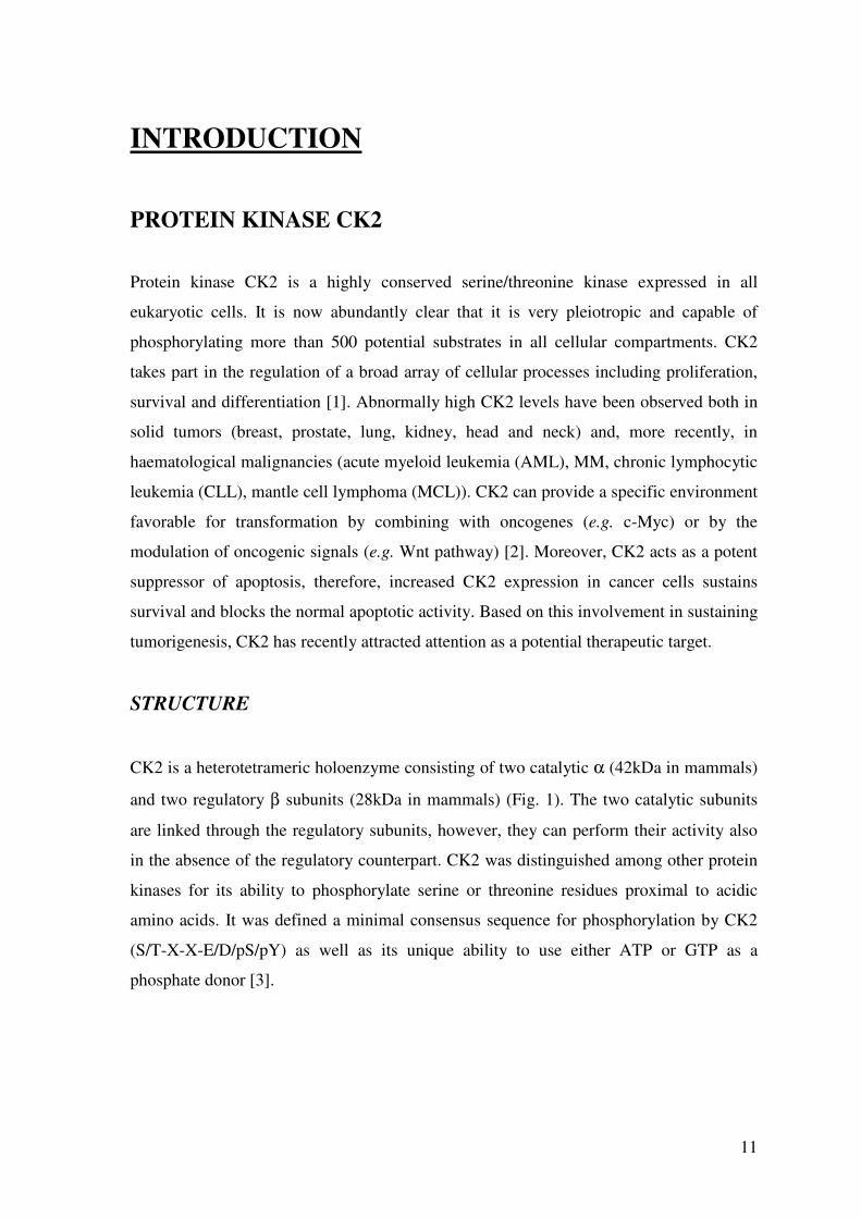
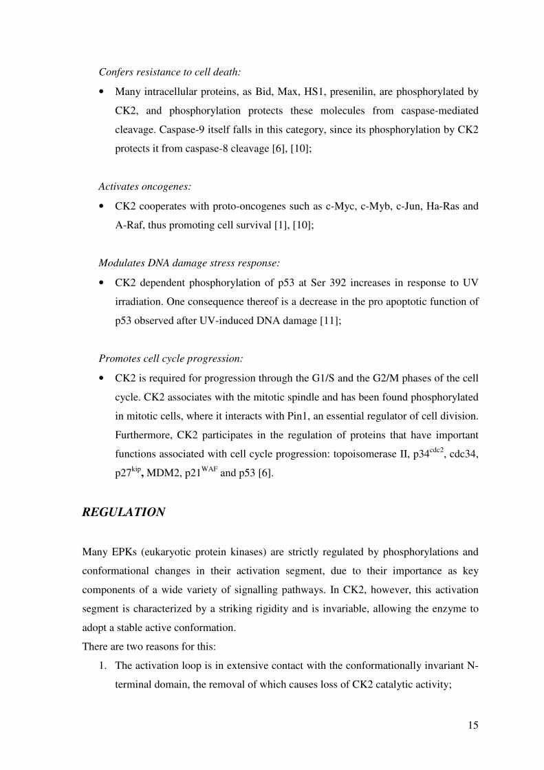
CK2 is a heterotetrameric holoenzyme consisting of two catalytic α (42kDa in mammals)
and two regulatory β subunits (28kDa in mammals) (Fig. 1). The two catalytic subunits
are linked through the regulatory subunits, however, they can perform their activity also
in the absence of the regulatory counterpart. CK2 was distinguished among other protein
kinases for its ability to phosphorylate serine or threonine residues proximal to acidic
amino acids. It was defined a minimal consensus sequence for phosphorylation by CK2
(S/T-X-X-E/D/pS/pY) as well as its unique ability to use either ATP or GTP as a
phosphate donor [3].
12
Figure 1. Ribbon diagram illustrating the high-resolution structure of tetrameric CK2 (modified from
[4]).
• CK2α: The α subunit is made up of a catalytic core composed of two major
folding domains (N- and C-terminal) harbouring the active site in between (Fig. 1).
In humans, two different isoforms of the catalytic subunit (designated CK2α and
CK2α'), encoded by distinct genes, were initially characterized. With the
exception of their unrelated C-terminal domains, these two isoforms are very
similar with approximately 90% identity within their catalytic domain. It is well
established that they are closely related and show considerable functional overlap;
indeed, knockout of the gene encoding CK2α' in mice results in viable offspring
when heterozygous mice are bred to homozygosity, suggesting that α has the
capacity to compensate for α' in the context of viability. However, male are sterile
and display defects in spermatogenesis, demonstrating that the functional
compensation is not absolute [4]–[6].
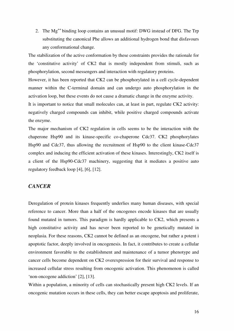
• CK2β: Only one known form of the regulatory β subunit has been identified in
mammals, but multiple forms have been identified in other organisms, such as
Saccharomyces cerevisiae. CK2β is highly conserved among species and x-ray
13
crystallography studies have determined that a dimer of the β subunits forms the
core of the tetramer (Fig. 1).
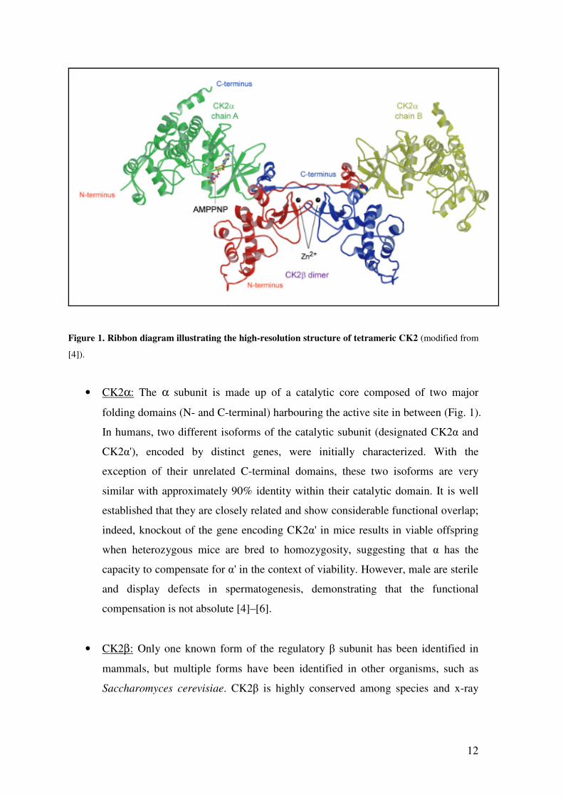
A large proportion of CK2β has been shown to be phosphorylated at an
autophosphorylation site consisting of Ser 2, 3 at its N-terminus and this may
regulate its proteasome-dependent degradation. CK2β is also phosphorylated at
Ser 209 in the C-terminus in a cell-cycle dependent manner by p34cdc2 (Fig. 2) [6].
Figure 2. The regulatory CK2β subunit. Linear representation of CK2β, illustrating the main
elements within its amino acid sequence (modified from [6]).
CK2β monomer has a ‘body’ consisting of the N-terminal domain and a
dimerization domain, the latter containing the zinc-finger region, characterized by
four Cys residues, which mediate the interaction allowing the β dimer to form the
core of the holoenzyme. CK2β dimerization precedes catalytic subunit binding
and is a prerequisite for the formation of the tetramer. The last 33 amino acids
form the ‘tail’ of the monomer and contain the CK2α interaction motif. This C-
terminal region is responsible for the ability of CK2β to enhance and stabilize
CK2 catalytic activity (Fig. 2) [6].
FUNCTIONS
CK2 is a signalling enzyme that behaves as an anti-apoptotic agent implying on different
cellular functions, kinase pathways and biochemical reactions, which ultimately
cooperate to promote cell survival.
14
Sustains proliferative signalling cascades:
• NF-κB. This transcription factor (TF) is normally sequestered in the cytosol by
binding to its inhibitor IκB. CK2 phosphorylates IκB thus promoting its
degradation through the proteasome machinery, promotes IKKβ mediated
phosphorylation of NF-κB RELA on Ser 356, as well as phosphorylates RELA in
Ser 529, increasing its transcriptional capability [7];
• PI3K/PTEN/AKT. Here again CK2 operates as a multisite regulator. The tumor
suppressor PTEN is the phosphatase that dephosphorylates PIP3
(phosphatidylinositol 3, 4, 5 triphosphate), thus maintaining the PI3K/AKT signal
down, under resting conditions. It has been demonstrated that phosphorylation of
PTEN at Ser 380 by CK2, while regulating PTEN protein stability, has an
inhibitory effect on its activity, with the final result of stimulating AKT-dependent
signalling. A second level of CK2 involvement in this pathway is represented by
AKT itself: beside a physical interaction between the two kinases, a direct
phosphorylation of Ser 129 by CK2 has been identified, which enhances the
catalytic activity of AKT. There is also an indirect effect of this CK2-mediated
phosphorylation, since it contributes to maintain high levels of phospho Thr 308
by PDK1, by ensuring a stable association with the chaperone protein Hsp90,
known to protect Thr 308 from dephosphorylation. Moreover, CK2 down
modulation reduces AKT activating phosphorylation at Ser473, mediated by
mTOR [8];
• Wnt. β-catenin is a transcriptional co-factor in the Wnt signalling pathway. CK2 is
a positive regulator of Wnt signalling through phosphorylation of β-catenin at Thr
393, leading to proteasome resistance and increased protein and co-transcriptional
activity. β-catenin can therefore enter the nucleus and interact with TFs such as
TCF (T-cell factor)/LEF (lymphoid enhancing factor), thus activating Wnt
responsive genes, like MYC and CCND1, that induce proliferation and resistance
to apoptosis [9];
15
Confers resistance to cell death:
• Many intracellular proteins, as Bid, Max, HS1, presenilin, are phosphorylated by
CK2, and phosphorylation protects these molecules from caspase-mediated
cleavage. Caspase-9 itself falls in this category, since its phosphorylation by CK2
protects it from caspase-8 cleavage [6], [10];
Activates oncogenes:
• CK2 cooperates with proto-oncogenes such as c-Myc, c-Myb, c-Jun, Ha-Ras and
A-Raf, thus promoting cell survival [1], [10];
Modulates DNA damage stress response:
• CK2 dependent phosphorylation of p53 at Ser 392 increases in response to UV
irradiation. One consequence thereof is a decrease in the pro apoptotic function of
p53 observed after UV-induced DNA damage [11];
Promotes cell cycle progression:
• CK2 is required for progression through the G1/S and the G2/M phases of the cell
cycle. CK2 associates with the mitotic spindle and has been found phosphorylated
in mitotic cells, where it interacts with Pin1, an essential regulator of cell division.
Furthermore, CK2 participates in the regulation of proteins that have important
functions associated with cell cycle progression: topoisomerase II, p34cdc2, cdc34,
p27kip, MDM2, p21WAF and p53 [6].
REGULATION
Many EPKs (eukaryotic protein kinases) are strictly regulated by phosphorylations and
conformational changes in their activation segment, due to their importance as key
components of a wide variety of signalling pathways. In CK2, however, this activation
segment is characterized by a striking rigidity and is invariable, allowing the enzyme to
adopt a stable active conformation.
There are two reasons for this:
1. The activation loop is in extensive contact with the conformationally invariant N-
terminal domain, the removal of which causes loss of CK2 catalytic activity;
16
2. The Mg++ binding loop contains an unusual motif: DWG instead of DFG. The Trp
substituting the canonical Phe allows an additional hydrogen bond that disfavours
any conformational change.
The stabilization of the active conformation by these constraints provides the rationale for
the ‘constitutive activity’ of CK2 that is mostly independent from stimuli, such as
phosphorylation, second messengers and interaction with regulatory proteins.
However, it has been reported that CK2 can be phosphorylated in a cell cycle-dependent
manner within the C-terminal domain and can undergo auto phosphorylation in the
activation loop, but these events do not cause a dramatic change in the enzyme activity.
It is important to notice that small molecules can, at least in part, regulate CK2 activity:
negatively charged compounds can inhibit, while positive charged compounds activate
the enzyme.
The major mechanism of CK2 regulation in cells seems to be the interaction with the
chaperone Hsp90 and its kinase-specific co-chaperone Cdc37. CK2 phosphorylates
Hsp90 and Cdc37, thus allowing the recruitment of Hsp90 to the client kinase-Cdc37
complex and inducing the efficient activation of these kinases. Interestingly, CK2 itself is
a client of the Hsp90-Cdc37 machinery, suggesting that it mediates a positive auto
regulatory feedback loop [4], [6], [12].
CANCER
Deregulation of protein kinases frequently underlies many human diseases, with special
reference to cancer. More than a half of the oncogenes encode kinases that are usually
found mutated in tumors. This paradigm is hardly applicable to CK2, which presents a
high constitutive activity and has never been reported to be genetically mutated in
neoplasia. For these reasons, CK2 cannot be defined as an oncogene, but rather a potent i
apoptotic factor, deeply involved in oncogenesis. In fact, it contributes to create a cellular
environment favorable to the establishment and maintenance of a tumor phenotype and
cancer cells become dependent on CK2 overexpression for their survival and response to
increased cellular stress resulting from oncogenic activation. This phenomenon is called
‘non-oncogene addiction’ [2], [13].
Within a population, a minority of cells can stochastically present high CK2 levels. If an
oncogenic mutation occurs in these cells, they can better escape apoptosis and proliferate,
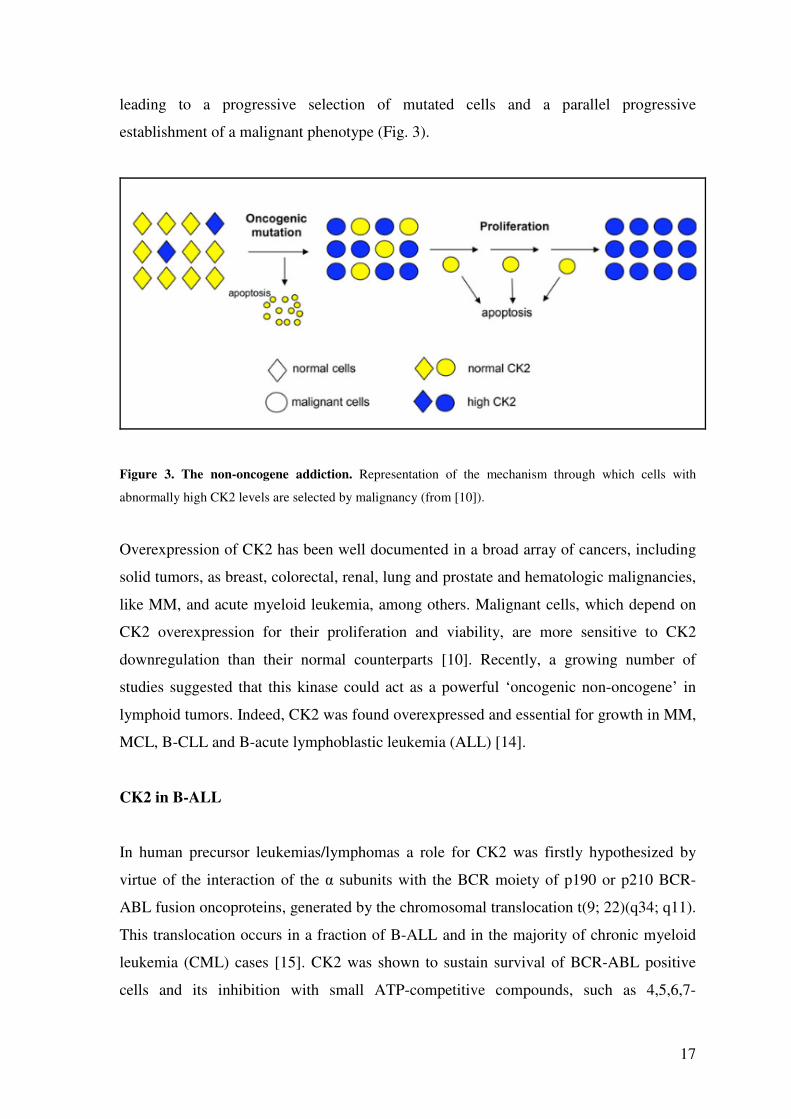
17
leading to a progressive selection of mutated cells and a parallel progressive
establishment of a malignant phenotype (Fig. 3).
Figure 3. The non-oncogene addiction. Representation of the mechanism through which cells with
abnormally high CK2 levels are selected by malignancy (from [10]).
Overexpression of CK2 has been well documented in a broad array of cancers, including
solid tumors, as breast, colorectal, renal, lung and prostate and hematologic malignancies,
like MM, and acute myeloid leukemia, among others. Malignant cells, which depend on
CK2 overexpression for their proliferation and viability, are more sensitive to CK2
downregulation than their normal counterparts [10]. Recently, a growing number of
studies suggested that this kinase could act as a powerful ‘oncogenic non-oncogene’ in
lymphoid tumors. Indeed, CK2 was found overexpressed and essential for growth in MM,
MCL, B-CLL and B-acute lymphoblastic leukemia (ALL) [14].
CK2 in B-ALL
In human precursor leukemias/lymphomas a role for CK2 was firstly hypothesized by
virtue of the interaction of the α subunits with the BCR moiety of p190 or p210 BCR-
ABL fusion oncoproteins, generated by the chromosomal translocation t(9; 22)(q34; q11).
This translocation occurs in a fraction of B-ALL and in the majority of chronic myeloid
leukemia (CML) cases [15]. CK2 was shown to sustain survival of BCR-ABL positive
cells and its inhibition with small ATP-competitive compounds, such as 4,5,6,7-
18
tetrabromobenzimidazole (TBB) or 2dimethylamino-4,5,6,7-tetrabromo-1H-
benzimidazole (DMAT), resulted in growth arrest [15], [16].
CK2 was demonstrated to be instrumental for a proper activation of the PI3K/PTEN/AKT
signalling cascade in B-ALL. Gomes et al. (2014) showed that in adult B-ALL cells CK2
is overexpressed/hyperactive [17]. Considering the well-known involvement of CK2 in
PTEN protein stability, being CK2-dependent phosphorylation a signal for PTEN
stabilization and functional inactivation, the authors challenged B-ALL cells with CK2
inhibitor CX-4945, with the remarkable result of causing growth arrest associated with a
drop of PI3K/AKT activity [18]. In addition, parallel research has identified a potential
role for CK2 in the regulation of the half-life of the TF Ikaros, which is mutated in
pediatric and adult B-ALL [19]. A balance between protein phosphatase PP1 and CK2
was found to determine Ikaros stability and transcriptional activity, with CK2-
phosphorylated Ikaros displaying reduced protein stability and functional activation [20]–
[22]. Taken together, these data suggest the relevance of investigating the role of CK2 in
Ikaros mutated B-ALL, in which this TF displays a loss of function in 5% of cases.
CK2 in MM and MCL
CK2 was shown to be overactive and overexpressed in MM and MCL cell lines and
primary tumor samples. CK2α knockdown or inhibition with TBB, TBB-derived agents
or with CX-4945 caused MM cell apoptosis, not counteracted by the addition of growth
factors, such as interleukin-6 (IL-6) and insulin-like growth factor-1 (IGF-1). Moreover,
CK2 silencing or inhibition was associated to IκBα stabilization and decreased NF-κB
transcriptional activity [23]. It was also demonstrated that CK2 positively regulates
STAT3 and NF-κB-dependent signalling, both in MM and in MCL cells. CK2 down
modulation is associated with a reduction in NF-κB phospho-Ser 529 and Ser 536, and
STAT3 phospho-Ser 727 levels [23]. Indeed, there is robust evidence that the
phosphorylation of these serine residues takes part in the modulation of the transcriptional
activation of both proteins. Furthermore, a role for CK2 in the sensitivity of MM as well
as MCL cells to novel therapeutic agents was clearly demonstrated. CK2 lies downstream
of the endoplasmic reticulum (ER)-stress induction by Hsp90 inhibitors as well as
proteasome inhibition by bortezomib. Double inhibition of Hsp90 and CK2 strongly
synergizes in vitro and in mouse xenotransplant in vivo models in inducing MM cell
apoptosis [24].
19
Moreover, downregulation of the kinase profoundly influences cellular response to
bortezomib. In conditions of CK2 blockade, MM and MCL cells became much more
prone to bortezomib-induced cytotoxicity and this was accompanied by an increase in the
proteotoxic response, as documented by the raise in ubiquitylated proteins found in these
cells [24], [25]. In addition to this, signals sustained by CK2 might promote the formation
of a pro-survival milieu between bone marrow (BM) stromal and MM cells. In stromal
cells, CK2 stimulates the expression of growth signals, including tumor necrosis factor-α
(TNF-α) and IL-6, which activate NF-κB and STAT3 TFs in MM cells [26]. In an in vitro
model of MM cells’ culture in the presence of BM stromal cells, the down modulation of
CK2 activity with CX-4945 induced a significant amount of MM cell death minimally
affecting the stroma. CX-4945 demonstrated its ability to counteract the stromal support
to malignant cells, which may widely contribute to resistance [26].
CK2 in B-CLL
Several lines of evidence suggest a central role for CK2 also in mature B lymphoid
tumors, including CLL. Jaeger et al. found high levels of CK2β phospho-Ser 209 in
primary samples derived from 44 CLL patients [27]. The phosphorylation of CK2β on
this residue is known to modulate CK2 enzymatic activity and target binding [6].
Similarly to ALL, CK2 inhibition was associated with a decrease in PI3K/AKT functional
activation, due to a reduction of PTEN phosphorylation at Ser 380 and AKT at Ser 473,
and with an increase in cell death [27]. Martins et al. confirmed the strong pro-survival
function of CK2 in CLL by showing that primary CLL cells are characterized by
increased levels of CK2 α and β subunits [28]. CK2 inhibition was found to be coupled
with inactivation of PKC, increased PTEN activity and apoptosis, especially in CLL cells
isolated from patients with advanced disease stage. Importantly, normal B lymphocytes
were only slightly affected by the treatment with CK2 inhibitors [28]. In another recent
work, Martins et al. confirmed the pro apoptotic effect of CX-4945 against primary CLL
cells and cell lines [29]. Remarkably, the cytotoxic effect of the drug was not reversed by
stromal co-culture. In vivo, the combination of CX-4945 with fludarabine, which is used
for the treatment of CLL, led to a significant reduction of tumor growth, as compared
with single treatments. Another work put into light that CX-4945 cooperates with two
compounds currently approved for the treatment of relapsed/refractory CLL and NHLs,
the BTK inhibitor ibrutinib and the PI3Kδ inhibitor idelalisib [30], providing strong
20
rationale for the introduction of CX-4945 in combination therapies also with inhibitors of
the BCR cascade.
CK2 INHIBITOR CX-4945 (SILMITASERTIB)
It has been frequently reported that malignant cells, with abnormally high CK2 levels, are
more susceptible to CK2 inhibitors-induced apoptosis, than their normal counterparts [10].
Interest in developing small molecule inhibitors of CK2 increased with the identification
of adenosine -5’-triphosphate (ATP)-binding sites specific chemotypes. However, as with
many inhibitors of other kinases, questions regarding their specificity arose immediately.
This cautionary note has to be considered especially for these kinds of compounds that
are competitors of ATP, since ATP is the substrate for all protein kinase family members
in addition to a vast array of other cellular enzymes. The ATP binding site of CK2 is
smaller than most of the other kinases, because of the presence of unique bulky residues,
which allow for the design of very selective and specific low molecular weight ATP-
competitive inhibitors [31].
Many ATP-competitive inhibitors of CK2 have already been reported in the literature, but
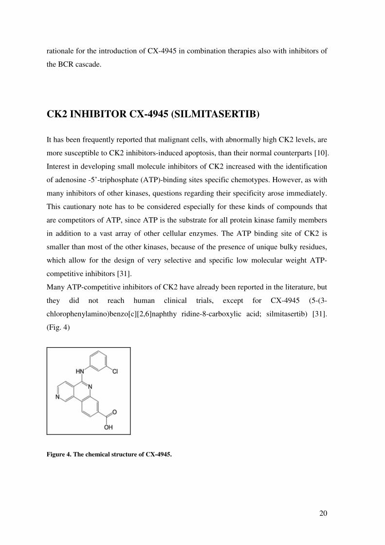
they did not reach human clinical trials, except for CX-4945 (5-(3-
chlorophenylamino)benzo[c][2,6]naphthy ridine-8-carboxylic acid; silmitasertib) [31].
(Fig. 4)
Figure 4. The chemical structure of CX-4945.
21
This compound is a selective, potent, orally bioavailable inhibitor of CK2α subunits,
whose anti-tumor activity has been validated in cancer cell lines and murine xenograft
models [32].
It was designated by Cylene Pharmaceuticals and entered phase I clinical trials for
advanced solid tumors and multiple myeloma (MM) (NCT00891280, ClinicalTrials.gov)
and, more recently, a randomized study that compares antitumor activity in
cholangiocarcinoma patients receiving the standard of care gemcitabine plus cisplatin
versus CX-4945 with gemcitabine plus cisplatin (NCT02128282, ClinicalTrials.gov).
During phase I trial, CX-4945 has been safe and well tolerated, demonstrated a clear
dose-dependent pharmacodynamic response and the capacity to kill tumor cells in
patients.
The crystal structure of human CK2α in complex with CX-4945 shows two direct protein-
inhibitor hydrogen bonds. Two well-ordered water molecules mediate additional contacts
between the carboxylate group of CX-4945 and CK2α. This extensive combination of
direct and water-mediated hydrogen bonds and van der Waals contacts between CX-4945
and CK2α establishes the structural basis for the high affinity binding of the inhibitor [33].
Downregulation of CK2 activity with CX-4945 boosts cytotoxicity in haematological
cancer cells; this points out that the kinase may be a valid druggable anti-cancer target to
be employed in the treatment of haematological malignancies [34].
B-CELLS
B lymphocytes are a population of cells expressing clonally diverse surface
immunoglobulin (Ig) receptors recognizing specific antigenic epitopes. These cells are
key components of the adaptive immunity, responding to pathogens by proliferation,
differentiation and Ab production [35]. Human B-cell development encompasses a
continuum of stages that begin in primary lymphoid tissues (fetal liver and fetal/adult
BM), with functional maturation in secondary lymphoid tissues (lymph nodes and spleen).
Early B-cell development is characterized by the rearrangement of the IgH and IgL chain
loci and the assembly of the pre-BCR, which is fundamental for B-cell development and
survival in periphery.
22
Ag-induced B-cell activation and differentiation in secondary lymphoid tissues are
mediated by changes in gene expression that give rise to the germinal center (GC)
reaction. The GC reaction is characterized by clonal expansion, class switch
recombination, somatic hypermutation and selection for a high affinity unique-antigenic-
epitope BCR. The result of this reaction is the generation of plasma blasts, secreting
antibodies while still dividing, and short-lived plasma cells (PC), secreting Ag-specific
germ line-encoded antibodies. Persistent Ag-specific antibody titres derive from long-
lived PCs that migrate to the BM and can persist without self-replenishment or turnover.
The aforementioned developmental stages have malignant counterparts that reflect the
expansion of a dominant subclone leading to development of leukemias and lymphomas
[36]. The majority of human B-cell tumors arise from mature B-cells recruited into the
GC reaction. GC B-cell lymphomas are divided into Hodgkin and NHLs. The latter group
comprises Burkitt’s lymphoma, follicular lymphoma (FL) and diffuse large B-cell
lymphoma (DLBCL).
Development and function of B-cells are principally affected by signalling via their BCR.
BCR SIGNALLING PATHWAY
BCR signal transduction is an intricate network involving multiple interconnected
pathways of effector molecules responsible for signal initiation, propagation, integration
and modulation, which culminates in activation of specific TFs and changes in gene
expression [37].
The BCR is an Ag-binding membrane Ig non-covalently associated with a disulfide-
linked heterodimer of CD79a (Igα) and CD79b (Igβ). Following ligation by an Ag, the
signal is transduced across the plasma membrane, leading to phosphorylation of the
immunoreceptor tyrosine-based activation motif (ITAM), present in the cytoplasmic tails
of CD79a/b, by the Src-family kinase LYN. Phosphorylated ITAM tyrosines bind and
activate SYK, thus leading to the formation of a multi-component signalling complex,
called ‘signalosome’ that propagates the signal inside the B-cell. Crucial in spreading the
the signal is the binding of the adaptor protein BLNK to CD79a and its phosphorylation
by SYK. BLNK serves as a scaffold for the assembly of the signalosome, by binding
23
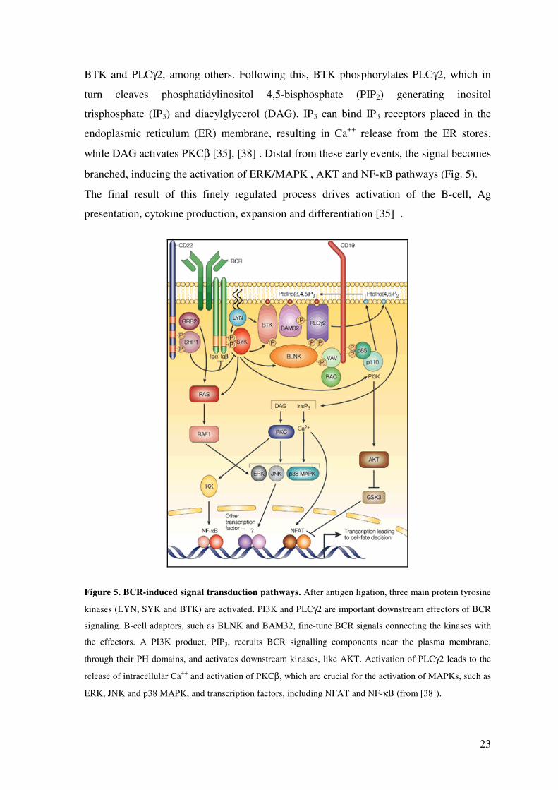
BTK and PLCγ2, among others. Following this, BTK phosphorylates PLCγ2, which in
turn cleaves phosphatidylinositol 4,5-bisphosphate (PIP2) generating inositol
trisphosphate (IP3) and diacylglycerol (DAG). IP3 can bind IP3 receptors placed in the
endoplasmic reticulum (ER) membrane, resulting in Ca++ release from the ER stores,
while DAG activates PKCβ [35], [38] . Distal from these early events, the signal becomes
branched, inducing the activation of ERK/MAPK , AKT and NF-κB pathways (Fig. 5).
The final result of this finely regulated process drives activation of the B-cell, Ag
presentation, cytokine production, expansion and differentiation [35] .
Figure 5. BCR-induced signal transduction pathways. After antigen ligation, three main protein tyrosine
kinases (LYN, SYK and BTK) are activated. PI3K and PLCγ2 are important downstream effectors of BCR
signaling. B-cell adaptors, such as BLNK and BAM32, fine-tune BCR signals connecting the kinases with
the effectors. A PI3K product, PIP3, recruits BCR signalling components near the plasma membrane,
through their PH domains, and activates downstream kinases, like AKT. Activation of PLCγ2 leads to the
release of intracellular Ca++ and activation of PKCβ, which are crucial for the activation of MAPKs, such as
ERK, JNK and p38 MAPK, and transcription factors, including NFAT and NF-κB (from [38]).
24
IP3 MEDIATED Ca++
RELEASE
Alterations in cytosolic Ca++, named Ca++ signals, affect a plurality of intracellular
processes central to cell-fate decisions in B lymphocytes. Among these processes are
kinase signalling, mitochondrial physiology, apoptosis, nucleocytoplasmic trafficking,
chromatin accessibility, cell adhesion and migration [39].
Ca++ signals are initiated after the formation of the signalosome and are mediated by lipid
phosphoinositides, which are abundant in the inner leaflet of the plasma membrane.
Briefly, PIP3 is produced by PI3K as a consequence of phosphorylation of PIP2 and, for
this reason, it accumulates in those areas of the membrane rich in PIP2. PIP3 binds with
high affinity to the pleckstrin-homology (PH) domain of many intracellular proteins,
including BTK, AKT and PLCγ2, which are therefore rapidly recruited near the plasma
membrane to form the signalosome. In this way, PLCγ2 gets in contact with its
physiological substrate PIP2 converting it into IP3 and DAG, hence transmitting the signal
downstream. PLC activation and cytosolic Ca++ differentially impact many key TF
pathways in B-cells, as exemplified by the nuclear factor of activated T cells (NFAT)
pathway, which is involved in the activation of cytokine genes such as interleukin-4 (IL-
4), and the NF-κB pathway, which in B-cells targets molecules, such as B-cell
lymphoma-6 (BCL-6), involved in avoiding apoptosis and supporting proliferation. The
NFAT pathway is activated in response to sustained Ca++ elevation. By contrast, the NF-
κB pathway is activated through DAG- and Ca++-dependent degradation of the inhibitor
IκB, and exhibits strong dependence on peak amplitude, rather than duration, of Ca++
signals. Differences in the level of BCR activation, and therefore PLCγ activity, may
produce markedly different B-cell-fate choices in the context of tolerizing versus non-
tolerizing Ag exposure: non-tolerizing Ags induce large sustained Ca++ responses able to
efficiently activate both pathways, whereas tolerizing Ags induce low-level sustained
responses able to activate NFAT, but not NF-κB, pathway.
NF-κB PATHWAY
The NF-κB TF family regulates the expression of a great variety of genes involved in
many diverse cellular processes, such as inflammatory and immune responses, growth,
survival and development. These factors are normally activated in a tightly regulated
25
manner, as a response to numerous signals, including cytokines, pathogens, and injuries
[40].
This family consists of five members: p50, p52, p65 (RelA), c-Rel and RelB, which share
an N-terminal domain responsible for DNA binding and dimerization. Dimers bind to κB
sites within promoters/enhancers of target genes and regulate transcription through the
recruitment of coactivators or corepressors. Only p65 (RelA), c-Rel and RelB present a
transcription activation domain (TAD) at the C-terminal, which is necessary for positive
regulation of gene expression [41].
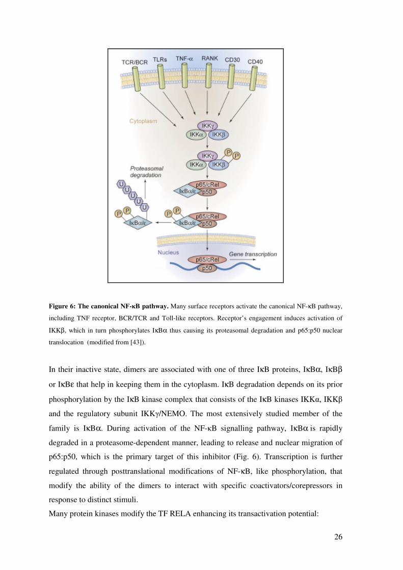
Three distinct NF-κB pathways have been described: the canonical, the alternative and
the atypical. In the canonical pathway, proinflammatory signals like cytokines (e.g.
TNFα), pathogen- and danger- associated molecular patterns, and the BCR/TCR activate
the dimer p50:p65 (Fig. 6), while the alternative pathway is triggered by BAFF or some
viruses that direct the activation of p52:RelB. The atypical pathway, on the other hand, is
activated in response to DNA damage [42].
26
Figure 6: The canonical NF-κB pathway. Many surface receptors activate the canonical NF-κB pathway,
including TNF receptor, BCR/TCR and Toll-like receptors. Receptor’s engagement induces activation of
IKKβ, which in turn phosphorylates IκBα thus causing its proteasomal degradation and p65:p50 nuclear
translocation (modified from [43]).
In their inactive state, dimers are associated with one of three IκB proteins, IκBα, IκBβ
or IκBε that help in keeping them in the cytoplasm. IκB degradation depends on its prior
phosphorylation by the IκB kinase complex that consists of the IκB kinases IKKα, IKKβ
and the regulatory subunit IKKγ/NEMO. The most extensively studied member of the
family is IκBα. During activation of the NF-κB signalling pathway, IκBα is rapidly
degraded in a proteasome-dependent manner, leading to release and nuclear migration of
p65:p50, which is the primary target of this inhibitor (Fig. 6). Transcription is further
regulated through posttranslational modifications of NF-κB, like phosphorylation, that
modify the ability of the dimers to interact with specific coactivators/corepressors in
response to distinct stimuli.
Many protein kinases modify the TF RELA enhancing its transactivation potential:
27
• PKA phosphorylates RELA at Ser 276, after IκBα degradation, promoting the
interaction of p65 with the transcriptional coactivators CBP and p300;
• IKKβ phosphorylates RELA at Ser 536 in the TAD. This phosphor-residue is
involved in the regulation of transcriptional activity, nuclear localization and
protein stability and mutations thereof disrupt the interaction of RELA with
CBP/p300;
• CK2 phosphorylates RELA at Ser 529 in the TAD. The phosphorylation of this
residue alters the association with basal components of the transcriptional
machinery and may therefore be involved in the regulation of gene expression [41].
It is currently largely unclear how NF-κB dimers control key parameters of the target
gene-specific response. Each individual NF-κB activating stimulus leads to the induction
of a specific overlapping and distinct subset of genes [44].
Constitutive NF-κB activation contributes to the growth and malignancy of cancer cells
and affects tumor response to chemotherapy. In particular, it has been demonstrated that it
can promote continuous lymphocyte cycling and survival and is a critical pathogenetic
factor in lymphomas [43].
It is well known that a wide variety of genetic alterations induce an aberrant activation of
the canonical NF-κB signalling pathway in human lymphomas, such as Hodgkin
lymphoma, mucosa associated lymphoid tissue lymphoma and activated B-cell like
(ABC)-DLBCL.
PI3K/AKT PATHWAY
Phosphorylated lipids are produced at cellular membranes during signalling events and
are responsible for the recruitment and activation of various cytoplasmic signalling
components [45].
PI3Ks are a family of lipid kinase enzymes that produce 3’-phosphorylated
phosphoinositides that act as second messenger to redirect intracellular proteins to
cellular membranes. Among these proteins, which present a PH domain that binds to PIP3,
are BTK, AKT and PDK1. The Ser/Thr kinase AKT is the major mediator of PI3K
28
signalling. Through the regulation of multiple distinct targets, AKT controls the
equilibrium between survival and apoptosis, quiescence and proliferation, as well as cell
metabolism and differentiation [46]. After its membrane recruitment, PDK1
phosphorylates AKT at Thr 308 and mTORC2 at Ser 473, thus enhancing AKT activity
[47].
It has been shown that CK2 can further phosphorylate AKT at Ser 129 enhancing Ser 473
phosphorylation, thus supporting the view that CK2 action induces conformational
changes in AKT that render the kinase more prone to activation [48].
AKT directly phosphorylates the kinase GSK3 and FOXO TFs, and indirectly stimulates
mTOR and NF-κB. Phosphorylation of GSK3 turns off the catalytic activity of this
enzyme; by inactivating this protein, which negatively regulates c-Myc and cyclin-D,
AKT supports cell cycle entry [46].
Phosphorylation of FOXOs, instead, induces relocalization of these TFs from the nucleus
to the cytoplasm, where they are degraded by the proteasome. Since active FOXOs
promote cell death, through the control of the expression of pro apoptotic proteins, like
BIM and FasL, reduction in FOXO factors is considered an important event in the
pathology of cancer [46].
The termination of PI3K/AKT signalling by degradation of PIP3 is mediated by SHIP and
PTEN phosphatases that are able to generate PIP2 [45].
Activation of the PI3K/AKT cascade is a common feature of most human cancers and B-
cell tumors represent no exception. Convincing evidence indicate a high AKT and mTOR
basal activation in B-cell leukemias, lymphomas and MM. ABC-DLBCL, for example,
displays constitutive AKT signalling through chronic active BCR [47].
In addition to this, PTEN is often inactivated in human cancers, supporting its role as a
fundamental tumor suppressor [45].
DIFFUSE LARGE B-CELL LYMPHOMA (DLBCL)
DLBCL is an aggressive B-cell non-Hodgkin lymphoma (NHL) characterized by large
heterogeneity in terms of clinicopathologic and molecular genetic features. The 2008
World Health Organization (WHO) classification defines DLBCL as a diffuse growth of
29
neoplastic large B lymphoid cells with a nuclear size equal to or exceeding normal
macrophage nuclei. It is the most frequent lymphoma subtype and accounts for 30-40% of
adult NHLs [49]. DLBCL is in general aggressive and affects patients of all ages with a
broad range of clinical presentations. Generally, it can be cured in more than 50% of
cases, even in the advanced stages. The chemotherapy currently used is R-CHOP, a
combination of the anti-CD20 antibody rituximab with cyclophosphamide, doxorubicin,
vincristine and prednisone. However, up to one-third of patients will not achieve cure
with initial therapy and have refractory disease or relapse after treatment. The standard
salvage treatment for these patients is autologous stem cell transplantation, but success
rates are poor and most of them will succumb to their disease [37].
Efforts to highlight the molecular heterogeneity of DLBCL rely on gene expression
profiling (GEP) that allowed to identify molecular subtypes, which correlate not only
with prognosis, but also with diverse genetic alterations and oncogenic signalling
pathways. In one approach, comparison of the genetic signatures across DLBCL allowed
to define three separate clusters. Groups of DLBCL identified by this consensus cluster
classification are:
• BCR-DLBCL, with upregulation of genes encoding BCR signalling components;
• OxPhos-DLBCL, enriched in genes involved in mitochondrial oxidative
phosphorylation;
• HR, host response DLBCL with host inflammatory infiltrate.
The cell of origin classification, instead, outlined three DLBCL subsets according to
similarities with the putative cell of origin:
• Germinal center B-cell like (GCB)-DLBCL, which derives from centroblasts;
• ABC-DLBCL, which derives from plasmablastic B-cells committed to terminal B
cell differentiation;
• Primary mediastinal large B-cell lymphoma (PMBL), which derives from thymic
B-cells.
These three groups present different mechanism of oncogenic activation and are
associated with diverse prognoses.
Anyhow, 15-30% of DLBCL cannot be included into any of the above groups [50], [51].
30
ABC-DLBCL
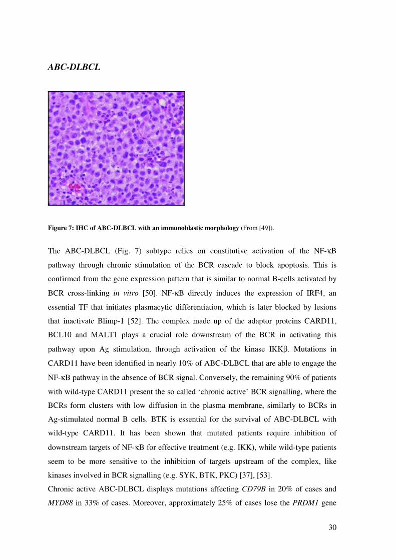
Figure 7: IHC of ABC-DLBCL with an immunoblastic morphology (From [49]).
The ABC-DLBCL (Fig. 7) subtype relies on constitutive activation of the NF-κB
pathway through chronic stimulation of the BCR cascade to block apoptosis. This is
confirmed from the gene expression pattern that is similar to normal B-cells activated by
BCR cross-linking in vitro [50]. NF-κB directly induces the expression of IRF4, an
essential TF that initiates plasmacytic differentiation, which is later blocked by lesions
that inactivate Blimp-1 [52]. The complex made up of the adaptor proteins CARD11,
BCL10 and MALT1 plays a crucial role downstream of the BCR in activating this
pathway upon Ag stimulation, through activation of the kinase IKKβ. Mutations in
CARD11 have been identified in nearly 10% of ABC-DLBCL that are able to engage the
NF-κB pathway in the absence of BCR signal. Conversely, the remaining 90% of patients
with wild-type CARD11 present the so called ‘chronic active’ BCR signalling, where the
BCRs form clusters with low diffusion in the plasma membrane, similarly to BCRs in
Ag-stimulated normal B cells. BTK is essential for the survival of ABC-DLBCL with
wild-type CARD11. It has been shown that mutated patients require inhibition of
downstream targets of NF-κB for effective treatment (e.g. IKK), while wild-type patients
seem to be more sensitive to the inhibition of targets upstream of the complex, like
kinases involved in BCR signalling (e.g. SYK, BTK, PKC) [37], [53].
Chronic active ABC-DLBCL displays mutations affecting CD79B in 20% of cases and
MYD88 in 33% of cases. Moreover, approximately 25% of cases lose the PRDM1 gene
31
encoding Blimp-1 that is essential for PCs formation, thus blocking terminal
differentiation [54].
This subtype remains less responsive to therapy than the GCB one [49].
GCB-DLBCL
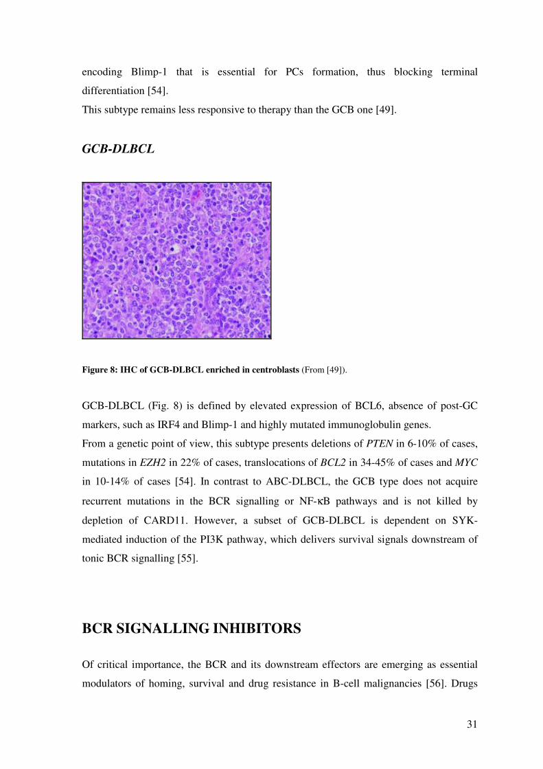
Figure 8: IHC of GCB-DLBCL enriched in centroblasts (From [49]).
GCB-DLBCL (Fig. 8) is defined by elevated expression of BCL6, absence of post-GC
markers, such as IRF4 and Blimp-1 and highly mutated immunoglobulin genes.
From a genetic point of view, this subtype presents deletions of PTEN in 6-10% of cases,
mutations in EZH2 in 22% of cases, translocations of BCL2 in 34-45% of cases and MYC
in 10-14% of cases [54]. In contrast to ABC-DLBCL, the GCB type does not acquire
recurrent mutations in the BCR signalling or NF-κB pathways and is not killed by
depletion of CARD11. However, a subset of GCB-DLBCL is dependent on SYK-
mediated induction of the PI3K pathway, which delivers survival signals downstream of
tonic BCR signalling [55].
BCR SIGNALLING INHIBITORS
Of critical importance, the BCR and its downstream effectors are emerging as essential
modulators of homing, survival and drug resistance in B-cell malignancies [56]. Drugs
32
that inhibit the transmission of signals throughout the BCR have been shown to disrupt
the microenvironment and promote cell death [30].
It is well known that many lymphoma subtypes subvert the BCR signalling to their
malignant purpose, suggesting that pharmacological inhibition of proteins acting
downstream of the BCR holds promise in these B-cell cancers [57].
It is important to underline that proper employ of BCR cascade inhibitors requires a deep
understanding of the type of BCR signal in action in each lymphoma subtype. Chronic
active BCR signalling in ABC-DLBCL engages SYK and BTK to activate PI3K and NF-
κB; in contrast, tonic BCR signalling in GCB-DLBCL depends upon SYK, but not on
BTK to activate PI3K and does not display a constitutive NF-κB pathway. Considering
these issues, SYK and BTK became, in the last few years, key target molecules for the
definition of novel therapeutic agents to be employed as single agents or in drug
combination therapies.
FOSTAMATINIB: A SYK INHIBITOR
SYK is an essential non-receptor tyrosine kinase recruited and activated by
phosphorylated ITAMs after BCR engagement to transmit the signal inside the B-cell.
For this reason, SYK is critical to Ag-dependent BCR signalling, including the chronic
active one, characteristic of ABC-DLBCL [57].
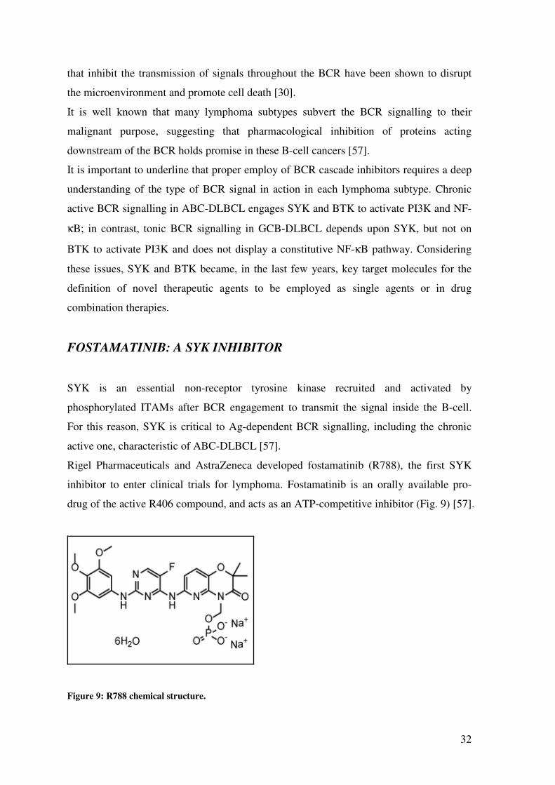
Rigel Pharmaceuticals and AstraZeneca developed fostamatinib (R788), the first SYK
inhibitor to enter clinical trials for lymphoma. Fostamatinib is an orally available pro-
drug of the active R406 compound, and acts as an ATP-competitive inhibitor (Fig. 9) [57].
Figure 9: R788 chemical structure.
33
A phase I/II trial of safety and efficacy of fostamatinib in patients with relapsed/refractory
B-cell NHL indicates significant clinical activity. Dose limiting toxicity in the phase I
portion was neutropenia, diarrhea, and thrombocytopenia. 200mg twice daily was chosen
for phase II testing, enrolling patients with DLBCL, FL, MCL, CLL. The study reports
objective response rates of 55% in CLL, 22% in DLBCL, 11% in MCL and 10% in FL.
DLBCL tumors were not divided into ABC and GCB types, thus it is not clear if the drug
has inhibited only chronic active BCR or not (NCT00446095, ClinicalTrials.gov) [58].
Fostamatinib has completed also a phase II trial to evaluate its efficacy in patients with
relapsed/refractory DLBCL (NCT01499303, ClinicalTrials.gov).
IBRUTINIB: A BTK INHIBITOR
BTK is a non-receptor intracellular kinase fundamental for antigen-stimulated BCR
signalling, including chronic active BCR in ABC-DLBCL. It is recruited to the
signalosome through its PH domain, which binds PIP3 in the inner leaflet of the plasma
membrane, and its Src-homology 2 domain, which binds phospho-BLNK. BTK is
essential for BCR-induced Ca++ release, proliferation and activation of the NF-κB
pathway [57], [59].
BTK has been found overexpressed in several B-cell derived tumors, suggesting its
importance as a non-oncogenic protein for malignant B-cell survival and growth [59]. To
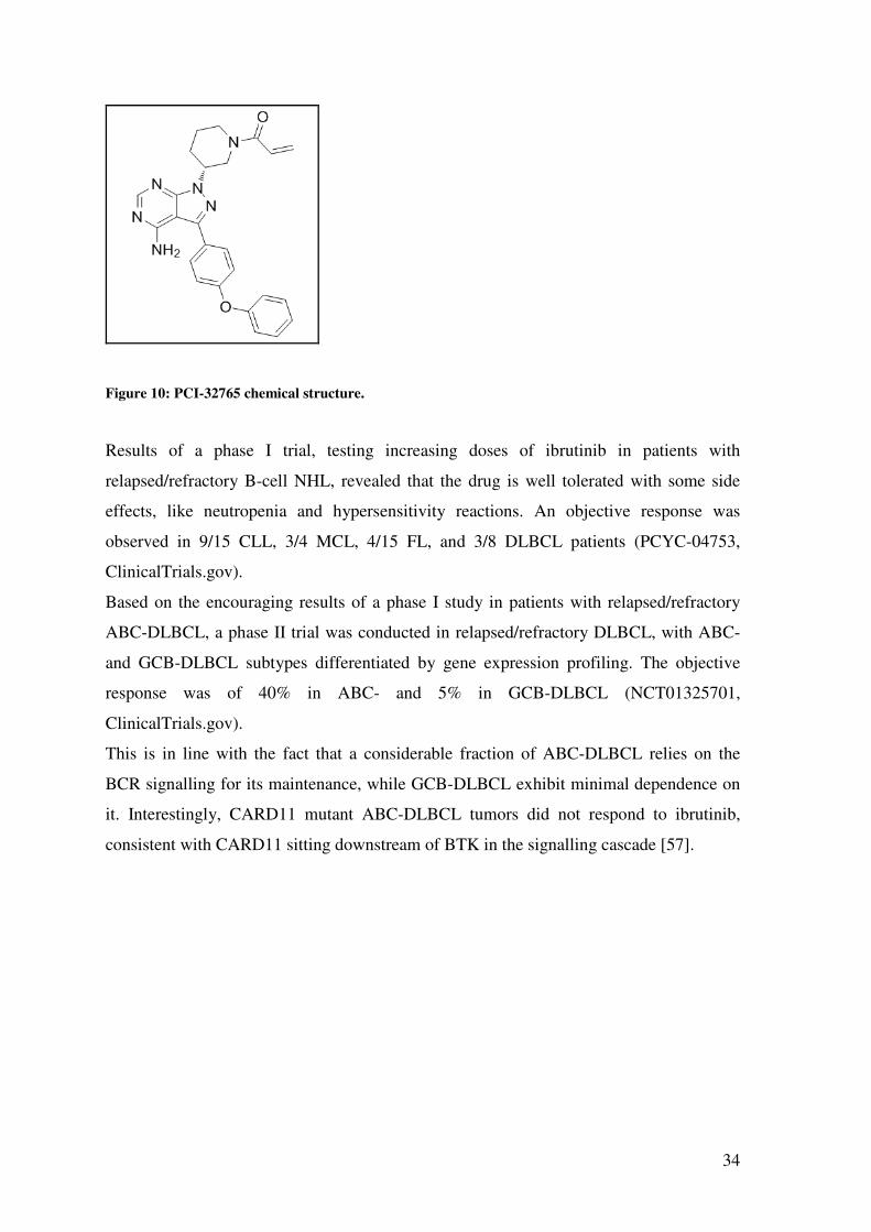
inhibit BTK, Pharmacyclics developed ibrutinib (PCI-32765), a selective and potent
molecule that irreversibly modifies the kinase, through a covalent bond with Cys 481 near
the active site of BTK. It is extremely rare among protein kinases to have a Cys residue in
that position, lending ibrutinib an elevated level of specificity (Fig. 10) [57].
34
Figure 10: PCI-32765 chemical structure.
Results of a phase I trial, testing increasing doses of ibrutinib in patients with
relapsed/refractory B-cell NHL, revealed that the drug is well tolerated with some side
effects, like neutropenia and hypersensitivity reactions. An objective response was
observed in 9/15 CLL, 3/4 MCL, 4/15 FL, and 3/8 DLBCL patients (PCYC-04753,
ClinicalTrials.gov).
Based on the encouraging results of a phase I study in patients with relapsed/refractory
ABC-DLBCL, a phase II trial was conducted in relapsed/refractory DLBCL, with ABC-
and GCB-DLBCL subtypes differentiated by gene expression profiling. The objective
response was of 40% in ABC- and 5% in GCB-DLBCL (NCT01325701,
ClinicalTrials.gov).
This is in line with the fact that a considerable fraction of ABC-DLBCL relies on the
BCR signalling for its maintenance, while GCB-DLBCL exhibit minimal dependence on
it. Interestingly, CARD11 mutant ABC-DLBCL tumors did not respond to ibrutinib,
consistent with CARD11 sitting downstream of BTK in the signalling cascade [57].
35
AIM OF THE STUDY
CK2 being involved in a wide variety of cellular processes, promotes cell survival and
proliferation. It has been found overexpressed in many hematologic malignancies, and it
has been shown that a downmodulation of its activity induces malignant cell death. With
the above as a background, we culled to investigate CK2 levels and activity, and the
consequences of its inhibition in the most common type of NHL, namely DLBCL, an
aggressive B-cell derived lymphoma that remains to date a therapeutic challenge.
It is well known that B-cells rely on the BCR signalling for their survival and activation
and that drugs able to hinder the transmission of the signal through the BCR have shown
promising therapeutic outcomes in patients with B-cell lymphomas, including DLBCL.
Considering these issues, we first intended to characterize CK2 potential implications
downstream from the BCR engagement. Moreover, we tested the effects of CK2
inhibition together with the BCR blockade in DLBCL, with the aim of shaping new
rational drug combination therapies with inhibitors of this pathway for the treatment of
this type of lymphoma.

36
37
MATERIALS AND METHODS
PROTEIN EXTRACTION
WHOLE PROTEIN EXTRACTION
All steps were performed at 4°C. Cells (1-2x10⁶) were collected, washed in PBS and
centrifuged at 5000 rpm for 5’. Pellets were resuspended in 40-50µl of lysis buffer
composed of: 150 mM NaCl, 2 mM EDTA, 2 mM EGTA supplemented with 0,5% Triton
X-100 (Sigma, Germany), protease inhibitor cocktail (Sigma, Germany), phosphatase
inhibitor cocktail (Thermo Scientific, USA), 1 mM phenyl-methyl-sulfonyl fluoride
(PMSF; Sigma, Germany), 1 μM okadaic acid (Sigma, Germany), dithiothreitol (DTT;
Sigma, Germany) in a buffer made up of TRIS (pH7.5) 20mM, NaCl 150mM, EDTA
2mM, EGTA 2mM to final volume. Samples were incubated in ice for 30’, vortexing
every 10’ and then centrifuged for 10’ at 13000 rpm. Supernatants were collected and
stored at -20°C or quantified immediately.
PROTEIN QUANTIFICATION
To measure the concentration of proteins after cell lysis we used Bradford
(Sigma,Germany) protein assay. It is based on an absorbance shift of the Comassie
Brilliant Blue G-250 dye. Under acidic conditions, the red form of the dye is converted
into its bluer form to bind to the protein being assayed. The bounded form of the dye has
the maximum absorption spectrum at 595 nm. The binding of the dye to the protein
stabilizes the blue anionic form, increasing the absorbance at 595nm in proportion to the
amount of bounded dye, and thus to the concentration of protein present in the sample.
Bradford was diluted 1:2 in distilled water and 1mL of diluted reagent was added to each
tube. Then, 1-1.5µl of cell lysate was added to the solution, mixed well and incubated 3’
in the dark. Using 1.5 mL cuvettes, absorbance at 595nm was red using a
spectrophotometer (Ultrospec 1100pro; Amersham).
Concentration values were obtained applying the Lambert-Beer formula:
A=ε x c ε=molar extinction coefficient
38
Molar extinction coefficient was derived from a calibration curve, obtained using known
concentrations of bovine serum albumine (BSA).
SDS-PAGE
Sodium dodecyl sulfate-polyacrylamide gel electrophoresis (SDS-PAGE) is a method that
allows the separation of proteins according to their size and no other physical feature.
SDS is a detergent that can dissolve hydrophobic molecules and has a negative charge
(sulfate) attached to it, so it can disrupt hydrophobic areas and coat proteins with many
negative charges, which overpower any positive charges the protein might present. The
resulting protein is denatured (reduced to its primary structure) and linearized. Moreover,
proteins having now a large negative charge will migrate towards the positive pole when
placed in an electric field.
When polyacrylamide, a polymer of acrylamide monomers, undergoes the process of
polymerization, it turns into a gel that can be placed in an electric field to pull the proteins
through it. The acrylamide concentration of the gel can vary, generally from 5% to 25%.
Lower percentage gels are better for separating high molecular weight proteins, while
higher percentages are needed for smaller proteins. Small molecules can move through
the polyacrylamide mesh faster than big molecules.
The polyacrylamide gel is composed of two phases: the upper phase is the stacking gel
(pH 6.8) and the lower phase is the separating gel (pH 8.8). The first one allows the
protein to compact and enter the separating phase simultaneously. The second one allows
the separation of proteins according to their molecular weight. We used fixed
concentrations of acrylamide (8% or 10% for separating gel; 5% for stacking gel). Protein
samples and a molecular weight reference (Seeblue Plus2 Prestained Standard 1X,
Invitrogen) were loaded into different wells in the stacking gel and separated using
Amersham electrophoretic chambers, a specific saline running buffer (pH 8.3) (25 mM
Tris, 192 mM glycine, 0.1% SDS) and an applied electric field of 25mA.
15-30 µg of protein lysates were mixed with sample buffer (1:4) composed by SDS 20%,
Tris (pH 6.8) 1,5M, bromophenol blu 0,05%, DTT 6%, and β-mercaptoethanol 1:20.
Samples were heated at 100°C for 4’ to favour denaturation.
39
WESTERN BLOT (WB)
After the electrophoresis, proteins must be transferred from the electrophoresis gel to a
membrane. The most commonly used transfer method is an electrophoretic transfer: this
method involves placing a protein-containing polyacrylamide gel in contact with a
membrane of polyvinylidene difluoride (PVDF) or other suitable material, and
"squeezing" these together between two electrodes in a conducting solution. Since the
PVDF is very hydrophobic, we previously activated it in methanol for 1’ to expose its full
protein binding capacity. The blotting sandwich is composed into a grid in the following
manner: sponge, watman paper, gel, PVDF membrane, watman paper, sponge. When an
electric field is applied, the proteins move out of the polyacrylamide gel onto the surface
of the membrane, where they become tightly attached. The result is a membrane with a
copy of the protein pattern that was originally in the gel. The transfer is performed in a
specific saline buffer containing Tris 250mM, glycine 1.92M and methanol 20%. After
the transfer the membrane is saturated to prevent unspecific binding of the detection
antibodies during subsequent steps. Saturation is performed for 1 hour in a solution
composed of non-fatty milk 5% (Ristora) and TBS (tris buffered saline) supplemented
with tween-20 0,05% (Sigma). Saturation is followed by washing in TBS plus Tween-20
0,05% in order to remove unbound reagents and reduce the background signal. The
membrane is then incubated overnight at 4°C with a primary antibody that recognizes a
specific protein or epitope on a group of proteins. The primary antibody is not directly
detectable. Therefore, tagged secondary antibodies that recognise the heavy chains of the
primary antibodies are used to detect the target Ag (indirect detection). Secondary
antibodies are enzymatically labelled with Horseradish peroxidase (HRP). After a final
series of washes to remove unattached antibodies, the antibodies on the membranes are
ready to be detected. An appropriate chemiluminescent substrate, which produces light, is
then added to the membrane. The light output can be captured using ImageQuant LAS500
machine (GE Healhcare Life Sciences).
We used different chemiluminescent substrates:
• Pierce ECL western blotting substrate (Thermo Scientific);
• LiteAblot PLUS Enhanced Chemioluminescent Substrate (EuroClone);
• LiteAblot EXTEND Long Lasting Chemiluminescent Substrate (EuroClone);
40
• LiteAblot Turbo Extra Sensitive Chemioluminescent Substrate (EuroClone).
In order to detect more antibodies with the same specificity and similar molecular weight
it is necessary to strip the membrane. Stripping buffer reagent (Thermo scientific) allows
the cleaning and the efficient removal of primary and secondary antibodies from
immunoblots without removing or damaging the immobilized Ag. This allows blots to be
re-probed with new antibodies. Membranes were covered with this buffer and incubated
for 20’-25’ at 37°C and then washed with TBS, afterwards the membranes were saturated
again with milk.
ANTIBODIES
Primary antibodies: anti-CK2α provided by Prof. Ruzzene, University of Padova, Italy;
anti-CK2β (Abcam, UK); anti-phospho-CK2β Ser 209 (Assay Biotech, USA); anti-
caspase3 (Alexis Biochemical, Switzerland); anti-cleaved PARP (Cell Signaling, USA);
anti-Mcl1 (Cell Signaling, USA); anti-FoxO3 (Cell Signaling, USA); anti-phospho-
FoxO3 Ser 253 (Cell Signaling, USA); anti-GSK3α/β (SantaCruz, USA), anti-phospho
GSK3β Ser 9, anti-RelA (Abcam, UK), anti-phospho-RelA Ser 529 (SantaCruz, USA),
anti-AKT (Cell Signaling, USA); anti-phospho-AKT Ser 473 (Cell Signaling, USA); anti-
phospho-BTK Tyr 223 (Cell Signaling, USA); anti-BTK (Cell Signaling, USA); anti-
phospho-PLCγ2 Tyr 1217 (Cell Signaling, USA); anti- PLCγ2 (Cell Signaling, USA);
anti-phospho-ERK1/2 Thr 202/Tyr 204 (Cell Signaling, USA); anti-ERK1/2 (Cell
Signaling, USA); anti-CDC37 (SantaCruz, USA); anti-GAPDH (Millipore, Germany);
anti-β-ACTIN (Sigma, Germany).
Secondary antibodies: anti-rabbit IgG HRP-linked antibody (Cell Signaling, USA); HRP
labeled goat anti-mouse IgG (KPL, USA).
RNA PURIFICATION
RNA was purified using RNeasy mini kit (Qiagen). This procedure represents a well-
established technology that combines the selective binding properties of a silica-based
41
membrane with the speed of microspin technology. Biological samples are first lysed and
homogenized in the presence of a highly denaturing guanidine-thiocyanate-containing
buffer, which immediately inactivates RNases to ensure purification of intact RNA.
Ethanol is added to provide appropriate binding conditions, and sample is then added to
an RNeasy Mini spin column, where the total RNA binds to the membrane and
contaminants are efficiently washed away. RNA is then eluted in water. The procedure
provides enrichment for mRNA since most RNAs <200 nucleotides are excluded.
Briefly, cells were collected and washed, removing the medium; then the appropriate
volume of RLT lysis buffer, that contains guanidine-thiocyanate, was added (350 µl for
<5x10⁶ cells, 600 µl for 5-9x10⁶ cells). RLT was supplemented with β-mercaptoethanol
1:100 v/v, which inhibits RNases further. Samples were homogenized by vortexing and
then 70% ethanol was added. After pipetting, lysed samples were transferred to RNeasy
spin columns and centrifuged at 11000rpm for 1’, discarding the flow-through. RNA
bound to the silica membrane was washed with buffer RW1 and centrifuged at 1000rpm
for 1’; a mix of DNase and buffer RDD (10µl and 70 µl respectively) were added directly
on the membrane and kept in incubation for 15’-30’, in order to remove contaminant
DNA. Afterwards a series of washes were performed, first of all with buffer RW1 (700µl)
and then with buffer RPE (500µl) (containing ethanol). Samples were centrifuged at
12800rpm for 2’, ensuring that the membrane was dry. At the end RNA was eluted using
30µl of RNase free water.
RNA was quantified by means of Nanodrop 1000 (Thermo Scientific).
REVERSE TRANSCRIPTION
Reverse transcription is a reaction exploited by a RNA-dependent polymerase capable of
synthesizing a complementary strand of DNA, called cDNA, using a RNA strand as
template.
RNA was reversely transcribed to cDNA by means of Reverse Transcription System
(Promega, USA). AMV, namely avian myeloblastoma virus, is the reverse transcriptase
enzyme used, which sythesizes single stranded cDNA from isolated mRNA; it shows
polymerase activity from 5’ to 3’ versus, and RnaseH activity from 3’ to 5’, degrading the
RNA strand when the hybrid cDNA/RNA is formed. The reaction was done in 20µl of
final volume:
42
• MgCl2 (25mM) 4µl
• reverse transcription 10X buffer 2µl
• dNTPs mix (10mM) 2µl
• Oligo dT primer (0,5mg/ml) 1µl
• RNasin RNase inhibitor 0,5µl
• AMV Reverse Transcriptase 0,6µl
• RNase free H20 to final volume
Then samples underwent the following thermal protocol:
• 42°C for 15’
• 95°C for 5’
• 4°C maintenance
REAL-TIME PCR
The real-time PCR is a method for gene quantification characterized by high sensibility
and specificity. It is called “real-time PCR” because it allows the scientist to actually
observe in “real time” the increase in the amount of DNA as it is amplified. This is
possible because the real-time PCR system combines a thermal cycler and an optical
reaction module that detects and quantifies fluorophores. Molecules added to the PCR
mix, as SYBR Green, bind the amplified DNA and emit a signal that increases in
proportion to the rise of the amplified DNA products. An amplification curve is obtained
where cycle numbers are found in abscissa and the fluorescence normalized on internal
fluorophore in ordinate. At the beginning of the reaction there are only little changes in
fluorescence and this is the baseline region; the increasing in fluorescence above this
threshold underlines amplified product formation. From this point on, the reaction
maintains an exponential course that degenerates in plateau at the end of the reaction.
In the midway cycles the curve has a linear course: this is the most important phase since
the amount of amplified DNA is correlated with the amount of cDNA expressed at the
beginning in the sample. In this linear region a threshold of fluorescence is chosen and
from this value it is possible to obtain the Ct (threshold cycle), namely the cycle that are
43
necessary, for the sample, to reach that threshold of emission. If the amount of cDNA
present at the beginning in the sample is high, the curve will rise earlier and Ct values will
be smaller.
As detector dye we used SYBR Green that emits low fluorescence if present in solution;
on the contrary the signal becomes stronger if the dye binds to double strand DNA.
However SYBR Green is not a selective dye and binds to all DNA, even to primer
dimers. For this reason it is recommended the introduction of a further step after
amplification, called dissociation protocol. During this step, temperature rises gradually
until all the double strands are denaturated. This method allows the identification of
contaminants or unspecific amplification products since they show different melting
points. There is also a dye called ROX that works as an internal reference used by the
instrument to normalize the SYBR Green fluorescence.
For the evaluation of gene expression we chose a relative quantification method, using the
ΔΔCt formula:
1) ΔCt = Ct (target gene) - Ct (reference gene)
2) ΔΔCt = ΔCt (of treated sample) - ΔCt (of untreated sample, the internal
calibrator)
3) 2^-ΔΔC
The “2”value in the last formula represents the higher efficiency for reaction that means a
doubling of the product at every cycle of amplification.
The thermal cycler used was the Sequence Detection System 7000 (Applied Biosystem)
and the software was ABI PRISM 7000.
The reagents of the reaction mix were:
• Roche FastStart Universal SYBR Green Master (ROX) 7,5µl
• Forward primer (4pmol/µl) 1µl
• Reverse primer (4pmol/µl) 1µl
• H2O 4,5µl
• cDNA 1µl
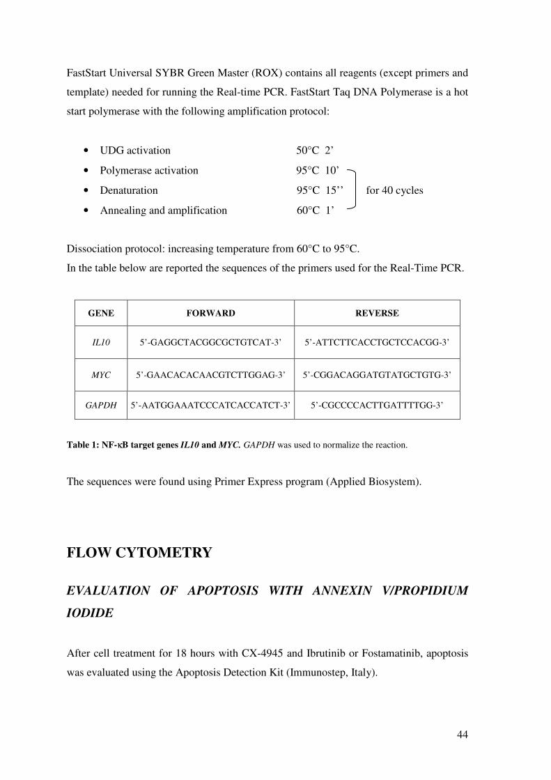
44
FastStart Universal SYBR Green Master (ROX) contains all reagents (except primers and
template) needed for running the Real-time PCR. FastStart Taq DNA Polymerase is a hot
start polymerase with the following amplification protocol:
• UDG activation 50°C 2’
• Polymerase activation 95°C 10’
• Denaturation 95°C 15’’ for 40 cycles
• Annealing and amplification 60°C 1’
Dissociation protocol: increasing temperature from 60°C to 95°C.
In the table below are reported the sequences of the primers used for the Real-Time PCR.
GENE FORWARD REVERSE
IL10 5’-GAGGCTACGGCGCTGTCAT-3’ 5’-ATTCTTCACCTGCTCCACGG-3’
MYC 5’-GAACACACAACGTCTTGGAG-3’ 5’-CGGACAGGATGTATGCTGTG-3’
GAPDH 5’-AATGGAAATCCCATCACCATCT-3’ 5’-CGCCCCACTTGATTTTGG-3’
Table 1: NF-κB target genes IL10 and MYC. GAPDH was used to normalize the reaction.
The sequences were found using Primer Express program (Applied Biosystem).
FLOW CYTOMETRY
EVALUATION OF APOPTOSIS WITH ANNEXIN V/PROPIDIUM
IODIDE
After cell treatment for 18 hours with CX-4945 and Ibrutinib or Fostamatinib, apoptosis
was evaluated using the Apoptosis Detection Kit (Immunostep, Italy).
45
AnnexinV (AV) is a member of a highly conserved protein family that binds acidic
phospholipids in a calcium-dependent manner. The protein presents a high affinity for
phosphatidylserine, which is translocated from the inner side of the plasma membrane to
the outer layer when cells undergo death by apoptosis or necrosis. Exposed
phosphatidylserine is one of several signals through which the cell, that is undergoing
apoptosis, can be recognized by phagocytes. AV binding to the cell surface indicates that
cell death is imminent. In order to differentiate apoptosis from necrosis, a dye exclusion
test with propidium iodide (PI) is performed, in order to establish if membrane integrity
has been conserved or not. A combination test measuring AV binding and dye exclusion
allows discrimination between live cells, apoptotic cells and necrotic cells.
2x10⁵ cells were washed in PBS to remove medium and resuspended in 100µl of binding
buffer. 1.7µl of AV-FITC were added and cells were incubated for 10’ at room
temperature in the dark. 100µl of binding buffer were further added to the cell suspension
and DNA was stained with 5µl of PI immediately before proceeding with flow cytometry
analysis. Fluorescence Activated Cell Sorting (FACS) analysis was performed using a
FACSCanto Cytometer and a FACSDiva 6.0 software (Becton-Dickinson, Italy).
CALCIUM FLUX ASSAY
1x10⁶ cells were cultured in RPMI or IMDM medium and incubated with 5 µM CX-4945
for 3 hours. The cells were then incubated in 500 µl of complete medium with 4 µg/ml
Fluo-4/AM (Invitrogen, USA), 16 µg/ml Fura Red/AM (Invitrogen, USA), 250 µM
sulfinpyrazone (Sigma, Germany) and 0.2 µl Pluronic acid in 20% DMSO for 30’ to
allow the loading of the probes. To quench extracellular Fluo-4/AM and Fura Red/AM
and allow de-esterification, cells were incubated in PBS for 10’ at room temperature in
the dark. The cells were analysed for 30’’, to record the basal fluorescence of the probes,
then stimulated with 20 µg/ml α-IgM and at 7’ 1 µg/ml of ionomycin (Sigma, Germany)
was added for the complete emptying of the stores and recorded for a total of 9’. The
record was done with a FACSCanto and the analysis with FlowJo software 10.1 and
Excel.
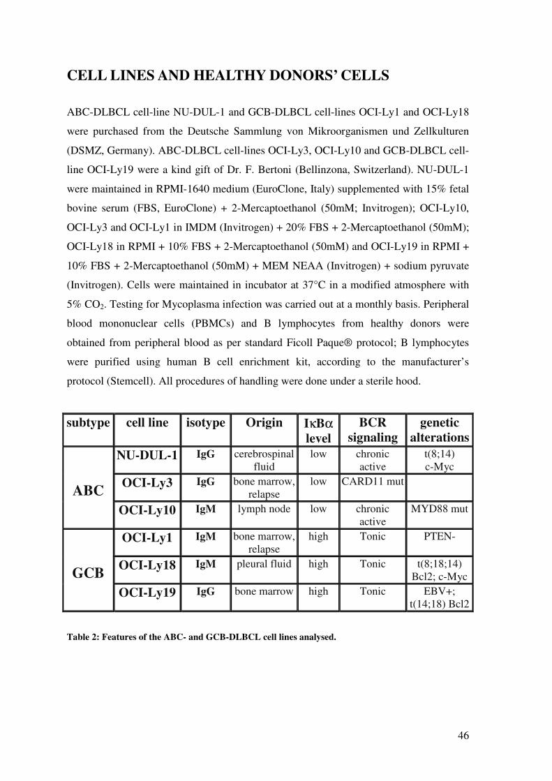
46
CELL LINES AND HEALTHY DONORS’ CELLS
ABC-DLBCL cell-line NU-DUL-1 and GCB-DLBCL cell-lines OCI-Ly1 and OCI-Ly18
were purchased from the Deutsche Sammlung von Mikroorganismen und Zellkulturen
(DSMZ, Germany). ABC-DLBCL cell-lines OCI-Ly3, OCI-Ly10 and GCB-DLBCL cell-
line OCI-Ly19 were a kind gift of Dr. F. Bertoni (Bellinzona, Switzerland). NU-DUL-1
were maintained in RPMI-1640 medium (EuroClone, Italy) supplemented with 15% fetal
bovine serum (FBS, EuroClone) + 2-Mercaptoethanol (50mM; Invitrogen); OCI-Ly10,
OCI-Ly3 and OCI-Ly1 in IMDM (Invitrogen) + 20% FBS + 2-Mercaptoethanol (50mM);
OCI-Ly18 in RPMI + 10% FBS + 2-Mercaptoethanol (50mM) and OCI-Ly19 in RPMI +
10% FBS + 2-Mercaptoethanol (50mM) + MEM NEAA (Invitrogen) + sodium pyruvate
(Invitrogen). Cells were maintained in incubator at 37°C in a modified atmosphere with
5% CO2. Testing for Mycoplasma infection was carried out at a monthly basis. Peripheral
blood mononuclear cells (PBMCs) and B lymphocytes from healthy donors were
obtained from peripheral blood as per standard Ficoll Paque® protocol; B lymphocytes
were purified using human B cell enrichment kit, according to the manufacturer’s
protocol (Stemcell). All procedures of handling were done under a sterile hood.
subtype cell line isotype Origin IκBα level
BCR
signaling genetic
alterations
ABC
NU-DUL-1 IgG cerebrospinal fluid
low chronic active
t(8;14) c-Myc
OCI-Ly3 IgG bone marrow, relapse
low CARD11 mut
OCI-Ly10 IgM lymph node low chronic active
MYD88 mut
GCB
OCI-Ly1 IgM bone marrow, relapse
high Tonic PTEN-
OCI-Ly18 IgM pleural fluid high Tonic t(8;18;14) Bcl2; c-Myc
OCI-Ly19 IgG bone marrow high Tonic EBV+; t(14;18) Bcl2
Table 2: Features of the ABC- and GCB-DLBCL cell lines analysed.
47
TISSUE SAMPLES
Cases were retrospectively collected from the archives of the Hematopathology Section,
Sant’ Orsola-Malpighi Hospital (University of Bologna, Bologna, Italy). 52 cases of
DLBCL (30 cases of GCB-type and 22 cases of ABC-type, as sub-classified according to
Hans’ algorithm [60]) were considered. Tissue microarray (TMA) blocks were prepared,
obtaining three tissue cores from each original sample. Adequate positive (lymphoid and
thymic tissue) and negative (myocardium) controls were also included.
None of the patients received radiation therapy or chemotherapy prior to biopsy sampling
and histological evaluation. The institutional and international (Declaration of Helsinki)
ethics regulations governing research conducted on human tissues were followed, and all
patients gave their informed consent.
CHEMICALS
Cells were stained with Trypan Blue (Sigma, Germany) and counted in a Neubauer
chamber, then centrifuged at 1000rpm for 5’ and plated with fresh medium.
We employed treatments with:
• CX-4945 (Silmitasertib, Activate-Scientific GmbH, Germany), CK2 inhibitor;
• PCI-32765 (Ibrutinib, SelleckChem, USA), BTK inhibitor;
• R788 (Fostamatinib, SelleckChem, USA), SYK inhibitor.
IMMUNOHISTOCHEMISTRY (IHC)
IHC was performed on 4 μm-thick formalin-fixed, paraffin-embedded sections, using
CK2α (EP1963Y, Epitomics, USA) and CK2β (6D5, Santa Cruz, USA) monoclonal
primary antibodies. Heat/EDTA-based Ag retrieval methods were applied, as previously
described. All sections were processed using the sensitive Bond Polymer Refine
Detection kit, a biotin-free, polymeric horseradish peroxidase–linker antibody conjugate
system, in an automated immunostainer (Bond maX, Menarini, Italy). Sections were then
48
slightly counterstained with hematoxylin. Appropriate positive and negative controls were
run concurrently.
CK2α and CK2β immunostain was semiquantitatively scored in a four-tiered scale, as
follows: score 0 = negative staining; score 1+ = weak positivity or positive staining in
<5% of tumor cells; score 2+ = moderate positivity or strong positive staining in <50% of
cells; score 3+ = strong positive staining in >50% of tumor cells. Immunohistochemical
reactions were independently scored by two investigators (agreement k>0.8). In case of
discrepancies, a consensus opinion was rendered. For the statistical interpretation of
immunohistochemical data, scores 0/1+ and 2+/3+ were lumped in “low-expression” and
“high-expression” groups, respectively.
IMMUNOFLUORESCENCE (IF)
Cells (4-5 x 10⁴) were seeded on polylysine-coated glass slides and incubated at 37°C for
30’ to let them adhere to the polylysine. After 3 washes in PBS, cells were fixed with
formaldehyde 3.7% for 20’ and permeabilized with Triton 0.1% for 4’ at room
temperature. After another three washes in PBS, cells were blocked with BSA 3% for 30’.
Samples were then stained with anti-CK2α primary antibody and Alexa Fluor 488 goat
anti-rabbit secondary antibody (Thermo Fisher, USA). Specimens were mounted in
Vectashield medium with DAPI (4′,6-diamidino-2-phenylindole) (Vector Laboratories,
USA), in order to distinguish the nuclei and analyzed using ZEISS LSM700 confocal
microscope with 63x magnification objective. Images were analyzed with ImageJ
software.
MTT ASSAY
MTT (3-(4,5-dimethylthiazol-2-yl)-2,5-diphenyl tetrazolium bromide, Sigma, Germany)
assay was performed in 96-well plates. Briefly, 0.5 x 10⁴ cells were plated in 96-well V-
bottom plates the day before the experiment. At the end of the treatment with the
inhibitors (48-72 hours), culture medium was removed and replaced with 100 µl of
49
complete medium + 50 µl of MTT to each well. During an incubation period of 1.5 hours
at 37°C, the MTT salt is metabolically reduced only by viable cells into an insoluble
purple formazan; the plate was centrifuged and 150 µl of DMSO (Sigma, Germany) were
added to each well to dissolve the formazan. Then, 100 µl of the purple solution were
transferred into a flat bottom plate and the absorbance read by a microplate reader at 570
nm. Values were expressed as percentage of viable cells, with untreated cells considered
as 100% viable.
STATISTICAL ANALYSIS
Data were evaluated for their statistical significance with the Student’s t test and
differences or correlations between groups were tested by applying analysis of variance
(ANOVA) or Fisher’s exact test (immunohistochemical results). p values below 0.05
were considered statistically significant. Analysis were performed using GraphPad Prism
5 or OriginPro 8.

50
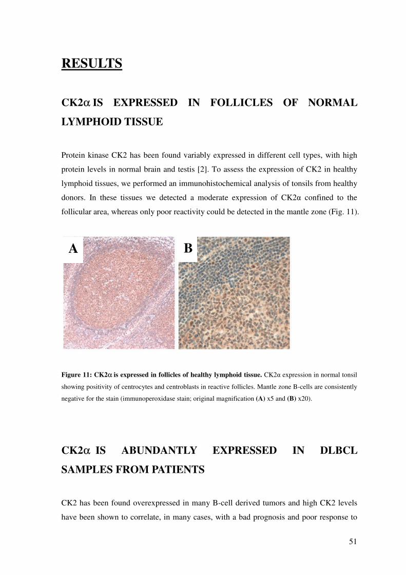
51
RESULTS
CK2α IS EXPRESSED IN FOLLICLES OF NORMAL
LYMPHOID TISSUE
Protein kinase CK2 has been found variably expressed in different cell types, with high
protein levels in normal brain and testis [2]. To assess the expression of CK2 in healthy
lymphoid tissues, we performed an immunohistochemical analysis of tonsils from healthy
donors. In these tissues we detected a moderate expression of CK2α confined to the
follicular area, whereas only poor reactivity could be detected in the mantle zone (Fig. 11).
Figure 11: CK2α is expressed in follicles of healthy lymphoid tissue. CK2α expression in normal tonsil
showing positivity of centrocytes and centroblasts in reactive follicles. Mantle zone B-cells are consistently
negative for the stain (immunoperoxidase stain; original magnification (A) x5 and (B) x20).
CK2α IS ABUNDANTLY EXPRESSED IN DLBCL
SAMPLES FROM PATIENTS
CK2 has been found overexpressed in many B-cell derived tumors and high CK2 levels
have been shown to correlate, in many cases, with a bad prognosis and poor response to
A B
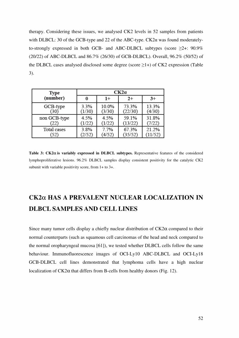
52
therapy. Considering these issues, we analysed CK2 levels in 52 samples from patients
with DLBCL: 30 of the GCB-type and 22 of the ABC-type. CK2α was found moderately-
to-strongly expressed in both GCB- and ABC-DLBCL subtypes (score ≥2+: 90.9%
(20/22) of ABC-DLBCL and 86.7% (26/30) of GCB-DLBCL). Overall, 96.2% (50/52) of
the DLBCL cases analysed disclosed some degree (score ≥1+) of CK2 expression (Table
3).
Table 3: CK2α is variably expressed in DLBCL subtypes. Representative features of the considered
lymphoproliferative lesions. 96.2% DLBCL samples display consistent positivity for the catalytic CK2
subunit with variable positivity score, from 1+ to 3+.
CK2α HAS A PREVALENT NUCLEAR LOCALIZATION IN
DLBCL SAMPLES AND CELL LINES
Since many tumor cells display a chiefly nuclear distribution of CK2α compared to their
normal counterparts (such as squamous cell carcinomas of the head and neck compared to
the normal oropharyngeal mucosa [61]), we tested whether DLBCL cells follow the same
behaviour. Immunofluorescence images of OCI-Ly10 ABC-DLBCL and OCI-Ly18
GCB-DLBCL cell lines demonstrated that lymphoma cells have a high nuclear
localization of CK2α that differs from B-cells from healthy donors (Fig. 12).
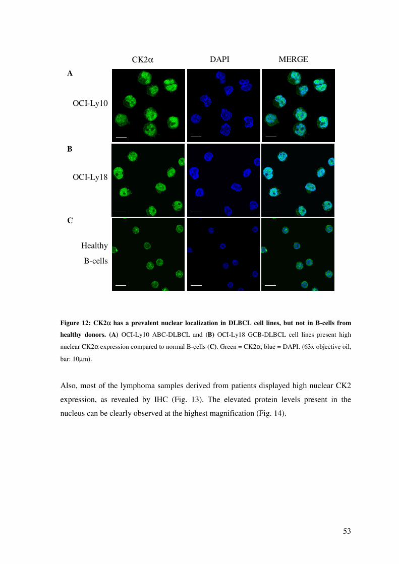
53
CK2α DAPI MERGE
OCI-Ly10
OCI-Ly18
Healthy
B-cells
Figure 12: CK2α has a prevalent nuclear localization in DLBCL cell lines, but not in B-cells from
healthy donors. (A) OCI-Ly10 ABC-DLBCL and (B) OCI-Ly18 GCB-DLBCL cell lines present high
nuclear CK2α expression compared to normal B-cells (C). Green = CK2α, blue = DAPI. (63x objective oil,
bar: 10µm).
Also, most of the lymphoma samples derived from patients displayed high nuclear CK2
expression, as revealed by IHC (Fig. 13). The elevated protein levels present in the
nucleus can be clearly observed at the highest magnification (Fig. 14).
A
B
C
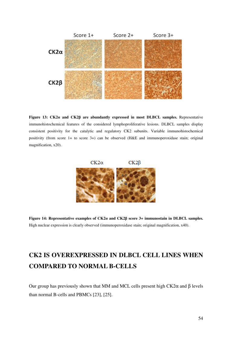
54
Figure 13: CK2α and CK2β are abundantly expressed in most DLBCL samples. Representative
immunohistochemical features of the considered lymphoproliferative lesions. DLBCL samples display
consistent positivity for the catalytic and regulatory CK2 subunits. Variable immunohistochemical
positivity (from score 1+ to score 3+) can be observed (H&E and immunoperoxidase stain; original
magnification, x20).
Figure 14: Representative examples of CK2α and CK2β score 3+ immunostain in DLBCL samples.
High nuclear expression is clearly observed (immunoperoxidase stain; original magnification, x40).
CK2 IS OVEREXPRESSED IN DLBCL CELL LINES WHEN
COMPARED TO NORMAL B-CELLS
Our group has previously shown that MM and MCL cells present high CK2α and β levels
than normal B-cells and PBMCs [23], [25].
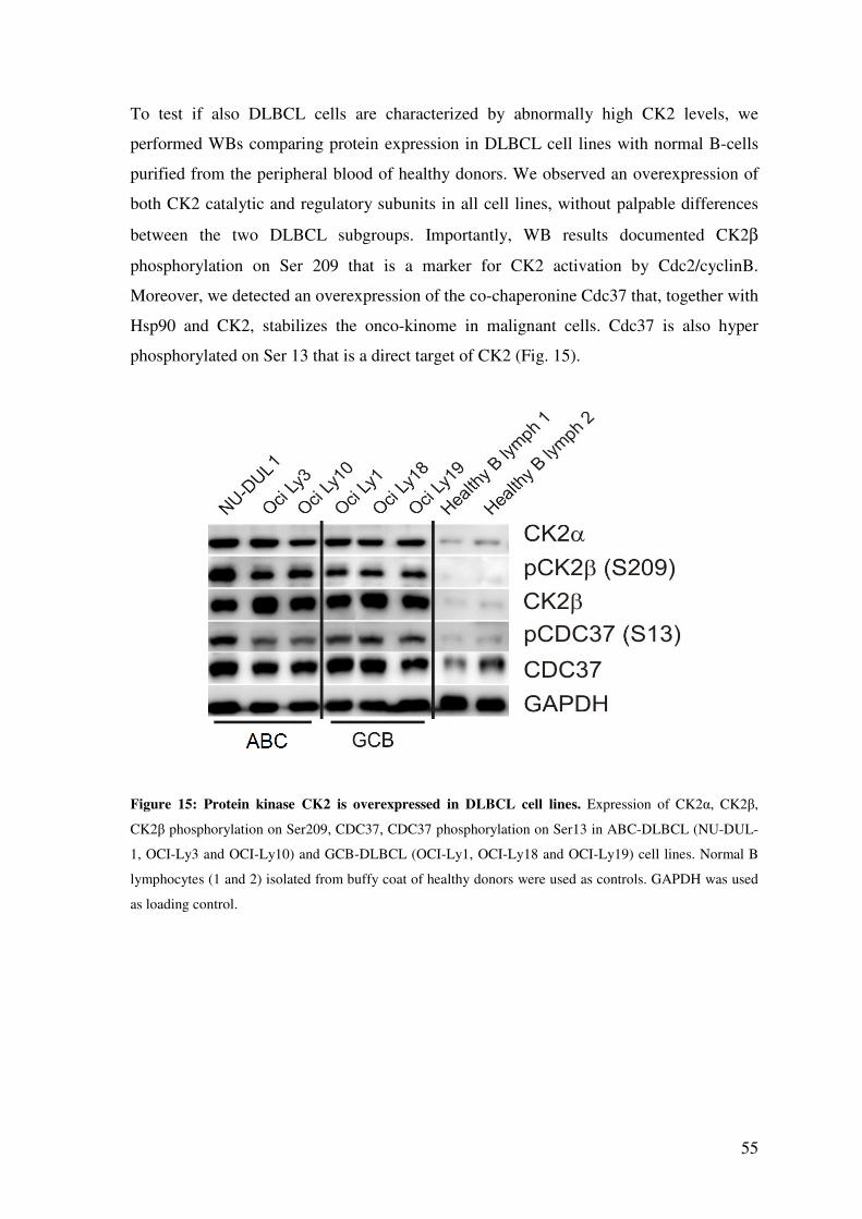
55
To test if also DLBCL cells are characterized by abnormally high CK2 levels, we
performed WBs comparing protein expression in DLBCL cell lines with normal B-cells
purified from the peripheral blood of healthy donors. We observed an overexpression of
both CK2 catalytic and regulatory subunits in all cell lines, without palpable differences
between the two DLBCL subgroups. Importantly, WB results documented CK2β
phosphorylation on Ser 209 that is a marker for CK2 activation by Cdc2/cyclinB.
Moreover, we detected an overexpression of the co-chaperonine Cdc37 that, together with
Hsp90 and CK2, stabilizes the onco-kinome in malignant cells. Cdc37 is also hyper
phosphorylated on Ser 13 that is a direct target of CK2 (Fig. 15).
Figure 15: Protein kinase CK2 is overexpressed in DLBCL cell lines. Expression of CK2α, CK2β,
CK2β phosphorylation on Ser209, CDC37, CDC37 phosphorylation on Ser13 in ABC-DLBCL (NU-DUL-
1, OCI-Ly3 and OCI-Ly10) and GCB-DLBCL (OCI-Ly1, OCI-Ly18 and OCI-Ly19) cell lines. Normal B
lymphocytes (1 and 2) isolated from buffy coat of healthy donors were used as controls. GAPDH was used
as loading control.
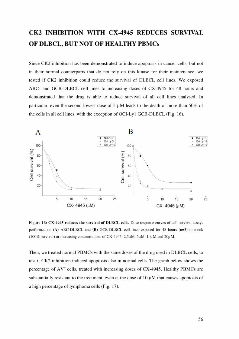
56
CK2 INHIBITION WITH CX-4945 REDUCES SURVIVAL
OF DLBCL, BUT NOT OF HEALTHY PBMCs
Since CK2 inhibition has been demonstrated to induce apoptosis in cancer cells, but not
in their normal counterparts that do not rely on this kinase for their maintenance, we
tested if CK2 inhibition could reduce the survival of DLBCL cell lines. We exposed
ABC- and GCB-DLBCL cell lines to increasing doses of CX-4945 for 48 hours and
demonstrated that the drug is able to reduce survival of all cell lines analysed. In
particular, even the second lowest dose of 5 µM leads to the death of more than 50% of
the cells in all cell lines, with the exception of OCI-Ly1 GCB-DLBCL (Fig. 16).
Figure 16: CX-4945 reduces the survival of DLBCL cells. Dose response curves of cell survival assays
performed on (A) ABC-DLBCL and (B) GCB-DLBCL cell lines exposed for 48 hours (n=3) to mock
(100% survival) or increasing concentrations of CX-4945: 2,5µM, 5µM, 10µM and 20µM.
Then, we treated normal PBMCs with the same doses of the drug used in DLBCL cells, to
test if CK2 inhibition induced apoptosis also in normal cells. The graph below shows the
percentage of AV+ cells, treated with increasing doses of CX-4945. Healthy PBMCs are
substantially resistant to the treatment, even at the dose of 10 µM that causes apoptosis of
a high percentage of lymphoma cells (Fig. 17).
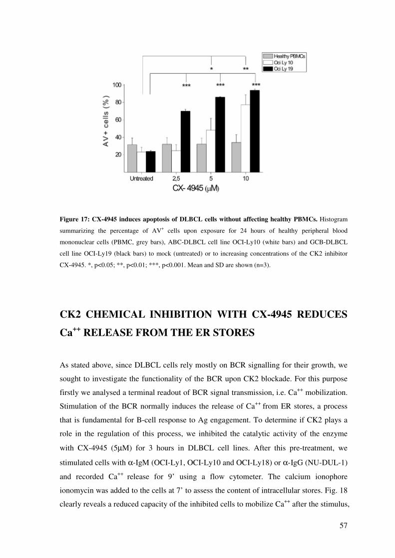
57
Figure 17: CX-4945 induces apoptosis of DLBCL cells without affecting healthy PBMCs. Histogram
summarizing the percentage of AV+ cells upon exposure for 24 hours of healthy peripheral blood
mononuclear cells (PBMC, grey bars), ABC-DLBCL cell line OCI-Ly10 (white bars) and GCB-DLBCL
cell line OCI-Ly19 (black bars) to mock (untreated) or to increasing concentrations of the CK2 inhibitor
CX-4945. *, p<0.05; **, p<0.01; ***, p<0.001. Mean and SD are shown (n=3).
CK2 CHEMICAL INHIBITION WITH CX-4945 REDUCES
Ca++
RELEASE FROM THE ER STORES
As stated above, since DLBCL cells rely mostly on BCR signalling for their growth, we
sought to investigate the functionality of the BCR upon CK2 blockade. For this purpose
firstly we analysed a terminal readout of BCR signal transmission, i.e. Ca++ mobilization.
Stimulation of the BCR normally induces the release of Ca++ from ER stores, a process
that is fundamental for B-cell response to Ag engagement. To determine if CK2 plays a
role in the regulation of this process, we inhibited the catalytic activity of the enzyme
with CX-4945 (5µM) for 3 hours in DLBCL cell lines. After this pre-treatment, we
stimulated cells with α-IgM (OCI-Ly1, OCI-Ly10 and OCI-Ly18) or α-IgG (NU-DUL-1)
and recorded Ca++ release for 9’ using a flow cytometer. The calcium ionophore
ionomycin was added to the cells at 7’ to assess the content of intracellular stores. Fig. 18
clearly reveals a reduced capacity of the inhibited cells to mobilize Ca++ after the stimulus,
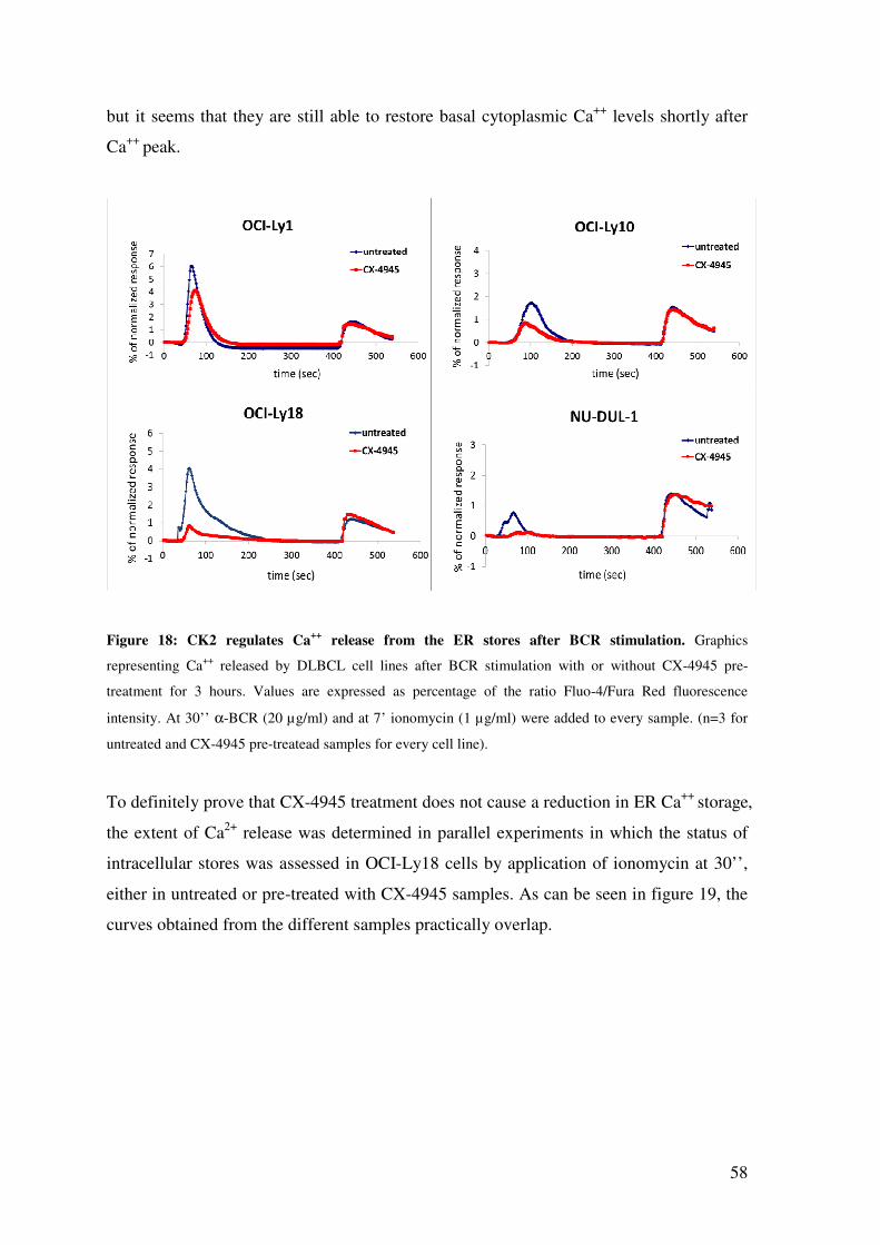
58
but it seems that they are still able to restore basal cytoplasmic Ca++ levels shortly after
Ca++ peak.
Figure 18: CK2 regulates Ca++
release from the ER stores after BCR stimulation. Graphics
representing Ca++ released by DLBCL cell lines after BCR stimulation with or without CX-4945 pre-
treatment for 3 hours. Values are expressed as percentage of the ratio Fluo-4/Fura Red fluorescence
intensity. At 30’’ α-BCR (20 µg/ml) and at 7’ ionomycin (1 µg/ml) were added to every sample. (n=3 for
untreated and CX-4945 pre-treatead samples for every cell line).
To definitely prove that CX-4945 treatment does not cause a reduction in ER Ca++ storage,
the extent of Ca2+ release was determined in parallel experiments in which the status of
intracellular stores was assessed in OCI-Ly18 cells by application of ionomycin at 30’’,
either in untreated or pre-treated with CX-4945 samples. As can be seen in figure 19, the
curves obtained from the different samples practically overlap.
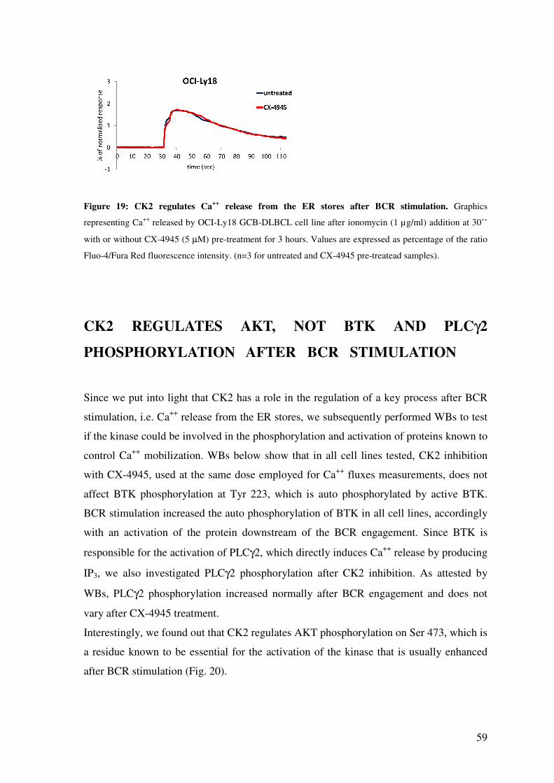
59
Figure 19: CK2 regulates Ca++
release from the ER stores after BCR stimulation. Graphics
representing Ca++ released by OCI-Ly18 GCB-DLBCL cell line after ionomycin (1 µg/ml) addition at 30’’
with or without CX-4945 (5 µM) pre-treatment for 3 hours. Values are expressed as percentage of the ratio
Fluo-4/Fura Red fluorescence intensity. (n=3 for untreated and CX-4945 pre-treatead samples).
CK2 REGULATES AKT, NOT BTK AND PLCγ2
PHOSPHORYLATION AFTER BCR STIMULATION
Since we put into light that CK2 has a role in the regulation of a key process after BCR
stimulation, i.e. Ca++ release from the ER stores, we subsequently performed WBs to test
if the kinase could be involved in the phosphorylation and activation of proteins known to
control Ca++ mobilization. WBs below show that in all cell lines tested, CK2 inhibition
with CX-4945, used at the same dose employed for Ca++ fluxes measurements, does not
affect BTK phosphorylation at Tyr 223, which is auto phosphorylated by active BTK.
BCR stimulation increased the auto phosphorylation of BTK in all cell lines, accordingly
with an activation of the protein downstream of the BCR engagement. Since BTK is
responsible for the activation of PLCγ2, which directly induces Ca++ release by producing
IP3, we also investigated PLCγ2 phosphorylation after CK2 inhibition. As attested by
WBs, PLCγ2 phosphorylation increased normally after BCR engagement and does not
vary after CX-4945 treatment.
Interestingly, we found out that CK2 regulates AKT phosphorylation on Ser 473, which is
a residue known to be essential for the activation of the kinase that is usually enhanced
after BCR stimulation (Fig. 20).

60
Figure 20: CK2 inhibition modulates AKT activation, but does not affect BTK and PLCγ2
phosphorylation after BCR stimulation. WB showing the phosphorylation of PLCγ2, BTK and AKT in
OCI-Ly1 and OCI-Ly18 GCB-DLBCL; OCI-Ly10 and NU-DUL-1 ABC-DLBCL cell lines pre-incubated
for 3 hours with CX-4945 (5µM) and then stimulated with α-IgM (OCI-Ly1, OCI-Ly10 and OCI-Ly18) or
α-IgG (NU-DUL-1) for 2’ (n=3). RELA was used as control of CK2 inhibition; ERK1/2 was used as
control of BCR stimulation; GAPDH was used as loading control.
CX-4945 SYNERGIZES WITH BCR INHIBITORS IN
INDUCING DLBCL CELL DEATH
BCR activation leads to survival and proliferation of B lymphocytes through signalling of
survival kinases, among which BTK and AKT. Drugs that inhibit the transmission of
signals through the BCR have been shown to disrupt the microenvironment and promote
apoptosis. Currently, many inhibitors of kinases activated after BCR engagement are in
clinical trials or have been approved for the treatment of B-cell derived disorders. Among
these inhibitors, fostamatinib, which targets SYK and ibrutinib, which targets BTK
demonstrated great efficacy in inducing lymphoma cell death. CK2 has an important role
61
in the regulation of oncogenic pathways shared with the BCR that include
PI3K/PTEN/AKT and NF-κB cascades.
Given that cell survival relies on the convergence of many regulatory inputs, including
those transmitted via the BCR, we hypothesized that inhibiting this growth and survival
pathway at more than one site would lead to enhanced apoptotic activity. We therefore
investigated the effect of CX-4945, in combination with fostamatinib or ibrutinib, in
terms of cell growth arrest. To this aim, we performed cell survival assays using the
constant drug ratio combination, according to the method described in [62]. Dose
response curves below show that the combined treatment is synergic in ABC- and GCB-
DLBCL cell lines, since the calculated CI values are in all cases lower than 1 (Fig. 21).
As we can see from the graphs, OCI-Ly10 ABC-DLBCL cells are the most sensitive to
the BCR blockade with ibrutinib, since we used 1:500 (CX:IBR) constant dose ratios to
perform the assay. There are no remarkable differences between the two DLBCL
subtypes (Fig. 21).
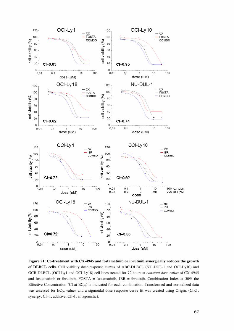
62
Figure 21: Co-treatment with CX-4945 and fostamatinib or ibrutinib synergically reduces the growth
of DLBCL cells. Cell viability dose-response curves of ABC-DLBCL (NU-DUL-1 and OCI-Ly10) and
GCB-DLBCL (OCI-Ly1 and OCI-Ly18) cell lines treated for 72 hours at constant dose ratios of CX-4945
and fostamatinib or ibrutinib. FOSTA = fostamatinib, IBR = ibrutinib. Combination Index at 50% the
Effective Concentration (CI at EC50) is indicated for each combination. Transformed and normalized data
was assessed for EC50 values and a sigmoidal dose response curve fit was created using Origin. (CI<1,
synergy; CI=1, additive, CI>1, antagonistic).
63
CK2 INHIBITION INCREASES APOPTOSIS INDUCED BY
THE BCR BLOCKADE
We better assessed the extent of apoptosis upon CK2 inhibition together with the BCR
blockade. The graphs below reveal a cooperation between CX-4945 and
fostamatinib/ibrutinib in increasing the apoptotic cells after 18 hours of incubation. As we
can see from the scatter plots, the combined treatment significantly increases the
percentage of AV+ cells compared not only with untreated samples, but also with samples
treated with one inhibitor at a time (Fig. 22).
OCI-Ly10 ABC-DLBCL cells are the most sensitive to fostamatinib and ibrutinib, among
the cell lines analysed. OCI-Ly18 GCB-DLBCL cells, instead, are the most resistant to
BCR blockade and are not killed by ibrutinib alone (even if the dose used was 10 µM), as
most of the GCB-DLBCL.
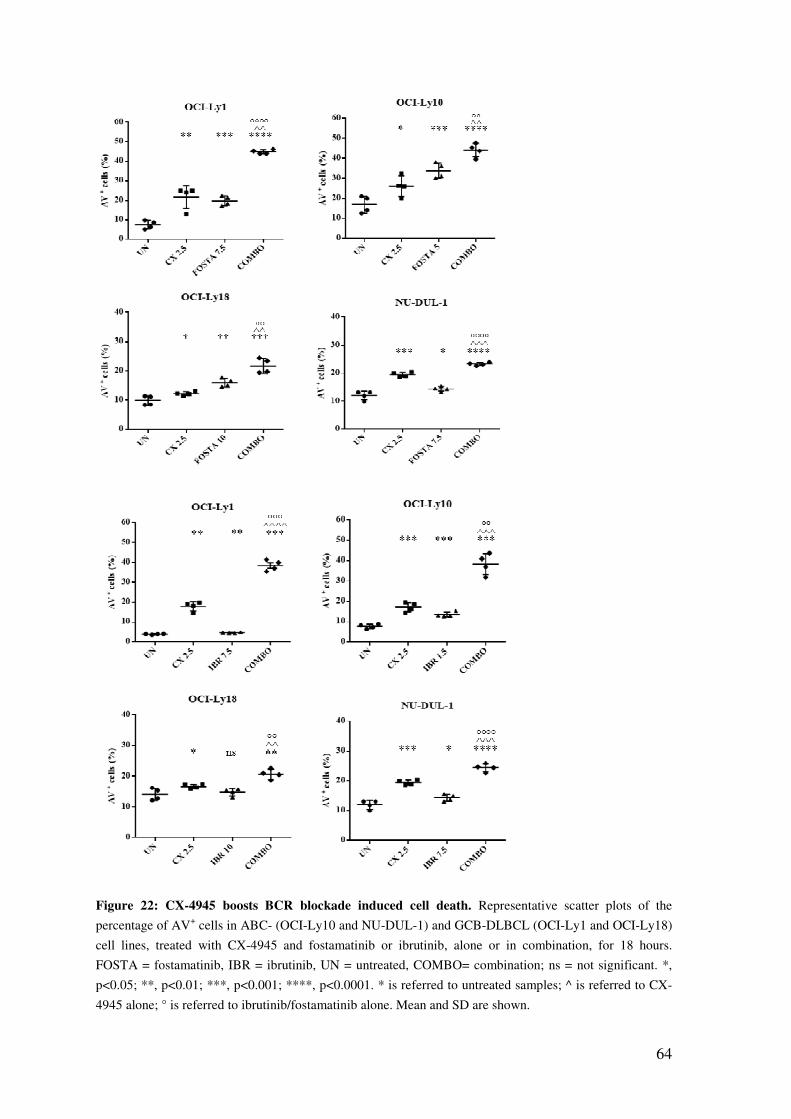
64
Figure 22: CX-4945 boosts BCR blockade induced cell death. Representative scatter plots of the
percentage of AV+ cells in ABC- (OCI-Ly10 and NU-DUL-1) and GCB-DLBCL (OCI-Ly1 and OCI-Ly18)
cell lines, treated with CX-4945 and fostamatinib or ibrutinib, alone or in combination, for 18 hours.
FOSTA = fostamatinib, IBR = ibrutinib, UN = untreated, COMBO= combination; ns = not significant. *,
p<0.05; **, p<0.01; ***, p<0.001; ****, p<0.0001. * is referred to untreated samples; ^ is referred to CX-
4945 alone; ° is referred to ibrutinib/fostamatinib alone. Mean and SD are shown.
65
To follow up we investigated the variation of proteins involved in the apoptotic process
upon combined treatment. By means of western blotting we confirmed the cooperation
between CK2 activity downmodulation and the BCR blockade. The combined treatment
with CX-4945 and either fostamatinib or ibrutinib increases the cleavage of PARP
proteins by effector caspases, like caspase-3, which is itself cleaved by initiator caspases
and, consequently, activated. Moreover, the combined treatments induce the degradation
of the anti-apoptotic protein Mcl-1 that is abundant in untreated cells. PARP and caspase
cleavage and anti-apoptotic proteins’ degradation are indisputable features of the
progression towards apoptosis (Fig. 23).
WBs reveal that OCI-Ly10 ABC-DLBCL cell line is the most sensitive to the combined
treatment, as revealed by the increase in cleaved PARP and caspase-3 after the
association of fostamatinib/ibrutinib with CX-4945, even if single agents cause only little
apoptotic effects.
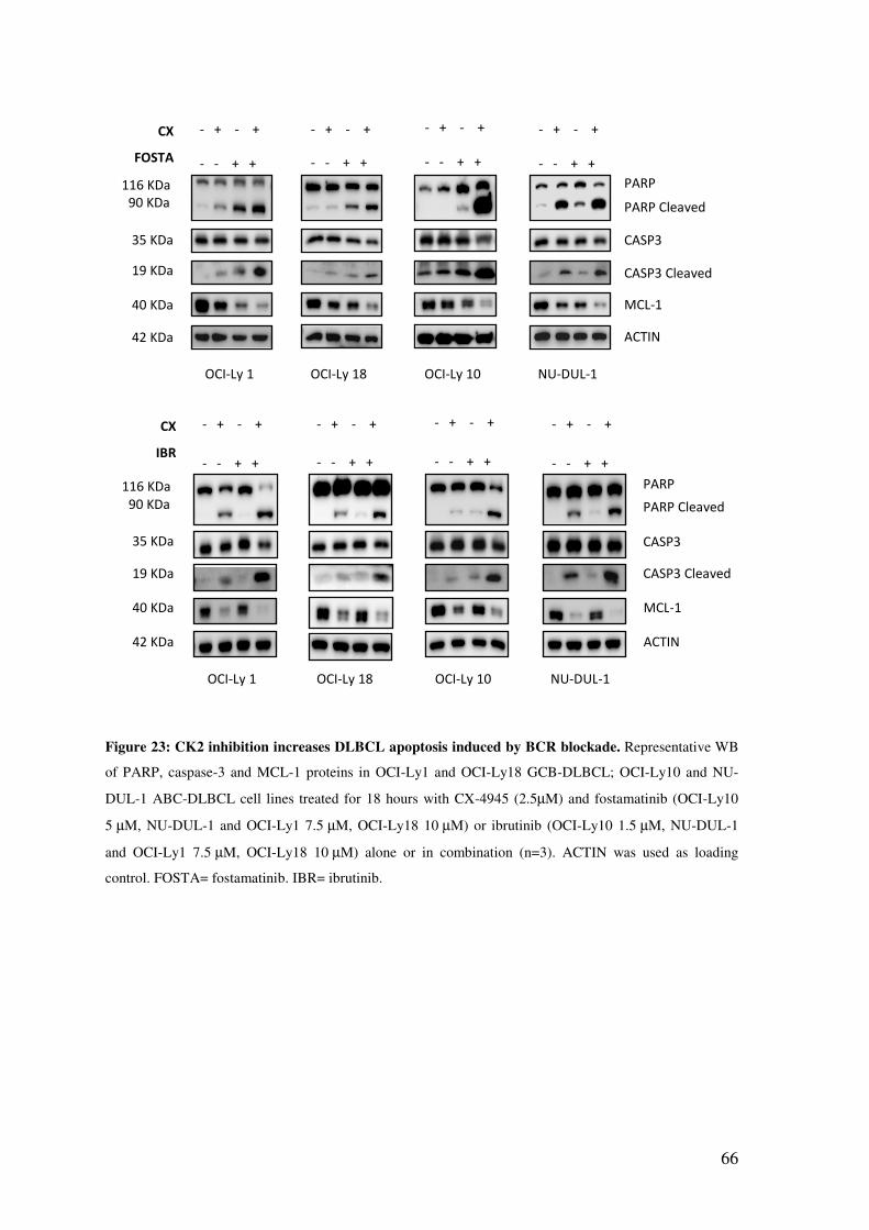
66
Figure 23: CK2 inhibition increases DLBCL apoptosis induced by BCR blockade. Representative WB
of PARP, caspase-3 and MCL-1 proteins in OCI-Ly1 and OCI-Ly18 GCB-DLBCL; OCI-Ly10 and NU-
DUL-1 ABC-DLBCL cell lines treated for 18 hours with CX-4945 (2.5µM) and fostamatinib (OCI-Ly10
5 µM, NU-DUL-1 and OCI-Ly1 7.5 µM, OCI-Ly18 10 µM) or ibrutinib (OCI-Ly10 1.5 µM, NU-DUL-1
and OCI-Ly1 7.5 µM, OCI-Ly18 10 µM) alone or in combination (n=3). ACTIN was used as loading
control. FOSTA= fostamatinib. IBR= ibrutinib.
OCI-Ly 1 OCI-Ly 18 OCI-Ly 10 NU-DUL-1
PARP PARP Cleaved
CASP3
CASP3 Cleaved
ACTIN
MCL-1
42 KDa
40 KDa
35 KDa
19 KDa
116 KDa 90 KDa
- + - +
- - + +
CX
FOSTA
- + - +
- - + +
- + - +
- - + +
- + - +
- - + +
OCI-Ly 1 OCI-Ly 18 OCI-Ly 10 NU-DUL-1
PARP PARP Cleaved
CASP3
CASP3 Cleaved
ACTIN
MCL-1
42 KDa
40 KDa
35 KDa
19 KDa
116 KDa 90 KDa
- + - +
- - + +
CX
IBR
- + - +
- - + +
- + - +
- - + +
- + - +
- - + +
67
CX-4945 POTENTIATES AKT INHIBITION INDUCED BY
THE BCR BLOCKADE
CK2 is known to act at different levels on the PI3K/PTEN/AKT survival pathway, by
promoting the activation of AKT, thus causing resistance to apoptosis and cell cycle
progression. To investigate the putative mechanisms leading to DLBCL cell apoptosis
upon exposure to CK2 inhibitor, BCR blockade and their combination, we analysed AKT
phosphorylation at Ser 473, a residue known to be crucial for the activation of the kinase.
WBs
show that the combined treatment reduces AKT phosphorylation, compared to untreated
samples. Interestingly, in OCI-Ly10, CX-4945 does not decrease AKT phosphorylation,
but the combination with the BCR blockade maintains low levels of phospho-AKT.
Noteworthy, in OCI-Ly1 GCB-DLBCL cells the combined treatment reduces not only the
phosphorylation, but also the total amount of AKT. Remarkably, a reduction in AKT
phosphorylation clearly correlates with a reduction in AKT activity, as suggested by the
decrease in AKT target proteins’ phosphorylation FOXO3a and GSK3β.
As can be seen in WBs below, the phosphorylation of AKT targets follows the pattern of
of AKT phosphorylation. In all cases, except for OCI-Ly10, the combination of CX-4945
and ibrutinib further reduces FOXO3a and GSK3β phosphorylation when compared to
single treatments (Fig. 24).
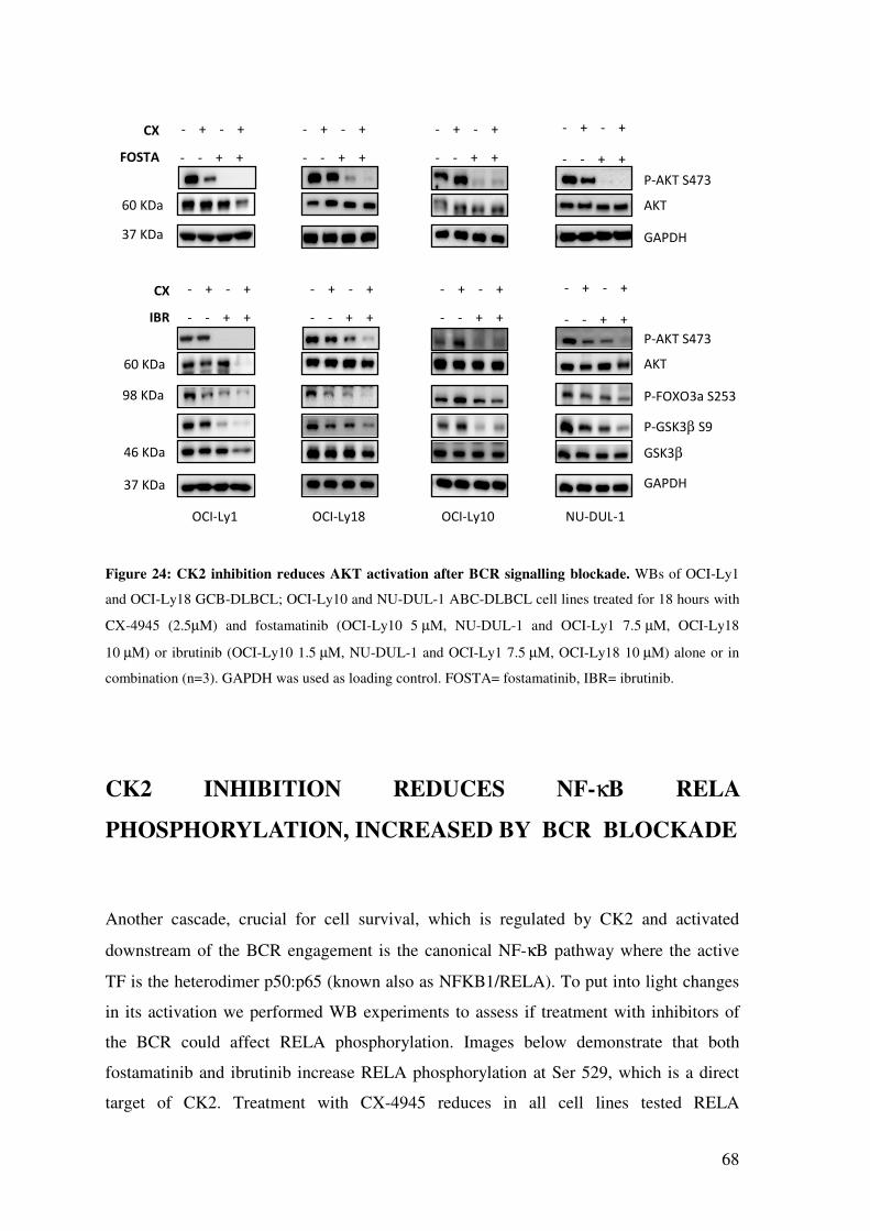
68
Figure 24: CK2 inhibition reduces AKT activation after BCR signalling blockade. WBs of OCI-Ly1
and OCI-Ly18 GCB-DLBCL; OCI-Ly10 and NU-DUL-1 ABC-DLBCL cell lines treated for 18 hours with
CX-4945 (2.5µM) and fostamatinib (OCI-Ly10 5 µM, NU-DUL-1 and OCI-Ly1 7.5 µM, OCI-Ly18
10 µM) or ibrutinib (OCI-Ly10 1.5 µM, NU-DUL-1 and OCI-Ly1 7.5 µM, OCI-Ly18 10 µM) alone or in
combination (n=3). GAPDH was used as loading control. FOSTA= fostamatinib, IBR= ibrutinib.
CK2 INHIBITION REDUCES NF-κB RELA
PHOSPHORYLATION, INCREASED BY BCR BLOCKADE
Another cascade, crucial for cell survival, which is regulated by CK2 and activated
downstream of the BCR engagement is the canonical NF-κB pathway where the active
TF is the heterodimer p50:p65 (known also as NFKB1/RELA). To put into light changes
in its activation we performed WB experiments to assess if treatment with inhibitors of
the BCR could affect RELA phosphorylation. Images below demonstrate that both
fostamatinib and ibrutinib increase RELA phosphorylation at Ser 529, which is a direct
target of CK2. Treatment with CX-4945 reduces in all cell lines tested RELA
OCI-Ly1 OCI-Ly18 OCI-Ly10 NU-DUL-1
P-AKT S473
AKT
37 KDa
60 KDa
GAPDH
P-FOXO3a S253
P-GSK3β S9
GSK3β
98 KDa
46 KDa
P-AKT S473
AKT
37 KDa
60 KDa
GAPDH
- + - +
- - + +
CX
IBR
- + - +
- - + +
- + - +
- - + +
- + - +
- - + +
- + - +
- - + +
CX
FOSTA
- + - +
- - + +
- + - +
- - + +
- + - +
- - + +
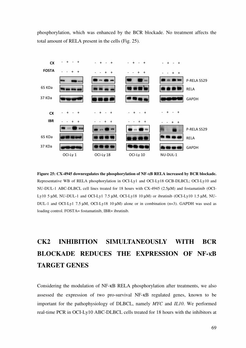
69
phosphorylation, which was enhanced by the BCR blockade. No treatment affects the
total amount of RELA present in the cells (Fig. 25).
Figure 25: CX-4945 downregulates the phosphorylation of NF-κB RELA increased by BCR blockade.
Representative WB of RELA phosphorylation in OCI-Ly1 and OCI-Ly18 GCB-DLBCL; OCI-Ly10 and
NU-DUL-1 ABC-DLBCL cell lines treated for 18 hours with CX-4945 (2.5µM) and fostamatinib (OCI-
Ly10 5 µM, NU-DUL-1 and OCI-Ly1 7.5 µM, OCI-Ly18 10 µM) or ibrutinib (OCI-Ly10 1.5 µM, NU-
DUL-1 and OCI-Ly1 7.5 µM, OCI-Ly18 10 µM) alone or in combination (n=3). GAPDH was used as
loading control. FOSTA= fostamatinib, IBR= ibrutinib.
CK2 INHIBITION SIMULTANEOUSLY WITH BCR
BLOCKADE REDUCES THE EXPRESSION OF NF-κB
TARGET GENES
Considering the modulation of NF-κB RELA phosphorylation after treatments, we also
assessed the expression of two pro-survival NF-κB regulated genes, known to be
important for the pathophysiology of DLBCL, namely MYC and IL10. We performed
real-time PCR in OCI-Ly10 ABC-DLBCL cells treated for 18 hours with the inhibitors at
OCI-Ly 1 OCI-Ly 18 OCI-Ly 10 NU-DUL-1
P-RELA S529
RELA 65 KDa
- + - +
- - + +
CX
FOSTA
- + - +
- - + +
- + - +
- - + +
- + - +
- - + +
37 KDa GAPDH
CX
IBR
P-RELA S529
RELA 65 KDa
- + - +
- - + +
- + - +
- - + +
- + - +
- - + +
- + - +
- - + +
37 KDa GAPDH
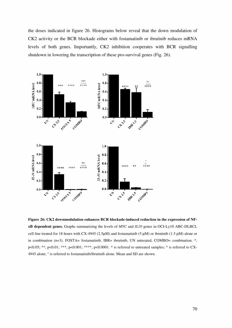
70
the doses indicated in figure 26. Histograms below reveal that the down modulation of
CK2 activity or the BCR blockade either with fostamatinib or ibrutinib reduces mRNA
levels of both genes. Importantly, CK2 inhibition cooperates with BCR signalling
shutdown in lowering the transcription of these pro-survival genes (Fig. 26).
Figure 26: CK2 downmodulation enhances BCR blockade-induced reduction in the expression of NF-
κB dependent genes. Graphs summarizing the levels of MYC and IL10 genes in OCI-Ly10 ABC-DLBCL
cell line treated for 18 hours with CX-4945 (2.5µM) and fostamatinib (5 µM) or ibrutinib (1.5 µM) alone or
in combination (n=3). FOSTA= fostamatinib, IBR= ibrutinib, UN untreated, COMBO= combination. *,
p<0.05; **, p<0.01; ***, p<0.001; ****, p<0.0001. * is referred to untreated samples; ^ is referred to CX-
4945 alone; ° is referred to fostamatinib/ibrutinib alone. Mean and SD are shown.
71
DISCUSSION
CK2 is a pleiotropic and constitutively active kinase that, being involved in a wide variety
of cellular protein networks, plays a crucial role in the regulation of survival and
proliferation. A number of studies revealed that cancer cells display abnormally high CK2
levels and activity, which confer resistance to chemotherapy, stabilize the oncokinome,
sustain pro survival signalling pathways and enhance the transforming potential of
oncogenes [2].
Malignant cells, which depend on CK2 overexpression for their proliferation and viability,
are more sensitive to CK2 downregulation than their normal counterparts. For these
reasons, this kinase has been proposed as a biomarker of cancer and its chemical
inhibition appears to be a good approach to achieve therapeutic effects [13].
To this extent, several inhibitors of CK2 have been designed, but only CX-4945
(silmitasertib) reached human clinical trials for cancer treatment [31].
It has been reported that many B-cell derived tumors like MM, MCL and CLL rely on
high CK2 activity and that its inhibition with CX-4945 induces cell death [25], [29].
Considering these issues, we investigated CK2 in DLBCL, an aggressive B-cell derived
neoplasia that is the most common type of NHL and is nowadays incurable in nearly one-
third of patients, suggesting the need for novel therapeutic approaches [37].
IHC performed on patients’ samples and WB of DLBCL cell lines revealed abnormally
high CK2 protein levels compared to normal lymphoid tissue or B-cells from healthy
donors. IF of GCB- and ABC-DLBCL cell lines and IHC of patients’ samples highlighted
that CK2α has a prevalent nuclear localization, in contrast with normal B-cells that
presented a diffuse pattern of expression. This is in line with a role for the kinase in the
promotion of cell survival in response to various growth stimuli [2]. This data revealed
that in DLBCL cells CK2 presents the same features that have been documented in other
cancer types.
The BCR pathway is fundamental for the survival of healthy and malignant B-cells, and
DLBCL, in particular ABC-DLBCL, presents an aberrant activation of this cascade. With
the above as a background, we investigated the functionality of the BCR signal upon CK2
inhibition, to evidence if CK2 overexpression and hyperactivity could be involved in
BCR-regulated processes. Therefore we employed the clinical grade compound CX-4945
to treat DLBCL cell lines and demonstrated that CK2 activity downregulation reduces the
72
amount of Ca++ released from the ER stores, after BCR engagement. Differences in the
peak of intracellular Ca++ are cell type-dependent, and could be a consequence of both the
BCR-surface expression and the ability to transduce the signal. WB revealed that
increased BTK and PLCγ2 phosphorylations after BCR stimulation, do not change after
CK2 inhibition. This makes us think that the signal is transmitted normally to PLCγ2. We
can suppose that CK2 acts downstream of PLC, maybe in phosphorylating IP3 receptors
on the ER membrane, thus altering their conformation and therefore their capacity to
release Ca++. Further experiments are needed to confirm the hypothesis. WB
demonstrated also that CX-4945 led to a decrease in AKT phosphorylation, thus
highlighting that CK2 contributes to the survival and proliferation of DLBCL cells also
through a positive action exerted on this kinase.
Cell survival assays demonstrated that CK2 inhibition reduces malignant B-cell survival,
but does not cause apoptosis of healthy PBMCs, suggesting that it is essential for cancer
maintenance and that there is the possibility of reducing its activity for therapeutic
purposes.
Inhibitors targeting kinases acting downstream of the BCR stimulation have shown
promising results in clinical trials, therefore we tested the combination of CK2 inhibition
with BCR blockade. We proved that CX-4945 synergizes with fostamatinib and ibrutinib
in inducing cell death. Increase in anti-apoptotic Mcl-1 degradation, PARP and caspase-3
cleavage, and AV+ cells after combined treatments, reinforced this data clearly suggesting
that CK2 plays a crucial role in the promotion of DLBCL cell survival, together with
signals generated by the BCR protein network. Hence, combining CK2 activity down
modulation with BCR signalling interruption could be an effective therapeutic strategy
for this disorder.
CK2 regulates two pro survival molecules, namely AKT and NF-κB, which are also
activated after BCR engagement. WB revealed that in all DLBCL cell lines analysed,
except for OCI-Ly10 ABC-DLBCL, combined treatments reduced AKT activity more
than BCR blockade alone. In OCI-Ly10 AKT is almost totally inhibited by
fostamatinib/ibrutinib and the addition of CX-4945 did not cause a further reduction in
AKT and its downstream targets’ phosphorylation. We can speculate that the down
modulation of the pro survival action exerted by AKT through the activation of GSK3β
and the inhibition of FOXO3a can explain, in some but not all DLBCL cells, the increase
in cell death observed after combined treatments. However, the fact that one cell line did
73
not follow the same behaviour suggests that other molecules are intimately involved in
the regulation of the equilibrium between survival and apoptosis. Consequently, we
analysed the modulation of the TF NF-κB RELA that is phosphorylated by CK2 and
together with NFKB1 takes part in the canonical pathway activated by the BCR signalling
cascade. It is well known that NF-κB is a key TF, which promotes survival, in particular
of ABC-DLBCL cells [52]. As expected, CK2 inhibition reduced RELA phosphorylation
on Ser 529, which is a direct target of this kinase. Surprisingly, the BCR blockade
increases RELA phosphorylation in this residue and combined treatments reduced the
stimulatory effect induced by fostamatinib/ibrutinib. A possible explanation for this can
be found considering that BCR blockade is a stressful event for B-cells and consequently
the activity of CK2, which is a stress-response kinase, increases and it phosphorylates its
targets to a higher extent, in order to allow the cell to react to stress. To assess if the
modulation of RELA phosphorylation on Ser 529, a residue of the C-terminal TAD
domain, could affect gene expression, we analysed the mRNA levels of MYC and IL-10,
which are NF-κB target genes crucial for DLBCL survival [63]. Either BCR blockade or
CK2 inhibition reduced MYC and IL10 transcription and their combination further
augmented this decrease. In light of these results, we can desume that this residue is
clearly not the only one that contributes to RELA transcriptional activity, since the pattern
of MYC and IL-10 mRNA levels does not follow RELA phosphorylation. Furthermore,
the reduction in MYC and IL-10 expression could, at least in part, explain the decrease in
DLBCL survival, attested by augmented PARP and caspase-3 cleavage.

74
75
CONCLUSIONS
Our study demonstrates that protein kinase CK2 is abnormally high both in terms of
protein quantity and catalytic activity in DLBCL cells, and that malignant B-cells are
more sensitive than their normal counterparts to CK2 inhibition. In the past few years,
this fact has been evidenced also in solid tumors and other hematologic malignancies,
suggesting that this protein could be an appealing target for many types of cancer.
With the present analysis in DLBCL, we demonstrate that CK2 inhibition reduces the
activation of AKT and the release of Ca++ from the ER stores after BCR stimulation,
disclosing the importance of this kinase in regulating pathways critical for malignant B-
cell activation, survival, and cell cycle progression. A downmodulation of CK2 activity
with the clinical grade CX-4945 induces malignant cell death and boosts the effects of
agents known to disrupt the BCR pathway, namely fostamatinib and ibrutinib, currently
under clinical trials in DLBCL patients.
These brand new findings suggest that CK2 could target multiple sites of the BCR-
dependent cascade, and could pave the way for the inclusion of CK2 inhibitors in drug
combination therapies with BCR signalling inhibitors. Targeting multiple molecules in
this pro survival cascade could indeed represent a promising strategy for the treatment of
DLBCL, and feasibly of many other B-cell derived lymphomas.

76
77
REFERENCES
[1] A. a Kramerov and A. V Ljubimov, “Focus on molecules: protein kinase CK2.,” Exp. Eye Res., vol. 101, pp. 111–2, Aug. 2012.
[2] J. H. Trembley, G. Wang, G. Unger, J. Slaton, and K. Ahmed, “Protein kinase CK2 in health and disease: CK2: a key player in cancer biology.,” Cell. Mol. Life Sci., vol. 66, no. 11–12, pp. 1858–67, Jun. 2009.
[3] N. a St-Denis and D. W. Litchfield, “Protein kinase CK2 in health and disease: From birth to death: the role of protein kinase CK2 in the regulation of cell proliferation and survival.,” Cell. Mol. Life Sci., vol. 66, no. 11–12, pp. 1817–29, Jun. 2009.
[4] K. Niefind, J. Raaf, and O.-G. Issinger, “Protein kinase CK2 in health and disease: Protein kinase CK2: from structures to insights.,” Cell. Mol. Life Sci., vol. 66, no. 11–12, pp. 1800–16, Jun. 2009.
[5] K. Niefind and B. Guerra, “Crystal structure of human protein kinase CK2: insights into basic properties of the CK2 holoenzyme,” EMBO J., vol. 20, no. 19, 2001.
[6] D. W. Litchfield, “Protein kinase CK2: structure, regulation and role in cellular decisions of life and death.,” Biochem. J., vol. 369, no. Pt 1, pp. 1–15, 2003.
[7] J. H. Trembley, G. M. Unger, V. L. Korman, D. K. Tobolt, Z. Kazimierczuk, L. a. Pinna, B. T. Kren, and K. Ahmed, “Nanoencapsulated anti-CK2 small molecule drug or siRNA specifically targets malignant cancer but not benign cells,” Cancer
Lett., vol. 315, no. 1, pp. 48–58, Feb. 2012.
[8] G. Di Maira, F. Brustolon, L. a Pinna, and M. Ruzzene, “Dephosphorylation and inactivation of Akt/PKB is counteracted by protein kinase CK2 in HEK 293T cells.,” Cell. Mol. Life Sci., vol. 66, no. 20, pp. 3363–73, Oct. 2009.
[9] D. H. Song, I. Dominguez, J. Mizuno, M. Kaut, S. C. Mohr, and D. C. Seldin, “CK2 phosphorylation of the armadillo repeat region of beta-catenin potentiates Wnt signaling.,” J. Biol. Chem., vol. 278, no. 26, pp. 24018–25, Jun. 2003.
[10] M. Ruzzene and L. a Pinna, “Addiction to protein kinase CK2: a common denominator of diverse cancer cells?,” Biochim. Biophys. Acta, vol. 1804, no. 3, pp. 499–504, Mar. 2010.
[11] K. Ahmed, D. Gerber, and C. Cochet, “Joining the cell survival squad: an emerging role for protein kinase CK2,” Trends Cell Biol., vol. 12, no. 5, pp. 226–230, 2002.
[12] Y. Miyata, “Protein kinase CK2 in health and disease: CK2: the kinase controlling the Hsp90 chaperone machinery.,” Cell. Mol. Life Sci., vol. 66, no. 11–12, pp. 1840–9, Jun. 2009.
[13] M. Ruzzene, K. Tosoni, S. Zanin, L. Cesaro, and L. a. Pinna, “Protein kinase CK2 accumulation in ‘oncophilic’ cells: causes and effects,” Mol. Cell. Biochem., vol. 356, no. 1–2, pp. 5–10, Jul. 2011.
[14] F. Piazza, S. Manni, M. Ruzzene, L. a Pinna, C. Gurrieri, and G. Semenzato, “Protein kinase CK2 in hematologic malignancies: reliance on a pivotal cell
78
survival regulator by oncogenic signaling pathways.,” Leukemia, vol. 26, no. 6, pp. 1174–9, Jun. 2012.
[15] S. Mishra, A. Reichert, J. Cunnick, D. Senadheera, B. Hemmeryckx, N. Heisterkamp, and J. Groffen, “Protein kinase CKIIalpha interacts with the Bcr moiety of Bcr/Abl and mediates proliferation of Bcr/Abl-expressing cells.,” Oncogene, vol. 22, no. 51, pp. 8255–62, Nov. 2003.
[16] S. Mishra, V. Pertz, B. Zhang, P. Kaur, H. Shimada, J. Groffen, Z. Kazimierczuk, L. a Pinna, and N. Heisterkamp, “Treatment of P190 Bcr/Abl lymphoblastic leukemia cells with inhibitors of the serine/threonine kinase CK2.,” Leukemia, vol. 21, no. 1, pp. 178–80, Jan. 2007.
[17] a. Margarida Gomes, M. V. D. Soares, P. Ribeiro, J. Caldas, V. Póvoa, L. R. Martins, A. Melão, A. Serra-Caetano, A. B. de Sousa, J. F. Lacerda, and J. T. Barata, “Adult B-cell acute lymphoblastic leukemia cells display decreased PTEN activity and constitutive hyperactivation of PI3K/Akt pathway despite high PTEN protein levels,” Haematologica, vol. 99, no. 6, pp. 1062–1068, 2014.
[18] A. Silva, J. A. Yunes, B. A. Cardoso, L. R. Martins, P. Y. Jotta, M. Abecasis, A. E. Nowill, N. R. Leslie, A. A. Cardoso, and J. T. Barata, “PTEN posttranslational inactivation and hyperactivation of the PI3K/Akt pathway sustain primary T cell leukemia viability.,” J. Clin. Invest., vol. 118, no. 11, pp. 3762–74, 2008.
[19] P. Kastner and A. Dupuis, “Function of Ikaros as a tumor suppressor in B cell acute lymphoblastic leukemia,” Am. J. …, vol. 3, no. 1, pp. 1–13, 2013.
[20] S. Dovat, C. Song, K. J. Payne, and Z. Li, “Ikaros, CK2 kinase, and the road to leukemia.,” Mol. Cell. Biochem., vol. 356, no. 1–2, pp. 201–7, Oct. 2011.
[21] M. Popescu, Z. Gurel, T. Ronni, C. Song, K. Y. Hung, K. J. Payne, and S. Dovat, “Ikaros Stability and Pericentromeric Localization Are Regulated by Protein Phosphatase 1,” J. Biol. Chem., vol. 284, no. 20, pp. 13869–13880, Mar. 2009.
[22] C. Song, Z. Li, A. K. Erbe, A. Savic, and S. Dovat, “Regulation of Ikaros function by casein kinase 2 and protein phosphatase 1.,” World J. Biol. Chem., vol. 2, pp. 126–131, 2011.
[23] F. a. Piazza, M. Ruzzene, C. Gurrieri, B. Montini, L. Bonanni, G. Chioetto, G. Di Maira, F. Barbon, A. Cabrelle, R. Zambello, F. Adami, L. Trentin, L. A. Pinna, and G. Semenzato, “Multiple myeloma cell survival relies on high activity of protein kinase CK2,” Blood, vol. 108, no. 5, pp. 1698–1707, 2006.
[24] S. Manni, A. Brancalion, L. Q. Tubi, A. Colpo, L. Pavan, A. Cabrelle, E. Ave, F. Zaffino, G. Di Maira, M. Ruzzene, F. Adami, R. Zambello, M. R. Pitari, P. Tassone, L. a. Pinna, C. Gurrieri, G. Semenzato, and F. Piazza, “Protein kinase CK2 protects multiple myeloma cells from ER stress-induced apoptosis and from the cytotoxic effect of HSP90 inhibition through regulation of the unfolded protein response,” Clin. Cancer Res., vol. 18, pp. 1888–1900, 2012.
[25] S. Manni, A. Brancalion, E. Mandato, L. Q. Tubi, A. Colpo, M. Pizzi, R. Cappellesso, F. Zaffino, S. A. Di Maggio, A. Cabrelle, F. Marino, R. Zambello, L. Trentin, F. Adami, C. Gurrieri, G. Semenzato, and F. Piazza, “Protein Kinase CK2 Inhibition Down Modulates the NF-κB and STAT3 Survival Pathways, Enhances the Cellular Proteotoxic Stress and Synergistically Boosts the Cytotoxic Effect of Bortezomib on Multiple Myeloma and Mantle Cell Lymphoma Cells,” PLoS One, vol. 8, no. 9, p. e75280, Sep. 2013.
79
[26] S. Manni, D. Toscani, E. Mandato, a Brancalion, L. Quotti Tubi, P. Macaccaro, a Cabrelle, F. Adami, R. Zambello, C. Gurrieri, G. Semenzato, N. Giuliani, and F. Piazza, “Bone marrow stromal cell-fueled multiple myeloma growth and osteoclastogenesis are sustained by protein kinase CK2,” Leukemia, vol. 28, no. 10, pp. 2094–2097, Jun. 2014.
[27] M. Shehata, S. Schnabl, D. Demirtas, M. Hilgarth, R. Hubmann, E. Ponath, S. Badrnya, C. Lehner, A. Hoelbl, M. Duechler, A. Gaiger, C. Zielinski, J. D. Schwarzmeier, and U. Jaeger, “Reconstitution of PTEN activity by CK2 inhibitors and interference with the PI3-K/Akt cascade counteract the antiapoptotic effect of human stromal cells in chronic lymphocytic leukemia,” Blood, vol. 116, pp. 2513–2521, 2010.
[28] L. Martins, P. Lúcio, M. Silva, and K. Anderes, “Targeting CK2 overexpression and hyperactivation as a novel therapeutic tool in chronic lymphocytic leukemia,” Blood, vol. 116, no. 15, pp. 2724–2732, 2010.
[29] L. R. Martins, P. Lúcio, a Melão, I. Antunes, B. a Cardoso, R. Stansfield, M. T. S. Bertilaccio, P. Ghia, D. Drygin, M. G. Silva, and J. T. Barata, “Activity of the clinical-stage CK2-specific inhibitor CX-4945 against chronic lymphocytic leukemia.,” Leukemia, vol. 28, no. 1, pp. 179–82, Jan. 2014.
[30] R. C. Prins, R. T. Burke, J. W. Tyner, B. J. Druker, M. M. Loriaux, and S. E. Spurgeon, “CX-4945, a selective inhibitor of casein kinase-2 (CK2), exhibits anti-tumor activity in hematologic malignancies including enhanced activity in chronic lymphocytic leukemia when combined with fludarabine and inhibitors of the B-cell receptor pathway.,” Leukemia, vol. 27, no. 10, pp. 2094–6, Oct. 2013.
[31] F. Pierre, P. C. Chua, S. E. O’Brien, A. Siddiqui-Jain, P. Bourbon, M. Haddach, J. Michaux, J. Nagasawa, M. K. Schwaebe, E. Stefan, A. Vialettes, J. P. Whitten, T. K. Chen, L. Darjania, R. Stansfield, K. Anderes, J. Bliesath, D. Drygin, C. Ho, M. Omori, C. Proffitt, N. Streiner, K. Trent, W. G. Rice, and D. M. Ryckman, “Discovery and SAR of 5-(3-chlorophenylamino)benzo[c][2,6]naphthyridine-8-carboxylic acid (CX-4945), the first clinical stage inhibitor of protein kinase CK2 for the treatment of cancer.,” J. Med. Chem., vol. 54, no. 2, pp. 635–54, Jan. 2011.
[32] a. Siddiqui-Jain, D. Drygin, N. Streiner, P. Chua, F. Pierre, S. E. O’Brien, J. Bliesath, M. Omori, N. Huser, C. Ho, C. Proffitt, M. K. Schwaebe, D. M. Ryckman, W. G. Rice, and K. Anderes, “CX-4945, an Orally Bioavailable Selective Inhibitor of Protein Kinase CK2, Inhibits Prosurvival and Angiogenic Signaling and Exhibits Antitumor Efficacy,” Cancer Res., vol. 70, no. 24, pp. 10288–10298, Dec. 2010.
[33] A. D. Ferguson, P. R. Sheth, A. D. Basso, S. Paliwal, K. Gray, T. O. Fischmann, and H. V Le, “Structural basis of CX-4945 binding to human protein kinase CK2.,” FEBS Lett., vol. 585, no. 1, pp. 104–10, Jan. 2011.
[34] H. J. Chon, K. J. Bae, Y. Lee, and J. Kim, “The casein kinase 2 inhibitor, CX-4945, as an anti-cancer drug in treatment of human hematological malignancies.,” Front.
Pharmacol., vol. 6, no. March, p. 70, Jan. 2015.
[35] T. a Packard and J. C. Cambier, “B lymphocyte antigen receptor signaling: initiation, amplification, and regulation.,” F1000Prime Rep., vol. 5, no. October, p. 40, Jan. 2013.
[36] T. LeBien and T. Tedder, “B lymphocytes: how they develop and function,” Blood,
80
vol. 112, no. 5, pp. 1570–1581, 2008.
[37] M. Roschewski, L. M. Staudt, and W. H. Wilson, “Diffuse large B-cell lymphoma-treatment approaches in the molecular era.,” Nat. Rev. Clin. Oncol., vol. 11, no. 1, pp. 12–23, Jan. 2014.
[38] H. Niiro and E. a Clark, “Regulation of B-cell fate by antigen-receptor signals.,” Nat. Rev. Immunol., vol. 2, no. 12, pp. 945–56, Dec. 2002.
[39] A. M. Scharenberg, L. a Humphries, and D. J. Rawlings, “Calcium signalling and cell-fate choice in B cells.,” Nat. Rev. Immunol., vol. 7, no. 10, pp. 778–89, 2007.
[40] J. Napetschnig and H. Wu, “Molecular Basis of NF-κB Signaling,” Annu. Rev.
Biophys., vol. 42, no. 1, pp. 443–468, 2013.
[41] M. S. Hayden and S. Ghosh, “Shared Principles in NF-kB Signaling,” Cell, vol. 132, no. 3, pp. 344–362, 2008.
[42] P. Viatour, M.-P. Merville, V. Bours, and A. Chariot, “Phosphorylation of NF-κB and IκB proteins: implications in cancer and inflammation,” Trends Biochem. Sci., vol. 30, no. 1, pp. 43–52, 2005.
[43] P. J. Jost and J. Ruland, “Aberrant NF-kappa B signaling in lymphoma: mechanisms, consequences, and therapeutic implications,” Blood, vol. 109, no. 7, pp. 2700–2707, 2007.
[44] R. Moreno, J.-M. Sobotzik, C. Schultz, and M. L. Schmitz, “Specification of the NF- B transcriptional response by p65 phosphorylation and TNF-induced nuclear translocation of IKK ,” Nucleic Acids Res., vol. 38, no. 18, pp. 6029–6044, 2010.
[45] L. C. Cantley, “The phosphoinositide 3-kinase pathway.,” Science, vol. 296, no. 5573, pp. 1655–1657, 2002.
[46] M. Werner, E. Hobeika, and H. Jumaa, “Role of PI3K in the generation and survival of B cells,” Immunol. Rev., vol. 237, no. 1, pp. 55–71, 2010.
[47] J. J. Limon and D. a Fruman, “Akt and mTOR in B Cell Activation and Differentiation.,” Front. Immunol., vol. 3, no. August, p. 228, Jan. 2012.
[48] G. Di Maira, M. Salvi, G. Arrigoni, O. Marin, S. Sarno, F. Brustolon, L. A. Pinna, and M. Ruzzene, “Protein kinase CK2 phosphorylates and upregulates Akt/PKB,” Cell Death Differ., vol. 12, no. 6, pp. 668–677, 2005.
[49] M. Menon, S. Pittaluga, and E. Jaffe, “The Histological and Biological Spectrum of Diffuse Large B-cell Lymphoma in the WHO Classification,” Cancer J, vol. 18, no. 5, pp. 411–420, 2012.
[50] C. Schneider, L. Pasqualucci, and R. Dalla-Favera, “Molecular Pathogenesis of Diffuse Large B-cell Lymphoma,” Semin Diagn PAtholChanges, vol. 29, no. 6, pp. 997–1003, 2012.
[51] P. Caro, A. U. Kishan, E. Norberg, I. A. Stanley, B. Chapuy, S. B. Ficarro, K. Polak, D. Tondera, J. Gounarides, H. Yin, F. Zhou, M. R. Green, L. Chen, S. Monti, J. A. Marto, M. A. Shipp, and N. N. Danial, “Metabolic Signatures Uncover Distinct Targets in Molecular Subsets of Diffuse Large B Cell Lymphoma,” Cancer Cell, vol. 22, no. 4, pp. 547–560, 2012.
[52] L. M. Staudt, “Oncogenic activation of NF-kappaB.,” Cold Spring Harb. Perspect.
Biol., vol. 2, no. 6, p. a000109, 2010.
[53] R. Davis, V. Ngo, G. Lenz, and P. Tolar, “Chronic Active B cell receptor signaling
81
in diffuse large B cell lymphoma,” Nature, vol. 463, no. 7277, pp. 88–92, 2010.
[54] L. Pasqualucci and R. Dalla-Favera, “The genetic landscape of diffuse large B-cell lymphoma.,” Semin. Hematol., vol. 52, no. 2, pp. 67–76, Apr. 2015.
[55] R. M. Young, A. L. Shaffer, J. D. Phelan, and L. M. Staudt, “B-cell receptor signaling in diffuse large B-cell lymphoma.,” Semin. Hematol., vol. 52, no. 2, pp. 77–85, Apr. 2015.
[56] K. H. Shain and J. Tao, “The B-cell receptor orchestrates environment-mediated lymphoma survival and drug resistance in B-cell malignancies.,” Oncogene, vol. 33, no. 32, pp. 4107–13, Aug. 2014.
[57] R. M. Young and L. M. Staudt, “Targeting pathological B cell receptor signalling in lymphoid malignancies.,” Nat. Rev. Drug Discov., vol. 12, no. 3, pp. 229–43, Mar. 2013.
[58] J. W. Friedberg, J. Sharman, J. Sweetenham, P. B. Johnston, J. M. Vose, A. Lacasce, J. Schaefer-Cutillo, S. De Vos, R. Sinha, J. P. Leonard, L. D. Cripe, S. a Gregory, M. P. Sterba, A. M. Lowe, R. Levy, and M. a Shipp, “Inhibition of Syk with fostamatinib disodium has significant clinical activity in non-Hodgkin lymphoma and chronic lymphocytic leukemia.,” Blood, vol. 115, no. 13, pp. 2578–85, Apr. 2010.
[59] C. B. Reeder and S. M. Ansell, “Novel therapeutic agents for B-cell lymphoma: developing rational combinations.,” Blood, vol. 117, no. 5, pp. 1453–62, Feb. 2011.
[60] C. P. Hans, D. D. Weisenburger, T. C. Greiner, R. D. Gascoyne, J. Delabie, G. Ott, H. K. Müller-Hermelink, E. Campo, R. M. Braziel, E. S. Jaffe, Z. Pan, P. Farinha, L. M. Smith, B. Falini, A. H. Banham, A. Rosenwald, L. M. Staudt, J. M. Connors, J. O. Armitage, and W. C. Chan, “Confirmation of the molecular classification of diffuse large B-cell lymphoma by immunohistochemistry using a tissue microarray.,” Blood, vol. 103, no. 1, pp. 275–82, 2004.
[61] R. a Faust, G. Niehans, M. Gapany, D. Hoistad, D. Knapp, D. Cherwitz, a Davis, G. L. Adams, and K. Ahmed, “Subcellular immunolocalization of protein kinase CK2 in normal and carcinoma cells.,” Int. J. Biochem. Cell Biol., vol. 31, no. 9, pp. 941–9, 1999.
[62] T. C. Chou and P. Talalay, “Quantitative analysis of dose-effect relationships: the combined effects of multiple drugs or enzyme inhibitors.,” Adv. Enzyme Regul., vol. 22, pp. 27–55, 1984.
[63] W. Béguelin, S. Sawh, N. Chambwe, F. C. Chan, Y. Jiang, J.-W. Choo, D. W. Scott, a Chalmers, H. Geng, L. Tsikitas, W. Tam, G. Bhagat, R. D. Gascoyne, and R. Shaknovich, “IL10 receptor is a novel therapeutic target in DLBCLs.,” Leukemia, no. February, pp. 1–11, 2015.

82
83
PUBLICATIONS
Manni S, Brancalion A, Mandato E, Tubi LQ, Colpo A, Pizzi M, Cappellesso R, Zaffino F, Di Maggio SA, Cabrelle A, Marino F, Zambello R, Trentin L, Adami F,Gurrieri C, Semenzato G, Piazza F. (PLoS One. 2013 Sep 27;8(9):e75280).
Protein Kinase CK2 Inhibition Down Modulates the NF-κB and STAT3 Survival
Pathways, Enhances the Cellular Proteotoxic Stress and Synergistically Boosts the
Cytotoxic Effect of Bortezomib on Multiple Myeloma and Mantle Cell Lymphoma
Cells.
Sabrina Manni, Denise Toscani, Elisa Mandato, Alessandra Brancalion, Laura Quotti Tubi, Paolo Macaccaro, Anna Cabrelle, Fausto Adami, Renato Zambello, Carmela Gurrieri, Gianpietro Semenzato, Nicola Giuliani and Francesco Piazza (Leukemia. 2014 Oct;28(10):2094-7).
Bone marrow stromal cell-fueled multiple myeloma growth and osteoclastogenesis
are sustained by protein kinase CK2.
Marco Pizzi, Francesco Piazza, Claudio Agostinelli, Fabio Fuligni, Pietro Benvenuti, Elisa Mandato, Alessandro Casellato, Massimo Rugge, Gianpietro Semenzato and Stefano A. Pileri. (Oncotarget. 2015 Mar 30;6(9):6544-52).
Protein kinase CK2 is widely expressed in Follicular, Burkitt and diffuse large B-cell
lymphomas and propels malignant B-cell growth.