Embed Size (px)
Citation preview
UNIVERSITA' DEGLI STUDI DI PADOVA
UNIVERSITA' DI PADOVA
FACOLTA' DI MEDICINA E CHIRURGIA
DIPARTIMENTO DI MEDICINA CLINICA E SPERIMENTALE
INTERNATIONAL DOCTORATE IN ARTERIAL HYPERTENSION AND
VASCULAR BIOLOGY
CICLO XXI
HUMAN PRIMARY ALDOSTERONISM;
INVESTIGATION OF THE MECHANISMS LEADING TO
“AUTONOMOUS” ALDOSTERONE EXCESS
Coordinatore: Ch.mo Prof. Gian Paolo Rossi
Supervisore: Ch.mo Prof. Gian Paolo Rossi
Dottorando: Enrico Aldighieri
Anno Accademico 2008-2009
2
Riassunto
Obiettivo: Alcuni studi hanno suggerito che l'espressione genica CYP11B2
è più elevata in pazienti con APA rispetto ai soggetti normali, tuttavia non
esistono anticorpi in grado di identificare questa proteina. Quindi abbiamo
deciso di effettuare studi di ibridazione in situ (ISH) per localizzare
CYP11B2 a livello tessutale. Un altro obiettivo del nostro studio è quello di
capire se la presenza di micronoduli possa influire sulla fisiopatologia
dell’adenoma. La nostra ipotesi è che un fattore umorale stimolante
potrebbe svolgere un ruolo nella genesi di APA.
Metodo: Abbiamo sviluppato un metodo non radioattivo basato sull'uso di
uno specifica sonda oligo per localizzare CYP11B2. Per la rilevazione
dell’housekeeping gene, abbiamo utilizzato una sonda a doppio filamento
per il gene PBGD, (NM_000190). Entrambe le sonde sono state marcate
con Digoxigenin. Abbiamo inoltre misurato i livelli di anticorpi anti-AT1 nel
siero di pazienti con APA e la loro localizzazione tessutale tramite IHC
nella ghiandola surrenalica.
Risultati: all’istopatologia si sono evidenziati noduli satelliti che
presentavano l’espressione del CYP11B2. L’ISH ha mostrato una
colorazione specifica di tutti i noduli a livello prevalentemente nucleare. La
presenza di Auto anticorpi anti AT1 è stata dimostrata con test ELISA nel
siero, ma la localizzazione nel tessuto non ha ancora mostrato risultati
apprezzabili.
Conclusione: questa tecnica non radioattiva di ISH dimostra in modo
inequivocabile la presenza di micro satelliti producenti aldosterone. Elevati
livelli di autoanticorpi nel siero dei pazienti con APA ci permettono di
ipotizzare che questo disordine della ghiandola surrenale possa essere di
origine autoimmune.
3
Summary
Objective: Due to its unique expression of aldosterone synthase
cytocromo P450 (CYP11B2), the enzyme required for the final steps of
aldosterone biosynthesis aldosterone is exclusively expressed in the
adrenalcortical zona glomerulosa cell. Some studies suggested that the
CYP11B2 gene expression can be higher in APAs than in normal
adrenocortical tissues, leading to postulate a transcriptional modulation of
aldosterone overproduction in these tumors but antibodies capable to
identify this protein do not exist. Thus we performed studies of in-situ
hybridization (ISH) to analyze CYP11B2 localization. Another aim of our
study is to understand whether the occurrence of micronodularity
peripheral to the main adenomatous mass in some of our patient could be
involved in the pathophysiology of the adenoma. Our hypothesis is that a
similar humoral stimulating factor could play a role in the genesis of APA.
Method: We developed a novel non radioactive in-situ hybridization
technique based on use of a CYP11B2 specific oligo probe to localize the
specific transcripts of the CYP11B2. Adrenocortical tissue from patients
was studied. As housekeeping gene, we used PBGD, (NM_000190) probe
labeled with Digoxigenin. The oligonucleotide probe for the CYP11B2 was
5’ conjugated with DIG. Negative controls was detected with probe not
labeled. The second approach was measuring of serum auto-antibodies
anti AT1 in patients with APA and their localization on adrenal gland tissue
by IHC.
Results: At hystopathology intensely labeled areas called “nodules
producing aldosterone” and negative areas were detected in the APA
analyzed. ISH showed specific staining of all nodules. The staining was
predominantly nuclear, a finding consistent with a good preservation of
the transcript at this level. The AAbodies’ anti AT1 presence has been
demonstrated with ELISA test but the tissue localization still did not show
appreciable results.
Conclusion: this novel non radioactive ISH technique unequivocally
demostrated the presence of aldosterone-producing microsatellites. High
levels of AutoAntibodies in the serum of patients with APA allow us to
hypothesize that this adrenal gland disorder is autoimmune.
4
INDEX
INTRODUCTION
Anatomy of the adrenal gland ………………………….....................6
Steroidogenesis……… ……….……………………………………………….9
Molecular regulation of steroidogenesis…………….…………….…12
Arterial Hypertension from Adrenal hyperfunction..…………....14
Clinic cases of Conn………………………………………………………....16
Bilateral adrenal hyperplasia versus CONN………………………...17
AIMS OF THE STUDY
Flow chart for testing the General Hypothesis…………………….20
Specific Hypothesis………………………………………………………….21
Study Approach……………………………………………………………….21
METHODS
Adrenal specimens……………………………………………………….….23
Choice of the technique for identification of aldosterone
synthase……………………………………………………………………......24
Work flow for the synthesis of probes for the housekeeping
gene Porphobilinogen Deaminase and for aldosterone
synthase …………………………………………………………………….….26
Production probes for hybridization probes……………………...…27
5
Work Flow for the In situ Hybridization for CYP11B2 and the
Housekeeping gene PBGD …………………………………….………...29
In situ Hybridization…………………………………………….….……….30
Immunohistochemisty CD56……………………….……………….…...32
Work Flow for the identification of Auto-Antibody on tissue
sections………………………………………………………………………….33
Measurement Auto Antibody serum APA………………………..…..34
Immunohistochemistry Auto Antibody……………………….……...35
RESULTS…………………………………………………………………….……….36
DISCUSSION………………………………………………………….……..…....41
REFERENCES……………………………………………………….…………..….46
6
INTRODUCTION
Anatomy of the Adrenal Gland
The two adrenal glands are located immediately anterior to the kidneys,
encased in a capsule of connective tissue and are usually found under a
layer of peri-adrenal fat. Like the kidneys, the adrenal glands lie beneath
the peritoneum, e.g. they are retroperitoneal. However, the exact location
compared to the kidney and the shape of the adrenal gland can vary
across species. The weight of normal glands is 4-6 grams each, after the
dissection of the fat, and concerning the size, the normal adrenal gland is
about 5 x 3 x 1 cm.
The adrenal arteries are multiple and result from many different
ramifications, which are mainly ramifications of the inferior diaphragmatic
arteries. This is a network of vessels that are located above the renal
vessels. Three main groups can be identified:
1. The superior adrenal artery
2. The media adrenal artery
3. The inferior adrenal artery
The adrenal veins, have a simpler organization. The right adrenal vein is
very short, with an almost horizontal course, and drains directly into the
inferior vena cava. The left adrenal vein is longer and has a nearly vertical
course. Often, the inferior phrenic vein joins the left adrenal vein forming
a common trunk, termed the pheric-adrenal that runs toward the left
renal vein. Other times, the left adrenal vein receives renal venous
ramifications. The gonad vein and lumbar veins may have connections
with the left adrenal vein as well this anatomic pattern has profound
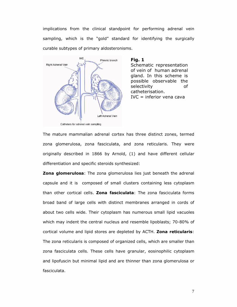
7
implications from the clinical standpoint for performing adrenal vein
sampling, which is the “gold” standard for identifying the surgically
curable subtypes of primary aldosteronisms.
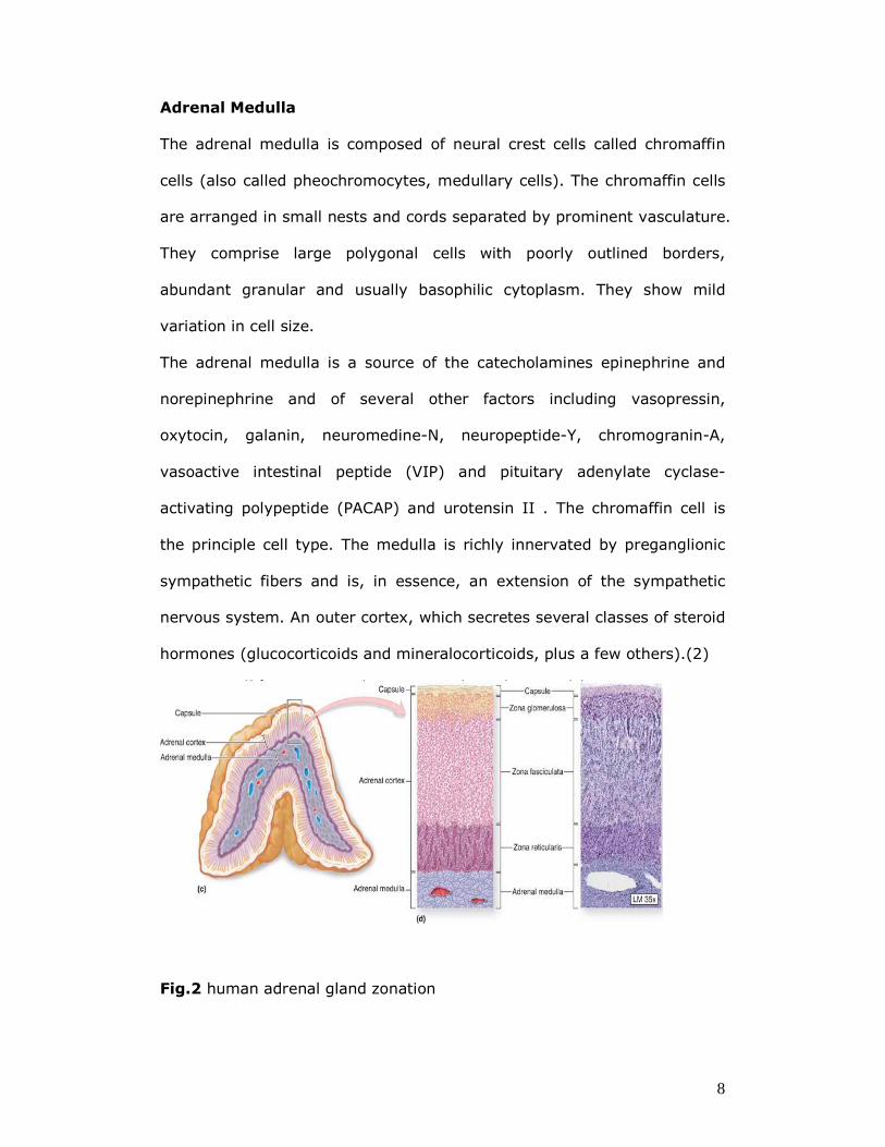
The mature mammalian adrenal cortex has three distinct zones, termed
zona glomerulosa, zona fasciculata, and zona reticularis. They were
originally described in 1866 by Arnold, (1) and have different cellular
differentiation and specific steroids synthesized:
Zona glomerulosa: The zona glomerulosa lies just beneath the adrenal
capsule and it is composed of small clusters containing less cytoplasm
than other cortical cells. Zona fasciculata: The zona fasciculata forms
broad band of large cells with distinct membranes arranged in cords of
about two cells wide. Their cytoplasm has numerous small lipid vacuoles
which may indent the central nucleus and resemble lipoblasts; 70-80% of
cortical volume and lipid stores are depleted by ACTH. Zona reticularis:
The zona reticularis is composed of organized cells, which are smaller than
zona fasciculata cells. These cells have granular, eosinophilic cytoplasm
and lipofuscin but minimal lipid and are thinner than zona glomerulosa or
fasciculata.
Fig. 1
Schematic representation of vein of human adrenal gland. In this scheme is possible observable the selectivity of catheterisation. IVC = inferior vena cava
8
Adrenal Medulla
The adrenal medulla is composed of neural crest cells called chromaffin
cells (also called pheochromocytes, medullary cells). The chromaffin cells
are arranged in small nests and cords separated by prominent vasculature.
They comprise large polygonal cells with poorly outlined borders,
abundant granular and usually basophilic cytoplasm. They show mild
variation in cell size.
The adrenal medulla is a source of the catecholamines epinephrine and
norepinephrine and of several other factors including vasopressin,
oxytocin, galanin, neuromedine-N, neuropeptide-Y, chromogranin-A,
vasoactive intestinal peptide (VIP) and pituitary adenylate cyclase-
activating polypeptide (PACAP) and urotensin II . The chromaffin cell is
the principle cell type. The medulla is richly innervated by preganglionic
sympathetic fibers and is, in essence, an extension of the sympathetic
nervous system. An outer cortex, which secretes several classes of steroid
hormones (glucocorticoids and mineralocorticoids, plus a few others).(2)
Fig.2 human adrenal gland zonation
9
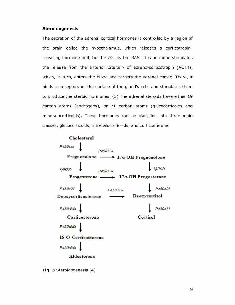
Steroidogenesis
The secretion of the adrenal cortical hormones is controlled by a region of
the brain called the hypothalamus, which releases a corticotropin-
releasing hormone and, for the ZG, by the RAS. This hormone stimulates
the release from the anterior pituitary of adreno-corticotropin (ACTH),
which, in turn, enters the blood and targets the adrenal cortex. There, it
binds to receptors on the surface of the gland's cells and stimulates them
to produce the steroid hormones. (3) The adrenal steroids have either 19
carbon atoms (androgens), or 21 carbon atoms (glucocorticoids and
mineralocorticoids). These hormones can be classified into three main
classes, glucocorticoids, mineralocorticoids, and corticosterone.
Fig. 3 Steroidogenesis (4)
10
The mineralocorticoids are essential for maintaining the balance of sodium
in the blood and body tissues and the volume of the extracellular fluid in
the body. Aldosterone, the principal mineralocorticoid produced by the
zona glomerulosa, enhances the uptake and retention of sodium in cells,
as well as the cells' release of potassium. This steroid also causes the duct
collectors cortical of the kidneys to retain sodium in exchange for
potassium of hydrogen, thus maintaining levels of this ion in the blood,
while increasing the excretion of potassium into the urine. Simultaneously,
aldosterone increases reabsorption of bicarbonate by the kidney, thereby
decreasing the acidity of body fluids and causing metabolic alkalosis,
which in measured by loss of hydrogen. A deficiency of adrenal cortical
hormone secretion causes Addison's disease.
Mineralocorticoid release is also influenced by factors circulating in the
blood. The most important of these factors is angiotensin II, the end
product of a series of steps starting in the kidney. When the body's blood
pressure declines, this change is sensed by a special structure in the
kidney called the juxtaglomerular apparatus. In response to decreased
pressure in kidney arterioles the juxtaglomerular apparatus releases an
enzyme called renin into the kidney's blood vessels. There, the renin turns
angiotensiongen to angiotensin I, which undergoes a further enzymatic
change in the bloodstream outside the kidney to angiotensin II.
Ang II stimulates the adrenal cortex to release aldosterone, the increased
concentration of sodium in the blood-filtering tubules of the kidney causes
an osmotic movement of water into the blood, thereby increasing the
blood pressure.
11
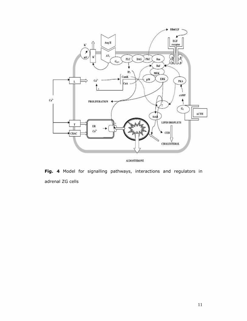
Fig. 4 Model for signalling pathways, interactions and regulators in
adrenal ZG cells
12
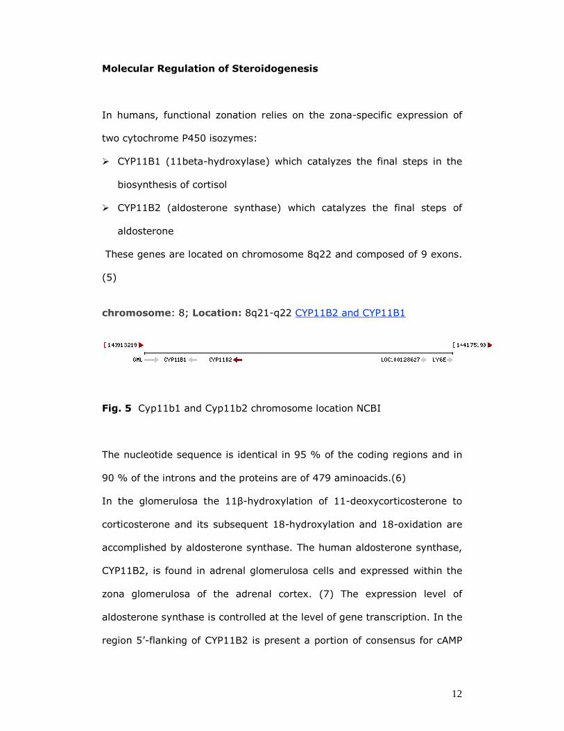
Molecular Regulation of Steroidogenesis
In humans, functional zonation relies on the zona-specific expression of
two cytochrome P450 isozymes:
� CYP11B1 (11beta-hydroxylase) which catalyzes the final steps in the
biosynthesis of cortisol
� CYP11B2 (aldosterone synthase) which catalyzes the final steps of
aldosterone
These genes are located on chromosome 8q22 and composed of 9 exons.
(5)
chromosome: 8; Location: 8q21-q22 CYP11B2 and CYP11B1
Fig. 5 Cyp11b1 and Cyp11b2 chromosome location NCBI
The nucleotide sequence is identical in 95 % of the coding regions and in
90 % of the introns and the proteins are of 479 aminoacids.(6)
In the glomerulosa the 11β-hydroxylation of 11-deoxycorticosterone to
corticosterone and its subsequent 18-hydroxylation and 18-oxidation are
accomplished by aldosterone synthase. The human aldosterone synthase,
CYP11B2, is found in adrenal glomerulosa cells and expressed within the
zona glomerulosa of the adrenal cortex. (7) The expression level of
aldosterone synthase is controlled at the level of gene transcription. In the
region 5’-flanking of CYP11B2 is present a portion of consensus for cAMP

13
response element (CRE) (position -74/-64), it plays a critical role in
transcriptional regulation of CYP11B2.
Angiotensin II is the major regulator of aldosterone synthesis in the
adrenal gland it can stimulate increase aldosterone synthase mRNA in
human primary adrenal zona glomerulosa cells. Study in-vitro
demonstrated Ang II stimulation of H295 cells results in an increase in
aldosterone production and aldosterone synthase mRNA levels. The effect
of Ang II on the synthesis of aldosterone has been shown to be through
the activation of CYP11B2 transcription common cis-elements and the CRE
site requires major induction in CYP11B2 promoter (8).
Ormones like Angiotensin II (Ang II), adrenocorticotrophin (ACTH) and
ione potassium (K+) stimulates aldosterone production by binding to the
melanocortin 2 receptor, a Gs-protein-coupled receptor that stimulates
adenylate cyclase and cAMP formation, and leads to the cAMP-dependent
protein kinase (PKA) activation. (9)
Variance of Ang II and increases in K+ concentration regulate the
aldosterone secretion by modulating the intracellular concentration of
calcium (Ca2+). (10) The binding of Ang II to AT1 receptors activates an
serial cascade of enzyme:
� Phospholipase C-dependent hydrolysis of plasma membrane
phosphatidylinositol 4,5- bisphosphate, which results in the formation
of inositol 1,4,5-trisphosphate (IP3) and diacylglycerol (DAG) (11)
� IP3 binds causes an increase in Ca2+ cytosolic concentration by the
rapid release of Ca2+ from endoplasmatic stores,
� DAG activates protein kinase C (PKC).
14
The increase in intracellular Ca2+, opening of the voltage-dependent (T-
and L-types) Ca2+ channels and the sustained entry of Ca2+ from the
external space.
Studies in-vitro demonstrated that these pathways are interrelated, ACTH
inhibits Ca2+ and Ang II stimulated aldosterone secretion via cAMP
formation in zona glomerulosa cells. (12,13)
Arterial Hypertension from Adrenal hyper function
The adrenal cortex plays a crucial role in the regulation of Na+ and water
homeostasis and of total peripheral resistance through the generic and
non generic actions of aldosterone. The two principal causes for the
arterial hypertension from adrenal disorder are:
a) Cushing’s syndrome
Corticosteroids are critically involved in blood pressure regulation and
glucocorticoid excess in Cushing’s syndrome invariably results in arterial
hypertension. Hypertension is one of the most distinguishing features of
endogenus Cusching’s Syndrome, as it is present in abouth 80% of adult
patients. (14,15)
Causes
(a) Exogenous glucocorticoids
(b) Small ACTH-producing pituitary adenoma or hyperplasia. Adrenals
usually exhibit nodular or diffuse hyperplasia, nodules are often multiple,
associated with hyperplastic cortex. Zona glomerulosa is difficult to
identify in adults, fasciculata has lipid-depleted cells, reticularis cells are
vacuolated.
15
(c) bilateral adrenal hyperplasia, adrenal adenoma or adrenal carcinoma
(d) Ectopic ACTH production by non-adrenal neoplasm.Tumors secrete
ACTH-like substance. In adults, usually due to small cell carcinoma of lung
or carcinoid tumors of lung or thymus; also medullary thyroid carcinoma,
pancreatic endocrine neoplasms, pheochromocytomas, ovarian tumors
(e) Rarely caused by tumors producing cortisol releasing factor
b) Hyperaldosteronism
The primary iperaldosteronismo (PA) is an endocrine disorder
characterized by excessive production of aldosterone, with the appearance
of arterial hypertension often resistant to therapy medical and, in a
proportion of cases of hypokalaemia and metabolic alkalosis. This disorder
can manifest itself with fatigue, muscle cramps, cardiac arrhythmias also
dangerous to the life and polyuria due to resistance tubular antidiuretic
hormone resulting in reduced ability to concentrate urine.
Dr. Jerome Conn, who first described the syndrome in 1954, recognized
this about ten years later, that not all individuals were affected by PA
ipokaliemici but for many decades in clinical practice is rooted in the use
and subjected to further diagnostic only those with hypertension who
presented ipokaliemia spontaneous or induced by diuretics. This has led to
claim that the PA was considered a rare cause of hypertension, comprising
less than 0.5-1% of all hypertensive subjects (16,17).
With the introduction of the screening test which considers the ARR
(aldosterone: renin ratio) which is the ratio of plasma aldosterone (PAC,
plasma aldosterone concentration) and plasma renin activity (PRA, plasma
renin activity) (18), was could identify cases in which the secretion of
16
aldosterone is inappropriate compared to that of renin. Thus leading to an
increase in the frequency with which the PA is diagnosed, revealing,
among other things, that only 20-40% of patients is ipokaliemico (19).
Clinic cases of CONN
The most common forms of PA are aldosterone-producing adenoma (APA)
and bilateral adrenocortical hyperplasia (BAH), also referred to as
idiopathic hyperaldosteronism (IHA). Is possible distinguish in the two
different cases (20):
1. Surgically curable
� Aldosterone-producing adenoma (aldosteronoma, APA)
� Multinodular unilateral adrenocortical hyperplasia (MUAN)
� Phaeochromocytoma causing Primary aldosteronism
2. Surgically not curable
� Bilateral adrenal hyperplasia (BAH)
� Unilateral APA with BAH
� Glucocorticoid-remediable aldosteronism (GRA): genetic disease
with autosomal transmission, is a crossing-over leading to the
formation of a chimeric gene where the promoter of CYP11B1,
regulates the expression of CYP11B2. This leads to an aldosterone
throughout the adrenal cortex
� Familial hyperaldosteronism type II (FH-II).
17
Bilateral adrenal hyperplasia versus CONN
APA and BHA are considered separate entities, but there is no border
between the two adrenal disorders, because there are not sharp criteria
to distinguish them. The only investigation that allows you to better define
the presence or absence of a hormonal hypersecretion, to discriminate
surgically curable forms (APA and UAH) from not surgically curable forms
(IHA) is the Adrenal Vein Sample (AVS). It consists in the measure of
aldosterone and cortisol in a sample of blood collected from the adrenal
veins and from the inferior vena cava.
However, these is no consensus on the cut-off used to define the
selectivity of the sample and the lateralization as yet.
The values we have used to prove for the lateralization are a ratio
between the dominant aldosterone and the controlateral aldosterone, each
normalized for the corresponding value of cortisol (21):
aldosteronedominant/cortisoldominant/
>2
aldosteronecontrolateral/cortisolcontrolateral
A problem for AVS is that it is an invasive test, it requires technical skill
and a “devoted” radiologist specific personnel and not always it is possible
to obtain a selective sampling due to the anatomy of the right adrenal
vein, which leads directly into the inferior vena cava. In spite of this,
many authors believe that this is an essential test for the diagnosis of
arterial hypertension from adrenal disorders (22,23,24).
At a pathological level the distinction between the two forms, APA and
BHA, is difficult because the Bilateral adrenal hyperplasia are not
surgically removed and therefore there aren’t enough case histories.
18
Fig.6 Flow chart for the diagnostic work-up of subtype identification of
primary aldosteronism (PA). APA = aldosterone-producing adenoma;
AVS = adrenal vein sampling; CT = computed tomography; IHA =
idiopathic hyperaldosteronism; MR = magnetic resonance; PAC =
plasma aldosterone concentration; PAH = primary adrenal
hyperplasia; PCC = plasma cortisol concentration; NP59 = 6b-[131I]
methyl-19-norcholesterol.
19
In the Primary Aldosteronism (PA) Prevalence in Italy (PAPY) study, a
prospective survey of 1125 consecutive newly diagnosed hypertensive
patients referred to specialized hypertension centers, aldosterone-
producing adenoma (APA) and idiopathic hyperaldosteronism (IHA) were
found in 4.8% and 6.4% of all patients(25,26), respectively, thus leading
to an overall prevalence of PA of 11.2% (20).
Notwithstanding were than 50 years of results the etiologic factor(s)
leading to primary hyperaldosteronism remain unknown. According to the
most credited hypothesis one or more factors have been contented to
chronically stimulate Aldosterone synthesis thereby leading to hyperplasic
changes in the adrenocortical ZG and thus to node formation. According to
one view, many nodules would be initially present, after which one would
take over therefore becoming the “ dominant” nodule and leading,
because of hyperaldosteronism. Body fluid expression and suppression of
the RAS, and therefore of Aldoserone synthesis in the other nodules. This
view is supported by earlier experience with tumorectomy for Conn’s
adenoma, which was abandoned because of early recurrence of PA and
nodule formation because of enlargement of one of the satellite nodules.
Although plausible, this hypothesis, which raises the contention that PA
can be an autonomon disorder like Graves’ disease has never been
formally investigated thus far.
20
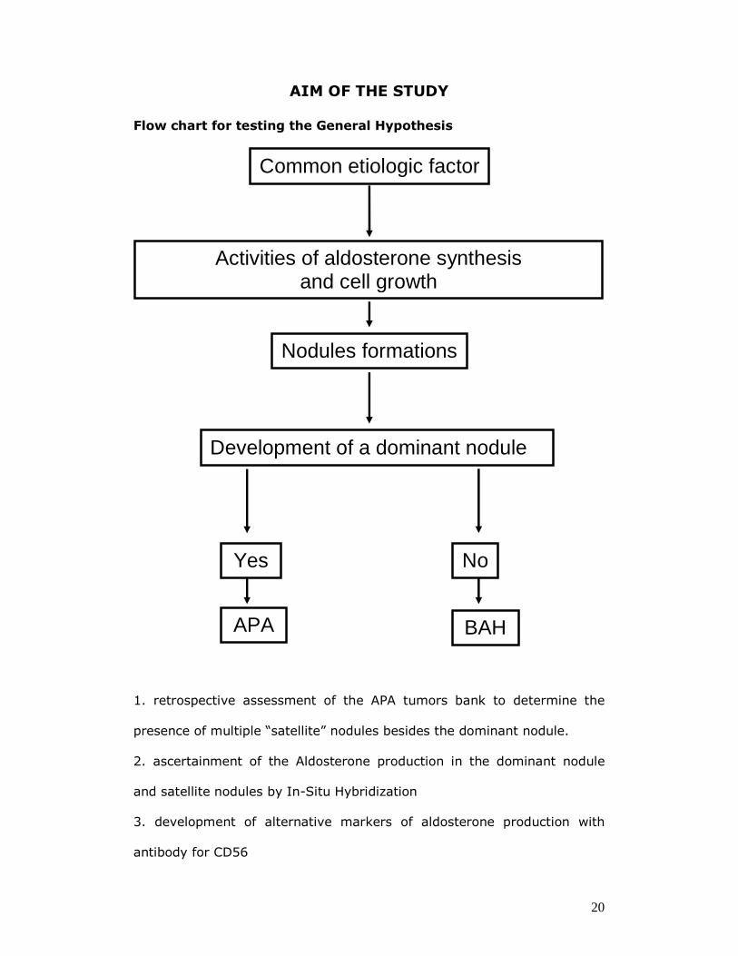
AIM OF THE STUDY
Flow chart for testing the General Hypothesis
1. retrospective assessment of the APA tumors bank to determine the
presence of multiple “satellite” nodules besides the dominant nodule.
2. ascertainment of the Aldosterone production in the dominant nodule
and satellite nodules by In-Situ Hybridization
3. development of alternative markers of aldosterone production with
antibody for CD56
Common etiologic factor
Activities of aldosterone synthesis and cell growth
Nodules formations
Development of a dominant nodule
Yes No
APA BAH
21
Specific Hypothesis
Considering the difficulty of discriminating APA from BAH and the likely
existence of a “continuum” between the two extreme of the spectrum we
hypothesize that a common etiologic factor through common mechanisms
can lead to either APA or BAH (IHA).
1. Is primary aldosteronism an autoimmune disorder (similar to Graves’
disease?)
Study Approach
The identification of aldosterone producing cells in surgically available
specimen from patients with primary aldosteronism (PA) has been
precluded thus mainly because of the steroid nature of aldosterone. Thus ,
because this liposolubility of the hormone is lost during the fixation-
dehydratation steps are required for immunocytochemistry. Moreover,
while antibodies specific for CYP11B1 and CYP11B2 exist for rat enzymes
but not for human enzymes. Thus, the lack of antibodies specifically
directed against the human aldosterone synthase, which is largely due to
large degree of homology of the protein with the 11beta-hydroxylase, has
precluded the development of reliable immuno histochemistry techniques
for assessing aldosterone productions in tissue sections. In fact, the
CYP11B2 gene encoding this enzyme shoves a 95% homology with the
CYP11B1 gene (Mornet E Journal of Biological Chemistry 1989) encoding
the 11-beta-hydroxilase, therefore explaining the aforementioned
difficulties in obtaining specific antibodies and molecular tools.
22
Preliminary studies have suggested the possibility of circumventing these
problems by designing molecular probes that can be used for in-situ
hybridization (ISH) (Sasano).
However, these techniques did not gain widespread use, because they
require radioactive labeling of the probe.
Hence, for this study we have decided to attempt to develop a novel non
radioactive ISH that could be applied to detect aldosterone synthase in
tissue sections and therefore to identify the sites and cells producing
aldosterone in aldosterone-producing adenoma and in adrenocortical
hyperplasia.
The second approach used in the identification of nodules in APA was to
investigate the presence and distribution of other markers of ZG,
aldosterone synthesizing cells. In this regard, we have undertaken a
scorch using a pool of different monoclonal antibodies which mostly
unspecific results. However, we came across a partial results with an
antibody for another protein the CD56.
The expression of CD56 is extensive in developing neuronal and endocrine
tissues, including the rat adrenal and also is expressed by NK cells and
some T cells. CD56 belongs to the Ig superfamily of adhesion molecules
and plays a role in the morphogenesis of several organ. Expression of
CD56 has been noted in both developing fetal organ systems and
physiologic and regenerative processes in adults.
Its role in these processes has been suggested to range from altering
migration to initiating differentiation and stimulation of signaling cascades.
(27) Thus, being a specific marker of ZG and adrena medullary cells we
hypothesize that an IHC technique using CD56 antibody will be used to
23
identify aldosterone producing cells and nodules in PA and adrenal gland
patients with APA.
MATERIALS AND METHODS
Adrenal specimens.
Adrenal gland tissues from ten patients with APA were studied. The
diagnosis of APA was based on strict predefined criteria that entail on
lateralization of aldosterone secretion at adrenal vein sampling, surgery,
pathology and, more importantly, follow-up data. For the latter,
demonstration of normokalemia and cure or improvement of hypertension
at least 120 days after adrenalectomy were required (PAPY). Cure was
defined as normotension without medications; improvement as a systolic
and diastolic blood pressure <140/90 mmHg, respectively, on the same or
reduced number of medications, and/or reduced defined daily doses ( GP
Rossi J Am Coll Cardiol 2006).
Tissue were immediately fixed in neutral formalin and included in paraffin.
All gave an informed consent to the study, which had been approved by
the local Ethical Committee.
24
Choice of the technique for identification of aldosterone synthase
We decided to use a technique of in situ hybridization, because the use of
an antibody against aldosterone is not possible as this hormone has a lipid
nature, and it is extracted during the fixation of the tissue.
Therefore, the only way to identify cells producing aldosterone is to study
the distribution of the enzyme which regulates its production (CYP11B2).
We chose to mark the probes with digosigenine (DIG) because this is a
vegetable steroid, absent in animal cells.
This detection system has the advantage of being relatively simple,
sensitive and specific without the use of radioactivity.
Furthermore, We adopted a highly specific oligo-nucleotide probe for the
CYP11B2 after blasting of the two enzymes and identification of not
homologous areas.
The probe was analyzed by a second blast on the human genome to
control cross-reactions with other genes. The specificity was demonstrated
by a selective staining of the zone glomerulosa of a normal human’s
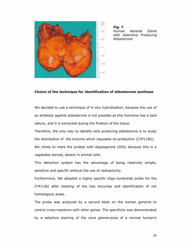
Fig. 7 Human Adrenal Gland with Adenoma Producing Aldosterone

25
adrenal gland. This oligo-nucleotide probe is resistant to RNAse and do
not require denaturation before use.
26
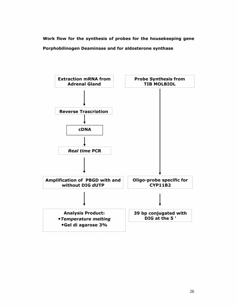
Work flow for the synthesis of probes for the housekeeping gene
Porphobilinogen Deaminase and for aldosterone synthase
Amplification of PBGD with and without DIG dUTP
Extraction mRNA from Adrenal Gland
Reverse Trascription
Analysis Product:
•Temperature melting •Gel di agarose 3%
Real time PCR
Oligo-probe specific for
CYP11B2
Probe Synthesis from TIB MOLBIOL
39 bp conjugated with
DIG at the 5 '
cDNA
27
Production probes for hybridization probes
For the hybridization have been used various types of probes:
� Oligonucleotide probe for the gene Cyp11b2.
The oligonucleotide probe specific for the CYP11B2 (28) have been
synthesized by TIB MOLBIOL on our design and is 39 bp long. It was
conjugated with DIG at the 5 '. Its sequence is:
5'-DIG-GCCTTGCTATTTGACAGCCTGGCAAGCCCCAGTCCTGG
For hybridization of the housekeeping gene (PBGD) transcript, we
designed a 92 bp probe labeled with Digoxigenin, corresponding to
position 406-498 of the mRNA sequence.
� Probe of double-strand DNA (dsDNA) for a housekeeping gene
(PBGD NM_000190) variant 2 is selectively expressed by erythroid
cells, whereas variant 1 is a housekeeping gene ideal, since it is
expressed in number of copies low and relatively constant in all
cells of the body (29)
The dsDNA probes must be denatured and are less sensitive; however,
they can be easily built by RT-PCR, and also they do not required
purification before using them (30).
The amplified stretch is 92 bp long. The following primers were used:
fw primer 5'-TGCCCTGGAGAAGAATGAAG
rev primer 3'-AGATGGCTCCGATGGTGA.
The mRNA extracted from normal adrenal tissue was reverse transcribed
with iScriptTMcDNA a cDNA Synthesis Kit (Biorad): 6 µl of mRNA were
28
mixed with 4 µl of 5X iScript Reaction Mix, 1 µl of iScript reverse
transcriptase and 9 µl of nuclease-free water to a final volume of 20 µl,
the reaction was done on a thermocycler. The thermal cicle profile
comprised 5 minutes at 25 ° C, 30 minutes at 42 ° C and 5 minutes at
85° C.
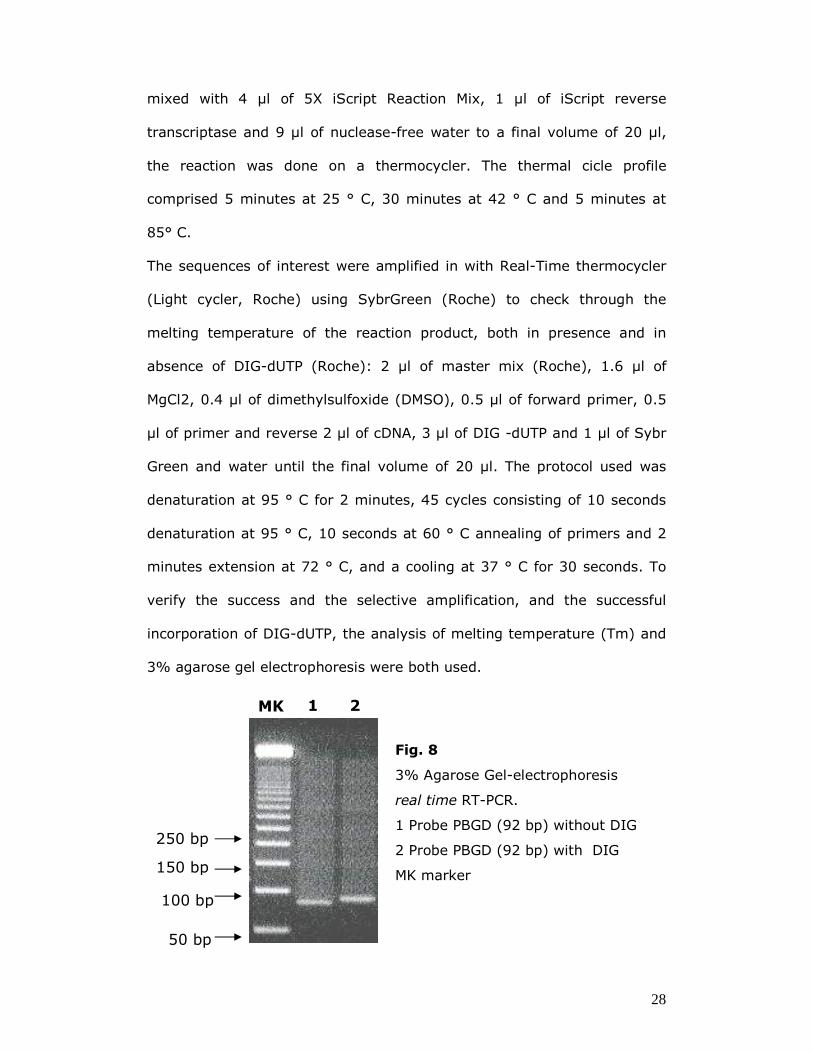
The sequences of interest were amplified in with Real-Time thermocycler
(Light cycler, Roche) using SybrGreen (Roche) to check through the
melting temperature of the reaction product, both in presence and in
absence of DIG-dUTP (Roche): 2 µl of master mix (Roche), 1.6 µl of
MgCl2, 0.4 µl of dimethylsulfoxide (DMSO), 0.5 µl of forward primer, 0.5
µl of primer and reverse 2 µl of cDNA, 3 µl of DIG -dUTP and 1 µl of Sybr
Green and water until the final volume of 20 µl. The protocol used was
denaturation at 95 ° C for 2 minutes, 45 cycles consisting of 10 seconds
denaturation at 95 ° C, 10 seconds at 60 ° C annealing of primers and 2
minutes extension at 72 ° C, and a cooling at 37 ° C for 30 seconds. To
verify the success and the selective amplification, and the successful
incorporation of DIG-dUTP, the analysis of melting temperature (Tm) and
3% agarose gel electrophoresis were both used.
MK
150 bp
100 bp
50 bp
250 bp
1 2
Fig. 8
3% Agarose Gel-electrophoresis
real time RT-PCR.
1 Probe PBGD (92 bp) without DIG
2 Probe PBGD (92 bp) with DIG
MK marker
29
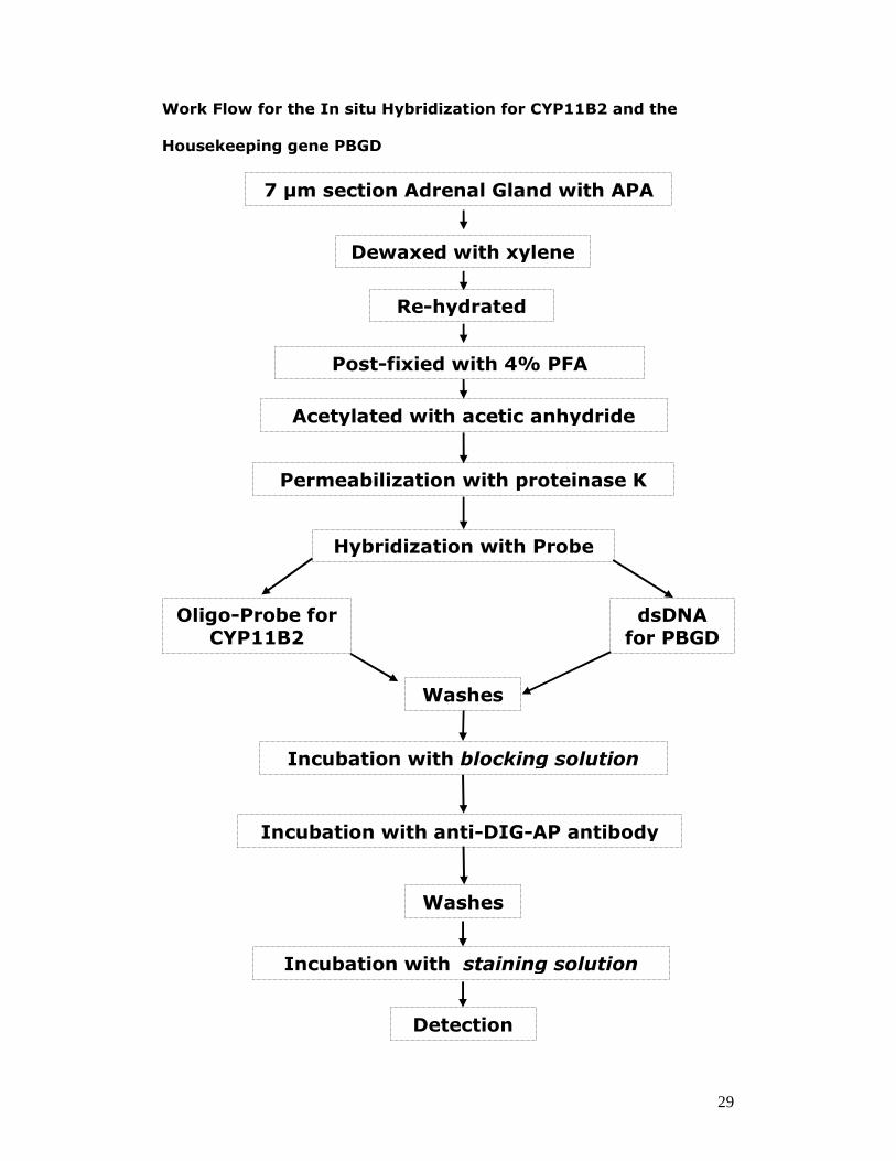
Work Flow for the In situ Hybridization for CYP11B2 and the
Housekeeping gene PBGD
7 µm section Adrenal Gland with APA
Hybridization with Probe
Dewaxed with xylene
Re-hydrated
Post-fixied with 4% PFA
Acetylated with acetic anhydride
Permeabilization with proteinase K
dsDNA
for PBGD
Oligo-Probe for
CYP11B2
Incubation with anti-DIG-AP antibody
Incubation with staining solution
Incubation with blocking solution
Washes
Washes
Detection
30
In situ Hybridization.
As preliminary studies showed that formalin and paraphormaldheid fixed
adrenocorticol tissue sections were unsuitable for ISH because of
inadequate preservation of the transcripts we had no choiche other than
used frozen (cryostate sections). This required the collection of all the APA
tissues directly in the operating room immediately after lapascopic
adrenalectomy.
The tissues were cut in 7 µm-thick slices and sections were mounted on
glass slide. All hybridization steps were conducted under RNase-free
conditions.
Sections were dewaxed with xylene (5min for 2 times), and re-hydrated in
different ethanol concentrations (5 min for 2 times), quickly washed in
DEPC-dH2O and phosphate buffered saline (PBS, Oxoid) (5 min for 2
times). After post-fixing the sections for 10 min with 4% PFA in
(Phosphate Buffer Saline)PBS and washing in PBS (5 min for 2 times),
they were acetylated for 5 min with 0.25% (v/v) acetic anhydride in 0.1 M
triethanolamine (TEA,Sigma) pH 8.0 and subsequently with acetic
anhydride (Fluka, Italy) 0.25% (v/v) in TEA and in SSC 2X (Sigma,
Italy )(3 min). The sections were treated with proteinase K (Sigma), 10
µg/ml in PBS, at 37°C for 20 min and then immersed in glycine (Fluka), 2
mg/ml in PBS, at 4°C for 1 min 30 seconds and PBS (5 min for 2 times).
The sections were incubated for 2 h at 37°C with pre-hybridization buffer
and washed in SSC 2X (5 min), then incubated with hybridization solution
containing DIG-labeled probes in hybridization buffer (50% deionized
formamide (Sigma), 20% (w/v) dextran sulfate, 1% Denhardt's 50X
31
solution (Sigma),20% SSC 2X, 1% Dithiothreitol (DTT, Fluka) and 1%
Salmon sperm DNA (19mg/ml, Invitrogen, Italy) at 37°C overnight.
Slides were washed with DTT (1.5mg/ml)/SSC 1X at room temperature (5
min), 45°C (2 times for 15 min) and DTT (1,5mg/ml)/SSC 0.5X at 45°C
(10 times for 2min), at room temperature (5min) and then transferred to
Tris-buffered saline (TBS) (100 mMTris HCl, 150 mM NaCl, pH 7,5) and
washed 5 min for 3 times. The sections were covered with blocking
solution (TBS, 0.1% tritonX-100 and 1% sheep serum, Sigma) at room
temperature for 30 min and subsequently incubated with anti-DIG
antibody (Roche, Italy) diluted 1:2000 overnight at 4°C. After washing
the sections 3 times for 5 min in TBS, they were incubated with staining
solution (FastTM NBT/BCIP in 10 ml of dH2O add 1% levamisol 1M, Sigma)
overnight at room temperature.
32
Immunohistochemisty CD56
For IHC the best results, often a long set of preliminary work, were
auctioned with formalin fixed and paraffin embedded. Specimen 4 µm
sections of the routinely processed paraffin blocks were stained with
hematoxylineosin (HE) for histopathological diagnosis. IHC staining was
performed using an indirect immunoperoxidase technique (Bond Polymer
Refine Detection; Vision BioSystems, UK) with a fully automated system
(Bond-maX; Vision BioSystems, UK). Four-micron-thick sections from
paraffin blocks were dewaxed and rehydrated by successive incubation in
Bond Dewax Solution (Vision BioSystems, UK), ethanol, and distilled water.
Antigen retrieval was performed by heating sections (100°C, 30 min) in
Bond Epitope Retrieval Solution 1 (Vision BioSystems, UK). Endogenous
peroxidase was blocked by treatment with 3% hydrogen peroxide before a
5 min incubation with a mouse monoclonal anti-CD56, clone 1B6 (diluited
1:100, Novocastra, Newcastle upon Tyne, UK). Antigen was detected by
incubation with labelled polymer (HRP) and diaminobenzidine. The
sections were then counterstained with hematoxylin, dehydrated, cleared,
and mounted.
33
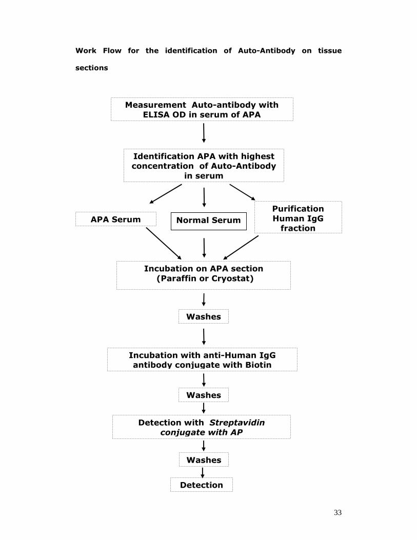
Work Flow for the identification of Auto-Antibody on tissue
sections
Measurement Auto-antibody with
ELISA OD in serum of APA
Identification APA with highest concentration of Auto-Antibody
in serum
Purification
Human IgG
fraction APA Serum
Incubation with anti-Human IgG antibody conjugate with Biotin
Detection with Streptavidin
conjugate with AP
Incubation on APA section
(Paraffin or Cryostat)
Washes
Washes
Detection
Washes
Normal Serum
34
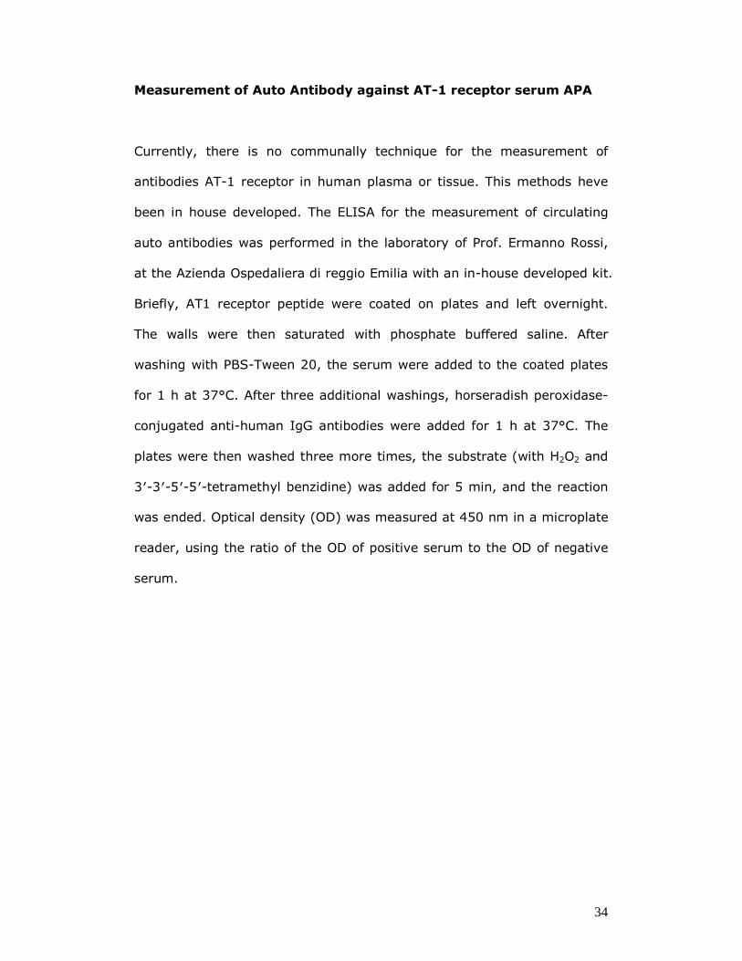
Measurement of Auto Antibody against AT-1 receptor serum APA
Currently, there is no communally technique for the measurement of
antibodies AT-1 receptor in human plasma or tissue. This methods heve
been in house developed. The ELISA for the measurement of circulating
auto antibodies was performed in the laboratory of Prof. Ermanno Rossi,
at the Azienda Ospedaliera di reggio Emilia with an in-house developed kit.
Briefly, AT1 receptor peptide were coated on plates and left overnight.
The walls were then saturated with phosphate buffered saline. After
washing with PBS-Tween 20, the serum were added to the coated plates
for 1 h at 37°C. After three additional washings, horseradish peroxidase-
conjugated anti-human IgG antibodies were added for 1 h at 37°C. The
plates were then washed three more times, the substrate (with H2O2 and
3′-3′-5′-5′-tetramethyl benzidine) was added for 5 min, and the reaction
was ended. Optical density (OD) was measured at 450 nm in a microplate
reader, using the ratio of the OD of positive serum to the OD of negative
serum.
35
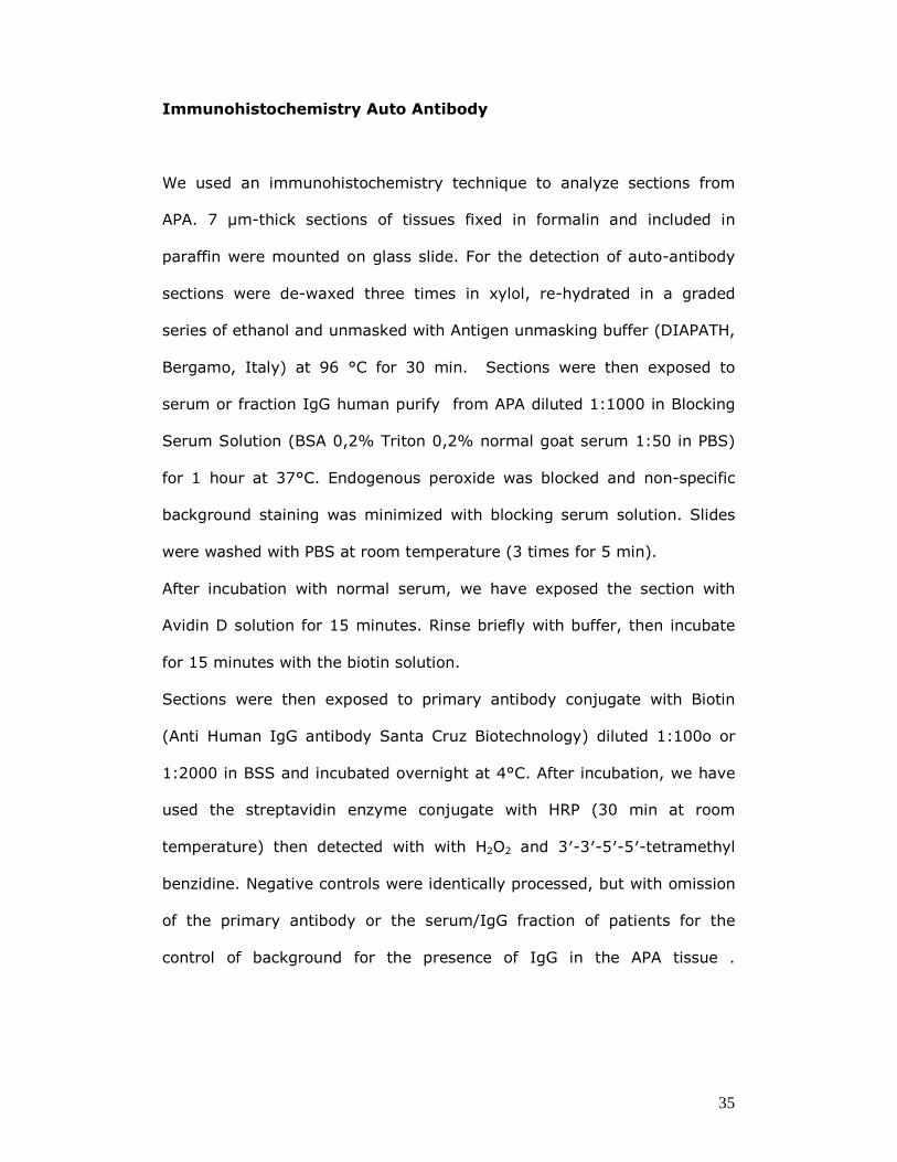
Immunohistochemistry Auto Antibody
We used an immunohistochemistry technique to analyze sections from
APA. 7 µm-thick sections of tissues fixed in formalin and included in
paraffin were mounted on glass slide. For the detection of auto-antibody
sections were de-waxed three times in xylol, re-hydrated in a graded
series of ethanol and unmasked with Antigen unmasking buffer (DIAPATH,
Bergamo, Italy) at 96 °C for 30 min. Sections were then exposed to
serum or fraction IgG human purify from APA diluted 1:1000 in Blocking
Serum Solution (BSA 0,2% Triton 0,2% normal goat serum 1:50 in PBS)
for 1 hour at 37°C. Endogenous peroxide was blocked and non-specific
background staining was minimized with blocking serum solution. Slides
were washed with PBS at room temperature (3 times for 5 min).
After incubation with normal serum, we have exposed the section with
Avidin D solution for 15 minutes. Rinse briefly with buffer, then incubate
for 15 minutes with the biotin solution.
Sections were then exposed to primary antibody conjugate with Biotin
(Anti Human IgG antibody Santa Cruz Biotechnology) diluted 1:100o or
1:2000 in BSS and incubated overnight at 4°C. After incubation, we have
used the streptavidin enzyme conjugate with HRP (30 min at room
temperature) then detected with with H2O2 and 3′-3′-5′-5′-tetramethyl
benzidine. Negative controls were identically processed, but with omission
of the primary antibody or the serum/IgG fraction of patients for the
control of background for the presence of IgG in the APA tissue .
36
RESULTS
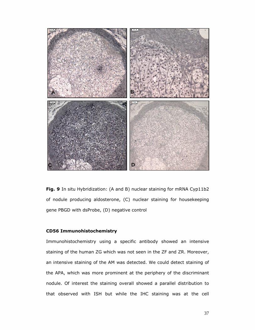
In situ hybridization
The APA analyzed showed intensely labeled areas called “nodules
producing aldosterone” and negative areas. In some cases, the positive
cells were localized under the capsule, while the central part of the mass
was not clearly marking. This experimental data has revealed an
expression of the gene Cyp11b2 reflecting considerable biological
variability of samples analyzed. It is therefore conceivable that some cells
have a quantity of mRNA for the gene Cyp11b2 and thus transcriptional
activity at the level of baseline adrenal gland that the methodology was
not sensitive enough to detect.
The presence of a nuclear marking with this method of hybridization is
readily explain when one considers that the transcription of DNA to mRNA
occurs in the nucleus and thus it is that the compartment when the
transcript is more better preserved. In the surrounding cytoplasm, the
staining is not appreciably different from that of cells. The nuclear staining
was obtained through specific bond oligo-probe to mRNA of Cyp11b2, and
not as an aspecific binding, since the probe was found only cells
corticosurrene, leaving those of the negative bone marrow or 'vascular
endothelium (Fig. 9 A, B). Moreover, samples treated with probes labeled
with Digosigenin or those in which the probe was omitted were completely
negative (Fig.9 D).
37
Fig. 9 In situ Hybridization: (A and B) nuclear staining for mRNA Cyp11b2
of nodule producing aldosterone, (C) nuclear staining for housekeeping
gene PBGD with dsProbe, (D) negative control
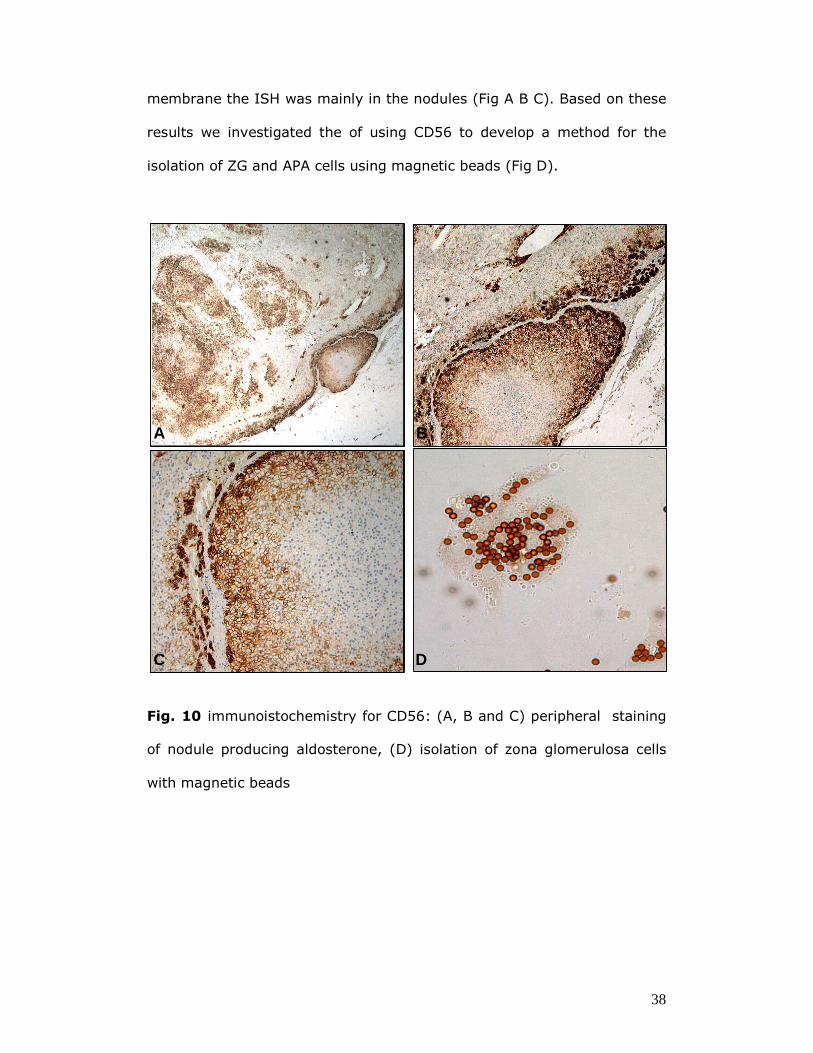
CD56 Immunohistochemistry
Immunohistochemistry using a specific antibody showed an intensive
staining of the human ZG which was not seen in the ZF and ZR. Moreover,
an intensive staining of the AM was detected. We could detect staining of
the APA, which was more prominent at the periphery of the discriminant
nodule. Of interest the staining overall showed a parallel distribution to
that observed with ISH but while the IHC staining was at the cell
A B
C D
38
membrane the ISH was mainly in the nodules (Fig A B C). Based on these
results we investigated the of using CD56 to develop a method for the
isolation of ZG and APA cells using magnetic beads (Fig D).
Fig. 10 immunoistochemistry for CD56: (A, B and C) peripheral staining
of nodule producing aldosterone, (D) isolation of zona glomerulosa cells
with magnetic beads
A B
C D
39
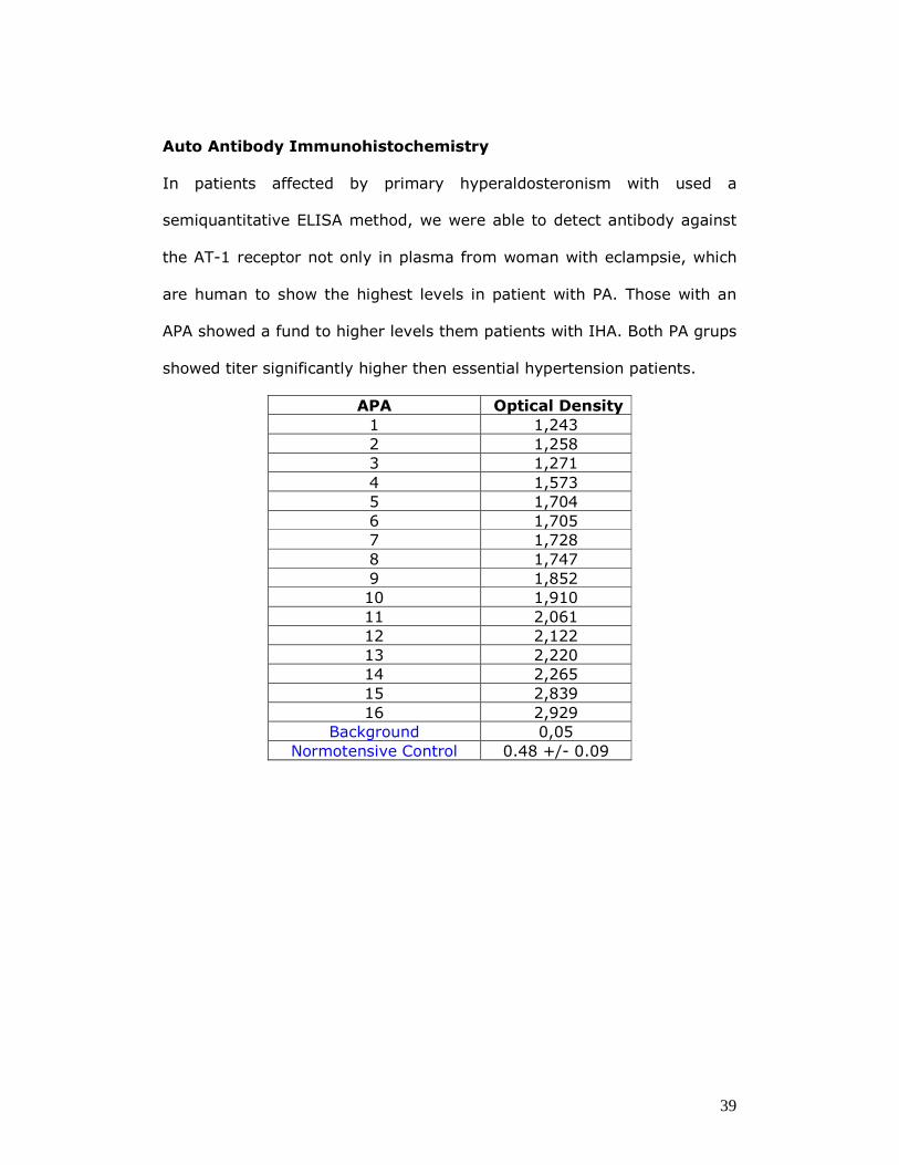
Auto Antibody Immunohistochemistry
In patients affected by primary hyperaldosteronism with used a
semiquantitative ELISA method, we were able to detect antibody against
the AT-1 receptor not only in plasma from woman with eclampsie, which
are human to show the highest levels in patient with PA. Those with an
APA showed a fund to higher levels them patients with IHA. Both PA grups
showed titer significantly higher then essential hypertension patients.
APA Optical Density
1 1,243 2 1,258 3 1,271 4 1,573 5 1,704 6 1,705 7 1,728 8 1,747 9 1,852 10 1,910 11 2,061 12 2,122 13 2,220 14 2,265 15 2,839 16 2,929
Background 0,05 Normotensive Control 0.48 +/- 0.09
40
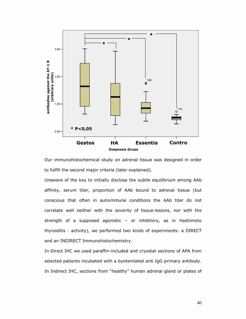
Our immunohistochemical study on adrenal tissue was designed in order
to fulfill the second major criteria (later explained).
Unaware of the key to initially disclose the subtle equilibrium among AAb
affinity, serum titer, proportion of AAb bound to adrenal tissue (but
conscious that often in autoimmune conditions the AAb titer do not
correlate well neither with the severity of tissue-lesions, nor with the
strength of a supposed agonistic – or inhibitory, as in Hashimoto
thyroiditis - activity), we performed two kinds of experiments: a DIRECT
and an INDIRECT Immunohistochemistry.
In Direct IHC we used paraffin-included and cryostat sections of APA from
selected patients incubated with a byotenilated anti IgG primary antibody.
In Indirect IHC, sections from “healthy” human adrenal gland or plates of
* *
*
* P<0,05
HA Gestos ControEssentia
Diagnosis Grups
antibodie
s a
gain
st th
e A
T-1
R
(arb
itrary u
nits)
41
cultured H295 cells were incubated with patients’ serum and the same
primary antibody described above.
Patients selection was performed on the basis of a semiquantitative
detection of anti angiotensin II AT1 receptor AAb in their serum.
DISCUSSION
In situ hybridization
By using a novel nonradioactive methodology for detection aldosterone
syntheses mRNA, which we developed we could show that Aldosterone
synthesis occurs only in the ZG in the normal human adrenal cortex.
This experimental evidence is consistent with other findings that reveal a
cytological heterogeneity of the APA, which may be made of phenotypes-
like cell clusters, fascicule-like and mixed in different proportions (32).
Moreover, given that the samples used were stored in paraffin, it may be
assumed that the mRNA content in the nucleus was better preserved
because it protected from the degradation than cytoplasmic.
It is also necessary to remember that the work so far published in which it
was studied the distribution to mRNA of Cyp11b2 through hybridization,
the subcellular localization is not indicated (33, 28, 34).
42
Auto Antibody Immunohistochemistry
In contrast with other tumorous condition, the exact pathway, mutations
and triggering factors leading to the development of aldosterone
producing adenomas have not been clarified yet.
The occurrence of micronodularity peripheral to the main adenomatous
mass in some of our patient and the frequent recurrence of APA in the
controlateral adrenal gland after therapeutic adrenalectomy aroused the
suspicion that a chronic proliferogenic stimulus could be involved in the
pathophysiology of the PA. Thus a continuum between hyperplasia,
micronodularity and adenomas could be hypothesized.
None of the actually known adrenothropic stimulating factors seems to be
able to play such a trophic role, and the possible culprit is unknown.
Nevertheless, we noted a similarity between the histopathological
behaviour of these APA with satellite micronodularity and a well known
clinical entity: Grave’s disease. This thyreotoxic condition associated with
hyperfunction of Thyroid gland is often referred to as a “diffuse
hyperplasia “ of the gland, but the natural history of the disease leads
often to a micronodular pattern in which the single nodes can grow
autonomously and a “dominant node” can develop. In this pathologic
hyperplasia, the primum movens consist in a peculiar autoimmune
reaction against the TSH-receptor as the autoantigen: what differentiates
this autoimmune disease from many others is the agonistic activity
expressed by the autoantibodies on the receptor. This provides the
necessary trophic and hormone release-inducing stimulus.
43
Our hypothesis is that a similar humoral stimulating factor could play a
role in the genesis of APA, with an implementation of the mithogenic
potential of adrenal cells and a consequent increased risk of adenoma
degeneration of a diffuse or nodular hyperplasia.
Stating an agonistic autoantibody-nature of this factor would consequently
make us assume APAgenesis as an autoimmune process: to validate this
suspicion, the criteria proposed by Witebsky and Rose to define a
condition as an autoimmune in nature should be fulfilled. Briefly, these
criteria are summarized as the following:
� Major criteria (Witebsky and Rose 1957)
1. Lympho-plasmocytic infiltration of the target organ or systemic
involvement.
2. Demonstration of circulating and/or tissue-localization of autoantibodies
(AAb) and/or autoreactive lymphocytic clones.
3. Identification of autoantigens involved in the autoimmune reaction,
induction of the disease in animals by injection of these autoantigens and
passive transfer of the disease by serum or lymphocytes.
� Minor criteria (Rose and Bona 1993)
1. Correlation with the MHC genes.
2. Response to immunosuppressive therapy.
3. Association with other autoimmune diseases.
Angiotensin II (Ang II), the main active peptide of the renin-angiotensin
system (RAS), is a potent vasoconstrictor hormone that is cleaved from
44
angiotensinogen by renina and the angiotensin-converting enzyme (ACE).
This octapeptide mediates its effects via two G-protein coupled receptors,
the angiotensin II type 1 (AT1) and type 2 (AT2) receptors. In particular,
the interaction of Ang II with the AT1 receptor stimulates
pathophysiological processes including hypertension.
Anti-AT1-receptor antibodies were first discovered by Fu et al. in the
serum of patients with malignant hypertension (35) They were then found
to exist with high titer in pre-eclampsia patients by Wallukat et al (36)
and in refractory hypertensive patients by Liao et al. (37), whereas only a
small percentage of patients with nonrefractory hypertension were
positive for AT1-AA. One may speculate that the presence of AT1-AA
contributes to a worse therapeutic response in the patients with refractory
hypertension. Interestingly, the authors identified even 7.5% of
normotensive controls as antibody-positive.
Many studies (38,39) have shown that these antibodies exhibit an
agonist-like activity similar to Angiotensin II, such as a stimulatory
positive chronotropic effect on neonatal rat cardiomyocytes; however an
agonistic activity also on aldosterone producing cells of adrenal zona
glomerulosa (mediated by the same receptor) has not been demonstrated
yet. The prosecution of our studies could clarify this point in future.
Our present knowledge about the role of these AAb in patients affected by
primary hyperaldosteronism is the detection, by a semiquantitative ELISA
method, of an Optical Density (OD) significantly higher than the same
parameter measured in the serum of control normotensive patients
described above (see table).
45
This measurements induced us to focus our attention on those patients
who revealed an higher OD, and we performed all the described IHCs on
sections of their APAs (for direct IHC) or using their serum or IgG-fraction
from serum (for indirect IHC).
To validate the third and last major criteria, we are planning to produce
an animal model of autoimmunity against the AT1 receptor, by
immunizing rats with the synthetic peptide corresponding to the second
extracelluar peptide of human AT1 as the antigen, identified in literature
as the binding site for antiAT1-AAb detected in preeclamptic women and
malignant-hypertension affected patients, and used in the ELISA assays to
detect the AAb. By raising them for two to six months, we’ll study Blood
pressure, Heart rate, biochemical profile including PRA and PAC, vascular
and cardiac remodeling and adrenal gland histopathology, in order to
reveal the in vivo effect of these AAb. Fu et al. (39) and Wang et al. (40)
already immunized rats with the synthetic peptide corresponding to the
second loop of human AT1receptor (residues 165–191) as antigen, and
then produced antibodies from the immunized serum: they demonstrated
a persistent agonistic activity of AAb and a vascular remodelling,
respectively.
Contemporarily to this study, a passive transfer of the possible alterations
enlisted above could be attempted by injecting other rats with the serum
of those previously immunized
46
Reference
1. Rainey,W.E. Adrenal zonation: clues from 11beta-hydroxylase and
aldosterone synthase. Mol. Cell Endocrinol. 151, 151-160 (1999)
2. Dr. Chris Doumen Lecture 4 2402 : Anatomy/Physiology Major
Endocrine Organs
3. William F. Ganong Book
4. Chapter 11. Adrenal Cortex, Development, Anatomy, Physiology I.
Vrezas Department of Endocrinology, Diabetes and Rheumatology,
University Hospital, , Duesseldorf, Germany H.S. Willenberg
Department of Endocrinology, Diabetes and Rheumatology,
University Hospital, , Duesseldorf, Germany S.R. Bornstein
Technical University of Dresden, Medical Department, ,
Fetscherstraße, Dresden, Germany
5. (Mornet,E., Dupont,J., Vitek,A. & White,P.C. Characterization of two
genes encoding human steroid 11 beta-hydroxylase (P-450(11)
beta). J. Biol. Chem. 264, 20961-20967 (1989) )
6. (Bassett,M.H., White,P.C. & Rainey,W.E. The regulation of
aldosterone synthase expression. Mol. Cell Endocrinol. 217, 67-74
(2004))
7. Curnow KM, Tusie-Luns M, Pascoe L, Natajaran R, Gu J, Nadler JL,
White PC 1991 The product of the CYP11B2 gene is required for
aldosterone biosynthesis in the human adrenal cortex. Mol
Endocrinol 5:1513–1522.
47
8. JIALI GU, YESHAO WEN, ANGELES MISON, AND JERRY L. NADLER
12-Lipoxygenase Pathway Increases Aldosterone Production,
� �3 ,5 -Cyclic Adenosine Monophosphate Response Element-
Binding Protein Phosphorylation, and p38 Mitogen-Activated Protein
Kinase Activation in H295R Human Adrenocortical Cells.
Endocrinology 144(2):534–543
9. Fakunding,J.L., Chow,R. & Catt,K.J. The role of calcium in the
stimulation of aldosterone production by adrenocorticotropin,
angiotensin II, and potassium in isolated glomerulosa cells.
Endocrinology 105, 327-333 (1979).
10.Foster,R.H. Reciprocal influences between the signalling pathways
regulating proliferation and steroidogenesis in adrenal glomerulosa
cells. J. Mol. Endocrinol. 32, 893-902 (2004).
11.Hajnoczky,G., Csordas,G., Bago,A., Chiu,A.T. & Spat,A. Angiotensin
II exerts its effect on aldosterone production and potassium
permeability through receptor subtype AT1 in rat adrenal
glomerulosa cells. Biochem. Pharmacol. 43, 1009-1012 (1992).
12.Yoshida,A., Nishikawa,T., Tamura,Y. & Yoshida,S. ACTH-induced
inhibition of the action of angiotensin II in bovine zona glomerulosa
cells. A modulatory effect of cyclic AMP on the angiotensin II
receptor. J. Biol. Chem. 266, 4288-4294 (1991).
13.Balla,T., Hollo,Z., Varnai,P. & Spat,A. Angiotensin II inhibits K(+)-
induced Ca2+ signal generation in rat adrenal glomerulosa cells.
Biochem. J. 273(Pt 2), 399-404 (1991).
14.Nakamura Y, Yano H, Nakashima T. False intranuclear inclusions in
adrenal cytomegaly Arch Pathol Lab Med. 1981 Jul;105(7):358-60
48
15.Deborah P. Merke. Approach to the adult with congenital adrenal
hyperplasia due to 21-hydroxylase deficiency. J Clin Endocrinol
Metab. 2008, 93: 653-660.
16.Gordon RD, Ziesak MD, Tunny TJ, Stowasser M, Klemm SA.
Evidence that primary aldosteronism may not be uncommon: 12%
incidence among antihypertensive drug trial volunteers. Clin Exp
Pharmacol Physiol. 1993;20:296-298.
17.Young WF, Jr. Minireview: primary aldosteronism--changing
concepts in diagnosis and treatment. Endocrinology.
2003;144:2208-2213.
18.Hiramatsu K, Yamada T, Yukimura Y, Komiya I, Ichikawa K,
Ishihara M, Nagata H, Izumiyama T. A screening test to identify
aldosterone-producing adenoma by measuring plasma renin activity.
Results in hypertensive patients. Arch Intern Med. 1981;141:1589-
1593.
19.Mulatero P, Stowasser M, Loh KC, Fardella CE, Gordon RD, Mosso L,
Gomez-Sanchez CE, Veglio F, Young WF, Jr. Increased diagnosis of
primary aldosteronism, including surgically correctable forms, in
centers from five continents. J Clin Endocrinol Metab.
2004;89:1045-1050.
20.a mazza c pessina at all. Endocrine arterial hypertension: diagnostic
approach in clinical practice. Minerva Endocrinol 2008 33, 127-43
21.Rossi GP, Sacchetto A, Chiesura-Corona M, De Toni R, Gallina M,
Feltrin GP, Pessina AC. Identification of the etiology of primary
aldosteronism with adrenal vein sampling in patients with equivocal
computed tomography and magnetic resonance findings: results in
49
104 consecutive cases. J Clin Endocrinol Metab. 2001;86:1083-
1090.
22.Stowasser M, Gordon RD. Primary aldosteronism. Best Pract Res
Clin Endocrinol Metab. 2003;17:591-605.
23.Young WF, Stanson AW, Thompson GB, Grant CS, Farley DR, van
Heerden JA. Role for adrenal venous sampling in primary
aldosteronism. Surgery. 2004;136:1227-1235.
24.Rossi GP, Ganzaroli C, Miotto D, De Toni R, Palumbo G, Feltrin GP,
Mantero F, Pessina AC. Dynamic testing with high-dose
adrenocorticotrophic hormone does not improve lateralization of
aldosterone oversecretion in primary aldosteronism patients. J
Hypertens. 2006;24:371-379.
25.Rossi,G.P. et al. Renal damage in primary aldosteronism: results of
the PAPY Study. Hypertension 48, 232-238 (2006).
26.Rossi,G.P. et al. A prospective study of the prevalence of primary
aldosteronism in 1,125 hypertensive patients. J. Am. Coll. Cardiol.
48, 2293-2300 (2006).
27. MARCUS O. The Journal of Clinical Endocrinology & Metabolism
88(8):3921–3930
28.Pascoe L, Jeunemaitre X, Lebrethon MC, Curnow KM, Gomez-
Sanchez CE, Gasc JM, Saez JM, Corvol P. Glucocorticoid-
suppressible hyperaldosteronism and adrenal tumors occurring in a
single French pedigree. J Clin Invest. 1995;96:2236-2246.
29.Grandchamp B, De Verneuil H, Beaumont C, Chretien S, Walter O,
Nordmann Y. Tissue-specific expression of porphobilinogen
50
deaminase. Two isoenzymes from a single gene. Eur J Biochem.
1987;162:105-110.
30.Alabi W, Cerghet M, Skoff RP, Ghandour MS. Detection of
oligodendrocytes in tissue sections using PCR synthesis of
digoxigenin-labeled probes. J Histochem Cytochem. 2003;51:913-
919.
31.Sasano H, Mason JI, Sasano N. Immunohistochemical study of
cytochrome P-45017 alpha in human adrenocortical disorders. Hum
Pathol. 1989;20:113-117
32.Neville AM, Symington T. Pathology of primary aldosteronism.
Cancer. 1966;19:1854-1868.
33.Enberg U, Volpe C, Hoog A, Wedell A, Farnebo LO, Thoren M,
Hamberger B. Postoperative differentiation between unilateral
adrenal adenoma and bilateral adrenal hyperplasia in primary
aldosteronism by mRNA expression of the gene CYP11B2. Eur J
Endocrinol. 2004;151:73-85.
34.Shigematsu K, Kawai K, Irie J, Sakai H, Nakashima O, Iguchi A,
Shimamatsu J, Shimamatsu K, Kusaba Y, Takahara O. Analysis of
unilateral adrenal hyperplasia with primary aldosteronism from the
aspect of messenger ribonucleic acid expression for steroidogenic
enzymes: a comparative study with adrenal cortices adhering to
aldosterone-producing adenoma. Endocrinology. 2006;147:999-
1006.
35.Fu ML, Herlitz H, Wallukat G, Hilme E, Hedner T, Hoebeke J,
Hjalmarson A (1996) Non-desensitized positive chronotropic effect
51
of anti-angiotension II receptor autoantibodies in a patient with
malignant hypertension (abstract). Circulation 94:4046a
36.Wallukat G, Homuth V, Fischer T, Lindschau C, Horstkamp B,
Jüpner A, Baur E, Nissen E, Vetter K, Neichel D, Dudenhausen JW,
Haller H, Luft FC (1999) Patients with preeclampsia develop
agonistic antibodies against the angiotension AT1 receptor. J Clin
Invest 103:945–952
37.Liao YH, Wei YM, Wang M, Wang ZH, Yuan HT, Cheng LX (2002)
Autoantibodies against AT1-receptor and alpha1- adrenergic
receptor in patients with hypertension. Hypertens Res 25:641–646
38.Fu ML, Leung PS, Wallukat G, Bergstrom G, Fu H, Schulze W,
Herlitz H (1999) Agonist-like activity of antibodies to angiotensin II
receptor subtype 1 (AT1) from rats immunized with AT1 receptor
peptide. Blood Press 8:317–324
39.Fu ML, Herlitz H, Schulze W, Wallukat G, Micke P, Eftekhari P,
Sjogren KG, Hjalmarson A, Muller-Esterl W, Hoebeke J (2000)
Autoantibodies against the angiotensin receptor (AT1) in patients
with hypertension. J Hypertens 18:945–953
40.Bin Wang, Yu-Hua Liao, Zihua Zhou, Liudong Li Fen Wei, Ming
Wang, Yumiao Wei (2004) Arterial structural changes in rats
immunized by AT1-receptor peptide. Heart Vessels (2005) 20:153–
158