Embed Size (px)
Citation preview

Unit 1: CELLS2.1 Cell Theory (p9-14)
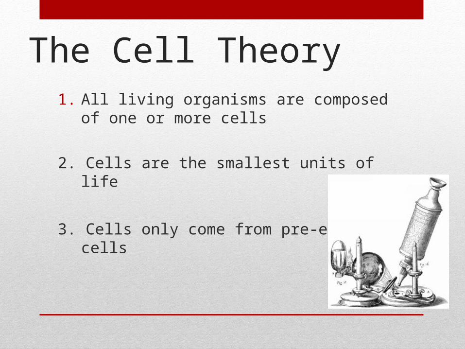
The Cell Theory
1. All living organisms are composed of one or more cells
2. Cells are the smallest units of life
3. Cells only come from pre-existing cells

Functions of Life• All organisms exist in either a unicellular or a
multicellular form. All organisms carry out all the functions of life. These functions include;• Metabolism
• Growth
• Reproduction
• Response to the environment
• Homeostasis
• Nutrition (excretion)
Functions together produce living things
• Metabolism includes all chemical reactions that occur in the body
• Growth may be limited but is always evident in one way or another
• Reproduction involves hereditary molecules that can be passed to offspring
• Response to the environment is imperative to the survival of the organism
• Homeostasis is maintaining a constant internal environment (e.g. temperature).
• Nutrition is providing a source of compounds with many chemical bonds that can be broken down to provide energy and nutrients.

Functions of paramecium
• Movement- hair like structures (cilia) on the outside beat rhythmically, and propel the cell through liquid.
• Nutrients-They consume small algae and plants, which they ingest through sweeping the food into mouth-like structures called buccal cavities( lined with cilia). Photosynthetic can make own food from the sun’s energy.
• Reproduction- can sexually reproduce and can asexually make exact copies of themselves.
• Response: has some sense of movement because it responds when it bumps into something. It also can sense certain chemicals
• Growth: evident up to a certain size.
• Homeostasis: the paramecium has a regulated internal environment aided by it membrane
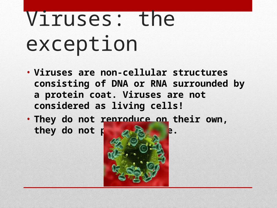
Viruses: the exception
• Viruses are non-cellular structures consisting of DNA or RNA surrounded by a protein coat. Viruses are not considered as living cells!
• They do not reproduce on their own, they do not produce waste.
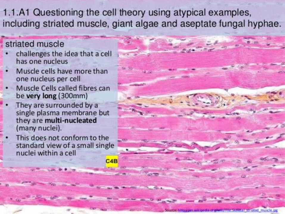
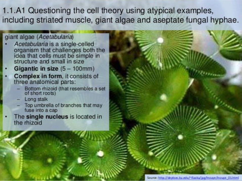
• Giant Algae:
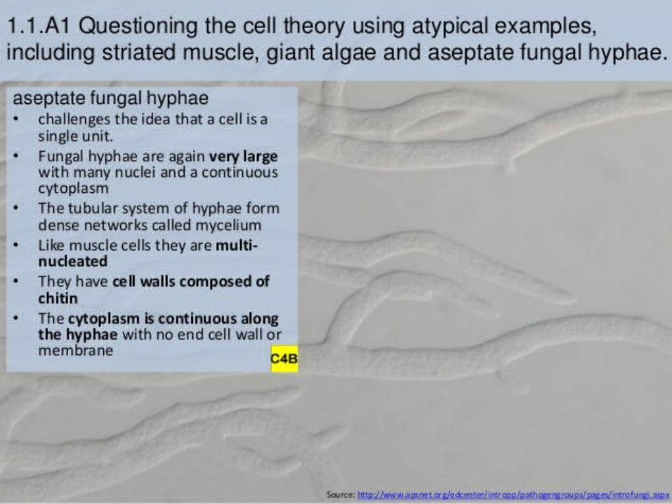
• Aseptate fungal hyphae:
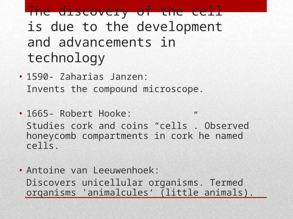
The discovery of the cell is due to the development and advancements in technology
• 1590- Zaharias Janzen: Invents the compound microscope.
• 1665- Robert Hooke:Studies cork and coins “cells”. Observed honeycomb compartments in cork he named cells.
• Antoine van Leeuwenhoek: Discovers unicellular organisms. Termed organisms 'animalcules‘ (little animals).
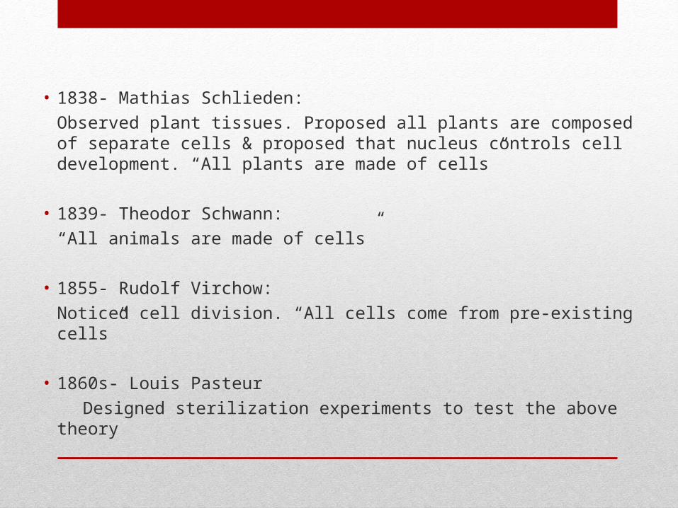
• 1838- Mathias Schlieden:
Observed plant tissues. Proposed all plants are composed of separate cells & proposed that nucleus controls cell development. “All plants are made of cells”
• 1839- Theodor Schwann:
“All animals are made of cells”
• 1855- Rudolf Virchow:
Noticed cell division. “All cells come from pre-existing cells”
• 1860s- Louis Pasteur
Designed sterilization experiments to test the above theory
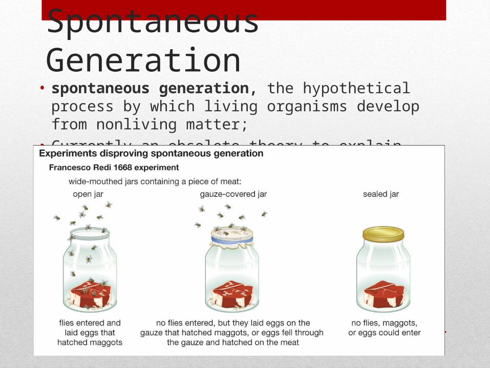
Spontaneous Generation
• spontaneous generation, the hypothetical process by which living organisms develop from nonliving matter;
• Currently an obsolete theory to explain the origin of life
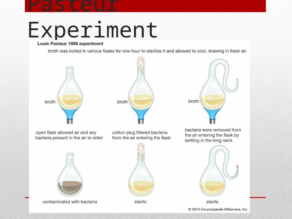
Pasteur Experiment
Findings
• If spontaneous generation had been a real phenomenon, Pasteur argued, the broth in the curved-neck flask would have eventually become re-infected because the germs would have spontaneously generated.
• The curved-neck flask never became infected, indicating that the germs could only come from other germs and spontaneous generation does NOT occur
• Pasteurization is the process of removing harmful pathogens from various types of food. Still used today for milk production
• http://science.howstuffworks.com/life/cellular-microscopic/pasteurization.htm
Unicellular Organisms
• Some organisms are unicellular meaning they are made of only a single cell.
(Examples: Amoeba, Chlorella, Scenedesmus)
• This single cell has to carry out all the functions of life
Multicellular Organisms
• Organisms comprised of more than one cell.
• The cells in a multicellular organism work together to carry out the various functions of life.
Emergent Properties
• Multicellular organisms show emergent properties.
• This means that the organism can achieve more than the sum of what each cell could achieve individually, because of cell interaction.
• Ex: anthill, human brain
Differentiation and Specialized Functions• In multicellular organisms such as humans, the DNA in
every somatic cell is identical.• ie, your stomach cells have the exact same DNA as your
heart cells
• Differentiated cells may contain identical DNA, but they differ in which genes are activated.
• Differentiation is when cells become specialized to perform one task, which is reflected in their structure and function.
• Organisms can use differentiation as they grow from a single cell to create all the necessary types of cells
STEM CELLS(plants-meristematic cells)
• Unspecialized cells These cells can become any type of cell or tissue(“pluripotent”).
• Embryo’s, the umbilical cord and bone marrow are sources of stem cells
• Adult stem cells are “multipotent”. For example, stem cells in the bone marrow can only give rise to all the various types of blood cells.
• A stem cells ability to replicate and differentiate along different pathways is necessary for embryonic development and therapeutic uses.
Utilization
• Adults still have some stem cells in their bone marrow which can be used to treat diseases such as leukemia
• Stems cells can differentiate into specialized cells when given a certain chemical signal. Therefore, the potential to grow a new organ in vitro exists.
• Potential uses in the future include treatments for Diabetes, heart disease, Multiple Sclerosis, Parkinson’s etc.
• Induced pluripotent stem (iPS) cells, discovered in 2007, are ”man-made”, reprogrammed, stem cells that share ES cells' ability to become other cell types.
Therapeutic use of Stem Cells• CELL THERAPY: non-functioning cells are replaced with
healthy, functioning cells
• Ex: Bone Marrow Transplants for leukemia patients.• Ex: Skin grafts for burn victims• Ex. Replenished brain cells for Parkinson’s and Alzheimers• Ex. Replace Insulin secreting pancreatic cells in diabetics• Ex. Help stop and reverse vision in people with Stargardt’s
disease• http://www.technologyreview.com/news/526591/stem-cell-treatment-for-blindness-moving-thro
ugh-patient-testing/
Ethical Implications
• Internationally there has been much sharing of data involving stem cell research.
• Most stem cells are generated in the lab, using in-vitro fertilization (IVF) techniques.
• National governments are influenced by local, cultural and religious traditions that impact the work of scientists and the use of stem cells in therapy.
• Research is restricted and regulated.
• Gathering stem cells requires the death of the embryo and opponents argue this is taking human life.
• WHAT DO YOU THINK?
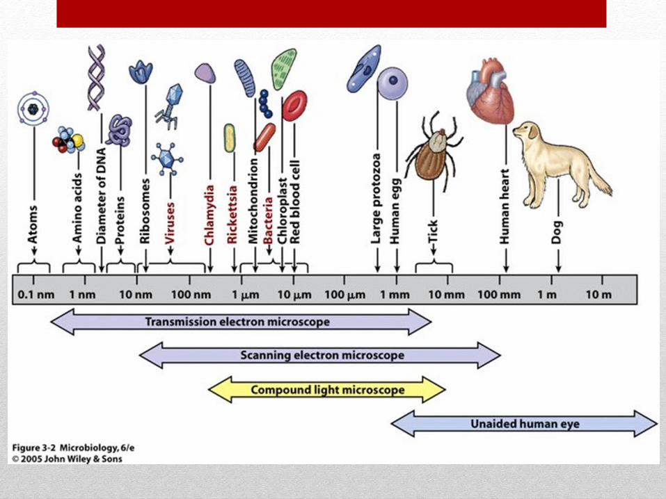
Cells and Sizes• Microscopes with high magnification and resolution (clarity)
are required to often visualize microorganisms and organelles
• An electron microscope (100 000x magnification) is used to study the internal structure of a cell (the organelles).
• Because these structures are very small, special units are used to measure them:
• Micrometre (μm) = 10-6 m
• Nanometer (nm) = 10-9 m
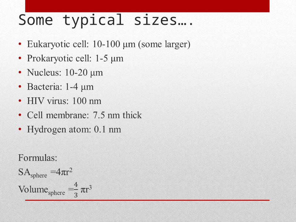
Some typical sizes….
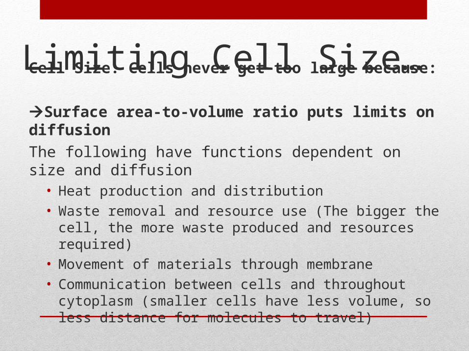
Cell Size: Cells never get too large because:
Surface area-to-volume ratio puts limits on diffusion
The following have functions dependent on size and diffusion• Heat production and distribution
• Waste removal and resource use (The bigger the cell, the more waste produced and resources required)
• Movement of materials through membrane
• Communication between cells and throughout cytoplasm (smaller cells have less volume, so less distance for molecules to travel)
Limiting Cell Size…
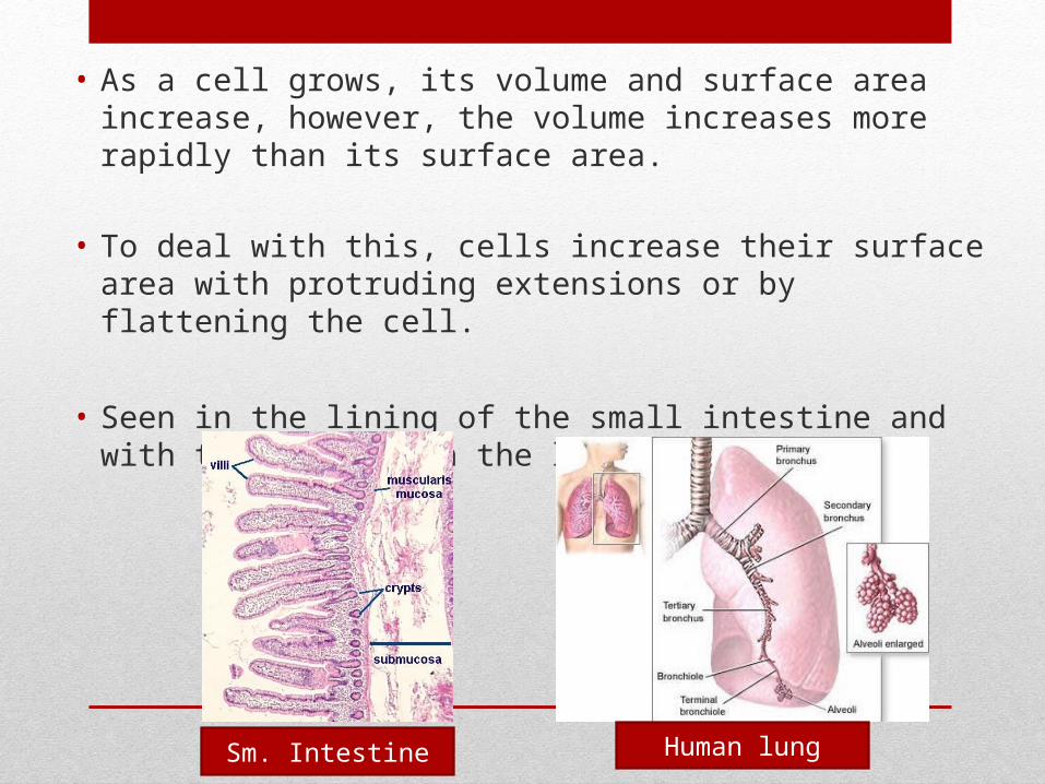
• As a cell grows, its volume and surface area increase, however, the volume increases more rapidly than its surface area.
• To deal with this, cells increase their surface area with protruding extensions or by flattening the cell.
• Seen in the lining of the small intestine and with the alveoli in the lungs
Sm. Intestine Human lung
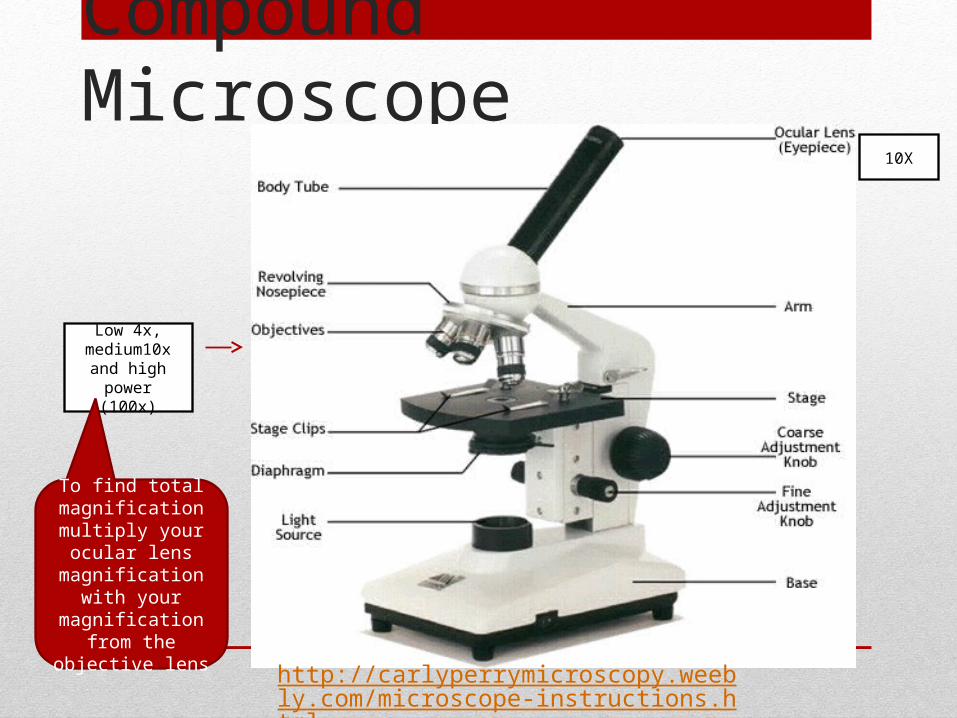
Compound Microscope
Low 4x, medium10x and
high power (100x)
10X
To find total magnification
multiply your ocular lens magnification
with your magnification from the objective lens
http://carlyperrymicroscopy.weebly.com/microscope-instructions.html
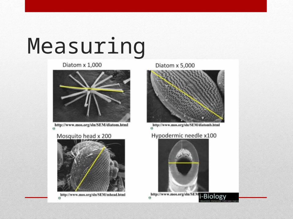
Measuring
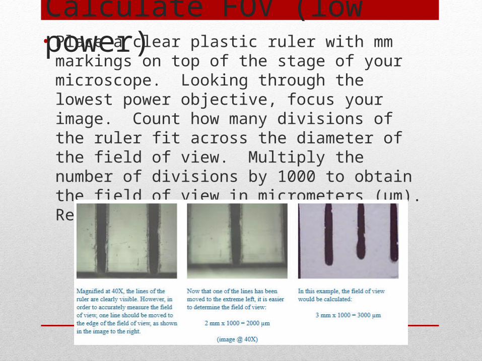
Calculate FOV (low power)• Place a clear plastic ruler with mm markings on top of the
stage of your microscope. Looking through the lowest power objective, focus your image. Count how many divisions of the ruler fit across the diameter of the field of view. Multiply the number of divisions by 1000 to obtain the field of view in micrometers (µm). Record this in µm (1mm = 1000 µm ).
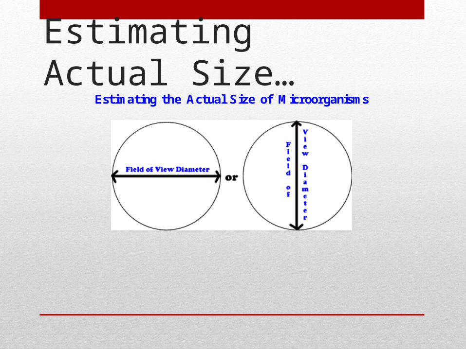
Estimating Actual Size…
Estimating the Actual Size of Microorganisms
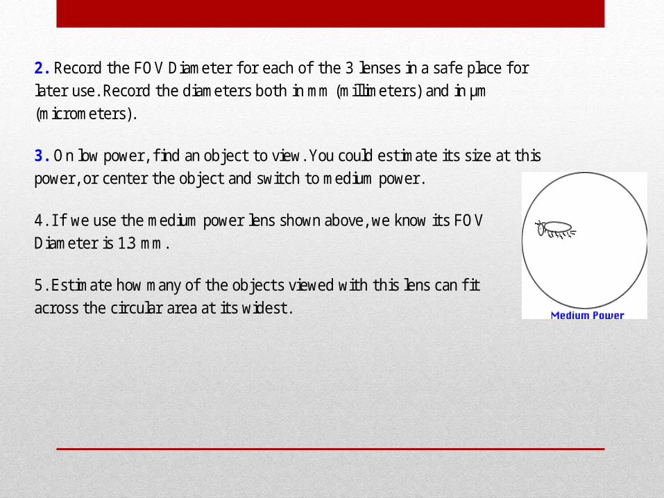
2. Record the FOV Diameter f or each of the 3 lenses in a safe place f or later use. Record the diameters both in mm (millimeters) and in µm (micrometers).
3. On low power, fi nd an object to view. You could estimate its size at this power, or center the object and switch to medium power.
4. I f we use the medium power lens shown above, we know its FOV Diameter is 1.3 mm.
5. Estimate how many of the objects viewed with this lens can fi t across the circular area at its widest.
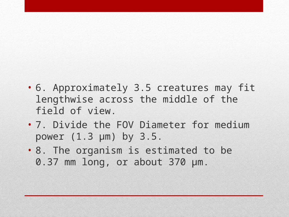
• 6. Approximately 3.5 creatures may fit lengthwise across the middle of the field of view.
• 7. Divide the FOV Diameter for medium power (1.3 µm) by 3.5.
• 8. The organism is estimated to be 0.37 mm long, or about 370 µm.
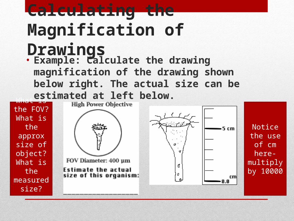
Calculating the Magnification of Drawings • Example: Calculate the drawing magnification of the
drawing shown below right. The actual size can be estimated at left below.
Notice the use of cm
here- multiply by
10000
What is the FOV?
What is the approx size of object?
What is the measured
size?
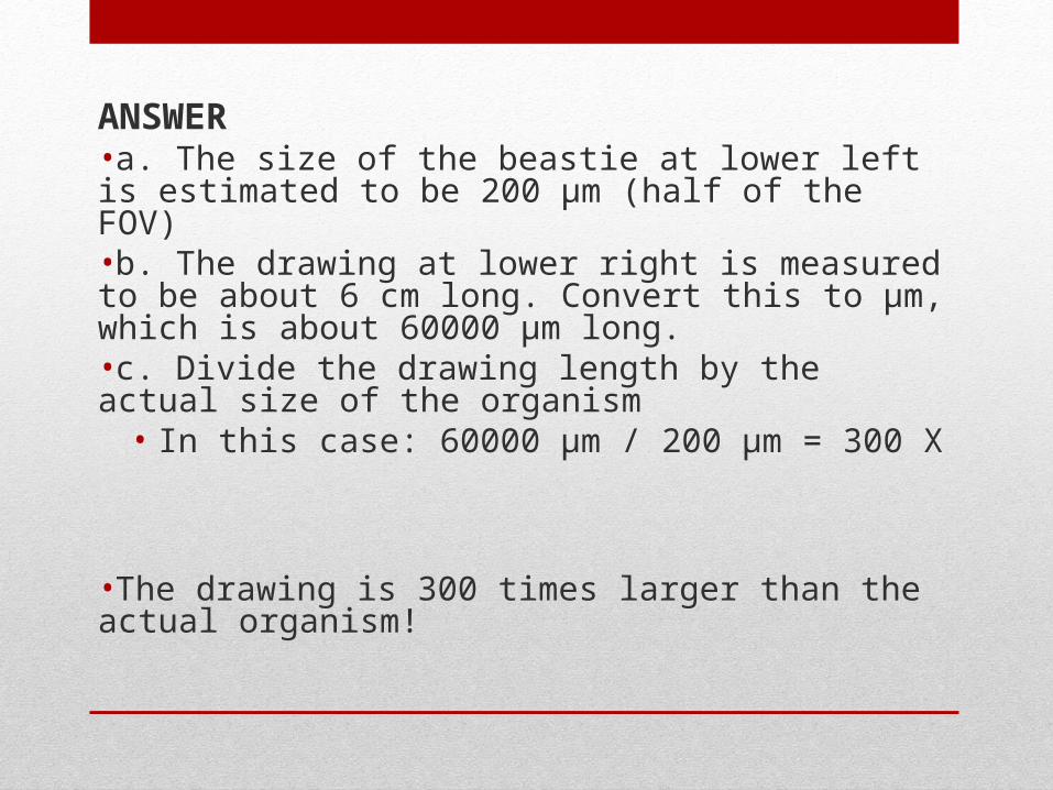
ANSWER•a. The size of the beastie at lower left is estimated to be 200 µm (half of the FOV)•b. The drawing at lower right is measured to be about 6 cm long. Convert this to µm, which is about 60000 µm long.•c. Divide the drawing length by the actual size of the organism
• In this case: 60000 µm / 200 µm = 300 X
•The drawing is 300 times larger than the actual organism!
Calculating magnification using the scale bar
• Diagrams and photographs can be shown larger or smaller than reality
• The magnification or a scale bar is given to indicate the real size of the object
• A scale bar is a line that shows the actual size of an image in relative proportion to its scale
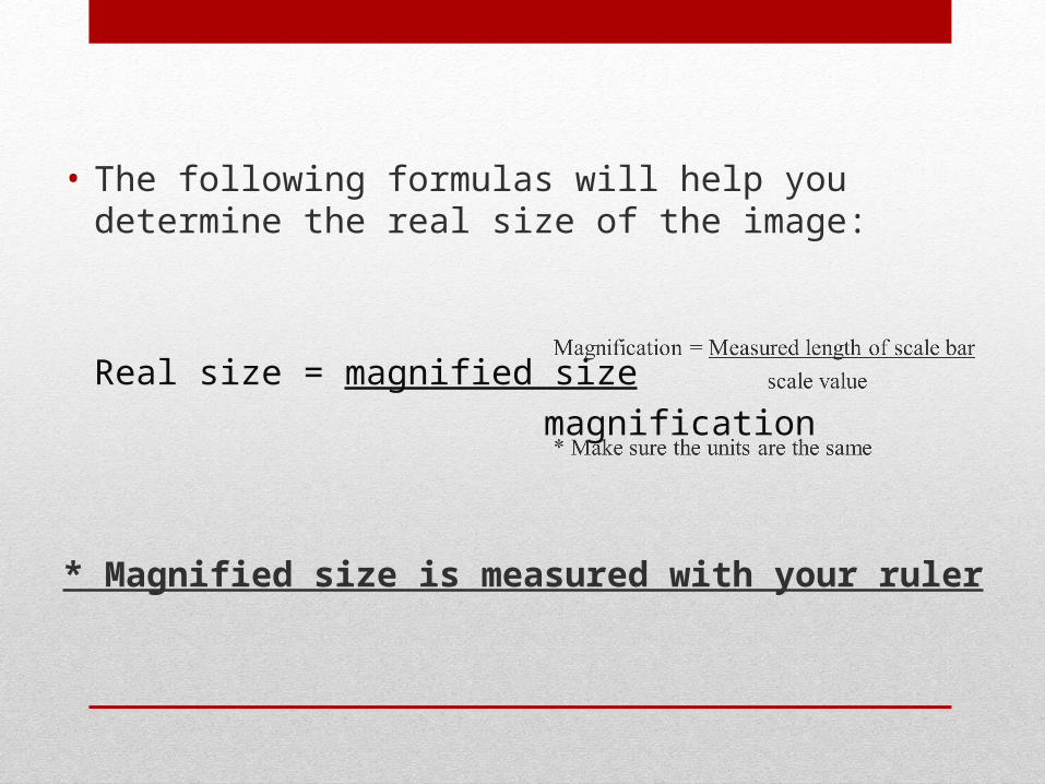
• The following formulas will help you determine the real size of the image:
Real size = magnified size
magnification
* Magnified size is measured with your ruler
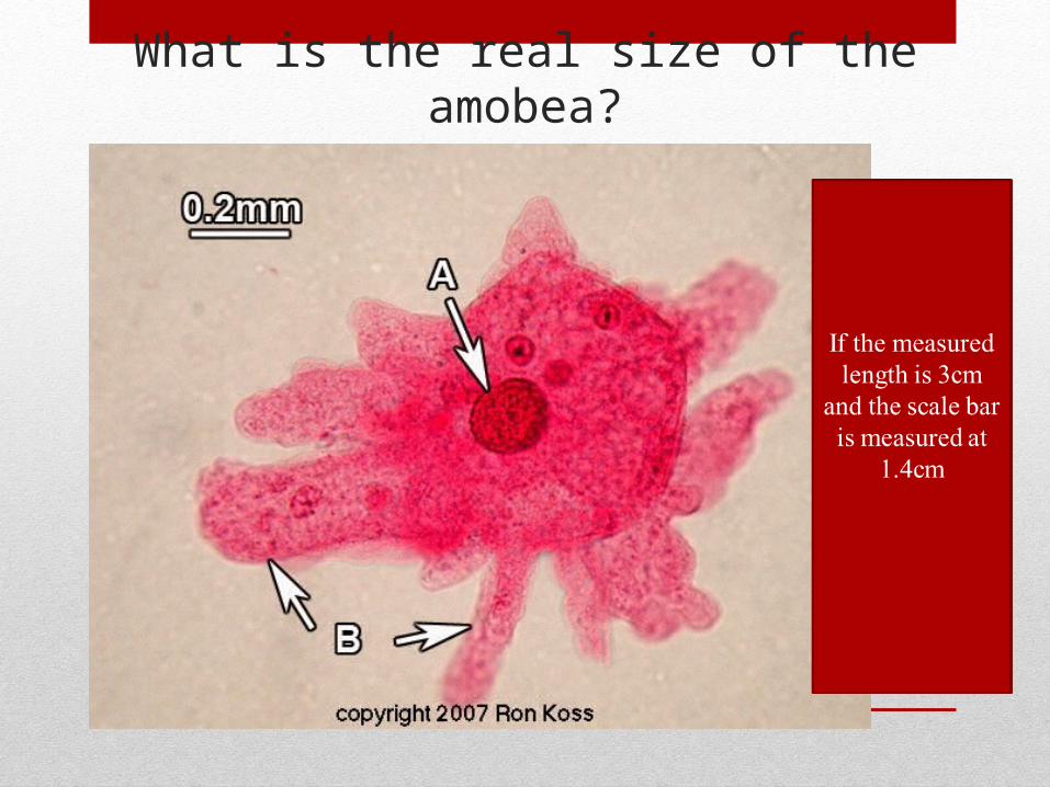
What is the real size of the amobea?
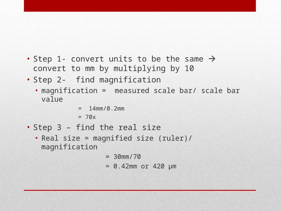
• Step 1- convert units to be the same convert to mm by multiplying by 10
• Step 2- find magnification • magnification = measured scale bar/ scale bar value
= 14mm/0.2mm
= 70x
• Step 3 – find the real size• Real size = magnified size (ruler)/ magnification
= 30mm/70
= 0.42mm or 420 µm
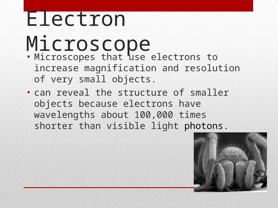
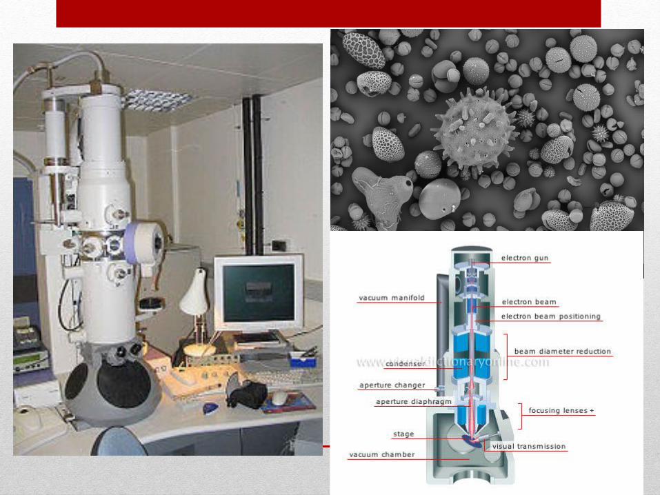
Electron Microscope• Microscopes that use electrons to increase magnification
and resolution of very small objects.
• can reveal the structure of smaller objects because electrons have wavelengths about 100,000 times shorter than visible light photons.
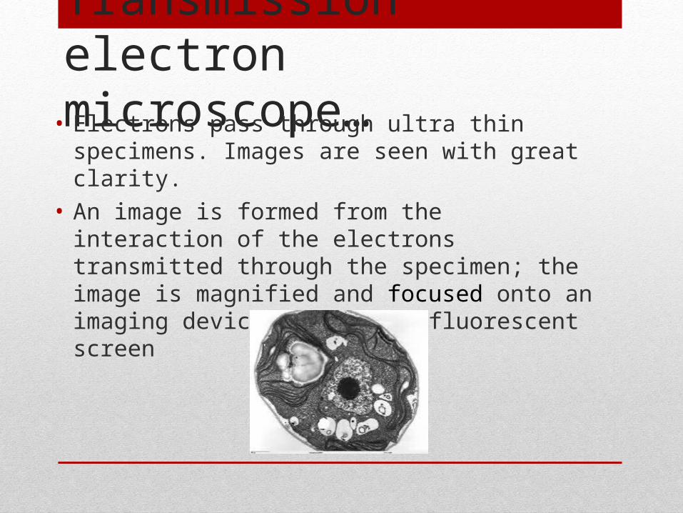
Transmission electron microscope…
• Electrons pass through ultra thin specimens. Images are seen with great clarity.
• An image is formed from the interaction of the electrons transmitted through the specimen; the image is magnified and focused onto an imaging device, such as a fluorescent screen
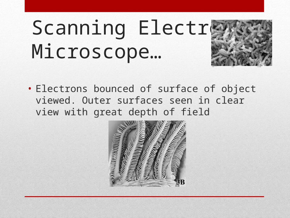
Scanning Electron Microscope…
• Electrons bounced of surface of object viewed. Outer surfaces seen in clear view with great depth of field

TO sum it up…
• http://www.biologycorner.com/quiz/qz_cell_theory.html
• http://www.pbs.org/wgbh/nova/sciencenow/0305/03.html
• (stem cell documentary)