Embed Size (px)
Citation preview

3022 Research Article
IntroductionActin filaments are intrinsically dynamic cytoskeletalstructures. At the cellular level, this dynamic behavior isenhanced or suppressed by a number of actin regulatoryproteins (Pollard and Borisy, 2003). Actin assembly isaccelerated by actin nucleation factors, such as the Arp2/3complex (Goley and Welch, 2006) and members of the forminfamily (Higgs, 2005), whereas disassembly is enhanced byactin severing and/or depolymerizing proteins, such as gelsolinand actin depolymerizing factor (ADF)/cofilin (Ono, 2007). Onthe one hand, coordinated regulation of actin assembly anddisassembly produces persistent actin treadmilling that drivesactin-based motility (Pollard and Borisy, 2003). On the otherhand, the actin cytoskeleton must be stabilized when actinsupports myosin-based contractility or when actin maintainsstable cellular structures. F-actin binding proteins can stabilizeactin filaments by preventing depolymerization or by cross-linking filaments to provide mechanical strength (McGough,1998). How actin stability is spatially and temporally regulatedin the cell is, however, is not completely understood.
ADF/cofilin enhances actin filament turnover byaccelerating the rate of depolymerization at the pointed endsand by severing filaments (Ono, 2007). The filament severingactivity of ADF/cofilin has been directly observed (Ichetovkinet al., 2000; Maciver et al., 1991b; Ono et al., 2004; Pavlov etal., 2007). However, the depolymerizing activity at the pointed
ends has been demonstrated indirectly by spectroscopicmethods (Carlier et al., 1997; Maciver et al., 1998), and arecent study has shown that ADF/cofilin does not significantlyincrease the rate of depolymerization at the pointed ends(Andrianantoandro and Pollard, 2006). In cultured cells,ADF/cofilin enhances depolymerization of actin filaments andincreases actin monomer concentrations (Hotulainen et al.,2005; Kiuchi et al., 2007). It also promotes actinpolymerization by nucleating filament assembly(Andrianantoandro and Pollard, 2006; Chen et al., 2004;Kudryashov et al., 2006) or by severing filaments, with aconcomitant increase in the number of exposed barbed ends(Ghosh et al., 2004). Thus, ADF/cofilin promotes overall actinfilament dynamics, but the precise mechanism by whichADF/cofilin regulates actin dynamics is complex and notcompletely understood.
ADF/cofilin is required for rapid actin turnover in vivo, andinhibition of ADF/cofilin activity stabilizes the actincytoskeleton (Lappalainen and Drubin, 1997). ADF/cofilin canbe inhibited by phosphorylation or by binding tophospholipids, and spatial and temporal control of theseregulatory mechanisms is important for coordinatedcytoskeletal function during directional cell migration(Mouneimne et al., 2006; Nishita et al., 2005). In addition,tropomyosin, a well-characterized F-actin binding protein,stabilizes actin filaments (Cooper, 2002) by preventing
Stabilization of actin filaments is critical for supportingactomyosin-based contractility and for maintaining stablecellular structures. Tropomyosin is a well-characterizedubiquitous actin stabilizer that inhibits ADF/cofilin-dependent actin depolymerization. Here, we show thatUNC-87, a calponin-related Caenorhabditis elegans proteinwith seven calponin-like repeats, competes withADF/cofilin for binding to actin filaments and inhibitsADF/cofilin-dependent filament severing anddepolymerization in vitro. Mutations in the unc-87 genesuppress the disorganized actin phenotype in anADF/cofilin mutant in the C. elegans body wall muscle,supporting their antagonistic roles in regulating actinstability in vivo. UNC-87 and tropomyosin exhibit
synergistic effects in stabilizing actin filaments againstADF/cofilin, and direct comparison reveals that UNC-87effectively stabilizes actin filaments at much lowerconcentrations than tropomyosin. However, the in vivofunctions of UNC-87 and tropomyosin appear different,suggesting their distinct roles in the regulation ofactomyosin assembly and cellular contractility. Our resultsdemonstrate that actin binding via calponin-like repeatscompetes with ADF/cofilin-driven cytoskeletal turnover,and is critical for providing the spatiotemporal regulationof actin filament stability.
Key words: Actin dynamics, ADF/cofilin, Calponin, Tropomyosin
Summary
UNC-87, a calponin-related protein in C. elegans,antagonizes ADF/cofilin-mediated actin filamentdynamicsSawako Yamashiro1, Mario Gimona2 and Shoichiro Ono1,*1Department of Pathology, Emory University, Atlanta, GA 30322, USA2Unit of Actin Cytoskeleton Regulation, Consorzio Mario Negri Sud, Department of Cell Biology and Oncology, Via Nazionale 8a, 66030 SantaMaria, Imbaro, Italy*Author for correspondence (e-mail: [email protected])
Accepted 26 June 2007Journal of Cell Science 120, 3022-3033 Published by The Company of Biologists 2007doi:10.1242/jcs.013516
Jour
nal o
f Cel
l Sci
ence
3023Actin stabilization by UNC-87
ADF/cofilin-dependent actin filament disassembly (Bernsteinand Bamburg, 1982; Ono and Ono, 2002). ADF/cofilin andtropomyosin often localize to different compartments of thecell and contribute to spatial separation of dynamic and stablepopulations of actin filaments (DesMarais et al., 2002; Guptonet al., 2005; Iwasa and Mullins, 2007). However, there are anumber of other less well-characterized F-actin-bindingproteins that also localize to different cytoskeletalcompartments. The mechanisms underlying their functionalinteractions with ADF/cofilin-dependent actin dynamicsremain elusive.
The body wall muscle of the nematode Caenorhabditiselegans has been used as a model to study assembly andmaintenance of striated muscle (Moerman and Fire, 1997). Wepreviously demonstrated that actin filament disassemblyactivities of UNC-60B (ADF/cofilin) (Ono et al., 2003; Ono etal., 1999) and actin-interacting protein 1 (UNC-78) (Mohri etal., 2006; Ono, 2001) are required for organized assembly ofactin filaments in C. elegans muscle, and that tropomyosinstabilizes actin filaments antagonistically (Ono and Ono, 2002;Yu and Ono, 2006). Tropomyosin is a major component ofisolated nematode thin filaments. Although removal oftropomyosin by high salt increased ADF/cofilin binding to thinfilaments in vitro, the binding still appeared partially inhibited.Therefore, additional factor(s) that prevent ADF/cofilin frombinding to actin must exist (Ono and Ono, 2002). Wehypothesized that UNC-87 is one of these additional actinstabilizers. The unc-87 gene is implicated in protectingmyofibrils from mechanical damage during contraction(Goetinck and Waterston, 1994b). UNC-87 is a calponin-related protein with seven calponin-like (CLIK) repeats, but itlacks a calponin-homology (CH) domain, which is present inthe calponin and SM22/transgelin families of actin bindingproteins (Gimona et al., 2002). UNC-87 localizes to the thinfilaments (Goetinck and Waterston, 1994a) and directly bindsto actin filaments in vitro (Kranewitter et al., 2001). However,the precise mode of actin binding and the nature of the actinstabilizing function of UNC-87 are not clearly understood.
The CLIK repeat is an actin-binding motif that is unique tocalponin-related proteins from yeast to vertebrates. CLIKrepeats are present in the C-terminal halves of calponins andSM22/transgelins and mediate the binding to F-actin, whereasthe N-terminal CH domains of calponin and SM22/transgelinare dispensable for actin binding (Gimona and Mital, 1998;Goodman et al., 2003). Ectopic expression of CLIK repeatsfrom calponin and UNC-87 in cultured mammalian cellsinhibits dynamic reorganization of actin filaments (Gimona etal., 2003; Lener et al., 2004), suggesting that CLIK repeatsnegatively regulate actin cytoskeleton turnover. In this study,we show that UNC-87 inhibits severing and depolymerizationactivities of ADF/cofilin in vitro and in vivo, and that this effectis more potent and stable than that of tropomyosin. From ourresults, we propose that actin-binding via CLIK repeatsantagonizes ADF/cofilin-driven actin cytoskeleton remodelingat specialized cellular regions.
ResultsUNC-87 and ADF/cofilin bind to F-actin in a mutuallyexclusive mannerTo test our initial hypothesis that UNC-87 stabilizes actinfilaments, we first examined whether UNC-87 inhibits
ADF/cofilin-mediated actin turnover in vitro, and whetherUNC-87 and ADF/cofilin compete for binding to actinfilaments, as was previously shown for tropomyosin andADF/cofilin (Bernstein and Bamburg, 1982; Ono and Ono,2002). To correlate in vitro and in vivo studies in C. elegans,we used actin, UNC-87 and UNC-60B (C. elegans muscle-specific ADF/cofilin) from C. elegans for most of thebiochemical assays. In an actin co-pelleting assay at highspeed, UNC-60B co-sedimented with actin filaments with asaturation at an approx. 1:1 molar ratio (Fig. 1Aa,b). However,when actin was preincubated with 10 �M UNC-87, the amountof UNC-60B in the pellets was decreased, indicating inhibitionof actin binding (Fig. 1A, compare a with b). The inhibitoryeffect of UNC-87 was concentration dependent, and a 30-50%reduction in actin-UNC-60B binding was detected at 2.5 �MUNC-87 (actin:UNC-87=4:1) (Fig. 1Ac). This inhibition was,however, not complete (Fig. 1Ac), because UNC-87 wasdissociated from actin and increased in the supernatants as theconcentrations of UNC-60B were increased (Fig. 1Ab). Theseresults indicate that UNC-87 and UNC-60B compete forbinding to F-actin.
The monomeric UNC-87 protein is capable of bundling actinfilaments (Kranewitter et al., 2001), suggesting that multipleactin-binding sites are present in a single molecule. When wetested the binding of 1.0-20 �M UNC-87 to 10 �M F-actin bythe co-sedimentation assay, binding did not reach saturationwithin this concentration range (Fig. 1Ba,c, closed circles).Similarly, binding did not reach saturation using 2 �M F-actin(our unpublished data). Higher concentrations of UNC-87 weretechnically difficult to analyze quantitatively because of theclose electrophoretic mobilities of UNC-87 and actin on SDS-PAGE (Fig. 1Ba). Nonetheless, Scatchard analysis of the datastrongly suggested the presence of a strong actin-bindingsite (Ka=6.5±0.32 �M–1) and a weak actin-binding site(Ka=0.31±0.0067 �M–1) (Fig. 1Bd, closed circles), supportingthe presence of two separate regions of interaction with F-actin.Preincubation of F-actin with UNC-60B (note the reverse orderof incubation as compared with Fig. 1A) inhibited binding ofUNC-87 to F-actin (Fig. 1Bb) in a concentration-dependentmanner (Fig. 1Bc). Scatchard analyses of these data indicatedthat the strong actin-binding site of UNC-87 was effectivelyinhibited by low concentrations of UNC-60B (Fig. 1Bd, see 5�M shown by open circles).
As a consequence of the above, we detected that UNC-60Binhibited the actin-bundling activity of UNC-87 (Fig. 2). In alow-speed sedimentation assay, UNC-87 induced the formationof actin bundles that sedimented under these conditions (Fig.2Aa), whereas, when actin was pre-incubated with UNC-60B,actin-bundling and hence actin filament pelleting at low gforces was efficiently inhibited (Fig. 2Ab,c). When UNC-87and UNC-60B were simultaneously added to F-actin, theinhibitory effect on bundling was apparently reduced, mostprobably due to their competitive binding to the side of actinfilaments (our unpublished data). We further confirmed theseeffects on actin bundling by direct observation of fluorescentlylabeled actin filaments (Fig. 2B). In the presence of UNC-87,thick actin bundles were formed (Fig. 2Bb, arrows), whereasUNC-60B induced the fragmentation of actin filamentswithout bundling (Fig. 2Bc). When UNC-87 was incubatedwith actin that had been pre-treated with UNC-60B, only shortindividual filaments were present, but actin bundles were not
Jour
nal o
f Cel
l Sci
ence

3024
detected (Fig. 2Bd). Gelsolin-severed short actin filaments (upto 1:50 molar ratios of gelsolin and actin) were efficientlybundled by UNC-87 (our unpublished data), suggesting thatthe inhibitory effect of UNC-60B is not simply due toshortening of the filaments. This antagonistic effect contraststhe previously reported synergistic effects on actin bundlingbetween ADF/cofilin and �-actinin (Maciver et al., 1991a) orelongation factor 1� (Gungabissoon et al., 2001).
UNC-87 Is a potent inhibitor of ADF/cofilin-mediatedactin turnoverThe competitive binding of UNC-87 and UNC-60B to F-actinsuggests that UNC-87 influences the rate of actin turnover thatis regulated by UNC-60B. Our preliminary experimentsindicated that UNC-87 alone inhibited latrunculin A-inducedactin depolymerization in vitro (our unpublished data).Furthermore, using fluorescence microscopy, we found thatUNC-87 has a strong protective effect against filament severingby UNC-60B (Fig. 3). Fluorescently labeled actin filaments
Journal of Cell Science 120 (17)
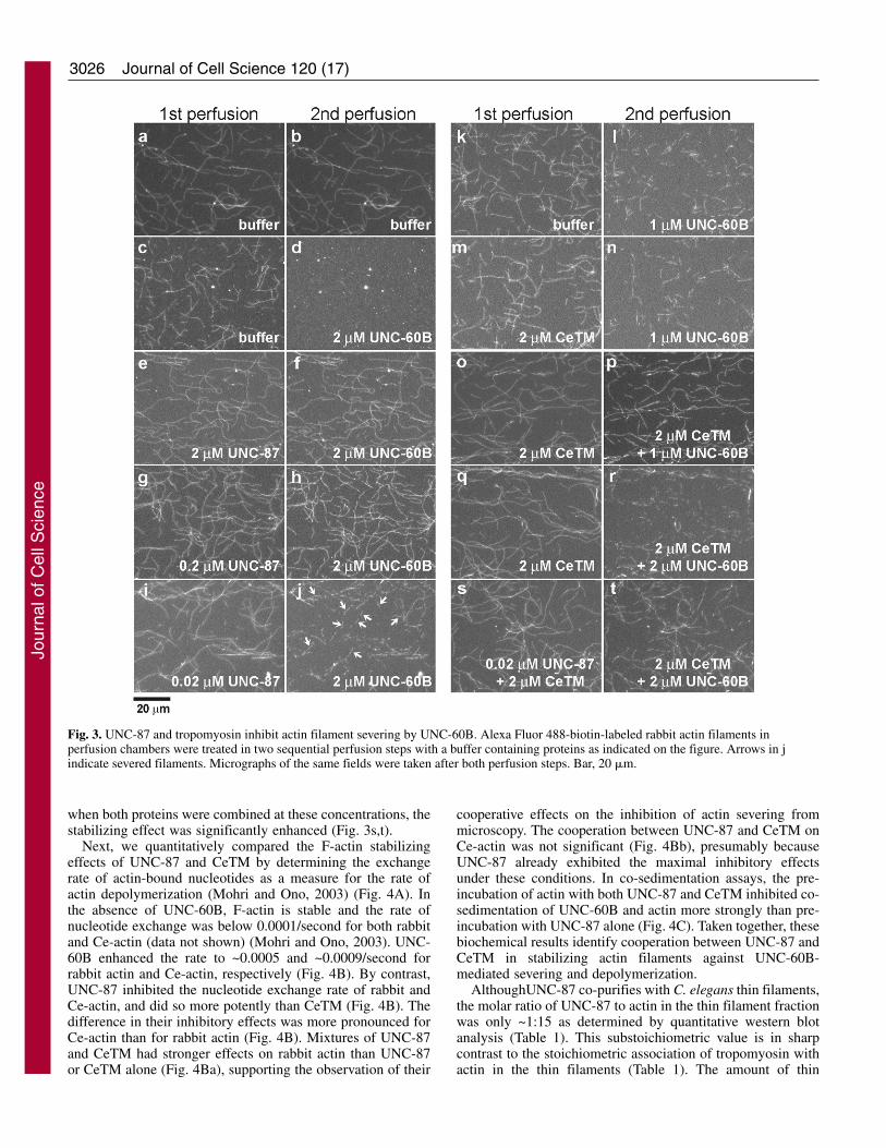
were immobilized to a glass surface in a perfusion chamber,exposed to these proteins in two sequential perfusion steps, andexamined for their stability. In control experiments, actinfilaments at 3 minutes after the first perfusion with buffer only(Fig. 3a) and at 3 minutes after the second perfusion with bufferonly (Fig. 3b) showed only minimal alterations in filamentmorphology. Addition of 2 �M UNC-60B in the secondperfusion step caused the severing of filaments (Fig. 3d) thathad been pre-treated with buffer only (Fig. 3c). However,incubation of filaments with 2 �M UNC-87 in the firstperfusion step (Fig. 3e) efficiently protected actin filamentsfrom severing by UNC-60B (Fig. 3f). A tenfold lowerconcentration (0.2 �M UNC-87) also had a strong protectiveeffect against severing by UNC-60B (Fig. 3h), and only at 0.02�M was the effect of UNC-87 was reduced (Fig. 3i, see arrowsfor severed filaments).
Tropomyosin is well-known to stabilize actin filamentsagainst depolymerization by ADF/cofilin in vitro and in vivo(Cooper, 2002). The C. elegans lev-11 gene (Y105E8B.1) is
Fig. 1. UNC-87 or UNC-60B interacts withCe-actin in a mutually exclusive manner.(A) 10 �M Ce-actin was pre-incubated with0-10 �M UNC-87 for 30 minutes, and then,various concentrations of UNC-60B (0-20�M) were added to the mixtures. After 30minutes, the samples were centrifuged at285,000 g for 20 minutes, and thesupernatant (s) and pellets (p) were analyzedby SDS-PAGE (12% acrylamide gel).Representative gels from experiments in theabsence (a) or in the presence of 10 �MUNC-87 (b) are shown. Quantitativeanalysis of the results by densitometry isshown in c, in which the amounts of actin-bound UNC-60B (mol/mol actin) wereplotted as a function of total UNC-60Bconcentrations (�M). (B) 10 �M Ce-actinwas pre-incubated with 0-20 �M UNC-60Bfor 30 minutes, and then, variousconcentrations of UNC-87 (0-20 �M) wereadded to the mixtures. After 30 minutes, thesamples were centrifuged at 285,000 g for20 minutes, and the supernatant (s) andpellets (p) were analyzed by SDS-PAGE.Representative gels of experiments in theabsence (a) and in the presence of 20 �MUNC-60B (b) are shown. Quantitativeanalysis of the results by densitometry isshown in c, in which the amounts of actin-bound UNC-87 (mol/mol actin) were plottedas a function of free UNC-87 concentrations(�M). (d) The quantitative results werefurther analyzed using a Scatchard plot.
Jour
nal o
f Cel
l Sci
ence

3025Actin stabilization by UNC-87
the single tropomyosin gene that encodes multiple spliceisoforms (Kagawa et al., 1995). C. elegans tropomyosin(CeTM) that was purified from wild-type worms consistsprimarily of high molecular mass isoforms, CeTMI and orCeTMII (Ono and Ono, 2002). A direct comparison of theeffects of UNC-87 and CeTM on actin severing revealed thatUNC-87 is a more potent stabilizer of actin filaments thantropomyosin. Pre-incubation of actin filaments with 2 �MCeTM had only a minimal protective effect against 1 �MUNC-60B (Fig. 3, compare l with n). The presence of 2 �MCeTM in both the first and second perfusion steps, however,elicited a significant protective effect against 1 �M UNC-60B(Fig. 3o,p), suggesting that CeTM is dissociated from thefilaments in the absence of free CeTM. However, when UNC-60B was increased to 2 �M, the protective effect of 2 �MCeTM was minimal (Fig. 3q,r), indicating that highconcentrations of tropomyosin are required to protect actinfilaments from severing by ADF/cofilin, whereas UNC-87
displays a robust and pronounced effect at significantly lowerprotein concentrations.
Recombinant UNC-87 and turkey smooth muscletropomyosin have been shown to simultaneously bind to F-actin in vitro (Kranewitter et al., 2001), and we confirmed thatUNC-87 and CeTM bind to C. elegans actin (Ce-actin) in asimilar manner (Fig. 4A). When Ce-actin (10 �M) was pre-incubated with 0-10 �M UNC-87 and subsequently with 2 �MCeTM, nearly the same amounts of CeTM co-sedimented withactin (Fig. 4Aa, compare lanes p for different UNC-87concentrations). Similarly, when Ce-actin (10 �M) was pre-incubated with 0-5 �M CeTM and subsequently with 5 �MUNC-87, nearly the same amounts of UNC-87 co-sedimentedwith actin (Fig. 4Ab, compare lanes p for different CeTMconcentrations). In addition, we detected that UNC-87 andCeTM showed cooperative stabilization of actin filaments.UNC-87 at 0.02 �M (Fig. 3i,j) or CeTM (2 �M) alone (Fig.3q,r) had only weak protective effects against UNC-60B, but,
Fig. 2. UNC-60B inhibits actin-bundlingactivity of UNC-87. (A) 10 �M rabbit F-actin was pre-incubated with 0-20 �MUNC-60B for 30 minutes, and variedconcentrations of UNC-87 (0-20 �M)were added to the mixtures. After 30minutes, the samples were centrifuged at18,000 g for 10 minutes, and thesupernatant (s) and pellets (p) wereanalyzed by SDS-PAGE. Representativegels of the experiments in the absence (a)and presence of 20 �M UNC-60B (b) areshown. Quantitative analysis of the resultsby densitometry is shown in (c), in whichthe percentages of sedimented (bundled)actin were plotted as a function of totalUNC-87 concentrations (�M). Data aremeans ± s.d. of three experiments. (B) 1�M Alexa Fluor 488-biotin-labeled rabbitF-actin was incubated with buffer only(a), 0.4 �M UNC-87 (b), 1 �M UNC-60B(c), or 1 �M UNC-60B and 0.4 �MUNC-87, and the filaments were observedby fluorescence microscopy. UNC-87induced actin bundling (b, arrows), but thebundling was inhibited by the presence ofUNC-60B (d). Bar, 20 �m.
Jour
nal o
f Cel
l Sci
ence
3026
when both proteins were combined at these concentrations, thestabilizing effect was significantly enhanced (Fig. 3s,t).
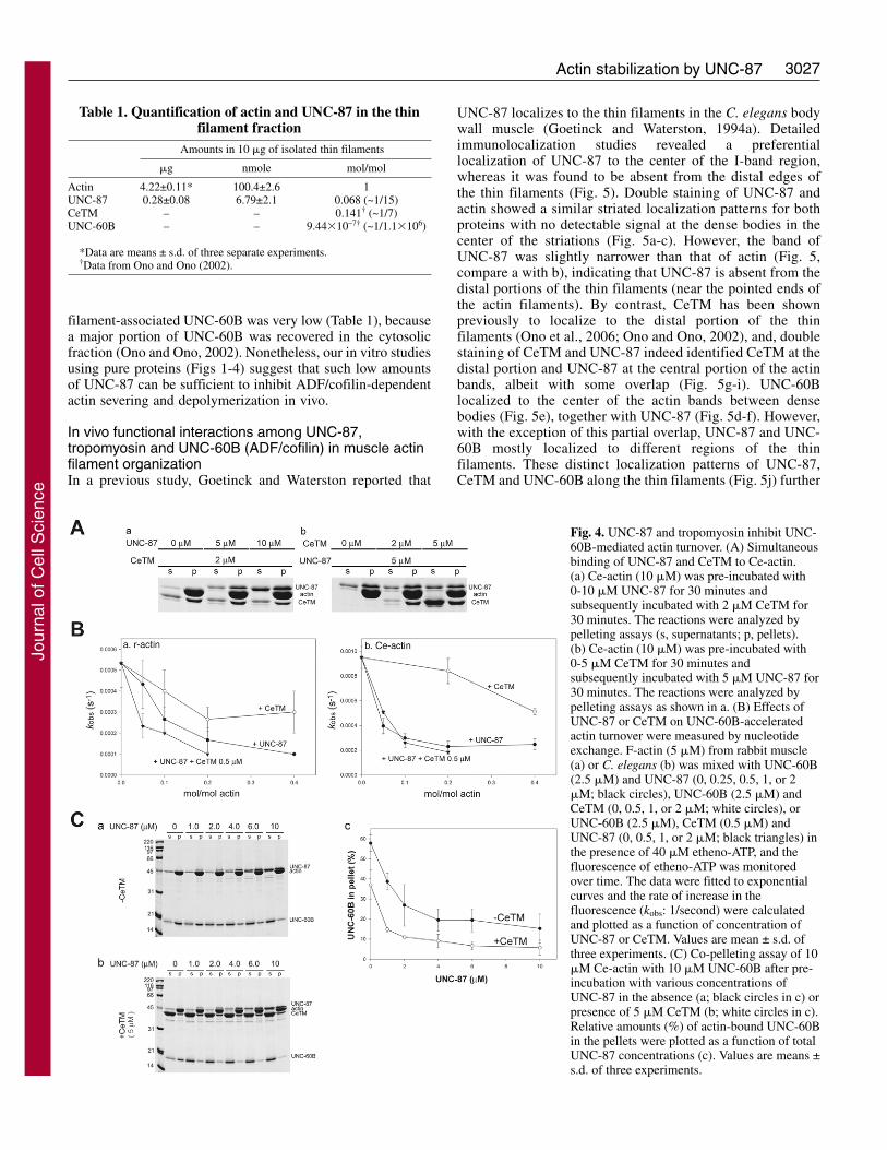
Next, we quantitatively compared the F-actin stabilizingeffects of UNC-87 and CeTM by determining the exchangerate of actin-bound nucleotides as a measure for the rate ofactin depolymerization (Mohri and Ono, 2003) (Fig. 4A). Inthe absence of UNC-60B, F-actin is stable and the rate ofnucleotide exchange was below 0.0001/second for both rabbitand Ce-actin (data not shown) (Mohri and Ono, 2003). UNC-60B enhanced the rate to ~0.0005 and ~0.0009/second forrabbit actin and Ce-actin, respectively (Fig. 4B). By contrast,UNC-87 inhibited the nucleotide exchange rate of rabbit andCe-actin, and did so more potently than CeTM (Fig. 4B). Thedifference in their inhibitory effects was more pronounced forCe-actin than for rabbit actin (Fig. 4B). Mixtures of UNC-87and CeTM had stronger effects on rabbit actin than UNC-87or CeTM alone (Fig. 4Ba), supporting the observation of their
Journal of Cell Science 120 (17)
cooperative effects on the inhibition of actin severing frommicroscopy. The cooperation between UNC-87 and CeTM onCe-actin was not significant (Fig. 4Bb), presumably becauseUNC-87 already exhibited the maximal inhibitory effectsunder these conditions. In co-sedimentation assays, the pre-incubation of actin with both UNC-87 and CeTM inhibited co-sedimentation of UNC-60B and actin more strongly than pre-incubation with UNC-87 alone (Fig. 4C). Taken together, thesebiochemical results identify cooperation between UNC-87 andCeTM in stabilizing actin filaments against UNC-60B-mediated severing and depolymerization.
AlthoughUNC-87 co-purifies with C. elegans thin filaments,the molar ratio of UNC-87 to actin in the thin filament fractionwas only ~1:15 as determined by quantitative western blotanalysis (Table 1). This substoichiometric value is in sharpcontrast to the stoichiometric association of tropomyosin withactin in the thin filaments (Table 1). The amount of thin
Fig. 3. UNC-87 and tropomyosin inhibit actin filament severing by UNC-60B. Alexa Fluor 488-biotin-labeled rabbit actin filaments inperfusion chambers were treated in two sequential perfusion steps with a buffer containing proteins as indicated on the figure. Arrows in jindicate severed filaments. Micrographs of the same fields were taken after both perfusion steps. Bar, 20 �m.
Jour
nal o
f Cel
l Sci
ence
3027Actin stabilization by UNC-87
filament-associated UNC-60B was very low (Table 1), becausea major portion of UNC-60B was recovered in the cytosolicfraction (Ono and Ono, 2002). Nonetheless, our in vitro studiesusing pure proteins (Figs 1-4) suggest that such low amountsof UNC-87 can be sufficient to inhibit ADF/cofilin-dependentactin severing and depolymerization in vivo.
In vivo functional interactions among UNC-87,tropomyosin and UNC-60B (ADF/cofilin) in muscle actinfilament organizationIn a previous study, Goetinck and Waterston reported that
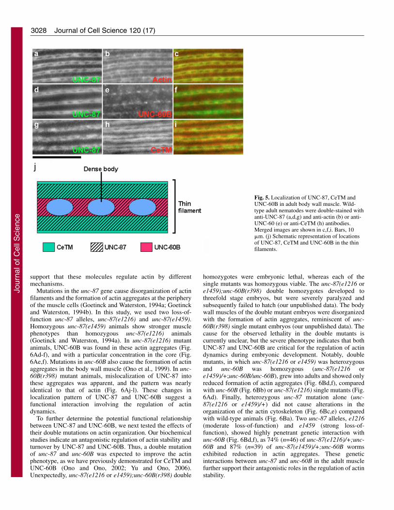
UNC-87 localizes to the thin filaments in the C. elegans bodywall muscle (Goetinck and Waterston, 1994a). Detailedimmunolocalization studies revealed a preferentiallocalization of UNC-87 to the center of the I-band region,whereas it was found to be absent from the distal edges ofthe thin filaments (Fig. 5). Double staining of UNC-87 andactin showed a similar striated localization patterns for bothproteins with no detectable signal at the dense bodies in thecenter of the striations (Fig. 5a-c). However, the band ofUNC-87 was slightly narrower than that of actin (Fig. 5,compare a with b), indicating that UNC-87 is absent from thedistal portions of the thin filaments (near the pointed ends ofthe actin filaments). By contrast, CeTM has been shownpreviously to localize to the distal portion of the thinfilaments (Ono et al., 2006; Ono and Ono, 2002), and, doublestaining of CeTM and UNC-87 indeed identified CeTM at thedistal portion and UNC-87 at the central portion of the actinbands, albeit with some overlap (Fig. 5g-i). UNC-60Blocalized to the center of the actin bands between densebodies (Fig. 5e), together with UNC-87 (Fig. 5d-f). However,with the exception of this partial overlap, UNC-87 and UNC-60B mostly localized to different regions of the thinfilaments. These distinct localization patterns of UNC-87,CeTM and UNC-60B along the thin filaments (Fig. 5j) further
Table 1. Quantification of actin and UNC-87 in the thinfilament fraction
Amounts in 10 �g of isolated thin filaments
�g nmole mol/mol
Actin 4.22±0.11* 100.4±2.6 1UNC-87 0.28±0.08 6.79±2.1 0.068 (~1/15)CeTM – – 0.141† (~1/7)UNC-60B – – 9.44�10–7† (~1/1.1�106)
*Data are means ± s.d. of three separate experiments.†Data from Ono and Ono (2002).
Fig. 4. UNC-87 and tropomyosin inhibit UNC-60B-mediated actin turnover. (A) Simultaneousbinding of UNC-87 and CeTM to Ce-actin.(a) Ce-actin (10 �M) was pre-incubated with0-10 �M UNC-87 for 30 minutes andsubsequently incubated with 2 �M CeTM for30 minutes. The reactions were analyzed bypelleting assays (s, supernatants; p, pellets).(b) Ce-actin (10 �M) was pre-incubated with0-5 �M CeTM for 30 minutes andsubsequently incubated with 5 �M UNC-87 for30 minutes. The reactions were analyzed bypelleting assays as shown in a. (B) Effects ofUNC-87 or CeTM on UNC-60B-acceleratedactin turnover were measured by nucleotideexchange. F-actin (5 �M) from rabbit muscle(a) or C. elegans (b) was mixed with UNC-60B(2.5 �M) and UNC-87 (0, 0.25, 0.5, 1, or 2�M; black circles), UNC-60B (2.5 �M) andCeTM (0, 0.5, 1, or 2 �M; white circles), orUNC-60B (2.5 �M), CeTM (0.5 �M) andUNC-87 (0, 0.5, 1, or 2 �M; black triangles) inthe presence of 40 �M etheno-ATP, and thefluorescence of etheno-ATP was monitoredover time. The data were fitted to exponentialcurves and the rate of increase in thefluorescence (kobs: 1/second) were calculatedand plotted as a function of concentration ofUNC-87 or CeTM. Values are mean ± s.d. ofthree experiments. (C) Co-pelleting assay of 10�M Ce-actin with 10 �M UNC-60B after pre-incubation with various concentrations ofUNC-87 in the absence (a; black circles in c) orpresence of 5 �M CeTM (b; white circles in c).Relative amounts (%) of actin-bound UNC-60Bin the pellets were plotted as a function of totalUNC-87 concentrations (c). Values are means ±s.d. of three experiments.
Jour
nal o
f Cel
l Sci
ence
3028
support that these molecules regulate actin by differentmechanisms.
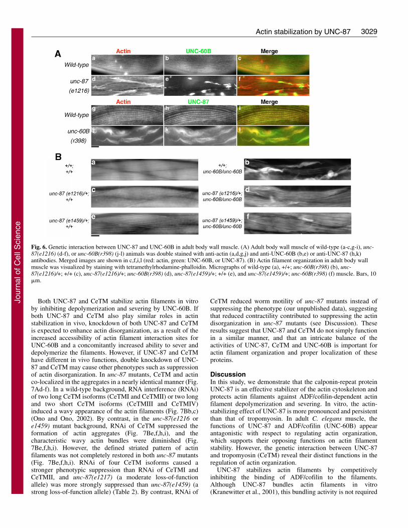
Mutations in the unc-87 gene cause disorganization of actinfilaments and the formation of actin aggregates at the peripheryof the muscle cells (Goetinck and Waterston, 1994a; Goetinckand Waterston, 1994b). In this study, we used two loss-of-function unc-87 alleles, unc-87(e1216) and unc-87(e1459).Homozygous unc-87(e1459) animals show stronger musclephenotypes than homozygous unc-87(e1216) animals(Goetinck and Waterston, 1994a). In unc-87(e1216) mutantanimals, UNC-60B was found in these actin aggregates (Fig.6Ad-f), and with a particular concentration in the core (Fig.6Ae,f). Mutations in unc-60B also cause the formation of actinaggregates in the body wall muscle (Ono et al., 1999). In unc-60B(r398) mutant animals, mislocalization of UNC-87 intothese aggregates was apparent, and the pattern was nearlyidentical to that of actin (Fig. 6Aj-l). These changes inlocalization pattern of UNC-87 and UNC-60B suggest afunctional interaction involving the regulation of actindynamics.
To further determine the potential functional relationshipbetween UNC-87 and UNC-60B, we next tested the effects oftheir double mutations on actin organization. Our biochemicalstudies indicate an antagonistic regulation of actin stability andturnover by UNC-87 and UNC-60B. Thus, a double mutationof unc-87 and unc-60B was expected to improve the actinphenotype, as we have previously demonstrated for CeTM andUNC-60B (Ono and Ono, 2002; Yu and Ono, 2006).Unexpectedly, unc-87(e1216 or e1459);unc-60B(r398) double
Journal of Cell Science 120 (17)
homozygotes were embryonic lethal, whereas each of thesingle mutants was homozygous viable. The unc-87(e1216 ore1459);unc-60B(r398) double homozygotes developed tothreefold stage embryos, but were severely paralyzed andsubsequently failed to hatch (our unpublished data). The bodywall muscles of the double mutant embryos were disorganizedwith the formation of actin aggregates, reminiscent of unc-60B(r398) single mutant embryos (our unpublished data). Thecause for the observed lethality in the double mutants iscurrently unclear, but the severe phenotype indicates that bothUNC-87 and UNC-60B are critical for the regulation of actindynamics during embryonic development. Notably, doublemutants, in which unc-87(e1216 or e1459) was heterozygousand unc-60B was homozygous (unc-87(e1216 ore1459)/+;unc-60B/unc-60B), grew into adults and showed onlyreduced formation of actin aggregates (Fig. 6Bd,f), comparedwith unc-60B (Fig. 6Bb) or unc-87(e1216) single mutants (Fig.6Ad). Finally, heterozygous unc-87 mutation alone (unc-87(e1216 or e1459)/+) did not cause alterations in theorganization of the actin cytoskeleton (Fig. 6Bc,e) comparedwith wild-type animals (Fig. 6Ba). Two unc-87 alleles, e1216(moderate loss-of-function) and e1459 (strong loss-of-function), showed highly penetrant genetic interaction withunc-60B (Fig. 6Bd,f), as 74% (n=46) of unc-87(e1216)/+;unc-60B and 87% (n=39) of unc-87(e1459)/+;unc-60B wormsexhibited reduction in actin aggregates. These geneticinteractions between unc-87 and unc-60B in the adult musclefurther support their antagonistic roles in the regulation of actinstability.
Fig. 5. Localization of UNC-87, CeTM andUNC-60B in adult body wall muscle. Wild-type adult nematodes were double-stained withanti-UNC-87 (a,d,g) and anti-actin (b) or anti-UNC-60 (e) or anti-CeTM (h) antibodies.Merged images are shown in c,f,i. Bars, 10�m. (j) Schematic representation of locationsof UNC-87, CeTM and UNC-60B in the thinfilaments.
Jour
nal o
f Cel
l Sci
ence
3029Actin stabilization by UNC-87
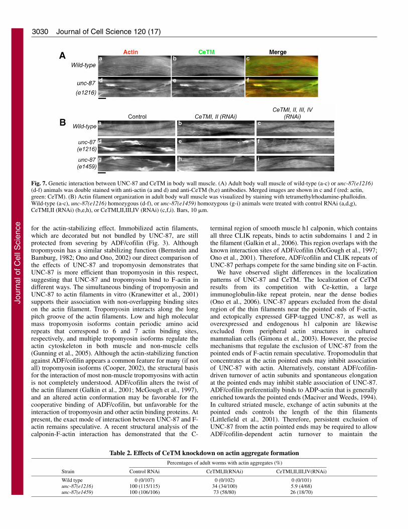
Both UNC-87 and CeTM stabilize actin filaments in vitroby inhibiting depolymerization and severing by UNC-60B. Ifboth UNC-87 and CeTM also play similar roles in actinstabilization in vivo, knockdown of both UNC-87 and CeTMis expected to enhance actin disorganization, as a result of theincreased accessibility of actin filament interaction sites forUNC-60B and a concomitantly increased ability to sever anddepolymerize the filaments. However, if UNC-87 and CeTMhave different in vivo functions, double knockdown of UNC-87 and CeTM may cause other phenotypes such as suppressionof actin disorganization. In unc-87 mutants, CeTM and actinco-localized in the aggregates in a nearly identical manner (Fig.7Ad-f). In a wild-type background, RNA interference (RNAi)of two long CeTM isoforms (CeTMI and CeTMII) or two longand two short CeTM isoforms (CeTMIII and CeTMIV)induced a wavy appearance of the actin filaments (Fig. 7Bb,c)(Ono and Ono, 2002). By contrast, in the unc-87(e1216 ore1459) mutant background, RNAi of CeTM suppressed theformation of actin aggregates (Fig. 7Be,f,h,i), and thecharacteristic wavy actin bundles were diminished (Fig.7Be,f,h,i). However, the defined striated pattern of actinfilaments was not completely restored in both unc-87 mutants(Fig. 7Be,f,h,i). RNAi of four CeTM isoforms caused astronger phenotypic suppression than RNAi of CeTMI andCeTMII, and unc-87(e1217) (a moderate loss-of-functionallele) was more strongly suppressed than unc-87(e1459) (astrong loss-of-function allele) (Table 2). By contrast, RNAi of
CeTM reduced worm motility of unc-87 mutants instead ofsuppressing the phenotype (our unpublished data), suggestingthat reduced contractility contributed to suppressing the actindisorganization in unc-87 mutants (see Discussion). Theseresults suggest that UNC-87 and CeTM do not simply functionin a similar manner, and that an intricate balance of theactivities of UNC-87, CeTM and UNC-60B is important foractin filament organization and proper localization of theseproteins.
DiscussionIn this study, we demonstrate that the calponin-repeat proteinUNC-87 is an effective stabilizer of the actin cytoskeleton andprotects actin filaments against ADF/cofilin-dependent actinfilament depolymerization and severing. In vitro, the actin-stabilizing effect of UNC-87 is more pronounced and persistentthan that of tropomyosin. In adult C. elegans muscle, thefunctions of UNC-87 and ADF/cofilin (UNC-60B) appearantagonistic with respect to regulating actin organization,which supports their opposing functions on actin filamentstability. However, the genetic interaction between UNC-87and tropomyosin (CeTM) reveal their distinct functions in theregulation of actin organization.
UNC-87 stabilizes actin filaments by competitivelyinhibiting the binding of ADF/cofilin to the filaments.Although UNC-87 bundles actin filaments in vitro(Kranewitter et al., 2001), this bundling activity is not required
Fig. 6. Genetic interaction between UNC-87 and UNC-60B in adult body wall muscle. (A) Adult body wall muscle of wild-type (a-c,g-i), unc-87(e1216) (d-f), or unc-60B(r398) (j-l) animals was double stained with anti-actin (a,d,g,j) and anti-UNC-60B (b,e) or anti-UNC-87 (h,k)antibodies. Merged images are shown in c,f,i,l (red: actin, green: UNC-60B, or UNC-87). (B) Actin filament organization in adult body wallmuscle was visualized by staining with tetramethylrhodamine-phalloidin. Micrographs of wild-type (a), +/+; unc-60B(r398) (b), unc-87(e1216)/+; +/+ (c), unc-87(e1216)/+; unc-60B(r398) (d), unc-87(e1459)/+; +/+ (e), and unc-87(e1459)/+; unc-60B(r398) (f) muscle. Bars, 10�m.
Jour
nal o
f Cel
l Sci
ence
3030
for the actin-stabilizing effect. Immobilized actin filaments,which are decorated but not bundled by UNC-87, are stillprotected from severing by ADF/cofilin (Fig. 3). Althoughtropomyosin has a similar stabilizing function (Bernstein andBamburg, 1982; Ono and Ono, 2002) our direct comparison ofthe effects of UNC-87 and tropomyosin demonstrates thatUNC-87 is more efficient than tropomyosin in this respect,suggesting that UNC-87 and tropomyosin bind to F-actin indifferent ways. The simultaneous binding of tropomyosin andUNC-87 to actin filaments in vitro (Kranewitter et al., 2001)supports their association with non-overlapping binding siteson the actin filament. Tropomyosin interacts along the longpitch groove of the actin filaments. Low and high molecularmass tropomyosin isoforms contain periodic amino acidrepeats that correspond to 6 and 7 actin binding sites,respectively, and multiple tropomyosin isoforms regulate theactin cytoskeleton in both muscle and non-muscle cells(Gunning et al., 2005). Although the actin-stabilizing functionagainst ADF/cofilin appears a common feature for many (if notall) tropomyosin isoforms (Cooper, 2002), the structural basisfor the interaction of most non-muscle tropomyosins with actinis not completely understood. ADF/cofilin alters the twist ofthe actin filament (Galkin et al., 2001; McGough et al., 1997),and an altered actin conformation may be favorable for thecooperative binding of ADF/cofilin, but unfavorable for theinteraction of tropomyosin and other actin binding proteins. Atpresent, the exact mode of interaction between UNC-87 and F-actin remains speculative. A recent structural analysis of thecalponin-F-actin interaction has demonstrated that the C-
Journal of Cell Science 120 (17)
terminal region of smooth muscle h1 calponin, which containsall three CLIK repeats, binds to actin subdomains 1 and 2 inthe filament (Galkin et al., 2006). This region overlaps with theknown interaction sites of ADF/cofilin (McGough et al., 1997;Ono et al., 2001). Therefore, ADF/cofilin and CLIK repeats ofUNC-87 perhaps compete for the same binding site on F-actin.
We have observed slight differences in the localizationpatterns of UNC-87 and CeTM. The localization of CeTMresults from its competition with Ce-kettin, a largeimmunoglobulin-like repeat protein, near the dense bodies(Ono et al., 2006). UNC-87 appears excluded from the distalregion of the thin filaments near the pointed ends of F-actin,and ectopically expressed GFP-tagged UNC-87, as well asoverexpressed and endogenous h1 calponin are likewiseexcluded from peripheral actin structures in culturedmammalian cells (Gimona et al., 2003). However, the precisemechanisms that regulate the exclusion of UNC-87 from thepointed ends of F-actin remain speculative. Tropomodulin thatconcentrates at the actin pointed ends may inhibit associationof UNC-87 with actin. Alternatively, constant ADF/cofilin-driven turnover of actin subunits and spontaneous elongationat the pointed ends may inhibit stable association of UNC-87.ADF/cofilin preferentially binds to ADP-actin that is generallyenriched towards the pointed ends (Maciver and Weeds, 1994).In cultured striated muscle, exchange of actin subunits at thepointed ends controls the length of the thin filaments(Littlefield et al., 2001). Therefore, persistent exclusion ofUNC-87 from the actin pointed ends may be required to allowADF/cofilin-dependent actin turnover to maintain the
Fig. 7. Genetic interaction between UNC-87 and CeTM in body wall muscle. (A) Adult body wall muscle of wild-type (a-c) or unc-87(e1216)(d-f) animals was double stained with anti-actin (a and d) and anti-CeTM (b,e) antibodies. Merged images are shown in c and f (red: actin,green: CeTM). (B) Actin filament organization in adult body wall muscle was visualized by staining with tetramethylrhodamine-phalloidin.Wild-type (a-c), unc-87(e1216) homozygous (d-f), or unc-87(e1459) homozygous (g-i) animals were treated with control RNAi (a,d,g),CeTMI,II (RNAi) (b,e,h), or CeTMI,II,III,IV (RNAi) (c,f,i). Bars, 10 �m.
Table 2. Effects of CeTM knockdown on actin aggregate formationPercentages of adult worms with actin aggregates (%)
Strain Control RNAi CeTMI,II(RNAi) CeTMI,II,III,IV(RNAi)
Wild type 0 (0/107) 0 (0/102) 0 (0/101)unc-87(e1216) 100 (115/115) 34 (34/100) 5.9 (4/68)unc-87(e1459) 100 (106/106) 73 (58/80) 26 (18/70)
Jour
nal o
f Cel
l Sci
ence
3031Actin stabilization by UNC-87
myofibril structure, whereas UNC-87 may be required forprolonged stabilization of F-actin at more proximal regions.This hypothesis is consistent with the phenotypes of the unc-87 mutants alone or in combination with the unc-60B mutation(Fig. 6). In the unc-87 mutants, actin filament stability isreduced and the filaments are more prone to rapid disassemblyby active UNC-60B, which may cause the disorganization ofactin filaments. When the activity of UNC-60B is low, actindisassembly is less frequent, and disorganization of thefilaments can be suppressed.
Although the adult muscle phenotypes of the unc-87 singleand unc-87;unc-60B double mutants are consistent with theproposed antagonistic roles of UNC-87 and UNC-60B on actinremodeling, the embryonic phenotypes were not. The unc-87;unc-60B double homozygous mutants were lethal at lateembryonic stages. By contrast, unc-87 or unc-60B singlemutants are homozygous viable (Goetinck and Waterston,1994b; Ono et al., 1999). Such enhancement in the phenotypecannot be explained by a simple antagonistic mechanism, butrather suggests additional cooperative functions between UNC-87 and UNC-60B. Indeed, under certain conditions, ADF/cofilinitself displays opposing functions: it nucleates actin assemblyand promotes polymerization (Andrianantoandro and Pollard,2006; Chen et al., 2004; Kudryashov et al., 2006). It isconceivable that, during embryonic stages when the actincytoskeleton is reorganized to form myofibrils, both UNC-60Band UNC-87 act to increase the amount of F-actin. Actinfilaments in the unc-87;unc-60B double mutant embryos weresimilarly disorganized to those in the unc-60B mutants, andcomparison of the actin phenotypes did not provide furtherinsight into the functional relationship between UNC-60B andUNC-87. Nonetheless, our results demonstrate that UNC-60Band UNC-87 play different yet profound roles in actinorganization, and further dissection of the functions of UNC-60B (severing/depolymerization and nucleation) and UNC-87 atdifferent developmental stages is required to clarify this issue.
The cooperative stabilization of F-actin by UNC-87 andtropomyosin antagonizes the activities of ADF/cofilin in vitro,albeit our results on the genetic interaction between UNC-87and CeTM also demonstrate distinct functions in vivo.Combination of unc-87 mutation and CeTM RNAi suppressesthe disorganization of actin filaments in body wall musclerather than enhancing the phenotype. In part, this can be dueto the known effects of muscle contractility on actinorganization, since active muscle contraction can damagemyofibrils (Friden and Lieber, 1992). Indeed, disorganizedactin filaments in unc-87 mutants is suppressed by a myosinmutation that reduces contraction frequency and magnitude(Goetinck and Waterston, 1994b), further suggesting that, inaddition to the action of UNC-60B, actomyosin contractilityalso contributes to the actin phenotype in unc-87 mutants.Knockdown of CeTM reduces the contractile activity of bodywall muscle (Ono and Ono, 2002; Yu and Ono, 2006), thussuppressing the phenotype of unc-87 mutants. However, theactin phenotype in unc-60B mutants is only weakly suppressedby reduced myosin activity (Yu and Ono, 2006), suggestingthat the mechanisms of actin disorganization in unc-87 andunc-60B mutants are different. By contrast, proper actomyosininteraction is required for the organized myofibril assembly(De Deyne, 2000; Kagawa et al., 2006). Currently, the role ofmyosin in actin dynamics is not completely understood, but
recent studies in dividing cells suggest that myosin II enhancesactin dynamics (Guha et al., 2005; Murthy and Wadsworth,2005). To better understand the function of UNC-87, we arecurrently investigating the potential role of UNC-87 inactomyosin contraction. Preliminary results indicate aninhibitory effect of UNC-87 on actomyosin contractility (ourunpublished data), suggesting a significant involvement in theregulation of actin organization. Notably, a similar function isattributed to calponin family members in mammalian muscleand non-muscle cells, and this function requires the interactionof calponin with the actin filaments via the CLIK repeats. Thethree CLIK repeats in h1 calponin reduce Arp2/3-mediatedactin polymerization during podosome formation (Gimona etal., 2003), inhibit cell motility and increase resistance topharmacological inhibitors of actin polymerization (Danningerand Gimona, 2000).
Taken together, our study provides evidence for theinhibition of ADF/cofilin-dependent actin dynamics by CLIKrepeats, and provides a first mechanistic insight into a possiblyconserved function of CLIK repeats in the stabilization of actinfilaments that has been observed in yeast (Goodman et al.,2003) and cultured mammalian cells (Gimona et al., 2003).Genetic analysis in budding yeast revealed that mutation of thecalponin homolog scp1 induces cooperative effects with thesac6 (fimbrin) mutation, but not with cofilin or tropomyosinmutation (Goodman et al., 2003). Notably, UNC-87 is a uniqueprotein that contains seven CLIK repeats, whereas, in yeastSCP1 and mammalian SM22/transgelin, only one repeat ispresent. A deletion analysis of UNC-87 has demonstrated thatmultiple CLIK repeats are required for strong F-actin bindingand bundling (Kranewitter et al., 2001), and that the effects onactin turnover directly correlate with the number of CLIKrepeats (Lener et al., 2004). CLIK repeats are intrinsicallyunfolded protein domains that adopt a stable conformationonly upon contact with actin filaments. Here, we describe theformation of two independent actin binding sites of differentaffinities by the seven CLIK repeats of UNC-87. Actin filamentstabilization appears independent of the bundling activity, andUNC-87 may use both filament interaction sites to bind thesame filament with different affinities and potentially also totwo different regions. It should be noted that also the prominentactin cross-linking protein �-actinin was shown recently toemploy both actin-binding sites for lateral binding to actinfilament (Hampton et al., 2007).
Although, at present, no calponin-like protein has beenidentified in vertebrate striated muscle, several actin-stabilizingproteins including nebulin (McElhinny et al., 2005), Xin(Pacholsky et al., 2004) and myotilin (Salmikangas et al., 2003)also have repetitive actin-binding motifs. By contrast, C. elegansdoes not have nebulin or Xin, but Ce-kettin (Ono et al., 2006)has immunoglobulin-like repeats and shows some similarity tomyotilin. Thus, UNC-87 might be functionally related to theseactin stabilizing proteins with repetitive actin-binding motifs.Importantly, mutations in nebulin and myotilin are associatedwith nemaline myopathy (Clarkson et al., 2004) and limb girdlemuscular dystrophy (Olive et al., 2005), respectively, suggestingthat impaired actin stabilizing activity may cause these inheritedmuscle diseases. Nebulin deficiency causes muscle defects inmice (Bang et al., 2006; Witt et al., 2006), whereas myotilin-deficient mice show no abnormal muscle phenotype (Moza etal., 2007). Thus, these actin-stabilizing proteins may have
Jour
nal o
f Cel
l Sci
ence
3032 Journal of Cell Science 120 (17)
different roles in specialized vertebrate muscles. Future studiesshould provide further insight into the spatial regulation of actindynamics and stability in vivo employing the C. elegans modelas a useful system to study the functions of actin-stabilizingproteins in striated muscle.
Materials and MethodsNematode strainsWild-type C. elegans strain N2 was obtained from the Caenorhabditis GeneticsCenter (Minneapolis, MN). unc-87(e1216) and unc-87(e1459) (Goetinck andWaterston, 1994a) were provided by Pamela Hoppe (Western Michigan University,Kalamazoo, MI) and Robert Waterston (University of Washington, Seattle, WA).unc-60B(r398) (McKim et al., 1988) was originally provided by David Baillie(Simon Fraser University, Burnaby, Canada), and a further outcrossed strain (Yuand Ono, 2006) was used. Double mutant strains, unc-87(e1216); unc-60B(r398)and unc-87(e1459); unc-60B(r398) were generated by standard crosses. However,the double mutants were homozygous lethal. Therefore, the strains were maintainedas unc-87/+ I; unc-60B/unc-60B V, where wild-type chromosome I was marked bydpy-5, and heterozygosity and homozygosity of the mutant alleles was confirmedby sequencing genomic DNA.
ProteinsC. elegans actin (Ce-actin) (Ono, 1999) and C. elegans tropomycin (CeTM) (Onoand Ono, 2002) were purified from wild-type strain N2 as described. Rabbit muscleactin was purified as described previously (Pardee and Spudich, 1982).Recombinant UNC-60B (Ono and Benian, 1998) and UNC-87 (Kranewitter et al.,2001) were expressed in E. coli and purified as described.
Assays for F-actin binding and bundling by pelletingCo-pelleting assays of rabbit muscle actin or Ce-actin with UNC-60B, UNC-87, andCeTM were performed as described previously in a buffer containing 0.1 M KCl,2 mM MgCl2, 20 mM Hepes-NaOH, 1 mM DTT, pH 7.5 (Ono and Ono, 2002).Ultracentrifugation was performed in a Beckman TLA100 rotor at 80,000 rpm(285,000 g) for 20 minutes at 20°C. To examine actin-bundling, the mixtures wereassembled in the same manner as the co-pelleting assays, and centrifuged at 18,000g for 10 minutes. The amounts of proteins that were contained in the supernatantsand pellets were analyzed by SDS-PAGE and densitometry as described previously.Scatchard analysis was performed with SigmaPlot 2000 (Systat Software), anddissociation constants were calculated by linear regression of the data.
Direct observation of actin filament severing and bundlingObservation of actin filament severing by fluorescence microscopy was performedas described previously (Ono et al., 2004) with slight modifications. Briefly, AlexaFluor 488 and biotin-labeled actin were attached to a perfusion chamber using anti-biotin antibody (Molecular Probes). After washing twice with wash buffer (50 mMKCl, 5 mM EGTA, 2 mM MgCl2, 1 mM ATP, 1 mM DTT, 0.5 mg/ml bovine serumalbumin and 20 mM Hepes-NaOH, pH 7.5), UNC-87 and/or CeTM in wash bufferwere perfused and incubated for 5 minutes (first perfusion). After washing twicewith anti-bleaching buffer (wash buffer plus 100 mM DTT, 0.036 mg/ml catalase,0.2 mg/ml glucose oxidase, 6 mg/ml glucose), UNC-60B in anti-bleaching bufferwas perfused into the chamber and incubated for 3 minutes (second perfusion).Micrographs of the fluorescent actin filaments were taken after the first and secondperfusions in the same field.
To examine actin bundling, 1 �M Alexa Fluor 488 and biotin-labeled actin wasincubated with or without 1 �M UNC-60B in wash buffer for 3 minutes, and thenincubated with or without 0.4 �M UNC-87 in wash buffer for 3 minutes at roomtemperature. The mixtures were diluted tenfold with wash buffer and the filamentswere attached to perfusion chambers using anti-biotin antibody. After washing twicewith anti-bleaching buffer, micrographs were taken.
Monitoring kinetics of actin turnover by nucleotide exchangeThe kinetics of actin turnover was monitored as described previously (Mohri andOno, 2003) by measuring exchange rate of actin-bound nucleotide.
Quantitative analysis of the thin filament proteins by westernblotNematode thin filaments were isolated as described previously (Ono and Ono,2002). Amounts of actin and UNC-87 were quantified by western blot with mousemonoclonal anti-actin (C4) and guinea pig anti-UNC-87 antibodies using purifiedactin and UNC-87 as standards, respectively. Densitometric quantification wasperformed in ranges where band intensity of the standard proteins was linearlycorrelated with their amounts.
Fluorescence microscopyImunofluorescent staining of adult nematodes was performed as describedpreviously (Finney and Ruvkun, 1990). Primary antibodies used were mouse anti-
actin monoclonal (C4, MP Biomedicals), rabbit anti-UNC-60B (Ono et al., 1999),guinea pig anti-CeTM (Ono and Ono, 2002), rabbit anti-UNC-87 (Goetinck andWaterston, 1994a) (provided by P. Hoppe and R. Waterston), and guinea pig anti-UNC-87 antibodies. Guinea pig anti-UNC-87 was prepared using purified UNC-87as immunogen at Quality Controlled Biochemicals (Hopkinton, MA). Secondaryantibodies were Alexa Fluor 488-goat anti-guinea pig IgG (Invitrogen), Cy3-donkeyanti-rabbit IgG and anti-mouse (Jackson ImmunoResearch). Staining of adultworms using tetramethylrhodamine-phalloidin was performed as describedelsewhere (Ono, 2001).
Samples were observed by epifluorescence using a Nikon Eclipse TE2000inverted microscope with a CFI Plan Fluor ELWD 40� (dry, NA 0.60) or a CFIPlan Apo 60� (oil, NA 1.40) objective. Images were captured by a SPOT RTMonochrome CCD camera (Diagnostic Instruments) and processed by the IPLabimaging software (BD Biosciences) and Adobe Photoshop 6.0.
RNA interference experimentsNematodes were treated with RNAi for CeTM by feeding them with E. coliexpressing double stranded RNA as described previously (Ono and Ono, 2002). L4larvae were treated with RNAi, and phenotypes were characterized in their F1generation.
We thank P. Hoppe and R. Waterston for antibody and worm strains,D. Baillie for worm strains, and J. H. Woo for technical support. Someworm strains were provided by the Caenorhabditis Genetics Center,which is funded by the NIH NCRR. This work was supported by aMarie Curie Excellence Grant (MEXT-CT-2003-002573) of theEuropean Union to M.G. and an NIH grant (R01 AR48615) to S.O.
ReferencesAndrianantoandro, E. and Pollard, T. D. (2006). Mechanism of actin filament turnover
by severing and nucleation at different concentrations of ADF/cofilin. Mol. Cell 24,13-23.
Bang, M. L., Li, X., Littlefield, R., Bremner, S., Thor, A., Knowlton, K. U., Lieber,R. L. and Chen, J. (2006). Nebulin-deficient mice exhibit shorter thin filament lengthsand reduced contractile function in skeletal muscle. J. Cell Biol. 173, 905-916.
Bernstein, B. W. and Bamburg, J. R. (1982). Tropomyosin binding to F-actin protectsthe F-actin from disassembly by brain actin-depolymerizing factor (ADF). Cell Motil.2, 1-8.
Carlier, M. F., Laurent, V., Santolini, J., Melki, R., Didry, D., Xia, G. X., Hong, Y.,Chua, N. H. and Pantaloni, D. (1997). Actin depolymerizing factor (ADF/cofilin)enhances the rate of filament turnover: implication in actin-based motility. J. Cell Biol.136, 1307-1322.
Chen, H., Bernstein, B. W., Sneider, J. M., Boyle, J. A., Minamide, L. S. andBamburg, J. R. (2004). In vitro activity differences between proteins of theADF/cofilin family define two distinct subgroups. Biochemistry 43, 7127-7142.
Clarkson, E., Costa, C. F. and Machesky, L. M. (2004). Congenital myopathies:diseases of the actin cytoskeleton. J. Pathol. 204, 407-417.
Cooper, J. (2002). Actin dynamics: tropomyosin provides stability. Curr. Biol. 12, R523-R525.
Danninger, C. and Gimona, M. (2000). Live dynamics of GFP-calponin: isoform-specific modulation of the actin cytoskeleton and autoregulation by C-terminalsequences. J. Cell Sci. 113, 3725-3736.
De Deyne, P. G. (2000). Formation of sarcomeres in developing myotubes: role ofmechanical stretch and contractile activation. Am. J. Physiol. Cell Physiol. 279, C1801-C1811.
DesMarais, V., Ichetovkin, I., Condeelis, J. and Hitchcock-DeGregori, S. E. (2002).Spatial regulation of actin dynamics: a tropomyosin-free, actin-rich compartment at theleading edge. J. Cell Sci. 115, 4649-4660.
Finney, M. and Ruvkun, G. (1990). The unc-86 gene product couples cell lineage andcell identity in C. elegans. Cell 63, 895-905.
Friden, J. and Lieber, R. L. (1992). Structural and mechanical basis of exercise-inducedmuscle injury. Med. Sci. Sports Exerc. 24, 521-530.
Galkin, V. E., Orlova, A., Lukoyanova, N., Wriggers, W. and Egelman, E. H. (2001).Actin depolymerizing factor stabilizes an existing state of F-actin and can change thetilt of F-actin subunits. J. Cell Biol. 153, 75-86.
Galkin, V. E., Orlova, A., Fattoum, A., Walsh, M. P. and Egelman, E. H. (2006). TheCH-domain of calponin does not determine the modes of calponin binding to F-actin.J. Mol. Biol. 359, 478-485.
Ghosh, M., Song, X., Mouneimne, G., Sidani, M., Lawrence, D. S. and Condeelis, J.S. (2004). Cofilin promotes actin polymerization and defines the direction of cellmotility. Science 304, 743-746.
Gimona, M. and Mital, R. (1998). The single CH domain of calponin is neither sufficientnor necessary for F-actin binding. J. Cell Sci. 111, 1813-1821.
Gimona, M., Djinovic-Carugo, K., Kranewitter, W. J. and Winder, S. J. (2002).Functional plasticity of CH domains. FEBS Lett. 513, 98-106.
Gimona, M., Kaverina, I., Resch, G. P., Vignal, E. and Burgstaller, G. (2003).Calponin repeats regulate actin filament stability and formation of podosomes insmooth muscle cells. Mol. Biol. Cell 14, 2482-2491.
Goetinck, S. and Waterston, R. H. (1994a). The Caenorhabditis elegans muscle-
Jour
nal o
f Cel
l Sci
ence

3033Actin stabilization by UNC-87
affecting gene unc-87 encodes a novel thin filament-associated protein. J. Cell Biol.127, 79-93.
Goetinck, S. and Waterston, R. H. (1994b). The Caenorhabditis elegans UNC-87protein is essential for maintenance, but not assembly, of bodywall muscle. J. Cell Biol.127, 71-78.
Goley, E. D. and Welch, M. D. (2006). The ARP2/3 complex: an actin nucleator comesof age. Nat. Rev. Mol. Cell Biol. 7, 713-726.
Goodman, A., Goode, B. L., Matsudaira, P. and Fink, G. R. (2003). TheSaccharomyces cerevisiae calponin/transgelin homolog Scp1 functions with fimbrin toregulate stability and organization of the actin cytoskeleton. Mol. Biol. Cell 14, 2617-2629.
Guha, M., Zhou, M. and Wang, Y. L. (2005). Cortical actin turnover during cytokinesisrequires myosin II. Curr. Biol. 15, 732-736.
Gungabissoon, R. A., Khan, S., Hussey, P. J. and Maciver, S. K. (2001). Interaction ofelongation factor 1� from Zea mays (ZmEF-1alpha) with F-actin and interplay withthe maize actin severing protein, ZmADF3. Cell Motil. Cytoskeleton 49, 104-111.
Gunning, P. W., Schevzov, G., Kee, A. J. and Hardeman, E. C. (2005). Tropomyosinisoforms: divining rods for actin cytoskeleton function. Trends Cell Biol. 15, 333-341.
Gupton, S. L., Anderson, K. L., Kole, T. P., Fischer, R. S., Ponti, A., Hitchcock-DeGregori, S. E., Danuser, G., Fowler, V. M., Wirtz, D., Hanein, D. et al. (2005).Cell migration without a lamellipodium: translation of actin dynamics into cellmovement mediated by tropomyosin. J. Cell Biol. 168, 619-631.
Hampton, C. M., Taylor, D. W. and Taylor, K. A. (2007). Novel structures for alpha-Actinin:F-Actin interactions and their implications for actin-membrane attachment andtension sensing in the cytoskeleton. J. Mol. Biol. 368, 92-104.
Higgs, H. N. (2005). Formin proteins: a domain-based approach. Trends Biochem. Sci.30, 342-353.
Hotulainen, P., Paunola, E., Vartiainen, M. K. and Lappalainen, P. (2005). Actin-depolymerizing factor and cofilin-1 play overlapping roles in promoting rapid F-actindepolymerization in mammalian nonmuscle cells. Mol. Biol. Cell 16, 649-664.
Ichetovkin, I., Han, J., Pang, K. M., Knecht, D. A. and Condeelis, J. S. (2000). Actinfilaments are severed by both native and recombinant Dictyostelium cofilin but todifferent extents. Cell Motil. Cytoskeleton 45, 293-306.
Iwasa, J. H. and Mullins, R. D. (2007). Spatial and temporal relationships between actin-filament nucleation, capping, and disassembly. Curr. Biol. 17, 395-406.
Kagawa, H., Sugimoto, K., Matsumoto, H., Inoue, T., Imadzu, H., Takuwa, K. andSakube, Y. (1995). Genome structure, mapping and expression of the tropomyosingene tmy-1 of Caenorhabditis elegans. J. Mol. Biol. 251, 603-613.
Kagawa, M., Sato, N. and Obinata, T. (2006). Effects of BTS (N-benzyl-p-toluenesulphonamide), an Inhibitor for myosin-actin Interaction, on myofibrillogenesis inskeletal muscle cells in culture. Zool. Sci. 23, 969-975.
Kiuchi, T., Ohashi, K., Kurita, S. and Mizuno, K. (2007). Cofilin promotes stimulus-induced lamellipodium formation by generating an abundant supply of actinmonomers. J. Cell Biol. 177, 465-476.
Kranewitter, W. J., Ylanne, J. and Gimona, M. (2001). UNC-87 is an actin-bundlingprotein. J. Biol. Chem. 276, 6306-6312.
Kudryashov, D. S., Galkin, V. E., Orlova, A., Phan, M., Egelman, E. H. and Reisler,E. (2006). Cofilin cross-bridges adjacent actin protomers and replaces part of thelongitudinal F-actin interface. J. Mol. Biol. 358, 785-797.
Lappalainen, P. and Drubin, D. G. (1997). Cofilin promotes rapid actin filamentturnover in vivo. Nature 388, 78-82.
Lener, T., Burgstaller, G. and Gimona, M. (2004). The role of calponin in the geneprofile of metastatic cells: inhibition of metastatic cell motility by multiple calponinrepeats. FEBS Lett. 556, 221-226.
Littlefield, R., Almenar-Queralt, A. and Fowler, V. M. (2001). Actin dynamics atpointed ends regulates thin filament length in striated muscle. Nat. Cell Biol. 3, 544-551.
Maciver, S. K. and Weeds, A. G. (1994). Actophorin preferentially binds monomericADP-actin over ATP-bound actin: consequences for cell locomotion. FEBS Lett. 347,251-256.
Maciver, S. K., Wachsstock, D. H., Schwarz, W. H. and Pollard, T. D. (1991a). Theactin filament severing protein actophorin promotes the formation of rigid bundles ofactin filaments crosslinked with alpha-actinin. J. Cell Biol. 115, 1621-1628.
Maciver, S. K., Zot, H. G. and Pollard, T. D. (1991b). Characterization of actin filamentsevering by actophorin from Acanthamoeba castellanii. J. Cell Biol. 115, 1611-1620.
Maciver, S. K., Pope, B. J., Whytock, S. and Weeds, A. G. (1998). The effect of twoactin depolymerizing factors (ADF/cofilins) on actin filament turnover: pH sensitivityof F-actin binding by human ADF, but not of Acanthamoeba actophorin. Eur. J.Biochem. 256, 388-397.
McElhinny, A. S., Schwach, C., Valichnac, M., Mount-Patrick, S. and Gregorio, C.C. (2005). Nebulin regulates the assembly and lengths of the thin filaments in striatedmuscle. J. Cell Biol. 170, 947-957.
McGough, A. (1998). F-actin-binding proteins. Curr. Opin. Struct. Biol. 8, 166-176.McGough, A., Pope, B., Chiu, W. and Weeds, A. (1997). Cofilin changes the twist of
F-actin: implications for actin filament dynamics and cellular function. J. Cell Biol.138, 771-781.
McKim, K. S., Heschl, M. F., Rosenbluth, R. E. and Baillie, D. L. (1988).Genetic organization of the unc-60 region in Caenorhabditis elegans. Genetics 118,49-59.
Moerman, D. G. and Fire, A. (1997). Muscle: Structure, function, and development. InC. elegans II (ed. D. L. Riddle, T. Blumenthal, B. J. Meyer and J. R. Priess), pp. 417-470. Plainview, NY: Cold Spring Harbor Laboratory Press.
Mohri, K. and Ono, S. (2003). Actin filament disassembling activity of Caenorhabditiselegans actin-interacting protein 1 (UNC-78) is dependent on filament binding by aspecific ADF/cofilin isoform. J. Cell Sci. 116, 4107-4118.
Mohri, K., Ono, K., Yu, R., Yamashiro, S. and Ono, S. (2006). Enhancement of actin-depolymerizing factor/cofilin-dependent actin disassembly by actin-interacting protein1 is required for organized actin filament assembly in the Caenorhabditis elegans bodywall muscle. Mol. Biol. Cell 17, 2190-2199.
Mouneimne, G., DesMarais, V., Sidani, M., Scemes, E., Wang, W., Song, X., Eddy,R. and Condeelis, J. (2006). Spatial and temporal control of cofilin activity is requiredfor directional sensing during chemotaxis. Curr. Biol. 16, 2193-2205.
Moza, M., Mologni, L., Trokovic, R., Faulkner, G., Partanen, J. and Carpen, O.(2007). Targeted deletion of the muscular dystrophy gene myotilin does not perturbmuscle structure or function in mice. Mol. Cell. Biol. 27, 244-252.
Murthy, K. and Wadsworth, P. (2005). Myosin-II-dependent localization and dynamicsof F-actin during cytokinesis. Curr. Biol. 15, 724-731.
Nishita, M., Tomizawa, C., Yamamoto, M., Horita, Y., Ohashi, K. and Mizuno, K.(2005). Spatial and temporal regulation of cofilin activity by LIM kinase and Slingshotis critical for directional cell migration. J. Cell Biol. 171, 349-359.
Olive, M., Goldfarb, L. G., Shatunov, A., Fischer, D. and Ferrer, I. (2005).Myotilinopathy: refining the clinical and myopathological phenotype. Brain 128, 2315-2326.
Ono, K., Parast, M., Alberico, C., Benian, G. M. and Ono, S. (2003). Specificrequirement for two ADF/cofilin isoforms in distinct actin-dependent processes inCaenorhabditis elegans. J. Cell Sci. 116, 2073-2085.
Ono, K., Yu, R., Mohri, K. and Ono, S. (2006). Caenorhabditis elegans kettin, a largeimmunoglobulin-like repeat protein, binds to filamentous actin and providesmechanical stability to the contractile apparatuses in body wall muscle. Mol. Biol. Cell17, 2722-2734.
Ono, S. (1999). Purification and biochemical characterization of actin fromCaenorhabditis elegans: its difference from rabbit muscle actin in the interaction withnematode ADF/cofilin. Cell Motil. Cytoskeleton 43, 128-136.
Ono, S. (2001). The Caenorhabditis elegans unc-78 gene encodes a homologue of actin-interacting protein 1 required for organized assembly of muscle actin filaments. J. CellBiol. 152, 1313-1319.
Ono, S. (2007). Mechanism of depolymerization and severing of actin filaments and itssignificance in cytoskeletal dynamics. Int. Rev. Cytol. 258, 1-82.
Ono, S. and Benian, G. M. (1998). Two Caenorhabditis elegans actin depolymerizingfactor/cofilin proteins, encoded by the unc-60 gene, differentially regulate actinfilament dynamics. J. Biol. Chem. 273, 3778-3783.
Ono, S. and Ono, K. (2002). Tropomyosin inhibits ADF/cofilin-dependent actin filamentdynamics. J. Cell Biol. 156, 1065-1076.
Ono, S., Baillie, D. L. and Benian, G. M. (1999). UNC-60B, an ADF/cofilin familyprotein, is required for proper assembly of actin into myofibrils in Caenorhabditiselegans body wall muscle. J. Cell Biol. 145, 491-502.
Ono, S., McGough, A., Pope, B. J., Tolbert, V. T., Bui, A., Pohl, J., Benian, G. M.,Gernert, K. M. and Weeds, A. G. (2001). The C-terminal tail of UNC-60B(ADF/cofilin) is critical for maintaining its stable association with F-actin and isimplicated in the second actin-binding site. J. Biol. Chem. 276, 5952-5958.
Ono, S., Mohri, K. and Ono, K. (2004). Microscopic evidence that actin-interactingprotein 1 actively disassembles actin-depolymerizing factor/cofilin-bound actinfilaments. J. Biol. Chem. 279, 14207-14212.
Pacholsky, D., Vakeel, P., Himmel, M., Lowe, T., Stradal, T., Rottner, K., Furst, D.O. and van der Ven, P. F. (2004). Xin repeats define a novel actin-binding motif. J.Cell Sci. 117, 5257-5268.
Pardee, J. D. and Spudich, J. A. (1982). Purification of muscle actin. Methods Enzymol.85, 164-181.
Pavlov, D., Muhlrad, A., Cooper, J., Wear, M. and Reisler, E. (2007). Actin filamentsevering by cofilin. J. Mol. Biol. 365, 1350-1358.
Pollard, T. D. and Borisy, G. G. (2003). Cellular motility driven by assembly anddisassembly of actin filaments. Cell 112, 453-465.
Salmikangas, P., van der Ven, P. F., Lalowski, M., Taivainen, A., Zhao, F., Suila, H.,Schroder, R., Lappalainen, P., Furst, D. O. and Carpen, O. (2003). Myotilin, thelimb-girdle muscular dystrophy 1A (LGMD1A) protein, cross-links actin filaments andcontrols sarcomere assembly. Hum. Mol. Genet. 12, 189-203.
Witt, C. C., Burkart, C., Labeit, D., McNabb, M., Wu, Y., Granzier, H. and Labeit,S. (2006). Nebulin regulates thin filament length, contractility, and Z-disk structure invivo. EMBO J. 25, 3843-3855.
Yu, R. and Ono, S. (2006). Dual roles of tropomyosin as an F-actin stabilizer and aregulator of muscle contraction in Caenorhabditis elegans body wall muscle. CellMotil. Cytoskeleton 63, 659-672.
Jour
nal o
f Cel
l Sci
ence


![[ A ] SPIRITS ADF [ADF] VODKA - BASIC](https://img.pdfslide.us/doc/110x75/6169d8c211a7b741a34c063e/-a-spirits-adf-adf-vodka-basic.jpg)