Embed Size (px)
Citation preview

ADF/Cofilin Is Not Essential but Is CriticallyImportant for Actin Activities duringPhagocytosis in Tetrahymena thermophila
著者 Shiozaki Nanami, Nakano Kentaro, KushidaYasuharu, Noguchi Taro Q. P., Uyeda Taro Q.P., Wloga Dorota, Dave Drashti, VasudevanKrishna Kumar, Gaertig Jacek, Numata Osamu
journal orpublication title
Eukaryotic cell
volume 12number 8page range 1080-1086year 2013-08権利 (C) 2013, American Society for MicrobiologyURL http://hdl.handle.net/2241/121359
doi: 10.1128/EC.00074-13

ADF/cofilin is not essential but critically important for actin activities
during phagocytosis in Tetrahymena thermophila
Nanami Shiozakia, Kentaro Nakanoa, Yasuharu Kushidaa, Taro QP Noguchib, Taro QP
Uyedaa,b, Dorota Wlogac, Drashti Davec, d, Krishna Kumar Vasudevanc, Jacek Gaertigc,
and Osamu Numataa
aStructural Biosciences, Graduate School of Life and Environmental Sciences,
University of Tsukuba, 1-1-1 Tennohdai, Tsukuba, Ibaraki 305-8577, Japan
bBiomedical Research Institute, National Institute of Advanced Industrial Science and
Technology (AIST), 1-1-1 Higashi, Tsukuba, Ibaraki 305-8562, Japan
cDepartment of Cellular Biology, University of Georgia, Athens, GA 30602-2607, USA
dCurrent address:Nencki Institute for Experimental Biology, Warsaw, Poland
*Corresponding authors: Kentaro Nakano, [email protected]; Osamu Numata,
Running title: Tetrahymena ADF/cofilin

Abstract
ADF/cofilin is a highly conserved actin-modulating protein. Reorganization of the actin
cytoskeleton in vivo through severing and depolymerizing F-actin by this protein is
essential for various cellular events such as endocytosis, phagocytosis, cytokinesis and
cell migration. We show that in the ciliate Tetrahymena thermophila, the
ADF/cofilin-homologue, Adf73p, associates with actin on nascent food vacuoles.
Overexpression of Adf73p disrupted the proper localization of actin and inhibited the
formation of food vacuoles. In vitro, recombinant Adf73p promoted the
depolymerization of filaments made of T. thermophila actin, Act1p. Knockout cells
lacking ADF73 gene are viable but grow extremely slowly and have a severely
decreased rate of food vacuole formation. Knockout cells have abnormal aggregates of
actin in the cytoplasm. Surprisingly, unlike in animals and yeasts, in Tetrahymena,
ADF/cofilin is not required for cytokinesis. Thus, the Tetrahymena model shows
promise for future studies on the role of ADF/cofilin in vivo.
(146 words)
1

Introduction
Dynamic reorganization of the actin cytoskeleton is associated with membrane
deformations such as phagocytosis, endocytosis, ameboid movement, and cytokinesis in
eukaryotes. ADF (actin-depolymerizing factor)/cofilin (AC) is the principal
actin-modulating protein involved in these events, and its biochemical activities have
been well studied in animal, yeasts, and higher plants (1). By binding to F-actin,
canonical AC induces a twist in the arrangements of subunits (2), which produces
distortion at the boundary between twisted and non-twisted polymer regions (3, 4).
Consequently, F-actin is severed, and dissociation of monomeric actin (G-actin) is
accelerated from the pointed end of filaments (5). On the other hand, at higher
concentration, AC increases the rate of actin nucleation and induces abnormal actin
structures, called bars or rods, probably because AC binding bridges longitudinal
contacts between actin subunits (3, 6, 7). Thus, it has been considered that AC exerts
distinct effects on F-actin depending on the AC:actin stoichiometry. In addition, AC
binds preferentially to ADP-G-actin and inhibits nucleotide exchange to form an
assembly competent ATP-actin (8). This activity may be involved in maintaining the
G-actin pool cooperatively with other G-actin-binding proteins such as thymosin β4 and
2

profilin.
Protists also show dynamic reorganization of the actin cytoskeleton
accompanying unique cell behaviors but neither their fundamental molecular
mechanisms nor regulatory systems have been uncovered. One exceptional is the
malaria parasite, a lineage of apicomplexa, which expresses AC-like protein ADF1 with
unusual biochemical properties (9). ADF1 does not bind or sever F-actin but strongly
stimulates the nucleotide exchange on G-actin to promote rapid actin polymerization
(10). These unique properties of ADF1 could co-evolve with malaria-specific actin
molecule that is extremely unstable as a polymer as compared with conventional actin
of other eukaryotes (11). Their functional relationship probably enables the malaria
parasite to rapidly turn over F-actin in gliding motility during host invasion (9).
Ciliates form the phylogenic group alveorates, together with apicomplexa and
dinoflagellates, are highly diverged from the animal and yeast lineage of opistkonts (12).
Tetrahymena thermophila is well studied model ciliates with a sequenced genome (13).
This unicellular organism vigorously internalizes extracellular solutions and particles as
nutrients by forming food vacuoles (FVs) at the funnel-shaped oral apparatus located in
the anterior region of the cell cortex (14). Electron microscopic observations revealed
3

that FVs are formed along the deep fiber, a fibrous microtubule-rich structure that
protrudes from the bottom of the oral apparatus (14). After absorption of nutrients,
undigested materials are egested at the organelle called the cytoproct in the posterior
region of the cell (14). One of the Tetrahymena actin Act1p (15) and actin-bundling
proteins including fimbrin (16) and eEF-1A (15) all localize near the deep fiber and
nascent FVs. Moreover, strong accumulation of actin is seen around the cytoproct (17).
Consistently, actin-binding drugs block the formation of FVs in the oral apparatus (18)
and membrane retrieval after FV ejection in the cytoproct (17). Thus, the actin
cytoskeleton is possible to play an important role in the deformation of membranes
associated with the formation and ejection of FVs. Meanwhile, profilin, which promotes
the nucleotide exchange on G-actin and hence is generally involved in accelerating actin
turnover, is not visibly associated with the FV formation (19). Overall, the exact
function of actin dynamics accompanying the FV cycle remains poorly understood. In
addition, the Tetrahymena actin is highly divergent from conventional actin in the
preservation of the amino acid sequence and shows unique biochemical characteristics
(20).
After completion of the macronuclear genome project on T. thermophila (13), it
4

has been revealed that this organism has only one gene encoding an AC homolog,
ADF73. Previously, we purified the gene product, Adf73p (21). Biochemical studies
using rabbit muscle actin showed that Adf73p has F-actin-severing activity, but
unexpectedly, accelerates the nucleotide exchange on G-actin in a manner similar to
malaria parasite ADF1. Here we investigated the cellular function of Adf73p by
determining its localization and the consequences of overexpression and deletion. We
show that Adf73p is required for phagocytosis, likely via its role in the reorganization of
the actin cytoskeleton during FV formation by severing and depolymerizing F-actin in
Tetrahymena thermophila. Surprisingly, unlike animal cells, Tetrahymena cells do not
require AC for cytokinesis.
5

Materials and Methods
Cell culture
Wild-type Tetrahymena thermophila was cultured in NEFF (0.25% proteose peptone,
0.25% yeast extract, 0.55% D-(+)-glucose, 33 μM FeCl2) or SPP (1% proteose peptone,
0.2% glucose, 0.1% yeast extract, 0.003% EDTA-ferric sodium salt) (22) medium at
30°C. To study the effects of ADF73 deletion, wild-type and adf73KO strains were
cultured in MEPP (2% proteose peptone, 0.06% sodium citrate·2H2O, 0.027%
FeCl3·6H2O, 0.0003% CuSO4·5H2O, and 0.0001% folinic acid) (23) medium, which
promotes the growth of cells lacking the ability to form FVs.
Mitotic index was defined as a percentage of the number of cells undergoing
micro- and macro-nuclear division against the total number of cells. Nuclear DNA was
microscopically observed by staining cells with 1 µM 4’, 6-diamidino-2-phenylindole
(DAPI, Invitrogen) after fixation with 1% formaldehyde (Wako).
Antiserum
To produce anti-Adf73p antibodies, recombinant GST-Adf73p was purified and Adf73p
was separated from GST using thrombin (21). Adf73p was dialyzed in the
6

phosphate-buffered saline (PBS) overnight at 4°C, mixed with adjuvant and emulsified
with Freund's complete adjuvant (Wako Pure Chemical Industries, Ltd.) for the first
injection and Freund's incomplete adjuvant (Wako Pure Chemical Industries, Ltd.) for
the following injections by sonication (UD-201; Tomy). The antigen was
hypodermically injected into the back of a rabbit. One month after the first injection,
additional injections were performed once every 2 weeks. After third additional
injection, the antisera were collected and diluted 10 times with PBS and incubated with
a PVDF membrane bound with recombinant Adf73p overnight at 4°C. The membrane
was washed 3 times with 20 mM Tris-HCl (pH 7.5), 0.5 M NaCl, 0.05% Tween 20 and
soaked in 50 mM glycine-HCl (pH 2.5). Eluted affinity-purified antibodies were
neutralized with 0.5 M Tris (pH 8.8). A guinea pig antiserum against T. thermophila
actin has previously been described (15). An anti-GFP antibody was purchased from
Roche Diagnostics. The mouse anti-chicken α-tubulin antibody was purchased from
Calbiochem.
Immunoblotting
T. thermophila cells cultured in NEFF medium at 30°C were collected by centrifugation
7

for 3 min at 750 × g and washed twice with NKC solution (34 mM NaCl, 1 mM KCl, 1
mM CaCl2). The cells were suspended in NKC containing 1 mM ATP, 0.5 mM DTT, 1
mM PMSF, 10 μg/ml leupeptin, 5 μg/ml pepstatin A, 5 μg/ml TLCK and smashed with
sonicater (UD-201; Tomy). After adding one-third volume of the loading buffer (0.25
mM Tris (pH 6.8), 4% SDS, 40% glycerol, and 8% β-mercaptoethanol) followed by
incubation at 95°C for 5 min, the cell lysate was subjected to SDS-PAGE and
transferred onto a PVDF membrane. The membrane was blocked with Tris-buffered
saline (TBS; 20 mM Tris-HCl, 0.9% NaCl, pH 7.5) containing 0.5% Tween-20 (TTBS)
with 1% skim milk and incubated with 0.1% affinity purified anti-Adf73p antiserum for
2 h. After washing with TTBS with 1% skim milk, the membrane was incubated with
1% goat anti-rabbit IgG antibodies conjugated to alkaline phosphatase (Biosource
International) for 1 h. Immunoblots were developed using the BCIP/NBT phosphatase
substrate kit (Kirkegaard & Perry Laboratories, Inc.).
Fluorescence microscopy
T. thermophila cells were fixed with cold methanol (−20°C) for at least 30 min. After
washing cells with PBS 3 times, cells were permeabilized with PBS containing 0.1%
8

Triton X-100 for 1 min and then washed with PBS 3 times. Cells were incubated with
PBS containing 1% skim milk for 30 min, and then with 1% anti-actin antiserum and/or
25% affinity purified anti-Adf73p antiserum in PBS containing 1% skim milk for more
than 6 h at room temperature. After washing cells with PBS containing 1% skim milk 3
times, cells were incubated with 1% Rhodamine-labeled goat anti-guinea pig IgG
(Kirkegaard & Perry Laboratories, Inc.) and/or 1% FITC-conjugated goat anti-rabbit
IgG (Jackson Immune Research Laboratories, Inc.) in PBS containing 1% skim milk for
more than 6 h at room temperature. After washing with PBS 3 times, cells were
observed by a confocal laser scanning microscope, LSM510 (Carl Zeiss, Inc.).
Immunofluorescence staining with an anti-α-tubulin was performed by the method of
Wloga et al. (24).
Gene overexpression
A plasmid vector for overexpressing ADF73, pMTT1-GFP-ADF73, was obtained by
inserting ADF73 cDNA into the BamHI-HindIII site of pMTT1-GFP (25). CU522 cells
were transformed with pMTT1-GFP-ADF73 as described (26). Overexpression of
ADF73 was induced by the addition of 2.5 μg/ml CdCl2 to the medium for 3 h.
9
Gene knockout
To obtain a targeting plasmid for knockout of ADF73, 1.5 kb DNA fragments of 5’ and
3’ untranslated regions of the ADF73 gene were amplified by PCR and cloned into the
ApaI-SmaI site and PstI-SacI sites of pNeo4 (27), respectively. Log-phase growing
B2086 and CU428 cells in SPP were washed, starved in 10 mM Tris-HCl (pH 7.4) for
16–24 hours at 30°C, and mixed for conjugation. pNeo4-ADF73 was introduced into
micronuclei of the conjugating cells by biolistic transformation (28). Germ-line
transformants were selected by the addition of 10 μg/ml paromomycin, 15 μg/ml
6-methylpurine and 1 μg/ml CdCl2. Disrupted loci in the macronuclei were eliminated
by phenotypic assortment in the absence of paromomycin (29). Heterozygous
heterokaryons were mated with a "star" strain (A*III) to make homozygous
heterokaryon strains with a double disrupted loci in the micronucleus and wild-type
genes in the macronucleus by uniparental pronuclear transfer. Two heterokaryons were
mated to obtain homozygous homokaryon strains (adf73KO).
Isolation and fractionation of cell bodies and cilia
10

Cell bodies and cilia were isolated by the method of Ueno et al. (30).
Co-sedimentation assay
Purification of recombinant Adf73p and the co-sedimentation assay were as described
by Shiozaki et al. (21).
Swimming behavior of cells
To investigate swimming behavior, 20 µL cell culture was placed in a basic chamber
assembled from a slide glass with two slits of thin vinyl tape arranged in parallel as a
spacer and covered with cover glass. In this chamber, cells can swim around freely.
Swimming behavior was recorded at 15 Hz for 30 sec using CCD camera (DFK41AU02,
Argo) under the inverted microscopy (Olympus IX70) equipped with ×10 objective lens.
Swimming velocity was determined using Manual tracking-plugin tool on Image J
software. To record the ciliary beating of swimming cells at 1 kHz for 1 s, ×100
objective lens and high-speed CCD camera (Focuscope SV-200i, Photron limited) were
used. All observations were performed at room temperature.
11

Results
Adf73p is a component of F-actin structures associated with FVs
In T. thermophila, dynamic reorganization of the actin cytoskeleton accompanies the
course of phagocytosis (Fig. S1). AC is known as the major actin-modulating protein
(1). The macronuclear genome contains only a single gene putatively encoding an AC
homologue (13, 21). To investigate the cellular localization of its gene product Adf73p,
we prepared an antiserum against the recombinant protein. After affinity purification,
immunoblotting against the total lysate of T. thermophila cells showed that the serum
most strongly recognized a band of 15 kDa, corresponding to the estimated molecular
weight of Adf73p (Fig. 1A). This signal is increased by Adf73p overexpression and
diminished by ADF73 gene knockout (Figs. 2A, 4C). In immunofluorescence
microscopy with an affinity-purified anti-Adf73p serum, a strong signal colocalized
with F-actin protruding from the oral apparatus, actin dots around the nascent FV, and
the actin clump in the posterior region of the cell, which would be consistent with
localization to an older FV ejecting its contents from the cytoproct (Fig. 1B). These
signals were gone by ADF73 gene knockout (Fig. 4D). Furthermore, genetically tagged
GFP-Adf73p showed a similar pattern of fluorescence (Fig. 1C). Taken together, we
12

concluded that Adf73p localized to actin structures associated with FVs around the oral
apparatus and a cytoproct. Consistently, brief incubation with the actin inhibitor
Latrunculin B (Lat-B), which sequestered actin monomer from polymerization, greatly
reduced the signal of both Adf73p and actin (Fig. 1B). In addition, F-actin associated
with Adf73p in those structures of T. thermophila cells appeared to possess a high
turnover rates of polymerization and depolymerization because of its disappearance by a
short period of incubation with Lat-B (Fig. 1B).
Adf73p affects actin in vivo and in vitro
To evaluate the activity of Adf73p against the actin cytoskeleton in vivo, we
constructed a strain that over expresses Adf73p under the Cd2+-inducible MTT1
promoter. The levels of Adf73p were markedly elevated after the addition of Cd2+ (Fig.
2A). Overproduction of Adf73p diminished actin associated with FVs and frequently
induced thick actin bars, probably because of the excessive amount of Adf73p
associated with F-actin (Fig. 2B). Importantly, the formation of FVs was strongly
suppressed by overproduction of Adf73p (Fig. 2C); thus, proper expression levels of
Adf73p are important for organization of actin structures and phagocytosis in T.
13
thermophila.
We have previously studied the biochemical activities of Adf73p against actin
derived from rabbit skeletal muscle; however, Tetrahymena actin isotypes are quite
evolutionarily divergent from skeletal actin and possess unique biochemical features
(20). T. thermophila has 4 actin genes in its genome (13), and Act1p is the most
abundant actin expressed in vegetative growing cells (KN and ON, manuscript in
preparation). Therefore, we investigated the biochemical activity of Adf73p on the
recombinant T. thermophila Act1p purified from the expression system using the slime
mold Dictyostelium discoideum (supplemental material and Fig. S2). Polymerized
Act1p was precipitated by ultracentrifugation (Fig. 3). After incubation with Adf73p,
the amount of Act1p in the supernatant was significantly increased. Thus, Adf73p
directly depolymerize F-actin made of Act1p.
Adf73p is required for vigorous cell growth
To investigate the cellular function of Adf73p, we made an adf73 gene knockout strain
by replacing the coding region with the neo marker in the micronucleus (see Fig. 4A).
Homozygous knockout cells were obtained as progeny of mating heterokaryons.
14

Initially, we failed to obtain viable progeny of knockout heterokaryons in SPP, the
standard medium for T. thermophila. However, putative gene knockout homozygotes
were isolated in MEPP, a medium that allows the growth of cells that lack the ability to
phagocytose (33). Gene replacement was confirmed by PCR using gene-specific
primers (Fig. 4B). Moreover, the gene product was not detected in the adf73 gene
knockout strain (adf73KO) by immunoblotting (Fig. 4C). It is generally thought that AC
is essential for cell viability in animals and yeast probably because its activity is
required for cytokinesis (31-35). In contrast, Tetrahymena adf73KO populations were
able to grow albeit extremely slowly as compared to wild-type cells (Fig. 4E). This
decrease in the multiplication rate is not caused by the prolonged cytokinesis process
since the mitotic index, which was generally elevated by cytokinesis defects, was
almost the same in adf73KO cells (15.8%) and control cells (16.8%)(n >200). Moreover,
a giant cell containing multiple nuclei, representing failed cytokinesis, was never seen in
adf73KO strains (data not shown). Consistently, we have not detected any signal of
Adf73p near the cleavage furrow in dividing cells (Fig. S3; our unpublished data).
Therefore, the function of Adf73p is dispensable for cytokinesis in Tetrahymena. On the
other hand, prolonged incubation of adf73KO cells entered the stationary phase at a
15

much lower cell densities than wild-type cells (see Fig. 4E). This difference suggests
that adf73KO cells are unable to take up enough of nutrients for cell proliferation as
compared with wild-type cells.
We then examined the ability of adf73KO cells to form FVs. Within 30 min after
incubation with India ink, the numbers of filled FVs was greatly increased in control
cells but not in adf73KO cells (Fig. 5A). Moreover, small black ball-shaped aggregates
of India ink, that represents FV materials ejected from cytoprocts after the phagocytic
process, were abundant in the cultures of wild-type cells but not in adf73KO cultures
after prolonged incubation with India ink (Fig. 5B). The result shown in Fig. 5C also
suggested that phagocytic activity was markedly reduced by gene knockout of ADF73.
Thus, Adf73p is required for efficient formation of FVs.
What is the primary defect in phagocytosis induced by loss of Adf73p function?
By examining the cellular localization of actin, we noticed large aggregates of actin in
the posterior region of adf73KO cells, which were not seen in wild-type cells in which
actin is mostly localized around a nascent FV near the oral apparatus located in the
anterior part of a cell (Fig. 6). It seems that the remodeling of the actin cytoskeleton
during FV formation (see Fig. S1) is severely affected in the absence of ADF73.
16

As mentioned previously, gene knockout of T. thermophila actin ACT1 abrogated
phagocytosis ability, potentially because of the dysfunction of ciliary motility that
affects the undulation of the oral membranelles that produces a current of extracellular
materials toward the bottom of the oral cavity (36). Actin is a component of the inner
dynein arm of cilia in T. thermophila (37). Interestingly, a homologue of AC is found in
flagella in Leishmania where it is required for axoneme assembly (38). However, we
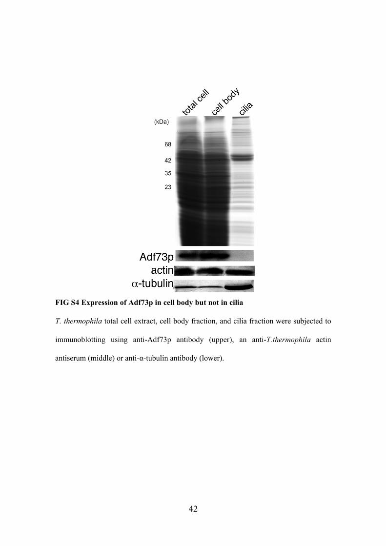
did not detect any Adf73p signal in isolated cilia by immunoblotting whereas actin was
detected in both the cell and cilia (Fig. S4). Also, the adf73KO cells are motile (see
supplemental movies), in contrast to act1KO cells that are paralysed (36). Although the
average swimming velocity was somewhat lower in adf73KO cells than wild-type cells,
the difference was not statistically significant (Table 1). Interestingly, adf73KO cells
turn more frequently than wild-type cells.
17

Discussion
Adf73p regulates the reorganization of the actin cytoskeleton required for
formation of FVs
Here we show that Adf73p colocalizes with actin dots associated with the nascent FVs
and a fibrous structure extending from the oral apparatus in T. thermophila.
Tetrahymena internalizes extracellular liquid with particles into a FVs every 1-2 min.
The actin cytoskeleton is implicated in the formation of FVs since actin inhibitors
potently block FV formation (18). Moreover, this study shows that the formation of FVs
is greatly reduced in cells that assemble abnormal actin structures due to overexpression
of GFP-Adf73p (Fig. 2). Taking into account that Adf73p has depolymerizing activity
against filaments made of Act1p (Fig. 3), a major isoform of T. thermophila actin, it is
conceivable that Adf73p-mediated turnover of the actin cytoskeleton is associated with
the formation of FVs. Importantly we found that the formation of FVs was significantly
inhibited in cells lacking Adf73p (Fig. 5A) and that the organization of the actin
cytoskeleton was abnormal in those cells (Fig. 6). Accordingly, the adf73KO cells failed
to proliferate in the standard SPP medium but grew in the MEPP medium that supported
mutants lacking ability to phagocytose (23). Thus, the phagocytosis-mediated uptake of
18

nutrients is severely affected in cells lacking Adf73p while the endocytosis-mediated
uptake of nutrients is probably functional. Indeed, it has been demonstrated that
endocytosis at the coated pits near basal bodies is not actin-dependent in T. thermophila
(39); however, in the natural environment, actin-based phagocytosis from the oral
apparatus is unambiguously indispensable for preying on microorganisms such as
bacteria for T. thermophila.
In other eukaryotic cells such as macrophages and slime molds, the actin
cytoskeleton promotes invagination or protrusion of the plasma membrane during
phagocytosis (40). In contrast, it has not been exactly understood how the actin
structures, such as actin dots and fibers, induce the formation of FVs, in part because
the source membrane for the nascent FV seems to be distinct from the plasma
membrane (41). Uncovering how the actin cytoskeleton functions in phagocytosis in T.
thermophila may shed light of the general question of how the actin cytoskeleton
regulates the plasma membrane dynamics.
In addition, we found that Adf73p colocalized with actin to FV near a cytoproct.
Sugita et al. (17) previously showed that the cytoproct-associated “actin clump” is a
transient structure engaged in the membrane recycling from old FV; therefore,
19

Adf73p-mediated reorganization of the actin cytoskeleton may be involved in this
process.
Adf73p is not essential for cytokinesis in T. thermophila
In the cytokinesis of animal and yeast cells, the cleavage furrow is induced by the
contraction of a ring-made of actin within the division plane (42-45). Many
actin-modulating proteins, including AC, are involved in the assembly and dynamics of
the contractile ring. Tetrahymena divides by binary fission and assembles an actin-rich
contractile ring (46). Several actin-modulating proteins, including fimbrin (16), eEF1A
(47), and profilin (48), localize to the cleavage furrow. On the other hand, we failed to
detect Adf73p in the cleavage furrow (Fig. S3 and our unpublished data). Supporting
this observation, adf73KO cells multiply without a major increase in the mitotic index
(Fig. 4E); therefore, Adf73p is dispensable for cytokinesis in T. thermophila. Moreover,
cells lacking Act1p can develop an advanced cleavage furrow (36) but fail to perform
scission, the terminal stage of cytokinesis, by which the cell bridge linking the two
daughter cells is broken by rotokinesis, a cilia-dependent rotation of daughter cells (49).
This is probably because of the essential function of Act1p in ciliary movement in the
20

inner arm dynein complex (36, 37). It is therefore likely that isotypes of actin distinct
from Act1p are involved in the contractile ring and that their functions do not require
Adf73p. Alternatively, it is possible that another type of actin-severing or
depolymerizing protein dominantly functions in Tetrahymena cytokinesis. Very recently,
it was reported that cyclase-associated protein CAP, known as a synergic factor for AC
function, can sever F-actin without AC, although the activity is weak (50). We have
found a gene encoding a CAP-homologous protein in the T. thermophila genome (our
unpublished data), although the function of this gene remains to be uncovered. In
addition, a myosin II heavy chain, an essential component of the contractile ring in
animal and yeast cells, is not present in T. thermophila (51, 52). Therefore, the
formation of the contractile ring in Tetrahymena is likely to involve divergent forms of
actin and actin regulators. Interestingly, it has been demonstrated that Giardia
intestinalis, the intestinal parasite lacking most of the genes encoding canonical
actin-modulating proteins including AC and myosin in the genome, requires actin
function for cytokinesis (53). Further studies on protists such as Giardia and
Tetrahymena will provide important evolutionary insights into the molecular
mechanism of cytokinesis. In addition, the divergence of the cytokinetic apparatus
21

creates an experimental opportunity in Tetrahymena. Namely, regulators of important
actin-dependent functions, such as AC, can be studied in vivo as their function is not
required for survival.
22

Acknowledgements
We are grateful to Dr. Mochizuki (Institute of Molecular Biotechnology of the Austrian
Academy of Sciences) for providing pNeo4 vector. The work in JG Laboratory was
supported by NSF grant MCB-033965 and NIH grant R01GM089912. This study was
supported by a Grant for Basic Science Research Projects from the Sumitomo
Foundation and by the Novartis Foundation (Japan) for the Promotion of Science.
23

References
1. Poukkula M, Kremneva E, Seriachius M, Lappalainen P. 2011.
Actin-depolymerizing factor homology domain: a conserved fold perfoming diverse
roles in cytoskeletal dynamics. Cytoskeleton 68:471-490.
2. McGough A, Chiu W. 1999. ADF/cofilin weakens lateral contacts in the actin
filament. J. Mol. Biol. 291:513-519.
3. Bobkov AA, Muhlrad A, Pavlov DA, Kokabi K, Yilmaz A, Reisler E. 2006.
Cooperative effects of cofilin (ADF) on actin structure suggest allosteric mechanism
of cofilin function. J. Mol. Biol. 356:325-334.
4. Bobkov AA, Muhlrad A, Shvetsov A, Benchaar S, Scoville D, Almo SC, Reisler
E. 2004. Cofilin (ADF) affects lateral contacts in F-actin. J. Mol. Biol. 337:93-104.
5. Bamburg JR. 1999. Proteins of the ADF/cofilin family: essential regulators of actin
dynamics. Annu. Rev. Cell. Dev. Biol. 15:185-230.
6. Andrianantoandro E, Pollard TD. 2006. Mechanism of actin filament turnover by
severing and nucleation at different concentrations of ADF/cofilin. Mol. Cell
24:13-23.
7. McGough A, Pope B, Chiu W, Weeds A. 1997. Cofilin changes the twist of F-actin:
24

implications for actin filament dynamics and cellular function. J. Cell Biol.
138:771-781.
8. Nishida E. 1985. Opposite effects of cofilin and profilin from porcine brain on rate
of exchange of actin-bound adenosine 5'-triphosphate. Biochemistry 24:1160-1164.
9. Sattler JM, Ganter M, Hliscs M, Matuschewski K. 2011. Actin regulation in the
malaria parasite. Eur. J. Cell Biol. 90:966-971.
10. Schüler H, Mueller AK, Matuschewski K. 2005. A Plasmodium
actin-depolymerizing factor that binds exclusively to actin monomers. Mol. Biol.
Cell 16:4013-4023.
11. Schmitz S, Grainger M, Howell S, Calder LJ, Gaeb M, Pinder JC, Holder AA,
Veigel C. 2005. Malaria parasite actin filaments are very short. J. Mol. Biol.
349:113-125.
12. Keeling P. 2009. Chromalveolates and the evolution of plastids by secondary
endosymbiosis. J. Eukaryot. Microbiol. 56:1-8.
13. Eisen JA, Coyne RS, Wu M, Wu D, Thiagarajan M, Wortman JR, Badger JH,
Ren Q, Amedeo P, Jones KM, Tallon LJ, Delcher AL, Salzberg SL, Silva JC,
Haas BJ, Majoros WH, Farzad M, Carlton JM, Smith RK Jr, Garg J, Pearlman
25

RE, Karrer KM, Sun L, Manning G, Elde NC, Turkewitz AP, Asai DJ, Wilkes
DE, Wang Y, Cai H, Collins K, Stewart BA, Lee SR, Wilamowska K, Weinberg
Z, Ruzzo WL, Wloga D, Gaertig J, Frankel J, Tsao CC, Gorovsky MA, Keeling
PJ, Waller RF, Patron NJ, Cherry JM, Stover NA, Krieger CJ, del Toro C,
Ryder HF, Williamson SC, Barbeau RA, Hamilton EP, Orias E. 2006.
Macronuclear genome sequence of the ciliate Tetrahymena thermophila, a model
eukaryote. PLoS Biol. 9:1620-1642.
14. Nilsson JR. 1979. Phagotrophy in Tetrahymena. In Biochemistry and Physiology of
Protozoa 2nd ed. (ed. M. Levandowsky and S. H. Huter). vol. 2, pp. 339-379. New
York: Academic Press.
15. Gonda K, Komatsu M, Numata O. 2000. Calmodulin and
Ca2+/calmodulin-binding proteins are involved in Tetrahymena thermophila
phagocytosis. Cell Struct. Funct. 25:243-251.
16. Watanabe A, Kurasawa Y, Watanabe Y, Numata O. 1998. A new Tetrahymena
actin-binding protein is localized in the division furrow. J. Biochem. 123:607-613.
17. Sugita M, Nakano K, Sato M, Toyooka K, Numata O. 2009. The roles of actin
cytoskeleton and microtubules for membrane recycling of a food vacuole in
26

Tetrahymena thermophila. Cell Motil. Cytoskeleton 66:371-377.
18. Zackroff RV, Hufnagel LA. 2002. Induction of anti-actin drug resistance in
Tetrahymena. J. Eukaryot. Microbiol. 49:475-477.
19. Wilkes DE, Otto JJ. 2003. Profilin functions in cytokinesis, nuclear positioning,
and stomatogenesis in Tetrahymena thermophila. J. Eukaryot. Microbiol.
50:252-262.
20. Hirono M, Kumagai Y, Numata O, Watanabe Y. 1989. Purification of
Tetrahymena actin reveals some unusual properties. Proc. Natl. Acad. Sci. USA.
86:75-79.
21. Shiozaki N, Nakano K, Takaine M, Abe H, Numata O. 2009. Usual and unusual
biochemical properties of ADF/cofilin-like protein Adf73p in ciliate Tetrahymena
thermophila. Biochem. Biophys. Res. Commun. 390:54-59.
22. Gorovsky M, Yao MC, Keevert J, Pleger G. 1975. Isolation of micro- and
macronuclei from Tetrahymena pyriformis. Methods Cell Biol. 9:311-327.
23. Orias E, Rasmussen L. 1976. Dual capacity for nutrient uptake in Tetrahymena. IV.
Growth without food vacuoles. Exp. Cell Res. 102:127–137.
24. Wloga D, Dave D, Meagley J, Rogowski K, Jerka-Dziadosz M, Gaertig J. 2010.
27

Hyperglutamylation of tubulin can either stabilize or destabilize microtubules in the
same cell. Eukaryot. Cell 9:184-193.
25. Wloga D, Camba A, Rogowski K, Manning G, Jerka-Dziadosz M, Gaertig J.
2006. Members of the NIMA-related kinase family promote disassembly of cilia by
multiple mechanisms. Mol. Biol. Cell 17:2799-2810.
26. Gaertig J, Gao Y, Tishgarten T, Clark TG, Dickerson HW. 1999. Surface
display of a parasite antigen in the ciliate Tetrahymena thermophila. Nat. Biotechnol.
17:462-465.
27. Mochizuki K. 2008. High efficiency transformation of Tetrahymena using a
codon-optimized neomycin resistance gene. Gene 425:79-83.
28. Cassidy-Hanley D, Bowen J, Lee JH, Cole E, VerPlank LA, Gaertig J,
Gorovsky MA, Bruns PJ. 1997. Germline and somatic transformation of mating
Tetrahymena thermophila by particle bombardment. Genetics 146:135–147.
29. Hai B, Gaertig J, Gorovsky MA. 2000. Knockout heterokaryons enable facile
mutagenic analysis of essential genes in Tetrahymena. Methods Cell Biol.
62:513-531.
30. Ueno H, Gonda K, Takeda T, Numata O. 2003. Elongation factor 1α and
28

calmodulin colocalize on the axonemal microtubules in Tetrahymena cilia. Cell
Motil. Cytoskeleton 55:51-60.
31. Abe H, Obinata T, Minamide LS, Bamburg JR. 1996. Xenopus laevis
actin-depolymerizing factor/cofilin: a phosphorylation-regulated protein essential for
development. J. Cell Biol. 132:871-885.
32. Gunsalus KC, Bonaccorsi S, Williams E, Verni F, Gatti M, Goldberg M. 1995.
Mutation in twinstar, a Drosophila gene encoding a cofilin/ADF homologue, result
in defects in centrosome migration and cytokinesis. J. Cell Biol. 131:1243-1259.
33. Kaji N, Ohashi K, Shuin M, Niwa R, Uemura T, Mizuno K. 2003. Cell
cycle-associated changes in Slingshot phosphatase activity and roles in cytokinesis in
animal cells. J. Biol. Chem. 278:33450-33455.
34. Nakano K, Mabuchi I. 2006. Actin-depolymerizing protein Adf1 is required for
formation and maintenance of the contractile ring during cytokinesis in fission yeast.
Mol. Biol. Cell. 17:1933-1945.
35. Ono K, Parast M, Alberico C, Benian GM, Ono S. 2003. Specific requirement for
two ADF/cofilin isoforms in distinct actin-dependent processes in Caenorhabditis
elegans. J. Cell Sci. 116:2073-2085.
29

36. Williams NE, Tsao CC, Bowen J, Hehman GL, Williams RJ, Frankel J. 2006.
The actin gene ACT1 is required for phagocytosis, motility, and cell separation of
Tetrahymena thermophila. Eukaryot. Cell 5:555-567.
37. Muto E, Edamatsu M, Hirono M, Kamiya R. 1994. Immunological detection of
actin in the 14S ciliary dynein of Tetrahymena. FEBS Lett. 343:173-177.
38. Tammana TV, Sahasrabuddhe AA, Mitra K, Bajpai VK, Gupta CM. 2008.
Actin-depolymerizing factor, ADF/cofilin, is essentially required in assembly of
Leishmania flagellum. Mol. Microbiol. 70:837-852.
39. Elde NC, Morgan G, Winey M, Sperling L, Turkewitz AP. 2005. Elucidation of
clathrin-mediated endocytosis in Tetrahymena reveals an evolutionarily convergent
recruitment of dynamin. PLoS Genet. 1:e52.
40. May RC, Machesky LM. 2001. Phagocytosis and the actin cytoskeleton. J Cell Sci.
114:1061-77.
41. Weidenbach AL, Thompson Jr GA. 1974. Studies of membrane formation in
Tetrahymena pyriformis. VIII. On the origin of membranes surrounding food
vacuoles. J. Protozool. 21:745-751.
42. Glotzer M. 2005. The molecular requirements for cytokinesis. Science 307,
30

1735-1739.
43. Goyal A, Takaine M, Simanis V, Nakano K. 2011. Dividing the spoils of growth
and the cell cycle: The fission yeast as a model for study of cytokinesis. Cytoskeleton
68:69-88.
44. Mabuchi I. 1986. Biochemical aspects of cytokinesis. Int. Rev. Cytol. 101:175-213.
45. Pollard TD. 2010. Mechanics of cytokinesis in eukaryotes. Curr. Opin. Cell Biol.
22:50-56.
46. Yasuda T, Numata O, Ohnishi K, Watanabe Y. 1980. A contractile ring and
cortical changes found in the dividing Tetrahymena pyriformis. Exp. Cell Res.
128:407-417.
47. Numata O, Kurasawa Y, Gonda K, Watanabe Y. 2000. Tetrahymena elongation
factor-1 alpha is localized with calmodulin in the division furrow. J. Biochem.
127:51-56.
48. Edamatsu M, Hirono M, Watanabe Y. (1992). Tetrahymena profilin is localized
in the division furrow. J. Biochem. 112:637-642.
49. Brown JM, Hardin C, Gaertig J. 1999. Rotokinesis, a novel phenomenon of cell
locomotion-assisted cytokinesis in the ciliate Tetrahymena thermophila. Cell Biol.
31

Int. 23:841-848.
50. Normoyle KPQ, Brieher WM. 2012. Cyclase-associated protein (CAP) acts
directly on F-actin to accelerate cofilin-mediated actin severing across the range of
physiological pH. J. Biol. Chem. 287:35722-35732.
51. Sugita M, Iwataki Y, Nakano K, Numata O. 2011. Unique sequences and
predicted functions of myosinsin Tetrahymena thermophila. Gene 480:10-20.
52. Williams SA, Gavin RH. 2005. Myosin genes in Tetrahymena. Cell Motil.
Cytoskeleton 61:237-243.
53. Paredez AR, Assaf ZJ, Sept D, Timofejeva L, Dawson SC, Wang CR, Cande Z.
2011. An actincytoskeleton with evolutionarily conserved functions in the absence of
canonical actin-binding proteins. Proc. Natl. Acad Sci. 108:6151-6156.
32

Figure Legends
Fig. 1. Adf73p colocalized with F-actin in T. thermophila
(A) Western blots with anti-Adf73p antiserum. Cell lysate of T. thermophila was
subjected to SDS-PAGF and transferred to PVDF membrane. The membrane was slit
and subjected to immunoblots with pre-immune serum (slit 1), anti-Adf73p antiserum
before (slit 2) and after (slit 3) affinity purification. Arrow indicates a band consistent
with the predicted size of Adf73p. Arrowheads indicate extra bands not eliminated after
the process of affinity purification. (B) Immunofluorescence microscopy for cellular
localization of Adf73p. Cells were processed for immunofluorescence staining after
black ink was loaded on their FVs for 30 min. Right column shows bright field
microscopic images of cells merged with immunofluorescence images (red, actin; green,
Adf73p). Blue dotted line shows outline of the cell. In top row, Adf73p localizes to
actin dots located on nascent FVs (small arrows) near the oral apparatus (OA) and an
actin clump near a cytoproct (CP) (large arrow). Mid-size arrow indicates an actin cable
protruding from OA. This cable-like structure seems to be transiently formed during
phagocytosis since it is found only in limited populations of cells forming FV. Adf73p
associates with this structure as well. Cells shown in the bottom two rows of panels
were treated with DMSO (solvent only) or actin-polymerization inhibitor Lat-B (final
conc. 10 μM) for 10 min, respectively. Note that localization of Adf73p was diminished
in cells incubated with Lat-B. (C) Cells transiently expressing GFP-Adf73p under
exogenous promoter of MTT1 were stained with an anti-actin antiserum and anti-GFP
antibody. Right column shows merged image (red, actin; green, GFP-Adf73p).
GFP-Adf73p showed similar localization with Adf73p observed in (B). Bars, 10 μm.
33
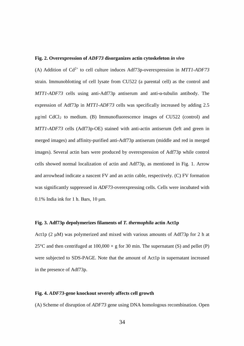
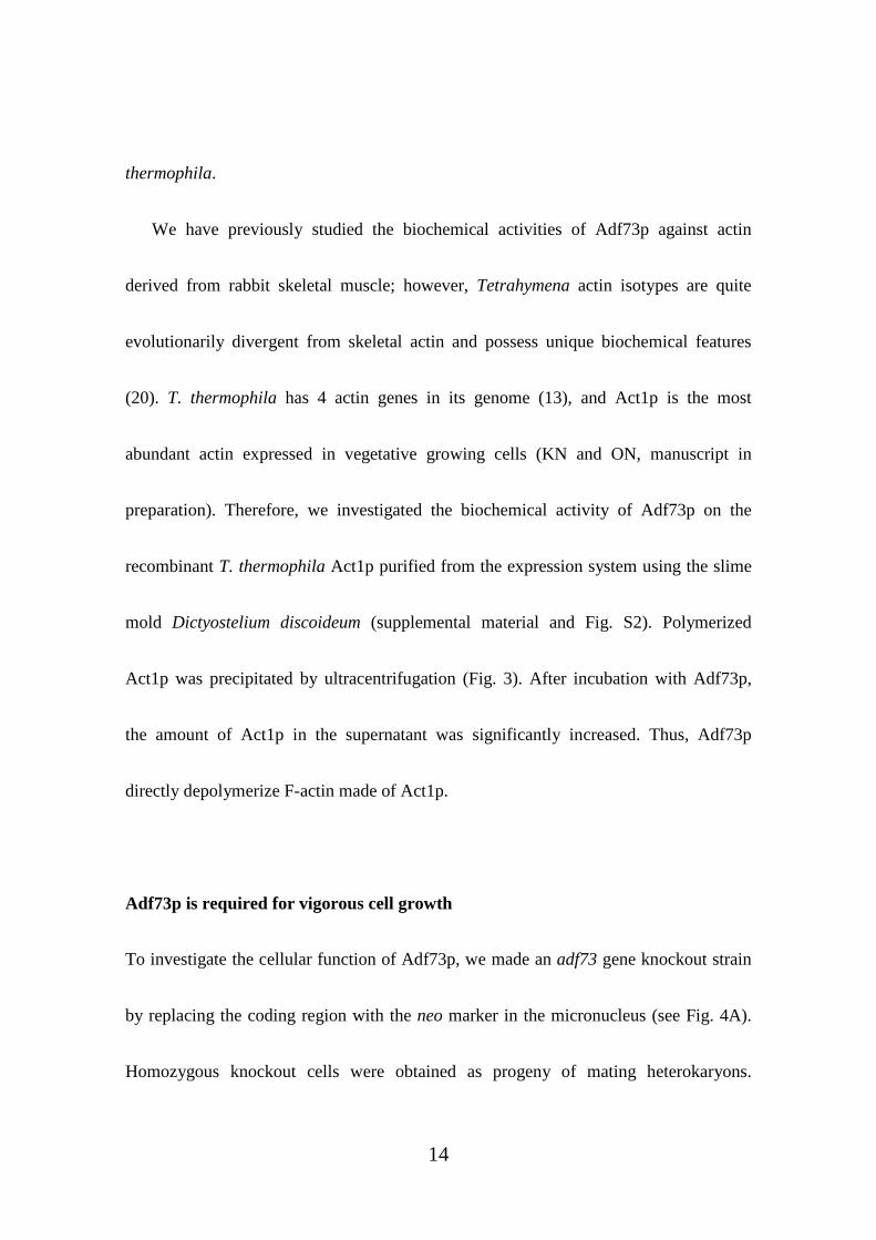
Fig. 2. Overexpression of ADF73 disorganizes actin cytoskeleton in vivo
(A) Addition of Cd2+ to cell culture induces Adf73p-overexpression in MTT1-ADF73
strain. Immunoblotting of cell lysate from CU522 (a parental cell) as the control and
MTT1-ADF73 cells using anti-Adf73p antiserum and anti-α-tubulin antibody. The
expression of Adf73p in MTT1-ADF73 cells was specifically increased by adding 2.5
μg/ml CdCl2 to medium. (B) Immunofluorescence images of CU522 (control) and
MTT1-ADF73 cells (Adf73p-OE) stained with anti-actin antiserum (left and green in
merged images) and affinity-purified anti-Adf73p antiserum (middle and red in merged
images). Several actin bars were produced by overexpression of Adf73p while control
cells showed normal localization of actin and Adf73p, as mentioned in Fig. 1. Arrow
and arrowhead indicate a nascent FV and an actin cable, respectively. (C) FV formation
was significantly suppressed in ADF73-overexpressing cells. Cells were incubated with
0.1% India ink for 1 h. Bars, 10 μm.
Fig. 3. Adf73p depolymerizes filaments of T. thermophila actin Act1p
Act1p (2 μM) was polymerized and mixed with various amounts of Adf73p for 2 h at
25°C and then centrifuged at 100,000 × g for 30 min. The supernatant (S) and pellet (P)
were subjected to SDS-PAGE. Note that the amount of Act1p in supernatant increased
in the presence of Adf73p.
Fig. 4. ADF73-gene knockout severely affects cell growth
(A) Scheme of disruption of ADF73 gene using DNA homologous recombination. Open
34

reading frame of ADF73 was completely replaced with NEO4 marker gene. (B)
Confirmation of gene replacement by PCR. Genomic DNAs prepared from a parental
strain (CU428) and two independent clones of ADF73 knockout cells (KO) were
amplified using two sets of PCR primers (primer 1 and 2 or primer 1 and 3). Annealing
sites of the primers are indicated in A. (C) Expression of Adf73p was abolished in the
knockout strain. Immunoblotting was performed against cell lysate using
affinity-purified anti-Adf73p serum. Membrane area corresponding to the molecular
weight of Adf73p was trimmed and shown. Expression level of actin was unaffected by
ADF73-gene knockout. (D) Immunofluorescence microscopy was performed against
adf73KO cells using affinity-purified anti-Adf73p serum. Right image is rather
overexposed for showing a non-specific fluorescence signal. Bar, 10 μm. (E) Cell
growth of CU428 strain as the wild-type (WT) and three independent clones of
adf73KO strains. Each strain was cultured in MEPP medium at 30°C. Note that both the
cell growth rate and maximum number of cells in the steady state were significantly
reduced in all of the adf73KO strains.
Fig. 5. Phagocytosis activity is severely affected in adf73KO cells
(A) FV-forming ability is significantly reduced by gene knockout of ADF73. Wild-type
and adf73KO cells were incubated with 0.1% India ink in MEPP medium at 30°C. At
0.5, 2, 4.5 and 7.5 h after the India ink was loaded, cells were fixed and the average
number of FVs per cell was evaluated (>10 cells). (B) Microscopic images of cells after
2 h of incubation with India ink were shown. Arrows indicate aggregates of India ink
that have been ejected from a cytoproct through a process of phagocytosis. (C) Colloid
35

of India ink was significantly reduced by long incubation with wild-type Tetrahymena
cells but not adf73KO cells. Microtubes containing cell culture with India ink as in A
were left to stand and observed. Note that almost all India ink settled into wild-type
cells but not in adf73KO after prolonged incubation for 24 h, since the colloid was
mostly precipitated with cells as the FV contents and India ink aggregated as a result of
phagocytosis.
Fig. 6. Abnormally large accumulation of actin was seen in adf73KO cells
Immunofluorescence images of a parental strain CU428 and adf73KO stained with
anti-actin antiserum and anti-α-tubulin antibody for MTs. Arrowheads indicate normal
localization of actin dots surrounding a nascent FV located close to the oral apparatus
(yellow arrows). White arrows indicate abnormal large accumulation of actin in
cytoplasm, which is frequently observed in adf73KO cells but never in wild-type cells.
Bar, 10 μm.
36

Fig.1

Fig.2

Fig.3

Fig.4

Fig.5

Fig.6
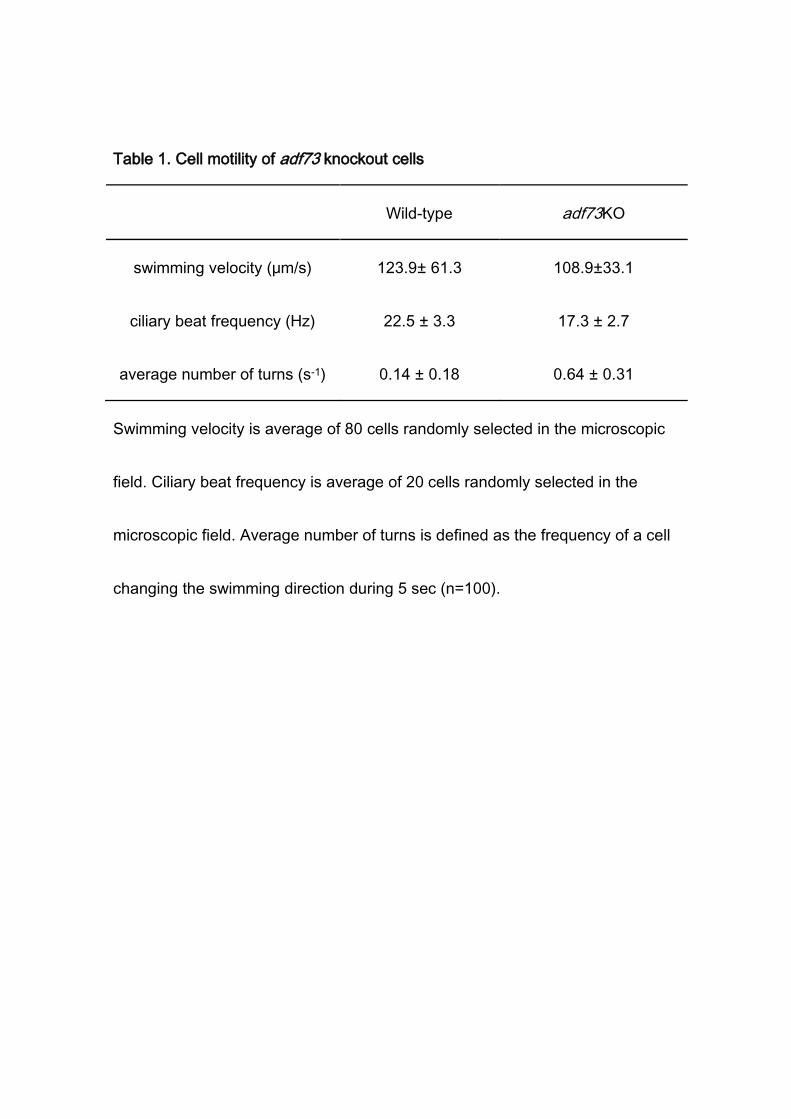
Table 1. Cell motility of adf73 knockout cells
Wild-type adf73KO
swimming velocity (μm/s) 123.9± 61.3 108.9±33.1
ciliary beat frequency (Hz) 22.5 ± 3.3 17.3 ± 2.7
average number of turns (s-1) 0.14 ± 0.18 0.64 ± 0.31
Swimming velocity is average of 80 cells randomly selected in the microscopic
field. Ciliary beat frequency is average of 20 cells randomly selected in the
microscopic field. Average number of turns is defined as the frequency of a cell
changing the swimming direction during 5 sec (n=100).
36
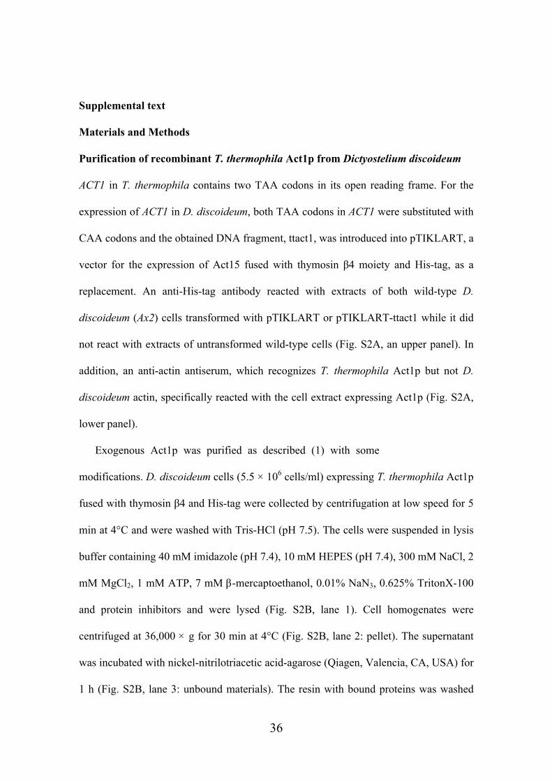
Supplemental text
Materials and Methods
Purification of recombinant T. thermophila Act1p from Dictyostelium discoideum
ACT1 in T. thermophila contains two TAA codons in its open reading frame. For the
expression of ACT1 in D. discoideum, both TAA codons in ACT1 were substituted with
CAA codons and the obtained DNA fragment, ttact1, was introduced into pTIKLART, a
vector for the expression of Act15 fused with thymosin β4 moiety and His-tag, as a
replacement. An anti-His-tag antibody reacted with extracts of both wild-type D.
discoideum (Ax2) cells transformed with pTIKLART or pTIKLART-ttact1 while it did
not react with extracts of untransformed wild-type cells (Fig. S2A, an upper panel). In
addition, an anti-actin antiserum, which recognizes T. thermophila Act1p but not D.
discoideum actin, specifically reacted with the cell extract expressing Act1p (Fig. S2A,
lower panel).
Exogenous Act1p was purified as described (1) with some
modifications. D. discoideum cells (5.5 × 10⁶ cells/ml) expressing T. thermophila Act1p
fused with thymosin β4 and His-tag were collected by centrifugation at low speed for 5
min at 4°C and were washed with Tris-HCl (pH 7.5). The cells were suspended in lysis
buffer containing 40 mM imidazole (pH 7.4), 10 mM HEPES (pH 7.4), 300 mM NaCl, 2
mM MgCl2, 1 mM ATP, 7 mM β-mercaptoethanol, 0.01% NaN3, 0.625% TritonX-100
and protein inhibitors and were lysed (Fig. S2B, lane 1). Cell homogenates were
centrifuged at 36,000 × g for 30 min at 4°C (Fig. S2B, lane 2: pellet). The supernatant
was incubated with nickel-nitrilotriacetic acid-agarose (Qiagen, Valencia, CA, USA) for
1 h (Fig. S2B, lane 3: unbound materials). The resin with bound proteins was washed

37
with buffer containing 40 mM imidazole (pH 7.4), 10 mM HEPES (pH 7.4), 300 mM
NaCl, 0.5 mM MgCl2, 0.1 mM ATP, 7 mM β-mercaptoethanol and 0.01% NaN3 (Fig.
S2B, lane 4) and then extracted with buffer containing 500 mM imidazole (pH 7.4), 10
mM HEPES (pH 7.4), 300 mM NaCl, 0.5 mM MgCl2, 0.1 mM ATP, 7 mM
β-mercaptoethanol and 0.01% NaN3 (Fig. S2B, lane 5: resin). The extract (Fig. S2B,
lane 6) was dialyzed against G-buffer (2 mM Tris-HCl (pH 7.4), 0.2 mM CaCl2, 0.2 mM
ATP, 0.5 mM DTT and 0.01% NaN3) overnight at 4°C. Then the fraction was treated
with 1-chloro-3-tosylamido-7-amino-2-heptanone-treated chymotrypsin (Sigma, St.
Louis, MO, USA) at a final concentration of 8.34 µg/ml for 20 min at 25°C (Fig. S2B,
lane 7) and the reaction was stopped by adding 0.4 mM PMSF. The digested
mixture was applied to an Econo-Pac HighQ Cartridge (Bio Rad, Hercules, CA, USA)
pre-equilibrated with G-buffer, and bound proteins were eluted with a linear 0-0.5 M
NaCl gradient in G-buffer (flow velocity: 1 ml/min). Fractions containing Act1p were
collected and dialyzed against G-buffer overnight at 4°C (Fig. S2B, lane 8). Act1p
monomers were polymerized by adding the final 2 mM ATP, 100 mM KCl and 4 mM
MgCl2 for 2 h at 25°C and centrifuged at 300,000 ×g for 30 min at 4°C (Fig. S2B,
lane 9: supernatant). F-actin pellet (Fig. S2B, lane 10) was suspended with G-buffer and
sonicated. After dialyzing against G-buffer overnight at 4°C, monomeric Act1p was
centrifuged at 300,000 × g for 15 min at 4°C (Fig. S2B, lane 12: pellet) and purified
recombinant Act1p was obtained in the supernatant (Fig. S2B, lane 11).

38
Supplemental references
1. Noguchi TQ, Kanzaki N, Ueno H, Hirose K, Uyeda TQ. 2007. A novel
system for expressing toxic actin mutants in Dictyostelium and purification and
characterization of a dominant lethal yeast actin mutant. J. Biol. Chem. 282:
27721-27727.
2. Sugita M, Nakano K, Sato M, Toyooka K, Numata O. 2009. The roles of
actin cytoskeleton and microtubules for membrane recycling of a food vacuole in
Tetrahymena thermophila. Cell Motil. Cytoskeleton 66: 371-377.
39
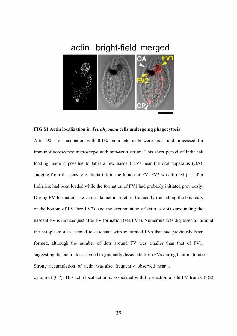
FIG S1 Actin localization in Tetrahymena cells undergoing phagocytosis
After 90 s of incubation with 0.1% India ink, cells were fixed and processed for
immunofluorescence microscopy with anti-actin serum. This short period of India ink
loading made it possible to label a few nascent FVs near the oral apparatus (OA).
Judging from the density of India ink in the lumen of FV, FV2 was formed just after
India ink had been loaded while the formation of FV1 had probably initiated previously.
During FV formation, the cable-like actin structure frequently runs along the boundary
of the bottom of FV (see FV2), and the accumulation of actin as dots surrounding the
nascent FV is induced just after FV formation (see FV1). Numerous dots dispersed all around
the cytoplasm also seemed to associate with maturated FVs that had previously been
formed, although the number of dots around FV was smaller than that of FV1,
suggesting that actin dots seemed to gradually dissociate from FVs during their maturation.
Strong accumulation of actin was also frequently observed near a
cytoproct (CP). This actin localization is associated with the ejection of old FV from CP (2).

40
FIG S2 Purification of T. thermophila Act1p from Dictyostelium discoideum
(A) Wild-type D. discoideum cells (lane 1), D. discoideum cells transformed with
pTIKLART (lane 2) or pTIKLART-ttact (lane 3) were lysed and subjected to Western
blotting using an anti-His antibody (upper) or an anti-Tetrahymena actin antiserum
(lower), respectively. (B) Tetrahymena Act1p was purified from D. discoideum. The
detailed procedure of the purification is described in Supplemental Materials and
Methods.

41
FIG S3 Localization of Adf73p in a mitotic cell
Cells transiently expressing GFP-Adf73p under exogenous promoter of the MTT1 gene
were observed. The dark region with less fluorescence signal seen on the left side of the
cell is the macronucleus elongated for division. No marked accumulation of
GFP-Adf73p was found in the division furrow initiated in the middle of the mitotic cell,
as indicated by an arrowhead, while GFP-Adf73p was dispersed throughout the
cytoplasm and several cytoplasmic dots were also observed. These dots were
occasionally observed as a cluster, as indicated by an arrow.
42
FIG S4 Expression of Adf73p in cell body but not in cilia
T. thermophila total cell extract, cell body fraction, and cilia fraction were subjected to
immunoblotting using anti-Adf73p antibody (upper), an anti-T.thermophila actin
antiserum (middle) or anti-α-tubulin antibody (lower).


![Review Actin-targeting natural products: structures ... · actin-binding proteins actively break or ‘sever’ actin filaments [e.g. actin-depolymerizing factor (ADF) and cofilin]](https://img.pdfslide.us/doc/110x75/5f0f85bd7e708231d44494d0/review-actin-targeting-natural-products-structures-actin-binding-proteins-actively.jpg)


![[ A ] SPIRITS ADF [ADF] VODKA - BASIC](https://img.pdfslide.us/doc/110x75/6169d8c211a7b741a34c063e/-a-spirits-adf-adf-vodka-basic.jpg)