Embed Size (px)
Citation preview
Tropical bat as mammalian model for skincarotenoid metabolismIsmael Galvána,1, Juan Garrido-Fernándezb, José Ríosc, Antonio Pérez-Gálvezb, Bernal Rodríguez-Herrerad,and Juan José Negroa
aDepartment of Evolutionary Ecology, Doñana Biological Station–Consejo Superior de Investigaciones Científicas (CSIC), 41092 Sevilla, Spain;bDepartment of Food Phytochemistry, Instituto de la Grasa–CSIC, 41013 Sevilla, Spain; cMass Spectrometry Laboratory, Instituto de la Grasa–CSIC,41013 Sevilla, Spain; and dSchool of Biology, University of Costa Rica, 2060 San José, Costa Rica
Edited by David W. Russell, University of Texas Southwestern Medical Center, Dallas, TX, and approved July 26, 2016 (received for review June 17, 2016)
Animals cannot synthesize carotenoid pigments de novo, and mustconsume them in their diet. Most mammals, including humans, areindiscriminate accumulators of carotenoids but inefficiently distrib-ute them to some tissues and organs, such as skin. This limits thepotential capacity of these organisms to benefit from the antioxi-dant and immunostimulatory functions that carotenoids fulfill.Indeed, to date, no mammal has been known to have evolvedphysiological mechanisms to incorporate and deposit carotenoidsin the skin or hair, and mammals have therefore been assumed torely entirely on other pigments such as melanins to color theirintegument. Here we use high-performance liquid chromatography(HPLC) in combination with time-of-flight mass spectrometry (HPLC-TOF/MS) to show that the frugivorous Honduran white batEctophylla alba colors its skin bright yellow with the deposition ofthe xanthophyll lutein. The Honduran white bat is thus a mamma-lian model that may help developing strategies to improve the as-similation of lutein in humans to avoid macular degeneration. Thisrepresents a change of paradigm in animal physiology showing thatsome mammals actually have the capacity to accumulate dietarycarotenoids in the integument. In addition, we have also discoveredthat the majority of the lutein in the skin of Honduran white bats ispresent in esterified form with fatty acids, thereby permittinglonger-lasting coloration and suggesting bright color traits mayhave an overlooked role in the visual communication of bats.
bats | carotenoids | macular degeneration | skin coloration
Carotenoids are photosynthetic pigments animals use to avoidcellular oxidative damage and to color external structures
(1–3). The inability of animals to synthesize de novo carotenoidsmeans these pigments must be obtained only from dietary sources(4). Among vertebrates, the limiting nature of carotenoids is par-ticularly marked in mammalian species, which exhibit a variable,but generally low, efficiency for carotenoid assimilation (5, 6).Here we show a mammal, the frugivorous Honduran white batEctophylla alba, that deposits amounts of carotenoids in the skinthat are high enough to generate a conspicuous coloration. In-deed, up to this time, no mammal has been known to incorporatesignificant visible amounts of carotenoids in the integument (7,8). This has prevented adequate animal models being found forcarotenoid assimilation in the skin that would enable improve-ment of the bioavailability of carotenoids in the diet of humans(9), where these pigments are of utmost importance for health byhaving provitamin A value and fulfilling antioxidant and immu-nostimulatory functions (10, 11). Using high-performance liquidchromatography (HPLC) in combination with time-of-flight massspectrometry (HPLC-TOF/MS), we found that the characteristicbright yellow coloration of the bare skin of the nose-leaf and earsin these bats is generated by the xanthophyll lutein in both freeand esterified forms. This indicates that the Honduran white batrepresents a change of paradigm in animal physiology, showingthat some mammals actually have the capacity to accumulate di-etary carotenoids in the integument. It is thus a mammalian modelthat may help in developing strategies to improve the assimilation
of lutein in humans (12), who depend on dietary contents to avoidmacular degeneration (13) and skin oxidative damage (14).The nocturnal Honduran white bat is a small phyllostomid
distributed exclusively in lowland rainforests of Mesoamericathat exhibits bright yellow coloration in the bare skin of the nose-leaf, ears, and wings (Fig. 1 A and B). This distinctive colorationresembles the one generated by some carotenoids, mainly luteinand zeaxanthin, in the integument of other vertebrates, such asthe bare integument and feathers of birds (4, 15). To the best ofour knowledge, such yellow hues are not found in other mam-mals, which mostly rely on darker endogenous melanins to colortheir pelage and skin (16), with the exception of other tropicalbats (see Evolutionary Implications). To determine the origin ofthe yellow color, we conducted in-depth chemical analyses of thebare skin of the ears and nose-leaf and of the liver of two maleHonduran white bat specimens from Costa Rica.The integument of mammals represents a striking example of
the difference in the bioavailability of carotenoids among animals.Carotenoids are widespread pigments in the integument of allvertebrates except mammals, as only small amounts of carotenoidsare found in the skin of humans and other mammalian species(7, 8), and in contrast to the feathers and scales of reptiles and fish,there seems to be a complete lack of carotenoids in mammalianhair (15). In humans, and aside from pathological conditions inwhich carotenoids accumulate in the skin (i.e., carotenodermia),these pigments only occur in low amounts in the skin (∼200 ng/gtissue) (7), where they interact with the color generated by keratin,melanins, and blood (17). Carotenoids are present at higher con-centrations in internal tissues (human macula: 1 mM; serum: 0.1–1 μM) (18). Therefore, the contribution of dietary carotenoidsto the perceived variation in human skin coloration is negligible.To date, the role of carotenoids as the primary pigments responsible
Significance
We have discovered that a mammalian species, a bat called theHonduran white bat Ectophylla alba, displays a yellow caroten-oid pigment called lutein in its bare skin. Even though carotenoid-based coloration has been found in birds, fish, amphibians, andreptiles, there are no reports of any extant mammals showingthese pigments in their skin or hair. The implications of thisfinding may be profound for human health, as carotenoids areessential micronutrients. Lutein in particular is involved in thepreservation of the macula of the eye. The Honduran white bat,with its ability to assimilate and deposit lutein in its bare skin,may be the sought-after mammalian model needed for en-hancing studies on carotenoid function and metabolism.
Author contributions: I.G. and J.J.N. designed research; I.G., B.R.-H., and J.J.N. performedresearch; J.G.-F., J.R., and A.P.-G. contributed new reagents/analytic tools; I.G., J.G.-F., J.R.,and A.P.-G. analyzed data; and I.G., A.P.-G., and J.J.N. wrote the paper.
The authors declare no conflict of interest.
This article is a PNAS Direct Submission.1To whom correspondence should be addressed. Email: [email protected].
10932–10937 | PNAS | September 27, 2016 | vol. 113 | no. 39 www.pnas.org/cgi/doi/10.1073/pnas.1609724113

for the generation of distinctive patches of bright color in the in-tegument, as in other vertebrates (4, 15), has never been reported inmammals. Beyond increasing our understanding of the evolution ofanimal coloration and appearance, finding mammals capable ofcoloring their integument with carotenoids would open new op-portunities to obtain mammalian models that help improve ourbioavailability of dietary carotenoids for a better fulfillment of hu-man physiological functions (19).Mammals obtain carotenoids through eating green plant products.
Once ingested, these pigments are released from the food matrix bythe action of bile salts and pancreatic lipases, which form lipid mi-celles that incorporate carotenoids and facilitate their transport tothe mucosal cells of the duodenum by passive diffusion or throughfacilitated transport via scavenger receptor class B, type 1 (20). Thecapacity to assemble micelles with incorporated carotenoids partlydetermines the efficiency of carotenoid absorption, which has led tothe advent of functional food technologies searching for food ma-trices that optimize the absorption of supplemented carotenoids infarmed fish (5, 21) and human diets (22). Once absorbed, caroten-oids are transported by lipoproteins through the bloodstream to thetarget tissues, a process in which the abundance and nature of li-poproteins limit the capacity to obtain carotenoids (15). Further-more, some animals can metabolize carotenoids into other forms,and occasionally esterify them with long-chain fatty acids (20). Theresult is that several factors, such as amounts and species of carot-enoids ingested, molecular linkage and food matrix composition,nutrient status of the host, and genetic components, influence the
bioavailability of carotenoids (23), creating large differences betweenspecies in their capacity to obtain these pigments from their diets andmake physiological use of them (15).
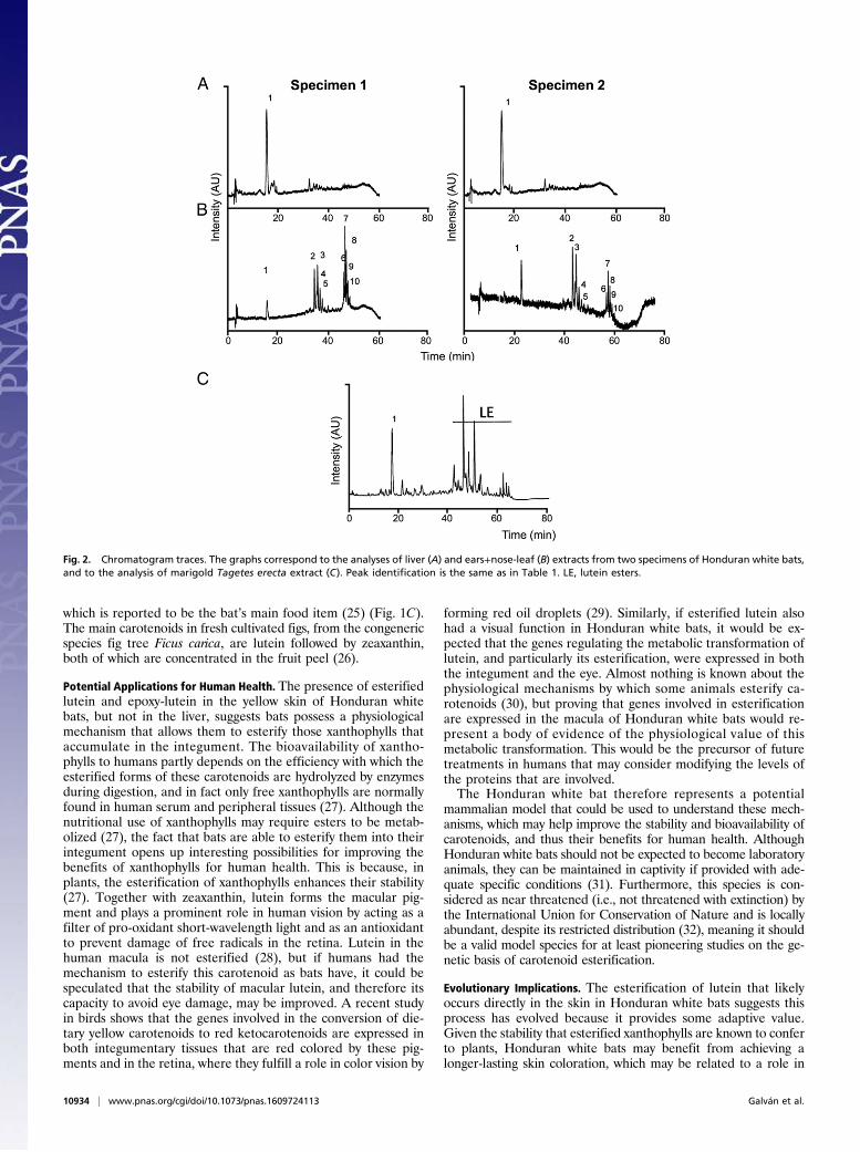
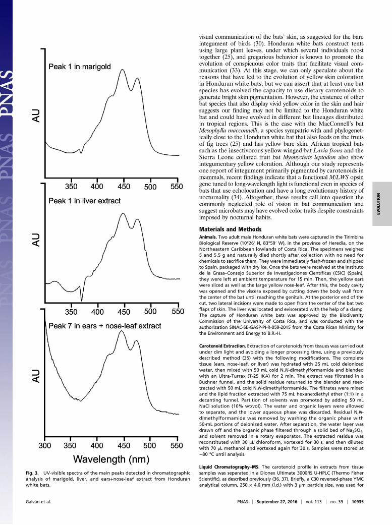
Results and DiscussionOur chromatographic analyses, performed with extracts from theears and nose-leaf of Honduran white bats, clearly showed thattheir bright yellow color is caused by the xanthophyll lutein, in-cluding trans and cis isomers, and its epoxy derivative, whichexclusively forms the carotenoid profile in the extracts of the twospecimens analyzed. Thus, both specimens contained the samequalitative carotenoid profile. The identification was based onthe chromatographic features, UV-visible absorbance spectra,and MS characteristics, including MS and MS2 spectral behavior,of the molecular ion, which is in line with the data available inthe literature (24). Fig. 2 depicts the HPLC analyses of the ex-tracts from liver (Fig. 2A), ears, and nose-leaf tissue (Fig. 2B) forboth specimens, whereas Fig. 3 shows the UV-visible absorbancespectra of the main peaks detected in the analyzed tissues. Wecompared the experimental data with those obtained from theanalysis of lutein standard isolated from a vegetable source,obtaining equivalent results (Figs. 2C and 3). Unexpectedly,most of the lutein content in the ears and nose-leaf is present asesterified forms with saturated and unsaturated fatty acids,whereas almost all lutein content in liver is present as free form(Table 1). The probable source of dietary lutein for Honduranwhite bats is the fig tree Ficus colubrinae, the ripe red fruit of
Fig. 1. Honduran white bat images. (A and B) Details of the bright yellow coloration of the bare skin in the ears, nose-leaf, and wings of a Honduran whitebat from Costa Rica. (C) A Honduran white bat feeding on the ripe fruit of a fig tree F. colubrinae. Photographs by Sharlene Santana (A and B) and MarcoTschapka (C).
Galván et al. PNAS | September 27, 2016 | vol. 113 | no. 39 | 10933
EVOLU
TION
which is reported to be the bat’s main food item (25) (Fig. 1C).The main carotenoids in fresh cultivated figs, from the congenericspecies fig tree Ficus carica, are lutein followed by zeaxanthin,both of which are concentrated in the fruit peel (26).
Potential Applications for Human Health. The presence of esterifiedlutein and epoxy-lutein in the yellow skin of Honduran whitebats, but not in the liver, suggests bats possess a physiologicalmechanism that allows them to esterify those xanthophylls thataccumulate in the integument. The bioavailability of xantho-phylls to humans partly depends on the efficiency with which theesterified forms of these carotenoids are hydrolyzed by enzymesduring digestion, and in fact only free xanthophylls are normallyfound in human serum and peripheral tissues (27). Although thenutritional use of xanthophylls may require esters to be metab-olized (27), the fact that bats are able to esterify them into theirintegument opens up interesting possibilities for improving thebenefits of xanthophylls for human health. This is because, inplants, the esterification of xanthophylls enhances their stability(27). Together with zeaxanthin, lutein forms the macular pig-ment and plays a prominent role in human vision by acting as afilter of pro-oxidant short-wavelength light and as an antioxidantto prevent damage of free radicals in the retina. Lutein in thehuman macula is not esterified (28), but if humans had themechanism to esterify this carotenoid as bats have, it could bespeculated that the stability of macular lutein, and therefore itscapacity to avoid eye damage, may be improved. A recent studyin birds shows that the genes involved in the conversion of die-tary yellow carotenoids to red ketocarotenoids are expressed inboth integumentary tissues that are red colored by these pig-ments and in the retina, where they fulfill a role in color vision by
forming red oil droplets (29). Similarly, if esterified lutein alsohad a visual function in Honduran white bats, it would be ex-pected that the genes regulating the metabolic transformation oflutein, and particularly its esterification, were expressed in boththe integument and the eye. Almost nothing is known about thephysiological mechanisms by which some animals esterify ca-rotenoids (30), but proving that genes involved in esterificationare expressed in the macula of Honduran white bats would re-present a body of evidence of the physiological value of thismetabolic transformation. This would be the precursor of futuretreatments in humans that may consider modifying the levels ofthe proteins that are involved.The Honduran white bat therefore represents a potential
mammalian model that could be used to understand these mech-anisms, which may help improve the stability and bioavailability ofcarotenoids, and thus their benefits for human health. AlthoughHonduran white bats should not be expected to become laboratoryanimals, they can be maintained in captivity if provided with ade-quate specific conditions (31). Furthermore, this species is con-sidered as near threatened (i.e., not threatened with extinction) bythe International Union for Conservation of Nature and is locallyabundant, despite its restricted distribution (32), meaning it shouldbe a valid model species for at least pioneering studies on the ge-netic basis of carotenoid esterification.
Evolutionary Implications. The esterification of lutein that likelyoccurs directly in the skin in Honduran white bats suggests thisprocess has evolved because it provides some adaptive value.Given the stability that esterified xanthophylls are known to conferto plants, Honduran white bats may benefit from achieving alonger-lasting skin coloration, which may be related to a role in
Fig. 2. Chromatogram traces. The graphs correspond to the analyses of liver (A) and ears+nose-leaf (B) extracts from two specimens of Honduran white bats,and to the analysis of marigold Tagetes erecta extract (C). Peak identification is the same as in Table 1. LE, lutein esters.
10934 | www.pnas.org/cgi/doi/10.1073/pnas.1609724113 Galván et al.
visual communication of the bats’ skin, as suggested for the bareintegument of birds (30). Honduran white bats construct tentsusing large plant leaves, under which several individuals roosttogether (25), and gregarious behavior is known to promote theevolution of conspicuous color traits that facilitate visual com-munication (33). At this stage, we can only speculate about thereasons that have led to the evolution of yellow skin colorationin Honduran white bats, but we can assert that at least one batspecies has evolved the capacity to use dietary carotenoids togenerate bright skin pigmentation. However, the existence of otherbat species that also display vivid yellow color in the skin and hairsuggests our finding may not be limited to the Honduran whitebat and could have evolved in different bat lineages distributedin tropical regions. This is the case with the MacConnell’s batMesophylla macconnelli, a species sympatric with and phylogenet-ically close to the Honduran white bat that also feeds on the fruitsof fig trees (25) and has yellow bare skin. African tropical batssuch as the insectivorous yellow-winged bat Lavia frons and theSierra Leone collared fruit bat Myonycteris leptodon also showintegumentary yellow coloration. Although our study representsone report of integument primarily pigmented by carotenoids inmammals, recent findings indicate that a functional M/LWS opsingene tuned to long-wavelength light is functional even in species ofbats that use echolocation and have a long evolutionary history ofnocturnality (34). Altogether, these results call into question thecommonly neglected role of vision in bat communication andsuggest microbats may have evolved color traits despite constraintsimposed by nocturnal habits.
Materials and MethodsAnimals. Two adult male Honduran white bats were captured in the TirimbinaBiological Reserve (10°26′ N, 83°59′ W), in the province of Heredia, on theNortheastern Caribbean lowlands of Costa Rica. The specimens weighed5 and 5.5 g and naturally died shortly after collection with no need forchemicals to sacrifice them. They were immediately flash-frozen and shippedto Spain, packaged with dry ice. Once the bats were received at the Institutode la Grasa–Consejo Superior de Investigaciones Científicas (CSIC) (Spain),they were left at ambient temperature for 15 min. Then, the yellow earswere sliced as well as the large yellow nose-leaf. After this, the body cavitywas opened and the viscera exposed by cutting down the body wall fromthe center of the bat until reaching the genitals. At the posterior end of thecut, two lateral incisions were made to open from the center of the bat twoflaps of skin. The liver was located and eviscerated with the help of a clamp.The capture of Honduran white bats was approved by the BiodiversityCommission of the University of Costa Rica, and was conducted with theauthorization SINAC-SE-GASP-PI-R-059-2015 from the Costa Rican Ministry forthe Environment and Energy to B.R.-H.
Carotenoid Extraction. Extraction of carotenoids from tissues was carried outunder dim light and avoiding a longer processing time, using a previouslydescribed method (35) with the following modifications. The completetissue (ears, nose-leaf, or liver) was hydrated with 25 mL cold deionizedwater, then mixed with 50 mL cold N,N-dimethylformamide and blendedwith an Ultra-Turrax (T-25 IKA) for 2 min. The extract was filtrated in aBuchner funnel, and the solid residue returned to the blender and reex-tracted with 50 mL cold N,N-dimethylformamide. The filtrates were mixedand the lipid fraction extracted with 75 mL hexane:diethyl ether (1:1) in adecanting funnel. Partition of solvents was promoted by adding 50 mLNaCl solution (10% wt/vol). The water and organic layers were allowedto separate, and the lower aqueous phase was discarded. Residual N,N-dimethylformamide was removed by washing the organic phase with50-mL portions of deionized water. After separation, the water layer wasdrawn off and the organic phase filtered through a solid bed of Na2SO4,and solvent removed in a rotary evaporator. The extracted residue wasreconstituted with 30 μL chloroform, vortexed for 30 s, and then dilutedwith 70 μL methanol and vortexed again for 30 s. Samples were stored at−80 °C until analysis.
Liquid Chromatography–MS. The carotenoid profile in extracts from tissuesamples was separated in a Dionex Ultimate 3000RS U-HPLC (Thermo FisherScientific), as described previously (36, 37). Briefly, a C30 reversed-phase YMCanalytical column, 250 × 4.6 mm (i.d.) with 3 μm particle size, was used for
Fig. 3. UV-visible spectra of the main peaks detected in chromatographicanalysis of marigold, liver, and ears+nose-leaf extract from Honduranwhite bats.
Galván et al. PNAS | September 27, 2016 | vol. 113 | no. 39 | 10935
EVOLU
TION
separation. The mobile phases were mixtures of methanol:tert-butyl methylether:water (A, 81:15:4 and B, 7:90:3), starting with 10 min isocratic A (100%),then a gradient to B (50%) at 40 min, B (100%) at 50 min, A (100%) at 55 min,and isocratic A (100%) from 55 to 60 min. The flow rate was 1 mL/min, andthe injection volume was 30 μL. UV-visible spectra were recorded from 350to 600 nm with a photodiode array spectrometer. A split postcolumn of0.4 mL/min was introduced directly onto the mass spectrometer ion source.MS was performed in a micrOTOF-QII high-resolution TOF mass spectrometer(UHR-TOF) with quadrupole (qQ)-TOF geometry (Bruker Daltonics) equippedwith atmospheric pressure chemical ionization (APCI) source. The instrumentwas operated in positive ion mode, using a scan range of m/z 50–1,200.Mass spectra were acquired through the broadband collision-induced dis-sociation mode, providing MS and MS/MS spectra simultaneously. The in-strument control was performed using Bruker Daltonics Hystar 3.2.
The strategy for the identification of pigment catabolites in plant tissueswas adapted to the identification of carotenoids in animal tissues (38). The in-house mass database created ex professo comprises monoisotopic masses,elemental composition, and optionally, retention time and characteristicproduct ions for 360 carotenes, xanthophylls, and xanthophyll esters. Dataevaluation was performed with Bruker Daltonics DataAnalysis 4.0. From the
HPLC/TOF-MS data, an automated peak detection on the extracted ionchromatograms expected for the [M+H]+ ion of each compound in thedatabase was performed with a script command editor. The identificationof carotenoids was performed on the basis of their chromatographic be-havior, UV-visible data, and high-resolution MS data. Thus, positive iden-tification rely on mass accuracy of the protonated molecule, mass error incomparison with the calculated mass for the target compound, and com-parison of the theoretical and experimental isotope patterns for the as-sumed protonated molecule. The latter was automatically calculated by thesoftware module SigmaFit, included in the data evaluation software. TheSigmaFit value is a measure of the goodness of fit between theoreticaland experimental isotopic patterns, with smaller values indicating a betterisotopic matching.
ACKNOWLEDGMENTS. Sharlene Santana and Marco Tschapka kindly al-lowed us to reproduce their Honduran white bat photographs, shown inFig. 1. I.G. is supported by a Ramón y Cajal Fellowship (RYC-2012-10237)from the Spanish Ministry of Economy and Competitiveness (MINECO).J.G.-F. and A.P.-G. benefited from Project AGL2013-42757-R from thesame ministry.
1. Blount JD, Metcalfe NB, Birkhead TR, Surai PF (2003) Carotenoid modulation of im-
mune function and sexual attractiveness in zebra finches. Science 300(5616):125–127.2. Stahl W, Sies H (2003) Antioxidant activity of carotenoids.Mol Aspects Med 24(6):345–351.3. Kaspar KL, et al. (2011) Pigmented potato consumption alters oxidative stress and
inflammatory damage in men. J Nutr 141(1):108–111.4. Negro JJ, et al. (2002) Coprophagy: An unusual source of essential carotenoids. Nature
416(6883):807–808.5. Meyers SM (1994) Developments in world aquaculture, feed formulations, and role of
carotenoids. Pure Appl Chem 66(5):1069–1076.6. Arab L, Steck-Scott S, Bowen P (2001) Participation of lycopene and beta-carotene in
carcinogenesis: Defenders, aggressors, or passive bystanders? Epidemiol Rev 23(2):
211–230.7. Hata TR, et al. (2000) Non-invasive Raman spectroscopic detection of carotenoids in
human skin. J Invest Dermatol 115(3):441–448.8. Lee EH, et al. (2004) Dietary lutein reduces ultraviolet radiation-induced inflammation
and immunosuppression. J Invest Dermatol 122(2):510–517.9. Lee CM, et al. (1999) Review of animal models in carotenoid research. J Nutr 129(12):
2271–2277.10. Demmig-Adams B, Adams WW, 3rd (2002) Antioxidants in photosynthesis and human
nutrition. Science 298(5601):2149–2153.11. Fiedor J, Burda K (2014) Potential role of carotenoids as antioxidants in human health
and disease. Nutrients 6(2):466–488.12. Lienau A, et al. (2003) Bioavailability of lutein in humans from intrinsically labeled
vegetables determined by LC-APCI-MS. J Nutr Biochem 14(11):663–670.
13. Age-Related Eye Disease Study 2 Research Group (2013) Lutein + zeaxanthin and
omega-3 fatty acids for age-related macular degeneration: The Age-Related Eye
Disease Study 2 (AREDS2) randomized clinical trial. JAMA 309(19):2005–2015.14. Palombo P, et al. (2007) Beneficial long-term effects of combined oral/topical anti-
oxidant treatment with the carotenoids lutein and zeaxanthin on human skin: A
double-blind, placebo-controlled study. Skin Pharmacol Physiol 20(4):199–210.15. McGraw KJ (2006) Mechanics of carotenoid-based coloration. Bird Coloration, Vol. I:
Mechanisms and Measurements, ed Hill GE, McGraw KJ (Harvard Univ. Press, Cam-
bridge, MA), pp 177–242.16. Bradley BJ, Mundy NI (2008) The primate palette: The evolution of primate colora-
tion. Evol Anthropol 17(2):97–111.17. Whitehead RD, Re D, Xiao D, Ozakinci G, Perrett DI (2012) You are what you eat:
Within-subject increases in fruit and vegetable consumption confer beneficial skin-
color changes. PLoS One 7(3):e32988.18. Krinsky NI, Mayne ST, Sies H (2004) Carotenoids in Health and Disease (Marcel Dekker,
New York, NY).19. Nwachukwu ID, Udenigwe CC, Aluko RE (2016) Lutein and zeaxanthin: Production
technology, bioavailability, mechanisms of action, visual function, and health claim
status. Trends Food Sci Technol 49:74–84.20. Eroglu A, Harrison EH (2013) Carotenoid metabolism in mammals, including man:
Formation, occurrence, and function of apocarotenoids. J Lipid Res 54(7):1719–1730.21. Barbosa MJ, Morais R, Choubert G (1999) Effect of carotenoid source and dietary lipid
content on blood astaxanthin concentration in rainbow trout (Oncorhynchus mykiss).
Aquaculture 176(3–4):331–341.
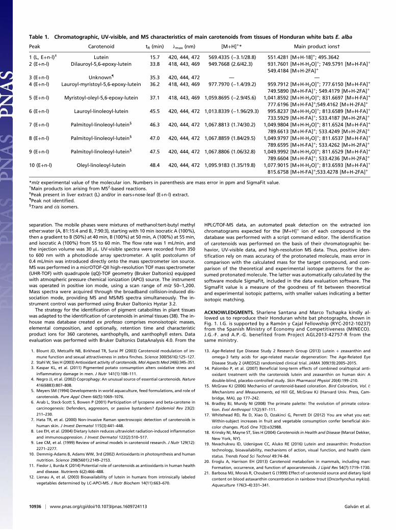
Table 1. Chromatographic, UV-visible, and MS characteristics of main carotenoids from tissues of Honduran white bats E. alba
Peak Carotenoid tR (min) λmax (nm) [M+H]+* Main product ions†
1 (L, E+n-l)‡ Lutein 15.7 420, 444, 472 569.4335 (−3.1/28.8) 551.4281 [M+H-18]+; 495.36422 (E+n-l) Dilauroyl-5,6-epoxy-lutein 33.8 418, 443, 469 949.7668 (2.6/42.3) 931.7601 [M+H-H2O]+; 749.5791 [M+H-FA]+
549.4184 [M+H-2FA]+
3 (E+n-l) Unknown{ 35.3 420, 444, 472 — —
4 (E+n-l) Lauroyl-myristoyl-5,6-epoxy-lutein 36.2 418, 443, 469 977.7970 (−1.4/39.2) 959.7912 [M+H-H2O]+; 777.6150 [M+H-FA]+
749.5890 [M+H-FA]+; 549.4179 [M+H-2FA]+
5 (E+n-l) Myristoyl-oleyl-5,6-epoxy-lutein 37.1 418, 443, 469 1,059.8695 (−2.9/45.6) 1,041.8592 [M+H-H2O]+; 831.6697 [M+H-FA]+
777.6196 [M+H-FA]+;549.4162 [M+H-2FA]+
6 (E+n-l) Lauroyl-linoleoyl-lutein 45.5 420, 444, 472 1,013.8339 (−1.96/29.3) 995.8237 [M+H-H2O]+; 813.6589 [M+H-FA]+
733.5929 [M+H-FA]+; 533.4187 [M+H-2FA]+
7 (E+n-l) Palmitoyl-linoleoyl-lutein§ 46.3 420, 444, 472 1,067.8813 (1.74/30.2) 1,049.9804 [M+H-H2O]+; 811.6524 [M+H-FA]+
789.6613 [M+H-FA]+; 533.4249 [M+H-2FA]+
8 (E+n-l) Palmitoyl-linoleoyl-lutein§ 47.0 420, 444, 472 1,067.8859 (1.84/29.5) 1,049.9797 [M+H-H2O]+; 811.6537 [M+H-FA]+
789.6595 [M+H-FA]+; 533.4262 [M+H-2FA]+
9 (E+n-l) Palmitoyl-linoleoyl-lutein§ 47.5 420, 444, 472 1,067.8806 (1.06/32.8) 1,049.9992 [M+H-H2O]+; 811.6529 [M+H-FA]+
789.6604 [M+H-FA]+; 533.4236 [M+H-2FA]+
10 (E+n-l) Oleyl-linoleoyl-lutein 48.4 420, 444, 472 1,095.9183 (1.35/19.8) 1,077.9015 [M+H-H2O]+; 813.6593 [M+H-FA]+
815.6758 [M+H-FA]+;533.4278 [M+H-2FA]+
*m/z experimental value of the molecular ion. Numbers in parenthesis are mass error in ppm and SigmaFit value.†Main products ion arising from MS2-based reactions.‡Peak present in liver extract (L) and/or in ears+nose-leaf (E+n-l) extract.{Peak not identified.§Trans and cis isomers.
10936 | www.pnas.org/cgi/doi/10.1073/pnas.1609724113 Galván et al.

22. Xavier AAO, Mercadante AZ, Garrido-Fernández J, Pérez-Gálvez A (2014) Fat content
affects bioaccessibility and efficiency of enzymatic hydrolysis of lutein esters added to
milk and yogurt. Food Res Int 65:171–176.23. West CE, Castenmiller JJ (1998) Quantification of the “SLAMENGHI” factors for ca-
rotenoid bioavailability and bioconversion. Int J Vitam Nutr Res 68(6):371–377.24. Britton G, Liaaen-Jensen S, Pfander H (2004) Carotenoids Handbook. Birkhäuser
(Switzerdland, Basel).25. Rodríguez-Herrera B, Medellín RA, Timm RM (2007) Neotropical Tent-Roosting Bats
(Instituto de Biodiversidad, Santo Domingo, Costa Rica).26. Su Q, Rowley KG, Itsiopoulos C, O’Dea K (2002) Identification and quantitation of
major carotenoids in selected components of the Mediterranean diet: Green leafy
vegetables, figs and olive oil. Eur J Clin Nutr 56(11):1149–1154.27. Pérez-Gálvez A, Mínguez-Mosquera MI (2005) Esterification of xanthophylls and its
effect on chemical behavior and bioavailability of carotenoids in the human. Nutr Res
25(7):631–640.28. Bone RA, Landrum JT, Hime GW, Cains A, Zamor J (1993) Stereochemistry of the
human macular carotenoids. Invest Ophthalmol Vis Sci 34(6):2033–2040.29. Mundy NI, et al. (2016) Red carotenoid coloration in the zebra finch is controlled by a
cytochrome P450 gene cluster. Curr Biol 26(11):1435–1440.30. García-de Blas E, Mateo R, Viñuela J, Pérez-Rodríguez L, Alonso-Alvarez C (2013) Free
and esterified carotenoids in ornaments of an avian species: The relationship to color
expression and sources of variability. Physiol Biochem Zool 86(5):483–498.
31. Rodríguez-Herrera B, Víquez-R L, Cordero-Schmidt E, Sandoval JM, Rodríguez-Durán A (2016) Energetics of tent roosting in bats: The case of Ectophylla alba andUroderma bilobatum (Chiroptera: Phyllostomidae). J Mammal 97:246–252.
32. Rodríguez-Herrera B, Medellín RA, Gamba-Rios M (2008) Roosting requirements ofwhite tent-making bat Ectophylla alba (Chiroptera: Phyllostomidae). Acta Chiropt10(1):89–95.
33. Leo Lester R, Grach C, Paul Pener M, Simpson SJ (2005) Stimuli inducing gregariouscolouration and behaviour in nymphs of Schistocerca gregaria. J Insect Physiol 51(7):737–747.
34. Zhao H, et al. (2009) The evolution of color vision in nocturnal mammals. Proc NatlAcad Sci USA 106(22):8980–8985.
35. del Val E, et al. (2009) The liver but not the skin is the site for conversion of a redcarotenoid in a passerine bird. Naturwissenschaften 96(7):797–801.
36. Emenhiser C, Simunovic N, Sander LC, Schwartz SJ (1996) Separation of geomet-rical carotenoid isomers in biological extracts using a polymeric C30 column inreversed-phase liquid chromatography. J Agric Food Chem 44(12):3887–3893.
37. Breithaupt DE, Wirt U, Bamedi A (2002) Differentiation between lutein monoesterregioisomers and detection of lutein diesters from marigold flowers (Tagetes erectaL.) and several fruits by liquid chromatography-mass spectrometry. J Agric Food Chem50(1):66–70.
38. Ríos JJ, Roca M, Pérez-Gálvez A (2015) Systematic HPLC/ESI-High resolution-q-TOF-MSmethodology for metabolomic studies in nonfluorescent chlorophyll catabolitespathway. J Anal Methods Chem 2015:490627.
Galván et al. PNAS | September 27, 2016 | vol. 113 | no. 39 | 10937
EVOLU
TION


![Simultaneous Detection of Multiple Carotenoid Pigments in Algae … · 2020. 8. 28. · and food colour enhancers [1,2,5]. The global market for carotenoids reached $1.5 billion in](https://img.pdfslide.us/doc/110x75/5fe9e5eec8dfc60e085f2cf4/simultaneous-detection-of-multiple-carotenoid-pigments-in-algae-2020-8-28-and.jpg)